












-
Colorectal cancer (CRC) is the third most frequent malignancy worldwide and one of the leading causes of cancer-related deaths. The burden of CRC is expected to rise to 60% by 2030[1,2]. In Iran, CRC ranks as the fourth most prevalent cancer[3], with its incidence expected to double by 2040. While CRC rates remain very low among the aging Iranian community, there is a concerning rise among younger Iranians[4]. CRC develops as genetic and epigenetic changes gradually accumulate in precursor lesions such as adenomas and serrated lesions. These changes lead to the inactivation of tumor suppressor genes, chromosomal instability, and oncogene activation[5,6]. CRC screening can remarkably improve tumor detection in the early stages[7]. Colonoscopy remains the gold standard tool for CRC diagnosis; however, its invasiveness and high cost limit its use for population screening[8,9]. The fecal occult blood test (FOBT) is a more affordable and noninvasive alternative, but results in a high rate of false-positive tests and requires frequent repetition[10]. Unfortunately, the majority of CRC patients are diagnosed at an advanced stage, underscoring the urgent requirement for the rapid identification of diagnostic and prognostic biomarkers[11,12].
Colorectal cancer can potentially be identified through the detection of genomic biomarkers in stool or blood samples, with varying sensitivity levels[13,14]. Biomarker-based screening offers significant advantages over classical methods such as imaging and endoscopy[15]. There is now widespread consensus that methylation markers serve as reliable indicators for identifying early-stage CRC patients[16]. One well-known epigenetic mechanism is the methylation of cytosine residues within CpG dinucleotide sequences, known as CpG islands, which are located in gene promoter regions. About 60% of human gene promoter regions contain CpG islands, mostly found at the 5' end of genes[17]. Alterations in DNA methylation patterns are a hallmark of CRC and many other cancers. Studies have revealed that numerous genes exhibit hypermethylation in early-stage CRC, directly contributing to tumor development and progression. Thus, various methylated genes may serve as valuable molecular markers for early tumor screening[18,19].
Various studies have demonstrated that numerous members of the FOX family play an active role in cancer progression[20]. FOXF1 is a forkhead box (FOX) transcription factor family member. Various FOX family members have been linked to various biological processes, including cell differentiation, development, cell cycle regulation, cell metabolism, aging, cell survival, immune responses, organogenesis, DNA repair, and embryonic development[21−23]. Early research demonstrated that FOXF1 was found to be a factor in the progression of multiple cancers, such as osteosarcoma, breast cancer, lung cancer, and colorectal cancer[24−28]. Notably, FOXF1 acts as both an oncogene and a tumor suppressor in cancer development[21].
Despite the increasing evidence pointing to FOXF1's impact on cancer development, its potential as a biomarker for CRC detection in stool remains unexplored. In this study, we aimed to assess, for the first time, the potential of utilizing FOXF1 gene promoter methylation in stool samples as a noninvasive biomarker for distinguishing CRC patients from healthy controls. Furthermore, we compared its diagnostic sensitivity and specificity with FOBT to evaluate its potential as an alternative or complementary screening method.
-
A total of 50 fecal specimens were obtained from CRC patients 7 d following colonoscopy and pathological confirmation of CRC. In addition, 50 stool samples were taken from healthy people 7 d after a colonoscopy to confirm their health. Consent forms were signed by all participants of the study. The studied population was made up of individuals who had been referred to Imam Reza Hospital, situated in Kermanshah, Iran. The study was approved by the Ethics Committee of Kermanshah University of Medical Sciences, Iran (Ethics code: No. 95285).
DNA extraction
-
DNA was extracted from fecal samples according to the manufacturer's instructions using the Gene All kit (South Korea, Cat. No. 115-150). In brief, 200 mg of the stool sample and 1 mL of PBS buffer were homogenized in a 2 mL microtube; the microtube was maintained at room temperature for several minutes, and the supernatant was separated into a 2 mL microtube, centrifuged at 13,000 r/min for 2 min, and the supernatant was discarded. Then, 1.3 mL of lysing buffer was added to the pellet and mixed. The tube was incubated at room temperature and then centrifuged at 12,000 r/min for 5 min. The supernatant was transferred to an EZ pass filter and centrifuged at 12,000 r/min for 1 min. The filter was placed on a 1.5 mL microtube, and 100 µL of elution buffer (EB) was added to the filter and centrifuged at 12,000 r/min for 5 min. We added 500 µL of PB to the microtube's contents, transferred them to a G-type spin column, and centrifuged them for 1 min at 12,000 r/min. A 500 µL sample of washing buffer was added to the spin column and centrifuged at 12,000 r/min for 1 min. We then added 50 µL of EB buffer to the column for centrifugation at 13,000 r/min for 1 min, and collected the DNA-containing solution in a new 1.5 mL microtube. The concentration of the extracted DNA was determined using a NanoDrop ND-1000 spectrophotometer (Thermo Fisher Scientific, Wilmington, DE, USA). DNA concentration ranged between 20 and 100 ng/µL.
Bisulfite treatment
-
DNA bisulfite treatment was performed to prepare DNA samples for MethyLight PCR using the EpiTect bisulfite kit in conformity with the manufacturer's protocol (Qiagen, Germany). The EpiTect bisulfite kit was used to modify 20–40 µL of DNA. DNA incubation with high concentrations of sodium bisulfite salt at low pH and high temperature leads to a change in unmethylated cytosine residues into uracil, leaving the methylcytosine unchanged.
Primer and probe design
-
Primers and a TaqMan probe were constructed using Beacon Designer™ (version 8.13;
www.premierbiosoft.com/molecular_beacons ) to specifically anneal full-methylated DNA at the FOXF1 promoter gene. The ALU-C4 sequences were used as a reference control. All primers and probes were synthesized by Metabion Biotechnology Company (Metabion, Steinkirchen, Germany). The primer and probe sequences are shown in Table 1.Table 1. Primer and probe sequences applied for MethyLight PCR.
Primers and probes Sequence (5'–3') FOXF1 Forward primer 5'-GTTAATCGTCGGCGGATTTAGTAG-3' Reverse primer 5'-AACTAACTACACCTCTACCGTCAT-3' Probe 5'-6-FAM-ACCACTTCTCCCGAAC
GAACCGC-TAMRA-3'ALU-C4 Forward primer 5'-GGTTAGGTATAGTGGTTTATATT
TGTAATTTTAGTA-3'Reverse primer 5'-ATTAACTAAACTAATCTTAAACT
CCTAACCTCA-3'Probe 5'-6-FAM-CCTACCTTAACCTCCC-TAMRA-3' MethyLight, a TaqMan-based methylation-specific real-time PCR technique
-
MethyLight PCR was carried out by an Applied Biosystems (ABI) 7500 Real-time device (Foster City, CA, USA), using the EpiTect MethyLight PCR + ROX Vial Kit (Qiagen, Germany) according to the manufacturer's protocol. Reactions were performed in a 20 µL volume including 10 µL of 1XEpiTect Methylation MasterMix, 5 µL mix including 0.4 µM forward and reverse primers and 0.2 µM TaqMan probe, 50x ROX Dye Solution 0.4 μL, 3–4 µL RNase–free water, and 1–2 µL converted DNA. Thermal cycling was performed for 45 cycles: 5 min at 95 °C for the initial PCR activation step, 15 s at 95 °C for denaturation, and 60 s at 60 °C for the annealing and extension step. Each plate consisted of 1 µL of patient DNA, full-methylated bisulfite-transformed DNA as a positive control (EpiTect PCR Control DNA, Qiagen, Germany), unmethylated human bisulfite-converted DNA (EpiTect PCR Control DNA, Qiagen, Germany), and RNase-free water, used as a negative control with each set of primers and probes. The standard curve was plotted using the Ct value of commercial methylated control DNA (EpiTect PCR Control DNA, Qiagen, Germany, Cat. No. 59655) with different concentrations vs the log value of input concentration DNA. We acquired an acceptable MethyLight PCR result, which is shown by the PCR efficiency (92%–105%) and correct slope value (−3.65 to −3.2). The standard curve was obtained by plotting the Ct values of methylated control DNA as a function of DNA concentrations.
FOBT
-
An alcohol pyrimidine-based FOBT was performed to assess the presence of blood in all of the stool samples. For this, we suspended a spatula of the stool in normal saline in a microtube and mixed it. Pyramidine alcohol and 4–5 drops of 10% acetic acid were added and mixed, and 5–6 drops of 10% H2O2 were added as a substrate. Pyrimidine alcohol oxidizes the oxygen released from H2O2 decomposition by the peroxidase enzyme, causing a color change. The color change to purple indicates the presence of blood in the medium. The activity of the peroxidase enzyme or blood content in the stool determines the extent of color change intensity.
Data analysis
-
The PCR data were evaluated using ABI 7500 SDS software (v1.3.1, Applied Biosystems, USA). For all samples, the percentage of methylation reference (PMR) was determined using the following formula: ([FOXF1/ALU] sample/[FOXF1/ALU] positive control) × 100. Statistical analysis was performed using SPSS software (v16.0, IBM, USA). A p-value < 0.05 was considered statistically significant. Receiver operating characteristic (ROC) curve analysis was used to evaluate the sensitivity and specificity of FOXF1 methylation.
-
Overall, 50 stool samples were obtained from patients with CRC and 50 samples from the control group. The average age of the patients was 56 ± 12.9 years. The healthy controls were matched with the patient group in terms of age and sex. There was no significant difference in age or gender between the CRC and control groups (Table 2).
Table 2. The demographic characteristics of the patient group with CRC and the healthy control.
Demographic features Patient group
(n = 50)Control group
(n = 50)p value* Age (years) 56 ± 12.9 54 ± 12 0.166 Gender
(female:male)3:2 9:7 0.686 * Mann–Whitney U test. FOXF1 gene promoter methylation
-
Median PMR values of stool samples from patients and healthy controls were 0.06 (95% CI 0.01–13.1) and 0.01 (95% CI 0.0–0.02), respectively (Table 3; Fig. 1). In stool samples from CRC patients, PMR values were higher than those of the control group (p < 0.001). PMR values showed no significant difference in demographic or pathological features between the cohorts used in the study (Table 4). The highest value of PMR in the stool samples from healthy controls was considered a threshold of methylation status, while a PMR value higher than that was considered a positive methylation status. Methylation of the FOXF1 gene (positive results) in the age group > 50 years was compared with the age group ≤ 50 years (73.5% vs 75%) and between female and male patients (80% vs 65%) (Table 5). There was no significant difference in the level of methylation between the gender and age subgroups (Table 5).
Table 3. Median PMR value results for FOXF1 gene promoter methylation in the stool samples of patients with CRC and healthy controls.
No. of patients Minimum Maximum Percentiles** p-value* 25th 50th 75th Stool PMR Patient
group50 0.0 73 0.01 0.06 13.1 < 0.001 Normal group 50 0.0 0.03 0.0 0.01 0.02 * Mann–Whitney U test. ** Explore. 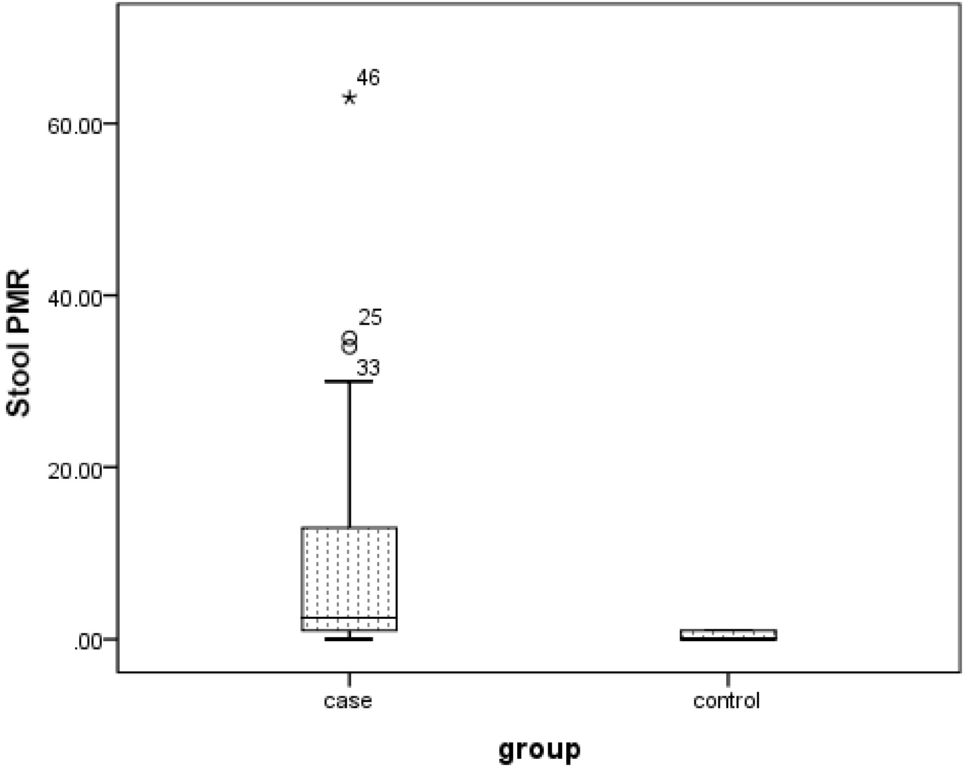
Figure 1.
Box plot for the FOXF1 gene PMR value of stool samples in case and control groups. The boxes show the interquartile range (the interval between the 25th and 75th percentiles). The lines within the boxes indicate the median values.
Table 4. Association of stool PMR values (percentiles) for the FOXF1 gene with demographic and pathological characteristics of the patient group.
Characteristics No. of
patientsStool PMR (percentiles)** p value* 25th 50th 75th Age ≤ 50 years 16 0.99 2.64 13.37 0.5 > 50 years 34 2.05 3.48 13.6 Gender Female 30 2.07 3.9 10.87 0.41 Male 20 1.1 2.55 14.95 Grade Poorly and moderate 10 1.91 3.1 9.15 0.97 Well 40 1.98 2.85 14.5 Location Distal 21 1.34 2.5 5.62 0.06 Proximal 29 2.04 4.6 20.25 Size ≤ 10 mm 25 1.91 2.65 9.02 0.31 > 10 mm 25 1.74 4.7 17.25 Stage Stage I and II 28 2.2 3.65 10.1 0.13 Stage III and IV 22 2.71 4.1 15.6 * Mann–Whitney U test. ** Explore. Table 5. Association between FOXF1 gene promoter hypermethylation and clinicopathological characteristics.
Characteristics No. of patients Hypermethylation status** p value* Positive
(%)Negative (%) Age ≤ 50 years 16 12 (75%) 4 (25%) 0.61 > 50 years 34 25 (73.5%) 9 (26.5%) Gender Female 30 24 (80%) 6 (20%) 0.14 Male 20 13 (65%) 7 (35%) Histological grade Poor and
moderate10 8 (80%) 2 (20%) 0.53 Well 40 29 (72.5%) 11 (27.5%) Tumor
locationDistal 21 15 (71.4%) 6 (28.6%) 0.74 Proximal 29 22 (75.9%) 7 (24.1%) Tumor size ≤ 10 mm 25 17 (68%) 8 (32%) 0.34 > 10 mm 25 20 (80%) 5 (20%) Tumor stage Stage I and II 28 23 (82.1%) 5 (17.9%) 0.15 Stage III and IV 22 19 (83.3%) 3 (16.7%) Methylation levels (PMR values) Median PMR
(CRC patients)0.06
(95% CI; 0.01–13.1)− < 0.001 Median PMR (healthy controls) 0.01
(95% CI; 0.0–0.02)− * Fisher's exact test. ** Crosstabs. The ability of the FOXF1 gene PMR level as a biomarker
-
Furthermore, we utilized ROC curve analysis to assess the ability of the FOXF1 gene PMR level in stool samples to distinguish between CRC patients and controls. ROC curve analysis showed that the sensitivity and specificity of FOXF1 gene promoter methylation at the optimal cut-off value, with an AUC of 0.744 (p < 0.001), were 59% and 96%, respectively (Fig. 2).
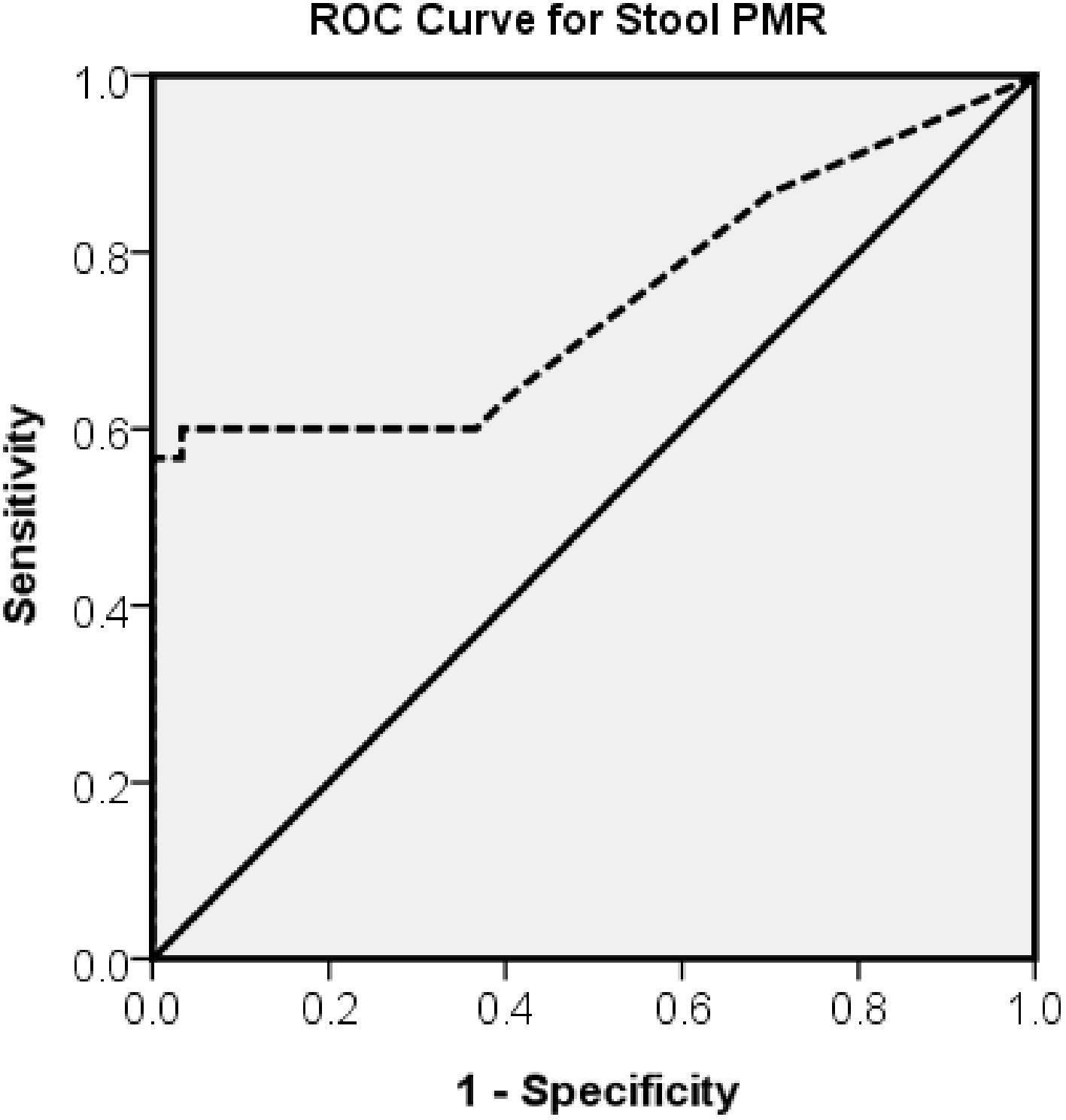
Figure 2.
ROC curve for FOXF1 gene promoter methylation in stool samples.
FOBT results
-
We also examined the FOBT as an early diagnostic test. The sensitivity of FOBT was 38% compared to 59% for FOXF1 gene promoter methylation. These results showed that the sensitivity of FOXF1 gene promoter methylation in stool samples was significantly higher than that of the FOBT.
-
Colorectal cancer is a major public health concern, accounting for about 10% of worldwide incidence and mortality[29]. In the development of CRC, there is a gradual accumulation of different genetic and epigenetic changes, with abnormal DNA methylation being a crucial mechanism[30]. Aberrant DNA methylation patterns are commonly observed in gastrointestinal neoplasms and could serve as valuable markers for screening, diagnosis, prognosis determination, and prediction of CRC. In addition, the plentiful presence in extracellular environments, resistance to degradation, and presence in plasma, serum, fecal specimens, and other noninvasive bodily fluids facilitate quantitative analysis of methylated DNA biomarkers[31]. Collecting bodily fluids is a non-intrusive process that allows for the early identification of cancer or disease monitoring[32]. Numerous studies have validated stool-based methylated DNA detection as an effective method for early CRC screening[31]. Promoter hypermethylation contributes to the inactivation of tumor suppressor genes and ultimately oncogenic processes by affecting key cellular pathways[16,33]. Common methylated and silenced genes often seen in CRC include vimentin (VIM), cadherin-1 (CDH1), MLH1, TIMP metallopeptidase inhibitor-3 (TIMP3), hypermethylated in cancer-1 (HIC1), and secreted frizzled related protein-1 (SFRP1)[33]. Recently, methylated syndecan-2 (mSDC2) and methylated SEPT9 (mSEPT9) have been identified as promising biomarkers for noninvasive detection of CRC[34−37]. Researchers have studied abnormal methylation of different genes in the tissues and bodily fluids of individuals with CRC, which could be used as valuable biomarkers in screening for CRC[16]. Cologuard is the first multitarget stool DNA test that has received FDA approval for CRC screening. It evaluates KRAS mutations, as well as BMP3 and NDRG4 methylation levels[13,38]. Moreover, NDRG4, BMP3, and SEPT9 are now incorporated as DNA methylation markers in FDA-approved CRC screening tests[39]. Therefore, we aimed to identify noninvasive biomarkers capable of detecting CRC in small stool samples. Analyzing single genes in epigenetic studies could provide a more cost-effective option for screening. Nevertheless, these studies have presented remarkable initial findings about these single epigenetic markers and their utility as stool DNA-based assays[40]. Moradi et al. reported that PMR values in the stool of CRC patients were higher than those of healthy controls. Also, the analysis of SOX21 gene promoter methylation using a stool-based assay shows a notably high level of sensitivity (72.5%) and specificity (100%)[41]. Zhang et al. showed that SPG20 promoter hypermethylation serves as a highly sensitive and specific biomarker in the detection of CRC using stool samples (80.2% and 100%, respectively)[42]. Glöckner et al. reported that, in stage I to III CRC patients, TFPI2 methylation was found in stool DNA with a sensitivity ranging from 76% to 89% and a specificity ranging from 79% to 93%. Recognition of TFPI2 methylation in fecal DNA may serve as a biomarker for CRC screening[40]. Muller et al. identified a sensitivity of 77% to 90% and a specificity of 77% for SFRP2 methylation as a sensitive single DNA-based biomarker for detecting CRC in stool samples from 49 patients[43]. According to research, using single-marker methylation-based stool markers for Vimentin and SFRP2, the sensitivity has been found to be 52% and 87% to 94%, while the specificity has been reported to be 90% and 85% to 90%, respectively[44−47]. Various studies have indicated that the FOX family plays a crucial role in cellular proliferation, apoptosis, metastasis, the cell cycle, cellular metabolism, progression, and cancer biology in various cancers[48−51]. In total, dysregulation of FOX proteins may impact cell fate, tumor growth, and metastasis of cancer[52]. FOXF1, a transcription factor, attaches to the promoter regions of target genes to control gene expression levels and influence the function of various signaling pathways crucial for physiological and pathological processes[20]. FOXF1 is critical for controlling angiogenesis during embryonic development and may also be involved in angiogenesis, impacting CRC progression. Increased FOXF1 expression has been linked to aggressive CRC features, including poor differentiation and advanced disease stages. There was a significant association between increased FOXF1 expression and poorer prognosis in CRC patients[28]. In contrast, FOXF1 expression is often silenced through DNA hypermethylation in breast cancer, where it acts as a tumor suppressor[53]. Herrera-Merchan et al. reported that FOXF1 expression could also be silenced via promoter methylation in lung adenocarcinoma and functions as a tumor suppressor[54]. In another study, Mitchell et al. analyzed a panel of 23 genes in the blood and tissue of CRC patients compared to a control group. They found increased DNA methylation in at least half of the CRC tissue samples compared to normal tissue. Additionally, 11 genes, including FOXF1, exhibited low methylation levels in peripheral blood DNA from healthy individuals[55]. On the other hand, FOXF1 promotes the development and maintenance of gastrointestinal stromal tumors through the regulation of KIT and ETV1 expression[56]. In hepatocellular carcinoma, FOXF1 serves as a suppressor of cell invasion and tumorigenesis, with its expression closely linked to patient survival rates[57]. Furthermore, in CRC cell lines with p53 inactivation, FOXF1 expression is silenced, leading to genomic instability in FOXF1-expressing CRC cells with a dysfunctional p53-p21WAF1 checkpoint. This underscores FOXF1's significance in CRC development[58]. Additionally, the competitive binding of miR-375-3p by LINC00022 could lead to the upregulation of FOXF1, potentially contributing to CRC progression[59]. Lo et al. reported cytoplasmic mislocalization of upregulated FOXF1 protein in CRC epithelial cells, which is associated with poor prognosis factors[21]. Furthermore, FOXF1 upregulation is related to poor outcomes, disease progression, invasion, and metastasis of CRC[20]. Recently, it was reported that FOXF1 gene methylation levels in the plasma of CRC patients are higher than in healthy controls[60]. In the present study, for the first time, we determined the FOXF1 gene promoter methylation for 50 patients with CRC and 50 healthy controls. Our results showed that the median PMR values of stool samples from patients and healthy controls were between 0.01 to 13.1 and 0.0 to 0.02, respectively. In addition, we examined the association of the PMR values obtained from stool samples with the demographic and pathologic features of the patients. There was no statistically significant correlation found between the methylation levels of the FOXF1 gene promoter and age, gender, histological grade, tumor stage, tumor location, or tumor size in CRC patients. In our research, FOXF1 gene promoter methylation position was assessed by the MethyLight technique, a quantitative method with high sensitivity and specificity capable of assaying a small amount of DNA. Our data revealed that the FOXF1 gene promoter methylation had higher sensitivity and specificity than FOBT in stool samples.
Although our results offer valuable information on the potential of FOXF1 methylation as a noninvasive biomarker for CRC detection in stool samples, certain limitations must be considered. The sample size is relatively small and may affect the generalizability of our results. Furthermore, potential biases in patient selection and sample collection could impact the observed methylation patterns. To validate our findings and better evaluate the clinical utility of FOXF1 methylation in CRC detection, future studies should include larger, more diverse cohorts.
-
In summary, as the first study to evaluate FOXF1 gene promoter methylation in stool samples of patients with CRC, it can be concluded that the PMR value for FOXF1 in CRC patients is higher than in healthy controls. The quantification of FOXF1 gene promoter methylation in stool samples shows promise as a noninvasive biomarker for CRC screening, with a sensitivity of 59% and a specificity of 96%. These results suggest potential sensitivity advantages over FOBT. Additionally, FOXF1 gene promoter methylation was found to be independent of age, sex, tumor grade, or stage, highlighting its potential utility in diverse patient populations. Future research with larger cohorts and more diverse populations is necessary to confirm these findings and explore their feasibility as a cost-effective screening tool.
The authors are grateful to the staff at the Medical Genetics Laboratory, Reference Laboratory, and Kermanshah University of Medical Sciences for their support throughout this research.
-
All individuals participating were tasked with signing the informed consent form. The study was approved by the Ethics Committee at Kermanshah University of Medical Sciences, Iran (Ethics Code: No. 95285) and conformed to the principles of the Helsinki Declaration.
-
The authors confirm their contribution to the paper as follows: study conception and design: Mehrjoei B, Rezvani N; sample and clinical data collection: Tahmasebi S; data analysis and interpretation: Rezvani N; manuscript preparation: Mehrjoei B; manuscript review and editing: Vaisi-Raygani A. All authors reviewed the results and approved the final version of the manuscript.
-
The data that support the findings of this study are available from the corresponding author upon reasonable request.
-
The authors declare that there is no conflict of interest.
- Copyright © 2026 by the author(s). European Journal of Cancer Care published by Maximum Academic Press on behalf of John Wiley & Sons Ltd. This is an open access article under the terms of the Creative Commons Attribution License, which permits use, distribution and reproduction in any medium, provided the original work is properly cited.
-
About this article
Cite this article
Tahmasebi S, Mehrjoei B, Vaisi-Raygani A, Rezvani N. 2026. Evaluation of FOXF1 promoter DNA methylation as a promising epigenetic biomarker for colorectal cancer in stool samples. European Journal of Cancer Care 2026: e013 doi: 10.48130/ejcc-0026-0013 

Evaluation of FOXF1 promoter DNA methylation as a promising epigenetic biomarker for colorectal cancer in stool samples
- Received: 19 October 2025
- Revised: 03 November 2025
- Accepted: 20 March 2026
- Published online: 16 September 2026
Abstract: Colorectal cancer pathogenesis is influenced by gene promoter region methylation, a key epigenetic mechanism. This study investigated whether FOXF1 promoter methylation in stool samples could serve as a biomarker for colorectal cancer. FOXF1 promoter methylation was analyzed in stool samples from 50 patients with colorectal cancer and 50 healthy controls. The percentage of methylation reference was determined for each sample. FOXF1 promoter methylation levels were higher in patients with colorectal cancer than in healthy controls (p < 0.001). The median percentage of methylation reference was 0.06 (95% confidence interval, 0.01–13.1) in patients and 0.01 (95% confidence interval, 0.0–0.02) in controls. Receiver operating characteristic curve analysis demonstrated a sensitivity of 59% and a specificity of 96% for FOXF1 promoter methylation in stool samples. Increased FOXF1 promoter methylation in stool samples suggests that it may serve as a noninvasive biomarker for the detection of colorectal cancer independent of sex, age, or disease stage.
-
Key words:
- Colorectal cancer /
- Stool samples /
- DNA methylation /
- FOXF1 gene /
- Epigenetic biomarker