













-
Coronaviruses have been the etiological agents in several major pandemic outbreaks over the last two decades, including the Severe Acute Respiratory Syndrome (SARS), Middle East Respiratory Syndrome (MERS), and Coronavirus Disease 2019 (COVID-19)[1−3]. Together, these viral infections caused millions of yearly infections around the globe, with an estimated case fatality rate of 9.6% (SARS), 35% (MERS), and 1%~5% (COVID-19). With the development of oral inhibitors (e.g., Paxlovid) against COVID-19 and the constant viral evolution towards less lethal strains, the impact of COVID-19 illness on public health has been alleviated[4,5]. From a retrospective point of view, however, a significant number of casualties during the pandemic were due to the overwhelming of health care systems, due to the lack of means to effectively distinguish false-positive cases from severe infections[6]. Specifically, the well-established Real-Time Quantitative PCR (RT-qPCR), as the standard method to monitor viral agents in a large population, may not demonstrate the differences between live viruses and debris of dead viruses[7]. Hence, the design of orthogonal analytical tools for detection and quantification of the conserved component of functional coronaviruses would be desirable for tracking more viral variants and providing hierarchical treatments in the future.
The common molecular diagnostic methods for various types of viruses could be categorized as nucleic acid-based, antibody-based, or antigen-based[8]. Among the nucleic acid amplification tests, the polymerase chain reaction (PCR) assays were recognized as the gold standard. The isothermal amplification (e.g., LAMP) and CRISPR-based tests were later developed to simplify the procedure[9]. Nonetheless, viral RNA may circulate in the patient's body for ~90 d, even after the virus loses infectivity, making it difficult to determine the stage of infection. The serologic test of antibodies (e.g., ELISA) was not primarily used to diagnose current infections, because the specific antibodies were produced after the immune system reacted to, and almost cleared up the viruses. To increase the sensitivity of immunoassays, a combined IgM/IgG antibody test was thus derived[10,11]. The antigen detection assays usually focus on the E, N, and S proteins of the Severe Acute Respiratory Syndrome Coronavirus 2 (SARS-CoV-2)[12]. While the viral surface glycoprotein S underwent constant mutations to escape the humoral immune system, the nucleocapsid protein N shares ~90% and ~49% sequence identity with that of SARS and MERS-CoV, respectively, making the N protein a preferred target for detection. However, these assays heavily relied on antibodies that recognized epitopes on N or S; thus, the read-outs reflected 'existence' instead of viral 'activity'.
To overcome this barrier, we hypothesized that an indispensable component for coronavirus replication, the 3C-Like protease (3CLPro, or main protease MPro), could become a useful indicator of viral infectivity[13,14]. Unlike the N protein and S protein, the enzymatic feature of 3CLPro could facilitate a direct readout of the 'activity signal'. By incorporating its substrate into a modular construct of luciferases or green fluorescent proteins (GFP), the activity of 3CLPro or the infectivity of SARS-CoV-2 could correlate with light signals. More importantly, by rationally optimizing the substrate octapeptide of 3CLPro, it is possible to increase 3CLPro-recognition specificity and the detection sensitivity. We set out to test this hypothesis by computationally designing substrates of SARS-CoV-2 3CLPro with elevated binding energies and assembling the molecular and cellular detection systems as a proof-of-concept for the potential detection of live viruses.
-
Throughout the life cycle of coronaviruses, 3CLPro performed the protease function after viral mRNA translation, but not before the assembly of nucleocapsid proteins into particles. It cleaved 12 sites within the polyprotein precursors pp1a and pp1ab[15]. Hence, the enzymatic activity of 3CLPro was directly linked to replication and maturation of coronaviruses. 3CLPro possesses > 96% sequence identity among global coronavirus strains (Supplementary Figs S1, S2). The most conserved region of 3CLPro was the substrate recognition pocket and the catalytic dyad (H41 and C145), (Fig. 1a)[4,13]. Thus, we hypothesized that a linear peptide that closely fits into the 3CLPro pocket would resist the majority of drifting mutations in virus evolution and be a starting point for deriving a 3CLPro detection tool.
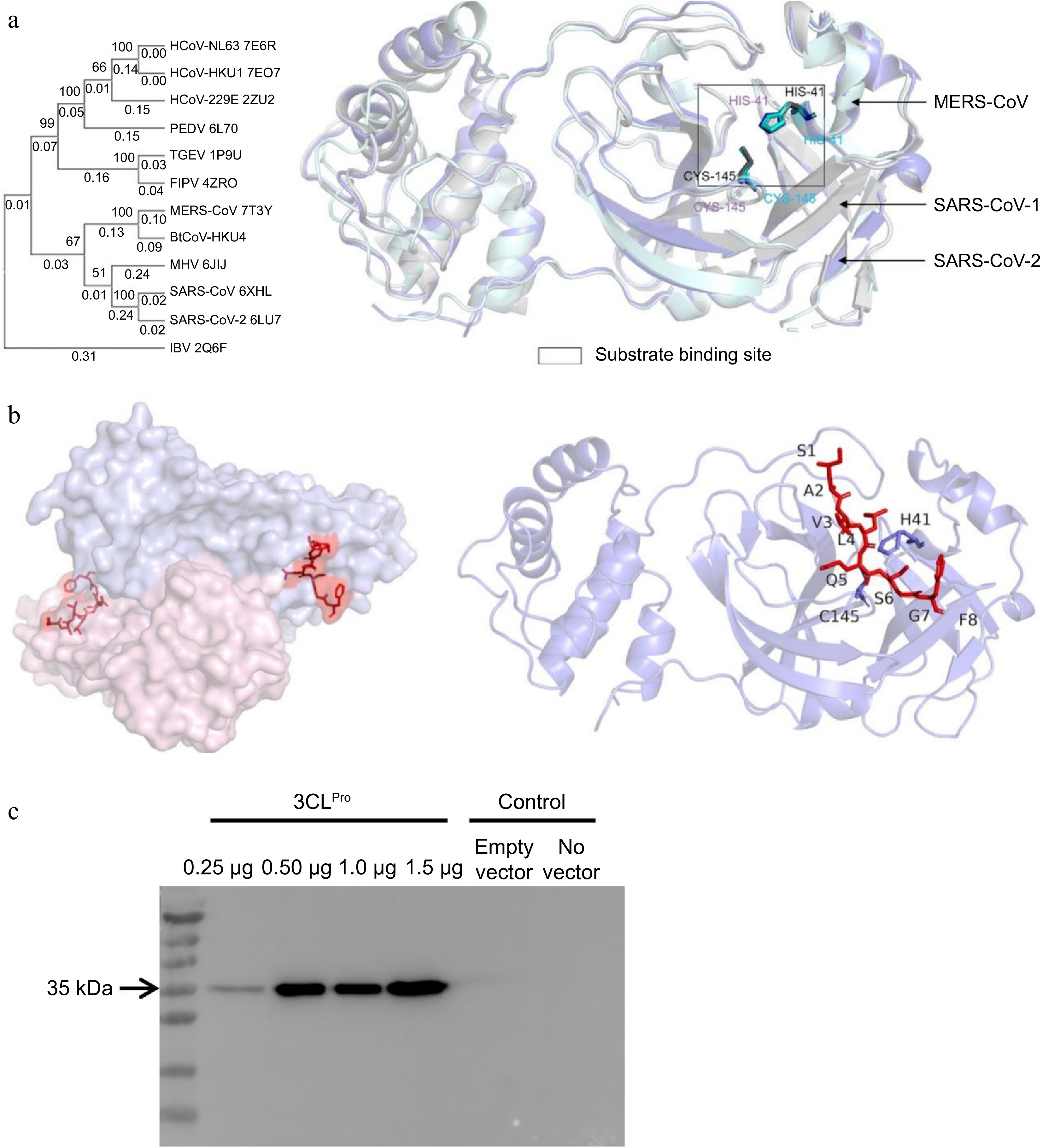
Figure 1.
The substrate binding pocket of 3CLPro. (a) Structural alignment of three coronaviruses 3CLPro (SARS-CoV-1, SARS-CoV-2, and MERS-CoV). The catalytic dyad was indicated in the black box. (b) The substrate peptide (red sticks) bound to 3CLPro. The protease was a homodimer in native conformations. In the design process, only one protomer (right panel) was used as the input model. (c) 3CLPro expression in HEK-293T cells. The cells were transfected with different amounts of pCMV vectors encoding 3CLPro (33.8 kD, without the Flag tag) and the protein expression levels were detected by Western blot using anti-Flag antibodies.
The catalytic pocket of SARS-CoV-2 3CLPro was a shallow groove accommodating a curved peptide conformation (Fig. 1b), composed of key interacting residues T24, N142, H163, E166, and Q189, with H41 and C145 flanking the cleavage site (Leu-Gln↓ [Ser, Ala, and Gly]). Based on the crystal structure of 3CLPro and the native peptide (SAVLQSGF, PDB 7N89)[16], we applied the deep learning sequence design model ProteinMPNN to optimize the peptide sequence while keeping the cleavage site intact[17]. To diversify the peptide sequences, we also applied Rosetta FastDesign to find optimal combinations of the residue rotamers with flexible backbones[18,19]. In total, we obtained three sequences from Rosetta FastDesign and one sequence from ProteinMPNN (out of 1,000 ranked designs) (Table 1). We predicted the complex structural models of 3CLPro and designed peptides using AlphaFold3, and the results indicated that all peptides bound at the pocket, adopting similar conformations as compared to the native models. To indicate the orthogonality and specificity of the P1 sequence, we performed a BLAST search for homologies and found that the closest protein segment was from the phosphohydroxythreonine transaminase of Bacillota bacterium. Hence, it is distinct from any eukaryotic proteins.
Table 1. Computational characterization of WT and designed sequences.
ID Sequence AlphaFold3 predictions Rosetta energy (REU) WT SAVLQSGF ipTM = 0.8 −56.1 P1 PVILQYTT ipTM = 0.72 −75.8 P2 MSRLQTSN ipTM = 0.73 −64.3 P3 PVILNYTH ipTM = 0.64 −71.5 P4 KPRLQAGN ipTM = 0.69 −47.5 To mimic the viral infections and set up a model system for examination of our biosensor designs, we expressed 3CLPro in the HEK-293T cell line (Fig. 1c) by plasmid transfections. We demonstrated that a varying concentration of 3CLPro could be achieved by varying the concentration of plasmids, thus semi-quantitatively mimicking the amounts of virus that infected the cell. We adopted this transcription system in the following experiments.
Modular design of luciferase and GFP-based 3CLPro detection tools
IN-GLuc designs
-
We first considered Gaussia Luciferase (GLuc, ~19.9 kD secreted enzyme), one of the brightest naturally occurring luminescent enzymes, as the scaffold for insertion of 3CLPro-recognition peptides[20−22]. When 3CLPro was present and active, the peptide-inserted Gluc proteins would result in a reduction of bioluminescent signals (Fig. 2a). Two factors were optimized when constructing the peptide-insertion proteins (denoted as IN-Gluc): the insertion sites and inserted sequence. We used SPELL, an online prediction tool for the identification of proper insertion sites on the surface of GLuc, avoiding disruption of the overall protein structure[23]. SPELL suggested the residue pairs 46/47, 103/104, and 151/152 for incorporating the designed sequences (the structural model in Fig. 2e). We first compared the effects of the native substrate (WT: SAVLQSGF) and all designed substrates when they were inserted into 46/47 (Fig. 2b, c). When 3CLPro was expressed in the cell, the WT-IN-Gluc featured a 23% reduction of Gluc signals, while P1-IN-Gluc featured a 67% reduction of signals (the highest among designed sequences, Fig. 2c), indicating that a tight binding between the peptide and 3CLPro would facilitate more thorough cleavage.
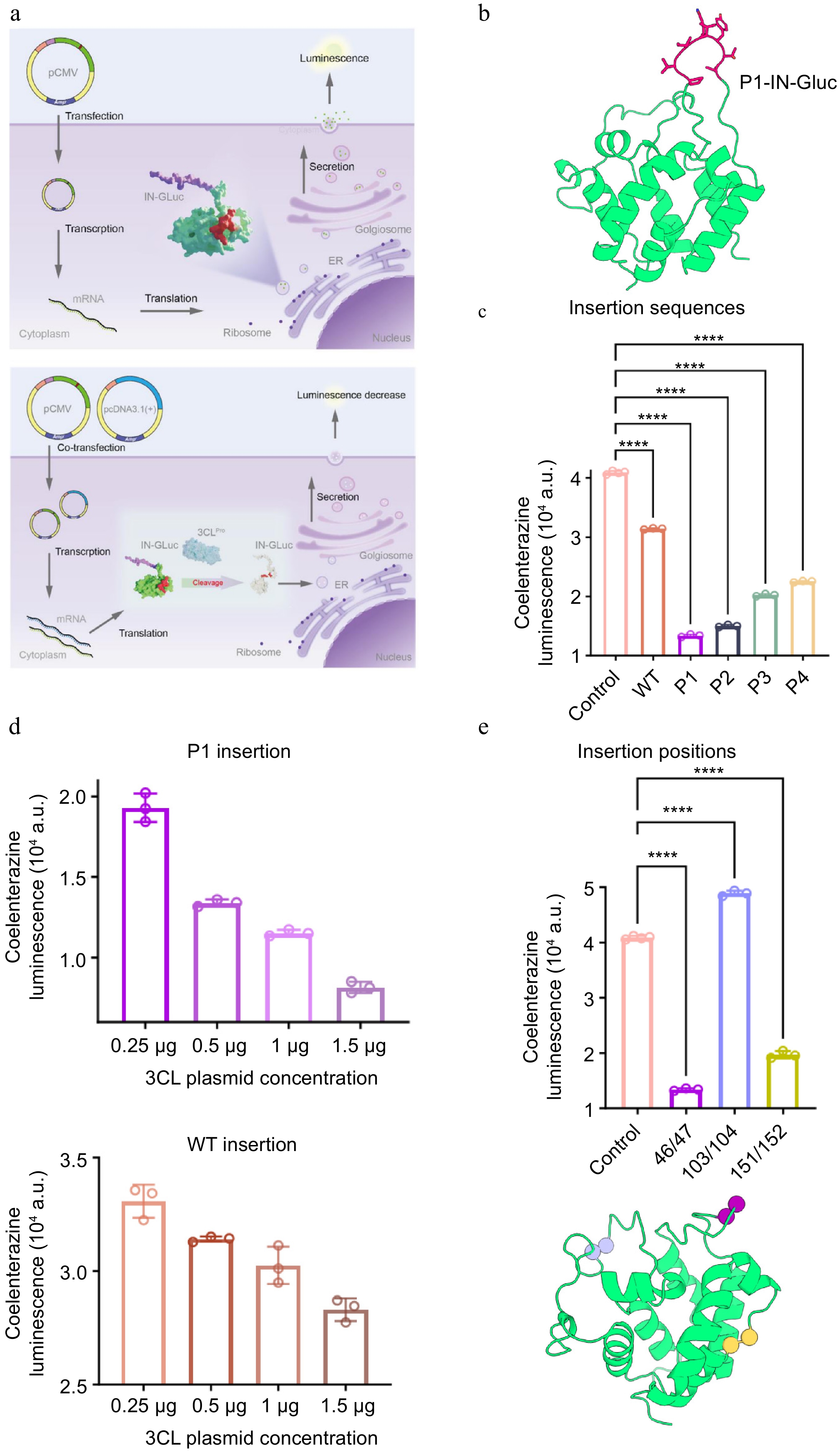
Figure 2.
The design of IN-Gluc. (a) The design principle of IN-Gluc. 3CLPro cleaved the peptide sequence inserted in the middle of Gluc, a secreted enzyme catalyzing the oxidation of coelenterazine to a luminescent molecule, leading to a reduction of luminescence (lower panel). In the absence of 3CLPro or infections, the Gluc was readily secreted out of the cytosol (upper panel). (b) The AlphaFold3 predicted structural model of P1-IN-Gluc. (c) The effects of different designed sequences when inserted into Gluc. The control was deactivated 3CLPro H41A/C145A expressed at the same level. (d) The 3CLPro dose-dependent signal change when P1 or WT peptide was inserted into Gluc. (e) The effects of different insertion positions and the locations of 46/47, 103/104, and 151/152 positions indicated as spheres. n = 3 as the number of replications (n = 4 for the control sample).
We then induced different expression levels of 3CLPro by transfecting various concentrations of vectors. The signals from P1-IN-Gluc semi-quantitatively corresponded to the amounts of 3CLPro with a dynamic range between 52% and 80%, wider than that of WT-IN-Gluc (18% to 30%). Hence, we focused on the P1 design in the following studies.
The 103/104 insertion did not result in signal reduction; by contrast, the luminescence increased 18% when 3CLPro was expressed (Fig. 2e). We think it is possible that the two halves of Gluc that split at the 103/104 site are associated with each other and function as a whole, releasing conformational tensions from the inserted P1 sequence. The 151/152 insertion resulted in 52% signal reduction. We also tested a combination of two different insertions, such that both 46/47 and 181/182 were inserted with P1 to increase the chance of recognition by 3CLPro. However, the double-insertion designs probably interfered with the catalytic function of Gluc, and only 35%~47% signal responses were recorded, less than the 67% reached by a single insertion at 46/47. Hence, a balance between 3CLPro recognition and the structural integrity of Gluc should be considered in these designs. In conclusion, P1-IN-Gluc was the best among the similar designs.
TM-Gluc designs
-
Instead of inducing a drop of light signal, we took advantage of the secretion property of Gluc and installed a transmembrane helix (TM) at the terminus of Gluc to retain it in the cytosol (Fig. 3a)[24]. We then put the P1 peptide between TM and Gluc (Fig. 3c). The active 3CLPro would release Gluc out of the cytosol so that we could measure the signals in the extracellular medium that reflected the amount of secreted Gluc. We consistently adopted the P1 sequence while optimizing one factor in the TM-Gluc design: the linker sequence (flexible or rigid).
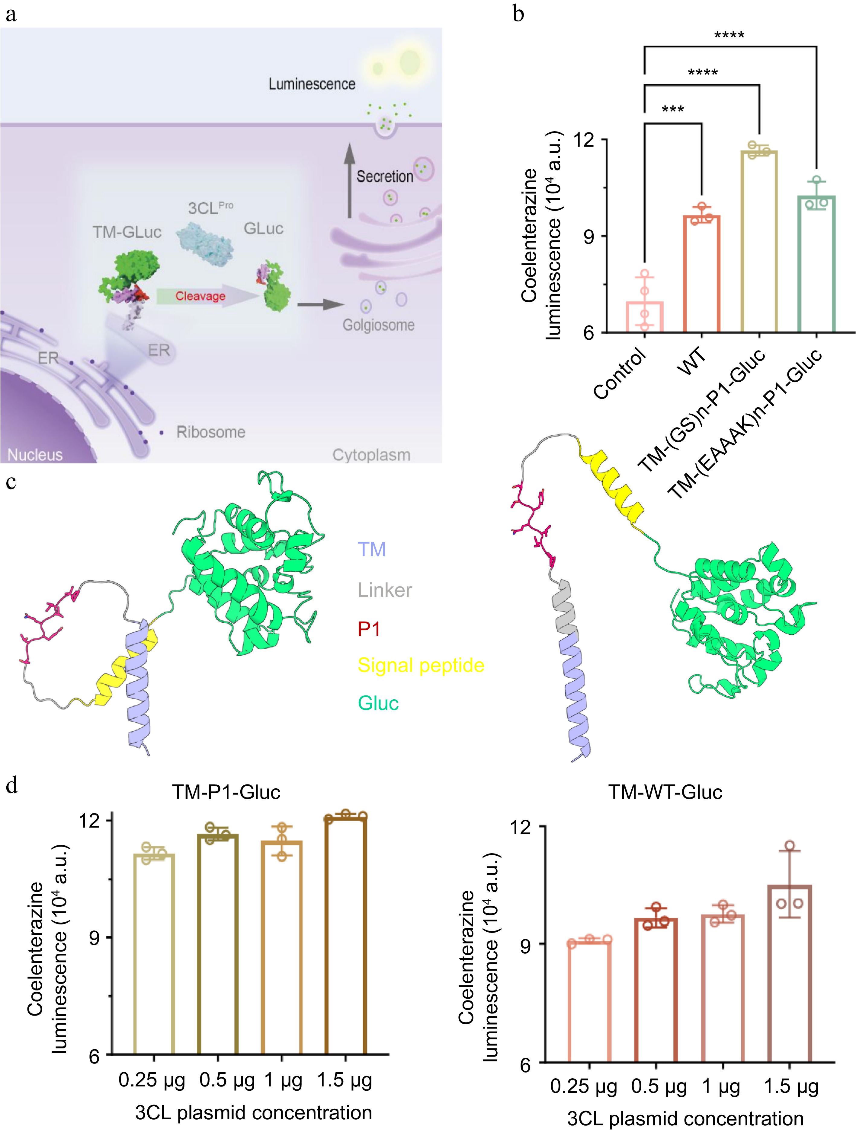
Figure 3.
The design of TM-Gluc. (a) The design principle of TM-Gluc. 3CLPro-mediated cleavage released Gluc for extracellular secretion, leading to elevation of luminescence. (b) The effects of flexible linker and rigid linker when fusing the P1 peptide with the membrane anchoring sequence TM. (c) The AlphaFold3 predicted structural model of P1-TM-Gluc (left: flexible linker, right: rigid linker). (d) The 3CLPro dose-dependent signal change when P1 or WT peptide was fused at the N-terminal of Gluc. n = 3 as the number of replications (n = 4 for the control sample).
We found that a flexible linker (GS)n combined with N-terminus TM-P1-Gluc could result in a 47% signal increase. A rigid linker (EAAAK)n resulted in less sensitive changes of signal (37% increase). The TM-(GS)n-P1-Gluc also quantitatively responded to the changing levels of 3CLPro with a dynamic range of 42% to 55%. Compared to the IN-P1-Gluc design, TM-(GS)n-P1-Gluc featured higher basal levels of fluorescein luminescence (0.8 × 10⁷ to 1.5 × 10⁷) at comparable Gluc expression levels, thus making it easier for detection. We postulated that the reason for this difference was that inserting P1 into the Gluc might interrupt its enzymatic activity, while fusing P1 at the N-terminus of Gluc minimizes this structural interruption. Compared to TM-(EAAK)n-P1-Gluc with the rigid linker, the sensitivity of TM-(GS)n-P1-Gluc was higher for the same amount of 3CLPro expression. Hence, for gain-of-signal detections, the TM-(GS)n-P1-Gluc was a suitable module.
IN-FlipGFP designs
-
As the second method to assemble 'signal elevation' systems, we inserted the P1 peptide into the previously reported FlipGFP[25,26]. The intrinsic fluorescence of enhanced Green Fluorescent Protein (eGFP) was abolished by flipping two β-strands with tightly bound coiled-coil helices (Fig. 4a). We inserted P1 and WT peptides into the coiled-coil region of FlipGFP, respectively (the structural model in Fig. 4e). Without 3CLPro expression, the cell cultures stay in a dark state. 3CLPro cleavage of the substrates restored FlipGFP to the light state, with the green fluorescence increasing 3.9 times for P1-IN-FlipGFP and 1.2 times for WT-IN-FlipGFP (Fig. 4b). This system is more sensitive than TM-P1-Gluc, spanning a 2.0- to 5.0-fold fluorescence elevation when expression of 3CLPro increased 6 times (Fig. 4c, d).
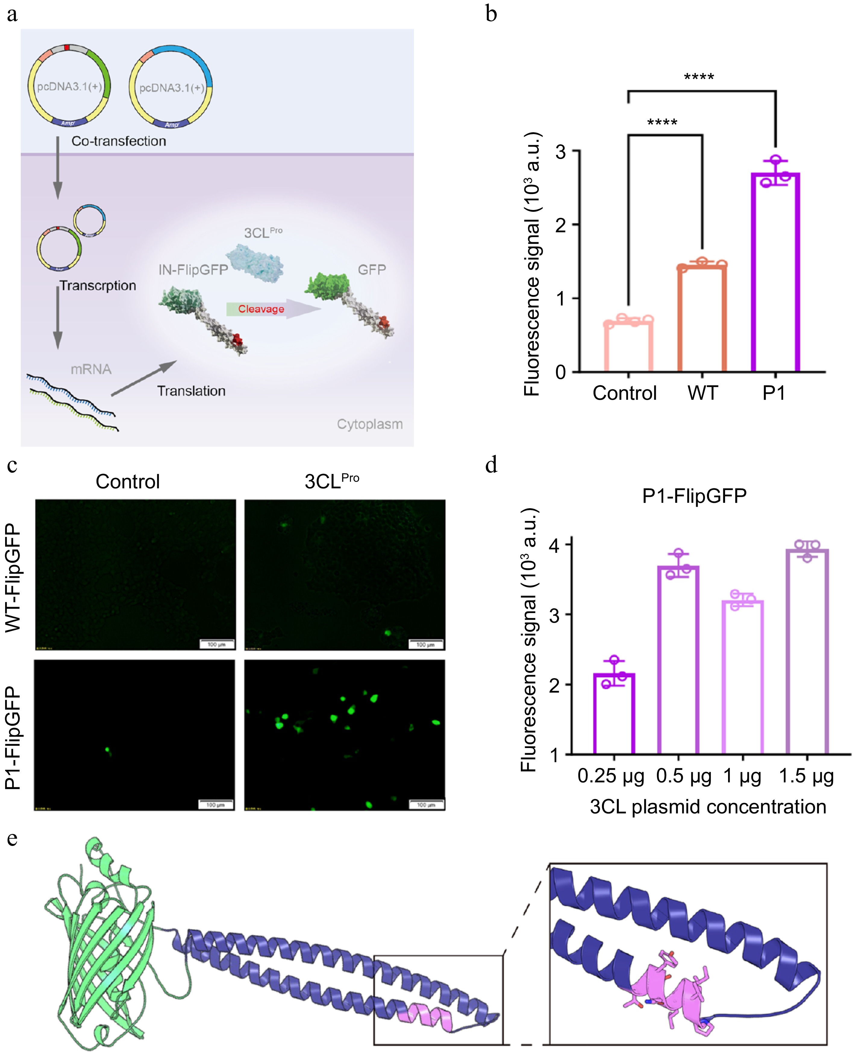
Figure 4.
The design of IN-FlipGFP. (a) The design principle of IN-FlipGFP. 3CLPro-mediated cleavage released conformational constraint of FlipGFP and restored the green fluorescence. (b) The comparison between WT peptide insertion and P1 peptide insertion in the FlipGFP construct. (c) The image of cells expressing 3CLPro and IN-FlipGFP indicating the brightest signal came from P1-IN-FlipGFP, with 3CLPro H41A/C145A expression as the control (left panel). (d) The 3CLPro dose-dependent signal change of the P1-IN-FlipGFP construct. (e) The AlphaFold3 predicted structural model of P1-IN-FlipGFP with the insertion site highlighted. n = 3 as the number of replications (n = 4 for the control sample).
Application of designed 3CLPro sensor
-
3CLPro was one of the major drug screening targets for the treatment of coronaviruses. Nirmatrelvir (USA), Ensitrelvir (Japan), and Leritrelvir (China) were 3CLPro-specific inhibitors prescribed in post-exposure prophylaxis[27]. We envisioned that molecular tools that detect the activity of 3CLPro would facilitate drug screening. As proof of principle, we used P1-IN-Gluc as the reporter, as the drug inhibition would lead to a signal increase. In the 3CLPro active cell line, the signals from P1-IN-Gluc remain at basal levels (7.5 × 10⁴). When 50 nM and 100 nM Paxlovid were added to the cell culture, we observed dose-dependent elevation of luciferase signals at 1.5 × 10⁵ and 3.0 × 10⁵ (Supplementary Fig. S3), respectively, suggesting a quantitative 3CLPro activity reporter was useful to demonstrate drug efficacy.
-
The crystal structure of 3CLPro and the native substrate SAVLQSGF (PDB 7N89) complex was relaxed by Rosetta and used as the input for substrate sequence design. The FastDesign mover of Rosetta was applied to find the optimal combinations of residues at each position (eight positions in total) in Cartesian mode. The backbones and side chains were flexible during the design process, allowing bond angles and bond lengths to be altered and sampled. The 3CLPro proteins were allowed to relax when the peptide sequence was altered. The canonical amino acids were sampled for the peptide except for cysteine (P1 in Table 1). The binding energy was evaluated by the Rosetta energy function with RigidBody TransMover. The design and evaluation scripts were based on PyRosetta4.Release. python312 and provided in the Supplementary Material.
To diversify the designed sequences, an enhanced sampling of the hydrogen bond network between the peptide and 3CLPro was also tested using HBNetStapleInterface movers. More polar residues were placed in the peptide by this mover (P3 and P4 in Table 1).
The deep neural network model ProteinMPNN was also applied to design the peptide. Five thousand sequences were designed with a sampling temperature of 0.1 and the vanilla_model_weights v_48_020. The designs were ranked by global score, and the top-ranking sequences were selected for experimental evaluations (P2 in Table 1).
Plasmid constructs
-
The gene encoding 3CLPro was synthesized by Tsingke Biotech (Beijing, China) and cloned into the pcDNA3.1 (+) vector. The codons were optimized for mammalian cell expression. As a control construct, the inactive version of 3CLPro was constructed by mutating H41 and C145 (active site) to alanine (H41A/C145A). The Gaussia luciferase gene sequence was recorded on
www.snapgene.com/plasmids/luciferase_vectors/pCMV-Gaussia_Luc and cloned into the pCMV vector for mammalian cell expression. The eGFP and FlipGFP genes were also obtained from SnapGene and cloned into pcDNA3.1(+) vectors (www.snapgene.com/plasmids/fluorescent_protein_genes_and_plasmids/EGFP ). The insertion of P1~P4 peptide sequences into luciferase or FlipGFP was conducted by TansGen Biotech PCR Kit with TransStart® FastPfu DNA Polymerase (Beijing, China) and a two-step overlapping PCR approach. The synthesized and purified plasmids were sequenced using fluorophore-based Sanger sequencing. The corresponding protein sequences were recorded in Supplementary Table S1.Cell cultures
-
The human embryonic kidney cells (HEK-293T, ATCC #CRL-3216) were cultured in Dulbecco's Modified Eagle Medium (DMEM) basic (Gibco, Cat#C11995500BT) containing 10% fetal bovine serum (FBS) (Gibco, Cat#10270-106), 100 μg/mL streptomycin, and 1% penicillin (MeilunBio, #MA0110). The cell cultures were typically grown on a 12-well plate at a 37 °C incubator with 5% CO2. At the 60%–80% confluency, the plasmids encoding 3CLPro or 3CLPro H41A/C145A (200–1,500 ng), IN-Gluc/TM-Gluc/IN-FlipGFP (100 ng) were transfected using Lipofectamine 3000 (Thermo Scientific, #L3000015). For the measurement of inhibitor effects, nirmatrelvir (PF-07321332) was purchased from MedChemExpress (99.83% purity) and applied to cell cultures at 50 or 100 nM end-point concentrations for 30 min before signal readout.
Measurement of Gluc bioluminescent signals
-
After 24 h of transfection, the luciferin bioluminescent signals were detected on a Centro microplate luminometer Centro XS3 LB 960 (Dixie Scientific), using Pierce™ Gaussia Luciferase Glow Assay Kit (Thermo). Typically, 15 μL of cell culture media was collected, and 50 μL of Gaussia Glow Assay Buffer containing coelenterazine was added for 10 min of incubation before the reading.
Measurement of eGFP signals
-
After 24 h of transfection, cells were fixed with 2% paraformaldehyde at room temperature for 15 min, then washed with PBS three times. The green fluorescence signals were observed on an Olympus IX71 Inverted Fluorescence Microscope (excitation at 489 nm and emission at 511 nm). The signal strength was quantified by ImageJ.
Statistical analysis
-
Quantification was performed using GraphPad Prism v8.0.2. Statistical significance was determined using Student's t-test to compare experimental and control groups and indicated as p values (p < 0.05 as significant). The error bars indicated standard deviations.
-
Computational protein design could be categorized as energy function-based ('classical') and deep neural network-based ('AI') methods. These methods have been applied to either design protein biosensors from scratch or optimize existing molecular tools. The AI-based methods, including ProteinMPNN and LigandMPNN, were advantageous in the de novo design of novel proteins[17,28,29]. In comparison, the classical design methods, through redesigning binding partners or fusing functional domains, were more suitable for creating biosensors through the assembly of protein fragments. Notably, a modular and tunable protein pair, lucCage/lucKey, was designed with Rosetta by splitting luciferase and fusing the two fragments into helical bundles. This protein biosensor switched conformations (between bound and unbound states) to detect the presence of antigens like SARS-CoV-2 RBD and Botulinum neurotoxin B. However, the lucCage/lucKey was not designed to reflect enzymatic activities (e.g., protease activities). In our study, we took the 'sequence optimization' to mean optimizing the peptide substrate of a viral protease, 3CLPro. This designed sequence (P1) became the foundation of new experimentally tested IN-Gluc, TM-Gluc, and IN-FlipGFP sensors. Based on the high-resolution crystal structures and the chemical environment of its catalytic pocket, the Rosetta protein design package was able to find optimal residues of the protease substrate and increase the sensitivity of these sensors, in comparison to the sensors equipped with a natural substrate (WT in Figs 2–4). We also compared the performance of Rosetta and ProteinMPNN (P1 and P2 in Table 1) and found that the classical design method performed better in this design task, possibly due to the limited length of the 3CLPro substrate (small search space) and explicit consideration of binding energies during the sequence design. Ideally, a more comprehensive screening and a broader search of designed sequences could lead to a peptide with suitable protease reaction kinetics (kcat/KM). The current work, although combining computational design and a quick screen with single read-outs (Fig. 2), may be limited in searching the vast peptide space.
An array of recent research was devoted to the development of 3CLPro-focused assays during the COVID-19 pandemic. These cell-based reporter assays demonstrated different aspects of advantages, including performing at BioSafety Level 2 lab settings, being fast and accurate, having elevated S/N ratios, or enabling high-throughput compatible protocols[20−22,24−26,30]. By focusing on the optimization of the cleavable peptide, we demonstrated the flexibility of various protein biosensors in terms of readily integrating a new P1 sequence into different detection tools. In drug screening campaigns, these assays accounted for membrane permeability and bioavailability in cells, compared to those of in vitro assays (e.g., RT-qPCR). The limitation of this work and our study is that we used single-target detection, while the coronavirus possessed both 3CLPro and PLPro (Papain-like protease) as indispensable proteases[31,32]. A combined detection of dual targets may further eliminate false positivity in the future, which could be achieved by using our computational design protocol (Supplementary Text 1, Supplementary Fig. S4) and replacing the substrate peptide among Gluc or FlipGFP modules, as demonstrated in this study. Another limitation of our work is the detection limit and dynamic response range of these platforms. We propose that the sensitivity of any Gluc or FlipGFP-based platform could be enhanced by using P1 as the substrate. To test the possible detection limit, we reduced the amount of 3CLPro expression vectors to 20 ng and detected the minimal fluorescence signals (Fig. 4c). In the future, a more rigorous relationship between live virus infection (pfu/mL) and 3CLPro expression levels (mg/mL) should be established for an accurate limit of detection value. In summary, these tools not only offer a complementary approach to existing nucleic acid and antibody-based diagnostics but also hold promise for revolutionizing the way we monitor and control a wide range of pathogens for public health.
-
In this study, we developed a modular and orthogonal biosensing strategy for detecting coronavirus activity by targeting the enzymatic function of 3CLPro. Through structure-guided computational design, we identified an optimized substrate peptide (P1) with enhanced binding affinity and specificity toward 3CLPro. Incorporation of this designed substrate into multiple reporter systems, including IN-Gluc, TM-Gluc, and IN-FlipGFP, enabled both loss-of-signal and gain-of-signal detection modalities with improved sensitivity compared to native substrates.
These biosensors demonstrated the ability to semi-quantitatively reflect 3CLPro expression levels in living cells and were further validated for evaluating the efficacy of protease inhibitors. By directly linking signal output to viral protease activity, this approach provides a functional readout of viral infectivity rather than mere presence.
Overall, our work establishes a versatile platform for activity-based viral detection and drug screening and highlights the potential of combining computational protein design with modular biosensor engineering for rapid response to emerging pathogens.
-
Not applicable.
-
The authors confirm contribution to the paper as follows: study conception and design: Zhu C, Wang T; data collection: Han Y, Guo W, Sun L, Li F; analysis and interpretation of results: Han Y, Du S, Guo W, Li F, Zhu C, Wang T; draft manuscript preparation: Zhu C. All authors reviewed the results and approved the final version of the manuscript.
-
All data supporting the findings of this manuscript are available from the corresponding author (Wang T and Zhu C) upon reasonable request.
-
The authors declare that they have no conflict of interest.
-
accompanies this paper online at: https://doi.org/10.48130/els-0026-0002.
- Supplementary Table S1 The protein sequences used in this study.
- Supplementary Fig. S1 The multi-sequence alignment and structural binding modes of 3CLpro among different viral strains.
- Supplementary Fig. S2 Schematics of plasmids construct used in this study.
- Supplementary Fig. S3 Application of P1-IN-Gluc for dose-dependent 3CLPro inhibitor measurements.
- Supplementary Fig. S4 Structural models of PLpro (papain-like protease) and its peptide substrate.
- Supplementary Text 1 Scripts for peptide sequence design.
- Copyright © 2026 by the author(s). Engineering in Life Sciences published by Maximum Academic Press on behalf of John Wiley & Sons Ltd. This is an open access article under the terms of the Creative Commons Attribution License, which permits use, distribution and reproduction in any medium, provided the original work is properly cited.
-
About this article
Cite this article
Han Y, Guo W, Sun L, Du S, Li F, et al. 2026. Computational design of 3C-like protease substrate peptide for modular detection of protease activity of coronavirus. Engineering in Life Sciences 26: e002 doi: 10.48130/els-0026-0002 

Computational design of 3C-like protease substrate peptide for modular detection of protease activity of coronavirus
- Received: 15 April 2026
- Revised: 22 May 2026
- Accepted: 25 May 2026
- Published online: 22 June 2026
Abstract: The severity of the recent Coronavirus Disease 2019 pandemic stresses the importance of analytical and biosensor research aimed at determining and curbing disease severity. It demanded an easy-to-adapt method to reflect the infectivity of viruses. The critical role of 3C-Like protease (3CLPro) in the replication cycle of coronaviruses, such as SARS-CoV-2, highlights its potential as an effective target correlated with viral activities. In this study, we aimed to develop a modular and orthogonal analytical tool for the detection and quantification of the essential protease component of coronaviruses, focusing on the enzymatic activity of the viral 3CLPro, a key component for coronavirus replication. Our approach leveraged the high sequence conservation of 3CLPro across global coronavirus strains, particularly in its substrate recognition pocket, to computationally design optimal substrate peptides. The designed sequences were orthogonal to any natural viral protein sequences. When incorporated into Gluc or FlipGFP proteins, our designs achieved modular gain-of-signal or loss-of-signal detections of 3CLPro expression levels. These antigen-focused molecular tools could facilitate the screening of effective treatments and quantitative visualization of virus-infected cells.
-
Key words:
- Protein design /
- Biosensor /
- Virus, Protease