












-
The high-density aquaculture sector encounters considerable challenges due to frequent disease outbreaks, with bacterial infections presenting particularly severe threats owing to their insidious onset and the substantial economic losses they incur[1]. Plesiomonas shigelloides, the sole species within the genus Plesiomonas of the Enterobacteriaceae, is a common pathogen in fish and can be transmitted to humans via contaminated water or seafood. P. shigelloides, isolated from rainbow trout displaying ulcerative skin lesions and hemorrhage[2], exhibits a high degree of pathogenicity and has the potential to induce severe inflammation, hemorrhage, and necrosis[3]. The infection of Silurus meridionalis by P. shigelloides is marked by significant epidermal ulceration and intestinal inflammation, underscoring its virulence[4]. Virulence is associated with a diverse array of pathogenic genes, including acrb, kata, csgg, lipa, htpb, msba, arcb, and baca[3−5]. However, the influence of the lipa gene on the host's physiological functions, especially immune and metabolic regulation during the pathogenic process of P. shigelloides, has not yet been systematically elucidated in fish models[6].
When facing bacterial infections, fish initiate a series of physiological and immune defense responses, including the activation of the coagulation and hemostasis system, the expression of acute-phase proteins, and the regulation of various immune effector molecules[7,8]. The coordination and intensity of these responses directly affect the outcome of the infection. Alterations in the lipoic acid synthesis pathway influence both the sensitivity of pathogens to host-derived reactive oxygen species and their surface structures[9,10], thereby potentially regulating immune recognition and inflammatory response by modifications to the pathogen-host interface. Therefore, elucidating the impact of the lipa gene on the physiological and biochemical responses of fish hosts during P. shigelloides infection will not only contribute to a deeper understanding of the pathogen's virulence mechanism, but also provide an entry point for exploring the metabolic immune regulation against infection from the perspective of host physiology.
This study aimed to elucidate the influence of the lipa gene on the pathogenicity of P. shigelloides, using S. meridionalis as a model host. We generated a lipa-overexpressing bacterial strain and assessed its virulence-associated phenotypes. Concurrently, we analyzed the dynamic host response by integrating transcriptomic and data-independent acquisition (DIA) proteomic data from infected head kidney tissue. lipa overexpression enhanced bacterial biofilm formation, adhesion, and enzymatic activity. In S. meridionalis, infection with this strain triggered significant increases in the expression of immune- and coagulation-related proteins, notably lysozyme C (LZMC), NACHT/LRR/PYD domain-containing protein (NLRP4), and complement component C6. Collectively, these data suggest that lipa regulates both bacterial virulence and the subsequent host immune response. The specific modulation of C6, A2ML1, and LZMC offers novel clues for deciphering the physio-biochemical mechanisms underlying the host's defense against this infection.
-
The P. shigelloides strain utilized in this study was previously isolated and purified from diseased Silurus meridionalis in our laboratory. The lipa target gene and its promoter (19-p-lipa) were amplified and seamlessly cloned into the pUC19 vector, resulting in the formation of the recombinant plasmid lipa-pUC19. Utilizing lipa-pUC19 as a template, both the lipa gene and its promoter were amplified, while the pBBR1MCS vector underwent inverse amplification. These fragments were subsequently ligated through seamless cloning and introduced into Escherichia coli DH5α competent cells via transformation. Positive clones were identified through sequencing to obtain the P. shigelloides lipa overexpression vector. The primer sequences employed in this study are detailed in Table 1.
Table 1. Primer information.
Primer name Sequence information lipa-F CCTGATAATCACAACTTTATGAGTAAACCTATTCAGATTG lipa-his-R TGGTGGTGGTGGTGGTGTTTAACTTCTTCGCCTTTGGCCT puc19-his-F CACCACCACCACCACTAAGGCGTAATCATGGTCATAGCTG 19-lipa-P-R TGAATAGGTTTACTCATAAAGTTGTGATTATCAGGTGCAT lipa-pBBR-F CACCACCACCACCACCACTAACTCGAGATCGATACCGTC
GACAGpBBR-his-F TACGATATCGATGGCACCACCGGTTTATTGACTACCGGAA ACTB-S CCAGCACTCCCACTTCACTTT ACTB-A GCTTTAGATTTTGGGTTTGTCG ccl20a.3-S GCAACAATAAATTCCAGACATGACC ccl20a.3-A ACACCAGACTTTATACCTCCCATGA LOC124381009-S TGAGGACGCCATTTCCACTG LOC124381009-A CAATTACAGAGTTGTGTGCTGATGC lgi1a-S GTGAAGACTCGTTTGGCAGCA lgi1a-A GAATGGAGGACGGTGCAATAA akap12b-S GGTCACCACAGAAAGCGAAGA akap12b-A GCTGGCTTTCCTCCTCAACA nlrp4-S CAACACATGAATTATCTCCATCTGC nlrp4-A ACAGCAGAACTTTACCTGGTTACTT c6-S AGACTGGAACCTTCGGAGACA c6-A CTCGCATCACAAACACTCCA lzmc-S TCAACCGAAACACTGATGGATCT lzmc-A GTGAGATGTTATCGGAAAGAAGTGC hpxa-S CATACCCATTCTGAGGCAAAACC hpxa-A ACAGCCGAGCAGTTCTTTGG Phenotypic assays
-
For the lipoyl synthase activity assay, overnight cultures of P. shigelloides underwent freeze-thaw cycles to lyse the cells. The lysate was centrifuged, and the supernatant was collected. Lipoyl synthase activity was subsequently quantified using a commercial ELISA kit (Shanghai Bohu Biotechnology Co., Ltd, Shanghai, China), with absorbance measured at 450 nm.
For the biofilm formation assay, overnight LB cultures were diluted in YP medium and dispensed into 96-well plates. Following 48-h incubation at 37 °C, biofilms were washed, fixed, stained with crystal violet, and quantified by measuring absorbance at 575 nm.
For the in vitro adhesion assay, fish surface mucus was collected, homogenized, and filtered. Bacterial adhesion was assessed following[8] and quantified using ImageJ software.
For the growth curve assay, overnight cultures were diluted to an OD600 of 0.05 in TSB broth and incubated at 37 °C. OD600 was measured every 2 h for 24 h using a microplate reader.
Animal infection experiment
-
S. meridionalis, each approximately 18 cm in length, were acclimated over an 8-week period at a temperature of 26 °C with a dissolved oxygen concentration of 7 mg/L. During this time, they were fed twice daily with a commercially available fish feed, purchased online (Shandong Shengsuo Feed Technology Co., Ltd.), which contained 50% crude protein and 7% crude fat. The fish were subsequently allocated into three experimental groups, each consisting of 20 individuals with three replicates per group: (1) a control group (control) receiving 0.2 mL of sterile saline; (2) a wild-type group (WT) administered 0.2 mL of WT P. shigelloides at a concentration of 1 × 10⁹ CFU/mL; and (3) an overexpression group (OE) receiving 0.2 mL of lipa-overexpressing P. shigelloides at the same concentration. After a period of 7 d, head kidney samples were collected from sick fish, rapidly frozen in liquid nitrogen, and stored at –80 °C for subsequent data-independent acquisition (DIA) proteomic analysis, conducted by Shanghai Majorbio Bio-pharm Technology Co., Ltd (Shanghai, China).
Sample collection and RNA extraction
-
Head kidney tissues were ground with liquid nitrogen and lysed in TRIzol. Following chloroform extraction and isopropanol precipitation, RNA pellets were washed with 75% ethanol, air-dried, and dissolved in DEPC-treated water.
Transcriptome and proteome analyses
-
For transcriptome analysis, total RNA (RIN ≥ 6.5, ≥ 1 μg) was subjected to poly(A) enrichment. Libraries were constructed and sequenced on an Illumina NovaSeq 6000 platform. Raw reads were quality-controlled using fastp and aligned to the reference genome using HISAT2. Differentially expressed genes were identified using DESeq2 (|log2FC| ≥ 1, FDR < 0.05), followed by GO and KEGG enrichment analyses.
For proteome analysis, head kidney tissues were lysed, and protein concentrations were determined by BCA assay. Proteins were digested with trypsin, and the resulting peptides were analyzed on a timsTOF Pro2 instrument in DIA-PASEF mode. Differentially expressed proteins were defined as FC ≥ 1.2 and p < 0.05. GO, KEGG, and protein-protein interaction analyses were performed using the Major platform and STRING database.
Validation experiments
-
For ELISA validation, five immune-related differentially expressed proteins (HA, NLRP4, C6, LZMC, A2ML1) were quantified using commercial ELISA kits.
For qRT-PCR validation, eight differentially expressed genes were selected. Reverse transcription was performed using the TransScript All-in-One kit. qRT-PCR was conducted with PerfectStart Green qPCR SuperMix on a real-time PCR system. Gene expression levels were calculated using the 2−ΔΔCᴛ method with ACTB as the internal control.
Statistical analysis
-
All experiments were biologically replicated three times independently, each with three technical replicates. Data are presented as mean ± standard deviation. Statistical differences were evaluated using one-way ANOVA (p < 0.05 considered significant). All analyses were performed using IBM SPSS Statistics 27.0.1.
-
Colony PCR analysis, conducted using 1% agarose gel electrophoresis, identified a target band of approximately 1,400 base pairs (Fig. 1). Subsequent sequencing confirmed a 99.53% identity with P. shigelloides lipa, thereby indicating successful overexpression.
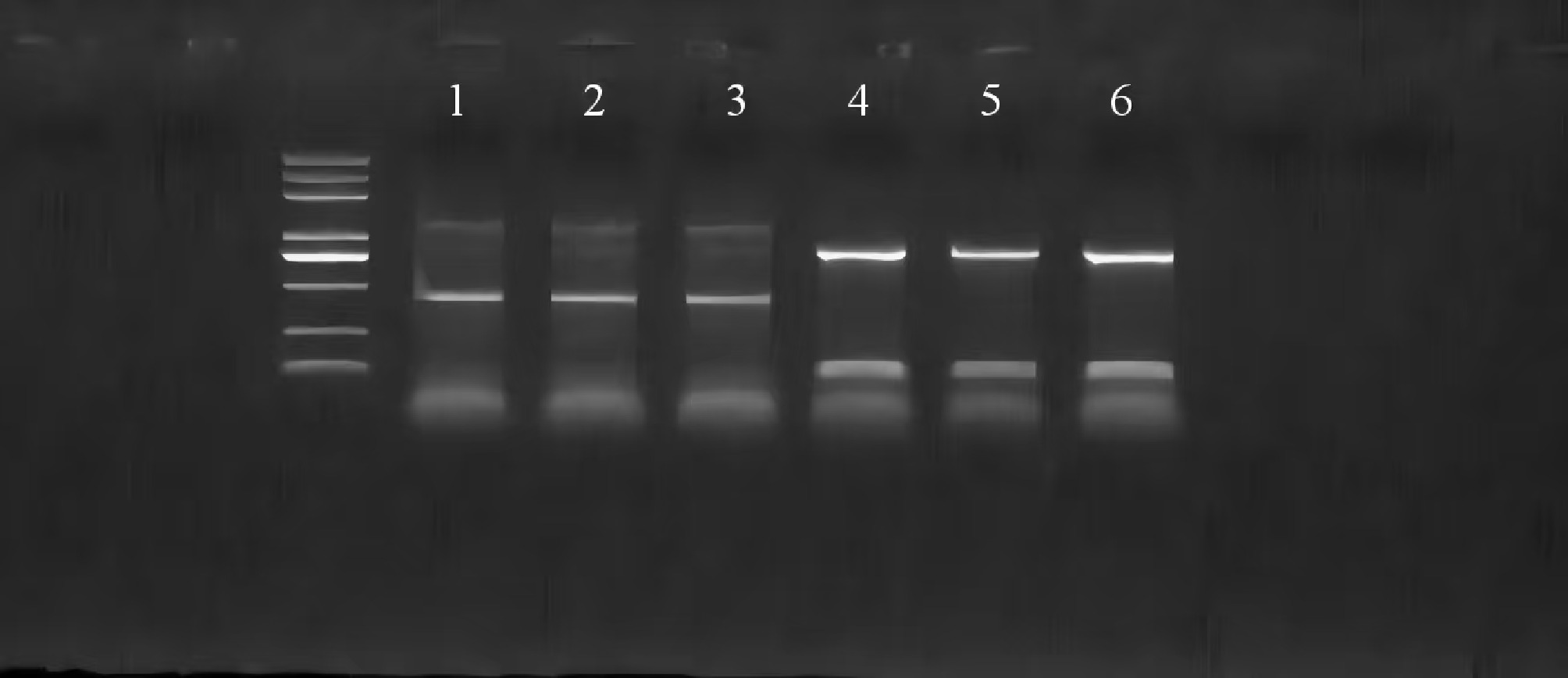
Figure 1.
PCR identification of Shiga-lipa-PBBR overexpressing colonies. Serial numbers 1–3 are wild-type P. shigelloides, and serial numbers 4–6 are P. shigelloides with overexpression of the lipa gene.
Lipoyl synthase activity assay
-
The linear regression analysis of the standard curve absorbance was employed to quantify lipoyl synthase levels. The wild-type (WT) strains exhibited an average activity of 50.5 ng/L. In contrast, the lipa-overexpressing (OE) strains demonstrated a significantly elevated activity level, averaging 61.2 ng/L (t = 9.418, p < 0.001).
Biofilm formation assay
-
Crystal violet staining revealed a significant increase in biofilm formation in the OE strains (OD575 = 0.19 ± 0.02) compared to the wild-type (WT) strains (OD575 = 0.17 ± 0.01; t = 4.5, p < 0.001), thereby corroborating the role of lipa in virulence.
Bacterial adhesion assay
-
Microscopic analysis demonstrated enhanced mucus adhesion in OE strains, characterized by purple-stained aggregates (Fig. 2b), in contrast to the dispersed, red-stained wild-type (WT) bacteria (Fig. 2a). Quantitative analysis revealed an average of 903 bacteria per field in the WT group, compared to 1,339 bacteria per field in the OE group. This observation suggests a modification in membrane permeability, potentially attributable to the overexpression of lipa.
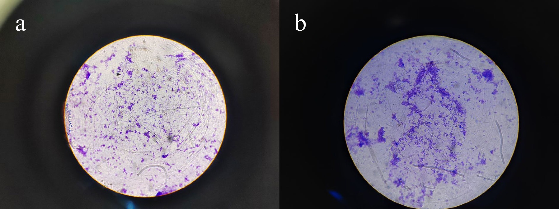
Figure 2.
The adhesion of wild-type P. shigelloides and lipa gene overexpressed P. shigelloides to the mucus on the body surface of the Silurus meridionalis. (a) staining of wild-type P. shigelloides, and (b) staining of P. shigelloides overexpressing the lipa gene.
Growth characteristics of the lipa-overexpressing strain
-
To exclude the possibility that enhanced virulence was due to altered bacterial fitness, we compared the growth curves of wild-type and lipa-overexpressing P. shigelloides. The results showed that overexpression of the lipa gene causes a 2-h growth delay in P. shigelloides, but it exhibits a higher plateau period (Fig. 3).
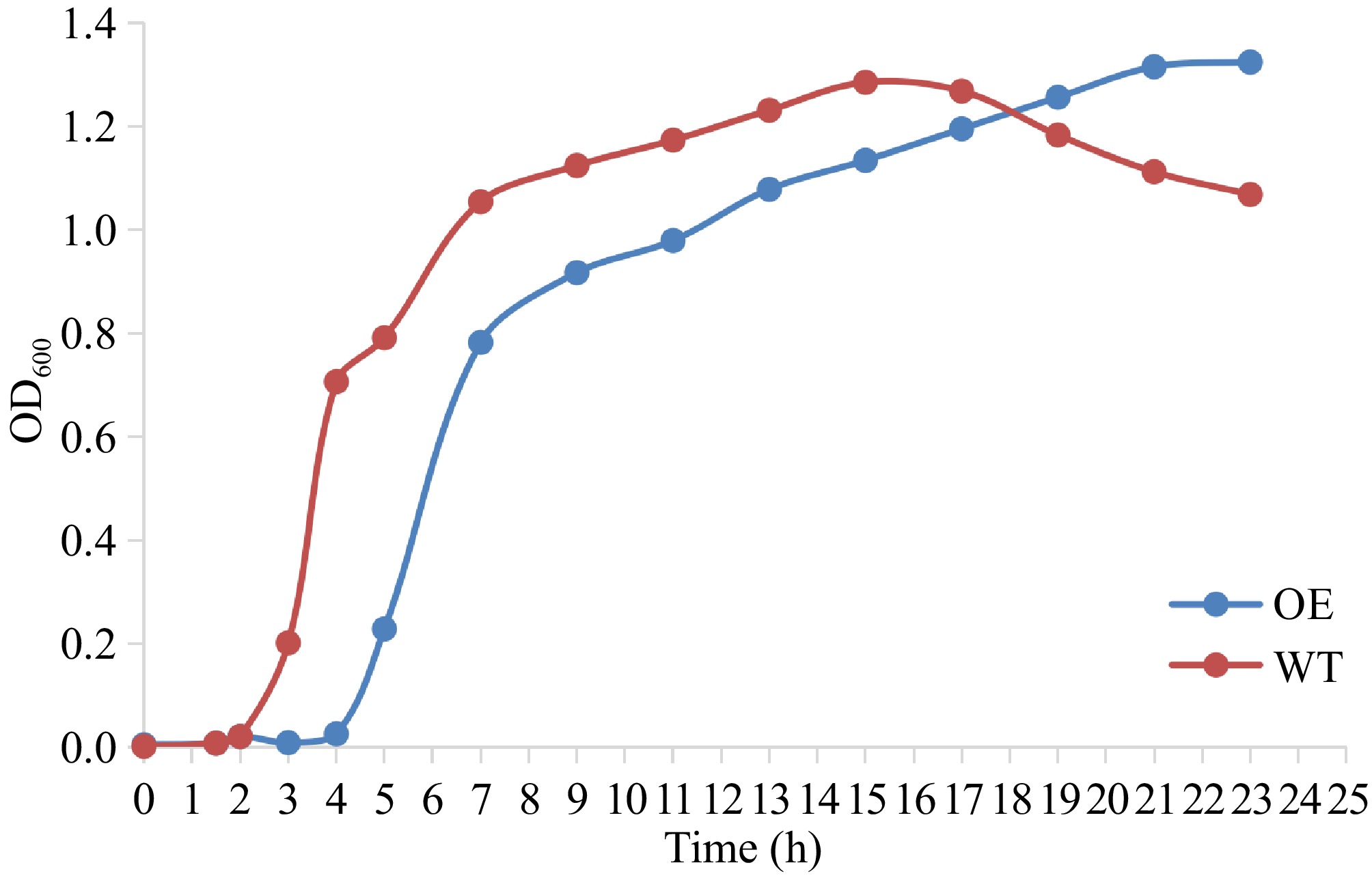
Figure 3.
Growth curves of wild-type (WT) and lipa-overexpressing (OE) P. shigelloides.
Clinical symptoms post-infection
-
Seventy-two hours post-infection, no lesions were observed in the control group (Fig. 4a). In contrast, the wild-type group demonstrated hemorrhaging in the anal and tail fins, accompanied by skin darkening (Fig. 4b). The overexpression group presented with severe symptoms, including excessive mucus secretion, fin hemorrhage, cloacal swelling, and necrotic lesions (Fig. 4c, d).
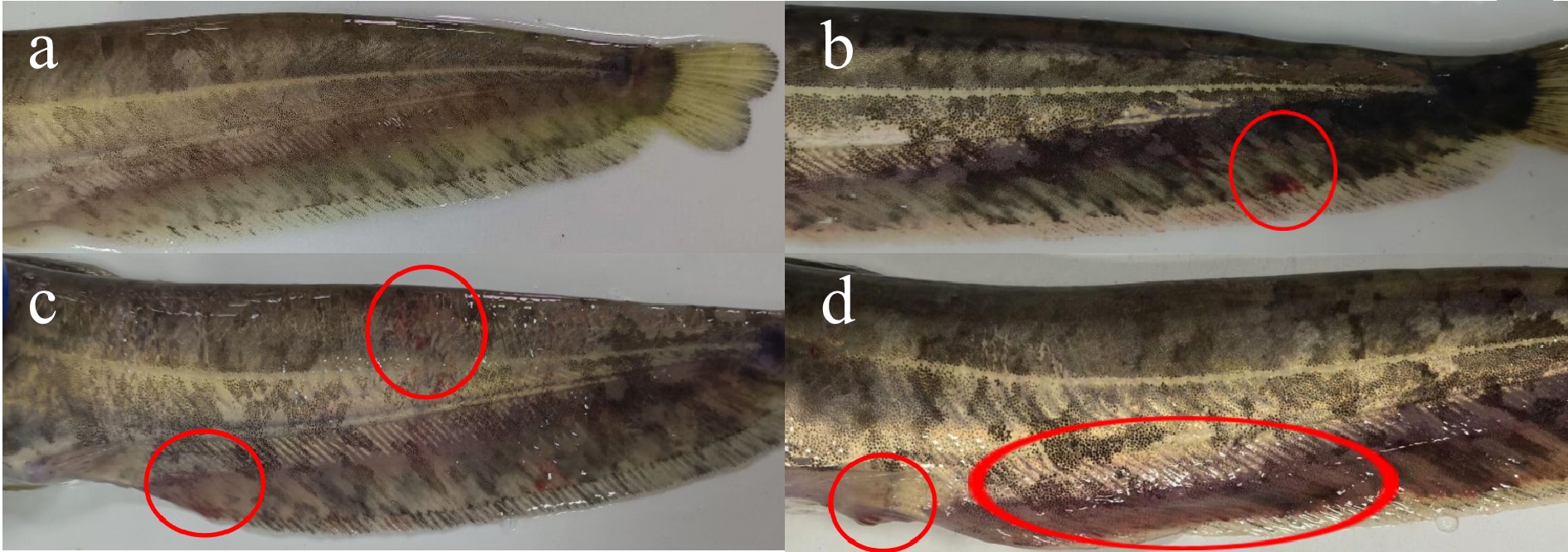
Figure 4.
Symptoms of P. shigelloides infection for 72 h. (a) asymptomatic Silurus meridionalis injected with sterile saline in the control group; (b) bleeding on the anal fin and tail fin with blackness on the body surface in the wild group; (c) obvious lesions on the body surface with bleeding on the tail fin in the overexpression group; and (d) redness and swelling of the genital orifice, black skin and bleeding on the anal fin and the tail fin in the overexpression group.
Venn diagram analysis
-
A total of 10,911 genes (77.56% of total) were commonly expressed between WT and OE groups, and 7,675 proteins (99.22% of total) were shared between the two groups (Fig. 5a, b).
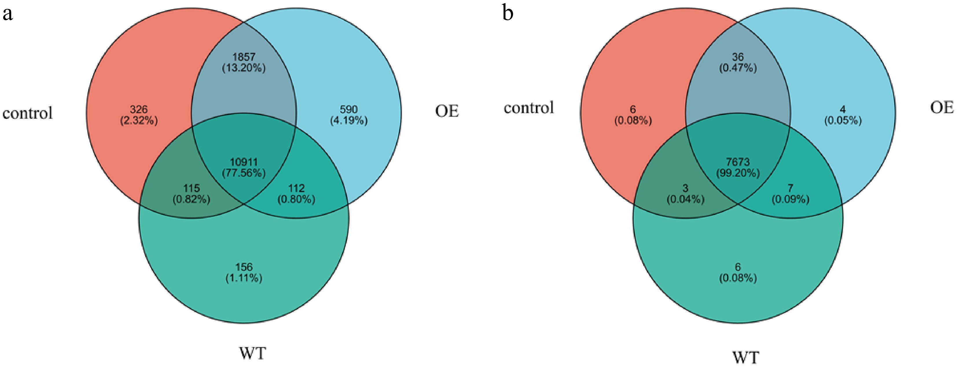
Figure 5.
Venn diagram of genes and proteins. The overlapped part of the figure represents the number of genes and proteins shared between groups; the part without overlap represents the number of genes and proteins unique to the group, and the numbers represent the corresponding gene and protein numbers. Among them, WT represents the wild-type strain group, and OE represents the overexpressed strain group.
Differentially expressed genes (DEGs) and proteins (DEPs)
-
Differential expression analysis between OE and WT groups identified 476 DEGs (179 up, 297 down) and 350 DEPs (164 up, 186 down), including 66 immune-related DEPs (Fig. 6). GO and KEGG enrichment analyses were performed to explore the potential functional roles of these differential molecules.
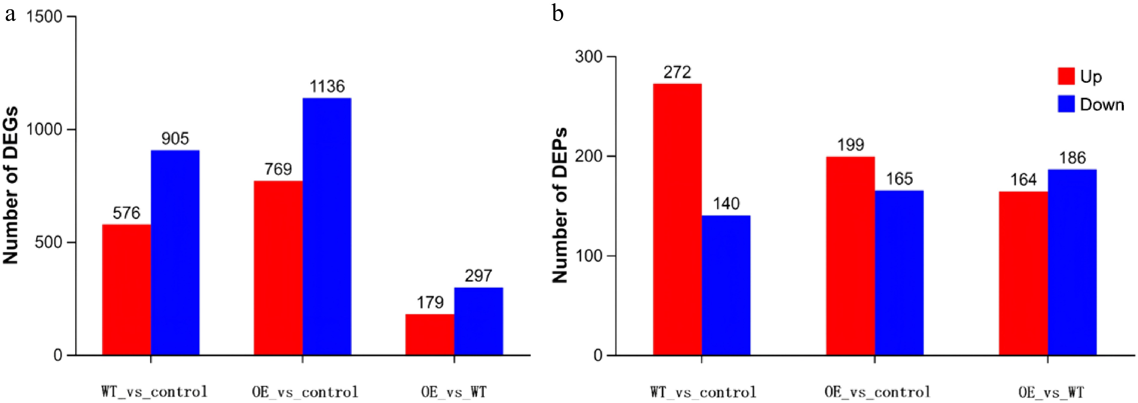
Figure 6.
Statistics of differential proteins and genes. Note: The horizontal coordinate is each difference comparison group. By default, red represents the upregulated gene and protein in the difference comparison group, and blue represents the downregulated gene and protein. Among them, WT represents the wild-type strain group, and OE represents the overexpressed strain group.
Integrated transcriptomic and proteomic analysis
-
By integrating DIA proteomic and transcriptomic data, the expression levels of immune-related proteins and genes were analyzed respectively through clustering heat maps. Among them, C6, NRPL4, and LZMC showed consistent expression trends in the transcriptome and proteome, and were all significantly upregulated in the OE group. The color gradient from blue to red represents the Spearman correlation coefficient (ρ) ranging from –1 to 1, with blue indicating negative correlation, white indicating no correlation, and red indicating positive correlation. X-axis denotes the identified proteins, and the Y-axis denotes the corresponding genes. Only significant correlations (p < 0.05) are displayed. In these visualizations, color intensity corresponds to expression level, facilitating the visual identification of molecular clusters potentially pivotal to the immune response and their expression dynamics (Fig. 7).
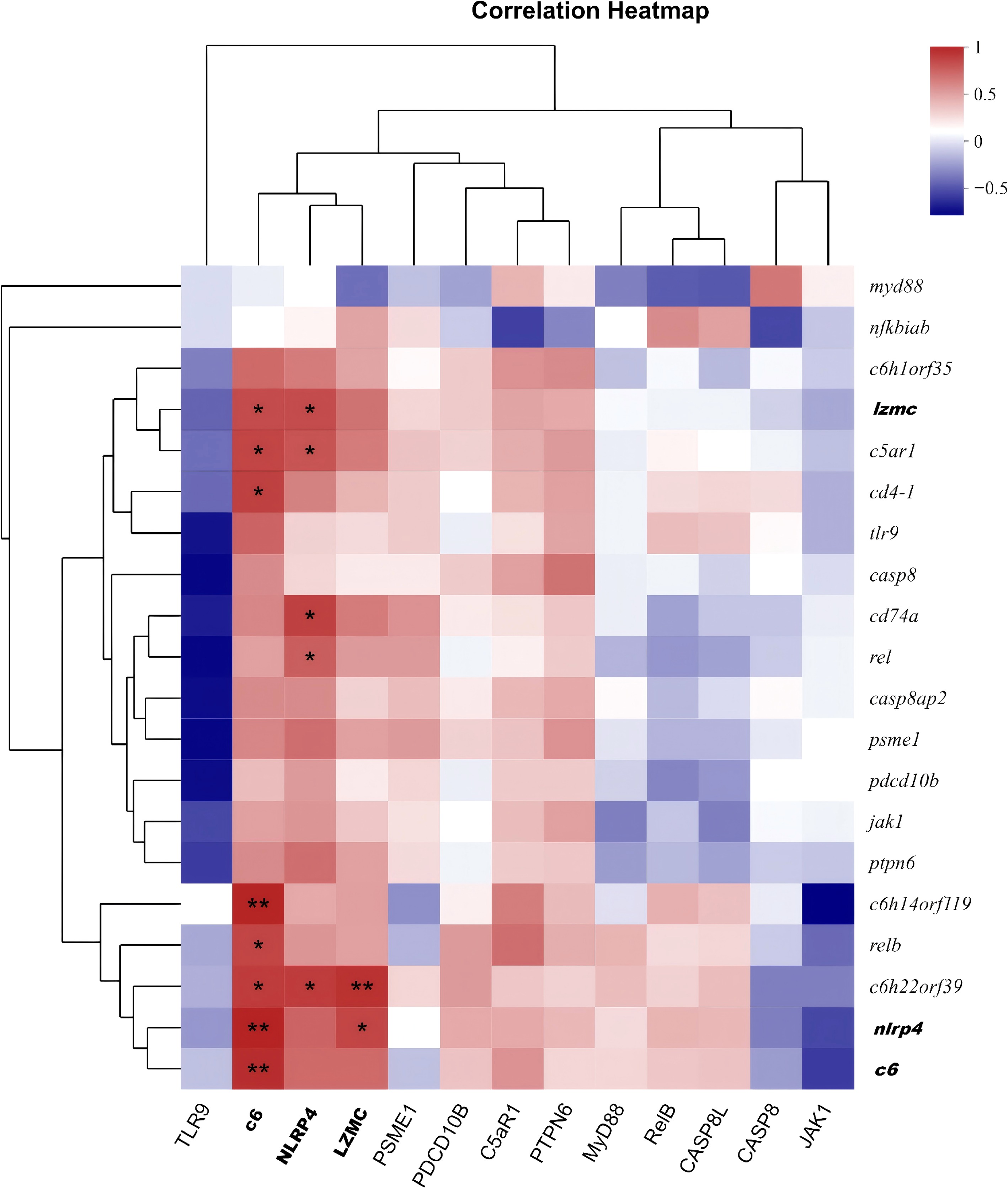
Figure 7.
Spearman correlation heatmap. Spearman correlation heatmap of immune-related molecules between the WT and OE groups. The color gradient from blue to red represents the Spearman correlation coefficient (ρ) ranging from –1 to 1; red indicates positive correlation, white indicates no correlation, and blue indicates negative correlation. The X-axis denotes proteins, and the Y-axis denotes genes. Only significant correlations (p < 0.05) are shown. Data are from three biological replicates per group.
Functional annotation of immune-related DEPs
-
GO enrichment analysis of immune-related DEPs indicated significant enrichment in biological processes such as negative regulation of wound response, hemostasis, and coagulation, as well as in the cellular component membrane attack complex (Fig. 8). Subsequent KEGG pathway analysis further revealed that these DEPs were predominantly involved in NOD-like receptor signaling, regulation of the actin cytoskeleton, and neuroactive ligand-receptor interaction (Fig. 9). Immune-related DEPs were enriched in negative regulation of wound response, hemostasis, and coagulation (biological processes), and membrane attack complex (cellular component) (Fig. 9).
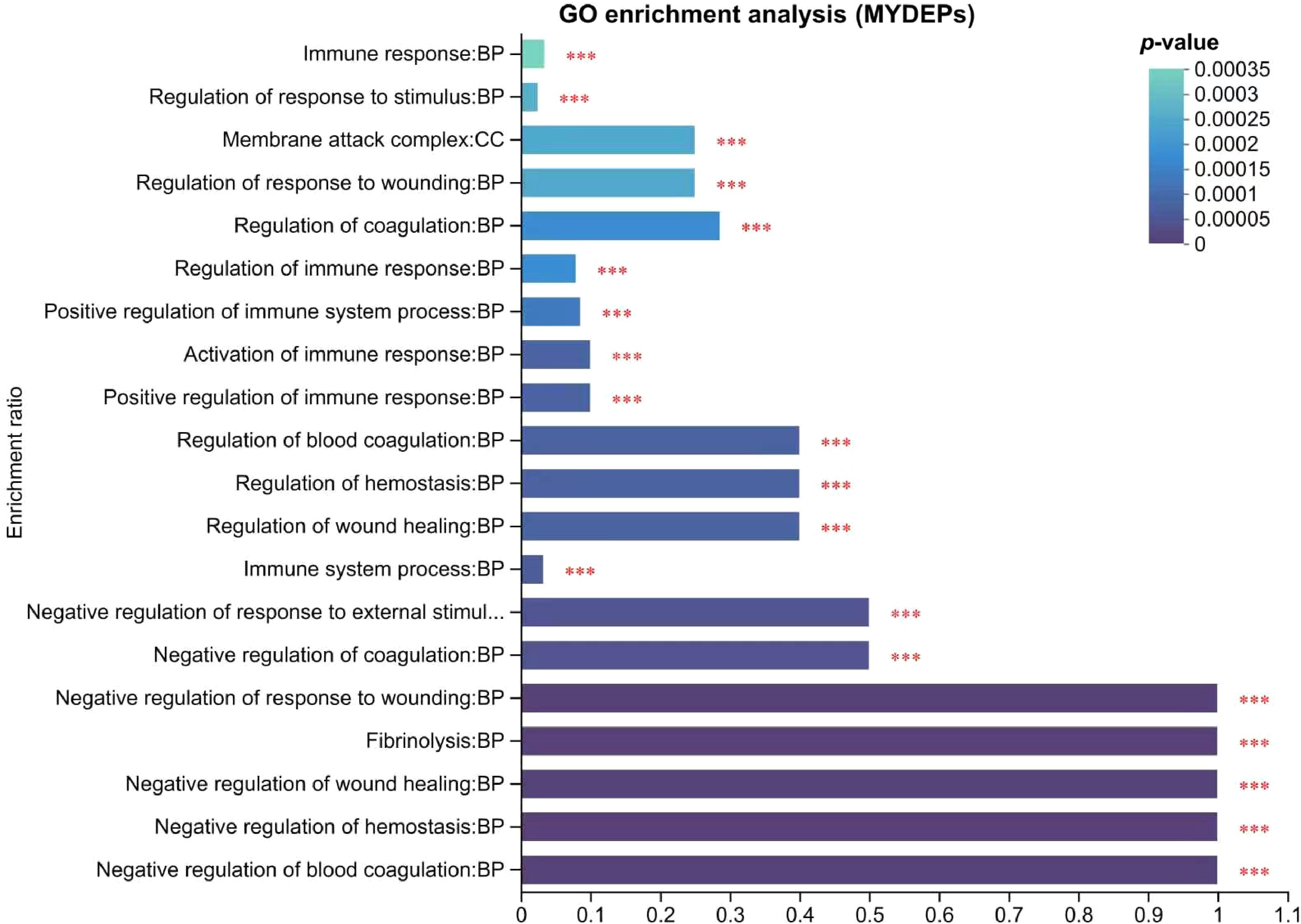
Figure 8.
Analysis of immune-related DEPs GO. Note: The abscissa represents the GO term, and the ordinate represents the enrichment rate (refers to the ratio of the number of proteins enriched in the GO term to the number of proteins annotated to the GO term; the greater the ratio, the greater the degree of enrichment). The column color gradient indicates the significance of enrichment, with p or FDR < 0.001 marked as ***, p or FDR < 0.01 marked as **, and p or FDR < 0.05 marked as *.
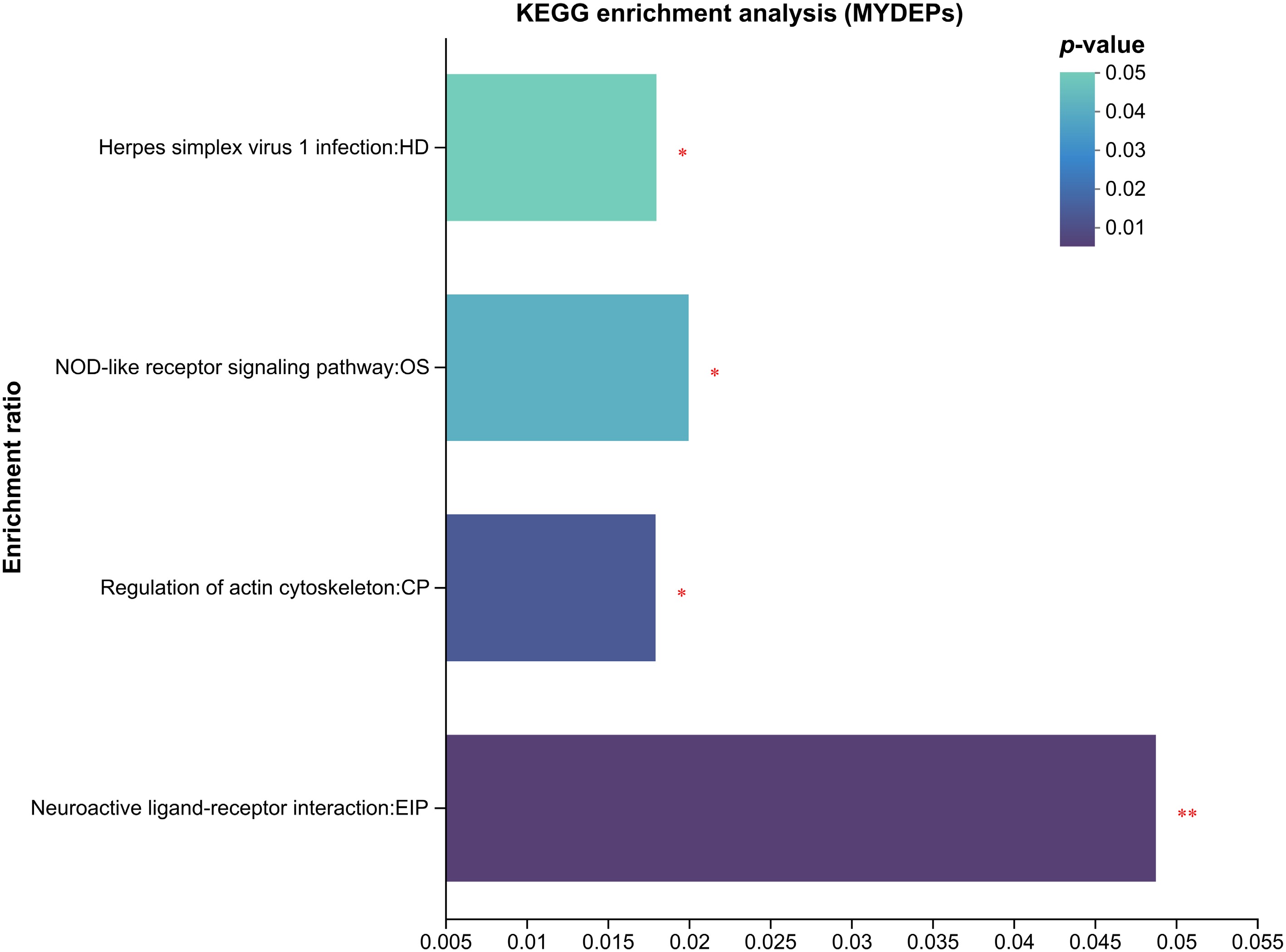
Figure 9.
Enrichment analysis of immune-related DEPs in KEGG. The abscissa represents the pathway name, and the ordinate represents the enrichment rate (refers to the ratio of the number of proteins enriched in the pathway to the number of proteins annotated to the pathway; the larger the ratio, the greater the enrichment). The column color gradient indicates the significance of the enrichment, and the darker the default color, the more significant the enrichment of the KEGG term, where p or FDR < 0.01 is marked as **, and p or FDR < 0.05 is marked as *.
ELISA validation
-
When comparing the OE and WT groups, PHA exhibited no significant change, whereas LZMC demonstrated a significant difference (p < 0.05). Notably, NLRP4, C6, and A2ML1 showed highly significant differences (p < 0.01). The ELISA results were consistent with the DIA proteomic data, thereby confirming the accuracy of our proteomic analysis (Fig. 10). This validation substantiates that lipa overexpression significantly altered the expression of key immune-related proteins in infected Silurus meridionalis.
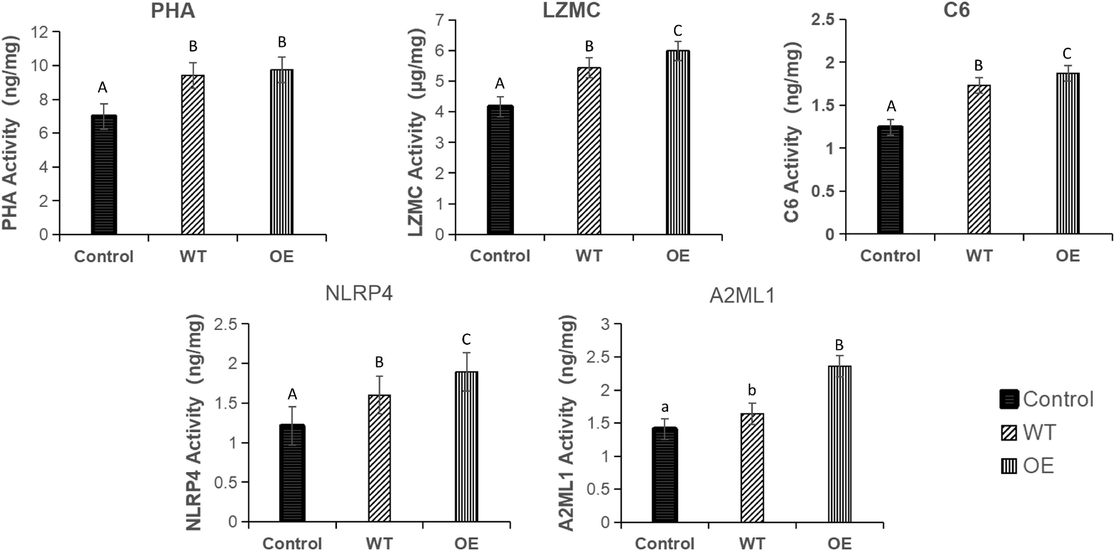
Figure 10.
Contents of PHA, LZMC, C6, NLRP4, and A2ML1 in the head kidney of Silurus meridionalis. There was no significant difference among groups represented by the same letter, while there were statistical differences at corresponding levels among groups represented by different letters. Among them, WT represents the wild strain group, and OE represents the overexpressed strain group.
qRT-PCR validation
-
To confirm RNA-Seq reliability, we analyzed the mRNA levels of the following eight differentially expressed genes (ccl20a.3, loc124381009, lgi1a, akap12B, c6, nlrp4, lzmc, and hpxa) by qRT-PCR. The results showed significant differences in ccl20a.3, akap128, c6, and lzmc genes among the three groups (p < 0.05). loc124381009 and lgi1a showed no differences between the WT and OE groups but differed significantly from the control group (p < 0.01). NLRP4 and hpxa showed no significant difference between the control p and WT groups but differed significantly from the OE group (p < 0.05) (Fig. 11).
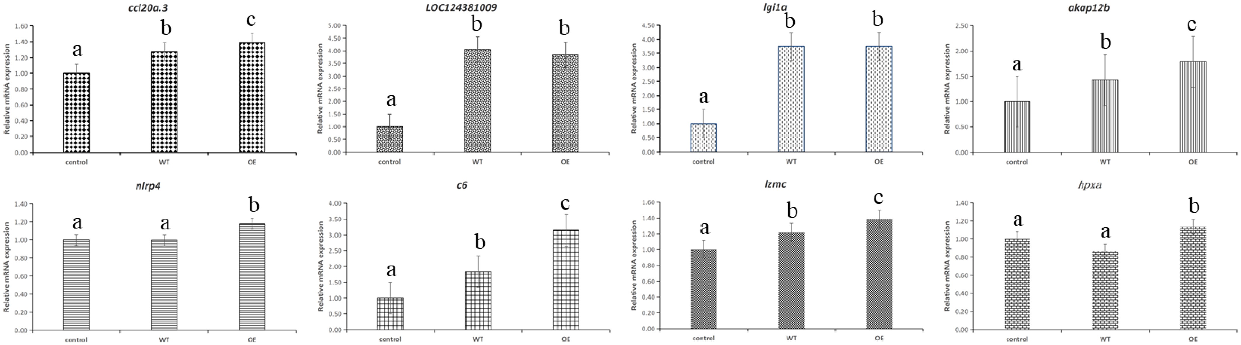
Figure 11.
Verification results of eight DEGs qRT-PCR.
-
STRING analysis of immune-related DEPs revealed interconnected nodes (e.g., complement C6, A2ML, NLRP) with high interaction scores (Fig. 12). The results indicate that C6 primarily interacts with C5 and C7, collectively participating in the assembly of the membrane attack complex (MAC). A2ML, an alpha-2-macroglobulin homolog, may synergize with complement proteins like C6 in immune signal transduction and shares a common immune pathway with HPXB. NLRP1, a member of the NLR family, forms inflammasome complexes with proteins such as IRAK4, contributing to the release of inflammatory cytokines.
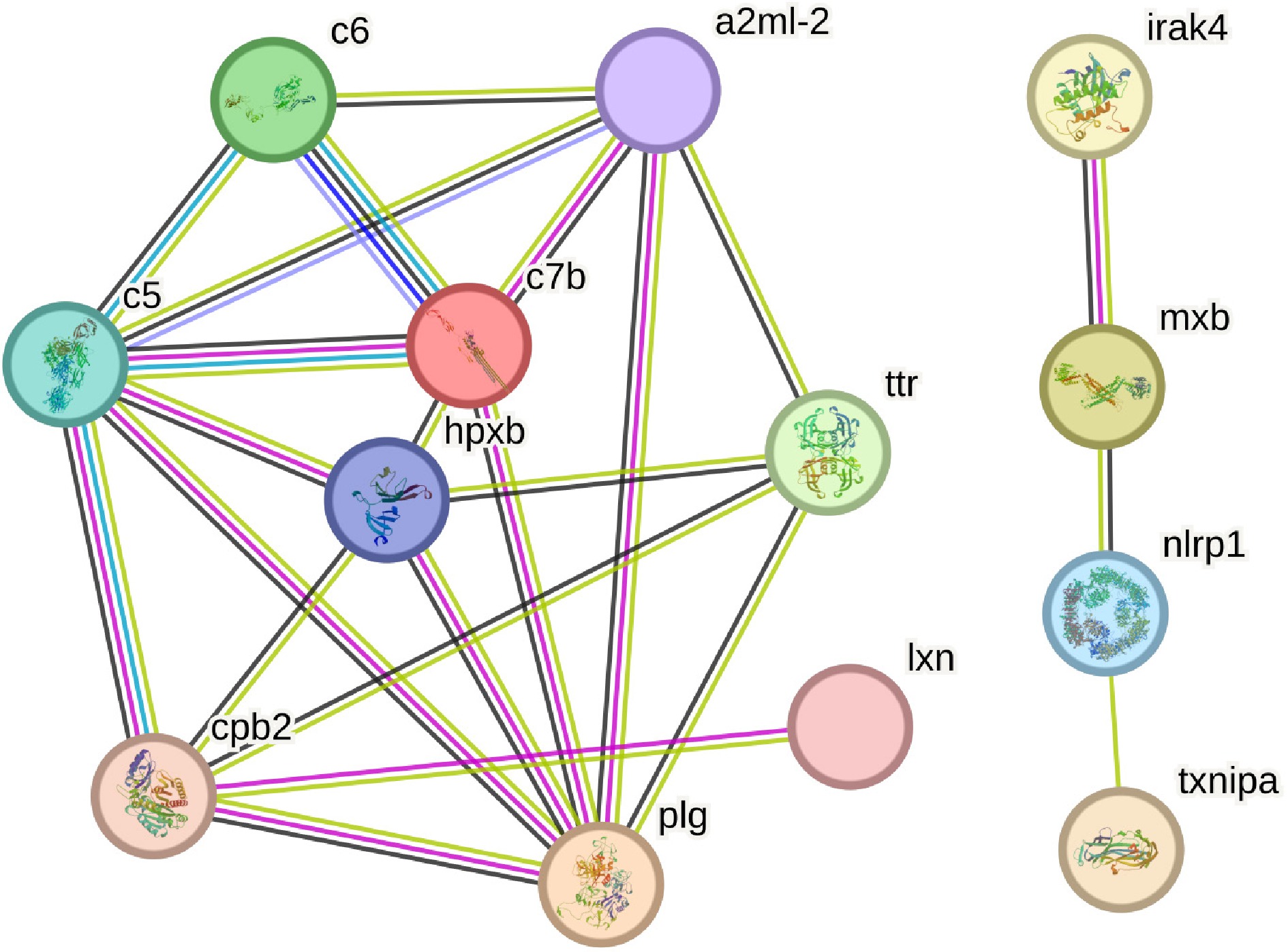
Figure 12.
Network diagram of immune-related differential protein interaction between the WT (wild) group and the OE (overexpression) group.
Coordinated regulation of complement C6 and Alpha-2-Macroglobulin Like-1
-
The interaction between the complement and coagulation cascades is illustrated (Fig. 13). During infection with lipa-overexpressing strains, both C6, crucial for MAC assembly, and the thrombin inhibitor A2ML1 were upregulated, suggesting a coordinated immune response. The elevation of C6 may be driven by two non-exclusive mechanisms: increased complement recognition of bacterial surfaces altered by lipa, or pathogen-induced interference with host regulatory pathways. Importantly, A2ML1 might help prevent thrombosis by enhancing plasminogen activator activity[11]. While acute infection models likely capture peak activation states, research on different infection periods is necessary to elucidate the timing between initial complement activation and subsequent coagulation changes.
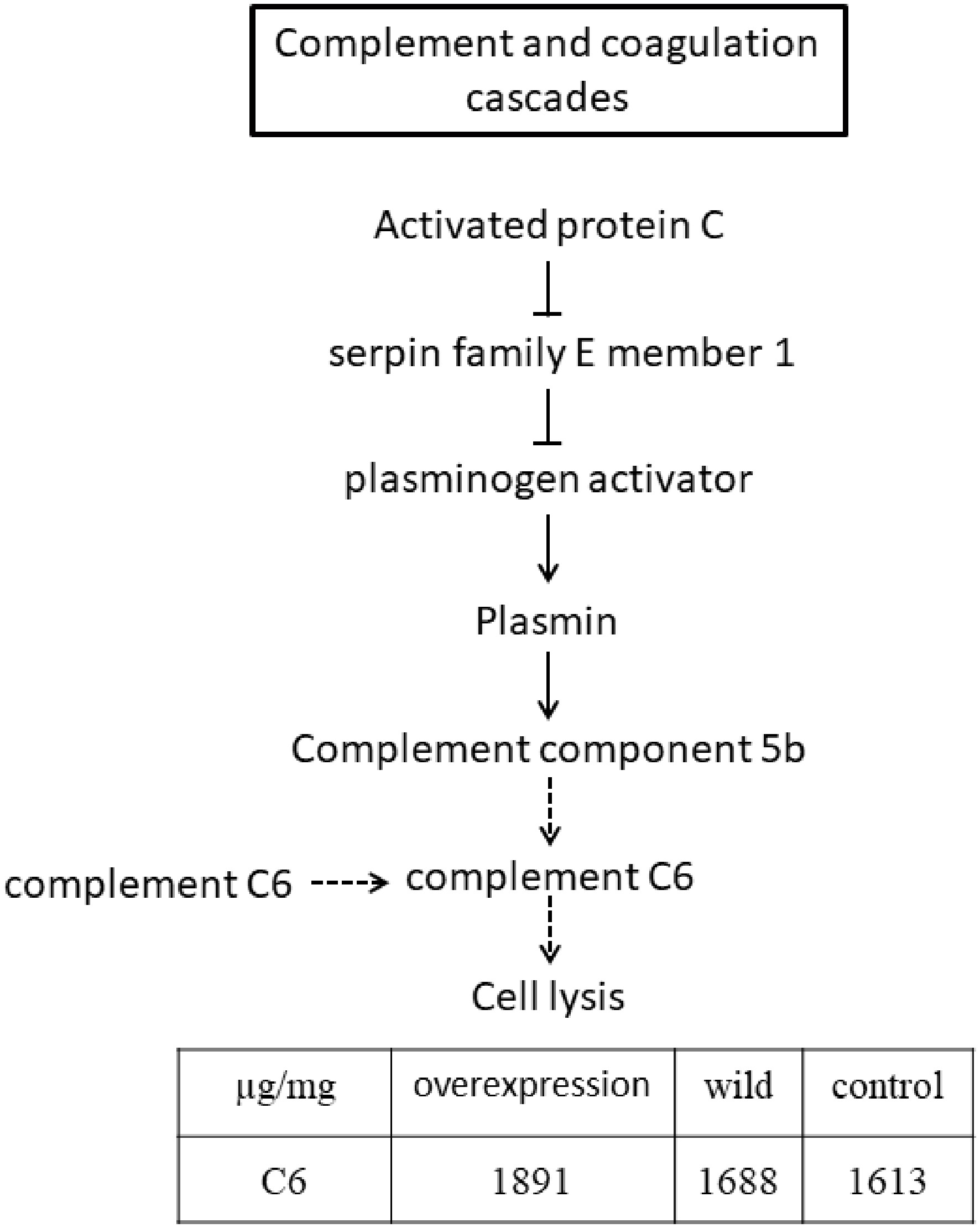
Figure 13.
Simplified complement and coagulation cascade pathway.
NLRP4 inflammasome activation in anti-bacterial defense
-
The NOD-like receptor (NLR) pathway (Fig. 14) triggers inflammation through caspase-1-dependent interleukin-1β (IL-1β) maturation. Notably, NLRP4 is significantly upregulated (p < 0.001) in infections with lipoyl synthase overexpression, suggesting that bacterial lipoic acid synthesis may modulate inflammasome responses. Emerging evidence indicates that the lipoylated metabolic protein lipoyl-E2-PDH, whose lipoylation depends on lipa, is released by bacteria and suppresses macrophage activation by reducing Toll-like receptor 1/2 (TLR1/2) recognition of bacterial lipopeptides[12]. A lipa-deficient strain induces heightened pro-inflammatory cytokine production, which is diminished in the absence of TLR2[12]. Furthermore, lipoic acid synthesis limits macrophage production of bactericidal reactive oxygen species (ROS) and reactive nitrogen species (RNS), promoting bacterial persistence during infection[13]. Although IL-1β plays a crucial role in recruiting neutrophils to clear bacterial infections[14], excessive inflammatory responses inadvertently benefit pathogens by generating nutrient-rich microenvironments[15], potentially explaining severe pathology despite strong immune responses.
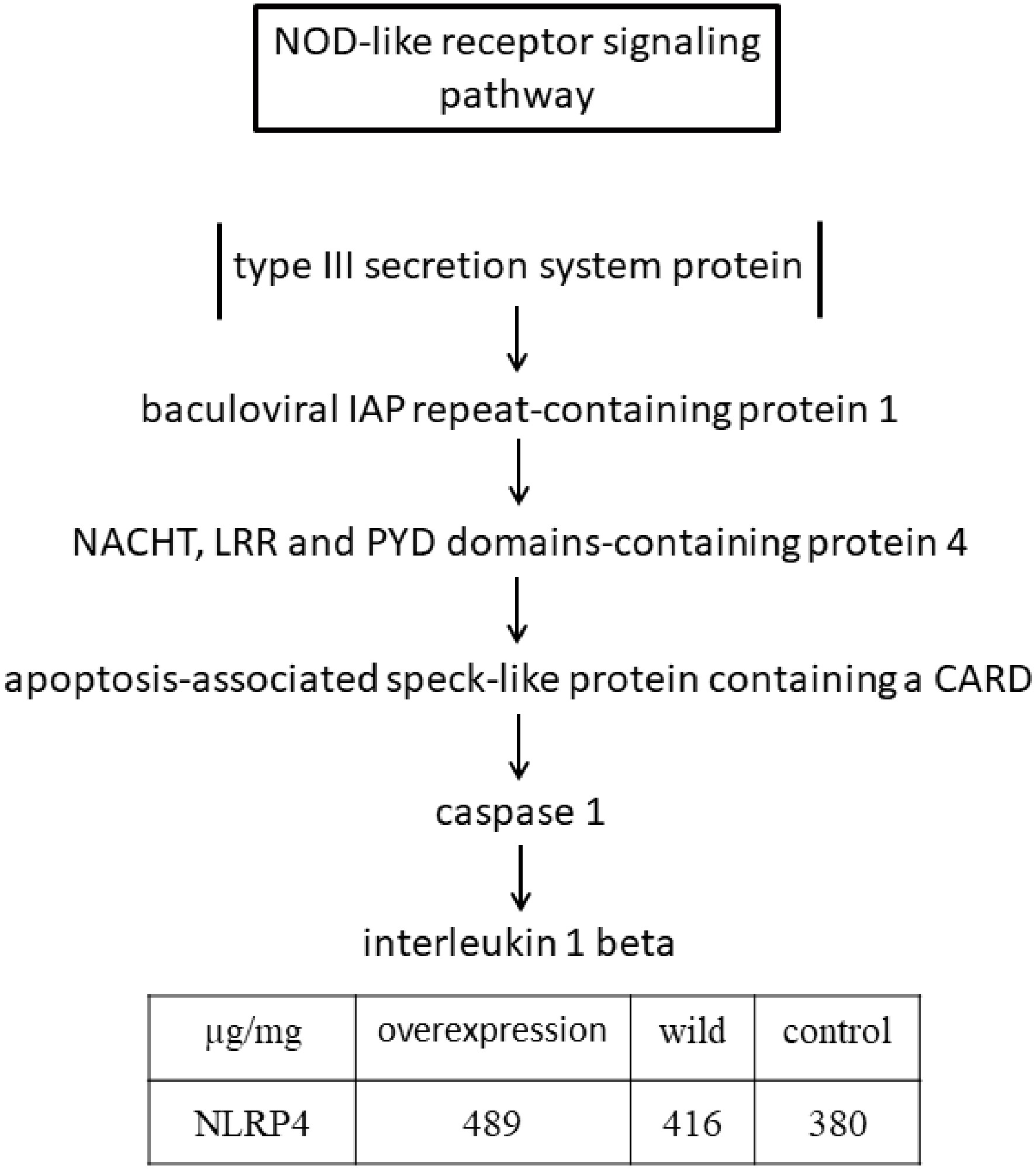
Figure 14.
Simplified diagram of the NOD-like receptor signaling pathway.
Enhanced biofilm formation capacity in lipa-overexpressing P. shigelloides
-
Our findings indicate that lipa overexpression significantly enhances biofilm formation in P. shigelloides. Bacterial biofilms, which consist of microbial communities embedded within an extracellular polymeric substance (EPS) matrix, confer resistance to environmental stressors and increase pathogenic potential[16]. However, due to the lack of a lipa knockout/complementation strain (attempts to generate a knockout were unsuccessful), causality cannot be firmly established from the current data. The lipa-mediated enhancement of biofilm formation may hypothetically involve several mechanisms: (1) the upregulation of adhesion-related proteins, such as pili, facilitating initial surface attachment; (2) the modulation of EPS synthesis to strengthen the biofilm structural integrity; and (3) potential interference with quorum sensing (QS) via the acyl-homoserine lactone (AHL) signaling pathway[17]. We acknowledge that direct experimental evidence supporting these proposed mechanisms (pili, EPS, QS) is currently lacking, and these interpretations remain speculative. The resulting fortified biofilm likely provides protection against host immune clearance and antibiotic treatments[18], highlighting lipa as a potential target for therapeutic intervention. Future research should aim to elucidate the precise roles of lipa in adhesion, EPS regulation, and QS to advance the development of anti-biofilm strategies.
lipa overexpression augments bacterial adhesion to S. meridionalis surface mucus
-
Fish epidermal mucus of fish serves as a primary immunological barrier, comprising antimicrobial peptides (AMPs), lysozymes, and immunoglobulins that neutralize invading pathogens[19,20]. Microscopic analysis demonstrated that lipa-overexpressing strains formed dense, purple-stained aggregates on S. meridionalis mucus, in contrast to the dispersed, red-stained wild-type bacteria. This phenotypic shift suggests that lipa expression alters membrane permeability, leading to: (1) the upregulation of specific adhesins[21]; and (2) modifications of metabolic pathways that facilitate sustained colonization. These adaptations might counter mucus clearance mechanisms mediated by viscosity and AMPs.
Limitations and future direction
-
Several limitations of this study should be acknowledged. First, although lipa overexpression significantly enhanced adhesion and biofilm formation, we were unable to generate a lipa knockout strain despite multiple attempts, precluding complementation experiments necessary to establish a direct causal relationship between lipa and the observed phenotypes. Second, while we have discussed potential mechanisms involving pili, EPS, and quorum sensing, these interpretations remain speculative, as direct experimental evidence—such as measurements of pilin expression, EPS production, or AHL signaling activity—is currently lacking. Consequently, our findings should be interpreted as correlative rather than mechanistic. Future studies should employ alternative strategies to disrupt lipa function (e.g., conditional knockdown or CRISPRi) and directly quantify the proposed downstream effectors to validate these hypotheses.
-
In conclusion, our in vitro experiments reveal that lipa overexpression in P. shigelloides significantly augments biofilm formation, lipoyl synthase activity, and host adhesion capacity, supporting its role in virulence through modulation of bacterial surface properties and metabolism. In S. meridionalis infection models, P. shigelloides overexpressing lipa induced more skin ulceration and systemic inflammation. Integrated transcriptomic and proteomic analysis of head kidney tissue revealed that the immune-related factors NLRP4, C6, and LZMC were significantly upregulated in the group treated with lipa-overexpressing P. shigelloides. This finding underscores that the coordinated defense response in S. meridionalis is likely mediated through the complement–coagulation cascade and NOD-like receptor signaling pathways. In addition, the aberrant downregulation of lysozyme C-like protein (LZMC) suggests efficient pathogen-mediated evasion of innate immunity. Collectively, these findings suggest that targeted inhibition of lipoyl synthase activity may represent a promising therapeutic strategy against P. shigelloides infections.
The data were analyzed using the free online majorbio cloud platform (cloud.majorbio.com). The authors express their most sincere gratitude to these individuals for their help.
-
The animal study protocol was approved by the Experimental Animal Ethics Committee of Guizhou University (protocol code EAE-GZU-2024-T103 and date of approval 12 March 2024).
-
The authors confirm their contributions to this study as follows: conceptualization: Dong R, Yang L; methodology: Yang L; investigation: Lu F; resources: Dong R; data curation: Luo D; writing—original draft preparation: Yang L; writing—review and editing, project administration, funding acquisition: Dong R; visualization: Lu F; supervision: Luo D. All authors have read and agreed to the published version of the manuscript.
-
The raw sequence data generated in this study have been deposited in the OMIX database at the National Genomics Data Center, China National Center for Bioinformation/Beijing Institute of Genomics, Chinese Academy of Sciences, under accession numbers OMIX011657 and CRA037443. The data are publicly accessible at https://ngdc.cncb.ac.cn/omix and https://ngdc.cncb.ac.cn/gsa.
-
The authors declare no conflict of interest.
- This is an open access article under the terms of the Creative Commons Attribution License, which permits use, distribution and reproduction in any medium, provided the original work is properly cited.
-
About this article
Cite this article
Yang L, Lu F, Luo D, Dong R. 2026. From gene to disease: the virulence amplification of Plesiomonas shigelloides via lipa overexpression. Journal of Applied Ichthyology 2026: e006 doi: 10.48130/jai-0026-0005 

From gene to disease: the virulence amplification of Plesiomonas shigelloides via lipa overexpression
- Received: 17 April 2026
- Revised: 17 June 2026
- Accepted: 10 August 2026
- Published online: 21 September 2026
Abstract: Frequent bacterial outbreaks in aquaculture threaten commercial fish, with Plesiomonas shigelloides being notably common due to its lipa gene, a critical virulence factor. This study overexpressed the lipa gene in fish-derived P. shigelloides to investigate its impact on biofilm formation, LIPA activity, and bacterial adhesion. We infected Silurus meridionalis for 7 d to assess pathogenicity, observing symptoms, and collected the head kidney for transcriptome and proteomic analysis. Results were confirmed by qPCR and ELISA, and we performed GO classification, KEGG pathway, and protein interaction network analysis on differentially expressed immune proteins. The findings revealed that lipa overexpression increased biofilm formation (p < 0.01), lipoyl synthase activity (p < 0.01), and bacterial adhesion compared to wild-type strains, offering novel insights into its function. In Silurus meridionalis, this overexpression led to increased hemorrhaging in the anal and caudal fins compared to the wild-type and control groups. Furthermore, q-PCR and ELISA results demonstrated that the expression levels of NACHT/LRR/PYD domain-containing protein 4 (NLRP4), complement component 6 (C6), and lysozyme C (LZMC) correlated with the transcriptome and proteomic data. Overall, lipa overexpression enhances P. shigelloides virulence and intensifies the immune responses in Silurus meridionalis, providing insights into its pathogenic mechanism and supporting healthy breeding of Silurus meridionalis.
-
Key words:
- lipa gene overexpression /
- Plesiomonas shigelloides /
- Proteomics /
- Silurus meridionalis