











-
Codonopsis pilosula (Franch.) Nannf., a commonly used medicinal herb in traditional Chinese medicine, is a perennial plant belonging to the Campanulaceae family and serves as one of the original botanical resources of Codonopsis Radix (Dangshen) in the Pharmacopoeia of the People's Republic of China[1]. It is primarily distributed across South Asia, Central Asia and East Asia, with its roots typically harvested in autumn[2]. C. pilosula is rich in various bioactive components, including polysaccharides, lobetyolin, atractylenolide III, syringin, and amino acids. These components endow it with multiple pharmacological functions, such as promoting blood circulation, nourishing blood, and promoting fluid production. Clinically, it is used to treat syndromes associated with spleen and lung deficiency, qi deficiency, and ischemia. Additionally, it exhibits therapeutic effects in expelling phlegm to alleviate cough, generating fluids to relieve thirst, and regulating gastrointestinal dysfunction. As a result, C. pilosula is frequently used as a substitute for ginseng (Panax spp.) in medical applications and is extensively utilized in Chinese herbal health products[3,4]. To date, more than 40 species of Codonopsis have been identified[5]. However, accurate species identification within the genus remains challenging because of the high morphological similarity among congeners.
Chloroplasts are a class of organelles found in most plants and algae, typically exhibiting a genome size ranging from 120 to 200 kb and encoding approximately 110−140 genes[6]. Compared with the mitochondrial and nuclear genomes, the chloroplast genome possesses a relatively compact genomic structure and its own transcription/translation systems. Owing to its relatively stable genome organization, highly conserved gene structure, moderate evolutionary rate, and predominant maternal inheritance in most angiosperms, the chloroplast genome has emerged as a powerful tool and is widely used in species identification and phylogenetic studies[7,8]. The majority of plant chloroplast genomes are circular and exhibit a typical quadripartite structure consisting of a large single-copy (LSC) region, a small single-copy (SSC) region, and a pair of inverted repeat (IR) regions[9]. Each of these structural regions plays a distinct role in defining chloroplasts' functionality, facilitating the transcription of external genetic information, and ultimately influencing the overall physiological activities of the chloroplast.
At present, most research on C. pilosula is predominantly focused on techniques of extracting its chemical components and exploration of its pharmacological activities. Although the chloroplast genome sequence of C. pilosula is available in public databases (GenBank accession number MT982394.1 or NC_060312.1), a comparative analysis of chloroplast genomes among the currently available Codonopsis species, which is useful for understanding its phylogenetic relationships and identifying candidate regions for genetic markers, remains limited. Moreover, the phylogenetic relationship between C. pilosula and its subspecies C. pilosula subsp. tangshen has not been adequately examined at the chloroplast genome level, despite their distinct medicinal applications in traditional Chinese medicine. This study therefore aims to (1) provide an independently assembled and annotated chloroplast genome of C. pilosula for in-depth characterization, and (2) perform a comparative genomic analysis of 11 Codonopsis chloroplast genomes (representing nine distinct species) to investigate their phylogenetic relationships, identify genomic variations, and determine highly divergent regions suitable for developing molecular markers.
-
Fresh leaves of C. pilosula were collected from the medicinal botanical garden of Shanghai University of Traditional Chinese Medicine. A voucher specimen (specimen number: SHUTCM-DS202501) was deposited in the herbarium of Shanghai University of Traditional Chinese Medicine. Total genomic DNA was isolated from the leaves using the cetrimonium bromide (CTAB) method[10]. The concentration and purity of the extracted DNA were quantified using a Nanodrop 2000 spectrophotometer (Thermo Scientific), and its integrity was verified by electrophoresis on a 1% agarose gel stained with ethidium bromide. The chloroplast genome of C. pilosula was sequenced using the Illumina HiSeq 2500 platform (Biozeron, Shanghai, China) with the total genomic DNA, and 150-bp paired-end reads were generated.
Assembly and annotation of the chloroplast genome
-
The raw sequencing data were assessed for quality using FastQC (version 0.11.9,
www.bioinformatics.babraham.ac.uk/projects/fastqc ), followed by trimming and filtering with Trimmomatic v. 0.39 under the default settings[11]. The filtered reads were assembled de novo using GetOrganelle v. 1.7.5 with the default parameters (k-mer 21, 45, 65, 85, 105)[12]. The high-quality paired-end reads were then aligned to the assembled chloroplast genome using BWA v. 0.7.17[13], and SAMtools v. 1.10[14] was used to process the alignment results to determine the average read coverage depth. IR boundaries were validated using read mapping in IGV v. 2.19.2 with the assembled genome as a reference, followed by manual inspection of coverage consistency at the LSC–IRb, IRb–SSC, SSC–IRa, and IRa–LSC junctions. The assembled chloroplast genome of C. pilosula was annotated using CPGAVAS2[15] (http://47.96.249.172:16019/analyzer/home ) with the default parameters and the 2,544-plastome reference dataset. For comparison, the chloroplast genomes of other Codonopsis plants retrieved from the National Center for Biotechnology Information (NCBI), with the original references provided in Table 1, were also annotated using the same pipeline. The start and stop codons of protein-coding genes were manually adjusted according to sequence alignment with homologous genes from closely related species available in NCBI to ensure the annotations' accuracy. The physical map of the C. pilosula chloroplast genome was generated by the Organellar Genome Draw (OGDRAW) v. 1.3.1[16].Table 1. Summary of the 11 Codonopsis chloroplast genomes analyzed in this study.
Species NCBI accession number Length
(bp)GC (%) LSC IRs SSC Coding regions Reference Size (bp) GC (%) Size (bp) GC (%) Size (bp) GC (%) Size (bp) GC (%) C. pilosula PQ541237 170,614 38.14 86,117 36.74 38,425 40.31 7,647 32.13 85,992 38.68 This study C. pilosula MT982394.1/NC_060312.1a 170,667 38.13 86,434 36.73 38,293 40.30 7,647 32.13 76,092 39.16 Unpublished Codonopsis bhutanica MW149120.1/NC_063738.1 169,718 38.38 84,771 37.13 38,621 40.36 7,705 32.29 84,258 38.83 Unpublished Codonopsis tsinlingensis MN122102.1/NC_056284.1 170,253 38.34 85,408 37.02 38,334 40.46 8,177 32.24 84,012 38.94 [27] Codonopsis minima KY587457.1/NC_036311.1 169,321 38.28 85,506 36.86 37,875 40.50 8,065 32.44 88,746 38.53 [28] Codonopsis lanceolata MH251613.1 170,075 38.24 85,241 36.87 38,387 40.37 8,060 32.34 88,659 38.52 Unpublished Codonopsis lanceolata MH018574.1 169,447 38.24 85,253 36.88 38,067 40.39 8,060 32.37 83,331 38.67 [29] Codonopsis javanica MW149125.1/NC_063743.1 172,874 37.97 87,389 36.76 38,698 39.99 8,089 31.66 69,894 39.23 Unpublished Codonopsis hongii MN849357.1/NC_049093.1 169,524 38.24 85,326 36.88 38,143 40.40 7,912 32.03 72,346 39.01 [30] C. pilosula subsp. tangshen MW415426.1 170,672 38.15 86,108 36.75 38,455 40.31 7,654 32.07 90,030 38.55 [31] Codonopsis convolvulacea NC_060685.1 183,616 38.72 96,234 37.59 38,454 40.74 10,474 34.27 75,537 39.41 [32] a In this table, accession numbers separated by a slash (e.g., MT982394.1/NC_060312.1) represent identical sequences confirmed by sequence alignment. Repeat sequence analysis
-
Simple sequence repeats (SSRs) in the chloroplast genomes of Codonopsis plants were identified using the MISA software (
https://webblast.ipk-gatersleben.de/misa/index.php?action=1 ). The minimum repeat thresholds were defined as follows: 10 for mononucleotides, 4 for dinucleotides, and 3 for trinucleotides, tetranucleotides, pentanucleotides, and hexanucleotides. Long repeat sequences in the chloroplast genomes were quantified using the REPuter software (https://bibiserv.cebitec.uni-bielefeld.de/reputer ). This analysis included the identification of palindromic repeats, forward repeats, reverse repeats, and complementary repeats. The parameters for REPuter were configured with a maximum of 5,000 repeats, a minimum of 30 repeats, and a Hamming distance of 3. Heatmap visualization was ultimately performed using TBtools 2.029[17].Comparative analysis of chloroplast genomes
-
To investigate the structural conservation and divergence among Codonopsis plants, a comparative genomic analysis was conducted using 11 complete chloroplast genomes. Among these, 10 sequences were obtained from the NCBI database, and 1 chloroplast genome was newly assembled in this study. The whole-genome alignment and visualization were performed with mVISTA (
https://genome.lbl.gov/vista/mvista/submit.shtml ) via the Shuffle-LAGAN model, with the newly assembled chloroplast genome serving as the reference. Structural dynamics at the IR boundaries were examined using CPJSdraw (http://cloud.genepioneer.com:9929/#/tool/alltool/detail/335 ) with the default parameters.Codon usage bias and sequence polymorphism analysis
-
Codon usage bias analysis was performed using CPStools[18]. To minimize potential biases, stringent filtering criteria were implemented: (1) Only coding sequences exceeding 300 bp in length were retained, (2) paralogous genes were removed, and (3) only genes containing canonical ATG initiation codons were selected. To investigate hypervariable regions in the chloroplast genomes of Codonopsis and establish a scientific identification system, multiple sequence alignment of conserved genes from 11 Codonopsis plants was conducted using MAFFT v7.505[19]. The aligned sequences were subsequently refined with Gblocks for quality control and concatenated into a supermatrix. Sliding window analysis was then performed using DnaSP v6[20] to calculate nucleotide diversity (Pi) across the concatenated alignment of conserved chloroplast genes, using a 600-bp window size with incremental steps of 200 bp[21]. For each window, the Pi was calculated, and the midpoint position was determined on the basis of the coordinates in the concatenated alignment.
Molecular evolution in Codonopsis chloroplast genomes
-
To investigate the evolutionary dynamics of chloroplast genomes in Codonopsis plants, a comprehensive analysis of the natural selection pressures acting on protein-coding sequences was conducted. A conserved set of orthologous genes was identified across all 11 Codonopsis chloroplast genomes, including the newly sequenced genome. For the analysis of selection pressure, the coding sequences of the chloroplast protein-coding genes were first aligned using MAFFT with the default parameters to ensure accurate codon-based alignment. The aligned sequences were then used as input for the yn00 program (implemented in the PAML package) according to Yang and Nielsen's method[22] to estimate nonsynonymous substitution rates (dN), synonymous substitution rates (dS), and their ratio (ω = dN/dS), with the newly assembled C. pilosula chloroplast genome designated as the reference. The ω ratio is a commonly used indicator of selective pressure in protein-coding genes, where ω < 1 generally reflects purifying selection, ω = 1 indicates neutral evolution, and ω > 1 may suggest positive selection[23].
Phylogenetic analysis
-
To infer the primary interspecific phylogenetic relationships, a maximum likelihood (ML) tree was constructed using the complete chloroplast genome sequences from 11 Codonopsis plants, with Lobelia chinensis and Platycodon grandiflorus as outgroup taxa. The ML approach was selected because it explicitly incorporates an evolutionary model and generally provides a more reliable topology for species-level phylogenetic inference. Sequence alignment was performed with MAFFT, followed by data refinement using trimAl v1.4 to trim ambiguous regions[24]. The ML tree was constructed using IQ-TREE 2[25], with the best-fit nucleotide substitution model automatically selected by the integrated ModelFinder Plus module on the basis of the Bayesian information criterion (BIC). Branch support was assessed using 5,000 ultrafast bootstrap replicates. The resulting ML tree was visualized and annotated using the iTOL online platform[26].
To evaluate the identification potential of candidate molecular markers, the complete ycf1 and ndhF genes were extracted from each of the 11 Codonopsis chloroplast genomes. The average lengths of ycf1 and ndhF were 6,794 and 2,246 bp, respectively. For each taxon, the two fragments were manually concatenated in the order ycf1–ndhF without a spacer, yielding an average concatenated length of 9,040 bp. Because these sequences were obtained directly from the assembled chloroplast genomes, no polymerase chain reaction (PCR) primers were used; for future experimental validation, primers can be designed from the conserved flanking regions. Three separate neighbor-joining (NJ) trees based on the ycf1 genes, the ndhF genes, and the combined ycf1–ndhF fragments were constructed using MEGA v 5.05 with the Kimura 2-parameter (K2P) model with pairwise deletion of gaps. Nodal stability was assessed with 1,000 bootstrap replicates. The NJ analysis was used as a distance-based exploratory method to evaluate the discriminatory power of the candidate markers, whereas the ML analysis was used as the primary model-based method for phylogenetic inference. The resulting NJ topologies were compared with the ML phylogeny to further validate the utility of these hypervariable regions for species identification within Codonopsis.
-
In this study, the complete chloroplast genome of C. pilosula was sequenced, assembled and compared with those of 10 other Codonopsis plants. After removing the adapter sequences and low-quality reads, approximately 8.08 Gb of clean reads was obtained from the total sequencing data for assembly. The newly assembled complete chloroplast genome of C. pilosula was 170,614 bp in length with a GC content of 38.14% (Fig. 1 and Table 1). The assembly was supported by a high average read coverage depth of 7,750× (Supplementary Fig. S1), indicating high confidence in the assembled sequence. The chloroplast genome exhibited a typical quadripartite structure, consisting of two IR regions of 38,425 bp each (40.31% GC content), separated by an SSC region of 7,647 bp (32.13% GC content) and an LSC region of 86,117 bp (36.74% GC content).
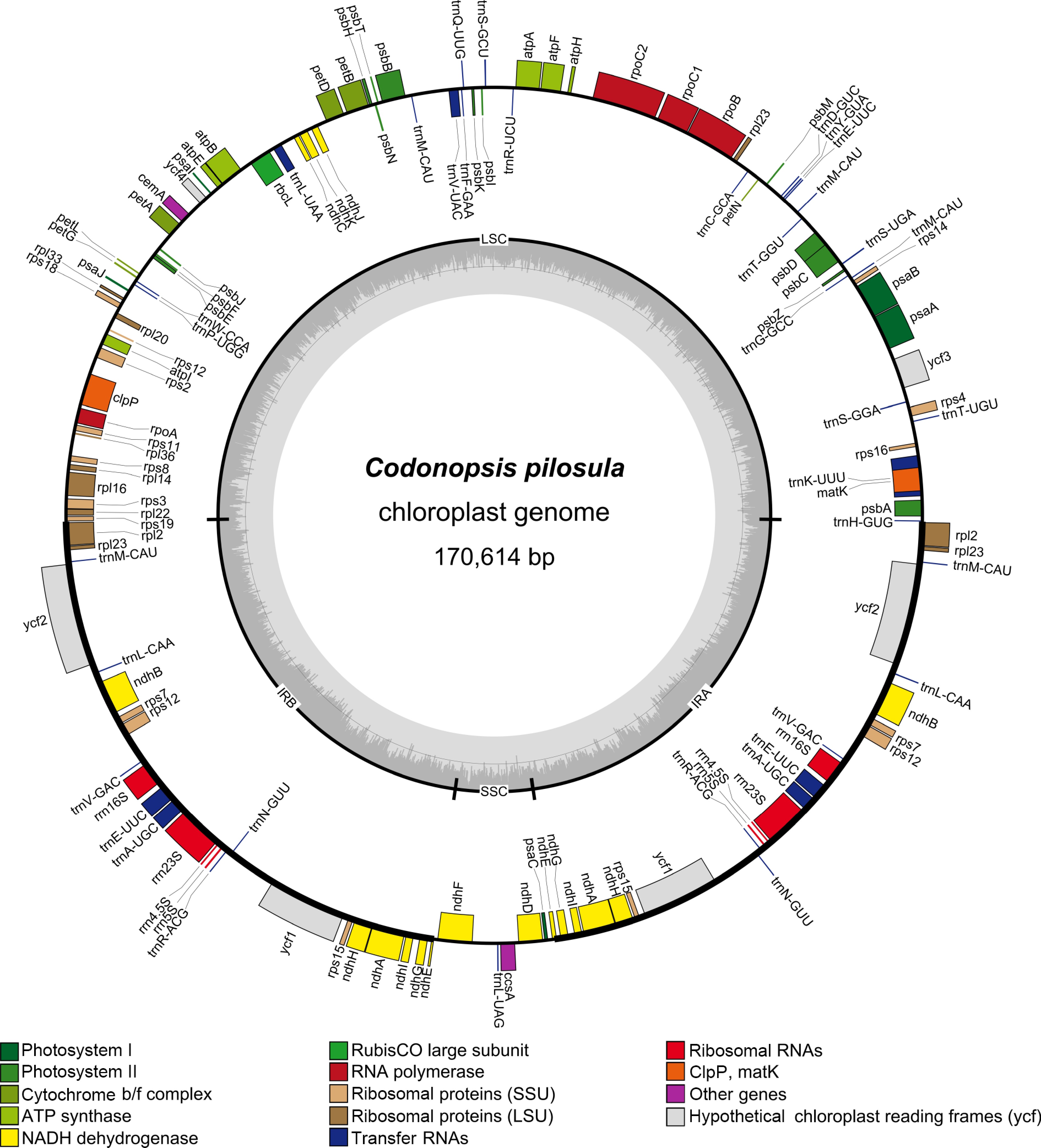
Figure 1.
Chloroplast genome map of Codonopsis pilosula. Genes shown outside the circle are transcribed counterclockwise, while those inside are transcribed clockwise. Functional gene categories are color-coded. The inner gray circle represents the GC content.
All the 11 chloroplast genomes of Codonopsis plants exhibited a typical quadripartite structure, consisting of an LSC region (84,771−96,234 bp), an SSC region (7,647−10,474 bp), and a pair of IR regions (37,875−38,698 bp). No large-scale deletions or rearrangements were observed. The chloroplast genomes spanned a length of 169,321 to 183,616 bp, and their GC contents varied between 37.97% and 38.72%. Notably, the GC contents of the IR regions (39.99%−40.74%) were consistently higher than those of the LSC (36.73%−37.59%) and SSC (31.66%−34.27%) regions (Table 1).
Functional annotation of the Codonopsis chloroplast genomes
-
To avoid redundancy, repeated genes were counted only once. As summarized in Table 2, a total of 104 genes were annotated from the assembled chloroplast genome of C. pilosula, comprising 74 protein-coding genes, 26 tRNA genes, and 4 rRNA genes. Furthermore, 16 genes were identified to harbor introns, with 2 of them containing two introns and the others containing one intron. All core chloroplast genes were present in the assembled C. pilosula chloroplast genome.
Table 2. Annotation of the C. pilosula chloroplast genome.
Category of genes Group of genes Name of genes Genes for photosynthesis Subunits of ATP synthase atpA, atpB, atpE, atpF*, atpH, atpI Subunits of Photosystem II psbA, psbB, psbC, psbD, psbE, psbF, psbI, psbJ, psbK, psbM, psbN, psbT, psbZ Subunits of NADH-dehydrogenase ndhA*(×2), ndhB*(×2), ndhC, ndhD, ndhE(×2), ndhF, ndhG(×2), ndhH(×2), ndhI(×2), ndhJ, ndhK Subunits of cytochrome b/f complex petA, petB*, petD*, petG, petL, petN Subunits of Photosystem I psaA, psaB, psaC, psaI, psaJ Subunit of rubisco rbcL Self-replication Large subunit of ribosome rpl14, rpl16*, rpl2*(×2), rpl20, rpl22, rpl23(×3), rpl33, rpl36 DNA dependent RNA polymerase rpoA, rpoB, rpoC1, rpoC2 Small subunit of ribosome rps11, rps12(×2), rps14, rps15(×2), rps16, rps18, rps19, rps2, rps3, rps4, rps7(×2), rps8 Ribosomal RNAs rrn16S(×2), rrn23S(×2), rrn5S(×2), rrn4.5S(×2) Transfer RNAs trnH-GUG, trnK-UUU*, trnT-UGU, trnS-GGA, trnM-CAU(×5), trnG-GCC, trnS-UGA, trnT-GGU, trnE-UUC*(×3), trnY-GUA, trnD-GUC, trnC-GCA, trnR-UCU, trnS-GCU, trnQ-UUG, trnF-GAA, trnV-UAC*,
trnL-UAA*, trnW-CCA, trnP-UGG, trnL-CAA(×2), trnV-GAC(×2), trnA-UGC*(×2), trnR-ACG(×2), trnN-GUU(×2), trnL-UAGOther genes C-type cytochrome synthesis gene ccsA Envelope membrane protein cemA Protease clpP** Maturase matK Unknown genes Conserved open reading frames ycf1*(×2), ycf2*(×2), ycf3**, ycf4 *: Gene contains one intron; **: Gene contains two introns; (×2): The number of repetitive units was 2; (×3): The number of repetitive units was 3; (×5): The number of repetitive units was 5. NADH: reduced nicotinamide adenine dinucleotide. Comparative analysis of chloroplast genome annotations across 11 Codonopsis plants revealed 103−106 genes per species, comprising 72−76 protein-coding genes, 26−27 tRNA genes, and 4 rRNA genes (Supplementary Table S1). The protein-coding genes were classified into four functional categories: Photosynthesis-related genes (42−44 genes), self-replication-related genes (53 or 54 genes), genes of unknown function (4 genes), and other genes (4 genes). Notably, certain genes, including ndhK, rpl23, and rpl32, were selectively annotated in specific species (Supplementary Table S1). Introns' presence/absence variation was also observed among the examined Codonopsis chloroplast genomes (Supplementary Table S1).
Codon usage bias
-
Codon preference analysis was conducted on the shared genes of the 11 Codonopsis chloroplast genomes, encompassing 21 amino acids and 64 codons, with 62 codons exhibiting usage bias. Among these, leucine (Leu), isoleucine (Ile), and serine (Ser) were the most abundant amino acids. Notably, analysis of the 11 chloroplast genomes revealed that nine codons displayed relative synonymous codon usage (RSCU) values > 1.5. Ser showed the strongest codon usage bias, with its TCT codon demonstrating the highest RSCU value of 1.91. In contrast, methionine (Met) and tryptophan (Trp) showed no codon preference, maintaining conserved RSCU values of 1.00 (Fig. 2 and Supplementary Table S2). In particular, codons ending with C or G had RSCU values < 1, accounting for 43.75% of the total, suggesting their relative rarity among the chloroplast genes of the 11 species.
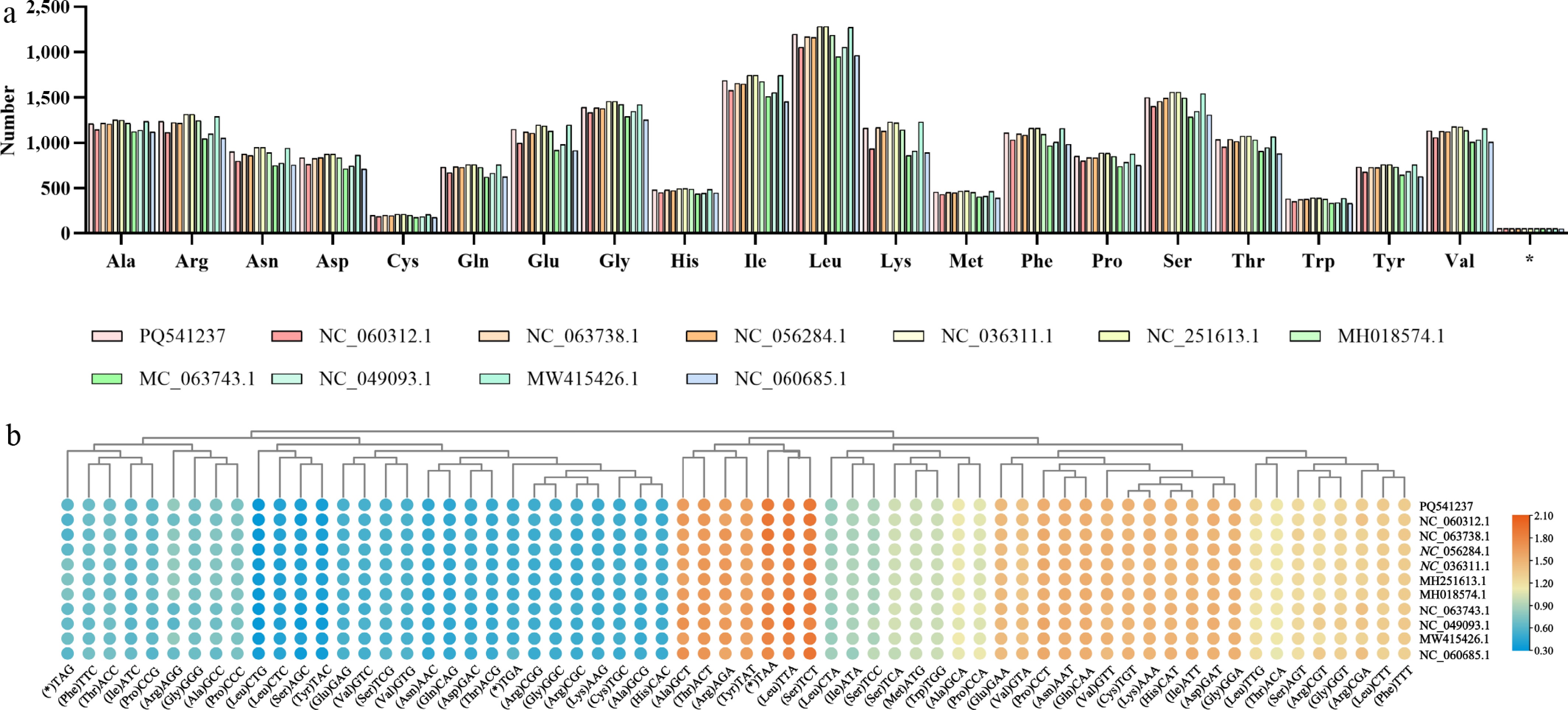
Figure 2.
Codon usage analysis of 11 Codonopsis chloroplast genomes. (a) Number of each amino acid. (b) Heatmap of relative synonymous codon usage (RSCU) values across 11 Codonopsis genomes. Rows represent species and columns represent codons. The color scale indicates RSCU values, with red representing higher bias (maximum 2.10) and blue representing lower bias (minimum 0.30).
Contraction or expansion of IR regions' boundaries
-
Through a comparative analysis of IR–SC (single-copy) junctions across 11 Codonopsis plants, consistent patterns of gene localization at the boundary regions were identified (Fig. 3). In the newly assembled chloroplast genome of C. pilosula, the genes rps19 and rpl2 were located at the LSC–IRb junction, with rps19 positioned 19 bp from the LSC–IRb boundary and rpl2 was 38 bp away from the same boundary. The ndhE genes spanned across the IRb–SSC and SSC–IRa boundaries, with extensions of 10 and 145 bp into the SSC region, respectively. Additionally, ndhF and psaC were situated 211 and 394 bp away from the IRb–SSC and SSC–IRa boundaries, respectively. Moreover, rpl2 and trnH were positioned on both sides of the IRa–LSC boundary, with distances of 38 and 3 bp from the boundaries, respectively.
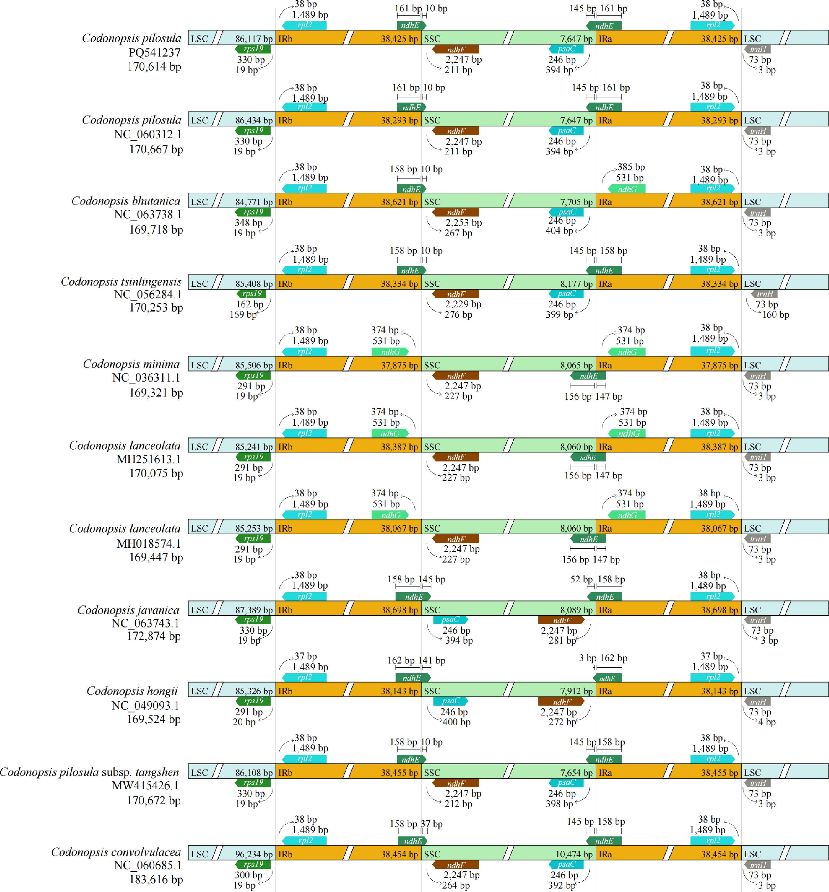
Figure 3.
Comparison of the LSC, SSC, and IR boundary regions across 11 Codonopsis chloroplast genomes. Genes near the boundaries are shown with their distances (in bp) from the junctions.
In the other 10 chloroplast genomes, the boundaries of the IRs remained stable without significant expansion or contraction. The LSC–IRb junction was consistently located between the genes rps19 and rpl2. Specifically, the IRb region extended 19−169 bp into the rps19 gene on the LSC side and 37 or 38 bp beyond rpl2 on the IRb side. Furthermore, the ndhE genes were positioned at the IRb–SSC and SSC–IRa boundaries, with extensions of 10−145 and 3−156 bp into the SSC region, respectively. However, in C. lanceolata and C. minima, ndhE was absent at the IRb–SSC boundary and replaced by ndhG. Similarly, ndhE was replaced by ndhG at the SSC–IRa region of C. bhutanica. The ndhF gene was located 211−276 bp downstream of the IRb–SSC boundary in nine species, whereas psaC was consistently positioned 392−404 bp left of the SSC–IRa junction in six species. Conversely, in C. javanica and C. hongii, the positions of ndhF and psaC were reversed. Additionally, the IRa–LSC boundaries of the chloroplast genomes in 11 Codonopsis plants were situated between rpl2 and trnH, with rpl2 positioned 37 or 38 bp into the IRa and trnH located 3−160 bp into the LSC region. This variation in boundary regions may contribute to the observed differences in chloroplast genomes' sizes. A comparison of the proximity of rpl2 to the LSC–IRb and IRa–LSC boundaries revealed consistent positioning of rpl2, indicating a high degree of conservation (Fig. 3).
SSRs in 11 Codonopsis chloroplast genomes
-
SSRs, known as simple sequence repeats, consist of tandem repeats one to six nucleotides long. These sequences are widely utilized as genetic markers for identifying species. In this study, the newly assembled sequence of C. pilosula chloroplast genome contained 400 SSRs (Fig. 4). Among these, 231 were mononucleotide repeats, with A/T repeats being the most prevalent, representing 28.5% and 29.25% of the total SSRs, respectively. Notably, no G/C SSR repeats were observed. Dinucleotide repeats (86, 21.50%), trinucleotide repeats (48, 12.00%), and tetranucleotide repeats (35, 8.75%) were also detected in C. pilosula chloroplast genome, but pentanucleotide and hexanucleotide repeats were absent. The majority of these SSRs were located in the LSC region, constituting 54.00% of the total SSRs.
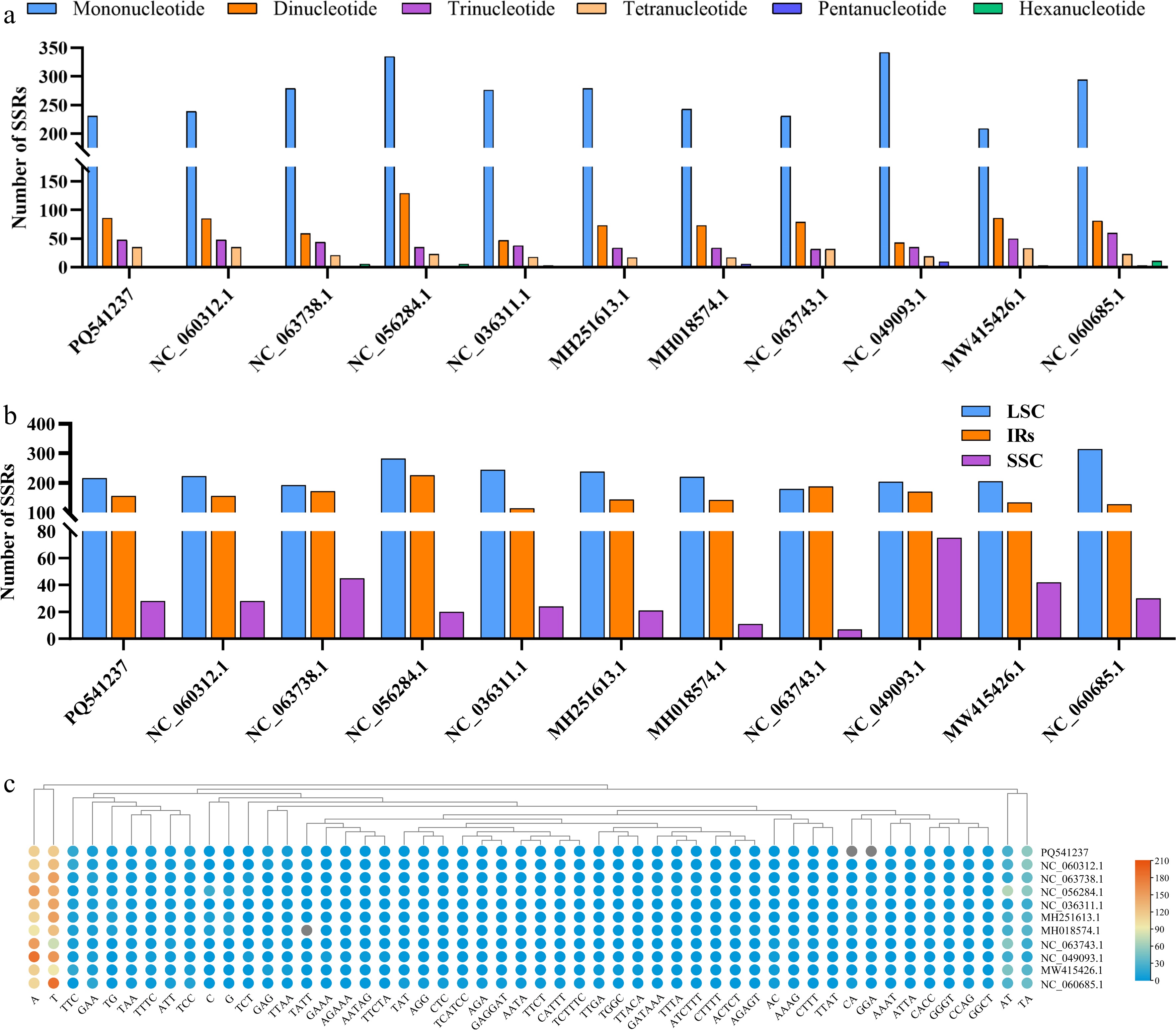
Figure 4.
Simple sequence repeat (SSR) analysis of 11 Codonopsis chloroplast genomes. (a) Number of SSRs by repeat type (mono-, di-, tri-, tetra-, penta-, hexanucleotide). (b) Distribution of SSRs across LSC, SSC, and IR regions. (c) The number of SSRs in each repeat type for each species.
Comparative analysis of SSRs across 11 Codonopsis plants revealed that mononucleotide repeats were the most abundant (209−342 per genome), followed by dinucleotides (43−129 per genome), trinucleotides (32−60 per genome), and tetranucleotides (17−35 per genome). Pentanucleotides and hexanucleotides were observed in C. convolvulacea, C. tsinlingensis, and C. bhutanica. A/T bias persisted in mononucleotides and dinucleotides (Fig. 4a, c and Supplementary Table S3), and SSRs were primarily enriched in LSC regions (46.94%−66.53%), followed by IR regions (27.12%−50.27%) (Fig. 4b and Supplementary Table S4).
Dispersed repeat sequence analysis
-
Dispersed repeat sequences in the 11 Codonopsis chloroplast genomes were analyzed using the online tool REPuter, with a minimum length threshold of 30 bp for four repeat types: Palindromic repeats, forward repeats, reverse repeats, and complementary repeats. In total, 762 repeat sequences were identified in the newly assembled C. pilosula chloroplast genome, consisting of 377 palindromic repeats (49.48%), 362 forward repeats (47.51%), 17 reverse repeats (2.23%), and 6 complementary repeats (0.79%), with most ranging between 30 and 39 bp in length (Fig. 5 and Supplementary Table S5). A comparative analysis of dispersed repeats across the 11 Codonopsis plants revealed variations in the repeat types and lengths among the different species. Palindromic repeats ranged from 301 to 671, whereas forward repeats ranged from 296 to 624. Distinctively, complementary repeats were absent in C. minima and C. javanica, but reverse repeats were undetected in C. hongii and C. javanica (Fig. 5a and Supplementary Table S6).
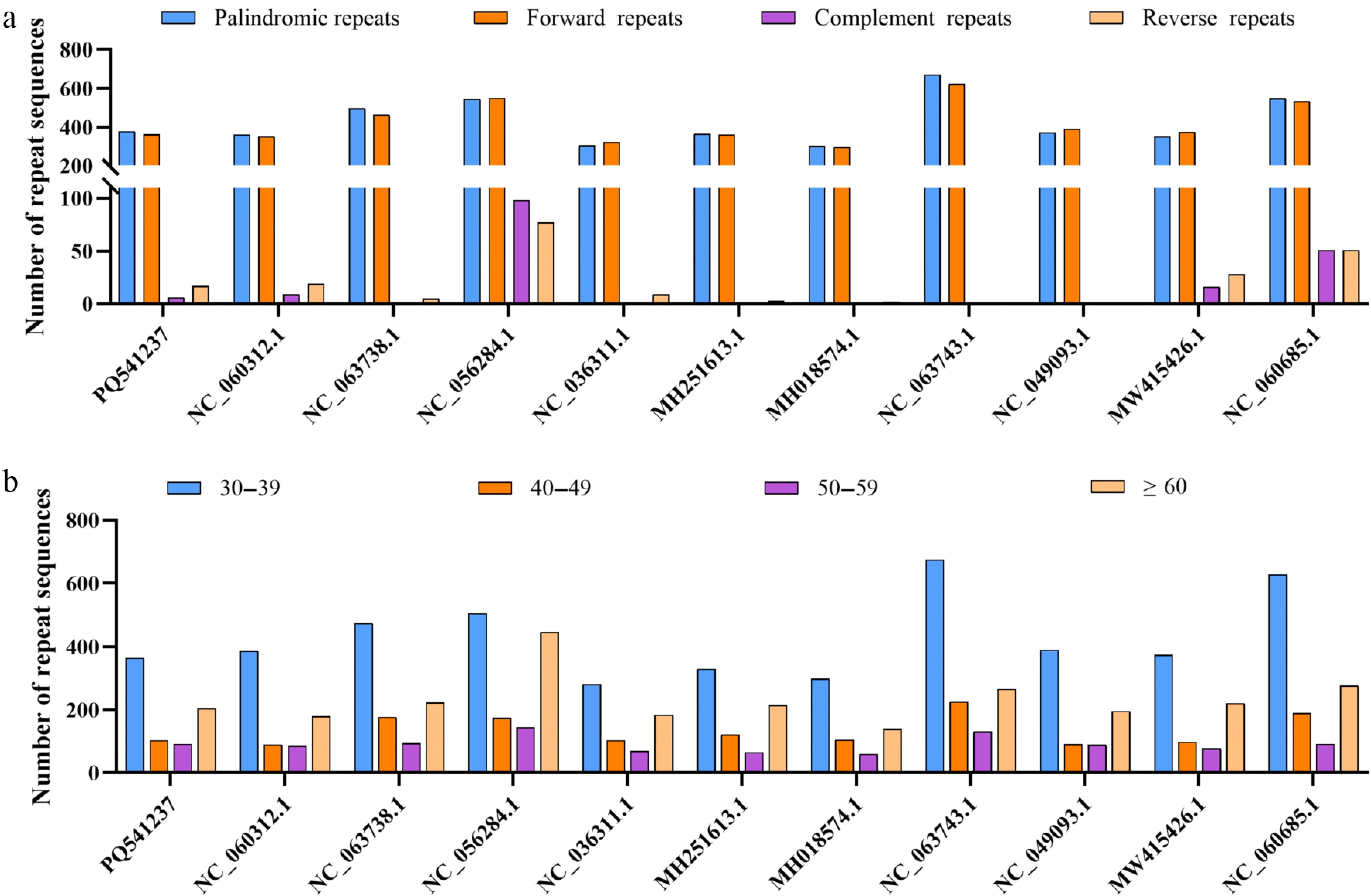
Figure 5.
Dispersed repeat analysis of 11 Codonopsis chloroplast genomes. (a). Classification and distribution of palindromic, forward, reverse, and complementary repeats. (b). Frequency distribution of dispersed repeats by length categories (30−39, 40−49, 50−59, and ≥ 60 bp).
Comparative chloroplast genome analysis of 11 Codonopsis plants
-
To assess the overall similarity of the chloroplast genomes among Codonopsis plants, genetic variants across 11 chloroplast genomes were compared and depicted using mVISTA (Supplementary Fig. S2). Notable sequence divergence was observed in six noncoding regions: rps16-trnT-UGU, psbD-rpl23, rpoC2-atpH, trnV-UAC-trnM-CAU, rps2-rpoA, and rpl36-rps8. These regions exhibit varying degrees of diversity and can be utilized for phylogenetic analysis of different species. Moreover, the variability in the IR regions was markedly lower compared with the LSC region, consistent with their evolutionary conservation in angiosperms. Furthermore, in contrast to the reference genome (the newly assembled chloroplast genome in this study), significant distinctions were observed in the coding regions of the ycf1 and ycf2 genes.
Evolutionary selection pressure analysis
-
An analysis of selection pressure was performed on 71 functionally important chloroplast protein-coding genes. The coding sequences were aligned using MAFFT with the default parameters to ensure accurate alignment. The aligned sequences were then used as input for the yn00 program to estimate dN, dS, and ω = dN/dS, with the newly assembled C. pilosula chloroplast genome as the reference. The complete dataset for all 71 genes is provided in Supplementary Table S7. Among the genes, rpl2 exhibited the highest nonsynonymous substitution rate (dN = 0.3681) and the most elevated synonymous substitution rate (dS = 2.729). This analysis demonstrates the predominance of synonymous substitutions over nonsynonymous substitutions across most genes, resulting in generally low ω values that are characteristic of purifying selection. However, potential positive selection signals (ω > 1) were detected in several genes: ycf1 and ycf2 showed elevated ω values across multiple species. These findings indicate that genes with signals of potential positive selection include both functionally uncharacterized genes and functionally characterized genes such as atpH and clpP. The remaining genes showed consistently low ω values, suggesting they are predominantly under purifying selection. Together, these findings provide insights into the molecular evolution of chloroplast genomes in Codonopsis plants.
Nucleotide diversity analysis and validation of hypervariable regions
-
A comparative analysis of the polymorphisms across 11 Codonopsis chloroplast genomes revealed 71 evolutionarily conserved genes. The ycf1 gene (6,944 bp), encoding a putative protein of unknown function, exhibited the highest nucleotide diversity (Pi = 0.06855), showing higher variability than ndhF (Pi = 0.03909, the second highest nucleotide diversity) (Supplementary Tables S8 and S9). These hypervariable regions showed approximately fivefold greater polymorphism than the average Pi value of conserved genes, suggesting their utility as potential phylogenetic markers for species discrimination within Codonopsis.
To validate the identification potential of these candidate regions, the hypervariable regions (ycf1 and ndhF) were extracted from the 11 Codonopsis chloroplast genomes and combined to generate the ycf1–ndhF fragment. Three NJ trees were built from the ycf1, ndhF, and ycf1–ndhF combined fragments (Supplementary Fig. S3). The three phylogenetic trees exhibited similar topological structures. The single-gene tree of ycf1 showed high bootstrap support for most clades and successfully resolved all sampled species into distinct monophyletic branches. The ndhF tree showed relatively low support at several nodes, indicating weaker identification potential than ycf1. The combined ycf1–ndhF fragment greatly increased the bootstrap values and achieved better phylogenetic resolution than each single locus. Across all trees, C. convolvulacea, C. javanica, and C. hongii formed well-separated independent clades. C. bhutanica and C. tsinlingensis were recovered as sister species with very close phylogenetic affinity, and C. minima was closely related to C. lanceolata. All accessions of C. pilosula (including the subspecies C. pilosula subsp. tangshen) constituted a monophyletic lineage.
Phylogenetic analysis of Codonopsis plants and their relatives
-
To resolve the phylogenetic position and evolutionary relationships of C. pilosula within the genus Codonopsis, a ML phylogenetic tree was reconstructed using all chloroplast genomes under the TVM + F + I + R8 nucleotide substitution model with ultrafast bootstrap support (5,000 replicates) (Fig. 6). The resulting phylogeny supported the monophyly of the sampled Codonopsis species relative to the outgroup taxa (L. chinensis and P. grandiflorus). Within the core Codonopsis clade, the tree resolved several well-supported subclades (Fig. 6). C. javanica and C. hongii formed an early-diverging lineage, whereas the remaining Codonopsis species were further divided into additional subclades. Interestingly, C. pilosula and its subspecies C. pilosula subsp. tangshen formed separate branches. The remaining taxa formed a topology similar to the NJ trees constructed from ycf1, ndhF and the combined ycf1–ndhF fragment. C. pilosula formed a distinct subclade, C. bhutanica was sister to C. tsinlingensis, C. minima clustered with C. lanceolata, and the two accessions of C. lanceolata grouped tightly.
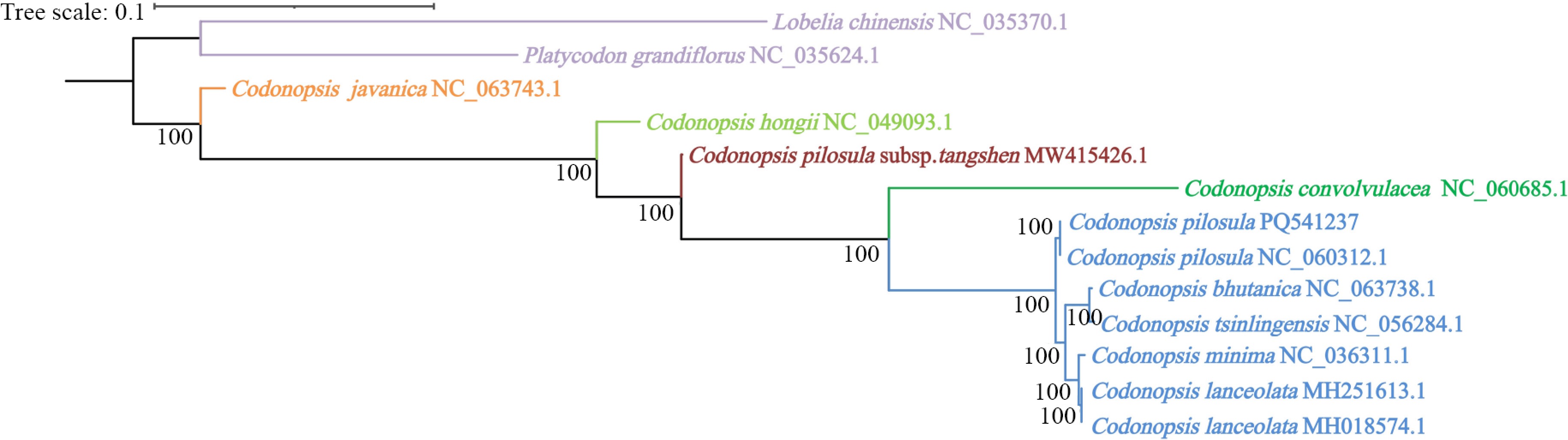
Figure 6.
The ML phylogenetic tree of 11 Codonopsis plants and allied plants. The tree was constructed using IQ-TREE 2 under the TVM + F + I + R8 model with 5,000 bootstrap replicates. Bootstrap support values (≥ 70%) are shown at the nodes.
-
C. pilosula is valued both as a traditional medicine and as an edible herb, and its market demand has steadily increased in recent years[33]. However, current research on this species has predominantly focused on its bioactive components, although its genetic resources remain underexplored. The chloroplast genome, owing to its conserved structure and moderate evolutionary rate, serves as an effective tool for investigating plants' phylogeny and genetic diversity[34]. In this study, we conducted a comparative analysis of chloroplast genomes across 11 Codonopsis plants, integrating a newly assembled genome of C. pilosula with publicly available data to elucidate their genomic architecture, phylogenetic relationships, and evolutionary dynamics. To ensure comparability, all genomes were uniformly reannotated using the same pipeline (CPGAVAS2), minimizing potential biases arising from inconsistent annotation standards.
Consistent with findings in other angiosperms, all 11 Codonopsis chloroplast genomes exhibited a typical quadripartite structure. Although the overall structural organization was conserved, the lengths and sequence composition of individual regions varied among species, with the LSC ranging from 84,771 to 96,234 bp, the SSC from 7,647 to 10,474 bp, and the IRs from 37,875 to 38,698 bp. The total GC contents across Codonopsis plants ranged from 37.97% to 38.72%, falling within the characteristic 30%−40% GC content range observed in angiosperm chloroplast genomes. Notably, the IR regions exhibited consistently higher GC contents (39.99%−40.74%) compared with the LSC (36.73%−37.59%) and SSC (31.66%−34.27%) regions, a phenomenon likely attributable to the presence of rRNA genes with an inherently high GC content in the IR regions[23]. These findings indicate that the overall quadripartite structure is conserved across the 11 Codonopsis plants, whereas the lengths and sequence composition of individual regions vary among species.
This variation in genome size (169,321−183,616 bp) was primarily attributed to length of the polymorphisms in the LSC region, particularly in the intergenic spacers, rather than in the coding regions. A key source of structural divergence was the expansion and contraction of the IR regions. Our analysis revealed that the LSC–IRb and IRa–LSC boundaries were highly conserved across most species. At the LSC–IRb boundary, rps19 was consistently located without extending into the IR region. At the IRa–LSC boundary, rpl2 and trnH were positioned on opposite sides. This configuration was nearly identical across all 11 species, underscoring the evolutionary constraint at these junctions. However, the distance between trnH and the IRa–LSC border varied from 3 bp in most species to 160 bp in C. tsinlingensis, indicating that shifts of the IR boundary represent an ongoing, albeit slow, evolutionary process within the genus. In contrast, the IRb–SSC boundary exhibited notable lineage-specific variations. In most Codonopsis species, the ndhE gene spanned this boundary. However, in C. lanceolata and C. minima, the ndhE copy was lost at this boundary and replaced by ndhG (Fig. 3). Consistent with this structural rearrangement, a reduction in the ndhE copy number was also observed. Two copies of ndhE are present in most species, whereas only a single copy was found in C. lanceolata, C. minima, and C. bhutanica. This lineage-specific loss of ndhE is particularly noteworthy from an evolutionary perspective and may reflect a functional redundancy, potentially related to their specific ecological adaptations. The pattern of gene loss and rearrangement suggests a shared evolutionary event within this clade, a hypothesis supported by the phylogenetic analysis (Fig. 6). Further structural polymorphism was observed in the early-diverging species C. javanica and C. hongii, where ndhF and psaC exhibited an inverted orientation at the IR–SSC junction relative to the typical configuration found in other Codonopsis species. Such rearrangements, though rare, contribute to diversity in the genome size and may influence gene expression by relocating regulatory elements near the borders. Similar IR boundary dynamics have been documented in other angiosperm lineages, including Orchidaceae, where they were associated with contraction of the SSC region and adaptive evolution[35].
The analysis of SSRs and long repeats provided additional insights into the genomes' plasticity and potential marker development. The predominance of A/T mononucleotide repeats across all species aligns with the overall AT-rich nature of chloroplast genomes and the observed base composition bias in the LSC region. The presence of species-specific repeat types, such as pentanucleotide and hexanucleotide repeats in C. convolvulacea, C. tsinlingensis, and C. bhutanica, suggests their potential utility as molecular markers.
Codon usage bias reflects evolutionary constraints on gene expression and has been widely documented in plant chloroplast genomes[36]. The RSCU value is a fundamental metric for measuring this bias, where values above 1 signify significant codon favoritism[37]. Analysis of the 11 Codonopsis chloroplast genomes revealed that Leu, Ser, and Ile were the most abundantly encoded amino acids. Among the 30 high-frequency codons (RSCU > 1), 13 terminated with A, 16 with T, and only 1 with G, reinforcing a pronounced A/T bias at the third codon position. This pattern is consistent with the findings of Yan et al.[38], who reported that 96.55% of high-frequency codons in Codonopsis chloroplast genomes ended with A/T, and aligns with observations in other medicinal plant chloroplast genomes, such as Aconitum and Delphinium grandiflorum[39,40], reflecting the overall AT-rich nature of the Codonopsis chloroplast genomes.
Identifying hypervariable regions with high nucleotide diversity is essential for developing molecular markers to differentiate closely related Codonopsis species. Nucleotide diversity analysis revealed that ycf1 exhibited the highest nucleotide diversity among all examined regions. Previous research has indicated that ycf1 demonstrates more variability than the matK locus in Orchidaceae, making it a promising plastid molecular marker for phylogeny at lower taxonomic levels[35]. Recent functional investigations have shown that ycf1 encodes Tic214, a core component of the translocon complex at the inner envelope membrane of chloroplasts, and plays a crucial role in regulating the synthesis of chloroplast-encoded photosynthetic complex subunits[41]. This functional importance, coupled with its high sequence variability, implies that ycf1 may serve as a candidate molecular marker for species identification in Codonopsis. Given the reported role of ycf1/Tic214 in chloroplast protein import and photosynthesis-related processes, future studies could further investigate whether sequence variation in ycf1 has functional or adaptive significance. Additionally, the combined ycf1–ndhF region has also proven to be effective for species identification in Prunus persica[42]. In this study, the NJ trees constructed from the combined ycf1–ndhF fragment showed improved bootstrap support for most clades compared with the single-locus ycf1 or ndhF trees (Supplementary Fig. S3). These findings suggest that ycf1, particularly when combined with ndhF, has the potential to serve as a candidate molecular marker for species identification in Codonopsis.
Investigating the evolutionary history of species requires an analysis of their adaptive evolution[43]. The ω ratio is a common indicator for evaluating the influence of selective pressures on protein-coding genes, and it is widely utilized to gauge species' adaptive evolutionary rates and the effects of positive selection. Most genes undergo negative selection, indicating a trend towards conservation in evolution[44]. Within the chloroplast genomes of Codonopsis, ycf2 consistently showed elevated ω values across multiple species, suggesting a potential signal of positive selection. Elevated ω values for ycf2 have also been documented in Berberis[45], although the functional basis remains unclear. The recurrent observation of elevated ω values for ycf2 across diverse plant groups suggests that this gene may warrant further functional characterization.
Phylogenetic analysis based on the complete chloroplast genome sequences resolved the interspecific relationships within the sampled Codonopsis species and supported their monophyly relative to the outgroups (Fig. 6). Notably, the overall phylogenetic framework recovered by complete chloroplast genomes was similar to the topology derived from the ycf1 genes, the ndhF genes, and the combined ycf1–ndhF fragments (Supplementary Fig. S3). These consistent phylogenetic patterns further confirm that the chloroplast genes ycf1, ndhF and their combined fragment may be suitable molecular markers for species identification within Codonopsis.
An intriguing finding of the phylogenetic analysis is the marked divergence between C. pilosula (PQ541237 and MT982394.1) and C. pilosula subsp. tangshen (MW415426.1). Despite their close taxonomic relationship, the two taxa were placed in distinct branches within the phylogenetic tree, suggesting that significant genetic differentiation has occurred between the typical variety and its subspecies. Notably, C. pilosula subsp. tangshen is recorded as C. tangshen in the Chinese Pharmacopoeia[1], where it is treated as an independent species rather than a subspecies, a treatment that is consistent with the phylogenetic divergence observed in this study.
The chloroplast genome-based ML phylogenetic tree also provides insights into the taxonomic status of two species with historical nomenclatural changes. Codonopsis hongii (originally described as Leptocodon hirsutus) constitutes an early-diverging lineage nested within the Codonopsis clade. This finding lends support to the taxonomic treatment transferring the former L. hirsutus into Codonopsis. By contrast, C. convolvulacea (recently renamed Pseudocodon convolvulaceus following taxonomic revision) forms a distinctly divergent sublineage in the phylogenetic tree. This deep genetic divergence supports its segregation into the newly recognized genus Pseudocodon. Nevertheless, both C. hongii and C. convolvulacea remain embedded within the broader Codonopsis clade in the plastid tree, implying that Codonopsis sensu lato may be paraphyletic.
-
This study presents a comparison of the chloroplast genomes of Codonopsis, encompassing 11 genomes (representing nine distinct species) and providing a newly assembled reference genome for C. pilosula. The analyses reveal both structural conservation and dynamics in the chloroplast genomes' architecture, identify the ycf1–ndhF combined region as a candidate molecular marker for species identification, and provide insights into the phylogenetic relationships among the sampled species, including the early divergence of C. javanica and C. hongii and the notable differentiation between C. pilosula and its subspecies C. pilosula subsp. tangshen. Collectively, these findings contribute to the growing genomic resources for Codonopsis and provide a foundation for future molecular marker development and phylogenetic studies with expanded sampling.
-
The authors confirm their contributions to the paper as follows: study conception and design: Li Q, Chen W; data collection: Liu J, Fang Z, Hu J; analysis and interpretation of results: Tian X; draft manuscript preparation: Tian X, Li Q. All authors reviewed the results and approved the final version of the manuscript.
-
The raw Illumina paired-end sequencing reads generated in this study are available in the NCBI Sequence Read Archive (SRA) (www.ncbi.nlm.nih.gov/sra) under the accession number SRP737974 (www.ncbi.nlm.nih.gov/sra/?term=SRP737974). The complete chloroplast genome of C. pilosula has been deposited in GenBank (www.ncbi.nlm.nih.gov/genbank) with the accession number PQ541237 (www.ncbi.nlm.nih.gov/nuccore/PQ541237).
-
The authors declare that they have no conflict of interest.
-
accompanies this paper online at: https://doi.org/10.48130/mpb-0026-0022.
- Supplementary Table S1 The annotation information of 11 Codonopsis chloroplast genomes.
- Supplementary Table S2 Codon preference analysis.
- Supplementary Table S3 SSR analysis of 11 Codonopsis plants.
- Supplementary Table S4 The SSR distribution regions of 11 Codonopsis plants.
- Supplementary Table S5 Frequency distribution of dispersed repeats by length categories.
- Supplementary Table S6 Classification and distribution of dispersed repeat types.
- Supplementary Table S7 dN,dS and dN/dS ratios of shared genes in Codonopsis plants.
- Supplementary Table S8 Detailed annotation of protein-coding genes in the Codonopsis chloroplast genomes.
- Supplementary Table S9 Nucleotide diversity (Pi) values of Codonopsis chloroplast genomes.
- Supplementary Fig. S1 The coverage depth of the chloroplast genome of C. pilosula.
- Supplementary Fig. S2 Whole-chloroplast genome alignment of 11 Codonopsis plants. The newly assembled C. pilosula genome is used as the reference.
- Supplementary Fig. S3 The neighbor-joining phylogenetic trees of 11 Codonopsis plants.
- Copyright: © 2026 by the author(s). Published by Maximum Academic Press, Fayetteville, GA. This article is an open access article distributed under Creative Commons Attribution License (CC BY 4.0), visit https://creativecommons.org/licenses/by/4.0/.
-
About this article
Cite this article
Tian X, Liu J, Fang Z, Hu J, Chen W, et al. 2026. The complete chloroplast genome of Codonopsis pilosula and comparative analysis with other Codonopsis plants: architecture, evolution, and candidate molecular markers. Medicinal Plant Biology 5: e027 doi: 10.48130/mpb-0026-0022 

The complete chloroplast genome of Codonopsis pilosula and comparative analysis with other Codonopsis plants: architecture, evolution, and candidate molecular markers
- Received: 16 August 2026
- Revised: 25 September 2026
- Accepted: 19 October 2026
- Published online: 10 November 2026
Abstract: Codonopsis pilosula (Franch.) Nannf., one of the botanical origins of the Chinese medicine "Dangshen", belongs to the genus Codonopsis (Campanulaceae). Morphological convergence among congeners makes species identification within this genus particularly difficult. The chloroplast genome, with its conserved structure and moderate evolutionary rate, has emerged as a powerful tool for species identification and plant phylogenetic studies. The complete chloroplast genome of C. pilosula was sequenced, assembled, and annotated in this study, and a comparative genomic analysis with 10 other Codonopsis chloroplast genomes representing nine species was performed. All chloroplast genomes exhibited a typical quadripartite structure, with lengths ranging from 169,321 to 183,616 bp and GC contents between 37.97% and 38.72%. Structural variations were primarily attributed to expansions and contractions at the inverted repeat boundaries and variations in noncoding regions. Repeat analysis revealed a higher frequency of palindromic repeats, whereas simple sequence repeat analysis showed a predominance of mononucleotide repeats, particularly A/T repeats, which may be useful for species identification and evolutionary studies. Phylogenetic analysis supported the monophyly of the sampled Codonopsis species relative to outgroups, resolving several well-supported subclades. Evolutionary selection pressure analysis indicated purifying selection as the dominant force, with potential positive selection signals detected in ycf2 and other genes. Nucleotide diversity analysis identified ycf1 and ndhF as hypervariable regions, suggesting that the combined ycf1–ndhF fragment may serve as a candidate molecular marker for species identification. The findings of this study provide critical genomic resources for evolutionary studies and species authentication of this medicinally valuable genus.