












-
China is expected to host many fungal species in its highly diverse landscapes[1−6]. Wood-decaying fungi play a fundamental ecological role as decomposers of plants in the fungal tree of life[7−17]. These fungi have been extensively studied in China recently, and more than 2,700 species have been reported from the country in total[5,6,18−26].
Traditional classification relied heavily on morphological features. However, molecular phylogenetics has revolutionized wood-decaying fungal systematics, leading to significant re-evaluations of genus, family, and order boundaries[9,11,27−32]. Among these redefined taxa, the genus Xenasmatella Oberw. historically placed within the Xenasmatales Jülich, Polyporales Gäum. or Russulales Kreisel ex P.M. Kirk et al.[28,33−35], has emerged as a focal point of taxonomic revision and species discovery in recent years. Subsequently, based on analyses of the internal transcribed spacer and nuclear large subunit (ITS + nLSU) as well as nLSU-only datasets across 13 orders, Luo & Zhao proposed that Xenasmatella should be assigned to the order level; therefore, Xenasmatales and Xenasmataceae Oberw. were established and accepted to accommodate species of Xenasmatella, which supports the previous placement. In their phylogenetic analyses, Xenasmatales formed a single clade and grouped with the orders Atheliales Jülich, Boletales E.J. Gilbert, and Hymenochaetales Oberw[36]. Xenasmatella is primarily characterized by corticioid hymenophores and soft membranous to ceraceous basidiomata. Morphologically, Xenasmatella (typified by X. subflavidogrisea (Litsch.) Oberw. ex Jülich) is distinguished by resupinate, gelatinous basidiomata; a monomitic hyphal system with clamped generative hyphae; the absence of cystidia; pleural basidia; and hyaline, thin-walled, warted, inamyloid, acyanophilous basidiospores[36,37].
The taxonomic history of Xenasmatella has been closely intertwined with the genus Phlebiella P. Karst. which was initially proposed by Karsten in 1890, but was not validly published due to the absence of a generic description; consequently, Xenasmatella has been accepted as the valid name for this group[38]. Donk formally established this invalidity[39]. Meanwhile, Oberwinkler validly circumscribed Xenasmatella, and Piątek proposed that Xenasmatella be recognized as the earliest valid name for this fungal group[40,41]. Despite the invalidity of Phlebiella, this genus has been used among many taxonomists from 1965 until around 2021, largely due to a continuation of morphological classification traditions where spore ornamentation was a key diagnostic feature[42−47]. During this period, species were gradually recombined, primarily from Phlebiella to Xenasmatella, e.g., Duhem proposed four combinations, transferring Phlebiella borealis K. H. Larsson & Hjortstam, P. caricis-pendulae P. Roberts, P. globigera Hjortstam & Ryvarden, and P. palmicola Hjortstam & Ryvarden into Xenasmatella[48]. Maekawa proposed a combination X. athelioidea (N. Maek.) N. Maek. from Japan[49]. Additionally, Phlebiella ailaoshanensis C.L. Zhao was similarly combined into this genus[50], and Phlebiella gossypina C.L. Zhao and P. wuliangshanensis C.L. Zhao were recombined into Xenasmatella by Gruhn & Trichies without any discussion[51].
Apart from the closely related genus Phlebiella, the taxonomic history of Xenasmatella has also been intricately linked with several other genera, reflecting evolving systematic concepts and the transition from morphology-based to molecular-informed classifications. Numerous species originally described in other genera have been recombined into Xenasmatella, while others initially placed in Xenasmatella have been transferred elsewhere based on revised phylogenetic understandings. The genus Trechispora P. Karst., for instance, shares morphological similarities with Xenasmatella, particularly in having ornamented basidiospores and similar basidioma structures. Notably, Trechispora sulphurea (Pers.) Rauschert transferred Xenasmatella to X. vaga[52,53], while X. tenuicula (Litsch.) Å. Strid was conversely placed in Trechispora as a heterotypic synonym[54,55], illustrating the practical difficulties in distinguishing these genera based solely on morphological features. Similarly, several species have been transferred between Aphanobasidium Jülich and Xenasmatella, e.g., X. bicornis (Boidin & Duhem) Piątek is currently accepted as Ap. bicorne (Boidin & Duhem) Duhem, indicating that not all species originally assigned to Xenasmatella have remained within the genus[41,48]. Conversely, Xenasmatella albida Hauerslev is now treated as a synonym of Aphanobasidium albidum (Hauerslev) Boidin & Gilles[56,57]. These transfers reflect ongoing refinements in generic delimitations within the corticioid fungi. In another case, the genus Amyloxenasma (Oberw.) Hjortstam & Ryvarden was established to accommodate species with amyloid hyphae, a feature not typically associated with Xenasmatella. Consequently, Xenasmatella allantospora Oberw. has been recombined as Amyloxenasma allantosporum (Oberw.) Hjortstam & Ryvarden, while X. grisella (Bourdot) Oberw. is now accepted as Am. grisellum (Bourdot) Hjortstam & Ryvarden[40,44]. These examples demonstrate how micromorphological and histochemical characters have led to generic segregation. The genus Xenasma Donk has also played a role in this taxonomic history, having historically served as a repository for species with similar morphological traits. Xenasma insperatum (H.S. Jacks.) Donk has been recombined as Xenasmatella insperata (H.S. Jacks.) Jülich, confirming its affinity with Xenasmatella based on re-evaluation of type material and consideration of the important character of the pleurobasidia[58,59]. Finally, several species originally described as Corticium Pers. sensu lato have found their placements in Xenasmatella, e.g., C. subflavidogriseum Litsch. is now accepted as X. subflavidogrisea (Litsch.) Oberw. ex Jülich[59,60]. These recombinations collectively reflect the progressive dismantling of Corticium as a 'catch-all' genus for corticioid fungi. It should be noted that many of these taxonomic adjustments occurred during a period when Phlebiella was also used as an alternative generic name. However, following the invalidation of the name Phlebiella, Xenasmatella has been accepted as the correct name for this group of fungi. Some species were placed in several genera; for example, Corticium grisellum Bourdot was successively assigned to Xenasma, Xenasmatella, Aphanobasidium, and Phlebiella before being finally accepted as Amyloxenasma grisellum[40,44,59,61,62]. This complex nomenclatural history underscores the importance of comprehensive type studies and molecular phylogenetics in resolving the natural relationships within this group of wood-inhabiting fungi.
Most recently, a mycological survey conducted in southern Xizang Autonomous Region, China, by Wang et al. revealed numerous additional taxa, further highlighting the adaptability of Xenasmatella to high-elevation ecosystems, and X. jilongensis S.L. Liu & L.W. Zhou was described from Xizang Autonomous Region[63].
Currently, Index Fungorum (
www.indexfungorum.org/Names/Names.asp ) and MycoBank (www.mycobank.org/page/Simple%20names%20search ) have registered 46 records in Xenasmatella. To date, 30 species are currently accepted in Xenasmatella worldwide, and 11 of these have been found in China so far by April 2026[36,46,47,50,63−66]. However, vouchered molecular sequences are publicly available for only a small fraction of these taxa in the NCBI database, especially for species discovered abroad, where available sequences are extremely scarce.This study is based on specimens collected in recent years from 15 provinces or autonomous regions across China: Anhui Province, Guangxi Autonomous Region, Guizhou, Hainan, Heilongjiang, Henan, and Hebei provinces, Inner Mongolia Autonomous Region, Jilin, Liaoning, Qinghai, and Sichuan provinces, Xizang Autonomous Region, Yunnan and Zhejiang provinces. Seventeen new species are identified based on morphology and molecular analyses using a dataset of ITS + nLSU.
-
The studied specimens were collected from various nature reserves and forest parks in China. The samples were photographed in situ, and macromorphological characters such as colour, shape and size, as well as other important collection details, were noted[67]. The specimens were then placed in net bags and taken to the field station, where they were dried using a portable mushroom drier (Evermat, Finland) at 35 °C for 7 h[68,69]. Dried specimens were labelled and then stored in a refrigerator at –80 °C for at least two weeks. The studied specimens are deposited in the Fungarium of the Institute of Microbiology, Beijing Forestry University (BJFC, China).
Morphological studies
-
Morphological descriptions are based on field notes and dried voucher specimens. The microscopic analysis followed the methods by Wu et al.[ 70] and Zhou et al.[26]. Sections were studied at magnifications up to 1,000× using a Nikon Eclipse 80i microscope with phase-contrast illumination. Line drawings were made with the aid of a drawing tube[71]. Microscopic features and measurements were made from slide preparations of dried specimens stained with Cotton Blue and Melzer's reagent to test cyanophilous, acyanophilous, amyloid, or dextrinoid reactions, respectively. To represent the variation in the size of spores, 5% of measurements were excluded from each end of the range and are given in parentheses. In the description, KOH = 5% potassium hydroxide, IKI = Melzer's reagent, IKI– = neither amyloid nor dextrinoid, CB = Cotton Blue, CB– = acyanophilous, L = arithmetic average of spore length, W = arithmetic average of spore width, Q = L/W ratios, and n = number of basidiospores/measured from a given number of specimens. Color terms followed by Anonymous[72] and Petersen[73].
DNA extraction, amplification, and sequencing
-
A CTAB rapid plant genome extraction kit-DN14 (Aidlab Biotechnologies Co., Ltd, Beijing) was used to obtain DNA from dried specimens and to perform the polymerase chain reaction (PCR) according to the manufacturer's instructions with some modifications[74]. The Internal Transcribed Spacer (ITS), nuclear Large Subunit ribosomal RNA gene (nLSU), and mitochondrial Small Subunit ribosomal RNA gene (mtSSU), RNA polymerase II largest subunit (rpb1), RNA polymerase II second largest subunit (rpb2), glyceraldehyde-3-phosphate dehydrogenase (gapdh), and translation elongation factor 1-alpha (tef1-α) sequences were amplified with different primer pairs. The ITS region was amplified with primer pair ITS5 and ITS4[75]. The nLSU region was amplified with primer pair LR0R and LR7[76]. The mtSSU region was amplified with primer pair MS1 and MS2[75]. The rpb1 region was amplified with primer pair RPB1-Af and RPB1-Cr[77]. The rpb2 region was amplified with primer pair bRPB2-6F[78] and bRPB2-7.1R[79]. The gapdh region was amplified with primer pair gapdh-F and gapdh-R[80]. The tef1 region was amplified with primer pair EF1-983F and EF1-1567R[81].
The primer pairs and PCR (the polymerase chain reaction) cycling procedures for the seven gene regions were followed as given by Liu et al.[65]. The PCR cycling schedule for ITS, mtSSU, and tef1 included an initial denaturation at 95 °C for 3 min, followed by 35 cycles at 94 °C for 40 s, 54 °C for ITS and mtSSU, 55 °C for tef1 for 45 s, 72 °C for 1 min, and a final extension at 72 °C for 10 min. The PCR procedure for nLSU was as follows: an initial denaturation at 94 °C for 1 min, followed by 34 cycles of denaturation at 94 °C for 30 s, annealing at 50 °C for 1 min, and extension at 72 °C for 1.5 min, and a final extension at 72 °C for 10 min. The PCR cycling schedule for rpb1 included an initial denaturation at 94 °C for 2 min, followed by 10 cycles at 94 °C for 45 s, 60 °C for 45 s (minus 1 °C per cycle) and 72 °C for 1.5 min, followed by 36 cycles at 94 °C for 45 s, 53 °C for 1 min and 72 °C for 1.5 min, and a final extension of 72 °C for 10 min. The PCR cycling schedule for rpb2 included an initial denaturation at 94 °C for 2 min, followed by 10 cycles at 94 °C for 40 s, 60 °C for 40 s and 72 °C for 2 min, then followed by 37 cycles at 94 °C for 45 s, 55 °C for 1.5 min and 72 °C for 2 min, and a final extension of 72 °C for 10 min. The PCR cycling schedule for GAPDH included an initial denaturation at 95 °C for 3 min, followed by 35 cycles at 94 °C for 40 s, 50 °C for 45 s and 72 °C for 1 min, and a final extension at 72 °C for 10 min.
The PCR products were purified and sequenced at the Beijing Genomics Institute (BGI) in China using the same primers. The newly generated sequences were deposited in GenBank (
www.ncbi.nlm.nih.gov/genbank ). All sequences analyzed in this study are listed in Supplementary Table S1.Phylogenetic analyses
-
Sequences generated from this study were aligned with additional sequences downloaded from GenBank using AliView version 1.27[82]. The final ITS and nLSU datasets were subsequently aligned using MAFFT v.7 under the E-INS-i strategy with no cost for opening gaps and equal cost for transformations (command line: mafft –genafpair –maxiterate 1000)[83] and visualised in AliView[82]. Alignments were spliced and transformed into formats in Mesquite v.3.2.[84]. Multi-locus sequence alignments were trimmed with trimAI v.1.2 using the -htmlout-gt 0.8 -st option to address gaps, when necessary[85].
Although some sequences of mtSSU, rpb1, rpb2, gapdh, and tef1-α from our studied samples are newly generated in this study, they are unavailable in most existing taxa of Xenasmatella, so the combined matrix from two loci, ITS and nLSU, is used for phylogenetic analyses to determine the position of the new species. The sequence alignments and the retrieved topologies were deposited in TreeBase (
www.treebase.org ), under accession ID: 32611 (study accession URL:https://purl.org/phylo/treebase/phylows/study/TB2:S32611 ). Sequences of Heterobasidion annosum (Fr.) Bref. and Xenasma rimicola (P. Karst.) Donk, obtained from GenBank, were was used as the outgroup following Liu et al.[65]. The phylogenetic analyses followed the approach of Mao et al.[86] and Zhou et al.[26]. Maximum Likelihood (ML) and Bayesian Inference (BI) analyses were performed, based on a dataset of ITS + nLSU.Maximum Likelihood (ML) and Bayesian Inference (BI) algorithms were used to perform phylogenetic analyses of the aligned sequences, as explained by Dissanayake et al.[87], conducted on the CIPRES Science Gateway portal (
https://www.phylo.org ). Sequences were analyzed using Maximum Likelihood (ML) with RAxML-HPC2 through the CIPRES Science Gateway[88]. Branch support (BT) for ML analysis was determined by 1,000 bootstrap replicates. Bayesian phylogenetic inference and Bayesian Posterior Probabilities (BPP) were computed with MrBayes 3.2.6[89]; Four Markov chains were run for five million generations for the combined two-marker dataset (ITS + nLSU, Fig. 1), until the split deviation frequency value was less than 0.01, and trees were sampled every 100 generations. The first 25% of the sampled trees were discarded as burn-in, and the remaining trees were used to reconstruct a majority-rule consensus and to calculate Bayesian Posterior Probabilities (BPP) for the clades. All trees were viewed in FigTree v. 1.4.3 (https://tree.bio.ed.ac.uk/software/figtree ). Branches that received bootstrap support for ML (≥ 75% (ML-BS)) and BPP (≥ 0.95 BPP) were considered as significantly supported. The ML bootstrap (ML) ≥ 50% and BBP (BPP) ≥ 0.90 are shown for the topologies from the ML analysis.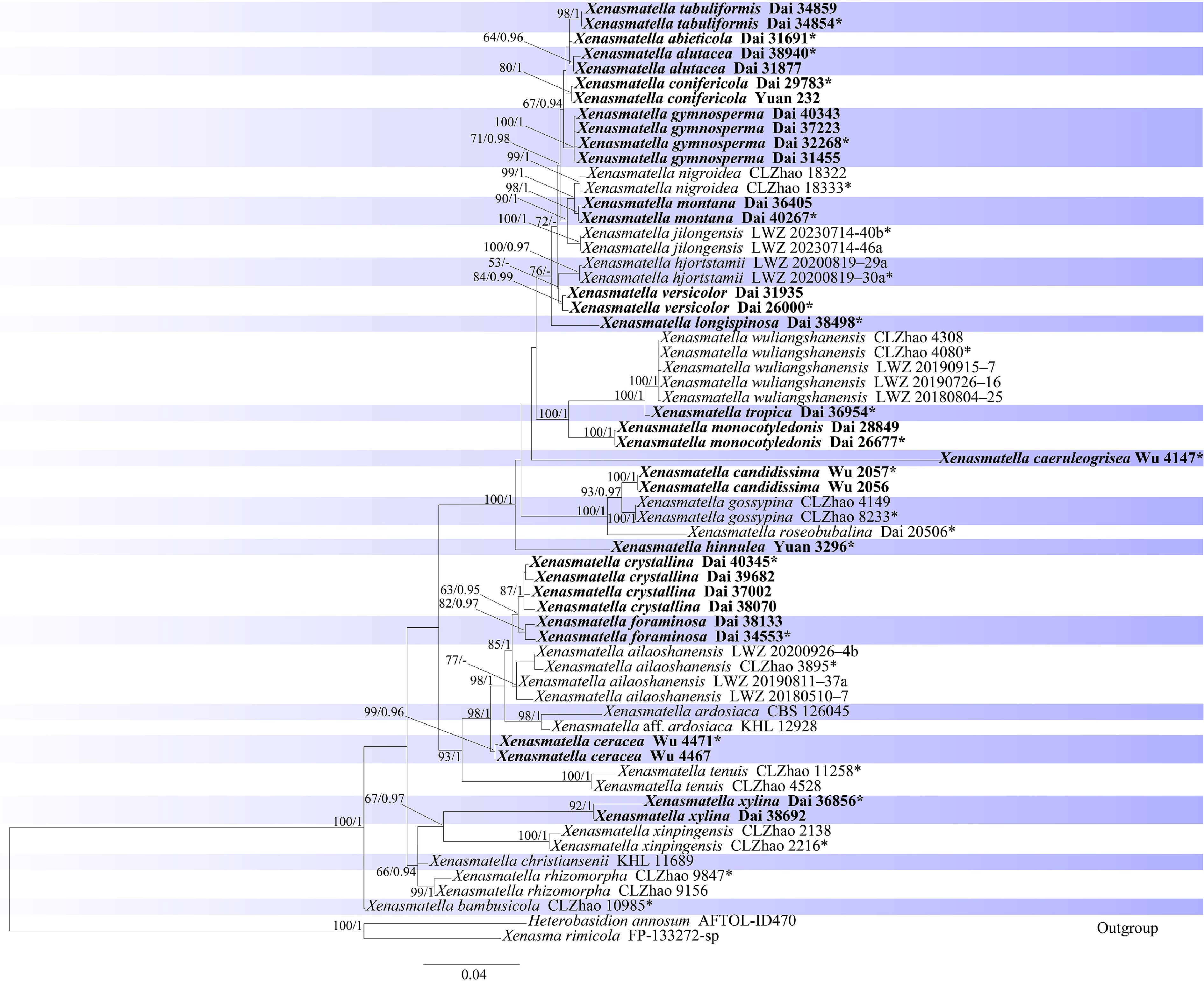
Figure 1.
Maximum likelihood strict consensus tree illustrating the phylogeny of species of order Xenasmatales based on a dataset of ITS + nLSU. ML bootstrap values higher than 60% and Bayesian posterior probability values more than 0.90 are shown. New species are in bold; * indicates type material.
-
The combined dataset of the two-locus (ITS + nLSU) included sequences from 63 samples representing 33 taxa. The dataset had an aligned length of 1,993 characters. The phylogenetic reconstruction using Maximum Likelihood (ML) and Bayesian Inference (BI) analyses of a single combined dataset showed a similar topology and a few differences in statistical support. The best model-fit applied in the Bayesian analysis was GTR + I + G, lset nst = 6, rates = invgamma, and prset statefreqpr = dirichlet (1, 1, 1, 1). Bayesian analysis yielded a nearly congruent topology, with an average standard deviation of split frequencies of 0.003931, similar to the ML analysis; thus, only the ML tree is provided (Fig. 1).
The phylogenetic tree inferred from the ITS + nLSU sequences showed that our samples clustered into 17 distinct lineages within Xenasmatella in Xenasmatales, which were identified as 17 possible new species. In our phylogeny, Xenasmatella abieticola is related to X. alutacea, X. conifericola, X. gymnosperma, and X. tabuliformis with support (ML = 67%, BPP = 0.94); X. candidissima grouped with X. gossypina (C.L. Zhao) G. Gruhn & Trichies with support (ML = 93%, BPP = 0.97), and then sister to X. roseobubalina Z.B. Liu & Yuan Yuan with support (ML = 100%, BPP = 1.00); X. ceracea is related to X. ailaoshanensis (C.L. Zhao) C.L. Zhao & T.K. Zong, X. ardosiaca (Bourdot & Galzin) Stalpers clade, X. crystallina and X. foraminosa with support (ML = 98%, BPP = 1.00); X. crystallina grouped together with X. foraminosa and X. ailaoshanensis with support (ML = 85%, BPP = 1.00); X. monocotyledonis grouped together with X. tropica and X. wuliangshanensis (C.L. Zhao) G. Gruhn & Trichies with support (ML = 100%, BPP = 1.00); X. montana is sister to X. nigroidea K.Y. Luo & C.L. Zhao with support (ML = 99%, BPP = 1.00); X. versicolor grouped together with X. hjortstamii S.L. Liu & L.W. Zhou with support (ML = 53%, BPP = 0.87); X. xylina grouped together with X. xinpingensis C.L. Zhao with support (ML = 67%, BPP = 0.97); X. caeruleogrisea, X. hinnulea and X. longispinosa formed three independent lineages.
A pairwise base-pair comparison was performed based on the ITS sequences obtained from the type material of the 17 new species. The detailed results, which compare the holotype sequences generated in this study with their closely related counterparts, are presented in Supplementary Table S2.
Taxonomy
-
Xenasmatella abieticola K.Y. Luo, Y.C. Dai, Y.J. Cui & Fang Wu, sp. nov. (Figs 2 and 3)
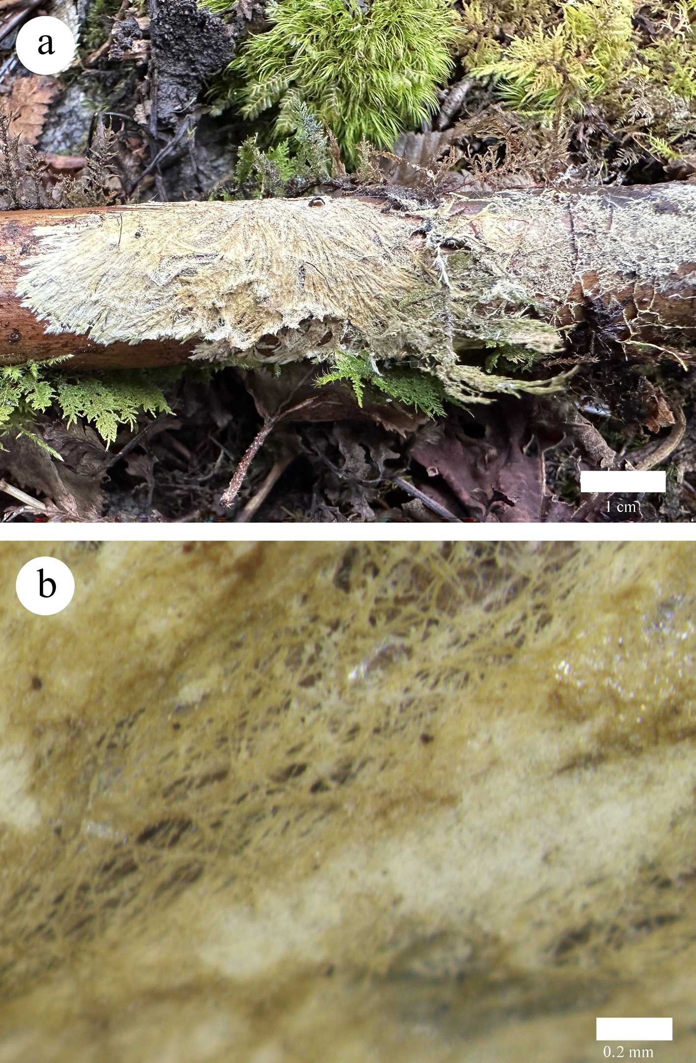
Figure 2.
Basidiomata of Xenasmatella abieticola (Dai 31691, holotype). (a) A fresh basidioma. (b) Dry hymenophore.
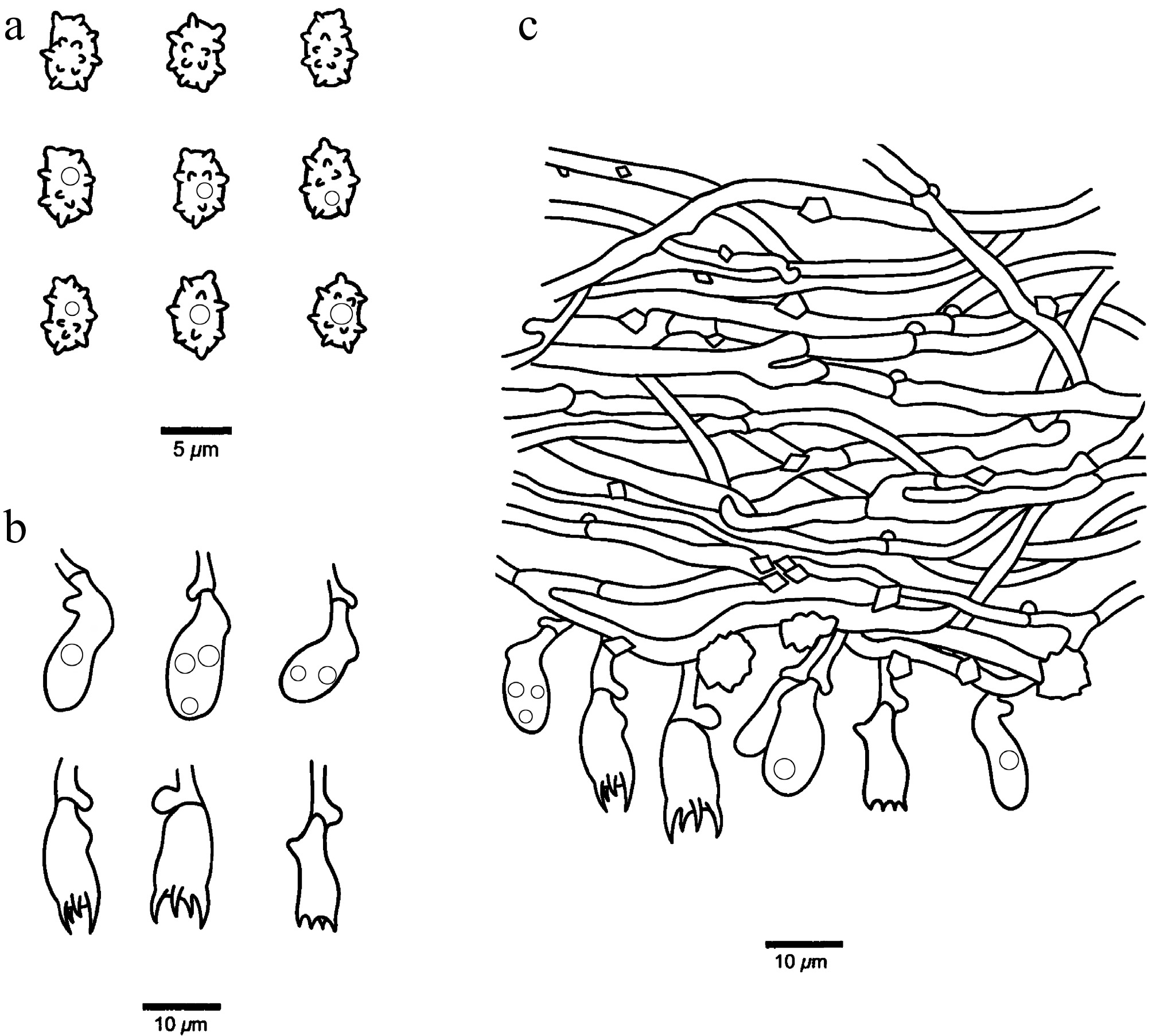
Figure 3.
Microscopic structures of Xenasmatella abieticola (Dai 31691, holotype). (a) Basidiospores. (b) Basidia and basidioles. (c) A section of hymenium.
MycoBank number: MB 863600.
Etymology — Abieticola (Lat.): refers to the species growing on Abies fabri.
Holotype — CHINA, Xizang Autonomous Region, Rikaze, Yadong County, Bacha Falls to Nathu La Gate. GPS coordinates: 27.419532° N, 88.953746° E; elevation: 2,800 m a.s.l.; on a fallen branch of A. fabri, 16 October 2024, Y.C. DAI, Dai 31691 (BJFC051950). DNA sequences: PV434792 (ITS).
Description — Basidiomata annual, resupinate, adnate, hardly separable from substrate, without odor or taste, soft, membranous, cream to buff when fresh and olivaceous buff upon drying, up to 6 cm long, 1.5 cm wide, 0.3 mm thick at center. Hymenophoral surface byssaceous, actinomorphous; sterile margin indistinct, cream; rhizomorphs concolorous with hymenophore; tissue unchanged in KOH. Hyphal system monomitic with clamp connections on generative hyphae; subicular hyphae hyaline, thin-walled, occasionally branched, flexuous, interwoven, rhomboid or irregular crystals present among hyphae, IKI–, CB–, 1.8–4.5 μm in diam. Cystidia and cystidioles absent. Basidia pleural, more or less barrel-shaped, with 4 sterigmata and a basal clamp connection, 9.5–20 × 6–7 μm; basidioles similar in shape to the basidia, but slightly smaller and with a few small guttules. Basidiospores ellipsoid, some with a constriction in the middle of one side, hyaline, thin-walled, warted throughout, usually with one small guttule, IKI–, CB–, 4.6–6.1(–6.4) × (2.8–)2.9–3.8(–4) µm, L = 5.41 μm, W = 3.23 μm, Q = 1.63 (n = 30/1); spore spines 0.6–0.9 µm long.
Notes — Xenasmatella abieticola was found on A. fabri from the Xizang Autonomous Region of southwestern China, with an alpine-plateau climate.
A BLAST search against the NCBI database based on ITS sequences revealed that Xenasmatella abieticola exhibited the highest sequence similarity to X. conifericola among the authoritative sequences, with the following alignment parameters: Max Score = 1,018; Total Score = 1,018; Query Cover = 95%; E-value = 0.0; Identity = 97.97%.
In our phylogeny (Fig. 1), Xenasmatella abieticola is related to X. alutacea, X. conifericola, X. gymnosperma, and X. tabuliformis with support (ML = 67%, BPP = 0.94). However, X. alutacea is readily distinguished from X. abieticola by its soft coriaceous basidiomata, smooth hymenophoral surface and thick-walled basidiospores; X. conifericola is different from X. abieticola by its coriaceous basidiomata, smooth to farinaceous hymenophoral surface and shorter basidiospores (3.2–4.4 vs 4.6–6.1 µm); X. gymnosperma differs from X. abieticola by its two types of subicular hyphae and shorter basidiospores (3.4–4.5 vs 4.6–6.1 µm); X. tabuliformis is readily distinguished from X. abieticola by its arachnoid or reticular hymenophoral surface, narrower basidia (4.5–6 vs 6–7 µm) and shorter blunt spines of the basidiospores (0.2–0.3 vs 0.6–0.9 µm). Morphologically, X. abieticola and X. rhizomorpha C.L. Zhao share membranous basidiomata with rhizomorphs, and similar-sized basidiospores. However, X. rhizomorpha differs from X. abieticola by the thick-walled generative hyphae and shorter blunt spines of the basidiospores (up to 0.2 vs 0.6–0.9 µm)[50].
Xenasmatella alutacea K.Y. Luo, Y.C. Dai, Y.J. Cui & Fang Wu, sp. nov. (Figs 4 and 5)
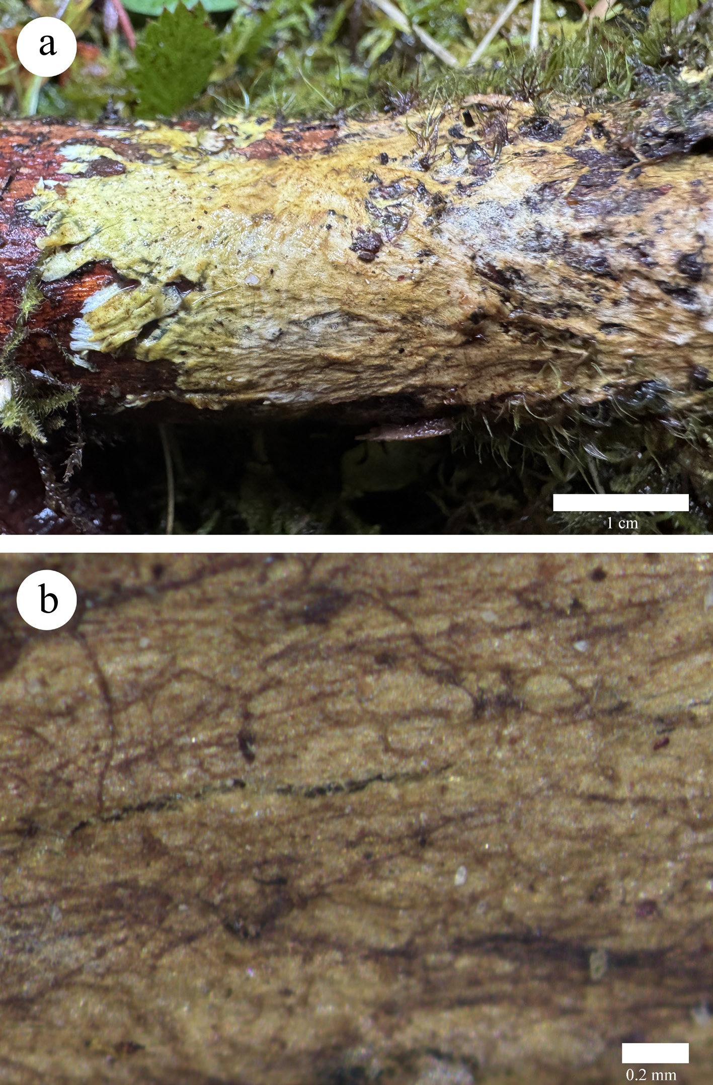
Figure 4.
Basidiomata of Xenasmatella alutacea (Dai 38940, holotype). (a) A fresh basidioma. (b) Dry hymenophore.
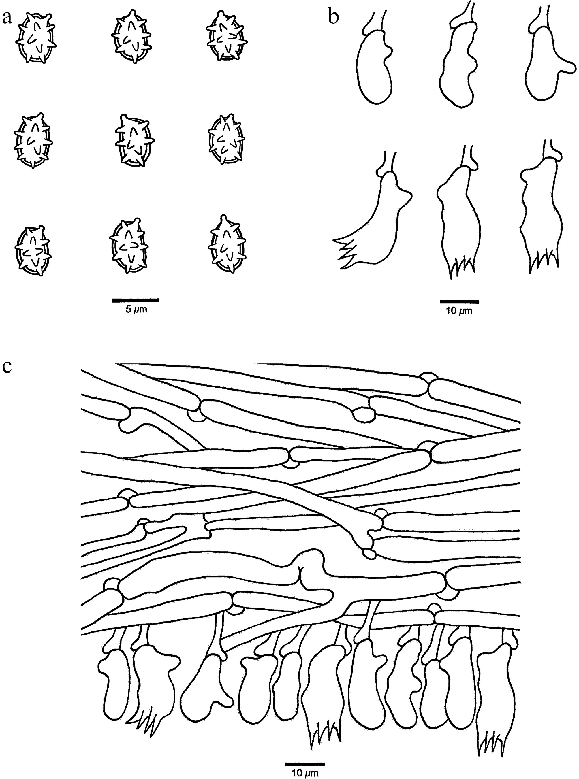
Figure 5.
Microscopic structures of Xenasmatella alutacea (Dai 38940, holotype). (a) Basidiospores. (b) Basidia and basidioles. (c) A section of hymenium.
MycoBank number: MB 863601.
Etymology — Alutacea (Lat.): refers to the species having leathery basidiomata when fresh.
Holotype — CHINA, Xizang Autonomous Region, Linzhi, Motuo County, along the Road Motuo. GPS coordinates: 29.705438° N, 95.572561° E; elevation: 2,760 m a.s.l.; on a fallen branch of Abies, 8 October 2025, Y.C. DAI, Dai 38940 (BJFC060199). DNA sequence: PZ101772 (ITS).
Description — Basidiomata annual, resupinate, adnate, hardly separable from substrate, without odor or taste, soft, coriaceous, buff to cinnamon buff when fresh and grayish brown upon drying, up to 7 cm long, 2 cm wide, 0.3 mm thick at center. Hymenophoral surface smooth; sterile margin distinct, fimbriate, white, up to 2 mm in width; tissue darkening in KOH. Hyphal system monomitic with clamp connections on generative hyphae; subicular hyphae hyaline, thin-walled, moderately branched, more or less straight, loosely interwoven, IKI–, CB–, 1.8–4.2 μm in diam. Cystidia and cystidioles absent. Basidia pleural, more or less barrel-shaped, with four sterigmata and a basal clamp connection, 18–25 × 5–8 μm; basidioles similar in shape to the basidia, but slightly smaller. Basidiospores ellipsoid, hyaline, thick-walled, warted throughout, IKI–, CB–, (4.1–)4.4–6(–6.8) × (2.7–)2.8–3.9(–4.1) µm, L = 5.16 μm, W = 3.37 μm, Q = 1.46–1.61 (n = 60/2); spore spines 0.4–1.1 µm long.
Additional specimen examined (paratype): CHINA, Xizang Autonomous Region, Shigatse, Yadong County, Rhododendron Scenic Spot. GPS coordinates: 27.396226° N, 88.828084° E; elevation: 4,232 m a.s.l.; on a fallen branch of Rhododendron, 17 October 2024, Y.C. DAI, Dai 31877 (BJFC052136). DNA sequences: PV434791 (ITS), PV434803 (LSU).
Notes — Xenasmatella alutacea was found on Abies and Rhododendron in the Xizang Autonomous Region of southwestern China, with an alpine-plateau climate.
A BLAST search against the NCBI database based on ITS sequences revealed that Xenasmatella alutacea exhibited the highest sequence similarity to X. abieticola among the authoritative sequences, with the following alignment parameters: Max Score = 996; Total Score = 996; Query Cover = 98%; E-value = 0.0; Identity = 97.92%.
In our phylogeny (Fig. 1), X. alutacea is related to X. abieticola, X. conifericola, X. gymnosperma and X. tabuliformis with support (ML = 67%, BPP = 0.94). However, morphological differences between X. abieticola and X. alutacea were listed under the notes of X. abieticola; X. conifericola is different from X. alutacea by its both shorter basidia (11.5–15 vs 18–25 µm) and basidiospores (3.2–4.4 vs 4.4–6 µm); X. gymnosperma differs from X. alutacea by its soft membranous basidiomata, byssaceous hymenophoral surface and thin-walled basidiospores; X. tabuliformis is readily distinguished from X. alutacea by its soft membranous basidiomata, arachnoid or reticular hymenophoral surface and thin-walled basidiospores. Morphologically, X. alutacea and X. hjortstamii share a smooth hymenophoral surface and similar-sized basidiospores. However, X. hjortstamii differs from X. alutacea by its soft membranous basidiomata and slightly thick-walled generative hyphae[65].
Xenasmatella caeruleogrisea K.Y. Luo, Y.C. Dai & Fang Wu, sp. nov. (Figs 6 and 7)
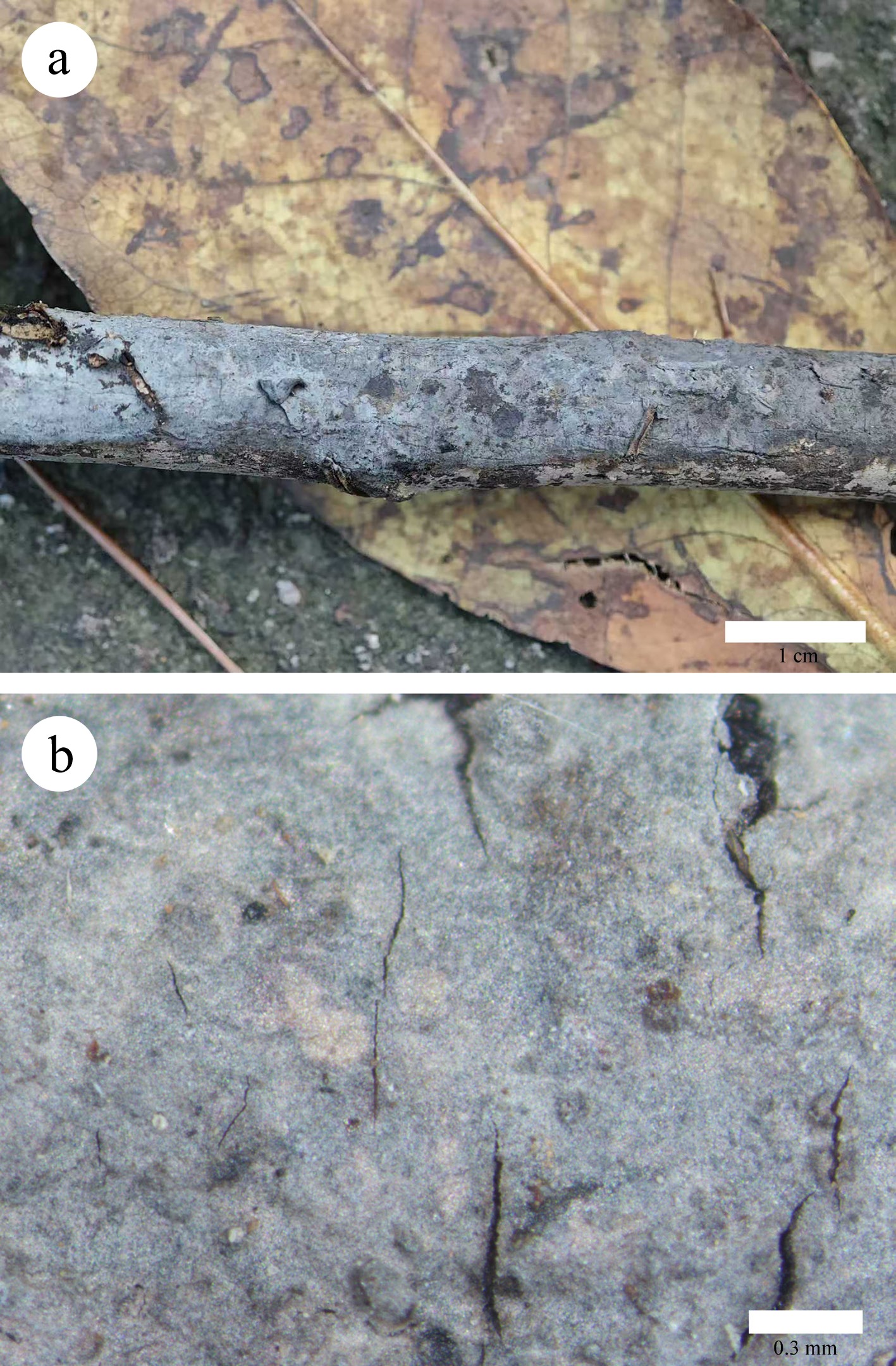
Figure 6.
Basidiomata of Xenasmatella caeruleogrisea (Wu 4147, holotype). (a) A fresh basidioma. (b) Dry hymenophore.
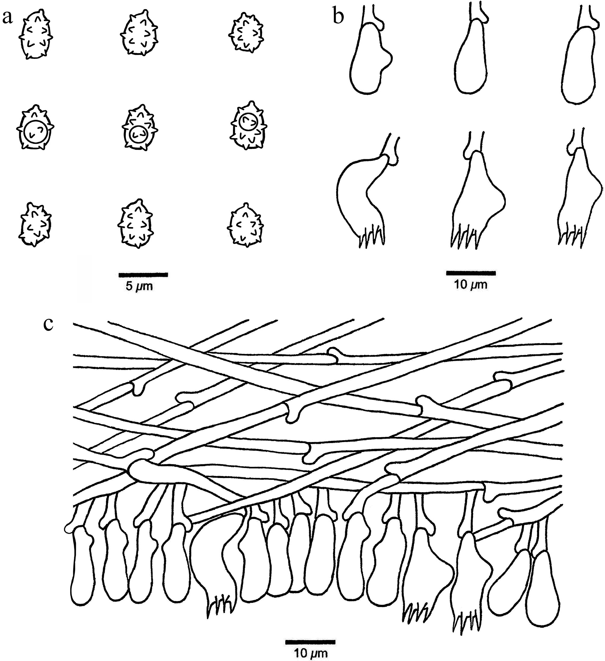
Figure 7.
Microscopic structures of Xenasmatella caeruleogrisea (Wu 4147, holotype). (a) Basidiospores. (b) Basidia and basidioles. (c) A section of hymenium.
MycoBank number: MB 863602.
Etymology — Caeruleogrisea (Lat.): refers to the species having a bluish-gray hymenophore.
Holotype — CHINA, Zhejiang Province, Hangzhou, Yuhang District, Daxiong Mountain. GPS coordinates: 30.362845° N, 120.013457° E; elevation: 72 m a.s.l.; on a fallen angiosperm branch, 13 September 2025, F. WU, Wu 4147 (BJFC064263). DNA sequences: PZ101759 (ITS), PZ112079 (LSU), PZ147911 (rpb2).
Description — Basidiomata annual, resupinate, adnate, thin, hardly separable from substrate, without odor or taste, coriaceous, bluish gray when fresh and cracked upon drying, up to 7 cm long, 1 cm wide, 0.1 mm thick at center. Hymenophoral surface smooth; sterile margin indistinct, concolorous with hymenophore; tissue darkening in KOH. Hyphal system monomitic with clamp connections on generative hyphae; subicular hyphae hyaline, thin-walled, unbranched, straight, loosely interwoven, IKI–, CB–, 2.5–4 μm in diam. Cystidia and cystidioles absent. Basidia pleural, mostly barrel-shaped, with four sterigmata and a basal clamp connection, 9–15 × 3–5 μm; basidioles similar in shape to the basidia, but slightly smaller. Basidiospores ellipsoid, hyaline, thin-walled, warted throughout, sometimes with one guttule, IKI–, CB–, (2.8–)3.1–4.4(–4.5) × (2.3–)2.4–3.3(–3.5) µm, L = 3.65 μm, W = 2.94 μm, Q = 1.24 (n = 30/1); spore spines 0.3–0.4 µm long.
Notes — Xenasmatella caeruleogrisea was found in the angiosperm forest from Zhejiang Province of eastern China with a subtropical climate.
A BLAST search against the NCBI database based on ITS sequences revealed that Xenasmatella caeruleogrisea exhibited the highest sequence similarity to X. longispinosa among the authoritative sequences, with the following alignment parameters: Max Score = 702; Total Score = 702; Query Cover = 96%; E-value = 0.0; Identity = 88.96%.
In our phylogeny (Fig. 1), X. caeruleogrisea formed an independent lineage. Morphologically, X. caeruleogrisea and X. insperata share the ash gray basidiomata, smooth hymenophoral surface, and similar-sized of basidiospores. However, X. insperata differs from X. caeruleogrisea by its subceraceous basidiomata and distribution in Canada and Norway[90,91].
Xenasmatella candidissima K.Y. Luo, Y.C. Dai, Fang Wu & Xin Zhang, sp. nov. (Figs 8 and 9)
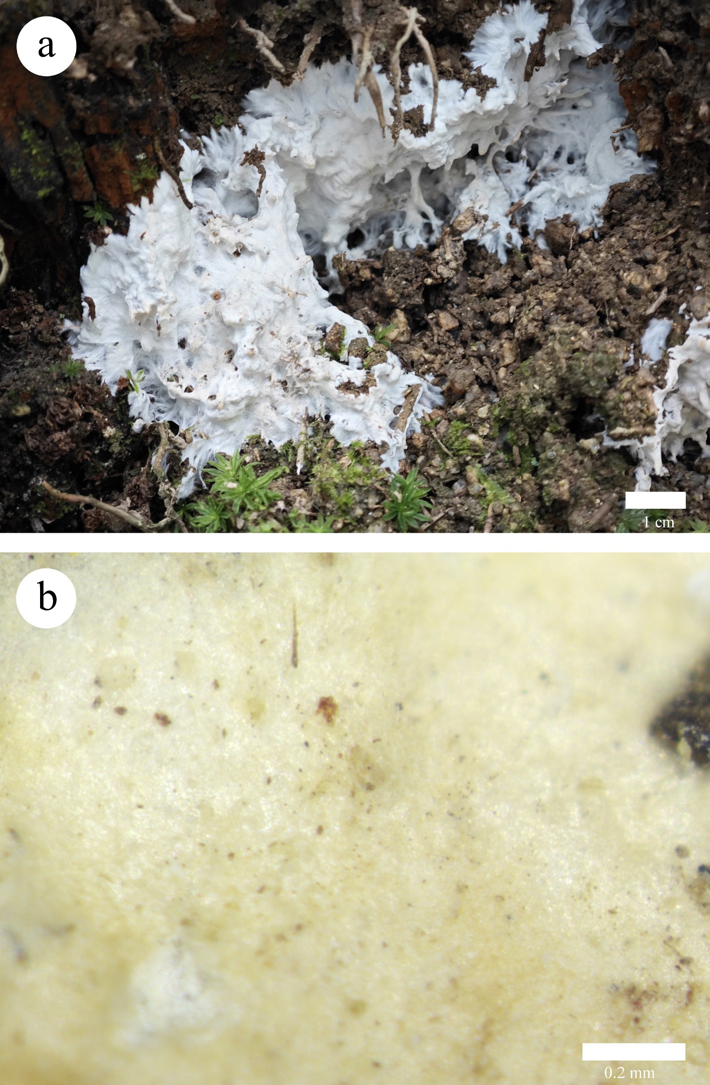
Figure 8.
Basidiomata of Xenasmatella candidissima (Wu 2057, holotype). (a) A fresh basidioma. (b) Dry hymenophore.
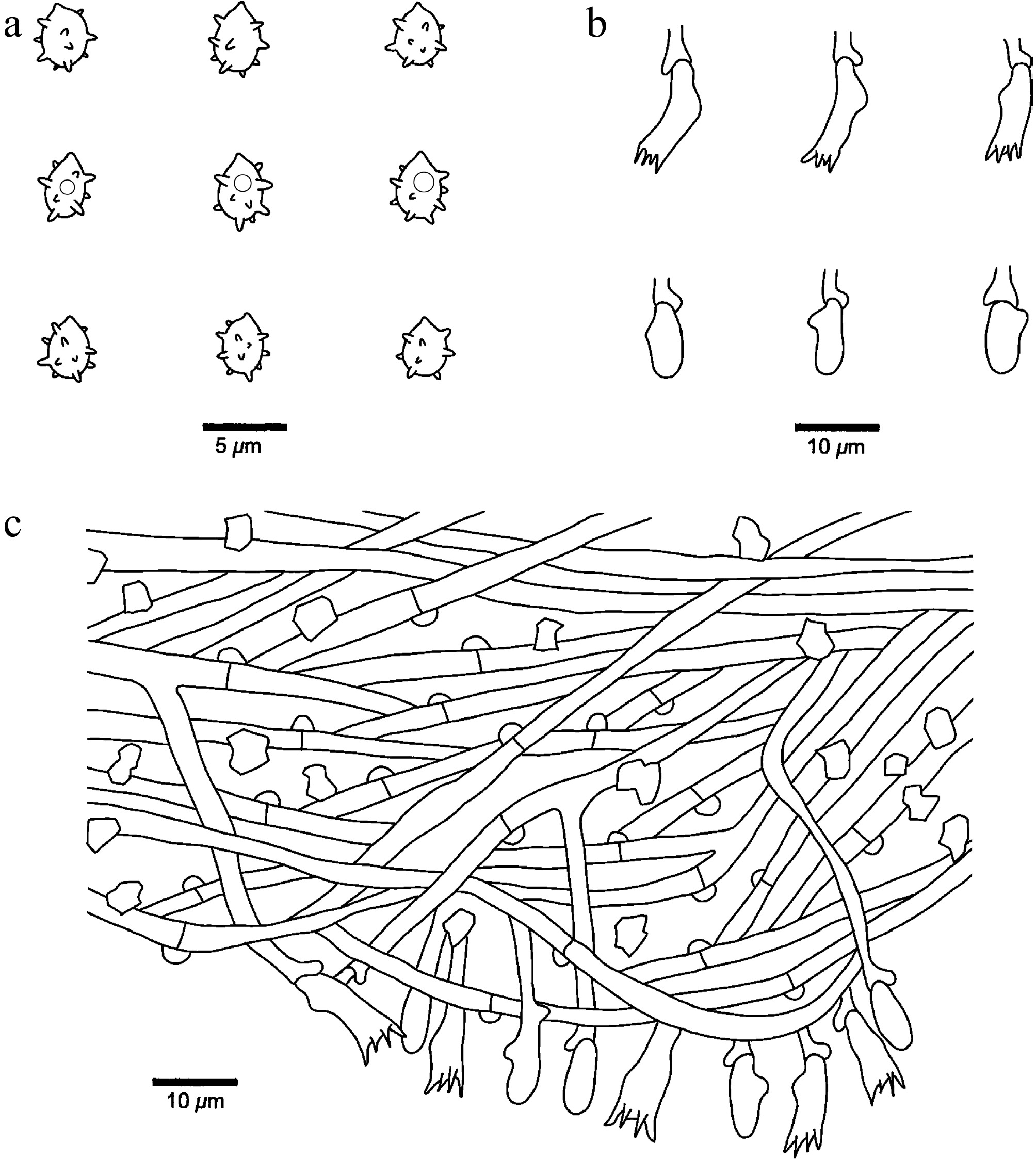
Figure 9.
Microscopic structures of Xenasmatella candidissima (Dai 2057, holotype). (a) Basidiospores. (b) Basidia and basidioles. (c) A section of hymenium.
MycoBank number: MB 863603.
Etymology — Candidissima (Lat.): refers to the species having a very white hymenophore when fresh.
Holotype — CHINA, Anhui Province, Anqing, Yuexi County, Laibang. GPS coordinates: 30.909502° N, 116.235284° E; elevation: 710 m a.s.l.; on bamboo root, 4 July 2024, F. WU, Wu 2057 (BJFC046365). DNA sequences: PV434797 (ITS), PV434808 (LSU), PZ158451 (mtSSU), PZ147913 (rpb2).
Description — Basidiomata annual, resupinate, adnate, slightly thick, hardly separable from substrate, without odor or taste, soft corky, white when fresh and upon drying, up to 9 cm long, 5 cm wide, 0.5 mm thick at center. Hymenophoral surface smooth under the lens; sterile margin distinct, fimbriate, white, up to 3 mm in width; tissue unchanged in KOH. Hyphal system monomitic with clamp connections on generative hyphae; subicular hyphae hyaline, thin-walled, occasionally branched, more or less straight, loosely interwoven, rhomboid or irregular crystals present among hyphae, IKI–, CB–, 1.4–3.5 μm in diam. Cystidia and cystidioles absent. Basidia pleural, barrel-shaped to subclavate, with four sterigmata and a basal clamp connection, 13–18 × 5–8 μm; basidioles similar in shape to the basidia, but distinctly smaller. Basidiospores ellipsoid, hyaline, thin-walled, warted throughout, sometimes with one small guttule, IKI–, CB–, (2.7–)3–4.2(–4.5) × (1.8–)2.5–3.5(–4) µm, L = 3.68 μm, W = 2.98 μm, Q = 1.22–1.24 (n = 60/2); spore spines 0.3–0.6 µm long.
Additional specimen examined (paratype): CHINA, Anhui Province, Anqing, Yuexi County, Laibang. GPS coordinates: 30.909495° N, 116.235279° E; elevation: 710 m a.s.l.; on the stump of a bamboo, 4 July 2024, F. WU, Wu 2056 (BJFC046364). DNA sequences: PV434796 (ITS), PV434807 (LSU), PZ158450 (mtSSU).
Notes — Xenasmatella candidissima was found on bamboo from Anhui Province, eastern China, with a subtropical to warm-temperate climate.
A BLAST search against the NCBI database based on ITS sequences revealed that Xenasmatella candidissima exhibited the highest sequence similarity to X. gossypina among the authoritative sequences, with the following alignment parameters: Max Score = 1,086; Total Score = 1,086; Query Cover = 99%; E-value = 0.0; Identity = 97.63%.
In our phylogeny (Fig. 1), X. candidissima grouped with X. gossypina with support (ML = 93%, BPP = 0.97), and was then sister to X. roseobubalina with support (ML = 100%, BPP = 1.00). However, X. gossypina is readily distinguished from X. candidissima by its gossypine-to-byssaceous hymenophoral surface and subglobose to globose basidiospores[47]; X. roseobubalina is different from X. candidissima by its pinkish buff hymenophoral surface and longer basidia (20–26 vs 13–18 µm)[64]. Morphologically, X. candidissima and X. tenuis C.L. Zhao share ellipsoid and similar-sized of basidiospores. However, X. tenuis differs from X. candidissima by its ceraceous to membranous basidiomata and shorter basidia (9–12.5 vs 13–18 µm)[50].
Xenasmatella ceracea K.Y. Luo, Y.C. Dai & Fang Wu, sp. nov. (Figs 10 and 11)
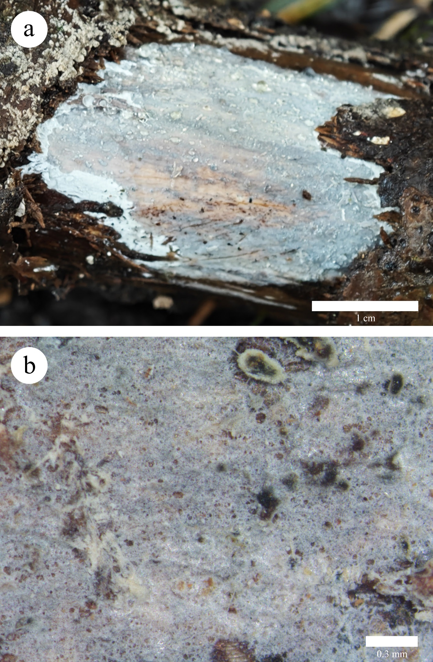
Figure 10.
Basidiomata of Xenasmatella ceracea (Wu 4471, holotype). (a) A fresh basidioma. (b) Dry hymenophore.
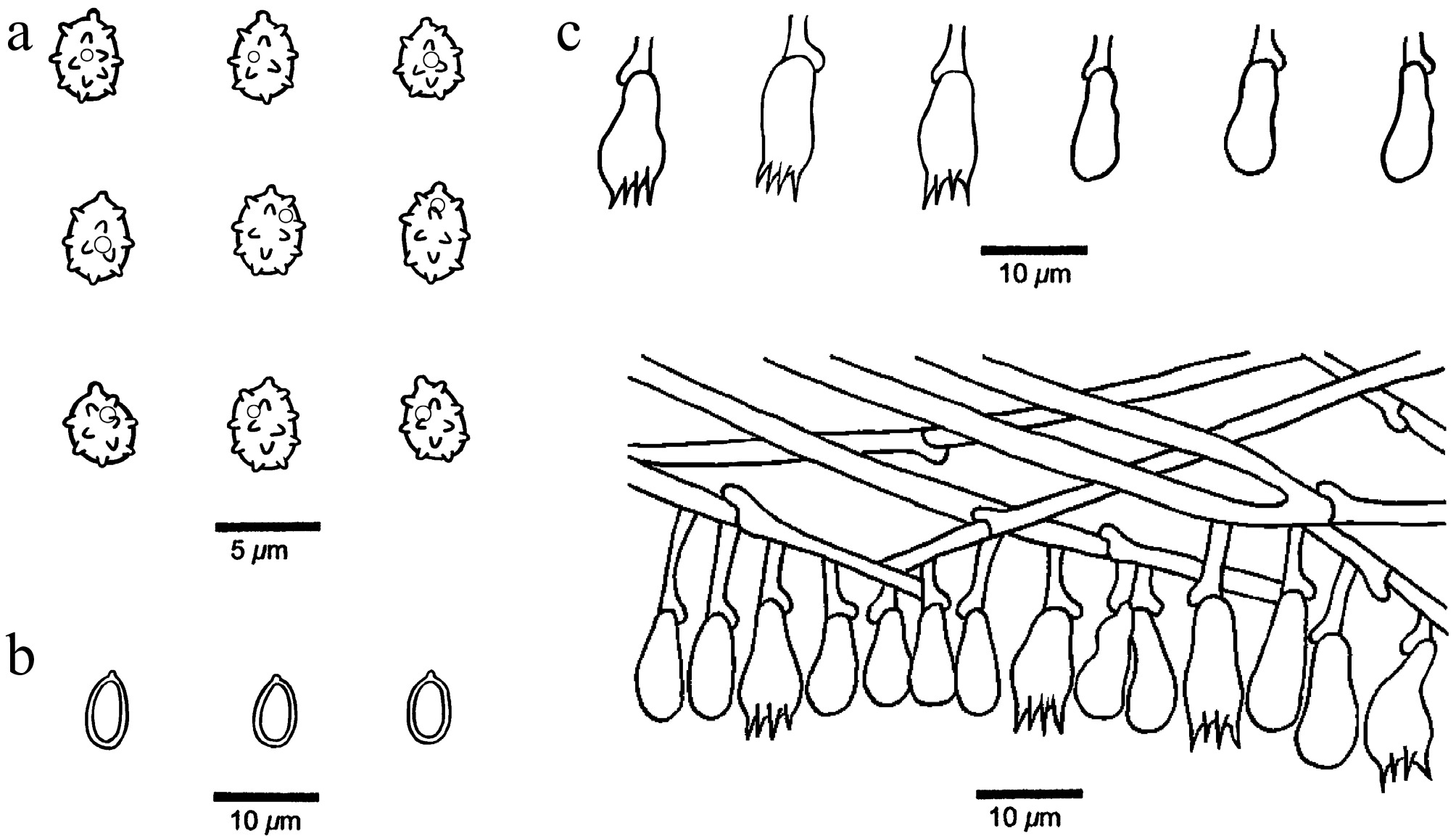
Figure 11.
Microscopic structures of Xenasmatella ceracea (Wu 4471, holotype). (a) Basidiospores. (b) Chlamydospores. (c) Basidia and basidioles. (d) A section of hymenium.
MycoBank number: MB 863604.
Etymology — Ceracea (Lat.): refers to the species having waxy basidiomata when fresh.
Holotype — CHINA, Hubei Province, Shennongjia Forestry District, Shennongjia National Forest Park. GPS coordinates: 31.447243° N, 110.193346° E; elevation: 2,577 m a.s.l.; on rotten angiosperm wood, 20 September 2025, F. WU, Wu 4471 (BJFC064587). DNA sequences: PZ101769 (ITS), PZ112088 (LSU), PZ158465 (mtSSU), PZ147916 (rpb2).
Description — Basidiomata annual, resupinate, adnate, hardly separable from substrate, without odor or taste, ceraceous, bluish gray when fresh and upon drying, up to 3 cm long, 2 cm wide, 0.2 mm thick at center. Hymenophoral surface smooth to holey under the lens; sterile margin distinct, white, up to 3 mm in width; tissue becoming dark gray in KOH. Hyphal system monomitic with clamp connections on generative hyphae; subicular hyphae hyaline, thin-walled, occasionally branched, straight, loosely interwoven, IKI–, CB–, 2.5–4 μm in diam. Cystidia and cystidioles absent. Basidia pleural, barrel-shaped to pyriform, with four sterigmata and a basal clamp connection, 9–15 × 5–7 μm; basidioles similar in shape to the basidia, but slightly smaller. Basidiospores ellipsoid, hyaline, thin-walled, warted throughout, usually with one small guttule, IKI–, CB–, (3.1–)3.3–4.8(–4.9) × 2.8–3.4(–3.7) µm, L = 4.15 μm, W = 3.11 μm, Q = 1.33 (n = 30/1); spore spines 0.5–0.6 µm long (the specimen Wu 4467 is sterile). Chlamydospores present, ellipsoid, abundant, hyaline, thick-walled, smooth, IKI–, CB–, 6–7.5 × 4–4.5 µm.
Additional specimen examined (paratype): CHINA, Hubei Province, Shennongjia Forestry District, Shennongjia National Forest Park. GPS coordinates: 31.447347° N, 110.192969° E; elevation: 2,578 m a.s.l.; on rotten angiosperm wood, 20 September 2025, F. WU, Wu 4467 (BJFC064583). DNA sequences: PZ101768 (ITS), PZ112087 (LSU), PZ158464 (mtSSU), PZ147917 (rpb2).
Notes — Xenasmatella ceracea was found on rotten wood from Hubei Province of central China with a subtropical climate.
A BLAST search against the NCBI database based on ITS sequences revealed that Xenasmatella ceracea exhibited the highest sequence similarity to X. ardosiaca among the authoritative sequences, with the following alignment parameters: Max Score = 937; Total Score = 937; Query Cover = 97%; E-value = 0.0; Identity = 96.81%.
In our phylogeny (Fig. 1), X. ceracea is related to X. ailaoshanensis, X. ardosiaca, X. crystallina and X. foraminosa with support (ML = 98%, BPP = 1.00). However, X. ailaoshanensis is readily distinguished from X. ceracea by its pruinose to farinaceous hymenophoral surface and wider basidiospores (3.5–4.5 vs 2.8–3.4 µm)[46]; X. ardosiaca differs from X. ceracea by its longer basidia (15–20 vs 9–15 µm), and globose, bigger basidiospores (5–6 µm in diam vs 3.3–4.8 × 2.8–3.4 µm)[37]; X. crystallina is different from X. ceracea by its coriaceous basidiomata and gossypine hymenophoral surface; X. foraminosa is readily distinguished from X. ceracea by its coriaceous basidiomata and thick-walled basidiospores. Morphologically, X. ceracea and X. xinpingensis share ceraceous basidiomata and similar-sized of basidiospores. However, X. xinpingensis differs from X. ceracea by its irregularly reticulate or somewhat wrinkled hymenophoral surface and subglobose basidiospores[50].
Xenasmatella conifericola K.Y. Luo, Y.C. Dai, Y.D. Wu & Fang Wu, sp. nov. (Figs 12 and 13)
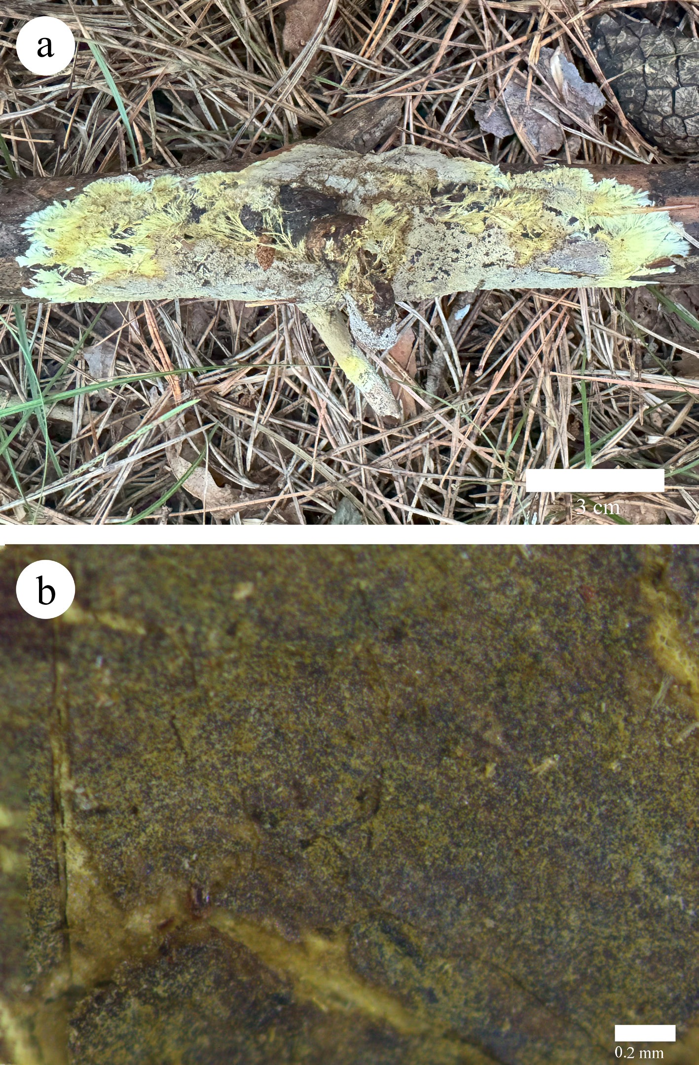
Figure 12.
Basidiomata of Xenasmatella conifericola (Dai 29783, holotype). (a) A fresh basidioma. (b) Dry hymenophore.
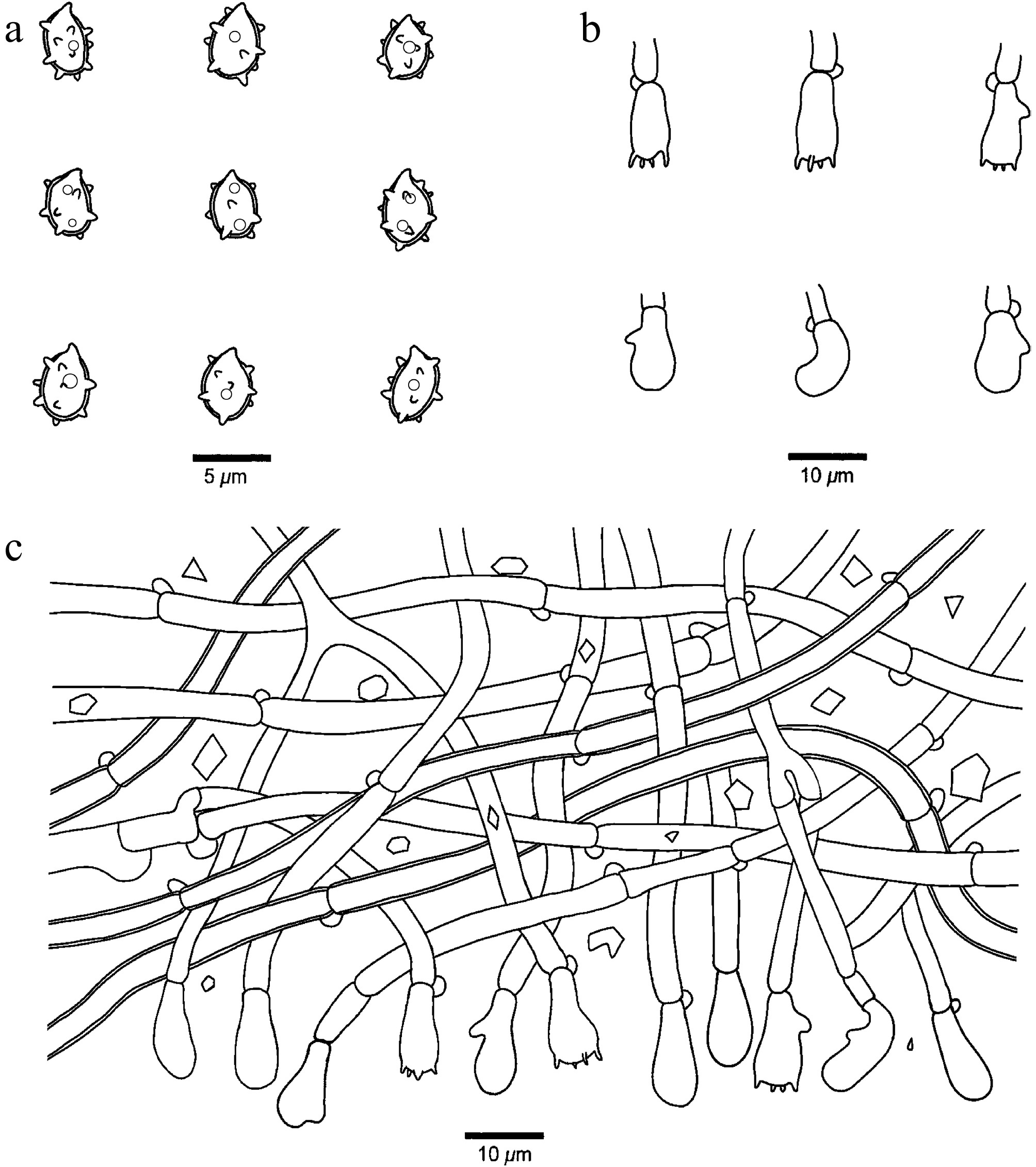
Figure 13.
Microscopic structures of Xenasmatella conifericola (Dai 29783, holotype). (a) Basidiospores. (b) Basidia and basidioles. (c) A section of hymenium.
MycoBank number: MB 863605.
Etymology — Conifericola (Lat.): refers to the species growing on coniferous wood.
Holotype — CHINA, Heilongjiang Province, Yichun, Lesser Hinggan Mountains Botanical Garden. GPS coordinates: 47.733336° N, 128.916723° E; elevation: 230 m a.s.l.; on a fallen branch of Pinus koraiensis, 16 August 2024, Y.C. DAI, Dai 29783 (BJFC050042). DNA sequences: PV434793 (ITS), PV434804 (LSU), PV455374 (mtSSU), PV468707 (tef1).
Description — Basidiomata annual, resupinate, adnate, thin, easily separable from substrate, without odor or taste, coriaceous, sulphur yellow to ash gray when fresh and grayish brown upon drying, up to 12 cm long, 3 cm wide, 0.3 mm thick at center. Hymenophoral surface smooth to farinaceous under the lens; sterile margin distinct, actinomorphous, straw yellow, up to 1 cm in width; rhizomorphs sulphur yellow to straw yellow; tissue darkening in KOH. Hyphal system monomitic with clamp connections on generative hyphae; subicular hyphae hyaline, thin- to slightly thick-walled, occasionally branched, flexuous, loosely interwoven, rhomboid crystals present among hyphae, IKI–, CB–, 2.8–4.7 μm in diam. Cystidia and cystidioles absent. Basidia pleural, barrel-shaped, with four sterigmata and a basal clamp connection, 11.5–15 × 5–6.5 μm; basidioles similar in shape to the basidia, but slightly smaller. Basidiospores ellipsoid, hyaline, slightly thick-walled, warted throughout, usually with one or two guttules, IKI–, CB–, (3.1–)3.2–4.4(–4.7) × (2.5–)2.7–3.4(–3.5) µm, L = 3.79 μm, W = 3.06 μm, Q = 1.21–1.27 (n = 60/2); spore spines 0.3–0.6 µm long.
Additional specimen examined (paratype): CHINA, Jilin Province, Yanbian, Antu County, Huangsongpu Forest Farm. GPS coordinates: 42.189052° N, 128.210271° E; elevation: 1,100 m a.s.l.; on a charred trunk of Larix, 9 October 2021, Y. YUAN, Yuan 232 (BJFC038495). DNA sequences: PV434795 (ITS), PV434806 (LSU), PZ158452 (mtSSU), PV468706 (tef1).
Notes — Xenasmatella conifericola was found on coniferous wood from Heilongjiang and Jilin Province of northeastern China with a temperate monsoon climate.
A BLAST search against the NCBI database based on ITS sequences revealed that Xenasmatella conifericola exhibited the highest sequence similarity to X. tabuliformis among the authoritative sequences, with the following alignment parameters: Max Score = 1,044; Total Score = 1,044; Query Cover = 99%; E-value = 0.0; Identity = 98.17%.
In our phylogeny (Fig. 1), X. conifericola is related to X. abieticola, X. alutacea, X. gymnosperma, and X. tabuliformis with support (ML = 67%, BPP = 0.94). However, X. abieticola is readily distinguished from X. conifericola by its soft, membranous basidiomata, byssaceous hymenophoral surface and longer basidiospores (4.6–6.1 vs 3.2–4.4 µm); X. alutacea is different from X. conifericola by its both longer basidia (18–25 vs 11.5–15 µm) and basidiospores (4.4–6 vs 3.2–4.4 µm); X. gymnosperma differs from X. conifericola by its soft, membranous basidiomata, byssaceous hymenophoral surface and longer basidia (18–25 vs 11.5–15 µm); X. tabuliformis is readily distinguished from X. conifericola by its soft, membranous basidiomata and arachnoid or reticular hymenophoral surface. Morphologically, X. conifericola and X. christiansenii share a smooth to farinaceous hymenophoral surface. However, X. christiansenii differs from X. conifericola by its white to grayish basidiomata and bigger basidiospores (6–7 × 4–4.5 vs 3.2–4.4 × 2.7–3.4 µm)[37].
Xenasmatella crystallina K.Y. Luo, Y.C. Dai & Fang Wu, sp. nov. (Figs 14 and 15)
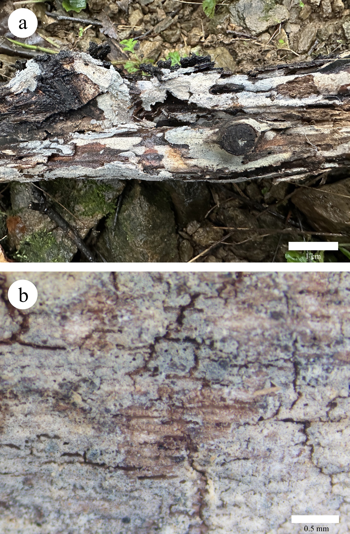
Figure 14.
Basidiomata of Xenasmatella crystallina (Dai 40345, holotype). (a) A fresh basidioma. (b) Dry hymenophore.
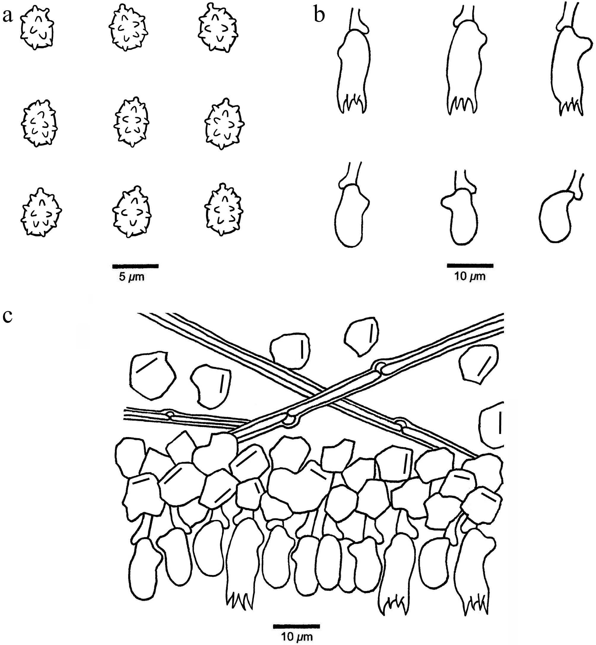
Figure 15.
Microscopic structures of Xenasmatella crystallina (Dai 40345, holotype). (a) Basidiospores. (b) Basidia and basidioles. (c) A section of hymenium.
MycoBank number: MB 863606.
Etymology — Crystallina (Lat.): refers to the species having abundant crystals.
Holotype — CHINA, Henan Province, Luoyang, Luanchuan County, Wangfu Bamboo Forest. GPS coordinates: 33.947143° N, 111.733304° E; elevation: 841 m a.s.l.; on a rotten trunk of Quercus aliena, 20 October 2025, Y.C. DAI, Dai 40345 (BJFC061601). DNA sequences: PZ101765 (ITS), PZ112085 (LSU), PZ158462 (mtSSU), PZ147914 (rpb2), PZ147906 (gapdh).
Description — Basidiomata annual, resupinate, adnate, hardly separable from substrate, without odor or taste, coriaceous, cream when fresh and ash gray upon drying, up to 10 cm long, 2 cm wide, 0.2 mm thick at center. Hymenophoral surface gossypine under the lens; sterile margin distinct, ash gray, up to 5 mm in width; tissue becoming dark gray in KOH. Hyphal system monomitic with clamp connections on generative hyphae; subicular hyphae infrequent, hyaline, thick-walled, unbranched, straight, loosely interwoven, abundant crystals present among hyphae, IKI–, CB–, 1.8–2.5 μm in diam. Cystidia and cystidioles absent. Basidia pleural, mostly barrel-shaped, with four sterigmata and a basal clamp connection, 10–15 × 4.5–6 μm; basidioles similar in shape to the basidia, but distinctly smaller. Basidiospores subglobose to broadly ellipsoid, hyaline, thin-walled, warted throughout, IKI–, CB–, (3.1–)3.4–4.8(–5.1) × (2.4–)2.8–4(–4.5) µm, L = 4.18 μm, W = 3.52 μm, Q = 1.17–1.26 (n = 120/4); spore spines 0.3–0.5 µm long.
Additional specimens examined (paratype): CHINA, Hainan Province, National Park of Hainan Tropical Rainforest, Yingge Mountain. GPS coordinates: 19.050340° N, 109.564783° E; elevation: 623 m a.s.l.; on dead rattan, 30 June 2025, Y.C. DAI, Dai 37002 (BJFC058261), DNA sequences: PZ101762 (ITS), PZ112082 (LSU), PZ158459 (mtSSU); Heilongjiang Province, Harbin, Tonghe County, Mamading Virgin Forest. GPS coordinates: 46.180548° N, 129.180753° E; elevation: 140 m a.s.l.; on a rotten trunk of Pinus koraiensis, 10 October 2025, Y.C. DAI, Dai 38070 (BJFC059329), DNA sequences: PZ101763 (ITS), PZ158460 (mtSSU), PZ147918 (rpb2), PZ147907 (gapdh); Guizhou Province, Qiannan, Libo County, Maolan National Nature Reserve. GPS coordinates: 25.220525° N, 108.019218° E; elevation: 500 m a.s.l.; on a fallen angiosperm branch, 8 June 2025, Y.C. DAI, Dai 39682 (BJFC060941), DNA sequences: PZ101764 (ITS), PZ112084 (LSU), PZ158461 (mtSSU).
Notes — Xenasmatella crystallina was found in virgin forest in Hainan, Heilongjiang, Henan, and Guizhou provinces of China, with temperate to tropical climates.
A BLAST search against the NCBI database based on ITS sequences revealed that Xenasmatella crystallina exhibited the highest sequence similarity to X. ailaoshanensis among the authoritative sequences, with the following alignment parameters: Max Score = 1,014; Total Score = 1,014; Query Cover = 97%; E-value = 0.0; Identity = 97.80%.
In our phylogeny (Fig. 1), X. crystallina grouped together with X. foraminosa and X. ailaoshanensis with support (ML = 85%, BPP = 1.00). However, X. foraminosa is different from X. crystallina by its holey hymenophoral surface and thick-walled basidiospores; X. ailaoshanensis is readily distinguished from X. crystallina by its membranous to soft ceraceous basidiomata, pruinose to farinaceous hymenophoral surface, and thin-walled generative hyphae[46]. Morphologically, X. crystallina and X. nigroidea share thick-walled generative hyphae and thin-walled, similar-sized basidiospores. However, X. nigroidea differs from X. crystallina by its gray to black basidiomata, with byssaceous to reticulate hymenophoral surface[36].
Xenasmatella foraminosa K.Y. Luo, Y.C. Dai, Xin Zhang & Fang Wu, sp. nov. (Figs 16 and 17)
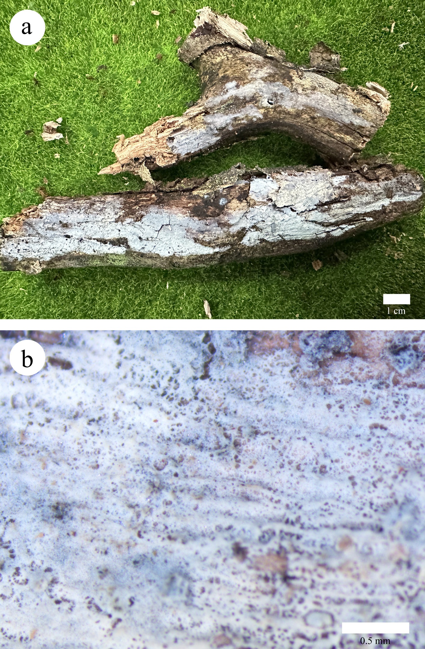
Figure 16.
Basidiomata of Xenasmatella foraminosa (Dai 34553, holotype). (a) A fresh basidioma. (b) Dry hymenophore.
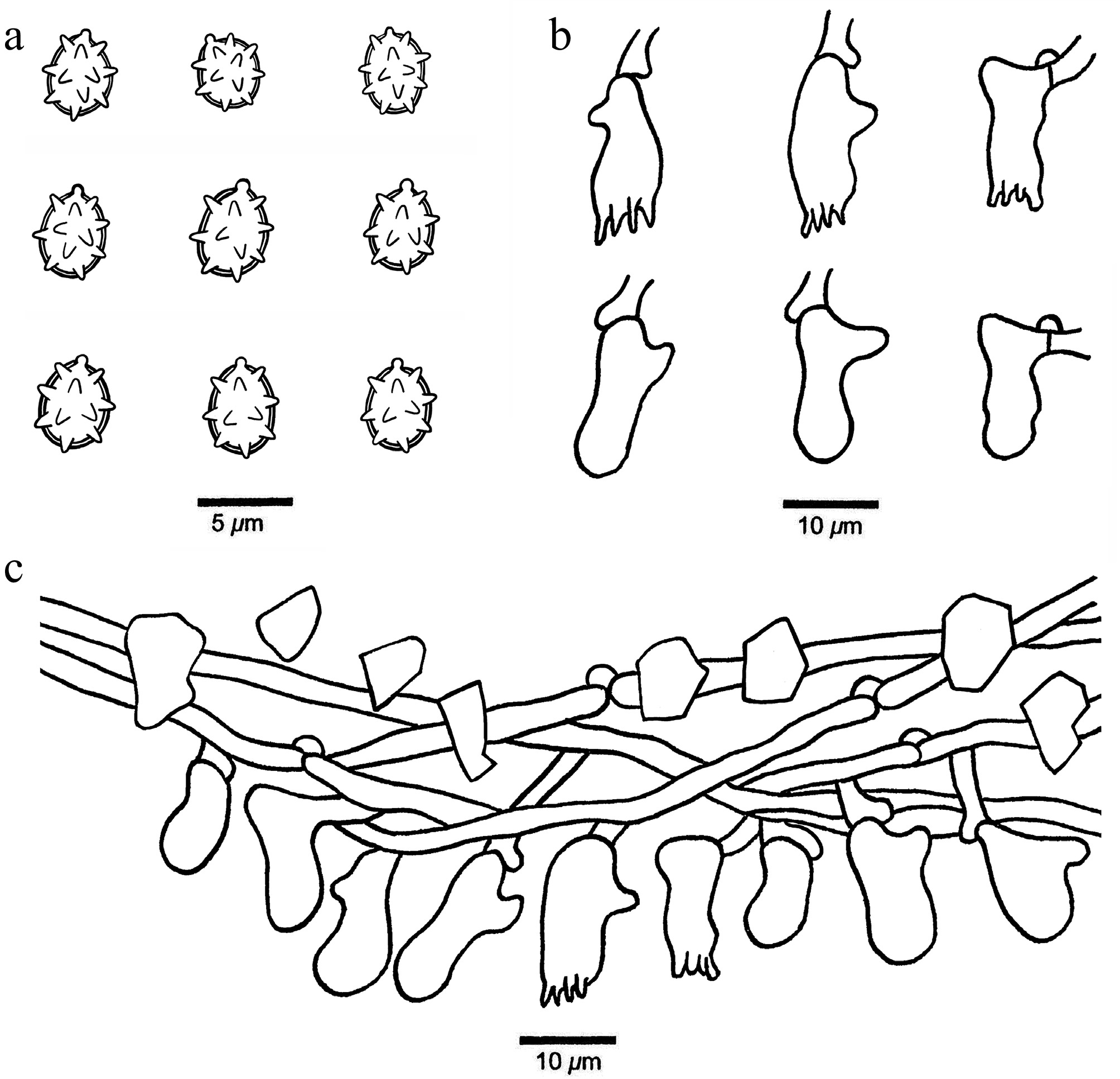
Figure 17.
Microscopic structures of Xenasmatella foraminosa (Dai 34553, holotype). (a) Basidiospores. (b) Basidia and basidioles. (c) A section of hymenium.
MycoBank number: MB 863607.
Etymology — Foraminosa (Lat.): refers to the species having a hymenophore full of holes.
Holotype — CHINA, Zhejiang Province, Jinhua, Panan County, Dapanshan National Nature Reserve. GPS coordinates: 28.985979° N, 120.539882° E; elevation: 676 m a.s.l.; on dead rattan, 6 November 2025, Y.C. DAI, Dai 34553 (BJFC055814). DNA sequences: PZ101766 (ITS), PZ112086 (LSU), PZ158463 (mtSSU), PZ147915 (rpb2), PZ147908 (gapdh).
Description — Basidiomata annual, resupinate, adnate, hardly separable from substrate, without odor or taste, coriaceous, white to smoke gray when fresh and bluish gray upon drying, up to 12 cm long, 2.5 cm wide, 0.2 mm thick at center. Hymenophoral surface holey under the lens; sterile margin indistinct, concolorous with hymenophoral surface; tissue darkening in KOH. Hyphal system monomitic with clamp connections on generative hyphae; subicular hyphae hyaline, thin-walled, unbranched, flexuous, loosely interwoven, irregular crystals present among hyphae, IKI–, CB–, 2–3 μm in diam. Cystidia and cystidioles absent. Basidia pleural, more or less barrel-shaped, with four sterigmata and a basal clamp connection, 9–18 × 5–6 μm; basidioles similar in shape to the basidia, but slightly smaller. Basidiospores subglobose to broadly ellipsoid, hyaline, thick-walled, warted throughout, IKI–, CB–, (3.6–)3.7–5(–5.1) × (2.7–)3.2–4.3(–4.6) µm, L = 4.38 μm, W = 3.66 μm, Q = 1.19–1.20 (n = 60/2); spore spines 0.4–0.8 µm long.
Additional specimen examined (paratype): CHINA, Heilongjiang Province, Harbin, Tonghe County, Mamading Virgin Forest. GPS coordinates: 46.180548° N, 129.180753° E; elevation: 140 m a.s.l.; on a rotten branch of Populus, 10 October 2025, Y.C. DAI, Dai 38133 (BJFC059392). DNA sequence: PZ101767 (ITS).
Notes — Xenasmatella foraminosa was found in virgin forest in Heilongjiang and Zhejiang Province of northeastern and eastern China, respectively.
A BLAST search against the NCBI database based on ITS sequences revealed that Xenasmatella foraminosa exhibited the highest sequence similarity to X. ailaoshanensis among the authoritative sequences, with the following alignment parameters: Max Score = 970; Total Score = 970; Query Cover = 97%; E-value = 0.0; Identity = 97.87%.
In our phylogeny (Fig. 1), X. foraminosa grouped with X. ailaoshanensis and X. crystallina with support (ML = 85%, BPP = 1.00). However, X. ailaoshanensis is readily distinguished from X. foraminosa by its membranous to soft ceraceous basidiomata, pruinose to farinaceous hymenophoral surface, and thin-walled basidiospores[46]; X. crystallina is different from X. foraminosa by its gossypine hymenophoral surface and thick-walled generative hyphae, and thin-walled basidiospores. Morphologically, X. foraminosa and X. globigera (Hjortstam & Ryvarden) Duhem share a fairly porulose hymenophoral surface and thick-walled basidiospores. However, X. globigera differs from X. foraminosa by its globose basidiospores[44].
Xenasmatella gymnosperma K.Y. Luo, Y.C. Dai, Yuan Yuan & Fang Wu, sp. nov. (Figs 18 and 19)
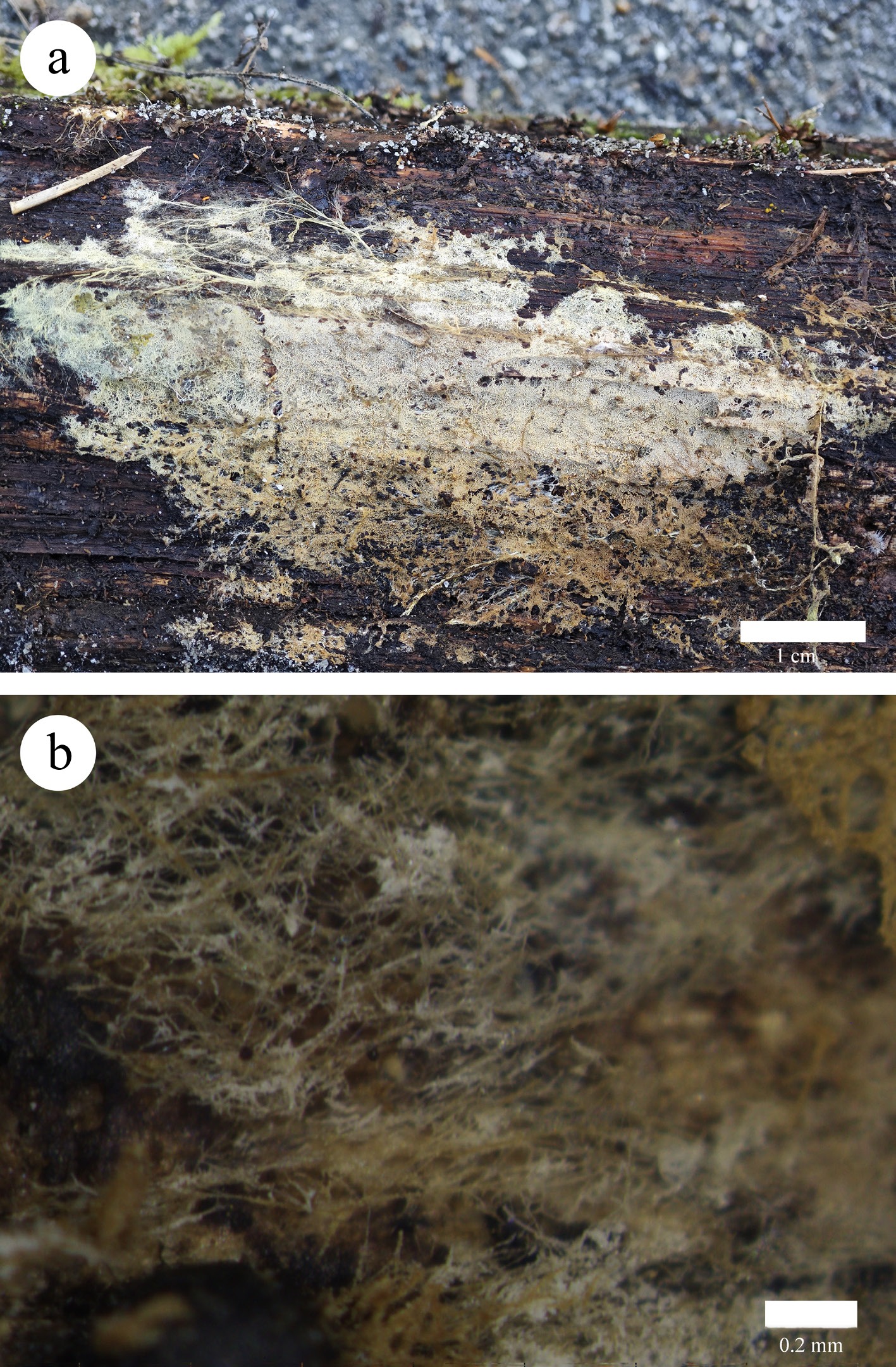
Figure 18.
Basidiomata of Xenasmatella gymnosperma (Dai 32268, holotype). (a) A fresh basidioma. (b) Dry hymenophore.
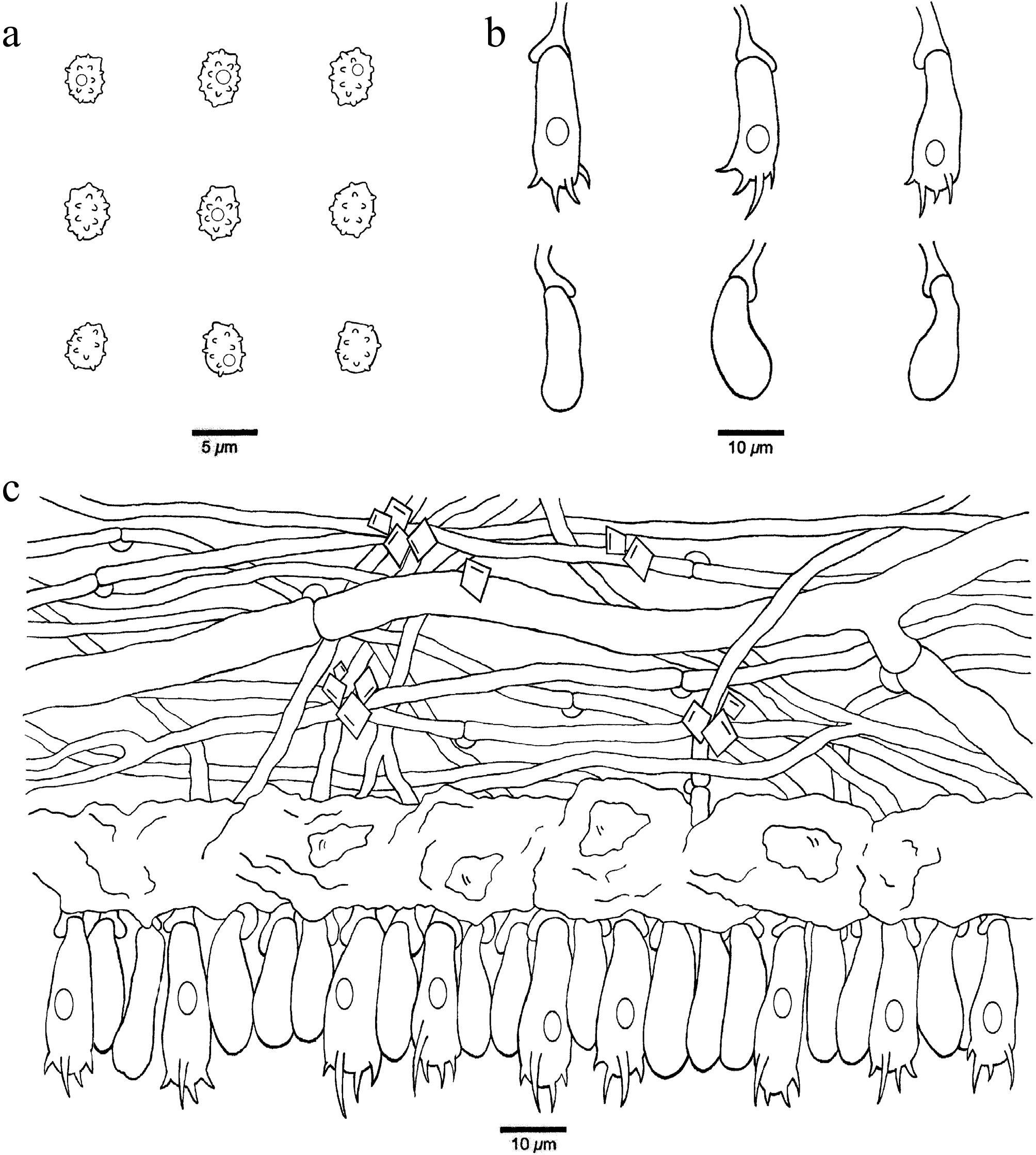
Figure 19.
Microscopic structures of Xenasmatella gymnosperma (Dai 32268, holotype). (a) Basidiospores. (b) Basidia and basidioles. (c) A section of hymenium.
MycoBank number: MB 863608.
Etymology — Gymnosperma (Lat.): refers to the species growing on gymnosperm wood.
Holotype — CHINA, Xizang Autonomous Region, Linzhi, Bomi County, Songrao Village. GPS coordinates: 29.904681° N, 95.503428° E; elevation: 2,559 m a.s.l.; on a fallen trunk of Pinus armandii, 22 October 2024, Y.C. DAI, Dai 32268 (BJFC052528). DNA sequences: PV434789 (ITS), PV434802 (LSU).
Description — Basidiomata annual, resupinate, adnate, thin, easily separable from substrate, without odor or taste, soft membranous, cream when fresh and buff upon drying, up to 13 cm long, 1 cm wide, 0.1 mm thick at center. Hymenophoral surface byssaceous under the lens; sterile margin indistinct, cream; rhizomorphs concolorous with hymenophoral surface; tissue darkening in KOH. Hyphal system monomitic with clamp connections on generative hyphae; subicular hyphae have two types; type one hyphae are hyaline, thin-walled, occasionally branched, flexuous, interwoven, IKI–, CB–, 2–3.5 μm in diam, and type two hyphae are distinctly wider than type one, IKI–, CB–, 5–7 μm in diam; rhomboid crystals present among hyphae. Subhymenium contains a gelatinized layer. Cystidia and cystidioles absent. Basidia more or less barrel-shaped, with four sterigmata, a basal clamp connection and one guttule, 18–25 × 5–7 μm; basidioles clavate to pyriform, slightly smaller than basidia. Basidiospores ellipsoid, hyaline, thin-walled, warted throughout, sometimes with one small guttule, IKI–, CB–, (3.0–)3.4–4.5(–5.1) × (2.2–)2.3–3.4(–3.7) µm, L = 3.87 μm, W = 2.89 μm, Q = 1.28–1.40 (n = 90/3); spore spines 0.3–0.6 µm long (the specimen Dai 37223 is sterile).
Additional specimens examined (paratype): CHINA, Xizang Autonomous Region, Rikaze, Yadong County, Xiayadong, Renqinggang Village. GPS coordinates: 27.396225° N, 88.933842° E; elevation: 2842 m a.s.l.; on a fallen twig of Pinus armandii, 15 October 2024, Y.C. DAI, Dai 31455 (BJFC051714), DNA sequences: PV434788 (ITS), PV434801 (LSU), PZ147919 (tef1); Liaoning Province, Benxi, Huanren County, Laotudingzi National Nature Reserve, Lamenligou. GPS coordinates: 41.322902° N, 124.887852° E; elevation: 700 m a.s.l.; on a fallen twig of Pinus koraiensis, 23 August 2025, Y.C. DAI, Dai 37223 (BJFC058482), DNA sequences: PZ101752 (ITS), PZ112074 (LSU), PZ158448 (mtSSU), PZ147921 (tef1); Fushun, Xinbin County, Laotudingzi National Nature Reserve, Liudaogou Protection Station. GPS coordinates: 41.343862° N, 124.840333° E; elevation: 593 m a.s.l.; on a fallen trunk of Larix olgensis, 14 October 2025, Y.C. DAI, Dai 40343 (BJFC061599), DNA sequences: PZ101753 (ITS), PZ112075 (LSU), PZ158449 (mtSSU), PZ147920 (tef1).
Notes — Xenasmatella gymnosperma was found in virgin forests from Xizang Autonomous Region and Liaoning Province in the southwestern and northeastern regions of China, respectively.
A BLAST search against the NCBI database based on ITS sequences revealed that Xenasmatella gymnosperma exhibited the highest sequence similarity to X. conifericola among the authoritative sequences, with the following alignment parameters: Max Score = 1,040; Total Score = 1,040; Query Cover = 99%; E-value = 0.0; Identity = 98.00%.
In our phylogeny (Fig. 1), X. gymnosperma is related to X. abieticola, X. alutacea, X. conifericola, and X. tabuliformis with support (ML = 67%, BPP = 0.94). However, X. abieticola is readily distinguished from X. gymnosperma by its longer basidiospores (4.6–6.1 vs 3.4–4.5 µm); X. alutacea is different from X. gymnosperma by its soft coriaceous basidiomata, smooth hymenophoral surface, and thick-walled basidiospores; X. conifericola differs from X. gymnosperma by its coriaceous basidiomata, smooth to farinaceous hymenophoral surface and shorter basidia (11.5–15 vs 18–25 µm); X. tabuliformis is readily distinguished from X. gymnosperma by its arachnoid or reticular hymenophoral surface. Morphologically, X. gymnosperma and X. rhizomorpha share membranous basidiomata with rhizomorphs, and similar-sized of basidiospores. However, X. rhizomorpha differs from X. gymnosperma by its thick-walled generative hyphae and pleural, shorter basidia (10.5–17.5 vs 18–25 µm)[50].
Xenasmatella hinnulea K.Y. Luo, Y.C. Dai, Xin Zhang & Fang Wu, sp. nov. (Figs 20 and 21)
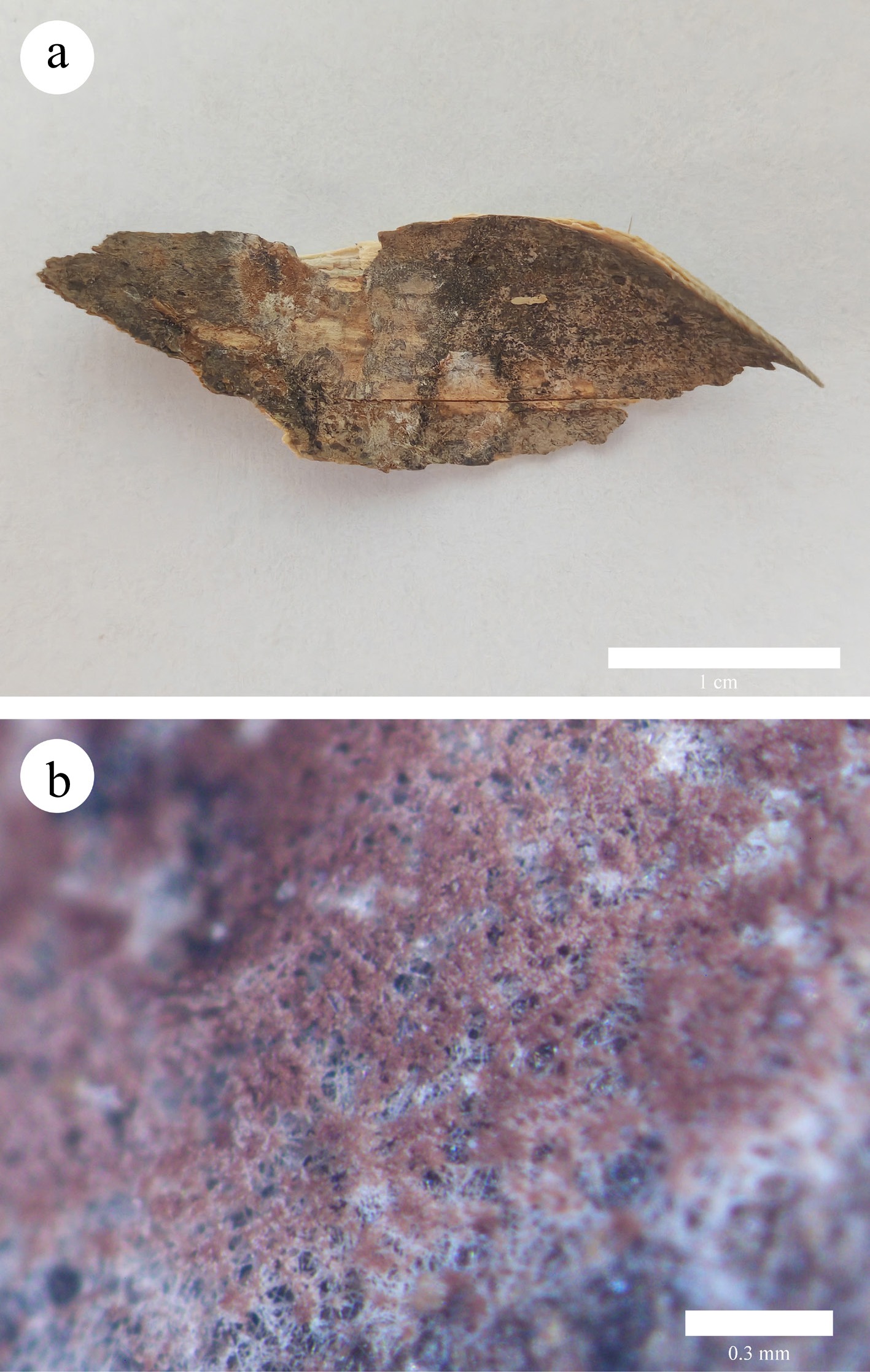
Figure 20.
Basidiomata of Xenasmatella hinnulea (Yuan 3296, holotype). (a) A fresh basidioma. (b) Dry hymenophore.
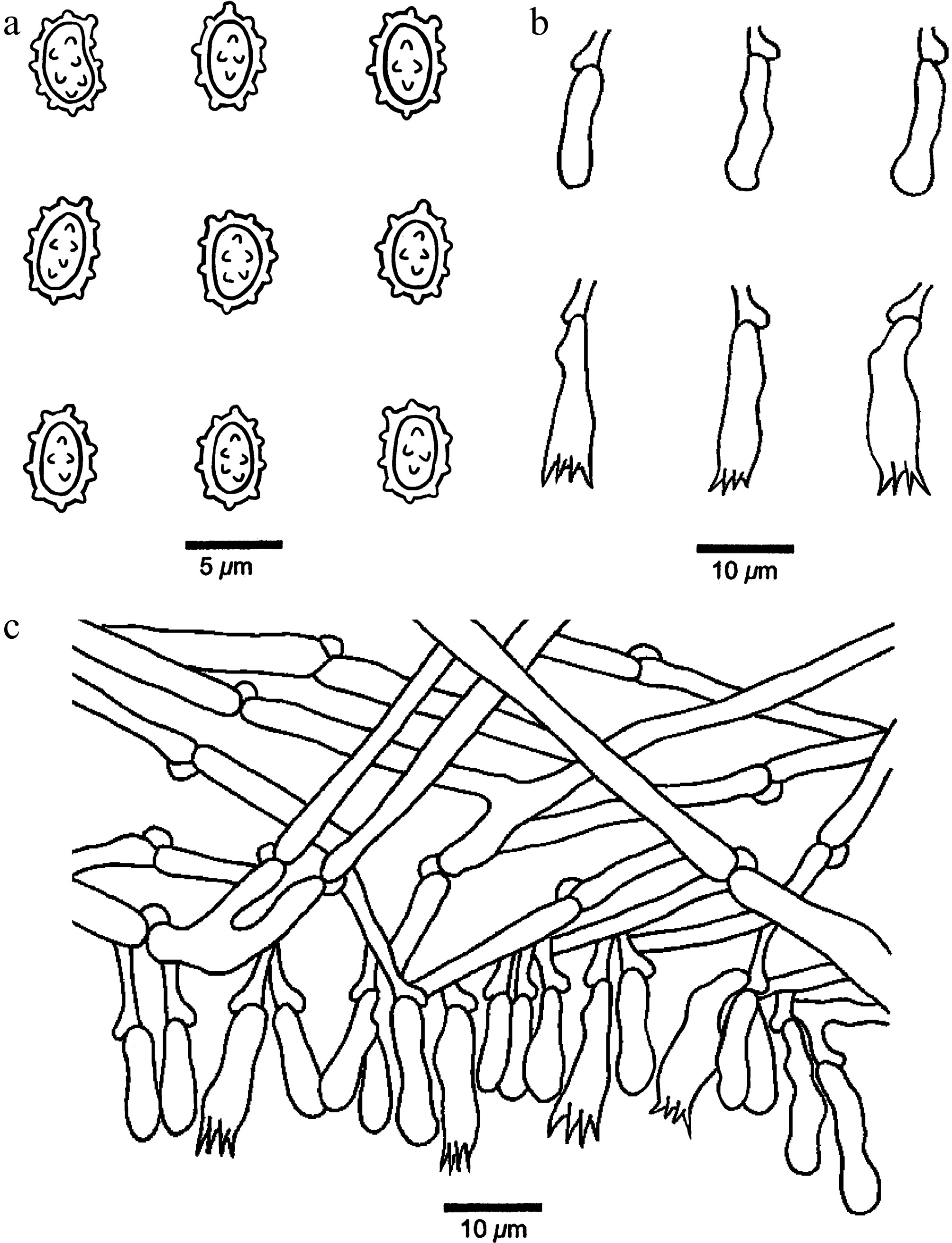
Figure 21.
Microscopic structures of Xenasmatella hinnulea (Yuan 3296, holotype). (a) Basidiospores. (b) Basidia and basidioles. (c) A section of hymenium.
MycoBank number: MB 863609.
Etymology — Hinnulea (Lat.): refers to the species having a fawn hymenophore when fresh.
Holotype — CHINA, Sichuan Province, Chendu, Wuhou District, Wangjianglou Park. GPS coordinates: 30.632563° N, 104.089396° E; elevation: 500 m a.s.l.; on stump of Bambusa chungii, 27 September 2025, Y. YUAN, Yuan 3296 (BJFC062842). DNA sequences: PZ101760 (ITS), PZ112080 (LSU), PZ158457 (mtSSU).
Description — Basidiomata annual, resupinate, adnate, thin, hardly separable from substrate, without odor or taste, membranous, fawn when fresh and darker brown upon drying, up to 3 cm long, 1 cm wide, 0.1 mm thick at center. Hymenophoral surface gossypine under the lens; sterile margin indistinct, white; tissue darkening in KOH. Hyphal system monomitic with clamp connections on generative hyphae; subicular hyphae hyaline, thin-walled, occasionally branched, more or less straight, loosely interwoven, IKI–, CB–, 2.5–3.5 μm in diam. Cystidia and cystidioles absent. Basidia pleural, more or less clavate, with four sterigmata and a basal clamp connection, 11–17 × 4–5.5 μm; basidioles similar in shape to the basidia, but slightly smaller. Basidiospores ellipsoid, hyaline, thick-walled, occasionally with a constriction in the middle of one side, warted throughout, IKI–, CB–, (4.2–)4.4–5.3(–5.7) × (2.6–)2.9–3.5(–3.6) µm, L = 4.86 μm, W = 3.17 μm, Q = 1.53 (n = 30/1); spore spines 0.2–0.3 µm long.
Notes — Xenasmatella hinnulea was found on bamboo from Sichuan Province of southwestern China with a subtropical climate.
A BLAST search against the NCBI database based on ITS sequences revealed that Xenasmatella hinnulea exhibited the highest sequence similarity to X. alutacea among the authoritative sequences, with the following alignment parameters: Max Score = 736; Total Score = 736; Query Cover = 99%; E-value = 0.0; Identity = 88.57%.
In our phylogeny (Fig. 1), X. hinnulea formed an independent lineage. Morphologically, X. hinnulea and X. jilongensis share membranous basidiomata and ellipsoid basidiospores. However, X. jilongensis differs from X. hinnulea by its smooth to grandinioid hymenophoral surface, and both wider basidia (6–9 vs 4–5.5 μm) and basidiospores (3.5–4.3 vs 2.9–3.5 μm)[63].
Xenasmatella longispinosa K.Y. Luo, Y.C. Dai, Yuna Yuan & Fang Wu, sp. nov. (Figs 22 and 23)
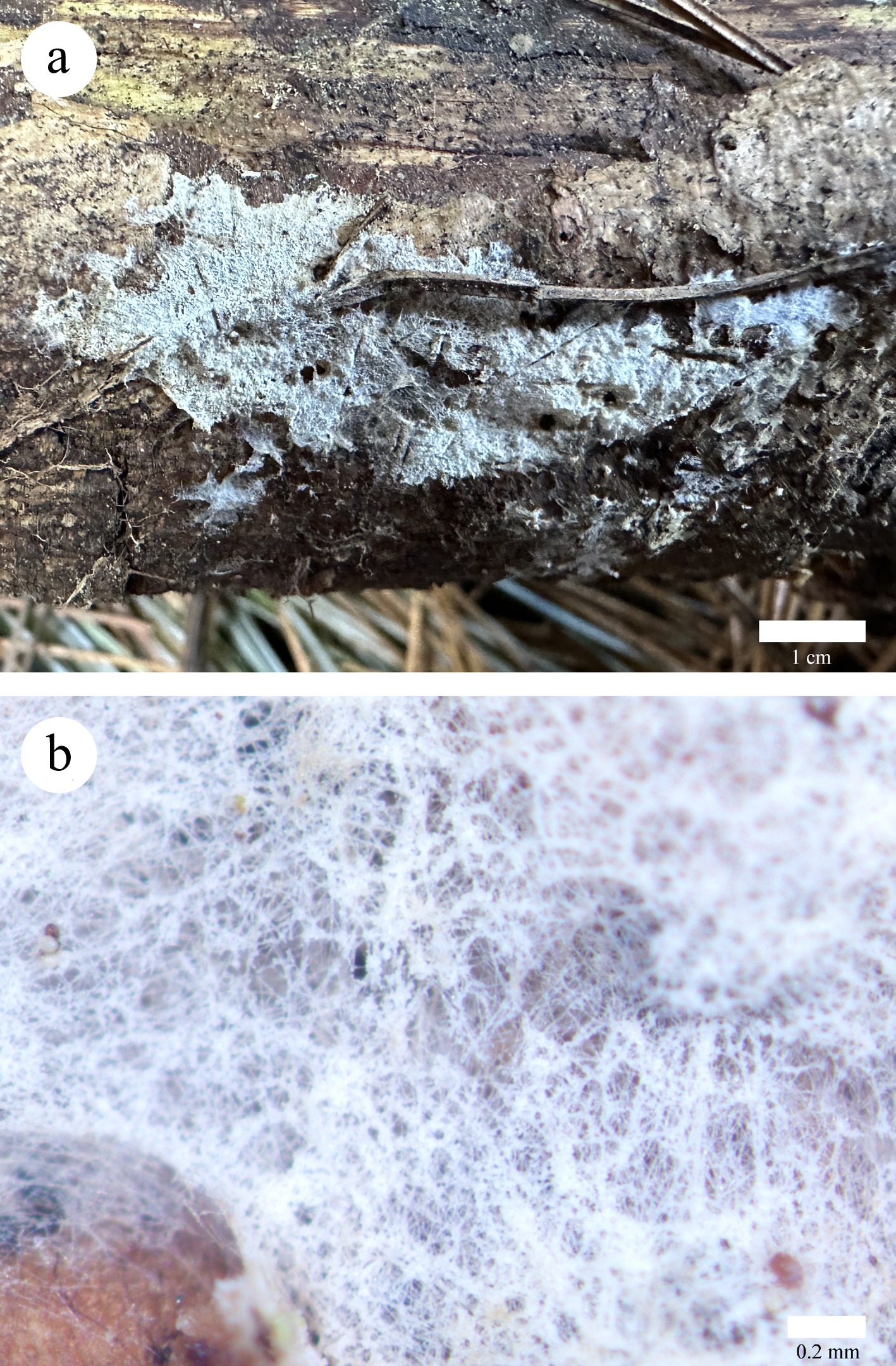
Figure 22.
Basidiomata of Xenasmatella longispinosa (Dai 38498, holotype). (a) A fresh basidioma. (b) Dry hymenophore.
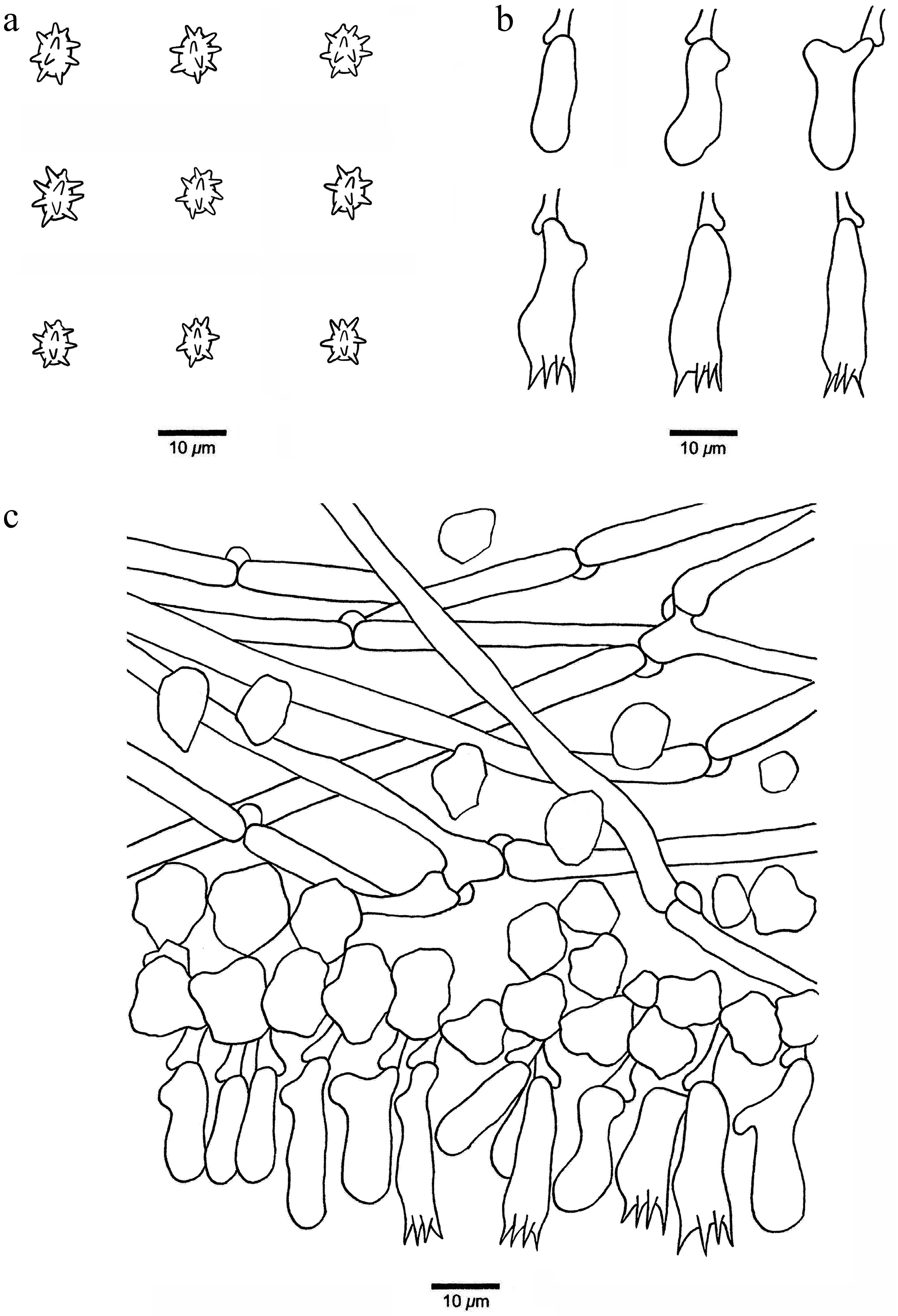
Figure 23.
Microscopic structures of Xenasmatella longispinosa (Dai 38498, holotype). (a) Basidiospores. (b) Basidia and basidioles. (c) A section of hymenium.
MycoBank number: MB 863610.
Etymology — Longispinosa (Lat.): refers to the species having basidiospores with long spines.
Holotype — CHINA, Liaoning Province, Benxi, Huanren County, Laotudingzi National Nature Reserve, Liudaogou Protection Station. GPS coordinates: 41.322902° N, 124.887852° E; elevation: 593 m a.s.l.; on a rotten trunk of Pinus koraiensis, 14 October 2025, Y.C. DAI, Dai 38498 (BJFC059757). DNA sequences: PZ101758 (ITS), PZ112078 (LSU), PZ158456 (mtSSU).
Description — Basidiomata annual, resupinate, adnate, thin, hardly separable from substrate, without odor or taste, membranous, bluish white when fresh and upon drying, up to 5 cm long, 2 cm wide, 0.1 mm thick at center. Hymenophoral surface byssaceous to reticulate under the lens; sterile margin indistinct, concolorous with hymenophoral surface; tissue unchanged in KOH. Hyphal system monomitic with clamp connections on generative hyphae; subicular hyphae hyaline, thin-walled, occasionally branched, more or less straight, loosely interwoven, abundant crystals present among hyphae, especially among subhymenium, IKI–, CB–, 3–4.5 μm in diam. Cystidia and cystidioles absent. Basidia pleural, barrel-shaped to subclavate, with four sterigmata and a basal clamp connection, 15–25 × 5–8 μm; basidioles similar in shape to the basidia, but slightly smaller. Basidiospores ellipsoid, hyaline, thin-walled, warted throughout, IKI–, CB–, (4.9–)5.1–6.2 × (3.6–)3.7–4.8(–4.9) µm, L = 5.69 μm, W = 4.32 μm, Q = 1.32 (n = 30/1); spore spines 0.8–1.6 µm long.
Notes — Xenasmatella longispinosa was found on Pinus koraiensis in the boreal forest of Liaoning Province, northeastern China, with a temperate monsoon climate.
A BLAST search against the NCBI database based on ITS sequences revealed that Xenasmatella longispinosa exhibited the highest sequence similarity to X. tabuliformis among the authoritative sequences, with the following alignment parameters: Max Score = 963; Total Score = 963; Query Cover = 95%; E-value = 0.0; Identity = 95.00%.
In our phylogeny (Fig. 1), X. longispinosa formed an independent lineage. Morphologically, X. longispinosa and X. rhizomorpha share a byssaceous to a reticulate hymenophoral surface and thin-walled basidiospores. However, X. rhizomorpha differs from X. longispinosa by its shorter blunt spines (up to 0.2 vs 0.8–1.6 µm) and smaller basidiospores (3.1–4.9 × 2.3–3.3 vs 5.1–6.2 × 3.7–4.8 µm)[50].
Xenasmatella monocotyledonis K.Y. Luo, Y.C. Dai, Xin Zhang & Fang Wu, sp. nov. (Figs 24 and 25).
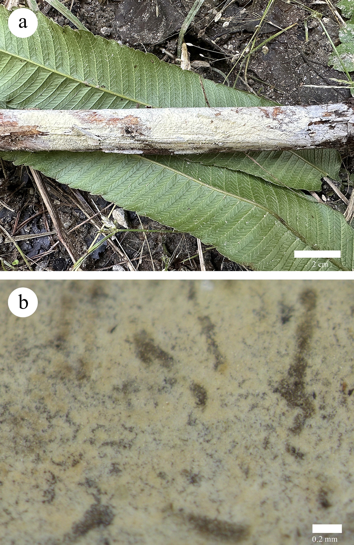
Figure 24.
Basidiomata of Xenasmatella monocotyledonis (Dai 26677, holotype). (a) A fresh basidioma. (b) Dry hymenophore.
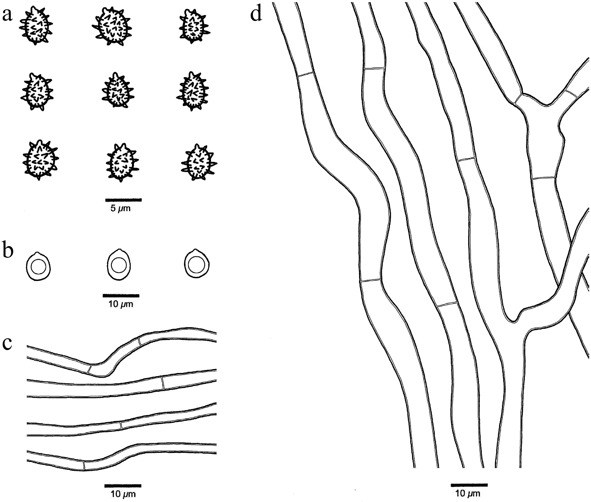
Figure 25.
Microscopic structures of Xenasmatella monocotyledonis (Dai 26677, holotype). (a) Basidiospores. (b) Chlamydospores. (c) Hyphae from subhymenium. (d) Hyphae from subiculum.
MycoBank number: MB 863611.
Etymology — monocotyledonis (Lat.): refers to the species growing on monocotyledonous plants.
Holotype — CHINA, Xizang Autonomous Region, Linzhi, Motuo County, the Road 219 from Motuo to Bomi. GPS coordinates: 29°18 ′N, 95°18 ′E; elevation: 1,000 m a.s.l.; on dead Miscanthus, 24 October 2023, Y.C. DAI, Dai 26677 (BJFC044227). DNA sequences: PP453842 (ITS), PP447930 (LSU), PV468694 (gapdh).
Description — Basidiomata annual, resupinate, adnate, hardly separable from substrate, without odor or taste, membranous, pale yellow when fresh and pale buff upon drying, up to 16 cm long, 2 cm wide, 0.3 mm thick at center. Hymenophoral surface gossypine under the lens; sterile margin distinct, white to pale yellow, up to 1.5 cm in width; tissue unchanged in KOH. Hyphal system monomitic with simple septa on generative hyphae; subicular hyphae hyaline, thin-walled, moderately branched, flexuous, more or less interwoven, IKI–, CB–, 4–8 µm in diam; subhymenium hyphae hyaline, thin-walled, unbranched, flexuous, more or less regularly arranged, IKI–, CB–, 2–3 µm in diam. Cystidia and cystidioles absent. Basidia and basidioles not observed. Basidiospores subglobose, hyaline, thin-walled, warted throughout, IKI–, CB–, (3.2–)3.3–4.6(–5) × (2.6–)3–4.2(–4.5) µm, L = 4.00 μm, W = 3.53 μm, Q = 1.12–1.15 (n = 60/2); spore spines 0.5–0.8 µm long. Chlamydospores present, globose to subglobose, abundant, hyaline, thick-walled, smooth, IKI–, CB–, 5–8 µm in diam.
Additional specimen examined (paratype): CHINA, Guangxi Autonomous Region, Laibin, Jinxiu County, Liugang, Daling Village. GPS coordinates: 23.942028° N, 110.073687° E; elevation: 800 m a.s.l.; on dead bamboo, 15 July 2024, Y.C. DAI, Dai 28849 (BJFC049108). DNA sequences: PV434785 (ITS), PV434798 (LSU), PZ147912 (rpb2), PV468695 (gapdh).
Notes — Xenasmatella monocotyledonis was found on monocotyledons (Miscanthus and bamboo) from Guangxi and Xizang Autonomous Region of China with subtropical and alpine-plateau climates, respectively.
A BLAST search against the NCBI database based on ITS sequences revealed that Xenasmatella monocotyledonis exhibited the highest sequence similarity to X. wuliangshanensis among the authoritative sequences, with the following alignment parameters: Max Score = 649; Total Score = 649; Query Cover = 97%; E-value = 0.0; Identity = 87.08%.
In our phylogeny (Fig. 1), X. monocotyledonis grouped together with X. tropica and X. wuliangshanensis with support (ML = 100%, BPP = 1.00). However, X. tropica is readily distinguished from X. monocotyledonis by its coriaceous basidiomata and generative hyphae with clamp connections; X. wuliangshanensis differs from X. monocotyledonis by its clay pink to saffron hymenophore and narrower basidiospores (2.5–3 vs 3–4.2 µm) and the absence of chlamydospores[47]. Morphologically, X. monocotyledonis and X. ailaoshanensis share membranous basidiomata and similar-sized basidiospores. However, X. ailaoshanensis is different from X. monocotyledonis by its generative hyphae with clamp connections and the absence of chlamydospores[46].
Xenasmatella montana K.Y. Luo, Y.C. Dai & Fang Wu, sp. nov. (Figs 26 and 27)
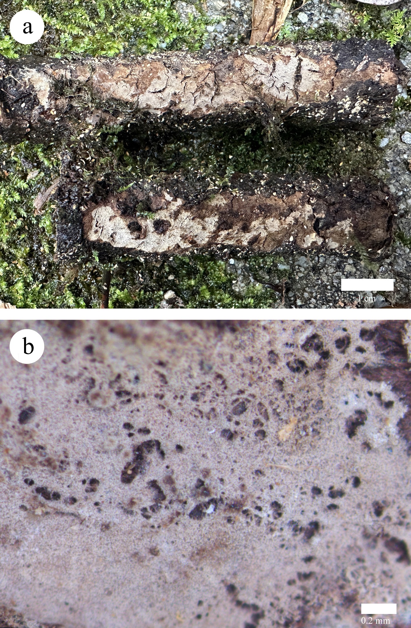
Figure 26.
Basidiomata of Xenasmatella montana (Dai 40267, holotype). (a) A fresh basidioma. (b) Dry hymenophore.
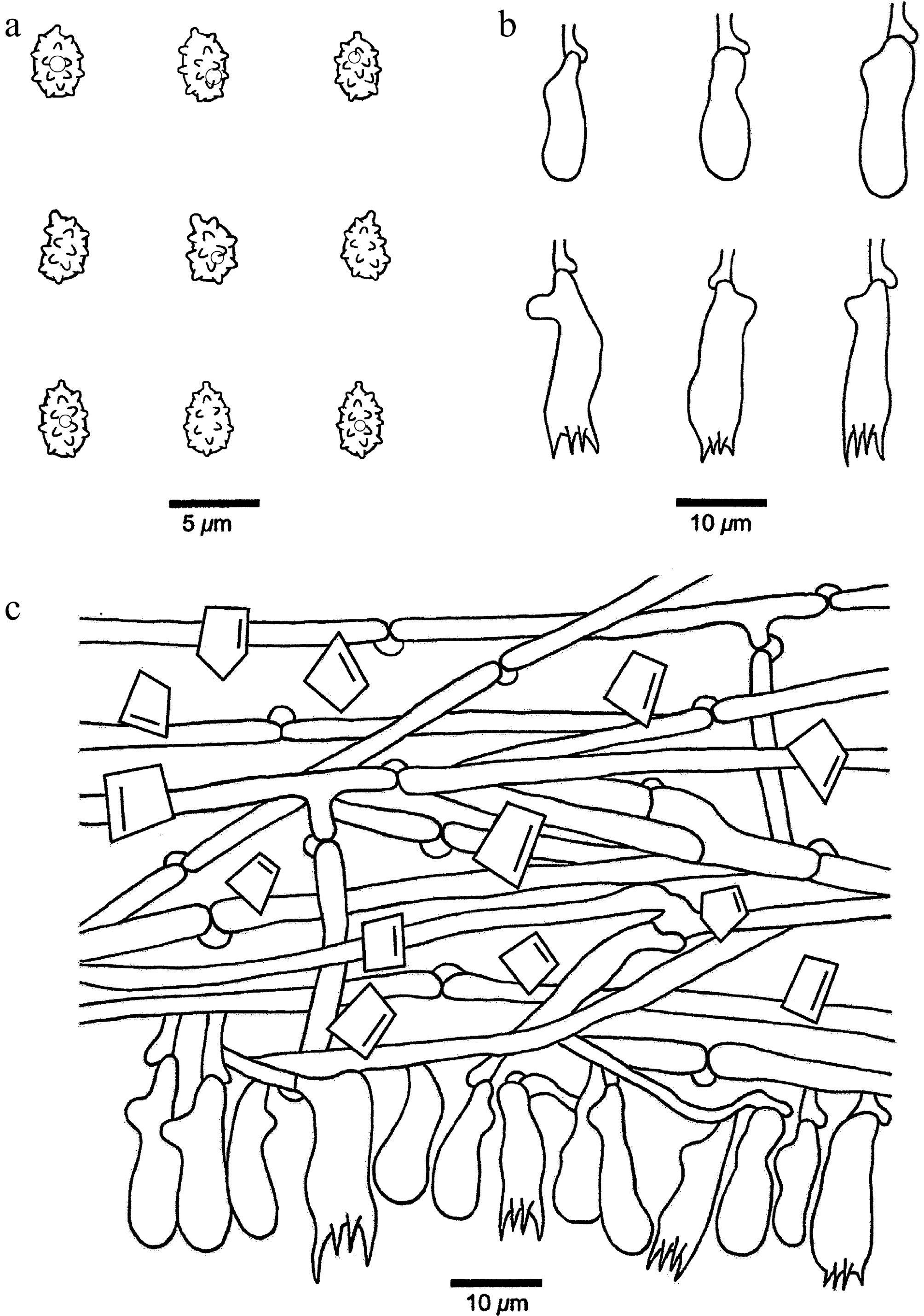
Figure 27.
Microscopic structures of Xenasmatella montana (Dai 40267, holotype). (a) Basidiospores. (b) Basidia and basidioles. (c) A section of hymenium.
MycoBank number: MB 863612.
Etymology — Montana (Lat.): refers to the species occurring in a high mountainous area.
Holotype — CHINA, Guangxi Autonomous Region, Guilin, Xingan County, Maoershan National Nature Reserve. GPS coordinates: 25.908587° N, 110.464042° E; elevation: 2,100 m a.s.l.; on a fallen branch of Tsuga tchekiangensis, 30 October 2025, Y.C. DAI, Dai 40267 (BJFC061524). DNA sequences: PZ101757 (ITS), PZ112077 (LSU), PZ158455 (mtSSU).
Description — Basidiomata annual, resupinate, adnate, thin, hardly separable from substrate, without odor or taste, membranous, vinaceous buff to clay buff when fresh and fawn and cracked upon drying, up to 8 cm long, 1 cm wide, 0.1 mm thick at center. Hymenophoral surface smooth to holey under the lens; sterile margin indistinct, concolorous with hymenophoral surface; tissue darkening in KOH. Hyphal system monomitic with clamp connections on generative hyphae; subicular hyphae hyaline, thin-walled, moderately branched, more or less straight, loosely interwoven, abundant rhomboid crystals present among hyphae, IKI–, CB–, 2–3.5 μm in diam. Cystidia and cystidioles absent. Basidia pleural, barrel-shaped, with four sterigmata and a basal clamp connection, 11–20 × 4–6 μm; basidioles similar in shape to the basidia, but slightly smaller. Basidiospores ellipsoid, hyaline, thin-walled, warted throughout, usually with one small guttule, IKI–, CB–, (3.2–)3.3–4.1(–4.2) × (2.1–)2.2–3.0(–3.1) µm, L = 3.73 μm, W = 2.60 μm, Q = 1.43 (n = 30/1); spore spines 0.3–0.5 µm long (the specimens Dai 36405 is sterile).
Additional specimen examined (paratype): CHINA, Guangxi Autonomous Region, Guilin, Xingan County, Maoershan National Nature Reserve. GPS coordinates: 25°44′ N, 110°19′ E; elevation: 2,100 m a.s.l.; on a fallen angiosperm branch, 10 May 2025, Y.C. DAI, Dai 36405 (BJFC057664). DNA sequences: PZ101756 (ITS), PZ112076 (LSU), PZ158454 (mtSSU), PZ147922 (tef1).
Notes — Xenasmatella montana was found in the high-mountain forest in the Guangxi Autonomous Region, southern China, with a temperate climate.
A BLAST search against the NCBI database based on ITS sequences revealed that Xenasmatella montana exhibited the highest sequence similarity to X. nigroidea among the authoritative sequences, with the following alignment parameters: Max Score = 1,051; Total Score = 1,051; Query Cover = 96%; E-value = 0.0; Identity = 98.18%.
In our phylogeny (Fig. 1), X. montana is sister to X. nigroidea with support (ML = 99%, BPP = 1.00). However, X. nigroidea is readily distinguished from X. montana by its gray to black basidiomata and thick-walled generative hyphae[36]. Morphologically, X. montana and X. bambusicola Qi Yuan & C.L. Zhao share membranous basidiomata and ellipsoid basidiospores. However, X. bambusicola differs from X. montana by its arachnoid hymenophoral surface and basidia with two sterigmata[66].
Xenasmatella tabuliformis K.Y. Luo, Y.C. Dai, Yuan Yuan & Fang Wu, sp. nov. (Figs 28 and 29)
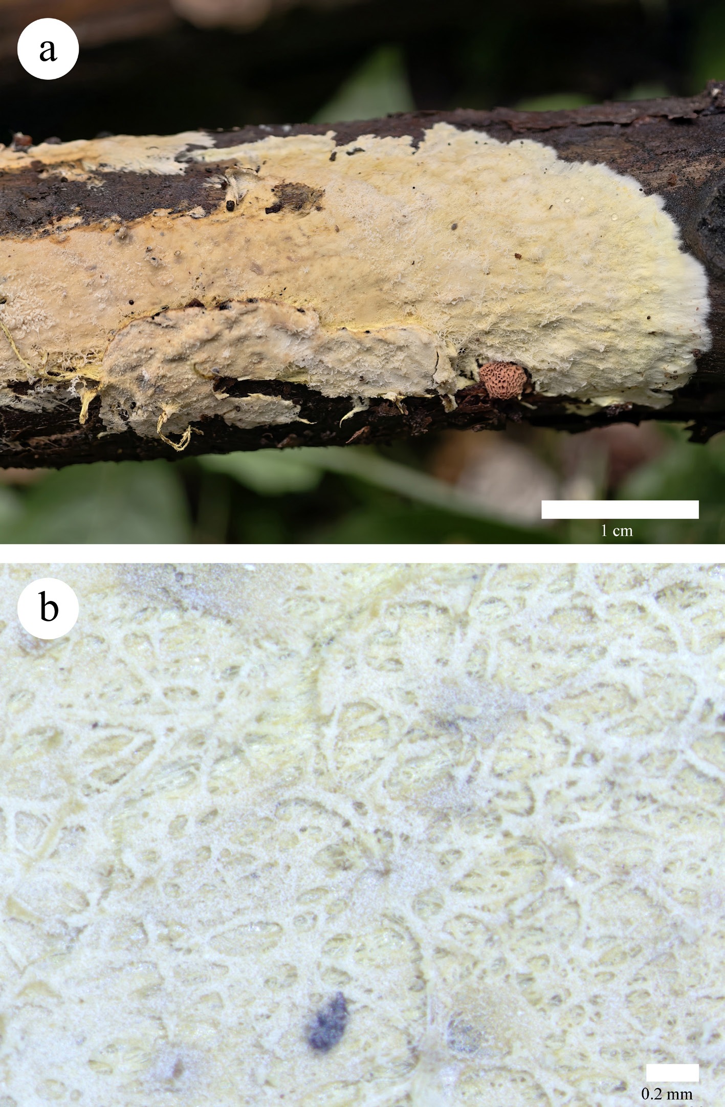
Figure 28.
Basidioma of Xenasmatella tabuliformis (Dai 34854, holotype). (a) A fresh basidioma. (b) Dry hymenophore.
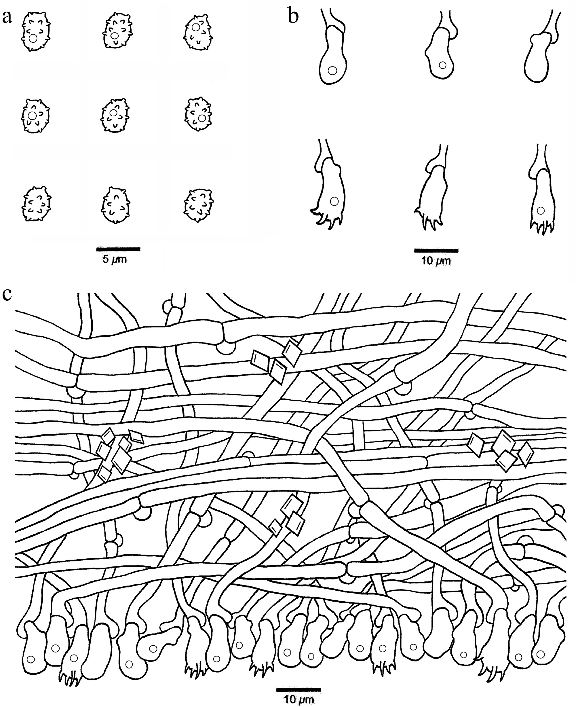
Figure 29.
Microscopic structures of Xenasmatella tabuliformis (Dai 34854, holotype). (a) Basidiospores. (b) Basidia and basidioles. (c) A section of hymenium.
MycoBank number: MB 863613.
Etymology — Tabuliformis (Lat.): refers to the species growing on Pinus tabuliformis.
Holotype — CHINA, Inner Mongolia Autonomous Region, Chifeng, Ningcheng County, Heilihe National Natural Reserve. GPS coordinates: 41.408971° N, 118.474290° E; elevation: 800 m a.s.l.; on a fallen branch of Pinus tabuliformis, 2 August 2025, Y.C. DAI, Dai 34854 (BJFC056115). DNA sequences: PZ101754 (ITS), PZ158453 (mtSSU).
Description — Basidiomata annual, resupinate, adnate, easily separable from substrate, without odor or taste, soft membranous, buff-yellow to salmon when fresh and becoming darker upon drying, up to 5 cm long, 2 cm wide, 0.2 mm thick. Hymenophoral surface arachnoid or reticular under the lens; sterile margin distinct, fimbriate, white, up to 1 mm in width; rhizomorphs saffron; tissue darkening in KOH. Hyphal system monomitic with clamp connections on generative hyphae; subicular hyphae hyaline, thin-walled, unbranched, flexuous, strongly interwoven, rhomboid crystals present among hyphae, IKI–, CB–, 2.5–4 μm in diam. Cystidia and cystidioles absent. Basidia pleural, barrel-shaped to pyriform, with four sterigmata and a basal clamp connection, sometimes with one small guttule, 9–21 × 4.5–6 μm; basidioles similar in shape to the basidia, but slightly smaller. Basidiospores ellipsoid, hyaline, thin-walled, warted throughout, usually with one small guttule, IKI–, CB–, (3.2–)3.5–4.7(–5.2) × (2.5–)2.7–3.6(–3.7) µm, L = 4.15 μm, W = 3.14 μm, Q = 1.29–1.35 (n = 60/2); spore spines 0.2–0.3 µm long.
Additional specimen examined (paratype): CHINA, Inner Mongolia Autonomous Region, Chifeng, Ningcheng County, Heilihe National Natural Reserve. GPS coordinates: 41.408971° N, 118.474290° E; elevation: 800 m a.s.l.; on a fallen branch of Pinus tabuliformis, 2 August 2025, Y.C. DAI, Dai 34859 (BJFC056120). DNA sequence: PZ101755 (ITS).
Notes — Xenasmatella tabuliformis was found on Pinus tabuliformis in the Inner Mongolia Autonomous Region of northeastern China, with a temperate continental climate.
A BLAST search against the NCBI database based on ITS sequences revealed that Xenasmatella tabuliformis exhibited the highest sequence similarity to X. alutacea among the authoritative sequences, with the following alignment parameters: Max Score = 1,057; Total Score = 1,057; Query Cover = 99%; E-value = 0.0; Identity = 98.04%.
In our phylogeny (Fig. 1), X. tabuliformis is related to X. abieticola, X. alutacea, X. conifericola, and X. gymnosperma with support (ML = 67%, BPP = 0.94). However, X. abieticola is readily distinguished from X. tabuliformis by its byssaceous hymenophoral surface and wider basidia (6–7 vs 4.5–6 µm); X. alutacea is different from X. tabuliformis by its soft coriaceous basidiomata, smooth hymenophoral surface and thick-walled basidiospores; X. conifericola differs from X. tabuliformis by its coriaceous basidiomata, smooth to farinaceous hymenophoral surface; X. gymnosperma is readily distinguished from X. tabuliformis by its byssaceous hymenophoral surface and two types of subicular hyphae. Morphologically, X. tabuliformis and X. borealis share an arachnoid hymenophoral surface and ellipsoid basidiospores. However, X. borealis differs from X. tabuliformis by its basidiomata with rhizomorphs and longer basidiospores (5–6 vs 3.5–4.7 µm)[90].
Xenasmatella tropica K.Y. Luo, Y.C. Dai & Fang Wu, sp. nov. (Figs 30 and 31)
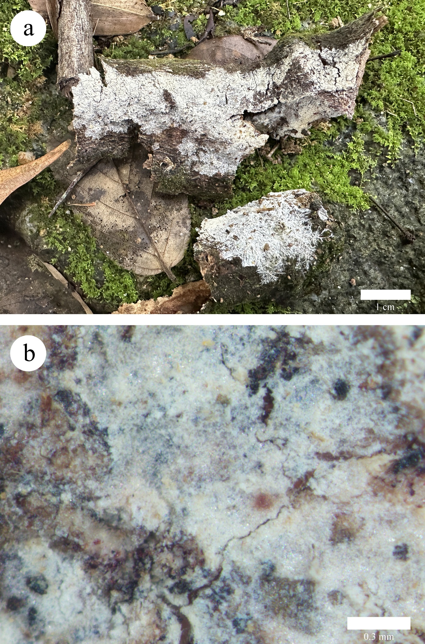
Figure 30.
Basidiomata of Xenasmatella tropica (Dai 36954, holotype). (a) A fresh basidioma. (b) Dry hymenophore.
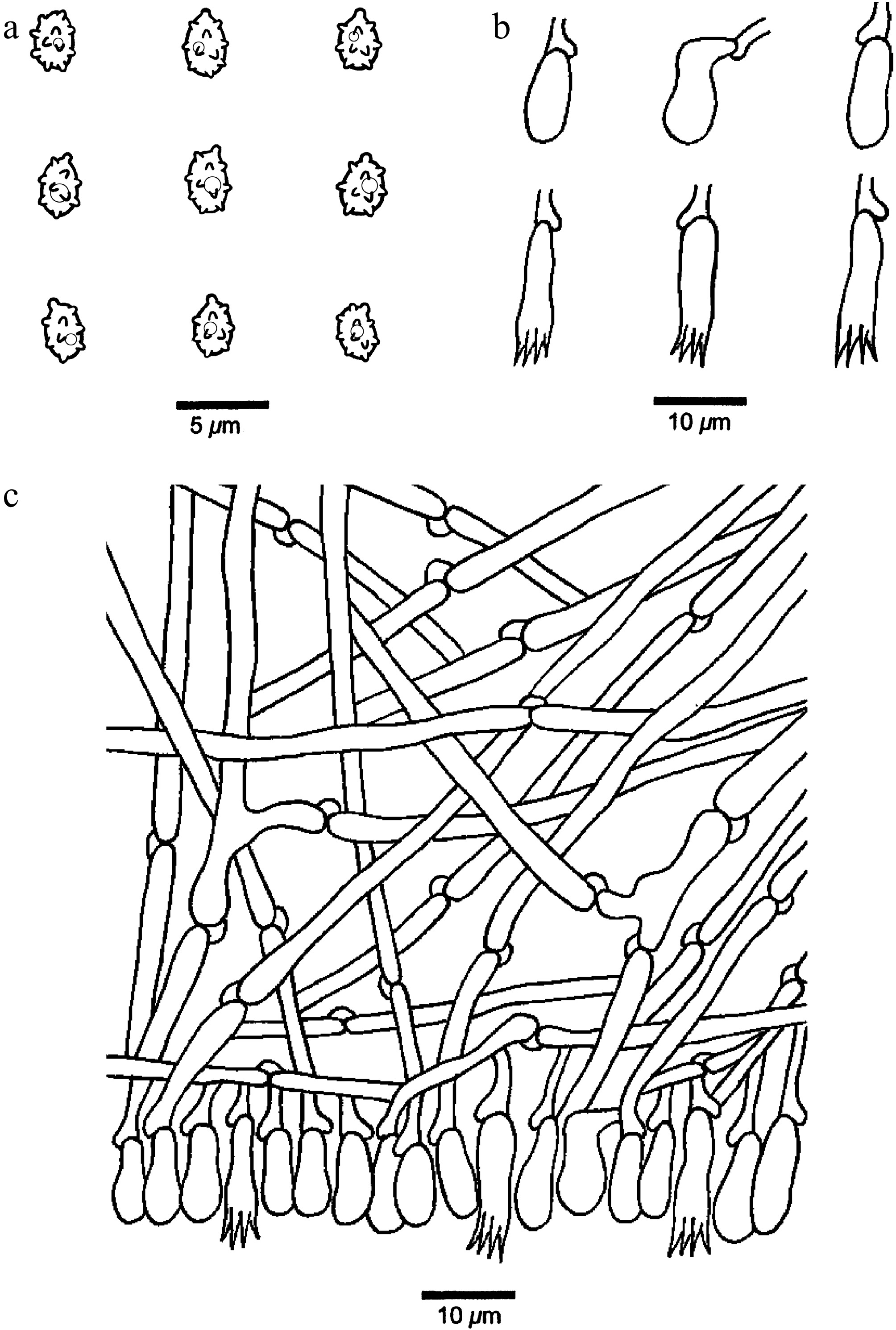
Figure 31.
Microscopic structures of Xenasmatella tropica (Dai 36954, holotype). (a) Basidiospores. (b) Basidia and basidioles. (c) A section of hymenium.
MycoBank number: MB 863614.
Etymology — Tropica (Lat.): refers to the species occurring in tropical area.
Holotype — CHINA, Hainan Province, National Park of Hainan Tropical Rainforest, Limu Mountain. GPS coordinates: 19.177884° N, 109.745181° E; elevation: 676 m a.s.l.; on rotten angiosperm wood, 29 June 2025, Y.C. DAI, Dai 36954 (BJFC058213). DNA sequences: PZ101761 (ITS), PZ112081 (LSU), PZ158458 (mtSSU).
Description — Basidiomata annual, resupinate, adnate, hardly separable from substrate, without odor or taste, coriaceous, brittle, white when fresh and upon drying, up to 7 cm long, 2 cm wide, 0.2 mm thick at center. Hymenophoral surface gossypine under the lens; sterile margin distinct, actinomorphous, concolorous with hymenophoral surface, up to 2 mm in width; rhizomorphs concolorous with hymenophoral surface; tissue unchanged in KOH. Hyphal system monomitic with clamp connections on generative hyphae; subicular hyphae hyaline, thin-walled, occasionally branched at a right angle, more or less straight, loosely interwoven, IKI–, CB–, 2.5–3.5 μm in diam. Cystidia and cystidioles absent. Basidia clavate, with four sterigmata and a basal clamp connection, 11–18 × 4–5 μm; basidioles similar in shape to the basidia, but slightly smaller. Basidiospores ellipsoid, hyaline, thin-walled, warted throughout, with one guttule, IKI–, CB–, (2.4–)2.6–3.4(–3.7) × (1.8–)1.9–2.6(–3.3) µm, L = 2.92 μm, W = 2.21 μm, Q = 1.32 (n = 30/1); spore spines 0.1–0.2 µm long.
Notes — Xenasmatella tropica was found in the Hainan Tropical Rainforest of China with a tropical climate.
A BLAST search against the NCBI database based on ITS sequences revealed that Xenasmatella tropica exhibited the highest sequence similarity to X. wuliangshanensis among the authoritative sequences, with the following alignment parameters: Max Score = 1,118; Total Score = 1,118; Query Cover = 97%; E-value = 0.0; Identity = 97.56%.
In our phylogeny (Fig. 1), X. tropica is sister to X. wuliangshanensis with support (ML = 100%, BPP = 1.00). However, X. wuliangshanensis is readily distinguished from X. tropica by its membranous to farinaceous basidiomata, smooth to tuberculate hymenophoral surface, and subglobose basidiospores[50]. Morphologically, X. tropica and X. fibrillosa (Hallenb.) Stalpers share basidiomata with rhizomorphs and ellipsoid basidiospores. However, X. fibrillosa differs from X. tropica by its porulose to reticulate hymenophoral surface and bigger basidiospores (4.5–5.5 × 3–3.5 vs 2.6–3.4 × 1.9–2.6 µm)[37].
Xenasmatella versicolor K.Y. Luo, Y.C. Dai & Fang Wu, sp. nov. (Figs 32 and 33)
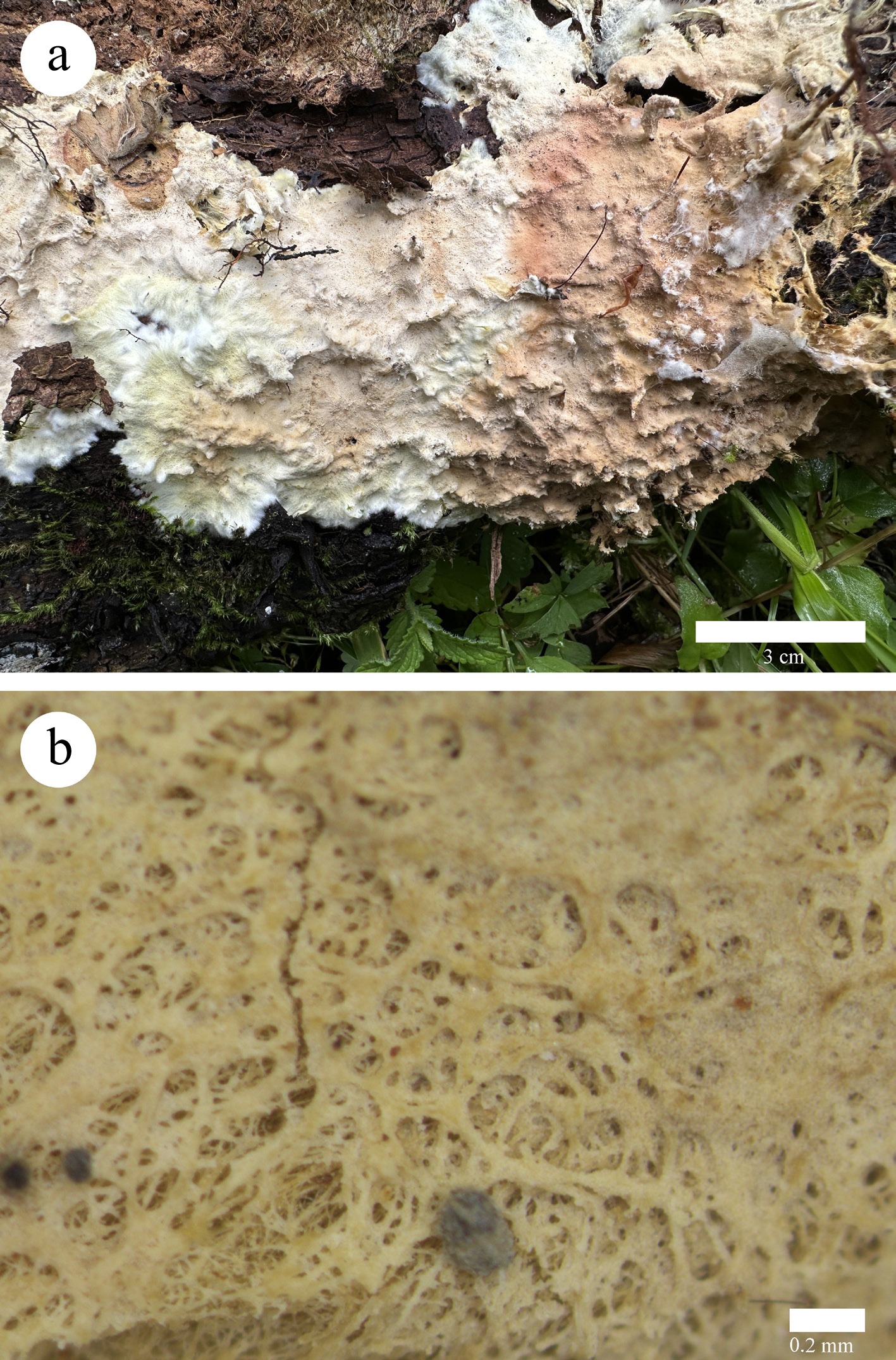
Figure 32.
Basidioma of Xenasmatella versicolor (Dai 26000, holotype). (a) A fresh basidioma. (b) Dry hymenophore.
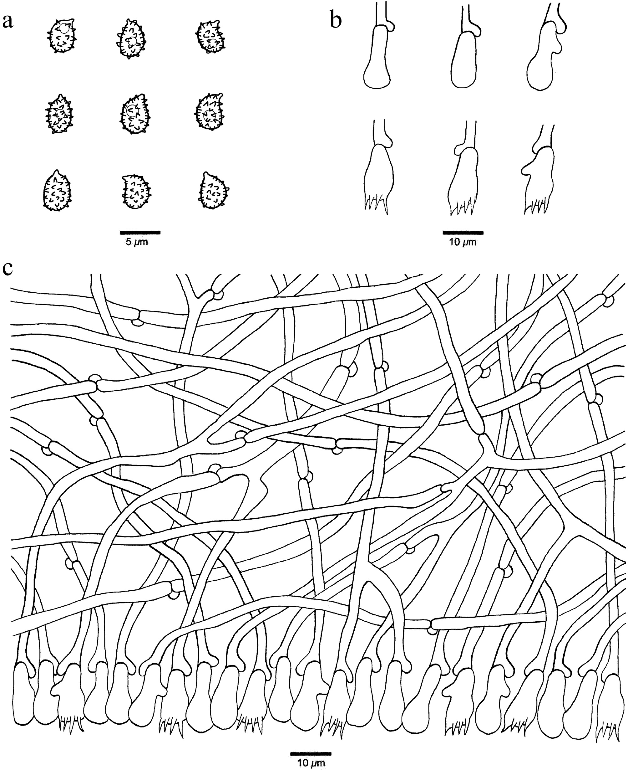
Figure 33.
Microscopic structures of Xenasmatella versicolor (Dai 26000, holotype). (a) Basidiospores. (b) Basidia and basidioles. (c) A section of hymenium.
MycoBank number: MB 863615.
Etymology — Versicolor (Lat.): refers to the basidiomata of the species having variable colors.
Holotype — CHINA, Qinghai Province, Guoluo, Banma County, Makehe Forest Park. GPS coordinates: 32.932799° N, 100.737001° E; elevation: 3,300 m a.s.l.; on rotten wood of Betula, 25 August 2023, Y.C. DAI, Dai 26000 (BJFC043549). DNA sequences: PP455443 (ITS), PP447931 (LSU), PV455373 (mtSSU), PV468704 (tef1).
Description — Basidiomata annual, resupinate, adnate, easily separable from substrate, without odor or taste, soft membranous, pale yellow to peach when fresh and olivaceous buff upon drying, up to 10 cm long, 8 cm wide, 0.2 mm thick at center. Hymenophoral surface reticulate under the lens; sterile margin distinct, fimbriate, white, up to 5 mm in width; tissue darkening in KOH. Hyphal system monomitic with clamp connections on generative hyphae; subicular hyphae hyaline, thin-walled, frequently branched, flexuous, loosely interwoven, IKI–, CB–, 2–4 μm in diam. Cystidia and cystidioles absent. Basidia pleural, barrel-shaped, with four sterigmata and a basal clamp connection, 9–14 × 4–5 μm; basidioles similar in shape to the basidia, but slightly smaller. Basidiospores ellipsoid, hyaline, thin-walled, warted throughout, usually with one guttule, IKI–, CB–, (3–)3.4–4.7(–4.9) × (2.1–)2.4–3.7(–3.9) µm, L = 4.08 μm, W = 3.09 μm, Q = 1.27–1.37 (n = 60/2); spore spines 0.2–0.6 µm long.
Additional specimen examined (paratype): CHINA, Xizang Autonomous Region, Rikaze, Yadong County, Bacha Falls to Yadong Customs. GPS coordinates: 27.316825° N, 88.969564° E; elevation: 4,067 m a.s.l.; on a fallen branch of Abies fabri, 18 October 2024, Y.C. DAI, Dai 31935 (BJFC052194). DNA sequences: PV434787 (ITS), PV434800 (LSU), PV468705 (tef1).
Notes — Xenasmatella versicolor was found on the Qinghai-Xizang Plateau of China, where it has an alpine-plateau climate.
A BLAST search against the NCBI database based on ITS sequences revealed that Xenasmatella versicolor exhibited the highest sequence similarity to X. alutacea among the authoritative sequences, with the following alignment parameters: Max Score = 965; Total Score = 965; Query Cover = 96%; E-value = 0.0; Identity = 97.21%.
In our phylogeny (Fig. 1), X. versicolor grouped with X. hjortstamii, with support (ML = 53%, BPP = 0.87). However, X. hjortstamii is different from X. versicolor by its smooth or somewhat wrinkled hymenophoral surface and bigger basidia (14–22 × 5–9 vs 9–14 × 4–5 μm)[65]. Morphologically, X. versicolor and X. romellii Hjortstam share the fimbriate sterile margin of the basidiomata. However, X. romellii differs from X. versicolor by its ceraceous basidiomata, farinaceous hymenophoral surface, and both bigger basidia (15–20 × 6–7 vs 9–14 × 4–5 μm) and basidiospores (6.5–7.5 × 3.5–4 μm vs 3.4–4.7 × 2.4–3.7 μm)[90].
Xenasmatella xylina K.Y. Luo, Y.C. Dai, Xin Zhang & Fang Wu, sp. nov. (Figs 34 and 35)
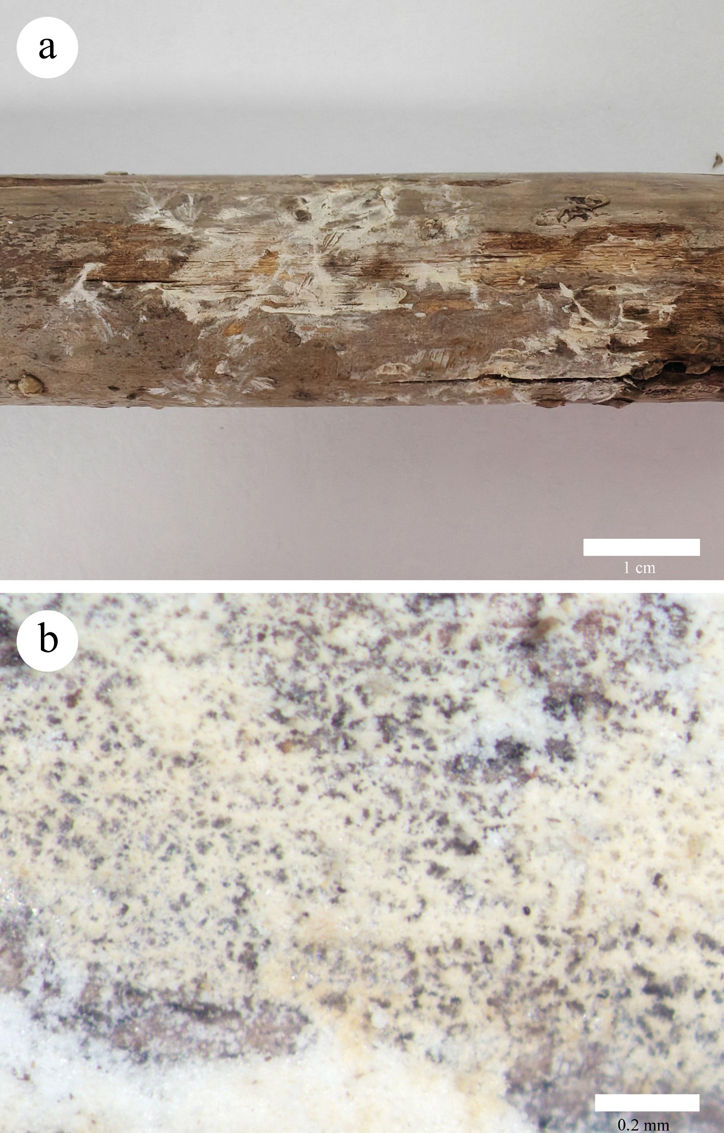
Figure 34.
Basidiomata of Xenasmatella xylina (Dai 36856, holotype). (a) A fresh basidioma. (b) Dry hymenophore.
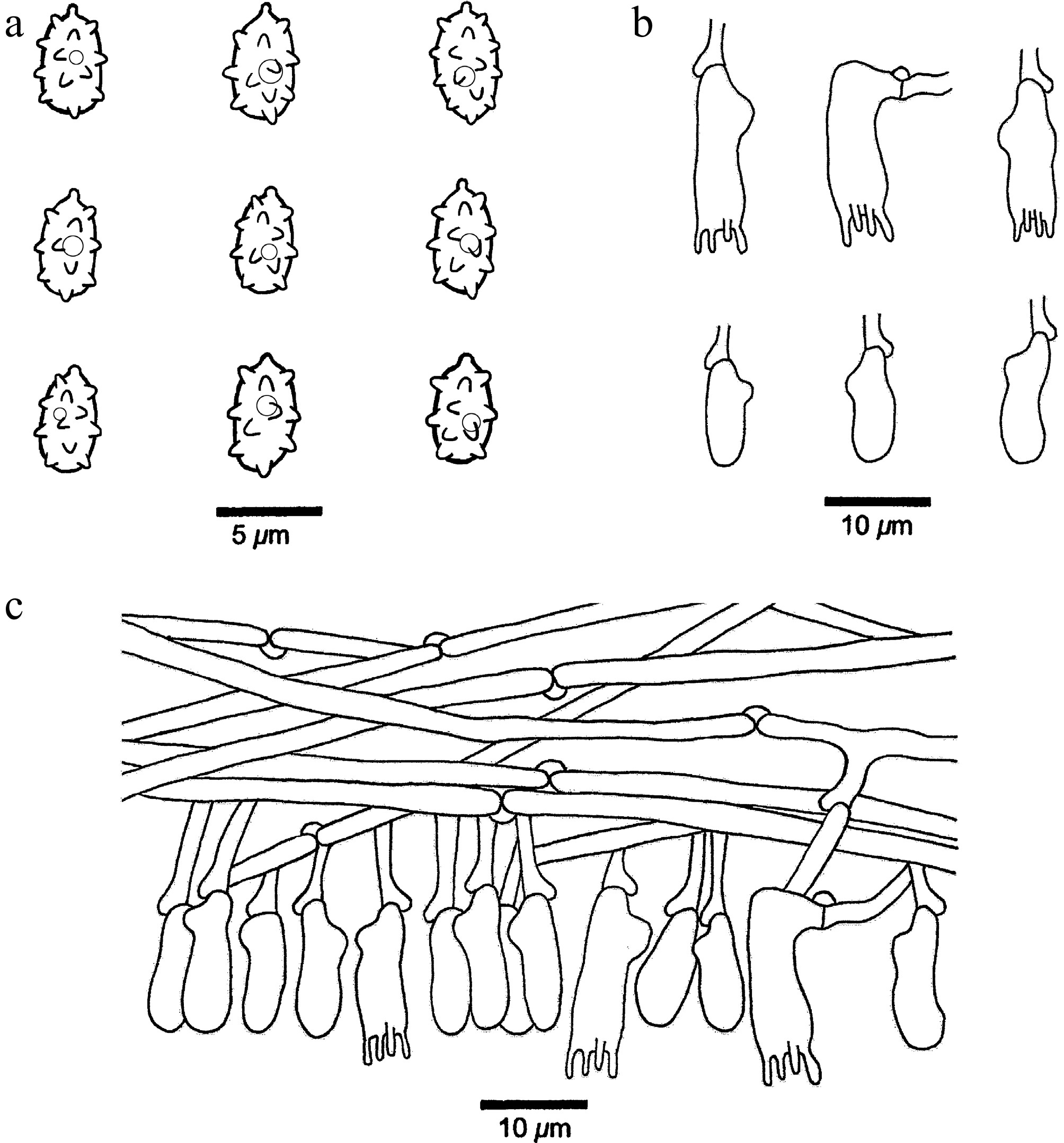
Figure 35.
Microscopic structures of Xenasmatella xylina (Dai 36856, holotype). (a) Basidiospores. (b) Basidia and basidioles. (c) A section of hymenium.
MycoBank number: MB 863616.
Etymology — Xylina (Lat.): refers to the species having a cottony hymenophore.
Holotype — CHINA, Guizhou Province, Guiyang, Qianlingshan Park. GPS coordinates: 26.602664° N, 106.694161° E; elevation: 1,200 m a.s.l.; on dead culm of Bambusa multiplex, 15 November 2025, Y.C. DAI, Dai 36856 (BJFC058115). DNA sequences: PZ101770 (ITS), PZ112089 (LSU), PZ158466 (mtSSU), PZ147910 (rpb1), PZ147909 (gapdh).
Description — Basidiomata annual, resupinate, adnate, very thin, hardly separable from substrate, without odor or taste, membranous, cream when fresh and upon drying, up to 13 cm long, 2 cm wide, 0.05 mm thick at center. Hymenophoral surface cottony; sterile margin indistinct, concolorous with hymenophoral surface; tissue unchanged in KOH. Hyphal system monomitic with clamp connections on generative hyphae; subicular hyphae hyaline, thin-walled, occasionally branched, more or less straight, loosely interwoven, IKI–, CB–, 1.8–3 μm in diam. Cystidia and cystidioles absent. Basidia pleural, mostly barrel-shaped, with four sterigmata and a basal clamp connection, 11–15 × 4–6 μm; basidioles similar in shape to the basidia, but distinctly smaller. Basidiospores ellipsoid, hyaline, thin-walled, warted throughout, with one small guttule, IKI–, CB–, (3.8–)4–5.4(–5.7) × 2.6–3.1(–3.3) µm, L = 4.7 μm, W = 2.85 μm, Q = 1.65 (n = 30/1); spore spines 0.4–0.5 µm long (the specimen Dai 38692 is sterile).
Additional specimen examined (paratype): CHINA, Xizang Autonomous Region, Linzhi, Gongbujiangda County, along the Road 318, 4,259–4,260 km. GPS coordinates: 29.887354° N, 93.623635° E; elevation: 3,250 m a.s.l.; on a fallen angiosperm branch, 5 October 2025, Y.C. DAI, Dai 38692 (BJFC059951). DNA sequence: PZ101771 (ITS).
Notes — Xenasmatella xylina was found on dead Bambusa multiplex and angiosperm wood from Guizhou Province and the Xizang Autonomous Region of southwestern China, with subtropical to alpine-plateau climates.
A BLAST search against the NCBI database based on ITS sequences revealed that Xenasmatella xylina exhibited the highest sequence similarity to X. rhizomorpha among the authoritative sequences, with the following alignment parameters: Max Score = 966; Total Score = 966; Query Cover = 98%; E-value = 0.0; Identity = 97.37%.
In our phylogeny (Fig. 1), X. xylina grouped together with X. xinpingensis with support (ML = 67%, BPP = 0.97). However, X. xinpingensis is readily distinguished from X. xylina by its soft ceraceous basidiomata, irregularly reticulate or somewhat wrinkled hymenophoral surface, longer basidia (15.5–20 vs 11–15 µm) and subglobose basidiospores[50]. Morphologically, X. xylina and X. gossypina share membranous basidiomata with a gossypine hymenophoral surface. However, X. gossypina is different from X. xylina by its subglobose basidiospores[47].
-
Morphologically, the macro- and micromorphological characteristics of the 17 newly discovered species conform to the definition of Xenasmatella, exhibiting membranous, ceraceous, or coriaceous basidiomata, a monomitic hyphal system with clamped generative hyphae, pleural basidia, and predominantly thin-walled, warted basidiospores[37]. Nevertheless, several species identified in this study deviate from these typical features. For example, Xenasmatella monocotyledonis possesses generative hyphae with simple septa; pleural basidia are absent in X. gymnosperma; and several species, including X. alutacea, X. conifericola, X. foraminosa, and X. hinnulea, have thick-walled basidiospores. Additionally, X. ceracea and X. monocotyledonis produce chlamydospores.
These findings further suggest that certain morphological traits are more variable within the genus than previously recognized, and the presence or the absences of clamp connections in the species of the same genus is not rare cases[92,93]. It is possible that such species represent alternative survival strategies on specialized hosts. Xenasmatella monocotyledonis is so far the only species in the genus grows on monocotyledonous plants. In addition, we have compiled the global distribution and substrate associations of Xenasmatella species (Supplementary Table S3).
Numerous species were described or transferred to the genus Xenasmatella based on morphological characteristics between the 1960s to 1990s; however, molecular data for these taxa remain unavailable. Xenasmatella species typically colonize angiosperm and gymnosperm wood, except for X. nasti (Boidin & Gilles) Stalpers, which was reported from soil[43]. The nomenclatural status of X. nasti is inconsistent, as it is considered invalid in Index Fungorum but legitimate in MycoBank. Due to the absence of available sequence data, the taxonomic placement of X. nasti remains uncertain. Consequently, this species is provisionally included in Xenasmatella.
In the present study, BLAST searches against the NCBI database consistently showed that sequences of a few species, such as X. caeruleogrisea, X. hinnulea, and X. monocotyledonis, were most closely related to Xenasmatella representatives, albeit with relatively low similarity scores. This is likely due, at least in part, to the extreme scarcity of authoritative reference sequences available in GenBank for the genus Xenasmatella, as well as for its family Xenasmataceae and order Xenasmatales. Given these limitations, alongside the fact that all single-gene and multi-locus phylogenetic analyses consistently place these species within the Xenasmatella clade, we consider it appropriate, at this stage, to tentatively assign them to this genus. Nevertheless, future studies incorporating broader taxon sampling, additional molecular markers, and more comprehensive morphological and ecological data will be essential to definitively resolve their higher-level phylogenetic positions.
Xenasmatella species are primarily distributed across Asia, the Americas, and Europe, with most Asian species documented in China. In this study, 17 new Xenasmatella species with 103 new sequences from China are described. These newly described species contribute to global fungal diversity and offer important insights into the biogeography and ecological functions of corticioid fungi in China. The country's significant environmental heterogeneity, encompassing a range of climates from boreal and temperate to subtropical, tropical, and alpine plateau, along with extensive pristine habitats, has facilitated the discovery of this previously unrecognized diversity. However, significant gaps persist in understanding the global distribution and evolutionary mechanisms underlying the genus.
Finally, a morphological identification key to the 28 accepted species of Xenasmatella in China is provided.
Key to 28 accepted species of Xenasmatella in China (new species in bold).
1.Rhizomorphspresent 2 1.Rhizomorphsabsent 7 2.Basidiomatacoriaceous 3 2.Basidiomatamembranous 4 3.Hymenophoralsurfacedrab X. conifericola 3.Hymenophoralsurfacewhite X tropica 4.Generativehyphaethick-walled X. rhizomorpha 4.Generativehyphaethin-walled 5 5.Hymenophoralsurfacearachnoidorreticular X. tabuliformis 5.Hymenophoralsurfacebyssaceous 6 6.Basidiospores4.6–6.1µminlength X. abieticola 6.Basidiospores3.4–4.5µminlength X. gymnosperma 7.Basidiosporesthick-walled 8 7.Basidiosporesthin-walled 10 8.Basidiomatamembranous X. hinnulea 8.Basidiomatacoriaceousorsoftcoriaceous 9 9.Basidia18–25µmlong X. alutacea 9.Basidia9–18µmlong X. foraminosa 10.Generativehyphaethick-walledorslightlythick-walled 11 10.Generativehyphaethin-walled 14 11.Hymeniumwithabundantcrystals X. crystallina 11.Hymeniumwithfewcrystalsorabsent 12 12.Basidiosporessubglobosetoglobose X. xinpingensis 12.Basidiosporesellipsoid 13 13.Basidiomatagraytoblack X. nigroidea 13.Basidiomatapalemousegraytoclaybuff,cinnamon X. hjortstamii 14.Hymenophoralsurfacewithtuberculateorgrandinioid 15 14.Hymenophoralsurfacewithouttuberculateorgrandinioid 16 15.Basidiospores5–6×3.5–4.3µm X. jilongensis 15.Basidiospores2.8–3.5×2.5–3µm X. wuliangshanensis 16.Basidiomatacoriaceousorsoftcorky 17 16.Basidiomatamembranousorceraceous 18 17.Hymenophorebluishgray X. caeruleogrisea 17.Hymenophoreverywhite X. candidissima 18.Chlamydosporespresent 19 18.Chlamydosporesabsent 20 19.Generativehyphaewithsimple-septa X. monocotyledonis 19.Generativehyphaebearingclampconnections X. ceracea 20.Basidiawith2sterigmata X. bambusicola 20.Basidiawith4sterigmata 21 21.Basidiomatawithvariablecolors X. versicolor 21.Basidiomatawithoutvariablecolors 22 22.Hymenophoralsurfacewhitetolilac X. tenuis 22.Hymenophoralsurfacewhitetogreyish,whitetocream,creamtobuff,pinkishbuff 23 23.Hymenophoralsurfacegossypineorcottony 24 23.Hymenophoralsurfacesmooth,pruinosetofarinaceous,holey,bassaceoustoreticulate 25 24.Basidiosporessubglobosetoglobose X. gossypina 24.Basidiosporesellipsoid X. xylina 25.Hymenophoralsurfacesmooth X. roseobubalina 25.Hymenophoralsurfacepruinosetofarinaceous,holey,bassaceoustoreticulate 26 26.Hymenophoralsurfacepruinaceoustofarinaceous X. ailaoshanensis 26.Hymenophoralsurfaceholeyorbyssaceoustoreticulate 27 27.Basidiospores5.1–6.2×3.7–4.8µm X. longispinosa 27.Basidiospores3.3–4.1×2.2–3.0µm X. montana The research is supported by the National Natural Science Foundation of China (Grant Nos 32570008 and U23A20142), the Fundamental Research Funds for the Central Universities (Grant No. QNTD202509), the National Natural Science Foundation of China (Grant Nos 32270011 and 32300013), the Macrofungal Diversity of Qinghai Province (Grant No. GHMB-2024-18) and the Yunnan Province expert workstation program (Grant No. 202205AF150014).
-
No ethical statement was reported.
-
The authors confirm contribution to the paper as follows: study conception and design: Wu F; data collection: Luo KY, Zhang X, Cui YJ, Zeng GY, Zhuang L; investigation: analysis and interpretation of results: Luo KY, Xie TY; draft manuscript preparation: Luo KY, Zhang X, Xie TY, Cui YJ, Zeng GY, Zhuang L, Wu YD, Li ZH, Wu F. All authors reviewed the results and approved the final version of the manuscript.
-
All data generated or analyzed during this study are included in this published article.
-
The authors declare that there is no conflict of interest.
-
accompanies this paper online at: https://doi.org/10.48130/mycosphere-0026-0014.
- Supplementary Table S1 A list of species, specimens and GenBank accession numbers of sequences used in this study (new species are in bold; * indicates the type material; — indicates no available sequence).
- Supplementary Table S2 The base pair comparison of sequences generated in this study (holotype) and closely related sequences.
- Supplementary Table S3 The global geographic distribution and host-substrate of Xenasmatella species (new species are in bold).
- Copyright: © 2026 by the author(s). Published by Maximum Academic Press, Fayetteville, GA. This article is an open access article distributed under Creative Commons Attribution License (CC BY 4.0), visit https://creativecommons.org/licenses/by/4.0/.
-
About this article
Cite this article
Luo K, Zhang X, Xie T, Cui Y, Zeng G, et al. 2026. Seventeen new species in Xenasmatales revealed by phylogenetic and morphological analyses. Mycosphere 17: e014 doi: 10.48130/mycosphere-0026-0014 

Seventeen new species in Xenasmatales revealed by phylogenetic and morphological analyses
- Received: 02 June 2026
- Revised: 22 June 2026
- Accepted: 29 June 2026
- Published online: 07 August 2026
Abstract: The order Xenasmatales comprises a diverse group of ecologically significant fungi, yet its species diversity remains insufficiently documented, particularly in understudied regions. Seventeen newly discovered species display macro- and micro-morphological characteristics consistent with the genus Xenasmatella, including membranous to coriaceous basidiomata, pleural basidia, and warted basidiospores. These species also exhibit distinctive morphological traits, such as variations in hymenophoral surface structure and basidiospore morphometrics, that distinguish them from previously described taxa. Phylogenetic analyses of ITS and nLSU sequences indicate that all the newly reported taxa form distinct, independent lineages within Xenasmatales. Phylogenetic trees based on a combined dataset of ITS and nLSU markers further confirm the placement of these taxa within the order. The new species were collected from 15 provinces or autonomous regions across China, encompassing a range of temperate, subtropical, tropical, and alpine-plateau climates. The diversity of climates and pristine habitats contributed to the discovery of this previously unrecognized diversity. The integration of morphological data with multi-locus phylogenetic analyses provides robust support for the recognition of these 17 new species. For each species, a description, illustrations, notes, phylogenetic analysis results, and comparisons with closely related taxa are presented. Additionally, an identification key to the 28 accepted species of Xenasmatella in China is provided.
-
Key words:
- Eco-geography /
- Morphology /
- Multi-locus phylogeny /
- Taxonomy /
- Wood-decaying fungi