













-
The escalating accumulation of anthropogenic pollutants, from persistent plastic wastes to radioactive heavy metals, has motivated intensive research into advanced technologies for environmental monitoring and remediation[1−3]. With the acceleration of industrialization and the overexploitation of nonrenewable resources (e.g., oil and coal), nuclear energy, as an efficient, clean, and safe emerging energy source, has made significant contributions to global sustainable, low-carbon, and reliable energy demand. Uranium (U), a key radionuclide and nuclear fuel, is widely used in nuclear power generation, military fields, and routine industrial processes, such as manufacturing nuclear weapons and producing biological stains[4−7]. However, force majeure events (e.g.,transportation of spent fuel, uranium mining, disposal of uranium-containing waste, and nuclear power plant accidents) during U's collection, utilization and reprocessing often lead to the unintended release of radioactive substances into the environment, causing radioactive contamination that endangers human health and ecological safety[8−11]. U is characterized by high toxicity, radioactivity, water mobility, and a long half-life, and its most stable form in aqueous environments, uranyl ion (UO22+), is the primary radioactive pollutant. UO22+ poses severe ecological risks because of its high chemical toxicity and radiotoxicity and mobility, and exposure to it can damage the kidneys and brain and cause disorders in the digestive, immune, hematopoietic, and reproductive systems[12−15]. The U.S. Environmental Protection Agency (EPA) and the World Health Organization (WHO) have set the uranium limit in drinking water at less than 30 μg/L. Thus, developing low-cost, easy-to-prepare, high-performance, and stable new materials for detecting UO22+ is crucial for environmental protection and the sustainable development of energy.
Sensors are the most desirable analytical methods for on-site field detection of UO22+. Although traditional instrumental techniques, such as total reflection X-ray fluorescence (TXRF)[16,17], atomic emission spectrometry (AES)[18], surface-enhanced Raman spectroscopy (SERS)[19,20], accelerator mass spectrometry (AMS)[21], inductively coupled plasma–optical emission spectrometry (ICP-OES)[22], and inductively coupled plasma mass spectrometry (ICP-MS)[23,24], offer high sensitivity and selectivity, their practical application are severely constrained by the prohibitive costs, cumbersome sample pretreatment, and the need for sophisticated, nonportable instrumentation. To overcome these limitations, various nanomaterial-based sensors have been developed utilizing colorimetric[25], electrochemical[26,27], and fluorescence detection strategies[28,29]. Among these, fluorescence detection has emerged as the most promising approach, owing to its operational simplicity, rapid response, cost-effectiveness, and high sensitivity for on-site detection.
In the field of fluorescent probes for detecting UO22+, significant progress has been made; however, the existing methods still face notable challenges. Early studies primarily focused on single-emission intensity-based probes. For instance, Niu et al.[30] prepared a fluorescent covalent organic framework that achieved selectivity through synergistic interactions within a π-conjugated framework, whereas Cheng et al.[31] synthesized a protein-based fluorescent sensor with good stability. More recently, Sun et al.[32] reported a porphyrin-based metal–organic framework (MOF) demonstrating remarkable sensitivity with a distinct fluorescence "turn-on" response. Despite their merits, these single-signal probes are inherently susceptible to environmental fluctuations, such as variations in the probe's concentration, instability in the excitation source, or matrix effects, which can lead to false readings. To mitigate these issues, ratiometric fluorescence sensors with built-in self-calibration capabilities have been developed. Wang et al.[33] synthesized a system using rhodamine-modified carbon dots based on fluorescence resonance energy transfer (FRET), achieving a color change from yellow-green to orange. Similarly, Nan et al.[34] designed a sensor based on the internal filtering effect (IFE) between gold nanoparticles and nanoclusters, realizing a red to blue transition. Although these ratiometric probes offer improved accuracy, they often rely on complex multicomponent assemblies or intricate energy transfer mechanisms that require precise spectral overlap, limiting their design flexibility and robustness in complex environments. Compared with the carbon dots, quantum dots, and nanoclusters reported in these studies, metal–organic coordination polymers offer distinct advantages: Their structure and performance can be precisely regulated by the rational design of metal centers and organic ligands, enabling superior stability, higher selectivity, and stronger fluorescence signals. This unique tunability provides a broad design space for constructing high-performance fluorescent probes.
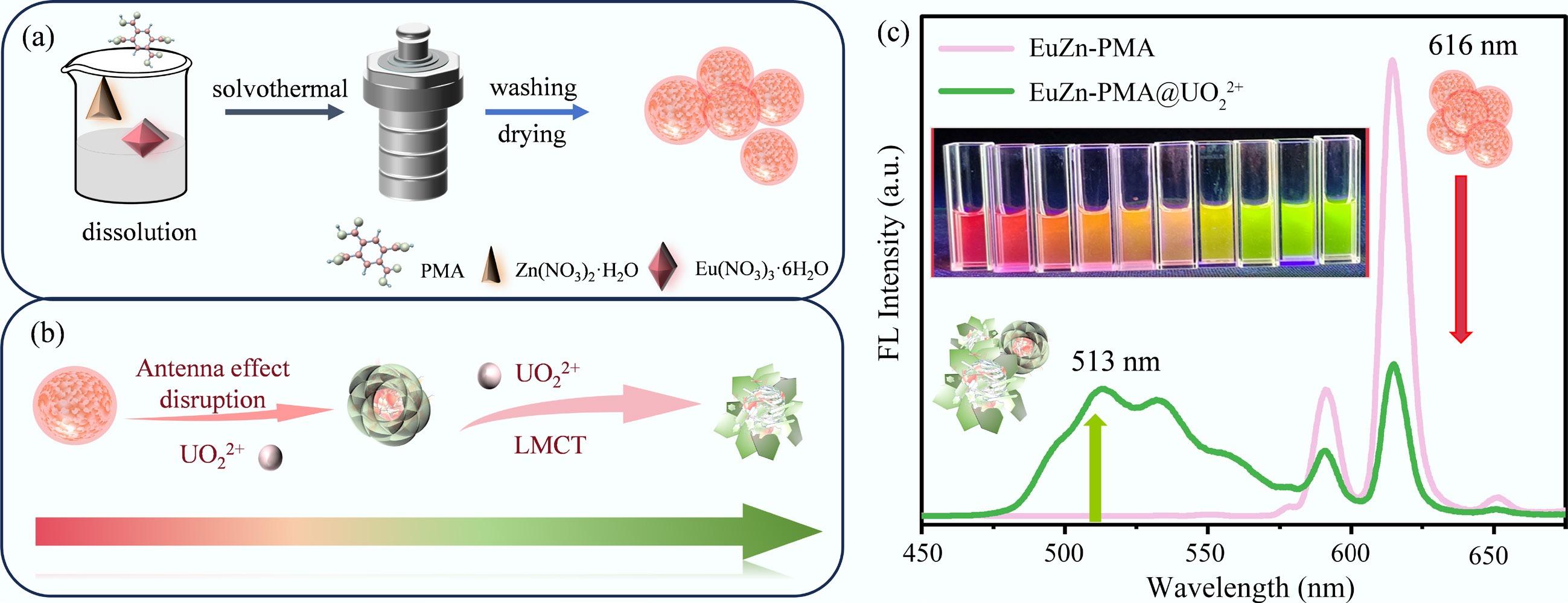
In this context, we designed a dual-metal coordination polymer, EuZn-PMA, which features Eu3+ and Zn2+ as the metal centers and pyromellitic acid (PMA) as a bifunctional ligand. Distinct from the sensors mentioned above, this probe leverages Eu3+ as the primary luminescent center, while Zn2+ serves as a cooperative metal ion that modulates the coordination environment. The PMA ligand plays a dual role: Its carboxyl groups function as recognition units for UO22+, and its conjugated framework simultaneously acts as an antenna to sensitize Eu3+ luminescence. Upon exposure to UO22+, the preferential coordination of UO22+ to the carboxyl groups blocks the antenna effect, leading to the attenuation of the characteristic Eu3+ emission at 616 nm. Concurrently, this coordination triggers a ligand to metal charge transfer (LMCT) process from PMA to UO22+, generating a new emission peak at 513 nm. This combined mechanism of disrupted antenna effect and the LMCT turn-on response enables a clearly distinguishable ratiometric fluorescence transition from red to green, providing an intuitive, reliable, and self-calibrated method for the visual detection of trace UO22+ in water.
-
The fluorescent probe EuZn-PMA was synthesized via a hydrothermal method. Briefly, PMA (0.3 mmol), zinc nitrate hexahydrate (0.3 mmol), and europium nitrate hexahydrate (0.15 mmol) were dissolved in a mixed solvent of anhydrous ethanol (20 mL) and N,N-Dimethylformamide (Dimethylformamide) (DMF) (20 mL). The molar ratio of PMA : Zn2+ : Eu3+ was maintained at 2:2:1. After ultrasonication for 10 min, the mixture was transferred to a Teflon-lined stainless-steel autoclave and heated at 160 °C for 12 h. The resulting precipitate was collected by centrifugation (8,000 rpm, 5 min), washed alternately with ultrapure water and anhydrous ethanol three times, and dried in a vacuum oven at 50 °C for 24 h. Finally, the product was dispersed in ultrapure water to form a suspension (1 mg/mL) for subsequent experiments.
For control experiments, single-metal Eu-PMA and Zn-PMA were synthesized under exactly the same hydrothermal conditions as EuZn-PMA, with only the europium source or the zinc source added separately. Detailed information on the raw materials and synthetic protocols are available in the Supplementary Text S1, S2 and S3.
Characterization
-
A series of characterization techniques including X-ray diffraction (XRD), scanning electron microscopy (SEM), Fourier transform infrared spectroscopy (FTIR), ultraviolet–visible (UV–vis) absorption spectroscopy, X-ray photoelectron spectroscopy (XPS), and fluorescence spectroscopy were used to analyze the structure, morphology, and optical properties of the as-prepared samples. Detailed instrument models, testing parameters and specific experimental conditions are fully listed in the Supplementary Text S4.
Fluorescence responses to UO22+
-
The fluorescence response to UO22+ was performed under optimal conditions. The specific steps were as follows. First, 200 μL of the 1 mg/mL fluorescent probe suspension was added to the buffer solution to prepare 2 mL of a 0.1 mg/mL EuZn-PMA suspension. Subsequently, different volumes of a standard solution of 1 mM UO22+ were sequentially injected into this suspension to examine the fluorescence response. Fluorescence spectra were collected in the range of 450–675 nm using a fluorescence spectrometer at an excitation wavelength of 272 nm with a 300-nm filter. All fluorescence detection experiments were carried out with no less than three parallel replicates (n ≥ 3) to guarantee experimental reproducibility and the data's credibility.
Fluorescence responses to other biological reagents and metal ions
-
To comprehensively evaluate the selectivity and immunity of EuZn-PMA, a series of interfering substances were selected according to their common coexistence in uranium-containing wastewater and natural water bodies. These included several common metal cations (Th4+, K+, Ni2+, Lu3+, Ba2+, Sm3+, Pb2+, Mn2+, Ca2+, Co3+, Dy3+, Na+, and Mg2+), as well as biological reagents and anions (CO32−, HCO3−, HS−, I−, S2−, Ac−, and Br−). For the selectivity experiments, the concentration of UO22+ and each individual interfering ion was fixed at 60 μM to allow for an equimolar comparison. In the anti-interference experiments, the concentration of the coexisting substance was set at three times that of UO22+ (180 μM) to simulate a harsher environment with high background interference. Fluorescence spectra were recorded from 450 to 675 nm upon excitation at 272 nm (n ≥ 3). Notably, although the probe demonstrated excellent selectivity against individual ions, the complex matrix effects present in real environmental samples (involving mixtures of these ions) were not simulated in this study. The potential interference in such mixed systems represents a limitation that should be addressed in future practical applications.
Analysis of UO22+ in real samples
-
Seawater was taken from Maoming, Guangdong Province, and filtered with a 0.22-μm microporous filter membrane and then prepared for use. Lake water was taken from the lakes in Maonan District, Maoming, and was also pretreated in the same way. To conform to the actual uranium pollution level in natural aquatic environments, gradient concentrations of a standard solution of UO22+ at 1, 2, and 4 μM were spiked into the two real water samples, and a blank group without added UO22+ was set simultaneously. The prepared sample solution was mixed with the probe dispersion, and the fluorescence spectra were recorded at an excitation wavelength of 272 nm with a 300-nm filter. All tests were carried out in no less than three parallel replicates (n ≥ 3).
-
To verify the successful synthesis and structural characteristics of the EuZn-PMA probe, a series of morphological, structural, and compositional characterizations were performed, as shown in Figs. 1 and 2.
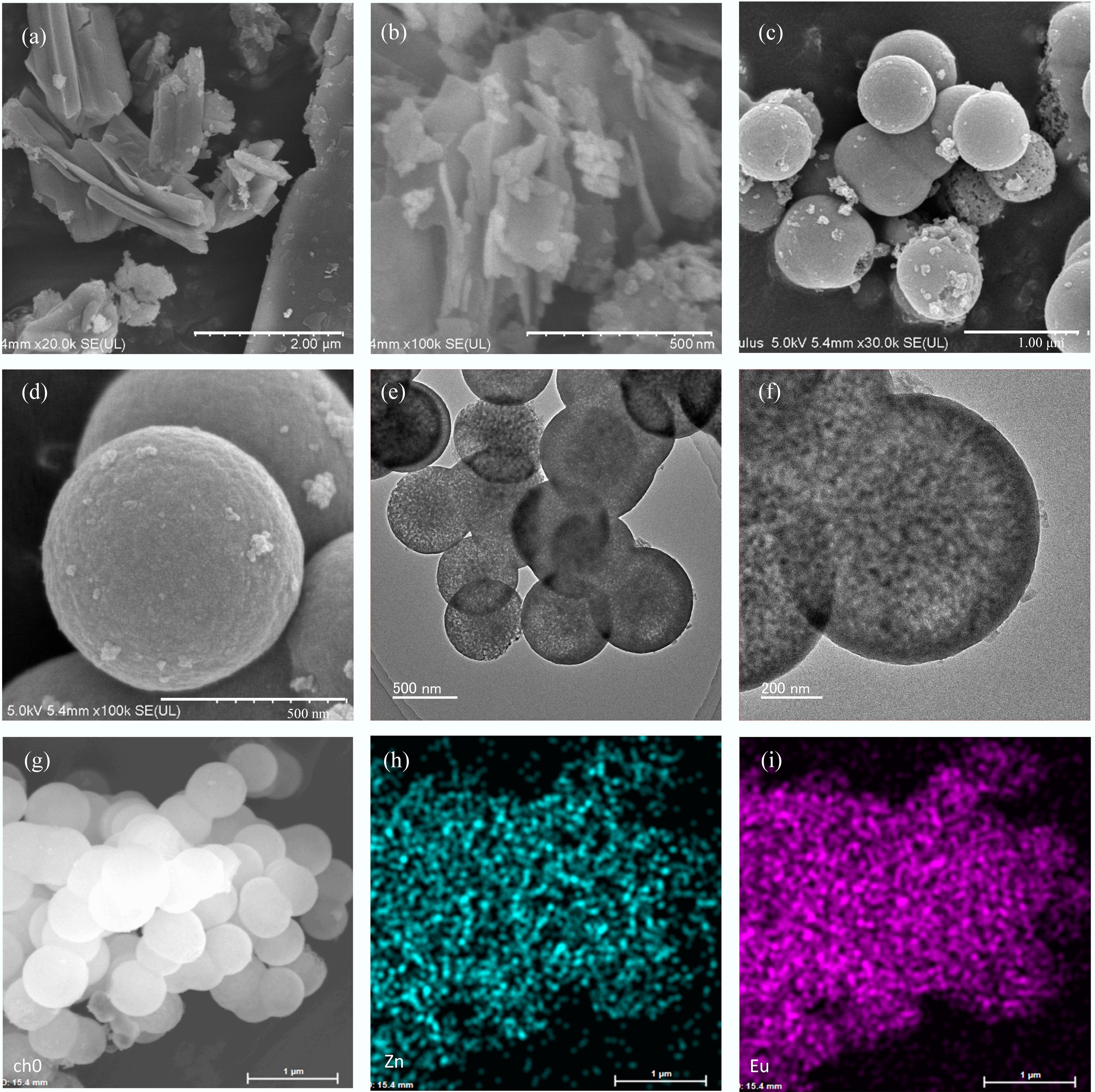
Figure 1.
(a) SEM images of Zn-PMA. (b) SEM images of Eu-PMA. (c), (d) SEM images of EuZn-PMA at different magnifications. (e), (f) Transmission electron microscopy (TEM) images of EuZn-PMA. (g) scanning transmission electron microscopy (STEM) image of EuZn-PMA and the corresponding elemental mapping of (h) Zn and (i) Eu.
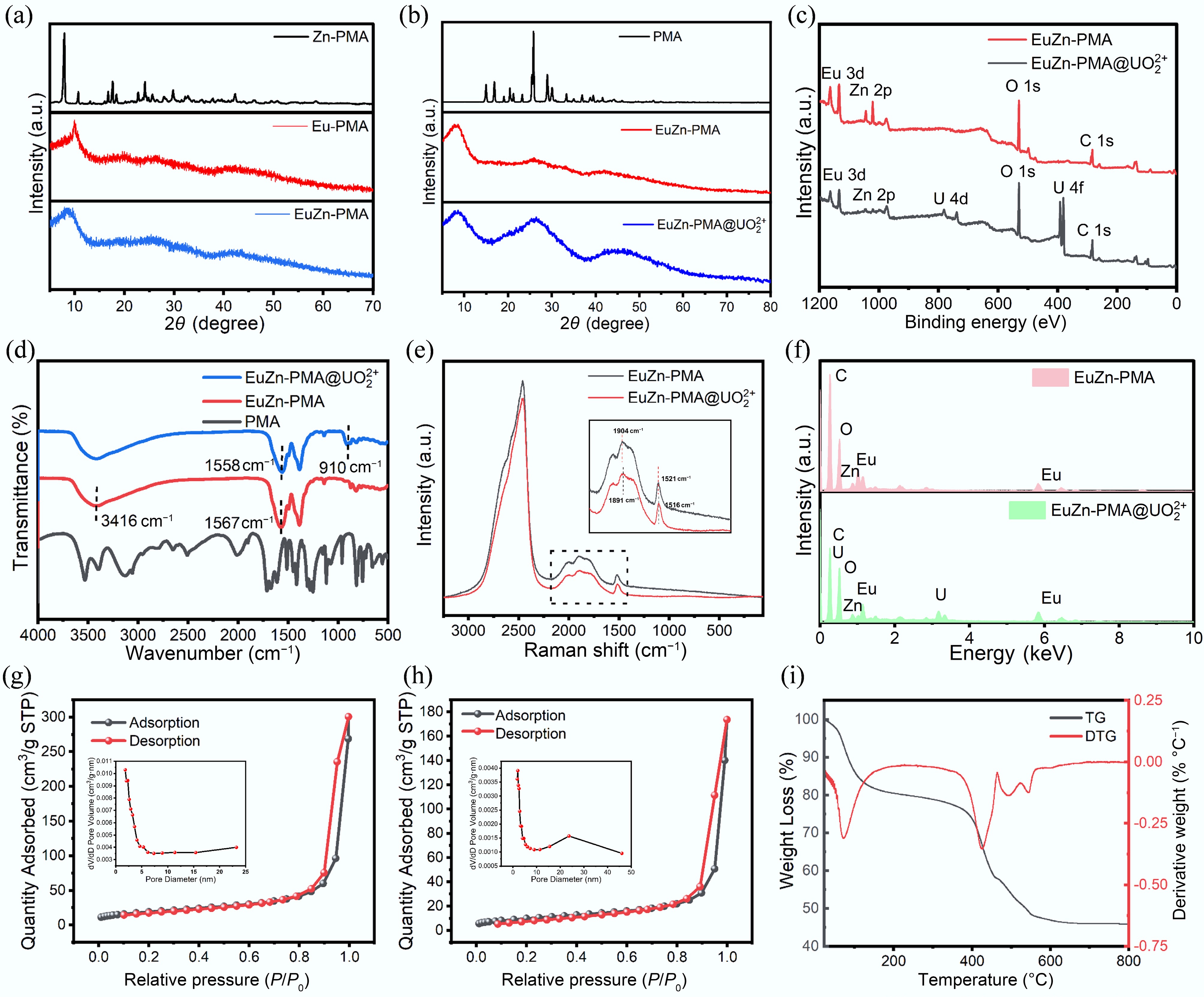
Figure 2.
(a), (b) XRD patterns of Zn-PMA, Eu-PMA, EuZn-PMA, PMA, and EuZn-PMA@UO22+. (c) Full XPS survey spectrum of EuZn-PMA. (d) FTIR spectra of PMA, EuZn-PMA, and EuZn-PMA@UO22+. (e) Raman spectra of EuZn-PMA and EuZn-PMA@UO22+. (f) Energy-dispersive X-ray spectroscopy (EDS) spectrum of EuZn-PMA and EuZn-PMA@UO22+. (g), (h) The 77.3 K nitrogen adsorption-desorption isotherms and pore size distribution of EuZn-PMA and EuZn-PMA@UO22+. (i) Thermogravimetric (TG) and derivative thermogravimetric (DTG) curves of EuZn-PMA.
First, the morphologies of the single-metal probe (Zn-PMA and Eu-PMA) and the bimetallic EuZn-PMA were compared by SEM. As shown in Fig. 1a, b, Zn-PMA exhibits a typical two-dimensional layered stacked structure with clear sheet edges, and Eu-PMA presents an aggregated and amorphous morphology. In contrast, the EuZn-PMA shows uniform spherical particles with a diameter of approximately 0.5 ± 0.1 μm (Fig. 1c, d). This indicates that the incorporation of Zn2+ effectively regulates the morphology, transforming the irregular aggregates into uniform microspheres.
The TEM images (Fig. 1e, f) further confirm the dense, homogeneous internal structure of EuZn-PMA microspheres, indicating that the bimetallic coordination effectively regulates the morphology and structural assembly kinetics and transforms the initial layered structure into a well-defined spherical morphology. This uniform spherical morphology ensures reproducible and stable dispersion in aqueous sensing media, which is critical for quantitative detection. Elemental distribution mapping (Figs. 1g−i and Supplementary Fig. S1) reveals that C, O, Zn, and Eu elements are uniformly distributed throughout the microspheres without obvious segregation, confirming the successful and homogeneous incorporation of both metal ions into the coordination polymer framework.
The XRD patterns of Zn-PMA, Eu-PMA, EuZn-PMA, and the pure PMA ligand are shown in Fig. 2a, b. Zn-PMA displays sharp and distinct diffraction peaks, indicating good crystallinity, whereas Eu-PMA shows no obvious crystalline peaks, presenting an amorphous state. EuZn-PMA exhibits broad diffraction humps in the wide-angle region, suggesting the formation of an amorphous coordination framework without long-range crystalline order. Despite the amorphous nature of the skeleton, the small-angle XRD pattern (Supplementary Fig. S2) reveals a distinct diffraction peak in the low-angle region. This characteristic peak corresponds to the (100) reflection, confirming the existence of a periodically ordered mesostructure within the EuZn-PMA framework[35]. Therefore, EuZn-PMA is identified as a mesoporous coordination polymer with short-range ordering and mesoscale periodicity.
FTIR was used to identify the functional groups and coordination modes (Fig. 2d). Compared with pure PMA, EuZn-PMA exhibits significant changes in the carboxylate group region: The characteristic peaks at 1,567 cm−1 (asymmetric stretching, νas) and 1,386 cm−1 (symmetric, νs) indicate that the carboxylate anions (COO−) of PMA have coordinated with Zn2+ and Eu3+[36]. The disappearance of the free C=O stretching vibration at 1,712 cm−1 further confirms the deprotonation and coordination of the carboxyl groups[37]. Raman spectroscopy (Fig. 2e) was used to further verify the coordination environment. As shown in the inset of Fig. 2e, the characteristic band at 1,521 cm−1, attributed to the stretching vibrations of the C=O/C=C groups of the PMA ligand, exhibits a distinct shift compared with the free ligand. This shift is consistent with the changes observed in the FTIR spectrum at 1,567 cm−1, confirming the successful coordination of the carboxylate groups with the metal centers.
The N2 adsorption–desorption isotherm of EuZn-PMA at 77.3 K (Fig. 2g) shows a Type IV isotherm with an H1 hysteresis loop, confirming the presence of uniform mesopores. The Brunauer–Emmett–Teller (BET) specific surface area is 68.61 m2/g, and the Barrett–Joyner–Halenda (BJH) pore size distribution reveals a narrow mesopore centered at 8.68 nm, consistent with the (100) peak in the small-angle XRD pattern (Supplementary Fig. S2). This confirms an ordered mesostructure within the amorphous framework. The thermogravimetric analysis of EuZn-PMA shows two main mass loss stages from room temperature to 800 °C (Fig. 2i). The first weight loss of ~20.5% occurs below 200 °C, which is attributed to the removal of adsorbed water and residual solvent. The second significant weight loss occurs between 450 and 800 °C, corresponding to the thermal decomposition of the organic ligand framework, with a total weight loss of ~33.4%[38]. The mass remains stable between 200 and 450 °C, indicating that EuZn-PMA exhibits good thermal stability. The framework structure will not undergo thermal decomposition under the conditions of room temperature detection, providing a reliable guarantee of stability for its practical application in environmental water samples.
XPS and EDS were used to analyze the elemental composition and oxidation states. The full XPS spectrum (Fig. 2c) confirms the presence of C, O, Zn, and Eu elements in EuZn-PMA. The high-resolution spectra (Fig. 3a−e) show the C 1s peaks at 284.85 eV (benzene ring), 288.1 eV (C=O), and 289 eV (O-C=O in the carboxylic groups)[36]; O 1s peaks at 531.85 eV (C=O) and 533.6 eV (C-O)[37]; Zn 2p peaks at 1,045.7 eV (Zn 2p1/2) and 1,022.65 eV (Zn 2p3/2), confirming the +2 oxidation state of Zn[39]; and Eu 3d peaks at 1,144.1 eV (Eu 3d3/2) and 1,135.85 eV (Eu 3d5/2), confirming the +3 oxidation state of Eu[40]. The EDS spectrum confirms the coexistence of C, O, Zn, and Eu (Fig. 2f).
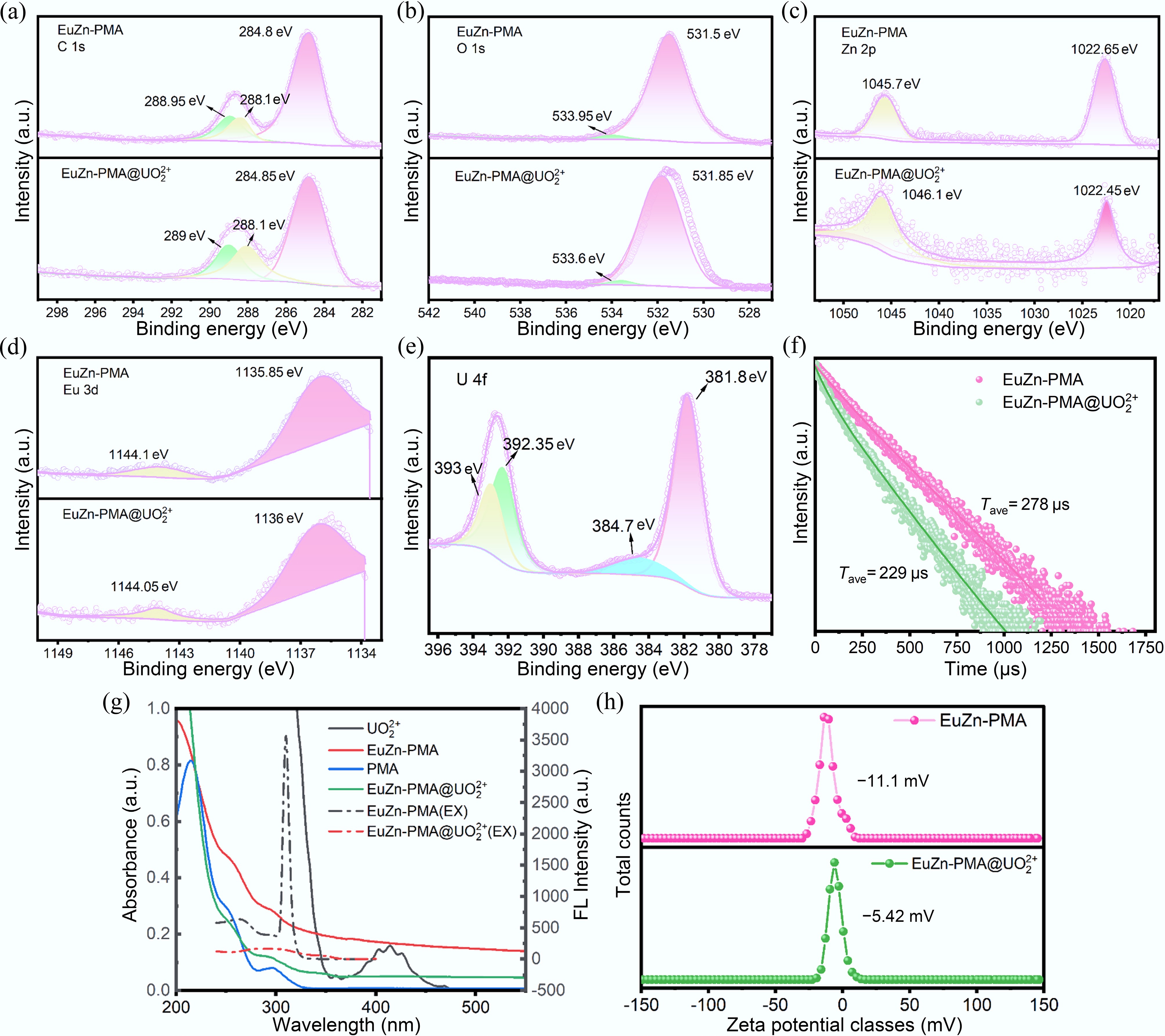
Figure 3.
Spectroscopic and interfacial characterization of EuZn-PMA before and after interaction with UO22+. (a)−(e) High-resolution XPS spectra of C 1s, O 1s, Zn 2p, Eu 3d, and U 4f. (f) Lifetime fluorescence decay curves of EuZn-PMA before and after adding UO22+. (g) UV–vis absorption spectra. (h) Zeta potential distribution.
In summary, all the abovementioned characterizations confirm the successful synthesis of the Eu–Zn bimetallic coordination polymer EuZn-PMA with well-defined morphology, stable structure, and uniform elemental distribution.
Optimization of sensing conditions
-
In order to achieve better UO22+ fluorescence ratio test performance, some experimental variables affecting the sensor's light intensity ratio ([F513/F0]/[F616/F0]) were optimized, including the buffer solution and pH value. This dual-emission ratiometric signal was chosen because it simultaneously captures the UO22+-induced emission enhancement at 513 nm and the attenuation of Eu3+ emission at 616 nm, closely reflecting the visually perceptible color transition and thus providing a robust basis for quantitative calibration. First, to investigate the fluorescence response of the EuZn-PMA fluorescent probe to UO22+ in different buffer solutions, a Britton–Robinson (BR) buffer solution, a 4-(2-hydroxyethyl)-1-piperazineethanesulfonic acid (HEPES) buffer solution, a phosphate-buffered saline (PBS) buffer solution, a Tris(hydroxymethyl)aminomethane hydrochloride (Tris-HCl) buffer solution, and an ultrapure water solution were prepared with a pH of 7. As indicated in Supplementary Fig. S3, the fluorescence intensity ratio of ([F513/F0]/[F616/F0]) was highest in ultrapure water, so the subsequent experiments were conducted in an ultrapure water solution. To optimize the effect of different pH values on the experimental results, the pH of the aqueous solution was adjusted using KOH and HCl, and ultrapure water buffers with pH values ranging from 4 to 9 were prepared. The fluorescence response phenomenon between the EuZn-PMA fluorescent probe and UO22+ was then investigated in these ultrapure water buffers at different pH values. A probe solution with a concentration of 0.25 mg/mL was prepared using buffer solutions with different pH values, and its fluorescence spectra were tested. The experimental results are shown in Supplementary Fig. S4. The fluorescence intensity of EuZn-PMA does not change significantly with pH, indicating that this fluorescent probe has good pH stability and is resistant to acids and alkalis. Subsequently, 120 μL of the 1 mM UO22+ solution was added separately. At pH = 7, the fluorescence ratio [(F513/F0)/(F616/F0)] was highest, facilitating the most distinct and measurable change in the fluorescence ratio. Therefore, this experiment selected an ultrapure water solution at a pH of 7 as the optimal experimental environment. As indicated in Supplementary Fig. S5, the fluorescence spectra under optimal excitation and different excitations were tested, and the excitation wavelength of 272 nm was ultimately selected to achieve the best fluorescence testing results.
Response of EuZn-PMA to UO22+
-
Under the optimized experimental conditions described above, the sensing response behavior of EuZn-PMA toward UO22+ was systematically investigated, and the results are displayed in Fig. 4. Prior to the concentration gradient test, the fluorescence responses of single-metal Zn-PMA, single-metal Eu-PMA, and bimetallic EuZn-PMA toUO22+ were first compared, and the relevant fluorescence spectra are shown in Fig. 4a. EuZn-PMA shows significantly enhanced Eu3+ emissions at 616 nm compared with single-metal Eu-PMA and Zn-PMA, indicating the synergistic effect of Zn2+ on luminescence[41]. Time-resolved fluorescence measurements further confirmed that the Eu3+ lifetime in EuZn-PMA (278 μs) is significantly longer than that of Eu-PMA, providing direct photophysical evidence for the role of Zn2+ as a luminescence sensitizer (Fig. 3f). Although Zn2+ does not act as a luminescent center or directly bind with UO22+, it effectively stabilizes the coordination framework, optimizes the material's morphology, and greatly boosts the antenna effect to sensitize the fluorescence of Eu3+. Upon the addition of UO22+, the emission at 616 nm was attenuated but a new emission appeared at 513 nm, forming the ratiometric response.
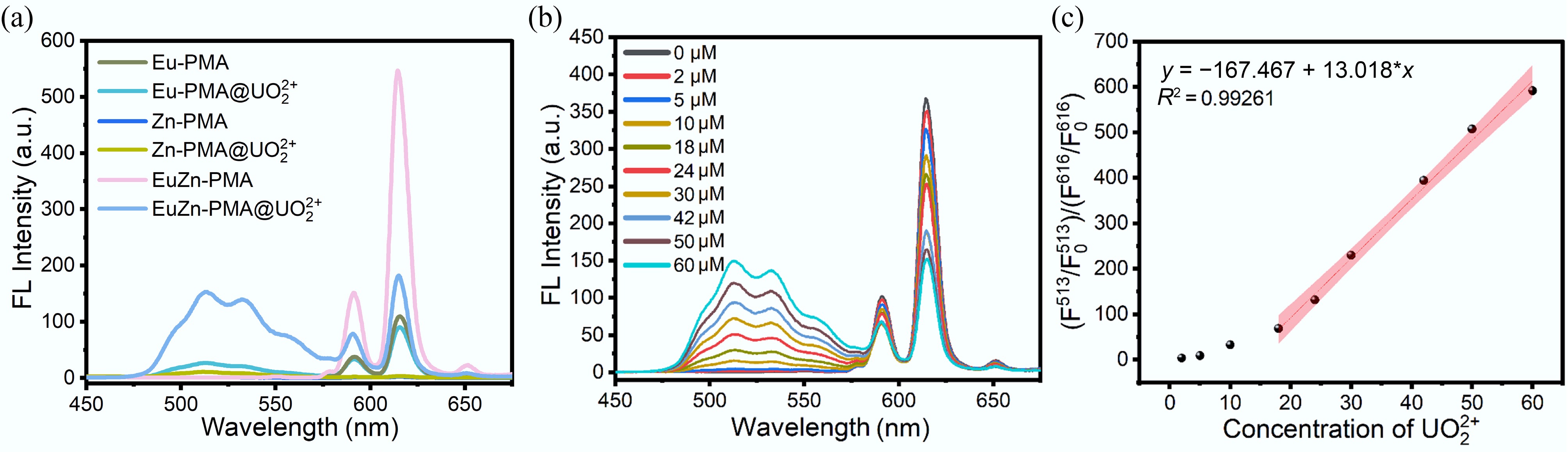
Figure 4.
(a) Fluorescence spectra of Eu-PMA, Zn-PMA, and EuZn-PMA before and after the addition of UO22+. (b) Fluorescence spectra of EuZn-PMA (0.1 mg/mL) in the presence of different concentrations of UO22+ (0−60 μM). (c) At UO22+ concentrations ranging from 0 to 60 μM, the linear relationship between ([F513/F0]/[F616/F0]) and different concentrations of UO22+.
Before adding UO22+, the fluorescent probe emits red fluorescence. As the concentration of UO22+ increases, the fluorescence peak intensity at 616 nm decreases, and a new fluorescence peak appears at 513 nm, exhibiting a distinct fluorescence ratio. Therefore, we calculate the ratio of enhanced fluorescence at 513 nm and the ratio of attenuated fluorescence at 616 nm, and divide the ratio of the enhanced fluorescence by the ratio of the attenuated fluorescence to obtain the ratio ([F513/F0]/[F616/F0]), which provides a more intuitive representation of the fluorescence changes. As shown in Fig. 4b, with increasing UO22+ concentration, the ratio ([F513/F0]/[F616/F0]) gradually increases, showing a significant change, and the probe exhibits an obvious fluorescence ratio. As depicted in Fig. 4c, within the UO22+ concentration range of 0–60 μM, the fluorescence ratio ([F513/F0]/[F616/F0]) exhibits a good linear correlation with the UO22+ concentration. The linear equation is y = 13.018 * x − 167.467, with a correlation coefficient R2 = 0.99261. The limit of detection (LOD) for UO22+ was calculated to be 51 nM, based on the equation LOD = 3σ/k (where σ is the standard deviation and k is the slope of the standard curve). This value is significantly lower than the maximum limit for uranium in drinking water set by the US EPA (30 μg/L, equivalent to 130 nM). Compared with the previously reported UO22+ detection sensor platform (as summarized in Supplementary Table S1), EuZn-PMA has a lower detection limit for UO22+, indicating that the EuZn-PMA fluorescent probe is highly sensitive and can achieve the desired goal.
Selectivity and anti-interference
-
The selectivity and anti-interference ability of EuZn-PMA were evaluated in the presence of interfering cations and anions. For cations, Th4+, Lu3+, Sm3+, Pb2+, Co2+, Dy3+, Ba2+, Ca2+, K+, Mg2+, Mn2+, and Na+ were selected as interfering substances; for anions, seven common anions (e.g., CO32−, HCO3−, HS−, Br−, I−, Ac−, S2−) were additionally tested to simulate the complex actual water environment. After adding interfering ions, the fluorescence ratio F513/F5130 of EuZn-PMA did not change significantly. In this selectivity study, the single-channel ratio at 513 nm was preferred because most coexisting interfering ions produce negligible emission at this wavelength, whereas the signal at 616 nm can be slightly perturbed by some cations; the dual-emission ratio would therefore yield near-zero values that mask the specific turn-on response to UO22+. As depicted in Fig. 5a, c, after adding UO22+ to the fluorescent probe, a fluorescence peak appeared at 513 nm, and the fluorescence ratio F513/F5130 increased, indicating that the fluorescent probe demonstrates good selectivity toward UO22+. To further verify the anti-interference performance of EuZn-PMA, interference ions at three times the concentration were added to a solution containing UO22+, and then UO22+ fluorescence detection was carried out. Fig. 5b, d reveals that the fluorescence response of EuZn-PMA to UO22+ remains largely unaffected, with minimal changes in the fluorescence ratio F513/F5130. This indicates that EuZn-PMA exhibits favorable anti-interference performance when detecting UO22+.
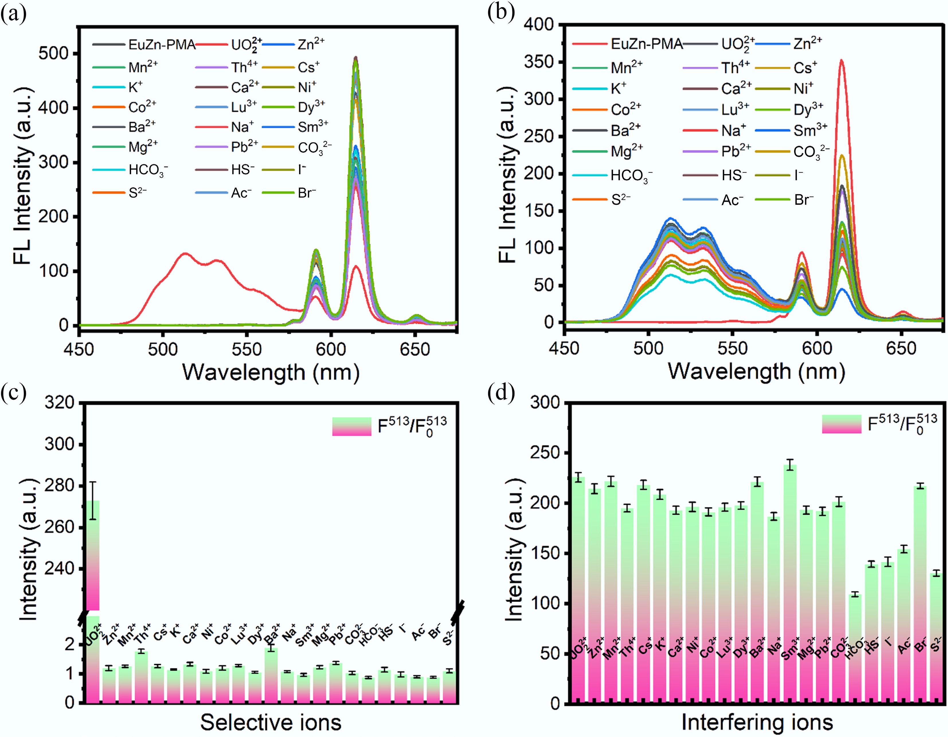
Figure 5.
Selectivity and anti-interference performance of EuZn-PMA. (a), (c) Fluorescence spectra and the corresponding fluorescence ratio F513/F5130 of EuZn-PMA in the presence of UO22+ and various common interfering ions. (b), (d) Fluorescence spectra and the corresponding fluorescence ratio F513/F5130 of EuZn-PMA in mixed solutions containing UO22+ and threefold excess concentrations of interfering ions.
Mechanism study
-
Upon interaction with UO22+, EuZn-PMA exhibits dual-channel ratiometric fluorescence behavior: The characteristic emission of Eu3+ at 616 nm is quenched and a new emission peak at 513 nm appears, accompanied by a visually perceptible red–green color change under 254-nm UV light. To elucidate the underlying mechanism, a series of characterizations were performed on EuZn-PMA before and after reaction with UO22+, including SEM/EDS, XRD, FTIR, BET, Raman, TG, XPS, zeta potential, UV–vis absorption, and fluorescence lifetime measurements.
As displayed in Fig. 6, obvious morphological changes can be observed after EuZn-PMA binds with UO22+. The original regular spherical structure gradually collapses and transforms into a loose layered stacked structure accompanied by surface sheet exfoliation. According to the EDS elemental mapping results, all elements are still uniformly distributed in the material system, proving that UO22+ achieves homogeneous binding rather than uneven surface adsorption. Notably, this morphological change does not represent irreversible structural degradation. Essentially, the coordination interaction between UO22+ and surface functional groups changes the interlayers' interaction force inside the assembled microspheres, resulting in rearrangement of the macroscopic morphology, although the internal basic crystal structure is well preserved.
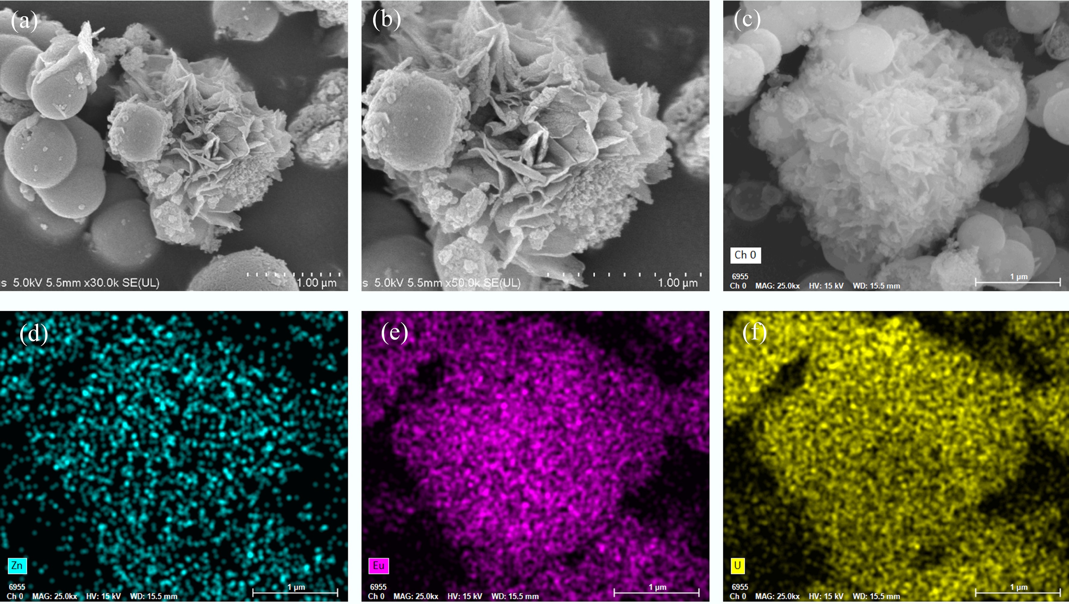
Figure 6.
SEM images and corresponding EDS elemental mapping of EuZn-PMA after reacting with UO22+. (a), (b) SEM morphology of the EuZn-PMA–UO22+ composite. (c) STEM image of EuZn-PMA@ UO22+ and the corresponding elemental mapping of (d) Zn, (e) Eu and (f) U.
To evaluate the structural stability during the process of recognizing UO22+, the XRD patterns of EuZn-PMA before and after interaction with UO22+ were compared (Fig. 2b and Supplementary Fig. S1). In the wide-angle XRD region, the broad amorphous diffraction peak of the matrix is well preserved, indicating that the local coordination environment remains intact. More importantly, the low-angle XRD pattern shows that the characteristic peak position remains unchanged after binding to UO22+. This demonstrates that the periodic mesoporous structure is perfectly preserved and no framework collapse occurs during the recognition process. The slight decrease in peak intensity can be attributed to the formation of PMA–UO22+ coordination complexes and their filling within pore channels. FTIR spectra further confirm the effective coordination behavior (Fig. 2d). The antisymmetric stretching vibration peak of the carboxylate group shifts from 1,567 to 1,558 cm−1, which is direct evidence that -COO− groups are combined with UO22+. Moreover, the newly appeared characteristic peak at 910 cm−1 is attributed to the U=O stretching vibration of UO22+, further confirming the successful formation of coordination complexes between the PMA ligand and UO22+[42]. Upon UO22+ binding, the characteristic Raman band at 1,521 cm−1 (attributed to carboxyl/benzene ring skeletal vibrations) undergoes a red shift to 1,516 cm−1 (Fig. 2e). This change confirms the coordination between UO22+ and the ligand framework via carboxylate oxygen atoms, resulting in a reduction in the electron density. After reaction with UO22+, the surface area decreases to 35.26 m2/g and the dominant pore size shifts to 24.20 nm (Fig. 2h), indicating pore filling and structural rearrangement upon binding with UO22+. The results of the zeta potential test show that the surface's negative potential changed from −11.1 to −5.42 mV after the reaction, suggesting electrostatic attraction between the negatively charged surface and the positively charged UO22+, which facilitates the subsequent coordination of the recognition process (Fig. 3h)[43].
The UV–vis spectrum in Fig. 3g shows no overlap between UO22+ absorption (≤ 500 nm) and EuZn-PMA emission (≥ 550 nm). This negligible spectral overlap rules out the FRET mechanism. Meanwhile, the UV–vis absorption exhibits an obvious intensity decrease in the range of 300–400 nm after UO22+ coordination, which is ascribed to redistribution of the electron density of the PMA ligand. XPS spectra before and after the reaction were used to analyze the changes in the chemical environment (Fig. 3a−e). The binding energies of Eu 3d and Zn 2p remain almost unchanged, demonstrating that the chemical valence states of Eu3+ and Zn2+ are stable throughout the detection process, and they do not directly participate in the coordination combination with UO22+. This indicates that the chemical states of Eu3+ and Zn2+ are unaffected, and that coordination is concentrated between PMA and UO22+. The obvious binding energy shift of C 1s and O 1s fully proves that the coordination reaction mainly occurs between the carboxylate groups of organic ligands and UO22+. The decrease in electron density around the oxygen atoms induces the change in the binding energy, which is consistent with the electronic redistribution expected for LMCT upon UO22+ coordination[44]. The successful detection of U's 4f characteristic peaks also directly proves the stable existence of UO22+ in the composite system. The average fluorescence lifetime of EuZn-PMA decreased from 278 to 229 μs upon binding to UO22+ (Fig. 3f). This shortened lifetime indicates that UO22+coordination perturbs the local environment of Eu3+ and accelerates nonradiative deactivation. Unlike classical static or dynamic quenching, where the quencher directly interacts with the fluorescent center, the attenuation of Eu3+ emissions here originates primarily from the disruption of the antenna effect: UO22+ competitively binds to PMA's carboxylate groups, thereby blocking ligand-to-Eu3+ energy transfer. The enhanced nonradiative decay, reflected by the reduced lifetime, further contributes to the decrease in emissions. This twofold mechanism is fully consistent with the dual-channel ratiometric response.
Given all these characterization results, the detailed UO22+ detection mechanism of EuZn-PMA is summarized as follows. In the initial state, the PMA ligand acts as an antenna, absorbing the excitation light and efficiently transferring energy to the Eu3+ center, thereby producing stable red emission at 616 nm. When UO22+ exists in the system, it preferentially binds to active carboxylate sites on the EuZn-PMA's surface through electrostatic attraction and the coordination effect. This binding behavior interrupts the original antenna effect's energy transfer channel, leading to the marked decrease in Eu3+ fluorescence emissions. At the same time, the formation of a PMA–UO22+ coordination complex triggers LMCT, thus producing a new stable fluorescence emission peak at 513 nm. The disrupted antenna effect is accompanied by perturbation of the Eu3+ coordination microenvironment, as reflected by the shortened fluorescence lifetime (from 278 to 229 μs), further contributing to the decrease in emissions. Together, these processes enable a sensitive dual-channel ratiometric fluorescence response with a clearly distinguishable red–green color transition.
Application to real water samples
-
To evaluate the practical application potential of EuZn-PMA, its performance in detecting UO22+ in real samples (lake water and seawater) was investigated. The accuracy and reliability of the probe were assessed using the standard addition method by spiking the samples with varying concentrations of UO22+ (1, 2, and 4 μM). As summarized in Supplementary Table S2, the recoveries ranged from 94.5% to 102.5%, with relative standard deviations (RSDs) between 1.9% and 3.9%. These results demonstrate the satisfactory accuracy and excellent anti-interference capability of the proposed method for detecting U in complex aquatic environments.
Smartphone-assisted portable device for visual assays of UO22+
-
To enable portable, on-site quantification of UO22+, a smartphone-based visual readout platform was constructed. After incubating EuZn-PMA with different UO22+ concentrations, the mixtures were photographed under 254-nm UV excitation. The acquired images were processed to obtain the red–green–blue (RGB) values (Supplementary Table S3). As the UO22+ concentrations increased, the fluorescence emission underwent a visually discernible transition from red to green (Fig. 7). The ratio of the green to red channel intensity (G/R) exhibited a good linear correlation with the UO22+ concentration over the range of 0–60 μM (R2 = 0.9760), according to the equation G/R = 0.24983 + 0.02217 * [UO22+]. The results confirm that the proposed smartphone-integrated sensing platform enables rapid, cost-effective, and instrument-free visual detection of UO22+ in field settings.
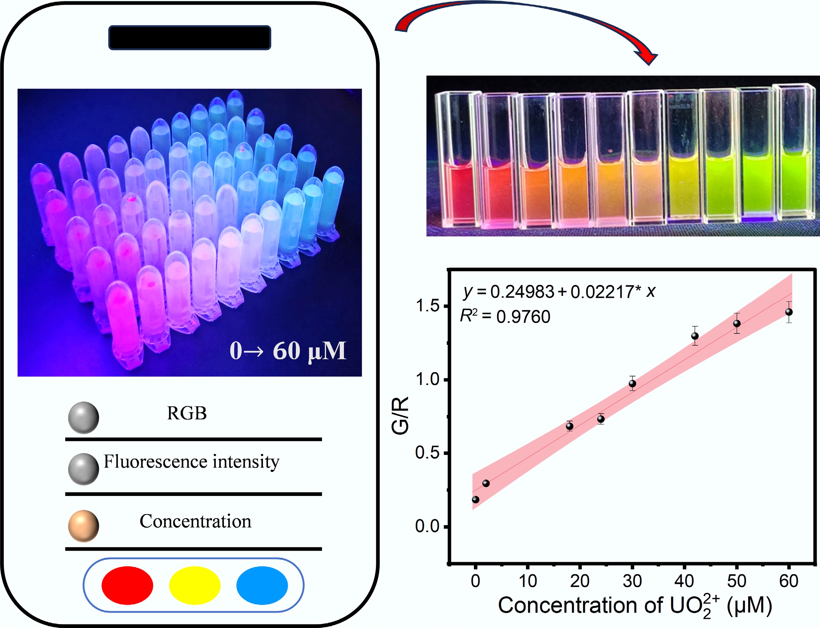
Figure 7.
Quantitative detection of UO22+ by a smartphone-assisted readout platform.
-
In summary, a ratiometric fluorescent sensing platform based on the synergistic effect of Eu–Zn bimetals was successfully developed for rapid, highly sensitive, and visual detection of UO22+. The morphological regulation by Zn2+ together with the excellent luminescent properties of Eu3+ gave the probe enhanced fluorescence performance and structural stability. Through the synergistic response of UO22+-induced suppression of the antenna effect and the LMCT process, significant dual-channel fluorescence changes were generated, thereby effectively improving the reliability and anti-interference capability of the sensing system. The proposed method exhibited a low detection limit of 51 nM and a satisfactory linear detection range of 0–60 μM, together with excellent accuracy and stability in practical analyses of water samples. Moreover, by integrating this with a smartphone-assisted RGB analysis platform, portable and on-site visual detection of UO22+ was also realized. The results demonstrate that EuZn-PMA holds great potential for monitoring radioactive uranium contaminants and also provides new insights into the design and application of lanthanide coordination polymers in environmental fluorescence sensing.
We gratefully acknowledge the support from the funding agencies mentioned below.
-
This accompanies this paper at https://doi.org/10.48130/scm-0026-0025.
-
This research did not involve human or animal samples.
-
The authors confirm their contributions to the paper as follows: Zhaojing Yuan: investigation, writing – original draft; Zhaojing Yuan, Xin Li: methodology; Chengtao He: conceptualization; Ziqing Liu, Hui Qiu, Xiangyang Hao: formal analysis; Qiushuo Zheng, Xiangyang Hao, Mingtai Sun: supervision; Long Yu: data curation; Chengtao He, Xin Li: software; Long Yu, Suhua Wang: writing – review, funding acquisition. All authors reviewed the results and approved the final version of the manuscript.
-
The data that support the findings of this study are available on request from the corresponding author.
-
The authors declare that they have no conflict of interest.
-
Full list of author information is available at the end of the article.
- The supplementary files can be downloaded from here.
- Copyright: © 2026 by the author(s). Published by Maximum Academic Press, Fayetteville, GA. This article is an open access article distributed under Creative Commons Attribution License (CC BY 4.0), visit https://creativecommons.org/licenses/by/4.0/.
-
About this article
Cite this article
Yuan Z, He C, Liu Z, Qiu H, Zheng Q, et al. 2026. Dual-metal synergistic coordination enables ultrasensitive and ratiometric multicolor fluorescent detection of uranyl ions. Sustainable Carbon Materials 2: e030 doi: 10.48130/scm-0026-0025 

Dual-metal synergistic coordination enables ultrasensitive and ratiometric multicolor fluorescent detection of uranyl ions
- Received: 01 April 2026
- Revised: 11 June 2026
- Accepted: 01 July 2026
- Published online: 02 September 2026
Abstract: The uranyl ion (UO22+), a key component in the nuclear fuel cycle, poses significant chemical toxicity and radioactive hazards. Herein, a Eu–Zn synergistic bimetallic coordination strategy was developed to construct a fluorescent probe for the rapid and highly sensitive detection of UO22+, based on the excellent luminescent properties of lanthanide ions. In this system, Zn2+ regulates the material morphology, while Eu3+ serves as the fluorescence signaling center. Their synergistic interaction significantly enhances the luminescence performance and structural stability of the probe. During the detection process, UO22+ coordinates with the carboxylate sites of pyromellitic acid (PMA) ligand, blocking the energy transfer from the ligand to Eu3+ (the antenna effect) and resulting in the quenching of the characteristic red emission of Eu3+. Meanwhile, the coordination interaction induces ligand-to-metal charge transfer (LMCT), generating a characteristic green emission. Based on variation in the dual-channel fluorescence signal, a ratiometric sensing mode was established, which effectively improves the anti-interference capability of the sensing system against environmental fluctuations. The proposed method achieved a detection limit as low as 51 nM with a linear range of 0–60 μM. Furthermore, a smartphone-assisted red–green–blue analysis platform was developed for on-site visual detection of UO22+ through the green–red channel ratio. In spiked lake water and seawater samples, satisfactory recoveries ranging from 94.5% to 102.5% were obtained, with relative standard deviations below 4%. This work provides a novel bimetallic fluorescent sensing strategy for the portable, visual, and reliable detection of radioactive uranium contaminants.
-
Key words:
- Fluorescent probe /
- Uranyl ion /
- Dual-metal synergy /
- Metal-organic coordination polymers