













-
Age-associated transcriptomic atlases of human microglia converge on a pattern that has received less attention than it warrants: early repression of cytoskeletal organisation, intracellular trafficking, and metabolic fitness emerges and persists across tissues, sexes, and cohorts[1−4]. This pattern has largely been interpreted as a downstream correlate of aging. We propose instead that it may reflect an upstream driver of microglial dysfunction. The implication extends beyond the observation that aged microglia exhibit altered phenotypes. Instead, a decline in mechanical competence may fundamentally reshape how these cells interpret, navigate, and respond to tissue demands.
Multi-dimensional analyses further show that microglial aging unfolds across coupled transcriptional, epigenetic, metabolic, and tissue-context axes, rather than following a single linear trajectory[3−5]. When framed as a problem of state regulation, this heterogeneity becomes mechanistically informative: the young-to-aged transition mapped in Fig. 1a reveals how aging progressively constrains microglial functional plasticity. With advancing age, microglia become less able to polarize appropriately, redistribute intracellular cargo, or adapt to spatially and temporally dynamic cues. In this context, inflammatory tone alone is insufficient to define the aging phenotype. Instead, dysfunction emerges from a progressive decoupling between molecular programs, such as gene expression changes, and mechanical behaviors, such as process extension and migration, required to execute them.
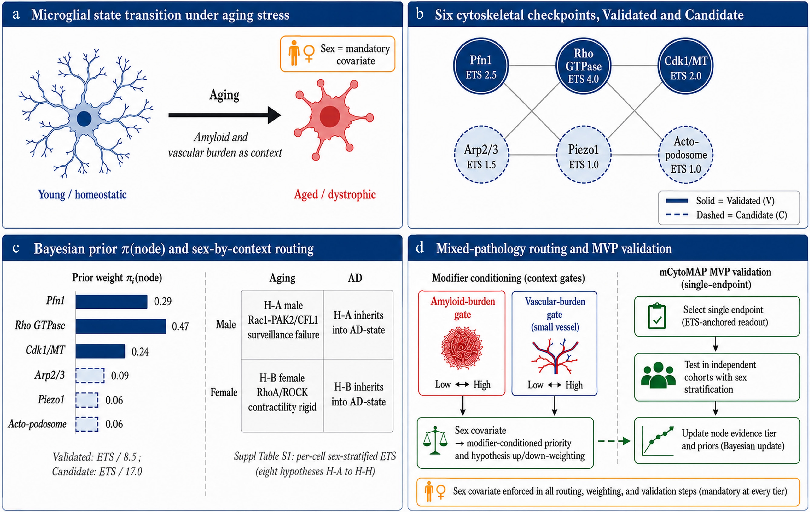
Figure 1.
Framework overview of cytoskeletal checkpoints as state-control nodes in the aging microglial targetome. (a) Microglial state transition under aging stress: young/homeostatic microglia (ramified, cool blue) shift to aged/dystrophic microglia (coral red) as aging proceeds, with amyloid and vascular burden acting as the disease-stage context. Sex is enforced as a mandatory covariate at every tier of the framework (gold-bordered badge). (b) Six cytoskeletal checkpoints organised by evidence tier: Pfn1, Rho GTPase network, and Cdk1/MT remodelling enter the Validated tier (solid borders) with Evidence-Tier Scores of 2.5, 4.0, and 2.0, respectively; Arp2/3 complex, Piezo1, and the actomyosin-podosome surveillance module enter the Candidate tier (dashed borders) with ETS values of 1.5, 1.0, and 1.0. (c) The Bayesian-style prior π(node) follows the two-tier normalisation rule (Validated: ETS/8.5; Candidate: ETS/17.0), giving per-node weights of 0.29 (Pfn1), 0.47 (Rho GTPase), 0.24 (Cdk1/MT), 0.09 (Arp2/3), 0.06 (Piezo1), and 0.06 (actomyosin–podosome). The sex-by-context routing grid (Male/Female × Aging/AD) maps to the H-A directional working hypothesis (male = Rac1-PAK2/CFL1 surveillance failure) and H-B (female = RhoA/ROCK contractility-rigid state), with the full per-cell sex-stratified ETS matrix and the eight directional hypotheses H-A through H-H in Supplementary Table S1. (d) Mixed-pathology routing and MVP validation: amyloid-burden and vascular-burden context gates re-weight the six-node ETS prior and up- or down-weight the relevant directional hypotheses; the resulting prior is then tested through the single-endpoint mCytoMAP MVP validation pathway, selection of a single sex-stratified ETS-anchored readout, testing in independent cohorts with sex stratification, and Bayesian update of node evidence tiers and priors. Sex covariate enforcement is mandatory in all routing, weighting, and validation steps.
Multi-omics profiling has additionally identified disease-restraining and resilience-associated microglial states that retain coordinated responses in both aged and disease contexts[6−8]. In Alzheimer's disease (AD), for example, proteomic signatures associated with resilience are enriched for actin filament organisation and injury-response pathways, rather than reflecting a simple absence of immune activation[7]. These findings suggest that aging does not represent a strictly irreversible trajectory. Rather, the preservation of structural plasticity is associated with adaptive microglial responses under pathological stress. Importantly, this adaptive capacity itself diminishes with age and disease progression, shifting the focus away from static molecular markers and toward the regulatory mechanisms that maintain functional competence.
Emerging evidence points to cytoskeletal regulation as a critical interface linking transcriptional programs, cellular mechanics, and circuit-facing microglial functions[9−12]. Within this framework, the cytoskeleton is not merely a structural scaffold but a dynamic regulator of whether microglia remain adaptively responsive or transition into mechanically constrained, maladaptive states. Accordingly, age-associated microglial heterogeneity may be better understood as reflecting varying degrees of retained structural and functional adaptability rather than simple differences in inflammatory activation alone.
Figure 1 summarises the conceptual framework developed in this perspective, the transition from descriptive accounts of microglial heterogeneity to a checkpoint-oriented model of state regulation. The entry point is the state-transition schematic in Fig. 1a, which casts the shift from young to aged microglia with sex carried explicitly as a biological covariate; framed this way, the central question becomes not what markers change but how aging reshapes the coordination and adaptability of microglial functional states.
From that framing, Fig. 1b organises the proposed checkpoint network into validated (Pfn1, Rho GTPase, Cdk1/MT) and candidate (Arp2/3, Piezo1, actomyosin-podosome) tiers, isolating the nodes most likely to govern adaptability across aging and disease. Because relevance is rarely uniform across individuals, the prioritisation grid in Fig. 1c then weights each checkpoint by sex-dependent and context-specific factors, providing a structured basis for target selection and experimental stratification. Figure 1d carries the same logic into disease, embedding amyloid and vascular pathology within a mixed-pathology model to show how context can redirect checkpoint engagement and reshape therapeutic responsiveness across distinct neurodegenerative trajectories. Taken together, these panels recast the central question: not whether the cytoskeleton contributes to microglial biology, but which regulatory nodes, in which biological context, and at which stage of disease, can be modulated to restore, rather than suppress, the functional states that maintain circuit integrity.
-
Actin and microtubules in microglia constitute a coupled regulatory system, rather than isolated subsystems, underpinning morphology, force generation, intracellular transport, and state transitions across homeostatic, reactive, and aging conditions[12]. Surveillance is not separable from structure: microglia interpret and respond to the parenchyma through protrusion dynamics, polarity changes, cargo redistribution, and contact-dependent signaling, all of which depend on coordinated actin-microtubule behavior[10,12]. Feedback coupling between cytoskeletal states and signaling transduction excitability further suggests that the cytoskeleton acts as a dynamic regulatory layer rather than solely a passive structural element[13].
Actin regulates cortical tension, membrane ruffling, phagocytic cup formation, and fine protrusive exploration, while microtubules organize polarity, long-range trafficking, and secretory logistics[9,12]. Rho-family GTPases such as RhoA, Rac1, and Cdc42 couple these processes to motility and contractility; Piezo1 translates extracellular stiffness into cytoskeletal remodeling; and reactive-state microglia actively remodel microtubule organization through centrosomal and Cdk1-dependent programs[12,14−16]. Taken together, the cytoskeleton functions not as a passive architecture but as an operational layer that constrains and enables cellular behavior.
Beyond profilin-regulated G-actin flux, branched actin network assembly controlled by the Arp2/3 complex constitutes a second architectural layer of cytoskeletal state control. Conditional depletion of the Arpc4 subunit in tissue-resident microglia demonstrates that Arp2/3 function is required not merely for protrusion mechanics, but for maintaining the full homeostatic transcriptional program: Arpc4-deficient microglia fail to adopt ramified morphology, lose P2RY12, TMEM119, Cx3cr1, and Hexb expression, acquire a DAM-like signature enriched for APOE and Ms4a7, an AD-risk gene cluster and exhibit impaired nuclear translocation of phospho-SMAD2/3, implicating actin-dependent TGFβ receptor trafficking as a key upstream regulator of homeostatic gene maintenance[12]. The implication is that cytoskeletal architecture does not merely execute transcriptional programs; it may also be required to sustain them. These are two distinct failure modes, execution failure (the homeostatic program intact but unexecutable) vs identity failure (the program itself eroded, as with Arpc4 loss), and distinguishing them separates chronological age from mechanical competence, so that a checkpoint perturbation can lower competence without advancing age and the framework avoids defining aging by the very plasticity loss it seeks to explain. Cytoskeletal remodeling is not inherently pathological. Microglia must traverse different mechanical configurations to clear debris, respond to focal injury, and exert circuit feedback[12,14,15,17]. Pathology arises when coordination and reversibility are lost, when cells can no longer return to a mechanically competent surveillance regime. Aging may therefore involve, in substantial part, progressive erosion of reversible cytoskeletal state transitions.
We define reversibility as the operational re-access of a competent microglial surveillance state, characterized by restoration of protrusion dynamics, Ca2+ microdomain signaling, and phagocytic capacity, rather than full transcriptomic or epigenomic rejuvenation. This definition is restricted to aged, non-terminally senescent microglia, in which chromatin accessibility at age-associated regulatory loci remains at least partially preserved[3], and where the CAM dissipation framework[18] supports recovery of functional state space. In contrast, in deep cellular senescence (p16/p21high, SAHF-positive states), functional recovery through checkpoint modulation is unlikely to be sufficient. In these contexts, clearance-and-repopulation strategies, such as senolytic elimination of senescent glia[19] or CSF1R inhibitor-mediated depletion followed by repopulation, which restores youthful microglial properties in the aged brain[20], represent more appropriate restorative approaches. Within this framework, mCytoMAP-defined reversibility operates at two levels: (i) node-level modifiability, referring to the capacity of individual cytoskeletal checkpoints to be functionally re-engaged, and (ii) state-level recovery, referring to the reoccupation of a coherent surveillance manifold associated with homeostatic microglial function.
Mechanical competence refers to the ability of microglia to couple environmental sensing to coordinated cellular action. This includes efficient extension and retraction of protrusions, polarization toward sites of injury, regulated intracellular trafficking, and sustained interactions with synapses and amyloid deposits. When mechanical competence declines, microglia may remain present and transcriptionally active, yet become functionally constrained in their responses.
Cytoskeletal dysfunction exhibits clear sex-dependent features. Transcriptomic and proteomic analyses of male and female microglia reveal differences in baseline GTPase signaling, antigen-presenting pathways, and purinergic receptor expression, suggesting distinct starting configurations of cytoskeletal regulation[21,22]. Male microglia tend to exhibit higher baseline contractility and stronger inflammatory responsiveness, whereas female microglia show greater engagement of neuroprotective and estrogen-sensitive programs that support cytoskeletal flexibility. These baseline differences establish sex-specific vulnerability profiles, although they do not rigidly determine outcomes under pathological stress. Within an integrative systems neuroscience model, pre-existing sex-dependent differences in microglial state influence how stress signals are processed through cytoskeletal regulatory networks, thereby biasing downstream cellular response trajectories. Under aging and Alzheimer's disease-related stress, male microglia are predicted to shift more frequently toward dysregulation involving the Rac1-PAK2/cofilin pathway, consistent with reduced buffering capacity in systems biased toward contractile tone. In contrast, female microglia are predicted to transition more often toward RhoA/ROCK-driven contractile states, particularly when estrogen-dependent modulation of cytoskeletal balance declines. These trajectories should be interpreted as working hypotheses rather than established causal routes: they represent a consilient inference drawn across independent transcriptomic, proteomic, and causal-perturbation datasets[21−31], not a direct head-to-head measurement, and the sex-balanced microglia-specific RhoA/Rac1/Cdc42 perturbation panel named in the Priority Research Agenda (item 6) is the experiment that would convert this inference into a measured assignment.
Organized this way, these associations form a unified systems-level account linking molecular regulators of the cytoskeleton to emergent cellular behavior under defined stress: the state-space problem (Fig. 1a), the six-module checkpoint inventory (Fig. 1b), sex- and context-dependent weighting (Fig. 1c), and the integrated amyloid-vascular disease model (Fig. 1d), with the underlying node-level detail tabulated in Supplementary Tables S2a, S2b; S3. Together, they provide a coherent framework for understanding how cytoskeletal regulation shapes microglial dysfunction across aging and disease.
-
A cytoskeletal checkpoint, as we define it, is a regulatory node at which perturbation reorganizes actin-microtubule coupling and, through that reorganization, propagates effects across signaling, metabolism, tissue sensing, and circuit interaction. This definition sets a deliberately high standard: checkpoints must operate at points of cross-scale leverage, alter dynamic behavior rather than simply report it, be supported by multi-layered evidence, and provide a plausible route for intervention. In targetome terms, such nodes are attractive because they reorganize the topology of accessible states; they do not merely shift individual downstream biomarkers.
Profilin-1 (Pfn1) provides the clearest current proof-of-principle; its monomer-flux cascade is laid out end-to-end in Fig. 2a. Age-associated decline of PFN1 transcript in human microglia first raised the possibility that cytoskeletal failure might be upstream of microglial senescence rather than a downstream consequence[2,11]. Using an adult-onset, microglia-specific Pfn1 knockout, we demonstrated that acute loss of this single actin regulator disrupts actin-microtubule coupling, impairs surveillance and injury-directed morphodynamics, activates an ERK/NF-κB-linked senescence-associated secretory phenotype (SASP), and reprograms the synaptic environment toward selective inhibitory vulnerability[11]. A single perturbation producing multiple coordinated multi-scale effects exemplifies a high-leverage checkpoint.
The mechanistic chain from Pfn1 loss to selective inhibitory synaptic vulnerability is best understood as a sequential cascade linking cytoskeletal disruption to circuit dysfunction. Pfn1 depletion disrupts profilin-gated monomer handling and actin-assembly dynamics, impairing the dendritic protrusion dynamics that underpin surveillance reach and injury sensing[11].
This disruption of monomer handling destabilizes actin–microtubule coordination, collapsing the polarity axis required for directed trafficking and secretion. The resulting cytoskeletal stress activates ERK, a convergence node for both RhoA/ROCK (contractility) and Rac1/PAK/cofilin (protrusion) signals[11], which in turn drives NF-κB nuclear translocation and a SASP-type secretory program[11,32]. We use 'SASP/senescence-like' operationally, sustained NF-κB-driven IL-1β/TNF-α/MMP9 secretion with stable morphodynamic arrest, as reported for the Pfn1 model[11], rather than as a claim of full replicative senescence.
The resulting output, dominated by IL-1β, TNF-α, and MMP9, drives selective GABAergic synaptic dysfunction through two convergent mechanisms[11]. First, SASP cytokines impose mitochondrial and metabolic stress on fast-spiking, parvalbumin-positive (PV+) interneurons, which are intrinsically vulnerable due to high bioenergetic stress. Second, MMP9-mediated degradation of perineuronal nets (PNNs) destabilizes inhibitory synaptic structure while amplifying inflammatory signaling. The selectivity for inhibitory synapses therefore emerges from the intersection of intrinsic neuronal vulnerability and targeted structural disruption.
This cascade, from actin monomer depletion to cytoskeletal collapse, ERK/NF-κB activation, SASP output, and circuit dysfunction, serves as a reference template for evaluating other candidate checkpoints. Importantly, the PV+ dual-hit signature should not be considered a Pfn1-specific effect but rather a downstream outcome of NF-κB/SASP-biased cascades.
In contrast, the Arpc4-associated cascade appears to channel cytoskeletal dysfunction toward TGFβ/SMAD signaling failure and loss of homeostatic transcriptional regulation, instead of ERK/NF-κB-driven SASP activation[12]. Accordingly, this pathway is not expected to fully reproduce the MMP9-PNN-PV+ interneuron axis, although partial convergence at shared endpoints may occur.
A discriminating test of this cascade-class distinction would involve a side-by-side comparison of adult-onset microglia-specific Pfn1 conditional knockout (cKO) and Arpc4 cKO models, with matched readouts of MMP9 activity (zymography), PNN integrity (WFA/aggrecan labeling around PV+ interneurons), PV+ interneuron physiology, mitochondrial function, and gamma-band local field potential (LFP) power. Such an experiment remains to be performed and represents a key falsification test of the framework.
Figure 2 assembles this cascade-class distinction into a single comparison. The Pfn1 arm (Fig. 2a) traces the monomer-flux remodeling cascade that culminates in PV+ interneuron dysfunction, while its branched-actin counterpart (Fig. 2b) follows Arp2/3-dependent remodeling toward a disease-associated microglial (DAM-like) state. Although their upstream biases diverge, NF-κB/SASP vs TGFβ/SMAD, the two cascades converge on the shared endpoints collected in Fig. 2c: loss of surveillance capacity, upregulation of Alzheimer's disease-associated gene programs, and impaired injury-directed motility. Whether that convergence is mechanistic or only phenotypic is left to a decisive test (Fig. 2d), which specifies a head-to-head Pfn1-cKO vs Arpc4-cKO design with a five-readout outcome matrix and predefined criteria for falsifying cascade-specific divergence.
Pfn1 sharpens the distinction between a target and a checkpoint: targets correlate with state, whereas checkpoints control access to state. By this criterion, other nodes acquire new relevance, not as isolated molecules but as elements of cytoskeletal control logic. Piezo1 functions as a mechanosensor of disease-associated stiffness; Rho GTPases regulate protrusion geometry and contractility; and Cdk1-dependent programs coordinate microtubule remodeling[12,14−16]. Their importance derives from their position within the control architecture rather than from expression changes alone.
Functional rescue studies further indicate that the system retains actionable plasticity when key control nodes are engaged[33], although current evidence derives from neurodevelopmental-deficit models (Shank3/Cnksr2-related synaptic pathology) rather than aged microglia. Direct node-level rescue in aged tissue remains untested and represents a central gap highlighted in the priority research agenda.
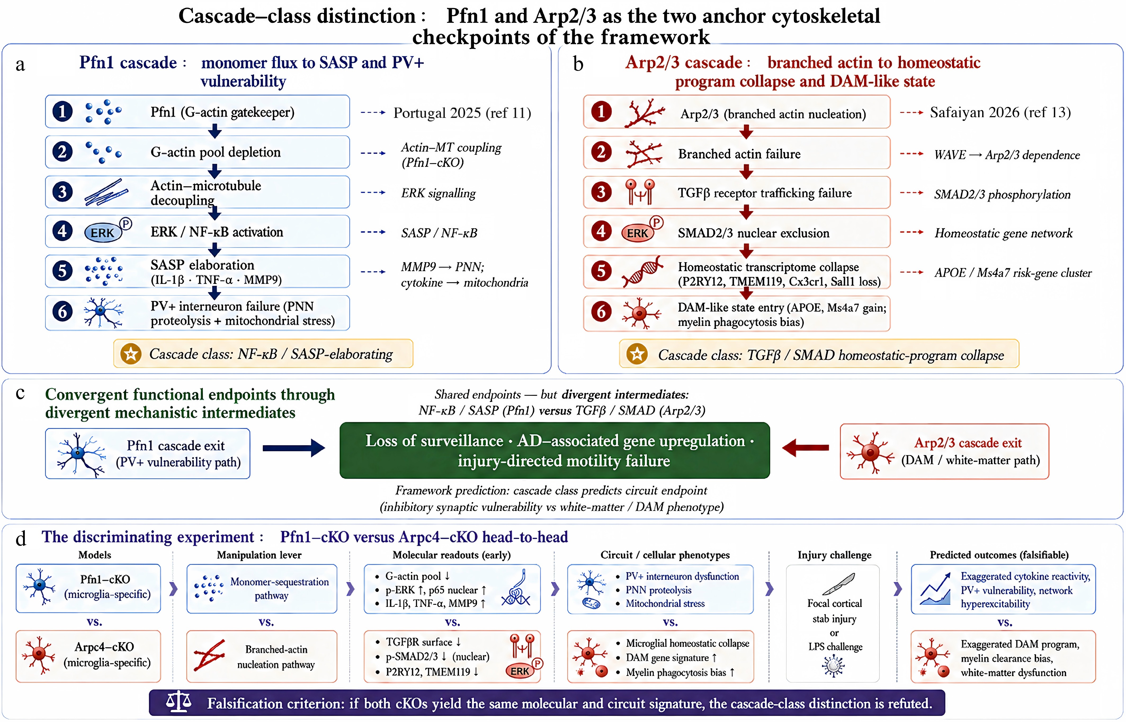
Figure 2.
Pfn1 and Arp2/3 as the two anchor cytoskeletal checkpoints of the cascade-class distinction framework. (a) The Pfn1 cascade: actin-monomer-handling failure drives sequential decoupling of actin-microtubule coordination, ERK/NF-κB activation, SASP elaboration (IL-1β, TNF-α, MMP9), and selective parvalbumin-positive (PV+) interneuron failure through combined MMP9-driven perineuronal-net proteolysis and cytokine-driven bioenergetic stress on fast-spiking interneurons; mechanistic anchors are the adult-onset microglia-specific Pfn1-cKO[11], microglial PNN proteolysis in AD brain[34], and PNN-buffered PV+ redox protection[35]. (b) The Arp2/3 cascade: branched-actin nucleation failure aborts TGFβ receptor trafficking, excludes phospho-SMAD2/3 from the nucleus, collapses the homeostatic transcriptional program (P2RY12, TMEM119, Cx3cr1, Sall1 loss), and drives DAM-like state entry with APOE/Ms4a7 gain and myelin-phagocytic bias; mechanistic anchor is conditional Arpc4-KO microglia[12]; constitutive Cx3cr1-Cre, so developmental and adult-onset contributions remain confounded, see Limitations. (c) The two cascades converge on shared functional endpoints, loss of surveillance, AD-associated gene upregulation, and injury-directed motility failure, through divergent mechanistic intermediates (NF-κB/SASP for Pfn1; TGFβ/SMAD for Arp2/3). The framework prediction is that cascade class, not the specific molecule, determines the circuit-level endpoint: inhibitory synaptic vulnerability for NF-κB/SASP-elaborating cascades, and white-matter/DAM phenotype for TGFβ/SMAD-collapsing cascades. (d) The discriminating experiment is a side-by-side adult-onset, microglia-specific Pfn1-cKO vs Arpc4-cKO comparison (sex-stratified) with five matched cortical readouts, MMP9 zymography, perineuronal-net integrity (WFA/aggrecan around PV+ cells), PV+ firing-fidelity together with mitochondrial respiration on PV-sorted cells, gamma-band local field potential power, and TGFβ/SMAD phosphorylation together with the homeostatic gene panel. The predicted-outcome matrix (cool blue = Pfn1-cKO cascade-positive; coral red = Arpc4-cKO cascade-positive) operationalises framework falsifiability: statistical equivalence of the two knockouts' multivariate readout profiles, tested within pre-specified margins under a genotype × sex model, would refute the cascade-class distinction. This experiment has not yet been performed.
The Arp2/3 complex defines a second, mechanistically distinct checkpoint class, the branched-actin cascade depicted in Fig. 2b. Whereas Pfn1 controls actin dynamics primarily through monomer availability and biases cytoskeletal stress responses toward ERK/NF-κB associated programs, the Arp2/3 complex regulates branched actin architecture and is linked to membrane trafficking and TGFβ/SMAD signaling[12]. This distinction reflects two orthogonal modes of cytoskeletal control: monomer-flux regulation vs filament architecture regulation.
Despite these mechanistic differences, the two cascades ultimately converge on a shared set of functional endpoints, reflecting partial overlap in downstream consequences rather than mechanistic redundancy and indicating that microglial function is governed by multiple entry points into a constrained state space. Combinatorial targeting across such nodes may therefore expand therapeutic flexibility, although this remains a conceptual inference.
At the level of downstream homeostatic regulation, disruption of Arp2/3-dependent TGFβ/SMAD signaling contributes to loss of homeostatic gene expression, including P2ry12, Tmem119, and Sall1[12]. In human cortical aging datasets, Arpc1a and Arpc1b decline alongside PFN1[2], suggesting coordinated vulnerability across actin-regulatory systems.
Compensatory increases in formin-mediated linear actin assembly observed in Arpc4-deficient microglia (including Daam1, Fmn1, and Fmnl2) further support a shift from surveillance to phagocytosis-biased states[12].
However, the Arpc4 dataset[12] lacks measurements of MMP9, PNN integrity, and PV+ function, meaning cross-cascade circuit effects remain unresolved. Accordingly, comparisons with the Pfn1 cascade should be considered hypothesis-generating rather than definitive.
To structure evaluation across studies, we propose an Evidence-Tier Score (ETS): ETS = Ncausal + Bcascade + Bstrat − Pdev. Here, Ncausal reflects the number of microglia-specific causal perturbation studies; Bcascade + 1.0 when three or more mechanistic levels downstream of a perturbation are supported; Bstrat + 0.5 when sex- or disease-stage-stratified effects are reported; and Pdev subtracts 0.5 when conclusions rely primarily on constitutive Cre drivers that may confound developmental and adult functions. This heuristic weighting system distinguishes causally supported checkpoints from emerging candidates, rather than serving as a fully formal statistical model. Nodes with ETS ≥ 2.0 are classified as higher-confidence checkpoints (Supplementary Table S2a; Fig. 1b), whereas nodes with ETS < 2.0 are considered lower-confidence or candidate checkpoints (Supplementary Table S2b; Fig. 1b).
The six-node Validated/Candidate (V/C) inventory is summarized in Fig. 1b, and ETS-derived weights are incorporated into a Bayesian-style prior (Fig. 1c). This inventory is an illustrative, evidence-prioritised starter set rather than an exhaustive partition of cytoskeletal control; the inclusion criteria, and nodes considered but not yet included (e.g., cofilin/LIMK, myosin II/MLCK, integrin-talin, TREM2-DAP12 coupling), are listed in Supplementary Table S1 (sheet S1.3). Together, this structure provides a transparent and falsifiable framework for prioritizing cytoskeletal checkpoints based on evidence strength and mechanistic leverage.
Collectively, Fig. 2 makes the case that diverging intermediate signaling, NF-κB/SASP vs TGFβ/SMAD, reflects partial convergence rather than mechanistic equivalence, and it is this distinction that supplies the empirical basis for the checkpoint taxonomy proposed here.
-
The Cellular Aging Map (CAM) framework provides a systems-level approach for operationalizing cytoskeletal checkpoint biology[18]. In CAM, aging is described as a data-driven dynamical process in which cellular trajectories can be classified as either recurrent and stable or wandering and dissipative. Using transformer-based masked language models trained on single-cell transcriptomes, where age is encoded as a learnable token, CAM generates an embedding space that captures molecular age, context-specific susceptibility, temporal drift, recurrence-divergence structure, and conditional entropy across the lifespan. For microglial biology, its value is therefore not only descriptive but also practical: it provides a principled way to define dissipation in a cell-type-specific manner and to ask whether, and under what conditions, that dissipation can be reversed.
Within this framework, a major substrate of microglial dissipation is cytoskeletal breakdown, specifically, the loss of coordinated actin-microtubule dynamics that prevents microglia from returning to a mechanically competent surveillance state. This idea is causally anchored by the Pfn1 phenotype, where disruption of cytoskeletal integrity leads to senescence-associated signaling, metabolic strain, and circuit-selective dysfunction[11,18,32]. CAM provides the language to quantify this loss, while mCytoMAP extends it by explicitly incorporating cytoskeletal state into the microglial aging landscape. Although chronological age remains an organizing variable, it is complemented by microglia-specific dimensions such as brain region, disease context, injury phase, sex, and cytoskeletal state, either experimentally defined (such as Pfn1 status) or inferred computationally (as a CytoState token).
With the four-axis decision framework established in Supplementary Box 1 (Fig. 1b), the next step is to apply it through the lens of sex stratification. Here, sex is not an optional refinement but a structural requirement. Transcriptomic and epigenetic trajectories of microglial aging differ between males and females[3,4], and these differences map directly onto the framework axes. Reversibility thresholds, dissipation load, and circuit consequences are not symmetric across sexes. The proposed assignment of sex-specific GTPase failure modes represents an integrative inference drawn from multiple independent lines of evidence[21−31,36]. However, it remains a working hypothesis that requires direct validation through sex-balanced, microglia-specific perturbations of RhoA, Rac1, and Cdc42. Supplementary Box 2 (Fig. 1c) formalizes this stratification across the four axes and embeds sex as a mandatory covariate throughout the framework.
Operationally, this stratification is implemented through sex- and context-specific Evidence-Tier Scores, defined as:
$ \mathrm{ETS\; (node|sex,context)=ETS}_{\mathrm{base}}-\mathrm{0.5\times\delta}_{\mathrm{sex}}-\mathrm{0.5\times\delta}_{\mathrm{stage}} $ In this formulation, penalties are applied when sex-stratified or context-specific causal evidence is lacking, with a minimum score of 0.5. The full directional sex × context matrix is presented in Supplementary Table S1, while Supplementary Table S3 provides a simplified, sex-aggregated version to maintain readability in the main text.
Once sex stratification is established, checkpoint prioritization can be resolved across biological contexts. The distinction between aging and Alzheimer's disease (AD) reflects different entry points into dysfunction rather than a simple linear progression. Both conditions involve the same cytoskeletal machinery, but they operate under different mechanical regimes. In aging, cytoskeletal erosion predominates: early PFN1 decline and widespread Rho GTPase dysregulation weaken structural adaptability before overt pathology appears. In AD, this already weakened system is exposed to additional mechanical and inflammatory stressors, including amyloid-associated stiffness, disease-associated microglial remodeling, and amplified inflammatory signaling.
Importantly, these contexts should not be interpreted as independent trajectories. Aging and AD engage the same underlying cytoskeletal systems under different constraints. Age-related reductions in monomer flux (PFN1) and branched actin architecture (ARPC1a/b) weaken the homeostatic baseline, while AD superimposes additional stressors such as amyloid fibril stiffness, vascular stiffening, and extracellular matrix remodeling. Crucially, the mechanical signals that activate microglial mechanosensing are not limited to amyloid pathology. Age-associated glycation cross-linking[37], lysyl oxidase activity[38], cerebrovascular stiffening[39], and extracellular matrix deposition during reactive astrogliosis[40] can all generate local stiffness gradients capable of activating Piezo1[36,41] independently of amyloid. Although whole-brain measurements suggest overall reduced stiffness with aging[31], these bulk values can obscure the local microenvironments sampled by microglial processes[42]. As a result, Piezo1 prioritization should be interpreted as dependent on context-specific modifiers, particularly vascular burden and extracellular matrix state, rather than as intrinsically amyloid-driven.
The integration of sex stratification (Supplementary Box 2) with context-dependent prioritization (Supplementary Box 3; Fig. 1d) yields a sex × context priority matrix that represents the central analytical output of the mCytoMAP framework. This matrix specifies which cytoskeletal checkpoint to target, in which population, and at which stage of disease, an intersection that is not explicitly resolved in current approaches. Supplementary Table S3 provides a simplified overview of these priorities across aging and AD contexts, while Supplementary Table S1 presents the full matrix across four strata (male/aging, female/aging, male/AD, female/AD). Each entry includes a sex-stratified Evidence-Tier Score, a governing hypothesis, a mechanistic rationale, and a proposed resolving experiment. Supporting worksheets in Supplementary Table S1 (sheets S1.2–S1.10) provide the operational state-metric set, the checkpoint inclusion criteria, the ETS bill-of-materials with independence count and sensitivity analysis, the sex-direction audit trail, the context-modifier proxies, the Pfn1 cascade evidence map, the likelihood-term and MVP design specification, and the biophysical parameter-anchoring table.
With this matrix established, the framework transitions from structured interpretation to quantitative implementation. Supplementary Box 4 (Fig. 3a) defines the mCytoMAP computational architecture that converts these priorities into a formal ranking system. At its biological foundation, this architecture distinguishes between two classes of cytoskeletal checkpoints. Monomer-flux regulators, such as Pfn1, control the availability of G-actin and link cytoskeletal disruption to inflammatory and senescence-associated outputs. Architecture-regulating nodes, such as the Arp2/3 complex, control filament organization and link cytoskeletal integrity to the maintenance of homeostatic gene expression through TGFβ/SMAD signaling. This distinction implies that restoring microglial competence requires coordinated intervention across both axes, rather than targeting individual nodes in isolation.
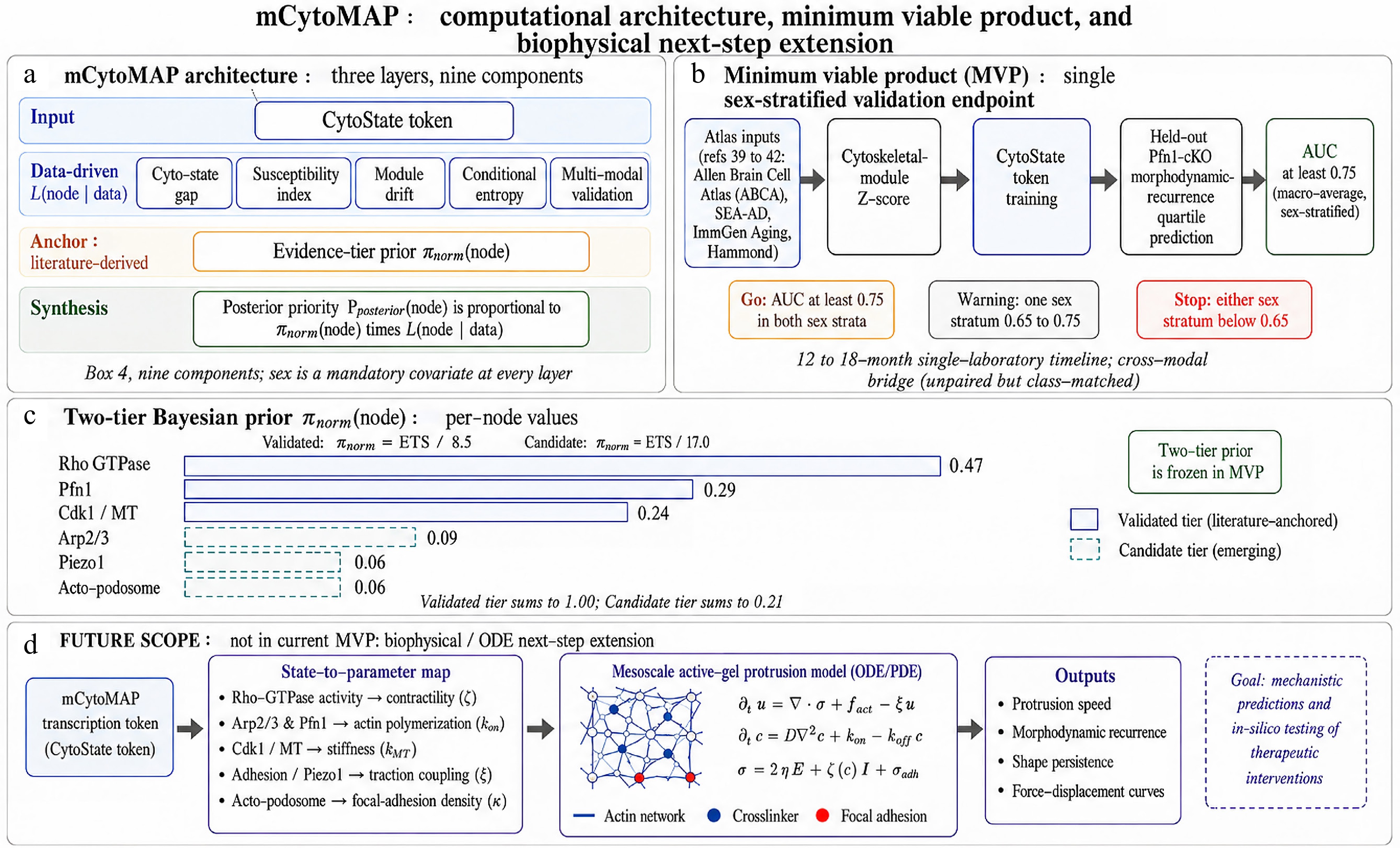
Figure 3.
Computational architecture, minimum viable product, and biophysical next-step extension of mCytoMAP. (a) mCytoMAP architecture organised in three layers and nine components (Supplementary Box 4). The Input layer carries the CytoState token (a learned multi-omic embedding of actin-microtubule coordination state). The Data-driven L(node|data) layer carries five components: cyto-state gap, susceptibility index, module drift, conditional entropy, and the multi-modal validation layer (live imaging + phosphoproteomics + electrophysiology). The Anchor layer carries the literature-derived Evidence-tier prior πnorm(node). The Synthesis layer carries the posterior priority Pposterior(node) $\varpropto $ πnorm(node) × L(node|data). Sex is a mandatory covariate at every layer. (b) The MVP single-endpoint validation pipeline (Supplementary Box 5b): four atlas inputs (Allen Brain Cell Atlas[43]; SEA-AD MTG[44]; ImmGen Aging[45]; Hammond developmental-adult mouse atlas[40]) → cytoskeletal-module Z-score → CytoState token training → held-out Pfn1-cKO morphodynamic-recurrence-quartile prediction (Portugal et al.[11]) → macro-average AUC ≥ 0.75 (sex-stratified, inverse-frequency class-weighted). Stop/go/ warning criteria are stated alongside the 12–18-month single-laboratory timeline; the bridge from snRNA-seq training to live-imaging validation is unpaired but class-matched (cross-modal). (c) The two-tier Bayesian-style prior πnorm(node) under the current rubric. Validated tier (cool blue solid bars; πnorm = ETS/ΣvalidatedETS = ETS/8.5): Rho GTPase network 0.47 (highest prior weight), Pfn1 0.29, Cdk1/MT 0.24 (sum = 1.00). Candidate tier (dashed teal bars; πnorm = ETS/(2 × Σvalidated ETS) = ETS/17.0): Arp2/3 0.09, Piezo1 0.06, actomyosin-podosome 0.06 (sum = 0.21). The two-tier prior is frozen across the MVP validation cycle; Bayesian updating resumes only after MVP closure. Falsifiability criterion: operational Spearman rank correlation ρ between ETS(node) and held-out Pposterior(node) across the six nodes must satisfy ρ ≥ 0.6 with P < 0.05 by two-sided permutation (n = 6 nodes; n_permutations = 720). (d) FUTURE SCOPE biophysical/ODE next-step extension, not in the current MVP. The mCytoMAP transcriptomic state (CytoState token) feeds a state-to-parameter map φ: CytoState → θ_bio = (σ_active, k_pol, κ_MT, γ_cortex, k_sub, n_adh), implemented as a supervised regression, a Gaussian-process emulator or neural-network surrogate trained on jointly acquired transcriptomic and biophysical reference cohorts, that maps each cytoskeletal-checkpoint node onto a unit-bearing mesoscale active-gel parameter: RhoA/ROCK activity → active cortical contractility σ_active (Pa); Arp2/3 and Pfn1 fluxes → effective filament polymerisation rate k_pol ([µmol·L−1]−1·s−1); Cdk1/MT remodelling → microtubule bending rigidity κ_MT (N m2); Piezo1 and adhesion activity → cortical tension γ_cortex (N m−1) and substrate-coupling stiffness k_sub (Pa m−1); actomyosin–podosome module → focal-adhesion density n_adh (µm−2). These parameters drive a coupled ODE/PDE active-gel protrusion model with explicit constitutive structure, overdamped force balance $\triangledown \cdot \sigma $_total + f_act − ξ u = 0 (inertia negligible in the cellular low-Reynolds-number regime), where u is the cytoplasmic velocity field (µm s−1), σ_total = σ_visc + σ_active the total stress tensor combining viscous (σ_visc = η$\triangledown $u) and active (σ_active = θ_bio[0]) contributions, f_act the local protrusive force density from barbed-end polymerisation, and ξ the cell-substrate friction; G-actin monomer-concentration dynamics ∂_t [G] = −k_pol·[G]·[B] + k_off·[B] + S_pool, where [B] is the free barbed-end concentration, k_pol the elongation rate, k_off the depolymerisation rate, and S_pool the Pfn1-gated monomer-release source, whose actin-network, crosslinker, and focal-adhesion topology produces outputs directly comparable to live morphodynamic readouts: protrusion speed, morphodynamic recurrence, shape persistence, and force-displacement curves. Polymerisation kinetics anchors: barbed-end addition rate k+ ≈ 11.6 (µmol·L−1)−1·s−1 for free ATP-actin[47]; k+(Pfn1-actin) ≈ 15–16 (µmol·L−1)−1·s−1 confirmed by single-filament TIRF[48]; Pfn1-G-actin Kd ≈ 0.1 µmol·L−1[46]; VCA-activated Arp2/3 nucleation rate knuc ≈ 2 × 10⁻⁴ (nmol·L−1)−1·s−1[45]; CapZ barbed-end capping rate kcap ≈ 3–4 (µmol·L−1)−1·s−1[49]. The goal is mechanistic predictions and in silico testing of therapeutic interventions; the biophysical layer is deferred as a multi-year deliverable outside the current Perspective scope, with explicit attention to (i) parameter identifiability under sloppy-manifold conditions typical of active-gel inference and (ii) the four-order-of-magnitude scale separation between monomer-kinetics timescales (ms–s) and morphodynamic-recurrence timescales (min–h), which is handled by quasi-steady-state reduction of the monomer-flux equations onto the slow protrusion manifold (Supplementary Box 5b closing paragraph; Section: Limitations and open questions).
The computational framework comprises seven core analytical components: the CytoState token, cyto-state gap, susceptibility index, module drift, conditional entropy, evidence-tier prior, and multi-modal validation layer, together with the minimum viable dataset specification and the explicit model-validation criteria, constitute the nine architectural components enumerated in Supplementary Box 4 (Fig. 3a). These components are organized into three layers. The input layer encodes cytoskeletal state through the CytoState token. The data-driven layer computes the likelihood term L(node|data) using molecular and functional information. The anchor layer incorporates prior knowledge from the literature through the Evidence-Tier Score. Together, these layers generate the posterior priority, Pposterior(node). Sex is incorporated as a mandatory covariate across all layers, ensuring that stratification is built into the system rather than added afterward.
A central feature of this architecture is the evidence-tier prior, π(node), which introduces a Bayesian-style weighting of each checkpoint. Because the two evidence tiers are normalised separately, validated-tier weights to a sum of 1.0 (π = ETS/8.5) and candidate-tier weights to a deliberately downweighted sum of 0.21 (π = ETS/17.0), π(node) is a tier-normalised weight applied up to proportionality in the posterior, not a single probability distribution summing to one across all six nodes; the six displayed weights therefore total 1.21 by construction rather than by arithmetic error. This prior is derived from the Evidence-Tier Score and interacts with the data-driven likelihood according to:
$ \mathrm{P}_{ \mathrm{posterior}} \mathrm{(node)\propto{\text{π}}(node)\times L(node|data)} $ In practice, π(node) increases with ETS. Nodes supported by stronger causal evidence (ETS ≥ 2.0) therefore have a greater influence on the final prioritization, affecting multiple components of the framework such as cyto-state gap estimation, susceptibility index allocation, module drift analysis, and entropy decomposition. By contrast, candidate nodes (ETS < 2.0) remain within the framework as testable hypotheses whose contribution can grow as new evidence becomes available. Because this prior is derived from the existing literature, it can be updated without generating new data, allowing the framework to evolve as the field progresses.
The framework can also be adapted to specific individuals or cohorts by defining conditional priors of the form π(node|sex, context, modifiers). These modifiers may include aging burden, amyloid burden, and vascular burden, which represent key features of mixed-pathology states. Introduced in Supplementary Box 3 and operationalized in the decision logic of Supplementary Box 6 (Fig. 1d), these modifiers allow checkpoint priorities to be dynamically adjusted. The resulting formulation,
Pposterior(node|profile) ∝ π(node|sex, aging burden, amyloid burden, vascular burden) × L (node|data), connects the conceptual framework directly to real-world decision-making.
Figure 3 turns the framework into an implementation plan. It opens with the full computational architecture (Fig. 3a) and then narrows to a single falsifiable test: the MVP validation pipeline in Fig. 3b couples atlas-based training and CytoState-token generation to one endpoint, prediction of morphodynamic recurrence in the Pfn1 conditional knockout model, held to a predefined macro-average AUC ≥ 0.75 on a projected 12–18-month timeline. The prior that weights this inference appears in Fig. 3c as the current two-tier Bayesian distribution of normalized node weights, while Fig. 3d sketches a future extension linking transcriptomic states to mesoscale biophysical models, explicitly designated as future scope, beyond the present implementation of the framework.
-
The mCytoMAP proposal and the cytoskeletal checkpoint paradigm carry important limitations that should be made explicit rather than implicit.
First, the generalizability of the checkpoint concept currently rests on a limited number of deeply characterized causal perturbations. At present, the most complete example is the Pfn1 knockout. The cascade defined for Pfn1, cytoskeletal collapse followed by ERK/NF-κB activation, induction of a senescence-associated secretory phenotype (SASP), and selective inhibitory synaptic vulnerability (Fig. 2a), is mechanistically coherent and well supported[11]. However, equivalent cascade-level evidence is still lacking for other key nodes, including Piezo1, the Rho GTPase network, and the Cdk1/microtubule module.
The Arp2/3 complex provides a second proof-of-principle perturbation through Arpc4 knockout, which links branched actin disruption to TGFβ receptor trafficking failure and collapse of the homeostatic transcriptional program (Fig. 2b)[12]. However, interpretation of this model is limited by the use of a constitutive Cx3cr1-Cre driver, which conflates developmental and adult-homeostatic failure. As a result, direct evidence that age-associated, adult-onset loss of Arp2/3 activity recapitulates this cascade remains inferential, supported indirectly by the decline of Arpc1a/b in aging human microglia[2]. A critical next step is therefore a tamoxifen-inducible, adult-onset Arpc4 knockout to isolate the relevant mechanism.
Additional limitations affect the interpretation of sex differences. The reported sex-neutral morphometric phenotype in the Arpc4 dataset is based on a very small sample size (approximately three animals per sex), which is underpowered for detecting meaningful sex effects. This limitation is particularly important given the central role of sex stratification in Supplementary Box 2 and should be treated as a hypothesis requiring replication in adequately powered, sex-balanced cohorts. The head-to-head discriminating experiment that would resolve the cascade-class vs molecule-specific question is depicted in Fig. 2d with its five-readout predicted-outcome matrix and falsification criterion.
More broadly, the Evidence-Tier Score (ETS) framework (Section: Cytoskeletal checkpoints as high-leverage Targetome nodes; Supplementary Box 4) makes this uneven evidence distribution explicit. The threshold defining 'validated' nodes (ETS ≥ 2.0) is a pragmatic calibration based on the current literature and is expected to evolve as new data accumulate.
Within this system, Pfn1 (ETS 2.5; cascade-depth-bonus-driven), Rho GTPase network (ETS 4.0; highest prior weight in the framework, anchored by three independent causal microglia-specific perturbation studies[26−28], and Cdk1/MT remodeling (ETS 2.0, at threshold) are classified as validated, whereas Arp2/3 (ETS 1.5; constitutive-Cre penalty applied), Piezo1 (ETS 1.0), and the actomyosin-podosome module (ETS 1.0) remain in the candidate tier. These assignments propagate directly into the Bayesian prior weights shown in Fig. 3c.
The elevated ranking of the Rho GTPase network deserves particular attention. Its positioning as the highest-priority node reflects the number of causal perturbation studies available[26−28] and the depth of downstream mechanistic evidence. However, these studies originate largely from the same laboratory, and therefore do not fully satisfy the criterion of independent replication. This is an acknowledged limitation. Importantly, the ETS ranking would remain stable even if only a subset of these studies were independently reproduced, but independent validation remains a priority. This concentration of evidence is disclosed transparently, including the contribution of the authors' laboratory to the relevant dataset.
A related limitation concerns the scope of downstream circuit effects. The Pfn1 cascade produces a characteristic 'dual-hit' signature affecting parvalbumin-positive (PV+) interneurons, combining MMP9-mediated degradation of perineuronal nets with cytokine-driven metabolic stress (Fig. 2a)[11,34,35]. The framework predicts that this pattern reflects a general property of NF-κB/SASP-driven cascades, rather than being specific to Pfn1 itself. However, this prediction has not yet been directly tested. The Arpc4 cascade, which operates through TGFβ/SMAD disruption (Fig. 2b) rather than NF-κB activation, is not expected to produce the same PV+ phenotype, but this distinction remains hypothetical. A direct comparison between adult-onset Pfn1 and Arpc4 perturbations (Fig. 2d), measuring MMP9 activity, perineuronal net integrity, PV+ interneuron physiology, mitochondrial function, and gamma oscillations, would provide a decisive test of this prediction. To keep that test falsifiable under realistic power, divergence is scored not as 'identical vs different' but as a pre-registered multivariate contrast, Pfn1-like vs Arpc4-like profile classification, with statistical equivalence on the shared intermediate markers evaluated within predefined margins (two one-sided tests) under a genotype × sex model, so that underpowered null results cannot be mistaken for cascade convergence.
A third limitation concerns context-dependent prioritization of Piezo1 across biological contexts. The framework distinguishes between a lower priority in aging and a higher priority in Alzheimer's disease, driven primarily by vascular and mechanical modifiers (Supplementary Box 3; Supplementary Table S3).
While several age-related processes, such as AGE/RAGE cross-linking of long-lived ECM, lysyl-oxidase activity, cerebrovascular and basement-membrane stiffening, reactive astrogliosis, and ECM deposition, could in principle generate local pericellular stiffness gradients in the kPa range that activate Piezo1[36] in the absence of amyloid, this has not been directly demonstrated in vivo. Existing evidence for microglial Piezo1 activation is largely restricted to Alzheimer’s disease models[16]. A key experiment would therefore test Piezo1 function in aged, amyloid-free systems, combining microglia-specific genetic perturbation with direct measurements of local tissue stiffness and Ca2+ signaling.
The concept of reversibility also remains incompletely resolved. In this framework, reversibility is defined operationally as the ability of microglia to re-enter a competent surveillance manifold state, restoration of process motility, Ca2+ microdomain surveillance, and phagocytic competence, rather than as full molecular rejuvenation. While state-level recovery is supported by approaches such as microglial depletion and repopulation[20] or senolytic clearance, direct evidence for node-level rescue restoring specific cytoskeletal regulators in aged microglia is lacking.
Existing rescue studies relate to developmental models rather than aging. A critical experiment would be the restoration of Pfn1 or Arpc1a/b function in aged microglia, combined with measurements of morphodynamics and inflammatory output, to determine whether checkpoint-level intervention can reopen functional state space.
The proposed sex-specific failure modes of the Rho GTPase network (Supplementary Box 2; Supplementary Table S3) also remain hypothetical. The framework suggests that males preferentially engage a Rac1-driven surveillance-failure axis, whereas females shift toward a RhoA/ROCK-driven contractile state. This assignment is based on convergent evidence[21−31] but has not yet been directly tested. Validation will require sex-balanced microglia-specific perturbation studies targeting RhoA, Rac1, and Cdc42, combined with matched structural, signaling, and circuit readouts, as specified in the Priority Research Agenda.
To maintain clarity in the main text, the full sex × context directional matrix, including per-cell, sex-stratified ETS (node|sex, context) values, governing H-tags, mechanistic rationale, and proposed resolving experiments, is provided in Supplementary Table S1, while Supplementary Table S3 (Section: mCytoMAP: A CAM-inspired framework for cytoskeletal target prioritization) presents a simplified, sex-aggregated version with sex included as a covariate. Two key experimental strategies are required to resolve this limitation: (i) sex-balanced, microglia-specific perturbations of RhoA, Rac1, and Cdc42 in adult cortex, with matched readouts of morphodynamics, contractility, protrusion structure, and inhibitory synapse interaction; and (ii) sex-stratified single-cell measurements of active Rho-family GTPase signaling in disease-stage human microglia (Priority Research Agenda items 2 and 6).
A further limitation concerns the practical constraints on CytoState token training (Supplementary Box 4). While the CAM age token was developed on large, well-annotated single-cell datasets with bulk-like coverage of cell populations, microglial datasets of comparable depth and consistent cytoskeletal metadata are not yet standardized across consortia. The minimum data specification in Supplementary Box 4 sets a clear standard, but achieving it for sex-stratified, region-resolved, multi-stage microglial cohorts requires coordinated data generation that exceeds what any single laboratory can provide. mCytoMAP is therefore also a call for open, harmonized data standards for microglial aging studies.
To address these constraints, the mCytoMAP Minimum Viable Product (MVP) protocol defined in Supplementary Box 5 (Fig. 3b) reduces validation to a single testable endpoint: sex-stratified prediction of morphodynamic recurrence in the Pfn1-cKO model (AUC ≥ 0.75), using existing atlas datasets (Allen Brain Cell Atlas, SEA-AD, ImmGen Aging, Hammond atlas[43−46]) for training and experimentally derived morphodynamic data for validation. This design makes the framework experimentally testable within a 12–18-month timeframe in a single laboratory setting.
The proposed biophysical extension (Fig. 3d; FUTURE SCOPE) introduces an additional layer of complexity and remains explicitly outside the current scope. This extension would couple mCytoMAP-derived transcriptomic states to a mesoscale active-gel model of protrusion dynamics, using established actin polymerization kinetics[47−51]. However, this approach is currently limited by insufficient data to robustly fit morphodynamic recurrence trajectories. In addition, two major theoretical constraints must be addressed: (i) parameter identifiability, as active-gel systems exhibit 'sloppy' parameter manifolds in which multiple parameter combinations produce indistinguishable outputs, necessitating formal sensitivity analysis (e.g., Sobol indices); and (ii) timescale separation, as fast monomer-level kinetics (milliseconds-seconds) must be reduced to slower morphodynamic dynamics (minutes-hours), typically through quasi-steady-state approximations. For these reasons, the biophysical layer remains a future extension contingent on MVP validation.
Finally, the distinction between aging and AD-context, as formalized in Supplementary Box 3 and Supplementary Table S3, relies on a stage-progression model that may not be universal: genetic risk backgrounds (APOE ε4, TREM2 variants), environment, and co-morbidities can compress or bypass disease trajectories, potentially shifting checkpoint priorities.
In addition, the supporting evidence base is asymmetric across species and experimental systems: age-related declines in PFN1 and Arpc1a/b are primarily observed in cross-sectional human datasets[2], whereas causal cascade evidence derives mainly from mouse models[11,12]. Consequently, it remains unresolved whether cytoskeletal dysfunction is a causal driver of disease progression or a permissive condition. More broadly, cytoskeleton-first ordering is one of several admissible causal graphs: mitochondrial/ATP limitation, lysosomal-lipid overload driving DAM entry, or epigenetic drift could each sit upstream of cytoskeletal remodelling rather than downstream of it. These orderings are discriminable; metabolic or lysosomal rescue that restores morphodynamic competence without cytoskeletal manipulation would favour them, whereas forced re-expression of a cytoskeletal node that overrides such deficits would favour the control-layer model, and the framework is meant to be tested against, not assumed over, these alternatives.
Two key experiments would resolve this uncertainty: (i) a longitudinal human dataset linking cytoskeletal state to disease-stage transitions in sex-stratified cohorts; and (ii) a pre-emptive rescue experiment, restoring cytoskeletal function (e.g., Pfn1 or Arpc1a/b) in aged microglia prior to amyloid challenge, to test whether preserved mechanical competence delays or prevents entry into disease-associated microglial (DAM) states.
Accordingly, the prioritization matrix presented in Supplementary Table S3 should be viewed as adaptive rather than fixed, and updated as stage-resolved and genetically stratified data become available, including explicit incorporation of genetic background in future iterations.
-
A cytoskeleton-centered view of the microglial targetome redefines therapeutic opportunity in aging and dementia. The aim is not to suppress terminal inflammatory output, but to preserve or restore the mechanical competence, the ability of microglia to maintain coordinated sensing, movement, and response, so that cells remain within an adaptive, recoverable state space. Because microglia operate at the intersection of plaque sensing, synaptic remodeling, metabolic support, and tissue repair, failure in cytoskeletal organization can propagate across multiple biological and pathological scales[7,11,16,18,33], making the cytoskeletal control layer both an upstream vulnerability and a high-leverage intervention point with broad downstream consequences.
Not all cytoskeletal molecules are suitable therapeutic entry points. Many regulators are pleiotropic and widely expressed. The checkpoint framework formalized across Supplementary materials Boxes 1–6 and synthesized in Supplementary Tables S2a, S2b; S3, with the full sex-stratified hypothesis matrix in Supplementary Table S1, imposes selectivity around intervention logic precisely because it demands more than abundance or age-sensitivity: it requires integration of structure and signaling, stage- or context-specific relevance, sex-stratified failure mode analysis, and functional rescue across scales.
This framework is made operational through clearly defined components. Supplementary Box 5 specifies the minimum viable product (MVP), a single, sex-stratified validation endpoint that allows the framework to be tested in 12–18 months. Supplementary Box 6 translates contextual modifiers (introduced in Supplementary Box 3) into a decision flowchart, enabling prioritization across mixed-pathology conditions (aging, amyloid, vascular burden).
At the implementation level, Fig. 3 serves as the central deliverable, integrating the nine-component mCytoMAP architecture (Fig. 3a), the MVP validation pipeline with go/warning/stop criteria (Fig. 3b), the two-tier Bayesian-style prior πnorm(node) (Fig. 3c), and the FUTURE-SCOPE biophysical extension (Fig. 3d). Together, these elements establish a framework that is not merely conceptual, but explicitly buildable and falsifiable.
Testing this architecture requires multi-modal integration across scales, including longitudinal single-cell and spatial transcriptomics, phosphoproteomics, biomechanical profiling, intravital imaging, and region-specific electrophysiology. Critically, validation criteria must go beyond normalization of molecular markers. Instead, interventions should be evaluated by their ability to restore core microglial functions: surveillance, recurrence, injury-directed responsiveness, and synaptic support.
Looking forward, the field should shift from ranking targets based on differential expression toward ranking based on functional control capacity. Specifically, targets should be ranked first according to their capacity to preserve reversibility, reduce dissipation, and protect circuit function, rather than by differential expression magnitude. Only after this functional prioritization should druggability be considered as a development filter rather than the discovery premise. This approach emphasizes restoring adaptive microglial plasticity, rather than pushing cells into artificially quiescent or suppressed states.
Within this framework, cytoskeletal checkpoints are emerging as state-control nodes whose relevance is supported by causal genetics[11,12,26−28], functional rescue[33], and sex-dimorphic profiling[21,22].
The design principle follows directly: rank targets by control leverage, reversibility, and access to functional state space; enforce sex stratification at every level; and organize intervention strategies around restoring the architecture that governs microglial state transitions. Evidence weighting is explicitly incorporated through the Evidence-Tier Score (ETS) and its associated Bayesian-style prior π(node) (Supplementary Box 4 row 6). In this system, Validated nodes (Pfn1, Rho GTPase network, Cdk1/MT) drive posterior prioritization, while Candidate nodes (Arp2/3, Piezo1, actomyosin-podosome) remain as structured, testable hypotheses. Because this prior is formalized and updateable, the framework is inherently auditable and falsifiable: any mismatch between prior expectation and data-driven posterior triggers a recalibration of the scoring system.
-
The following six experimental priorities derive directly from the framework logic (Supplementary Boxes 1–6; Supplementary Tables S1, S2a, S2b and S3:
Sex-stratified Pfn1 threshold determination
-
Define the PFN1 expression trajectory and morphodynamic collapse threshold in male and female human microglial aging cohorts using paired scRNA-seq and phosphoproteomics to identify the sex-specific cyto-state gap at which SASP activation becomes irreversible.
Sex-resolved mapping of Rho GTPase signaling in DAM states
-
Apply the phosphoproteomic workflow of Socodato & Relvas[12] to sex-stratified DAM vs homeostatic microglia from AD mouse models and human donor tissue, to compare contractility (ROCK axis) vs protrusion (PAK/cofilin axis) signaling biases in male and female microglia, without pre-assigning directional hypotheses.
CytoState token training and MVP validation
-
Train the mCytoMAP CytoState token using the atlas datasets specified in Supplementary Box 4, and validate predictions using the Pfn1-cKO morphodynamic dataset[11], following the MVP protocol defined in Supplementary Box 5b (AUC ≥ 0.75 endpoint).
Piezo1 context-priority validation
-
Conduct head-to-head comparisons of Piezo1 function in sex-stratified aging-only vs early-AD contexts (as defined in Supplementary Box 3; Supplementary Table S3), including amyloid-independent stiffness sources, with paired cytoskeletal and circuit-level readouts.
mCytoMAP-guided functional rescue testing
-
Test whether reducing the cyto-state gap (Supplementary Box 4) restores surveillance and synaptic support in aged microglia, using node-level perturbations (e.g., Pfn1 or Arpc1a/b re-expression) across different senescence depths.
Sex-balanced Rho GTPase perturbation panel
-
Experimentally resolve the directional hypotheses outlined in Supplementary Box 2; Supplementary Table S3 by performing sex-balanced perturbations of RhoA, Rac1, and Cdc42, with matched structural, signaling, and circuit readouts.
-
The framework presented here is intentionally incomplete, but explicitly designed to be testable. Its primary contribution is not to offer a final model of microglial aging, but to reframe the problem in a way that is mechanistically structured and experimentally actionable. As shown in Fig. 4. By shifting the field away from descriptive lists of age-associated molecular changes toward a state-control architecture centered on cytoskeletal checkpoints, it provides a unifying logic that links molecular alterations to cellular behavior and, ultimately, to circuit-level consequences. Crucially, the framework makes its assumptions transparent and anchors each major claim to defined experimental tests, including validation of checkpoint nodes, resolution of sex-specific failure modes, context-dependent prioritization across aging and disease, and demonstration of functional rescue. This explicit coupling of concept and experiment allows the field to move from descriptive accumulation toward iterative hypothesis testing and systematic refinement. In this sense, mCytoMAP should be understood not as a fixed model, but as a living scaffold that is expected to evolve as new causal evidence emerges. More broadly, the framework implies a shift in therapeutic logic: rather than asking which molecules change with age, the critical question becomes which regulatory nodes control access to functional cellular states, and under what conditions that access can be restored. This reorientation places intervention upstream, at the level of mechanisms that preserve or recover adaptive plasticity, rather than downstream suppression of pathological outputs. Ultimately, the central prediction is both simple and falsifiable: if cytoskeletal checkpoint modulation can reopen constrained state space in aged microglia, then meaningful recovery of function, extending from cellular behavior to circuit integrity, should be achievable. If borne out experimentally, this would establish cytoskeletal control architecture not only as a conceptual framework for understanding aging, but as a practical foundation for mechanism-driven therapeutic strategies in neurodegenerative disease.
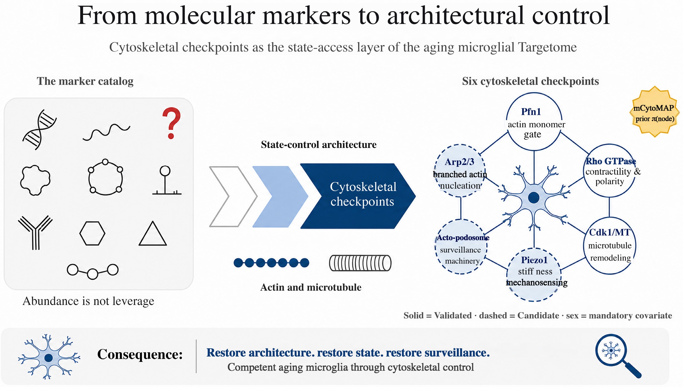
Figure 4.
From molecular markers to architectural control.
-
Not applicable.
-
The authors confirm their contributions to the paper as follows: conceptualization: Mendes Pinto I, Correia M, Relvas JB, Socodato R; methodology: Mendes Pinto I, Socodato R; data curation, visualization: Beça P, Galvão J; validation: Portugal CC; resources: Relvas JB, Socodato R; writing-original draft: Mendes Pinto I, Socodato R; writing-review & editing: Mendes Pinto I, Beça P, Correia M, Moreira JT, Galvão J, Portugal CC, Relvas JB, Socodato R; funding acquisition: Mendes Pinto I, Portugal CC, Relvas JB, Socodato R; supervision: Mendes Pinto I, Relvas JB, Socodato R. All authors reviewed the results and approved the final version of the manuscript.
-
No new primary data are reported in this perspective. The public datasets named in the mCytoMAP minimum-data specification (Supplementary Box 4) and Minimum Viable Product protocol (Supplementary Box 5) are openly accessible through their respective consortia and citations: Allen Brain Cell Atlas, single-cell transcriptomic data for the human cortex[43]; SEA-AD MTG multimodal Alzheimer's-disease atlas[44]; ImmGen Aging human microglial atlas[45]; Hammond developmental-adult mouse microglial atlas[46]. Adult-onset, microglia-specific Pfn1 knockout morphodynamic data[11] are available upon reasonable request; to enable independent benchmarking of the MVP, the derived morphodynamic features, recurrence scores and quartile labels, per-cell metadata (sex, age, region), and the locked train/validation/test split underlying the AUC ≥ 0.75 endpoint will be released with the first MVP validation study even where raw imaging volumes preclude direct hosting. Conditional Arp2/3 (Arpc4-cKO) data are available through the corresponding author of Safaiyan et al. 2026 (EMBO Reports 27:1696–1719). The mCytoMAP architecture, Evidence-Tier Score (ETS) rubric, two-tier Bayesian prior πnorm(node), and the MVP validation protocol are fully specified in this Perspective (Supplementary materials Boxes 1–6; Supplementary Tables S1, S2a, S2b and S3). A reference implementation of the literature-derived layer of mCytoMAP, the Evidence-Tier Score (ETS) rubric and the two-tier Bayesian-style prior πnorm(node), is provided as a standalone, MIT-licensed code archive (Python ≥ 3.10, standard library only), deposited at https://github.com/renatosocodato/mcytomap-core (release v1.2.0, commit 0e83bbe) and archived at Zenodo https://doi.org/10.5281/zenodo.20848905; running its single 'reproduce_values' entry point regenerates the underlying per-node values, not merely the plotted figures in Supplementary Tables S2a, S2b; Fig. 3c, and the per-cell ETS (node|sex, context) values in Supplementary Table S1. The data-dependent mCytoMAP components (CytoState token, cyto-state gap, susceptibility index, module drift, conditional entropy) require the snRNA-seq atlas training specified in the Minimum Viable Product protocol (Supplementary Box 5), and a complete reference implementation will accompany the first MVP validation study.
-
The authors declare that they have no conflict of interest.
-
accompanies this paper online at: https://doi.org/10.48130/targetome-0026-0035.
- Supplementary Table S1 Sex × context hypothesis matrix for cytoskeletal checkpoints in the aging microglial Targetome.
- Supplementary Table S2 (a) Validated cytoskeletal checkpoints in the aging microglial Targetome (ETS ≥ 2.0). (b) Candidate cytoskeletal checkpoints in the aging microglial Targetome (ETS < 2.0).
- Supplementary Table S3 Sex-aggregated context-priority table for cytoskeletal checkpoints (sex as covariate).
- Supplementary Box 1 A four-axis targetome decision framework.
- Supplementary Box 2 Sex stratification as a hypothesis-generating framework in cytoskeletal checkpoint biology.
- Supplementary Box 3 Cytoskeletal checkpoint priorities: aging, alzheimer's, and contextual modifiers.
- Supplementary Box 4 mCytoMAP computational architecture, data specifications, and validation criteria.
- Supplementary Box 5 Data-requirement → existing-resource mapping for the mCytoMAP MVP.
- Supplementary Box 6 Decision flowchart for mixed-pathology checkpoint prioritization (aging + amyloid + vascular).
- Copyright: © 2026 by the author(s). Published by Maximum Academic Press on behalf of China Pharmaceutical University. This article is an open access article distributed under Creative Commons Attribution License (CC BY 4.0), visit https://creativecommons.org/licenses/by/4.0/.
-
About this article
Cite this article
Mendes Pinto I, Beça P, Correia M, Moreira JT, Galvão J, et al. 2026. From molecular markers to architectural control: cytoskeletal checkpoints as the decisive state-access layer of the aging microglial targetome. Targetome 2(4): e038 doi: 10.48130/targetome-0026-0035 

From molecular markers to architectural control: cytoskeletal checkpoints as the decisive state-access layer of the aging microglial targetome
- Received: 14 March 2026
- Revised: 29 June 2026
- Accepted: 17 July 2026
- Published online: 20 August 2026
Abstract: The molecular complexity of microglial aging, extensively catalogued across transcriptomes, proteomes, and single-cell atlases, has not translated to mechanistic clarity about what to target or when. If anything, the growing list of altered molecules has made the prioritisation problem more difficult. We argue this reflects a systematic mismatch between the level at which dysfunction is measured and the level at which it is organised. In this perspective, the microglial cytoskeleton, the coordinated actin-microtubule machinery sustaining morphodynamic plasticity, mechanosensing, phagocytic capacity, and synaptic support, should not be viewed as a downstream readout of aging. Rather, it constitutes the control architecture whose failure, we propose, may initiate rather than merely accompany it. Accordingly, the most informative dementia targets are not age-associated molecules per se, but cytoskeletal checkpoints: regulatory nodes whose perturbation reorganises actin-microtubule coupling and, through this organization, propagates across signalling, metabolism, and circuit output. The microglial targetome should therefore be organised not as a catalogue of altered molecules but as a map of control leverage embedded within cytoskeletal architecture. Two proof-of-principle checkpoints establish this logic. Acute, microglia-specific loss of Profilin-1 (Pfn1), an actin-availability gatekeeper, is sufficient to collapse morphodynamic responsiveness and trigger an ERK/NF-κB-driven senescence-associated secretory phenotype, whose cytokine and MMP9 output selectively disrupts GABAergic synaptic function. By contrast, deletion of Arpc4 defines a mechanistically orthogonal axis: loss of branched actin nucleation impairs TGFβ receptor trafficking, prevents nuclear accumulation of SMAD2/3, and drives entry into a DAM-like state. This cascade appears to operate independently of ERK/NF-κB signaling, a prediction that remains to be tested directly, and establishes a distinct class of cytoskeletal checkpoint. Together, these two examples anchor a two-axis checkpoint taxonomy and underpin mCytoMAP, a sex-stratified, context-resolved decision framework ranking cytoskeletal targets according to reversibility, dissipation load, cell-state specificity, and circuit consequence. Within this framework, the central challenge becomes tractable: identifying which checkpoints can redirect the aging microglial state, and determining the biological contexts, including sex and disease stage, in which such interventions are most effective.
-
Key words:
- Microglia /
- Aging /
- Cytoskeleton /
- Profilin-1 /
- Dementia /
- Mechanobiology /
- Targetome /
- mCytoMAP /
- Perineuronal nets /
- Senescence-associated secretory phenotype /
- Sex stratification