












-
The respiratory chain has long been recognized as a central bioenergetic system, yet its potential as a therapeutic target is only beginning to be fully realized[1,2]. Once viewed primarily as a linear electron transfer pathway sustaining ATP production, it is now emerging as a highly druggable and programmable network capable of regulating cellular fate[2]. In aerobic organisms, the respiratory chain orchestrates the transfer of electrons from metabolic substrates to molecular oxygen, generating a proton motive force that drives ATP synthesis through oxidative phosphorylation (OXPHOS)[3]. In mitochondria and many aerobic bacteria, this membrane-embedded electron transport system maintains cellular homeostasis by coupling electron transfer to ATP synthesis[3]. Importantly, perturbations of respiratory function produce fundamentally different outcomes depending on biological context: while disruption is often lethal to pathogens, in mammalian cells it can rewire metabolism, modulate signaling pathways, or induce programmed cell death[4]. These dual consequences position the respiratory chain not merely as a metabolic engine, but as a versatile therapeutic control system.
Historically, pharmacological targeting of the respiratory chain originated from natural toxins. Classical inhibitors such as rotenone and piericidin A (Complex I), antimycin A (Complex III Qi-site), and oligomycin (ATP synthase Fo domain) provided early mechanistic insights into OXPHOS[5−7]. These molecules established a foundational paradigm: interruption of electron flow frequently leads to ATP depletion, increased reactive oxygen species (ROS) production, and metabolic failure[8,9]. This principle was later translated into antimicrobial therapy with the development and subsequent clinical adoption of bedaquiline for multidrug-resistant tuberculosis, which demonstrated that respiratory complexes can serve as clinically viable drug targets when sufficient selectivity is achieved[10−12]. In this traditional framework, therapeutic efficacy is primarily driven by bioenergetic deprivation.
More recently, however, a conceptual shift has reshaped the field. Small molecules targeting the respiratory chain are no longer limited to inducing energy collapse but are increasingly exploited to reprogram cellular metabolism[2,4]. In cancer, where glycolysis-centric models once dominated, it is now evident that many tumors retain functional mitochondrial respiration to support biosynthesis and redox homeostasis, often in a context-dependent and dynamically regulated manner[2,13]. Compounds such as Ophiobolin A transiently hyperactivate mitochondrial respiration, leading to selective bioenergetic collapse in cancer cells[14], while widely used drugs such as metformin act as mild but clinically relevant Complex I (CI) modulators, altering the NADH/NAD+ balance[15], AMPK signaling, and systemic metabolic homeostasis[16−18], thereby producing systemic glucose-lowering effects[19]. These findings highlight a modern pharmacological paradigm in which respiratory modulation, rather than outright inhibition, serves as a strategy for precision metabolic intervention.
Advances in cryo-electron microscopy (cryo-EM) and cryo-electron tomography (cryo-ET) have played a transformative role in enabling structure-guided targeting of the respiratory chain[20,21]. High-resolution structures of individual complexes, including mitochondrial bc1, mammalian CI, and ATP synthase dimers[22−24], as well as fully assembled supercomplexes (SCs) such as I–III–IV[25,26], have revealed a wealth of previously inaccessible features, including quinone-binding pockets, lipid-mediated interfaces, proton channels, and allosteric cavities[27,28]. These structural insights are redefining druggability by exposing non-canonical binding sites beyond traditional active centers[29,30].
Despite these advances, major challenges remain in translating respiratory targeting into safe and effective therapies. The high evolutionary conservation of respiratory complexes raises significant concerns regarding host toxicity[31,32], particularly when targeting mitochondrial systems in eukaryotic cells. In addition, microorganisms can exploit alternative respiratory pathways, such as alternative oxidase (AOX)[33] and cytochrome bd oxidase[34], to bypass inhibited complexes and develop drug resistance. Furthermore, mitochondrial perturbation elicits pleiotropic downstream responses, including oxidative stress, integrated stress signaling, immunometabolic remodeling, and transcriptional reprogramming[35,36]. Addressing these challenges requires an integrated understanding that combines structural biology, biophysics, chemical biology, systems-level analysis, and rational drug design.
In this review, we present a comprehensive framework for understanding pharmacological targeting of the respiratory chain across CI–V, encompassing antibacterial, antifungal, antiprotozoal, anticancer, and metabolic applications. We examine the mechanistic principles underlying small-molecule action, discuss determinants of selectivity between microbial and mammalian systems, and evaluate strategies to overcome bypass respiration and drug resistance. We further explore emerging opportunities in targeting respiratory SCs and allosteric regulatory sites, integrating insights from high-resolution structural studies and in situ mitochondrial architecture. Finally, we outline future directions for precision metabolic therapies, highlighting how modulation of the respiratory chain may enable tailored interventions across a broad spectrum of diseases. Taken together, the respiratory chain emerges not simply as a metabolic machine, but as a programmable bioenergetic switchboard, one that can be inhibited to eliminate cells, modulated to reshape metabolism, or rewired to control cellular identity.
-
The respiratory chain has traditionally been viewed as a central bioenergetic system responsible for ATP production through OXPHOS[3]. However, growing evidence now supports a broader conceptualization in which the respiratory chain functions not merely as an energy-producing system, but as a dynamic regulatory hub that integrates metabolism, redox balance, and signaling pathways[1,37]. This expanded perspective has profound implications for pharmacology[2], as it reframes the respiratory chain from a passive metabolic process into an actively druggable system capable of controlling cellular states.
A fundamental shift has therefore emerged in the therapeutic targeting of respiration[4]. Targeting the respiratory chain is no longer solely about inhibiting energy production but about controlling cellular metabolic states. In this context, small molecules acting on respiratory complexes can be broadly categorized into three functional modes, each associated with distinct mechanistic and biological outcomes (Fig. 1).
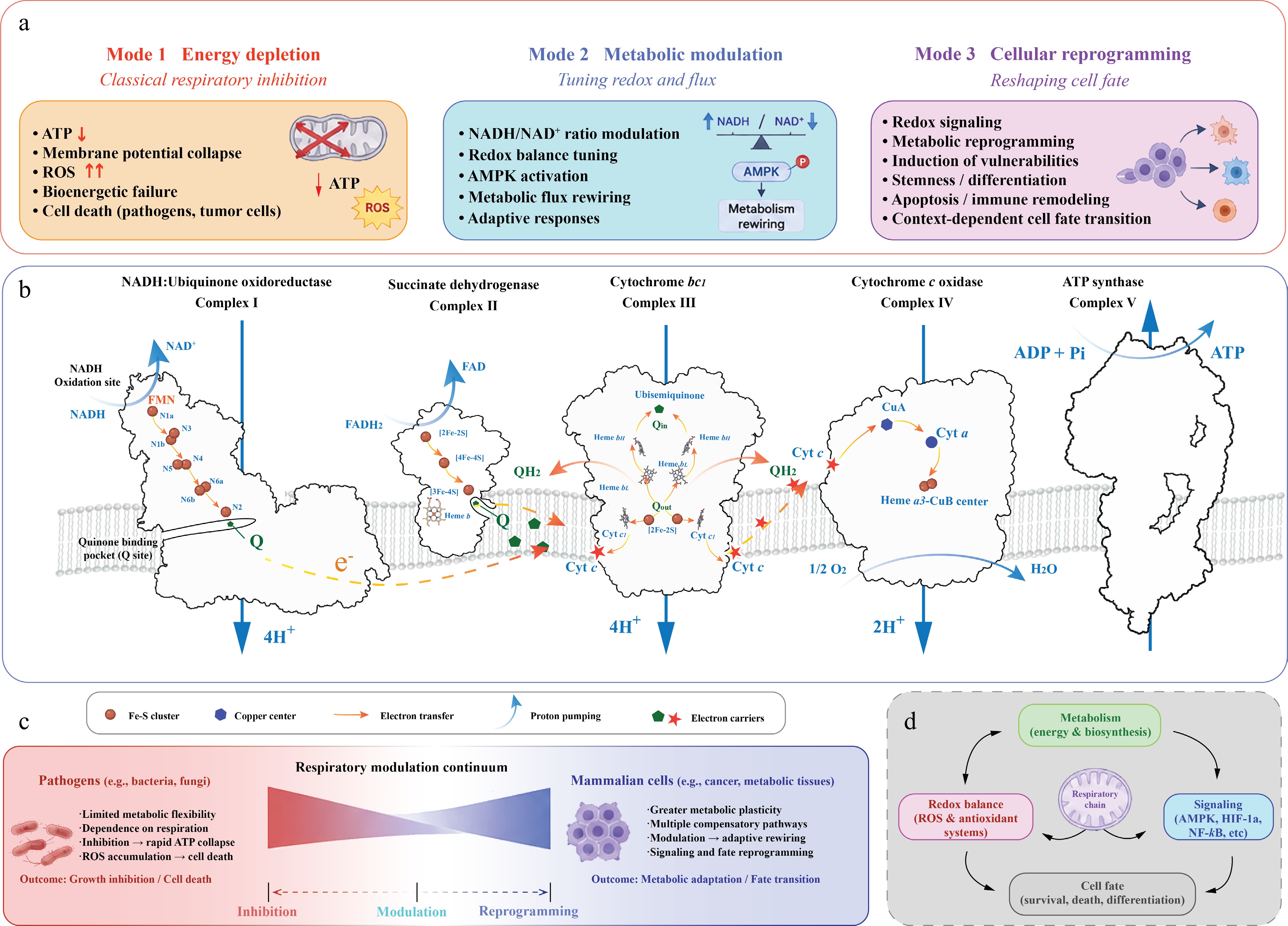
Figure 1.
The respiratory chain as a programmable therapeutic system. (a) Three modes of therapeutic targeting. Small molecules targeting the respiratory chain can be broadly categorized into three functional modes with distinct biological consequences. Mode 1: Energy depletion represents the classical paradigm of respiratory inhibition, in which disruption of electron transfer or proton coupling leads to ATP depletion, membrane-potential collapse, ROS accumulation, and cell death. Mode 2: Metabolic modulation involves partial or adaptive modulation of respiratory activity, leading to altered redox balance, NADH/NAD+ ratio, AMPK activation, and metabolic rewiring without immediate cytotoxicity. Mode 3: Cellular reprogramming extends beyond metabolic control to influence signaling pathways, oxidative stress responses, differentiation states, immune remodeling, and context-dependent cell-fate transitions. CI (NADH:ubiquinone oxidoreductase) transfers electrons from NADH to ubiquinone through a series of iron-sulfur (Fe-S) clusters (N1a, N3, N1b, N4, N5, N6a, N6b, and N2). CII (succinate dehydrogenase) links the tricarboxylic acid cycle to the respiratory chain through FAD-dependent electron transfer to ubiquinone. CIII (cytochrome bc1 complex) mediates the Q cycle and transfers electrons from ubiquinol to cytochrome c, whereas CIV (cytochrome c oxidase) catalyzes oxygen reduction to water. Proton pumping by CI, III, and IV establishes the proton motive force that drives ATP synthesis by CV. (b) Respiratory chain overview. Overview of the mitochondrial respiratory chain illustrating electron transfer and proton translocation across CI–V. Electrons derived from NADH enter through CI, whereas electrons from succinate oxidation enter through CII. Reduced ubiquinone (QH2) transfers electrons to CIII, followed by cytochrome c-mediated transfer to CIV, where molecular oxygen is reduced to water. Coupled proton translocation across the inner mitochondrial membrane generates the proton motive force used by CV for ATP production. (c) Context-dependent biological outcomes. The biological consequences of respiratory modulation differ substantially between pathogens and mammalian systems. Pathogens such as bacteria and fungi often exhibit limited metabolic flexibility and strong dependence on respiration, making them highly susceptible to respiratory inhibition and ATP collapse. In contrast, mammalian cells, particularly cancer and metabolic tissues, possess greater metabolic plasticity and compensatory capacity, allowing respiratory modulation to induce adaptive rewiring, signaling responses, and cellular state transitions rather than immediate lethality. These context-dependent outcomes define a continuum ranging from classical inhibition to metabolic modulation and cellular reprogramming. (d) Respiratory chain as a regulatory hub. The respiratory chain functions as a central regulatory hub coordinating metabolism, redox balance, signaling pathways, and cell-fate determination. Respiratory activity influences biosynthetic metabolism, ROS and antioxidant systems, and signaling pathways including AMPK, HIF-1α, and NF-κB, thereby integrating mitochondrial function with broader cellular regulatory networks controlling survival, differentiation, and stress adaptation.
Energy depletion: classical respiratory inhibition
-
The first mode (Fig. 1a), energy depletion, represents the classical paradigm of respiratory inhibition. In this framework, small molecules disrupt electron transfer or proton coupling (Fig. 1b), leading to collapse of the proton motive force, reduced ATP synthesis, and accumulation of ROS[3,8,38]. This mode is particularly effective in antimicrobial contexts, where the high energetic demand and limited metabolic flexibility of pathogens render them highly sensitive to respiratory blockade[39]. The success of inhibitors targeting complexes such as CI, CIII, or ATP synthase exemplifies this strategy[10,40], in which therapeutic efficacy is achieved through bioenergetic failure and subsequent cell death.
Metabolic modulation: tuning redox and flux
-
The second mode (Fig. 1a), metabolic modulation, reflects a more nuanced form of intervention in which respiratory activity is altered rather than completely suppressed. Instead of inducing catastrophic ATP depletion, these agents fine-tune redox balance, electron flux, and signaling pathways[1]. For example, partial inhibition of CI can shift the NADH/NAD+ ratio, activate AMP-activated protein kinase (AMPK), and reshape metabolic fluxes without immediately compromising cell viability[16,18,41]. This mode is particularly relevant in chronic diseases, where gradual reprogramming of cellular metabolism, rather than acute cytotoxicity, underlies therapeutic benefit. Importantly, metabolic modulation highlights that respiratory complexes can act as regulatory nodes within broader cellular networks.
Cellular reprogramming: reshaping cell fate
-
The third mode (Fig. 1a), cellular reprogramming, extends this concept further by leveraging mitochondrial function to actively reshape cell fate. In this framework, small molecules induce coordinated changes in bioenergetics, redox signaling, and metabolic pathways, leading to transitions between cellular states[4,42]. In cancer, for instance, modulation of mitochondrial activity can trigger selective vulnerabilities, driving metabolic collapse, oxidative stress, or apoptosis in tumor cells that rely on mitochondrial function for biosynthesis and redox homeostasis[4,14,42]. Unlike classical inhibitors, these agents do not simply shut down respiration but instead exploit its plasticity to induce context-dependent outcomes.
These three modes are not mutually exclusive but rather exist along a continuum, reflecting the diverse ways in which respiratory function can be pharmacologically manipulated. Crucially, the biological consequences of respiratory targeting are highly context-dependent (Fig. 1c). In pathogens, inhibition of respiration typically results in rapid cell death due to limited compensatory pathways. In contrast, in mammalian systems, particularly in cancer or metabolic tissues, modulation of respiration often leads to adaptive or rewired metabolic states rather than immediate lethality. This divergence underscores the importance of selectivity and highlights the need to understand respiratory function not only at the level of individual complexes but also within the broader metabolic and cellular context.
Together, these insights establish a unifying framework in which the respiratory chain is viewed as a programmable bioenergetic system (Fig. 1). Rather than serving solely as a target for inhibition, it represents a versatile platform for therapeutic intervention, where small molecules can be designed to selectively suppress, modulate, or reprogram cellular metabolism. This conceptual foundation provides the basis for understanding the diverse mechanisms, structural determinants, and therapeutic applications discussed in the following sections.
-
Building upon the conceptual framework outlined above, small molecules targeting the respiratory chain exert their effects through diverse yet interconnected mechanisms that extend beyond individual complexes. Rather than acting solely as binary inhibitors, these compounds modulate electron flow, proton translocation, redox balance, and metabolic signaling, collectively shaping cellular bioenergetics[43]. Importantly, these mechanisms map onto the three functional modes of respiratory targeting, energy depletion, metabolic modulation, and cellular reprogramming, highlighting how molecular interactions at specific sites translate into system-level outcomes.
Electron transfer inhibition
-
Electron transfer inhibition represents the most direct and well-characterized mechanism of respiratory targeting (Fig. 2a). In this mode, small molecules block the flow of electrons along the respiratory chain, typically at key redox-active sites within CI or CIII. Such inhibition disrupts the sequential transfer of electrons to molecular oxygen, leading to upstream accumulation of reduced cofactors and downstream collapse of oxidative phosphorylation[44].
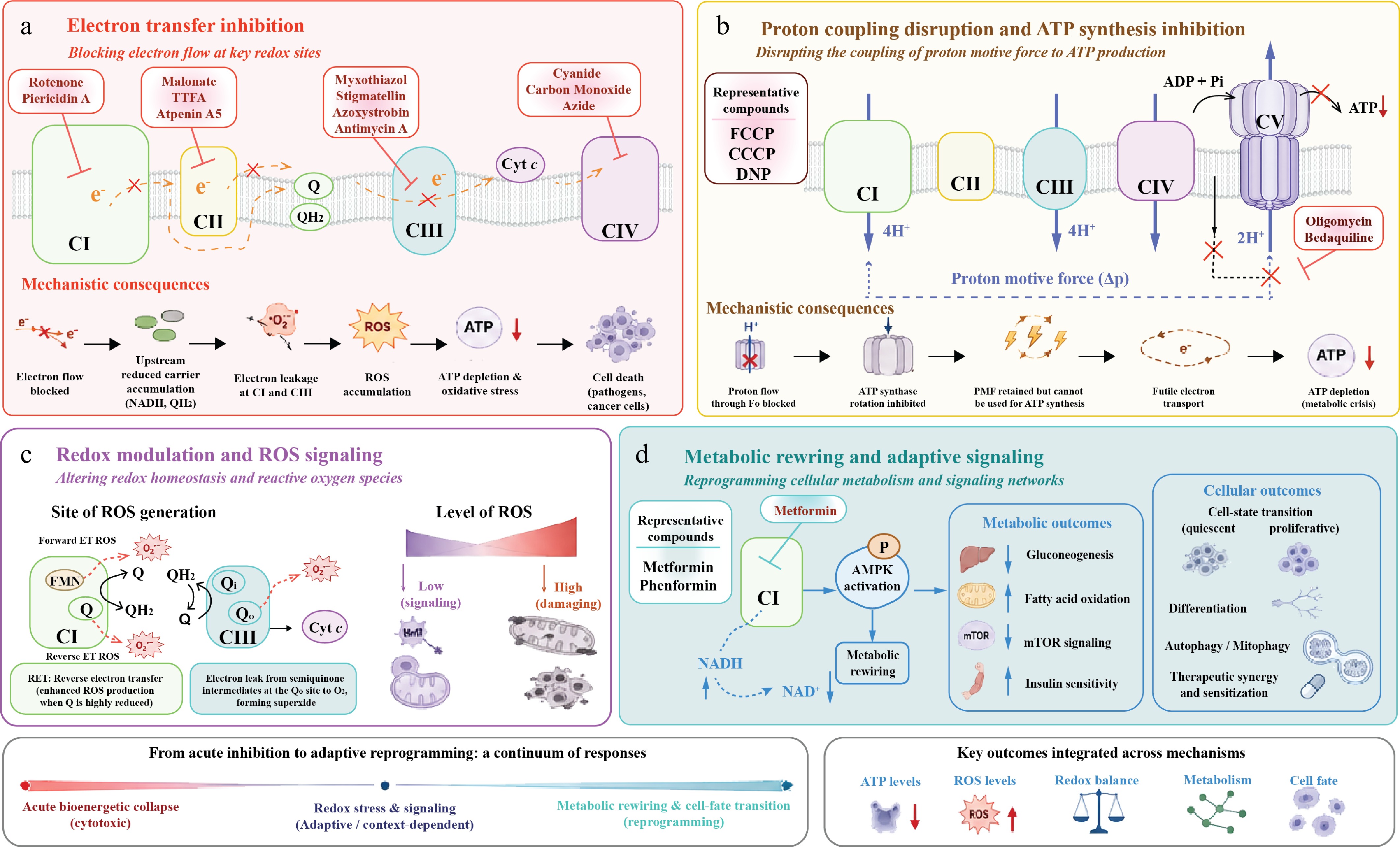
Figure 2.
Mechanisms of small-molecule modulation across the respiratory chain. (a) Electron transfer inhibition. Classical respiratory inhibitors block electron flow at specific redox-active sites within the respiratory chain. Representative inhibitors targeting CI, CII, CIII, and CIV are shown, including Qo-site inhibitors (myxothiazol, stigmatellin, azoxystrobin) and the Qi-site inhibitor antimycin A within CIII. CII inhibitors include substrate-site inhibitors such as malonate and quinone-site inhibitors such as TTFA and Atpenin A5. Inhibition of electron transfer leads to accumulation of upstream reduced carriers, enhanced electron leakage, ROS generation, impaired oxidative phosphorylation, ATP depletion, ultimately resulting in severe energetic stress, impaired cellular viability, or cell death depending on the magnitude and context of inhibition. (b) Proton coupling disruption and ATP synthesis inhibition. Small molecules can disrupt the coupling between proton translocation and ATP production by targeting proton motive force utilization or ATP synthase activity. Representative uncoupling compounds and ATP synthase inhibitors are illustrated. Perturbation of proton coupling compromises ATP generation despite continued or partially preserved respiratory activity, leading to energetic stress and metabolic crisis. (c) Redox modulation and ROS signaling. The respiratory chain functions as a major intracellular source of ROS, particularly at CI and CIII. Forward electron transfer and reverse electron transfer (RET) through CI, as well as semiquinone-mediated electron leakage at the Qo site of CIII, contribute to superoxide production. Moderate ROS levels can function as signaling molecules involved in stress adaptation and metabolic regulation, whereas excessive ROS accumulation induces oxidative damage and mitochondrial dysfunction. (d) Metabolic rewiring and adaptive signaling. Partial respiratory perturbation can induce adaptive metabolic reprogramming rather than acute cytotoxicity. Metformin and phenformin are shown as representative CI-targeting compounds that alter the NADH/NAD+ ratio and activate AMPK, leading to metabolic rewiring and downstream effects on gluconeogenesis, fatty acid oxidation, mTOR signaling, and insulin sensitivity. These changes can further influence cell-state transitions, differentiation, autophagy, mitophagy, and therapeutic sensitization.
Classical inhibitors of CI, such as rotenone and piericidin A, bind within or near the ubiquinone-binding pocket[5], preventing electron transfer from NADH-derived substrates to ubiquinone. Similarly, antimycin A targets the Qi site of CIII, blocking quinone reduction and interrupting the Q cycle[45]. These interactions not only halt electron flow but also alter the redox state of electron carriers, increasing the likelihood of electron leakage and formation of ROS[8].
At the cellular level, electron transfer inhibition results in rapid depletion of ATP due to impaired proton gradient formation, coupled with oxidative stress arising from ROS accumulation. This dual impact underlies the potent cytotoxicity of such compounds, particularly in microorganisms with limited metabolic flexibility. However, in eukaryotic cells, partial inhibition can produce more complex outcomes, including redox imbalance and signaling perturbation, indicating that even classical inhibitors may operate beyond simple energy deprivation depending on dose and context (Table 1).
Table 1. Structural and mechanistic basis of respiratory chain inhibition.
Pharmacological category Representative inhibitor ETC target Binding site/
Structural regionMechanism of action Structural effect/
Conformational stateRepresentative structure Quinone-site competitors Piericidin A CI Upper ubiquinone-binding channel near the NDUFS2-ND1 interface Mimics ubiquinone and competitively inhibits electron transfer at the
Q-reduction siteOccupies the upper Q-channel between NDUFS2-Tyr108 and NDUFS2-His59, stabilizing an inhibitor-bound catalytic state 6ZTQ Quinone-site competitors Rotenone CI Ubiquinone-binding cavity extending from the ND1/NDUFS2 region toward the membrane arm Blocks electron transfer from cluster N2 to ubiquinone by occupying the Q-channel Stabilizes an open/deactive-like inhibitor-bound state; rotenone is additionally observed near ND4 in the membrane arm 6ZKM Quinone-site inhibitors Atpenin A5 CII Ubiquinone-binding site in the membrane domain of succinate dehydrogenase Blocks electron transfer from succinate/FAD/Fe–S relay to ubiquinone by occupying the quinone-binding pocket Stabilizes an Atpenin A5-bound inhibited state of porcine mitochondrial CII 3AEE Quinone-site inhibitors TTFA CII Ubiquinone-binding site in the membrane domain of succinate dehydrogenase Blocks electron transfer from the FAD/Fe–S relay to ubiquinone by occupying the quinone-binding pocket Stabilizes a TTFA-bound inhibited state of avian mitochondrial CII 6MYP Qi-site blockers Antimycin A1 CIII Qi site within cytochrome b near heme bH Blocks quinone reduction at the Qi site and interrupts the Q cycle Occupies the Qi pocket and prevents quinone/quinol exchange, stabilizing a
Qi-inhibited state of
cytochrome bc11NTK Rieske-domain locking Qo-site inhibitors Stigmatellin CIII Qo site of cytochrome b near the Rieske [2Fe–2S] protein interface Mimics quinol binding at the Qo site and blocks bifurcated electron transfer Forms a hydrogen-bonding interaction with the Rieske His residue, locking the Rieske domain in the b-position and stabilizing a Qo-inhibited state 1PP9 Qo-site inhibitors Myxothiazol CIII Proximal Qo site of cytochrome b Blocks quinol oxidation at the Qo site and interrupts bifurcated electron transfer Occupies the proximal Qo pocket; unlike stigmatellin, it does not lock the Rieske domain tightly at the Qo site 1SQP ATP synthase inhibitors Oligomycin A CV Fo proton channel at the interface between the c-ring and subunit a Blocks proton translocation through the Fo motor and inhibits rotary ATP synthesis Immobilizes the membrane rotor machinery and stabilizes a proton-conduction-blocked state of ATP synthase 6CP3
6CP5ATP synthase inhibitors Bedaquiline CV Mycobacterial ATP synthase Fo c-ring/rotor region Selectively inhibits proton-driven rotary catalysis in mycobacterial ATP synthase Captures bedaquiline-saturated ATP synthase in rotational state 1 7JG8 Proton coupling and ATP synthesis
-
In contrast to electron transfer inhibitors, a second class of small molecules targets the coupling between electron transport and ATP synthesis (Fig. 2b). This mechanism primarily involves interference with proton translocation or utilization, most notably at the level of CV.
Oligomycin, a canonical inhibitor of ATP synthase, binds to the Fo subunit and blocks proton flow through the membrane-embedded channel, thereby preventing rotation of the enzyme and ATP production[46]. Bedaquiline, a clinically approved drug for multidrug-resistant tuberculosis, similarly targets the proton-conducting machinery of mycobacterial ATP synthase, but with enhanced selectivity for bacterial over mitochondrial enzymes[10,11]. By directly uncoupling proton motive force from ATP synthesis, these compounds effectively convert respiratory activity into a futile process, where electron transport may continue transiently but fails to generate usable energy[47].
This mode of action highlights an important distinction: while electron transfer inhibition disrupts upstream redox reactions, targeting proton coupling directly compromises the energetic output of the system. The resulting bioenergetic crisis can be particularly effective in pathogens that rely heavily on oxidative phosphorylation, while selective targeting of ATP synthase offers a viable strategy to minimize host toxicity. Furthermore, modulation of coupling efficiency can influence not only ATP levels but also mitochondrial membrane potential and metabolite transport, linking this mechanism to broader metabolic regulation.
Redox modulation and reactive oxygen species
-
Beyond direct inhibition of electron transfer or proton coupling, many small molecules exert their effects by modulating redox homeostasis and ROS production (Fig. 2c). The respiratory chain is a major source of intracellular ROS[8], primarily generated through electron leakage at complexes such as CI and CIII. Pharmacological perturbation of these sites can therefore amplify or redirect ROS generation, with significant downstream consequences.
Increased ROS levels can induce oxidative damage to proteins, lipids, and DNA, ultimately triggering apoptosis or other forms of cell death[48]. In cancer cells, which often operate near the threshold of oxidative stress, further elevation of ROS can selectively drive cytotoxicity. Conversely, moderate changes in ROS can act as signaling cues, activating pathways involved in stress response, inflammation, or metabolic adaptation[37].
Importantly, redox modulation is not solely a byproduct of respiratory inhibition but can represent a targeted mechanism of action. Compounds that subtly alter electron flow or stabilize specific redox states can fine-tune ROS production without completely shutting down respiration. This dual role of ROS as both a damaging agent and a signaling molecule places redox regulation at the intersection of energy metabolism and cellular decision-making, reinforcing the idea that respiratory complexes function as regulatory nodes rather than passive conduits.
Metabolic rewiring
-
The most advanced mode of respiratory targeting involves the reprogramming of cellular metabolism through coordinated modulation of mitochondrial function (Fig. 2d). In this context, small molecules do not simply inhibit respiration but reshape metabolic fluxes, redox balance, and signaling networks to induce sustained changes in cellular state.
A prominent example is metformin[15−18,49], which acts as a mild inhibitor of CI. Rather than causing acute ATP depletion, metformin shifts the intracellular NADH/NAD+ ratio[19], leading to activation of AMPK[50] and downstream suppression of anabolic processes. This metabolic rewiring contributes to its therapeutic effects in type 2 diabetes and has also been implicated in anticancer activity[42]. Crucially, the relatively weak inhibition of CI allows cells to adapt, transforming a potentially cytotoxic mechanism into a regulatory one.
More broadly, metabolic rewiring reflects the integration of respiratory function with cellular signaling and biosynthetic pathways[4]. Changes in mitochondrial activity can influence glycolysis, lipid metabolism, and nucleotide synthesis, as well as transcriptional programs that define cell identity. In cancer and stem cell biology, such reprogramming can determine proliferation, differentiation, or survival outcomes[42].
Taken together, these observations illustrate that respiratory targeting operates along a spectrum, from acute inhibition to subtle modulation and long-term reprogramming. The specific mechanism of action depends on both the molecular properties of the compound and the metabolic context of the target cell. Understanding these mechanisms in a unified framework is essential for rational drug design, as it enables the selection of strategies that maximize therapeutic efficacy while minimizing toxicity (Fig. 2a–d).
-
Advances in structural biology, particularly cryo-EM and cryo-ET, have fundamentally reshaped our understanding of respiratory chain druggability[20,21,26]. High-resolution structures of individual complexes and intact supercomplexes (SCs) now reveal that the respiratory chain is not a collection of isolated catalytic units, but a spatially organized and dynamically regulated network[27]. This architectural complexity provides multiple layers of pharmacological intervention, extending from classical active sites to previously unrecognized regulatory features embedded within membrane environments and higher-order assemblies (Fig. 3).
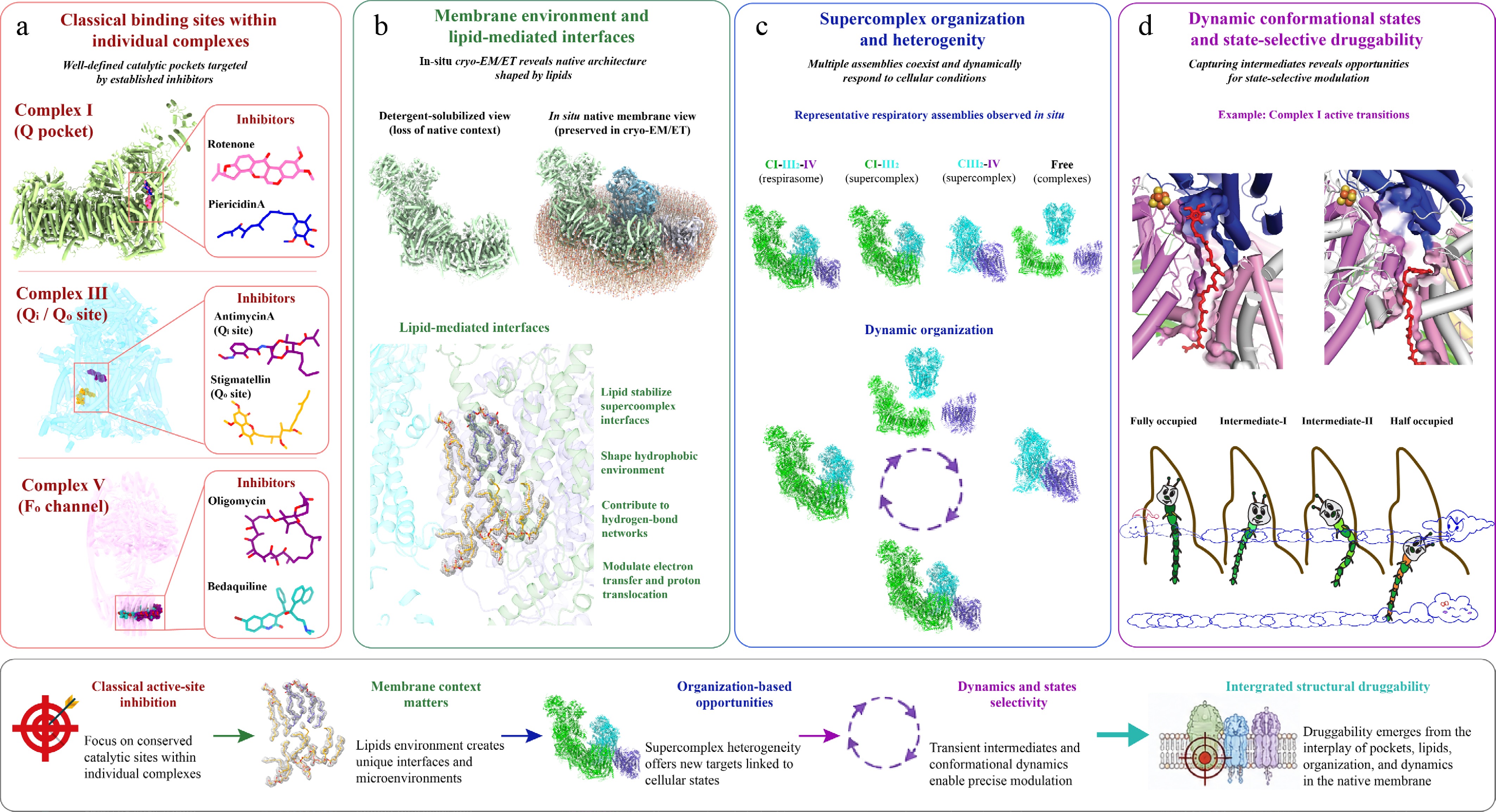
Figure 3.
Structural basis of respiratory chain druggability: from active sites to SC organization. (a) Classical binding sites within individual complexes. Established respiratory inhibitors primarily target conserved catalytic regions involved in electron transfer and proton translocation. Representative inhibitor-binding sites are illustrated for CI ubiquinone-binding pocket, CIII Qi/Qo sites, and the membrane-embedded Fo proton channel of CV. Inhibitors such as rotenone, piericidin A, antimycin A, stigmatellin, oligomycin, and bedaquiline occupy regions overlapping endogenous substrate pathways, thereby blocking electron transfer or proton translocation. Although targeting conserved catalytic centers can achieve potent inhibition, evolutionary conservation often limits selectivity. (b) Membrane environment and lipid-mediated interfaces. In contrast to detergent-solubilized structures, in situ cryo-EM/cryo-ET preserves the native membrane environment and reveals extensive lipid-mediated interactions between respiratory complexes. Structured lipids, including cardiolipin and phospholipids, stabilize SC interfaces, shape hydrophobic microenvironments, and contribute to hydrogen-bonding networks important for electron transfer and proton translocation. These observations highlight that respiratory druggability emerges not only from protein pockets but also from membrane organization and protein-lipid-protein interfaces. (c) SC organization and heterogeneity. Multiple respiratory assemblies coexist within native membranes, including respirasomes, partial SCs, and free complexes. In situ structural analyses reveal that respiratory organization is highly heterogeneous and dynamically responds to metabolic state, substrate availability, and cellular stress. Such organizational plasticity creates context-dependent vulnerabilities and opportunities for selective targeting of specific SC states or assembly-dependent functional behaviors. (d) Dynamic conformational states and state-selective modulation. Respiratory complexes undergo substantial conformational rearrangements during catalysis, generating transient structural intermediates that may serve as selective pharmacological targets. Representative active and deactive conformations of CI are shown to illustrate how substrate occupancy and structural rearrangement reshape catalytic architecture and accessibility. These dynamic states provide opportunities for state-selective modulation, in which small molecules stabilize specific conformations or alter conformational equilibria without necessarily inducing complete respiratory inhibition.
Classical binding sites
-
Early efforts in targeting the respiratory chain focused on well-defined catalytic centers responsible for electron transfer and substrate turnover (Fig. 3a). These classical binding sites, typically located within conserved active regions of respiratory complexes, remain the primary targets of many established inhibitors.
In CI and CIII, the ubiquinone-binding pocket represents a central hub for small-molecule interaction (Fig. 3a). Structural studies have shown that inhibitors such as rotenone and piericidin A occupy regions overlapping with endogenous quinone substrates, thereby blocking electron transfer at its entry or intermediate steps[23, 51]. Similarly, antimycin A binds to the Qi[52] site of CIII, preventing quinone reduction and disrupting the Q cycle. These interactions are often characterized by high affinity and direct competition with physiological substrates, making them effective but sometimes limited by lack of selectivity due to evolutionary conservation[22].
CV provides another classic example, where inhibitors such as oligomycin bind within the membrane-embedded Fo domain[46], obstructing proton translocation (Fig. 3a). Structural analyses have revealed how these compounds physically occlude proton channels, thereby uncoupling electron transport from ATP synthesis. More recently, high-resolution structures of mycobacterial ATP synthase have clarified the binding mode of bedaquiline, highlighting subtle structural differences that enable selective targeting of bacterial enzymes over their mitochondrial counterparts[10,53].
Collectively, these classical binding sites illustrate a foundational principle of respiratory druggability: targeting conserved catalytic regions can yield potent inhibition but often requires precise structural discrimination to achieve therapeutic selectivity.
Membrane environment and supercomplex organization: in situ insights into respiratory druggability
-
In contrast to structures derived from detergent-solubilized samples, in situ cryo-EM and cryo-ET have provided a fundamentally different view of respiratory chain organization and druggability[26] (Fig. 3b). Conventional in vitro purification approaches typically rely on detergents to extract membrane proteins, inevitably disrupting the native lipid environment. This process often leads to the loss of protein-lipid-protein interactions and can alter both the assembly state and conformational landscape of respiratory complexes. By preserving the native mitochondrial membrane, in situ approaches overcome these limitations and enable direct visualization of respiratory SCs under physiological conditions.
High-resolution in situ structures reveal that interactions between respiratory complexes are extensively mediated by lipids rather than solely by direct protein-protein contacts (Fig. 3b). Numerous structured lipid molecules occupy the interfacial regions between complexes, stabilizing SC architecture while simultaneously shaping local hydrophobic environments and hydrogen-bonding networks that are critical for electron transfer and proton translocation. These lipid-mediated interfaces are largely absent in detergent-solubilized preparations, underscoring a major limitation of traditional structural approaches.
Beyond structural stabilization, in situ analyses uncover a high degree of heterogeneity and dynamic organization within the respiratory chain (Fig. 3c). Multiple SC assemblies with distinct stoichiometries and spatial arrangements coexist within the native membrane, forming a structurally and functionally diverse network. Such diversity is typically underrepresented in purified systems, which tend to favor a limited subset of stable conformations.
A key advantage of in situ structural approaches is their ability to capture substrate-binding events across different stages of the catalytic cycle (Fig. 3d). High-resolution maps directly visualize multiple binding states of endogenous ligands (Fig. 3d), such as ubiquinone/ubiquinol, within respiratory complexes, revealing coordinated conformational changes associated with electron transfer. These dynamic intermediates, which are difficult to resolve using conventional approaches, provide a structural basis for understanding how electron flow is regulated in real time within the membrane environment.
Collectively, these findings redefine the landscape of respiratory druggability. First, lipid-mediated interfaces represent a previously underexplored class of targets, where small molecules may modulate function by perturbing local membrane organization or competing with structural lipids. Second, the existence of multiple SC assemblies introduces opportunities for selective targeting of specific organizational states, particularly under disease-relevant conditions. Third, the presence of multiple substrate-binding intermediates suggests that transient catalytic states can be selectively stabilized or perturbed, offering a strategy to modulate electron transfer pathways without complete inhibition.
Thus, druggability in the respiratory chain is not solely determined by static active sites within individual complexes, but emerges from the interplay between membrane environment, lipid interactions, SC architecture, and dynamic catalytic states. This in situ perspective shifts the focus of therapeutic design from isolated proteins to the organization and dynamics of the respiratory network as a whole.
-
The mechanistic and structural principles described above have enabled the development of diverse therapeutic strategies targeting the respiratory chain across multiple disease contexts. Importantly, the pharmacological outcome of respiratory modulation is highly dependent on cellular and organismal context, reflecting differences in metabolic demand, pathway redundancy, and structural organization of respiratory complexes. As a result, achieving therapeutic efficacy requires not only potent modulation of respiratory activity but also selective exploitation of vulnerabilities unique to specific pathogens or disease states (Fig. 4).
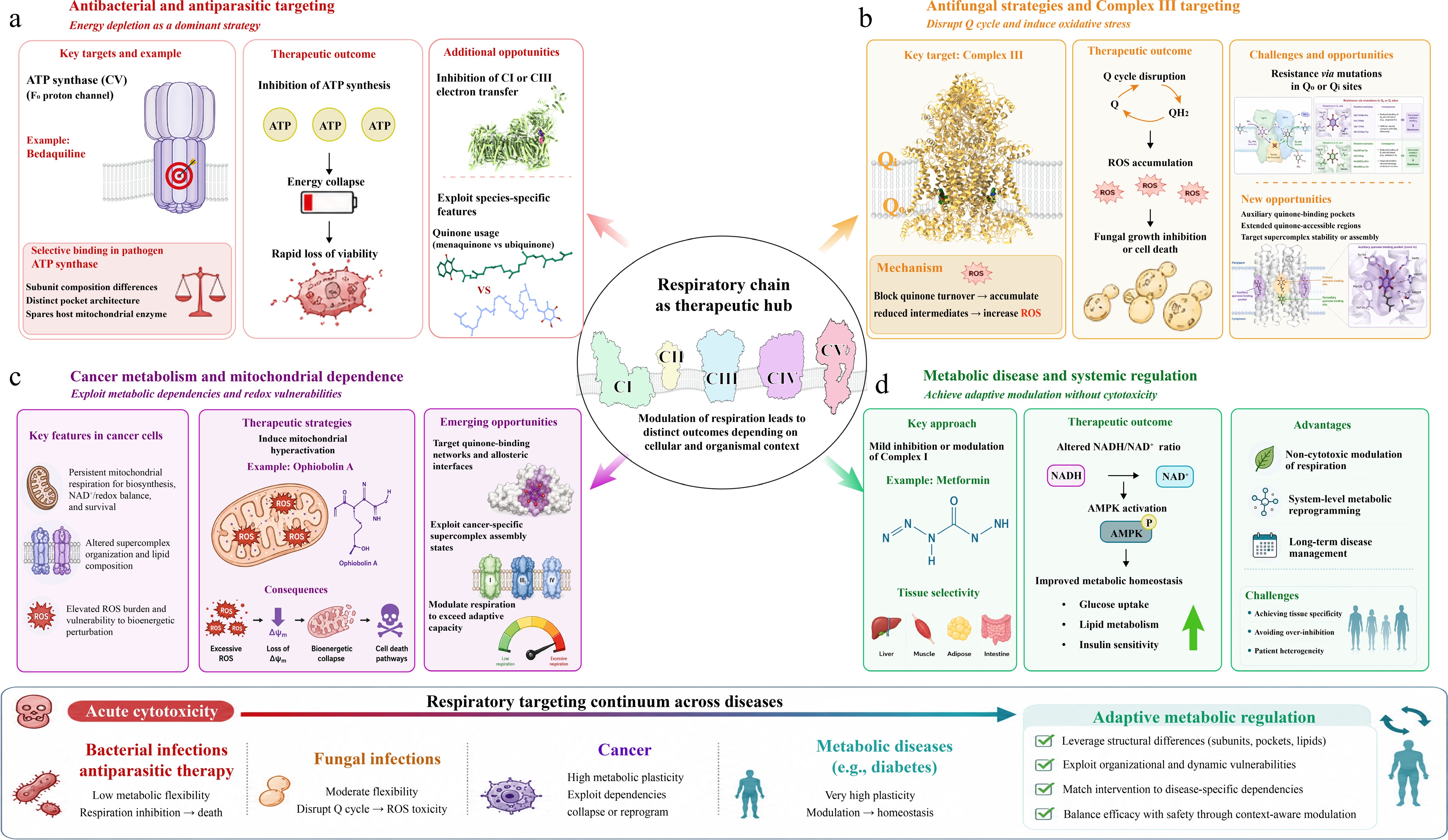
Figure 4.
Therapeutic applications and selectivity across diseases. (a) Antibacterial and antiparasitic targeting. In bacterial and parasitic pathogens, OXPHOS frequently serves as a dominant energy-producing pathway, rendering these organisms highly sensitive to respiratory inhibition. CV represents a major therapeutic target, exemplified by bedaquiline, which selectively inhibits the Fo proton channel of mycobacterial ATP synthase. Structural differences in subunit composition and binding-pocket architecture enable pathogen-selective inhibition while minimizing effects on the host mitochondrial enzyme. Respiratory inhibition leads to rapid ATP depletion, energetic collapse, and loss of viability. Additional opportunities for selective targeting arise from species-specific quinone usage, electron-transfer pathways, and respiratory SC organization. (b) Antifungal strategies and CIII targeting. In fungal pathogens, CIII is a major target of antifungal intervention. Inhibitors acting at the Qo or Qi sites disrupt the Q cycle, block quinone turnover, and promote accumulation of reduced intermediates and ROS, resulting in oxidative stress and fungal growth inhibition. Resistance frequently emerges through mutations in canonical quinone-binding regions. Structural studies further suggest the presence of auxiliary quinone-binding pockets and extended quinone-accessible regions that may provide alternative sites for therapeutic targeting. Modulation of SC stability or assembly may additionally create opportunities for fungal-selective respiratory disruption. (c) Cancer metabolism and mitochondrial dependence. Many tumor cells retain substantial dependence on mitochondrial respiration for biosynthesis, redox homeostasis, and survival despite elevated glycolytic activity. This metabolic reliance creates selective vulnerabilities that can be exploited therapeutically. Compounds such as Ophiobolin A induce mitochondrial hyperactivation, excessive ROS accumulation, membrane-potential disruption, and bioenergetic collapse, ultimately activating cell death pathways. Emerging therapeutic strategies aim to target quinone-binding networks, allosteric interfaces, and cancer-specific SC states in order to selectively perturb tumor metabolism while sparing normal tissues. (d) Metabolic disease and systemic regulation. In metabolic disorders such as type 2 diabetes, respiratory modulation is used not to induce cytotoxicity but to achieve adaptive metabolic regulation. Metformin exemplifies this strategy through mild inhibition or modulation of CI, leading to altered NADH/NAD+ balance, activation of AMPK, and downstream improvement of metabolic homeostasis. These effects contribute to regulation of glucose uptake, lipid metabolism, and insulin sensitivity while avoiding acute energetic collapse. Achieving tissue selectivity and avoiding excessive respiratory inhibition remain major challenges for long-term therapeutic application.
Antibacterial and antiparasitic targeting
-
Targeting the respiratory chain has proven particularly effective in antibacterial and antiparasitic therapy (Fig. 4a), where organisms often rely heavily on oxidative phosphorylation[40, 54] and possess limited metabolic flexibility. In these systems, energy depletion remains a dominant therapeutic strategy, with inhibition of ATP production leading to rapid loss of viability.
A prominent example is bedaquiline[10,53,55] (Fig. 4a; Table 2), which selectively targets the ATP synthase of Mycobacterium tuberculosis. Structural and biochemical studies have demonstrated that bedaquiline binds within the proton-translocating Fo domain, disrupting ATP synthesis while sparing the host mitochondrial enzyme. This selectivity arises from subtle but critical differences in subunit composition and binding pocket architecture, underscoring the importance of structure-guided drug design. Clinically, this mechanism translates into potent bactericidal activity against multidrug-resistant strains, validating respiratory complexes as viable antimicrobial targets. Another notable example is telacebec (Q203)[56] (Table 2), a first-in-class inhibitor targeting the cytochrome bc1 complex through the mycobacterial QcrB subunit. Unlike ATP synthase inhibitors, Q203 blocks electron transfer within CIII, resulting in rapid disruption of OXPHOS and ATP production. Owing to its high selectivity for the mycobacterial respiratory chain, Q203 has demonstrated potent activity against both drug-sensitive and multidrug-resistant M. tuberculosis and has advanced into late-stage clinical development[57], further validating respiratory complexes as attractive antimicrobial targets.
Table 2. Representative respiratory-chain-targeting therapeutics with clinical or translational potential.
Compound Chemical
structureTarget complex Primary indication Development stage Key pharmacological features Metformin 
CI Type 2 diabetes; oncology (investigational) Approved Weak but clinically relevant CI modulator; activates AMPK and induces metabolic reprogramming with an excellent safety profile. IACS-010759 
CI Relapsed/refractory AML; advanced solid tumors Phase I Potent and selective OXPHOS inhibitor developed through structure-guided optimization; preferentially targets OXPHOS-dependent tumors. IM156 
CI Advanced solid tumors Phase I Second-generation biguanide with improved potency, pharmacokinetics,
and oral bioavailability compared with metformin.EVT-701 
CI Solid tumors Phase I Highly selective CI inhibitor with an improved therapeutic window and potent activity against OXPHOS-dependent tumors. BAY 87-2243 
CI Solid tumors Phase I (terminated) Potent inhibitor of mitochondrial respiration that suppresses HIF signaling under hypoxic conditions and exhibits antitumor activity. Bedaquiline 
CV (ATP synthase) Multidrug-resistant tuberculosis Approved Selective bacterial ATP synthase inhibitor with minimal activity toward mammalian ATP synthase; first-in-class respiratory-chain antibiotic. Telacebec (Q203) 
CIII (QcrB) Tuberculosis Phase II/III Highly selective inhibitor of mycobacterial cytochrome bc1 complex with potent activity against drug-resistant
M. tuberculosis.AML, acute myeloid leukemia; AMPK, AMP-activated protein kinase; OXPHOS, oxidative phosphorylation. Beyond ATP synthase, inhibitors of electron transfer at CI and CIII have also been explored for antiparasitic applications. However, achieving selectivity remains a major challenge due to the evolutionary conservation of catalytic cores. Emerging insights into non-canonical binding sites and species-specific structural features, including variations in quinone-binding pockets and SC organization, suggest new opportunities for selective targeting. In particular, differences in quinone usage (e.g., ubiquinone vs menaquinone) and SC architecture may be exploited to design compounds that preferentially disrupt pathogen respiration without affecting host mitochondria (Fig. 4a).
Antifungal strategies and CIII targeting
-
In fungal pathogens, respiratory targeting has been widely applied in agricultural and clinical contexts, with CIII serving as a major site of intervention (Fig. 4b). Antifungal agents often act by disrupting the Q cycle[44,58], thereby impairing electron transfer and inducing oxidative stress.
Inhibitors targeting the Qo or Qi sites of CIII can effectively block quinone turnover, leading to accumulation of reduced intermediates and increased ROS production[8] (Fig. 4b). This dual mechanism, combining energy deprivation with oxidative damage, contributes to their antifungal efficacy. However, resistance frequently arises through mutations in quinone-binding sites, highlighting the limitations of targeting conserved catalytic regions[58].
Recent structural insights provide a framework for overcoming these challenges. The identification of auxiliary quinone-binding pockets[59] and extended quinone-accessible regions suggests that fungal respiratory complexes may harbor additional regulatory sites beyond canonical Qo/Qi positions (Fig. 4b). Targeting such non-canonical sites could reduce susceptibility to resistance while enabling more subtle modulation of respiratory activity. Furthermore, interference with SC stability or assembly may represent an alternative strategy, particularly in species where respiratory organization differs significantly from that of host cells.
Cancer metabolism and mitochondrial dependence
-
In contrast to antimicrobial applications, targeting the respiratory chain in cancer requires a fundamentally different strategy (Fig. 4c). Rather than inducing immediate cell death through energy depletion, therapeutic efficacy often depends on exploiting metabolic dependencies and redox vulnerabilities unique to tumor cells. Consequently, respiratory modulation has emerged as a promising strategy to selectively impair cancer cell fitness while minimizing toxicity to normal tissues.
Although many cancers exhibit enhanced glycolysis, increasing evidence indicates that mitochondrial respiration remains essential for biosynthesis, redox balance, and survival in diverse tumor types[4,42]. This persistent dependence on OXPHOS creates a therapeutic window for selectively targeting mitochondrial respiration in tumors. As a result, the development of OXPHOS-targeting agents, particularly selective CI inhibitors, has become an active area of anticancer drug discovery.
Unlike classical respiratory inhibitors such as rotenone and piericidin A, which primarily serve as experimental tool compounds, recent years have witnessed the emergence of a new generation of drug-like CI inhibitors developed through structure-guided optimization, phenotypic screening, and cancer-focused OXPHOS-targeting strategies. Representative compounds, including IACS-010759[60], IM156[61], EVT-701[62], BAY 87-2243[63] (Table 2), and other recently developed analogs, exhibit improved pharmacokinetic properties, enhanced CI selectivity, and greater therapeutic potential compared with classical inhibitors. Structural studies indicate that although many of these molecules ultimately interfere with ubiquinone reduction, differences in molecular scaffolds and binding interactions generate distinct pharmacological profiles and therapeutic windows. Importantly, the greater reliance of many tumors on mitochondrial respiration enables these inhibitors to preferentially suppress tumor bioenergetics while sparing normal tissues, and several compounds have advanced into clinical evaluation, highlighting the translational potential of selective respiratory modulation in oncology.
In addition to direct CI inhibition, alternative strategies have also emerged to exploit mitochondrial dependence in cancer. Compounds such as Ophiobolin A[64] exemplify this approach by inducing mitochondrial hyperactivation and subsequent bioenergetic collapse (Fig. 4c). Instead of directly inhibiting electron transfer, these agents perturb mitochondrial homeostasis, leading to excessive ROS production[48], disruption of membrane potential, and activation of cell death pathways. This mode of action aligns with the concept of cellular reprogramming, in which respiratory modulation drives cancer cells beyond their adaptive capacity.
Importantly, structural organization of the respiratory chain may further influence therapeutic outcomes in cancer. Variations in SC assembly, lipid composition, and quinone dynamics can affect electron transfer efficiency and ROS generation[26] (Fig. 4c). Targeting these higher-order features, such as quinone-binding networks or allosteric interfaces, may enable selective modulation of tumor metabolism without fully suppressing respiration in normal tissues. Such strategies highlight the potential of leveraging structural heterogeneity to achieve context-specific therapeutic effects.
Metabolic disease and systemic regulation
-
In metabolic diseases, including type 2 diabetes, respiratory targeting is employed not to induce cytotoxicity but to achieve systemic metabolic regulation (Fig. 4d). In this context, mild and sustained modulation of mitochondrial function can produce beneficial effects on glucose homeostasis and energy balance.
Metformin represents the prototypical example of this strategy (Fig. 4d). As a weak inhibitor of CI, metformin reduces mitochondrial respiration sufficiently to alter the intracellular NADH/NAD+ ratio, leading to activation of AMPK and downstream inhibition of gluconeogenesis. Unlike classical inhibitors, metformin does not cause acute ATP depletion under physiological conditions, allowing cells to adapt while shifting metabolic fluxes toward improved energy utilization[15,16,49].
This mode of action underscores the importance of dose-dependent and context-dependent effects in respiratory targeting. Low-level modulation can act as a signaling mechanism, whereas stronger inhibition may lead to an energy crisis and cell death. The ability to fine-tune respiratory activity is therefore critical for therapeutic success in metabolic diseases.
Emerging evidence suggests that structural features of respiratory complexes, including SC organization and lipid interactions, may influence sensitivity to metabolic modulators. Understanding how these structural parameters vary across tissues and disease states could inform the development of next-generation therapeutics with improved efficacy and reduced side effects (Fig. 4).
-
The emerging view of the respiratory chain as a dynamic, multi-layered regulatory system opens new avenues for therapeutic intervention, but also highlights key challenges that must be addressed to enable precise and safe targeting. Moving forward, advances in structural biology, chemical design, and systems-level analysis will need to converge to fully realize the therapeutic potential of respiratory modulation[1].
A central priority is to achieve context-specific selectivity. Given the high evolutionary conservation of respiratory complexes, indiscriminate inhibition often leads to host toxicity. Future strategies will therefore need to exploit subtle differences in structural organization, including species-specific variations in quinone usage, lipid composition, and SC architecture. In particular, non-canonical binding sites, such as lipid-mediated interfaces and allosteric cavities, offer promising opportunities for selective targeting without directly competing with conserved catalytic centers[26].
Another critical direction lies in leveraging dynamic and transient states of the respiratory chain. In situ structural approaches have revealed that respiratory complexes operate across multiple conformational and catalytic states, including distinct substrate-binding intermediates and active-inactive transitions[26]. These findings suggest that drug design can move beyond static inhibition toward state-selective modulation, in which small molecules stabilize or bias specific functional states to achieve desired biological outcomes. Such strategies may enable fine control of electron flux, redox balance, and metabolic signaling without complete disruption of respiration.
The integration of membrane biology into drug discovery represents an additional frontier. The functional roles of lipids in stabilizing SCs, shaping local environments, and regulating electron transfer highlight the importance of considering the membrane as an active participant in respiratory function[25,26]. Targeting protein-lipid-protein interfaces or modulating local membrane properties may provide new mechanisms to control respiratory activity with improved specificity. This perspective calls for the development of drug discovery platforms that better recapitulate native membrane environments, moving beyond detergent-based systems.
At a higher level, systems-level understanding of respiratory organization will be essential. The respiratory chain operates not as isolated complexes but as an integrated network whose function emerges from spatial organization, cooperative interactions, and metabolic context. Future efforts combining structural biology with metabolomics, live-cell imaging, and computational modeling will be required to map how perturbations at specific sites propagate across the system. Such approaches will be particularly important for understanding disease-specific vulnerabilities and predicting therapeutic responses.
Finally, the concept of the respiratory chain as a programmable bioenergetic system suggests new possibilities for precision medicine. Rather than applying uniform inhibition, future therapies may tailor respiratory modulation to specific cell types, disease states, or metabolic conditions. In cancer, this could involve exploiting metabolic plasticity and redox thresholds; in infectious diseases, selectively collapsing pathogen bioenergetics; and in metabolic disorders, fine-tuning systemic energy balance. Achieving this level of precision will require not only improved molecular targeting but also a deeper understanding of how respiratory function is integrated with cellular identity and environmental cues.
Taken together, these directions point toward a shift from conventional inhibition strategies to a new paradigm of precision bioenergetic control, in which the respiratory chain is manipulated as a dynamic and adaptable therapeutic system. Continued integration of in situ structural insights with functional and pharmacological studies will be critical in translating this vision into clinically effective interventions.
-
Not applicable. This is a review article and does not involve original research with human participants or animals.
-
The author confirms sole responsibility for all aspects of this work and approved the final version of the manuscript.
-
Data sharing not applicable to this review as no datasets were generated or analyzed.
-
The author declares that there is no conflict of interest.
- Copyright: © 2026 by the author(s). Published by Maximum Academic Press on behalf of China Pharmaceutical University. This article is an open access article distributed under Creative Commons Attribution License (CC BY 4.0), visit https://creativecommons.org/licenses/by/4.0/.
-
About this article
Cite this article
Zheng W. 2026. Targeting the respiratory chain: from bioenergetic inhibition to metabolic reprogramming. Targetome 2(4): e040 doi: 10.48130/targetome-0026-0037 

Targeting the respiratory chain: from bioenergetic inhibition to metabolic reprogramming
- Received: 05 June 2026
- Revised: 22 July 2026
- Accepted: 24 July 2026
- Published online: 24 August 2026
Abstract: The respiratory chain has traditionally been viewed as a central bioenergetic system responsible for ATP production through oxidative phosphorylation. However, emerging evidence now supports a broader framework in which respiratory complexes function not merely as energy-generating machinery, but as dynamic regulatory nodes integrating metabolism, redox balance, signaling, and cellular fate decisions. This conceptual shift has transformed the respiratory chain from a classical target of bioenergetic inhibition into a programmable therapeutic system capable of inducing context-dependent biological outcomes. Small molecules targeting the respiratory chain act through multiple mechanistic modes, including inhibition of electron transfer, disruption of proton coupling, modulation of reactive oxygen species, and large-scale metabolic rewiring. Depending on cellular context and degree of respiratory perturbation, these interventions can produce outcomes ranging from acute energetic collapse to adaptive metabolic remodeling and cell-state transitions. Such mechanisms have broad therapeutic relevance across antibacterial, antifungal, antiparasitic, anticancer, and metabolic diseases. Recent advances in cryo-electron microscopy (cryo-EM) and cryo-electron tomography (cryo-ET) have further redefined respiratory-chain druggability by revealing high-resolution architectures of individual complexes, supercomplexes, lipid-mediated interfaces, quinone-binding networks, and transient conformational states. These structural insights expose previously inaccessible regulatory features and provide a framework for state-selective and context-specific therapeutic targeting. In this review, we synthesize current understanding of respiratory-chain pharmacology from mechanistic, structural, and systems-level perspectives. We discuss how classical inhibition paradigms are evolving toward precision bioenergetic modulation, highlight emerging opportunities in targeting dynamic respiratory states and supercomplex organization, and outline future strategies for selective therapeutic intervention across diverse disease contexts.
-
Key words:
- Respiratory chain /
- Oxidative phosphorylation /
- Metabolic reprogramming /
- Drug discovery