










-
Ischemic stroke is the second leading cause of death in the world, and it contributes to a growing burden of disability and socioeconomic costs. Current treatment methods, such as thrombolysis and thrombectomy, are still constrained by a narrow time window and some contraindications, underscoring the need for new treatments. Although traditional research mainly focuses on neuronal damage, blood-brain barrier (BBB) destruction, and local neuroinflammation, the new concept of brain-organ axis redefines ischemic stroke as a systemic disease involving dynamic multi-organ crosstalk[1]. More and more evidence has shown that there is a two-way communication between the brain and peripheral organs, including the gut, heart, liver, spleen, lungs, and kidneys. This crosstalk is mediated by the neuro-immune-metabolic network (NIMN), an integrated system in which neural signals modulate immune responses and metabolic processes, while immune and metabolic factors reciprocally influence neural function. This network operates through shared molecular mediators, including cytokines, neuropeptides, and metabolites, forming a dynamic regulatory axis that maintains physiological homeostasis and contributes to the pathogenesis, progression, and repair of ischemic stroke.
The clinical studies have shown that ischemic stroke causes intestinal inflammation[2], intestinal microbial disorder[3], cardiovascular complications[4], hyperglycemia[5], splenic atrophy[6,7], lung injury[8], and renal dysfunction[9]. These systemic reactions can further aggravate the damage to the central nervous system (CNS), and more and more studies have corroborated this notion, for example, that metabolites produced by intestinal microbiota can affect neuroinflammation[10]. These findings position peripheral organs as promising intervention targets for ischemic stroke.
The core of the multi-axis crosstalk between the brain and peripheral organs lies in the integrated network composed of the nervous, immunity, immune, and metabolic systems. Autonomic signals drive changes in the phenotype of immune cells[11], and metabolic reprogramming such as glycolysis regulates the function of immune cells[12]. Immune mediators, namely interleukin-1β (IL-1β) and transforming growth factor-β (TGF-β), in turn can affect neural and metabolic activities[13]. This dynamic network provides a mechanistic basis for multi-target intervention. It should be noted that certain small molecule compounds, whether natural or synthetic rely on their multi-pharmacological properties to restore the homeostatic state of the NIMN. Fingolimod enhanced the effect of delayed alteplase by promoting anterograde reperfusion and preventing collateral failure, rather than by directly affecting recanalization[14]. Similarly, a phase II trial of IL-1 receptor antagonist in acute stroke reported promising trends in 3-month functional outcomes, as reflected by shifts in the National Institutes of Health Stroke Scale (NIHSS) and the modified Rankin scale (mRS) score distributions, supporting its potential neuroprotective effects[15]. Furthermore, emerging treatments such as vagus nerve stimulation (VNS), microbiome transplantation, and stem cell therapy are leveraging organ-axis crosstalk to reshape treatment methods.
This review systematically summarizes the physiological and pathological effects of the brain-gut-microbiota axis, brain-heart axis, brain-liver axis, brain-spleen axis, brain-lung axis and brain-kidney axis after ischemic stroke. The crosstalk of the brain with peripheral organs is interpreted within the NIMN framework, and pharmacological evidence is used to propose a new treatment based on the organ axes. This study should guide the direction of transition from single-target intervention to multi-axis collaborative treatment, and promote precision medicine for ischemic stroke.
-
Beyond the CNS, peripheral organs engage in vital bidirectional communication with the brain through specialized axes that critically influence the outcomes of ischemic stroke. The brain-gut-microbiota axis, brain-heart axis, brain-liver axis, brain-spleen axis, brain-lung axis, and brain-kidney axis together form a multi-organ network, and the pathological crosstalk can amplify secondary injury. Each axis affects different organ-brain interactions via the NIMN, thereby making ischemic stroke a systemic disease. The following sections explain the contribution of these six axes to the pathogenesis of ischemic stroke (Fig. 1).
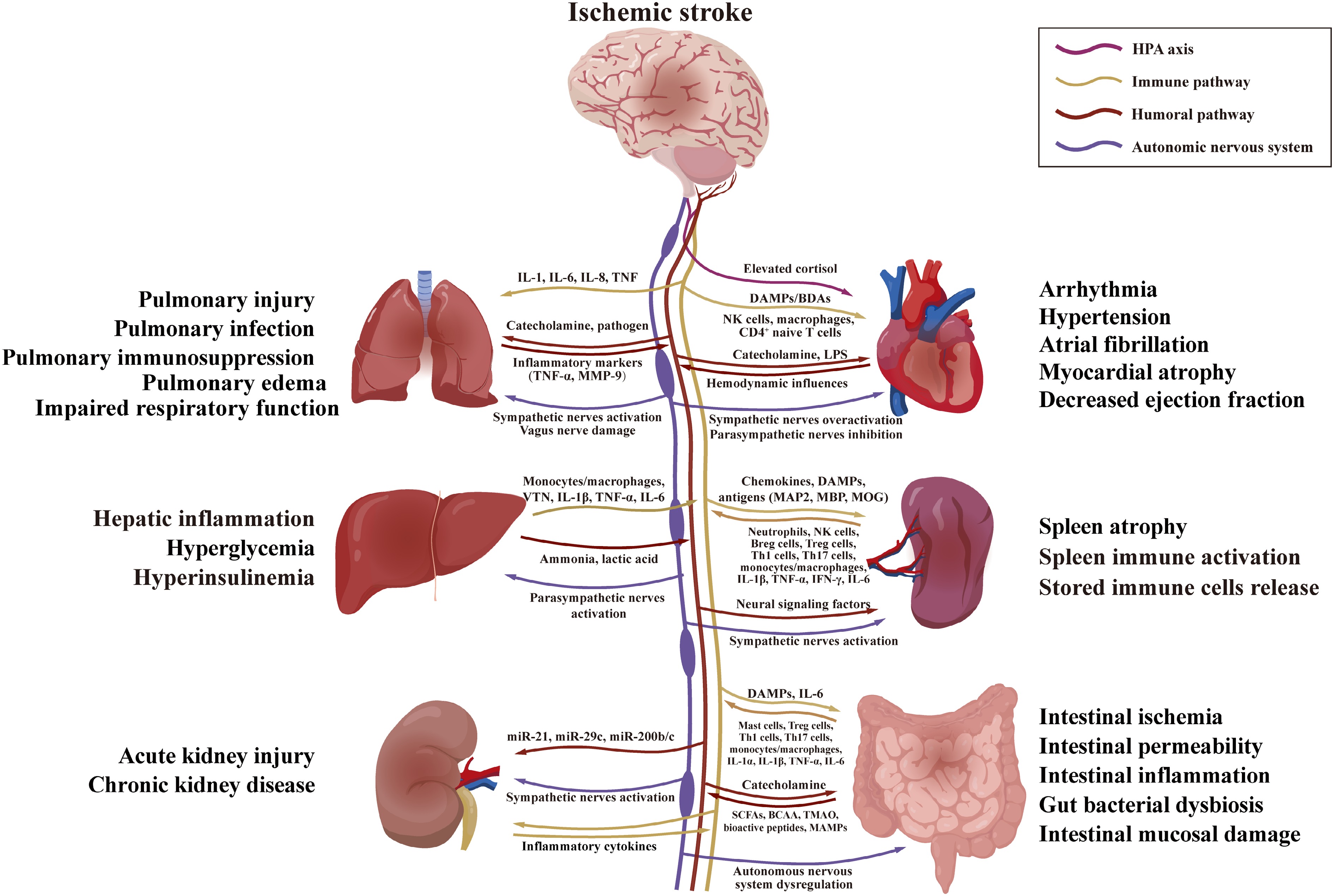
Figure 1.
Brain-organ interaction axes. Following ischemic stroke, the brain influences peripheral organs through autonomic, HPA, inflammatory, and humoral pathways, while organ dysfunction reciprocally impacts the brain. The figure was independently drawn using Adobe Illustrator. BCAA, branched-chain amino acid; DAMPs, damage-associated molecular patterns; HPA, hypothalamic-pituitary-adrenal; LPS, lipopolysaccharide; SCFAs, short-chain fatty acids; TMAO, trimethylamine-N-oxide; VTN, vitronectin.
The brain-gut-microbiota axis
-
The brain-gut-microbiota axis acts as a reciprocal communication pathway, including the brain, the intestine, and the intestinal microbiome (Fig. 1). The brain relies on the autonomic nervous system and humoral pathways to regulate intestinal motility, permeability, mucus secretion, and immune responses, thus affecting the intestinal microbiota. Clinical research showed that individuals with ischemic stroke presented with marked dysregulation of gut microbiota. One of the important characteristics is the higher abundance of short-chain fatty acids (SCFAs)-producing bacteria, as by Odoribacter and Akkermansia[16]. Ischemic stroke triggers the death of intestinal epithelial cells by activating the sympathetic nervous system, thereby resulting in increased intestinal permeability[17]. In addition, the brain can release signaling molecules like damage-associated molecular patterns (DAMPs), thus affecting the composition and metabolism of microorganisms[18]. In return, intestinal microbiota communicate with the brain via multiple routes. These include nutrient metabolism, production of bioactive peptides and the activation of microbial-associated molecular patterns, humoral pathways, the vagus nerves, and immune pathways[19]. Changes in the intestinal barrier permeability, inflammatory factor levels, and immune cell infiltration also affect the brain[20].
The brain-heart axis
-
There are multiple ways to maintain bidirectional crosstalk between the brain and the heart, including the hypothalamic-pituitary-adrenal (HPA) axis, the autonomic nervous system, immune signaling, humoral mechanisms, and hemodynamic changes[21] (Fig. 1). Cardiovascular issues rank as the second most common cause of death following ischemic stroke[4]. The complications, including myocardial atrophy, arrhythmia, and atrial fibrillation (AF), impair cerebral perfusion, which in turn, exacerbates secondary brain injury. Clinical evidence indicates a high probability of cardiac abnormalities after ischemic stroke, about 7.4% of patients showing decreased left ventricular ejection fraction[22]. Moreover, the larger the acute stroke infarction, the more serious the cardiac dysfunction[23].
Consistent with clinical observations, the preclinical animal models provide a basis for the mechanism explanation. It was found that persistent ischemic stroke induced cardiac dysfunction and decreased left ventricular ejection fraction in transient middle cerebral artery occlusion (tMCAO) models[24,25]. In MCAO rats, the incidence of arrhythmia-increased after ischemic stroke [26], and the average arterial pressure response decreased[27]. In addition, ischemic stroke was also confirmed to cause AF in canine models[28]. Therefore, the brain-heart axis leads to various cardiovascular dysfunctions after ischemic stroke, which affects neurological outcomes.
After ischemic stroke, heart injury aggravates brain damage due to changes in heart rate and blood flow. In a post hoc analysis of the International Head Positioning in Acute Stroke Trial (HeadPoST) study, low systolic blood pressure (SBP) and diastolic blood pressure (DBP) were associated with worse ischemic stroke results[29]. Another study showed that low SBP increased the risk of in-hospital mortality and complications for patients with acute ischemic stroke[30]. Tachycardia after acute non-cardioembolic stroke is thought to aggravate neurological functional deterioration[31]. Like the negative impact of low blood pressure, SBP ≥ 180 mmHg or DBP ≥ 100 mmHg can also increase the risk of early neurological deterioration and adverse consequences[32], which shows that cardiovascular management after ischemic stroke should be grasped as a delicate balance.
The brain-liver axis
-
Neural and immune pathways enable crosstalk between the brain and liver, forming the brain-liver axis (Fig. 1). Hepatic vagal sensory afferent nerves relay microenvironmental information to the brainstem nucleus tractus solitarius. This brainstem nucleus then feeds back to regulate intestinal peripheral regulatory T cell homeostasis via vagal and enteric neurons[33]. In addition, a quarter of non-diabetic patients in the stable period after ischemic stroke developed hyperglycemia[34]. This led to increased mortality, worsened prognosis, and impaired neurological function recovery[5]. Animal experiments using the permanent middle cerebral artery occlusion (pMCAO) model also showed hyperglycemia, accompanied by hyperinsulinemia[35]. Clinically, women with a fatty liver index above the 90th percentile have an elevated risk of ischemic stroke. Likewise, an aspartate aminotransferase to alanine aminotransferase ratio exceeding 2 increases the risk of ischemic stroke among white individuals[36].
The brain-spleen axis
-
The brain-spleen axis mediates two-way communication through neural, immune, and endocrine pathways, which mainly includes the autonomic nervous system, immune system, and the HPA axis (Fig. 1). After cerebral ischemia, the spleen releases monocytes, neutrophils, lymphocytes, and natural killer cells (NK cells) into the circulation, accompanied by contraction of the spleen. Splenic atrophy was observed in both pMCAO and tMCAO models[6, 37]. In patients with acute ischemic stroke, splenic atrophy and a reduction in peripheral lymphocytes were also observed[7].
The brain-lung axis
-
The brain-lung axis relies on autonomic, immune, and microbial pathways to connect the two organs[38] (Fig. 1). Ischemic stroke can cause lung damage, immunosuppression, and infection. Clinical studies showed that 15.6% of stroke patients had acute lung injury after hospitalization, and 7.8% had pneumonia or bronchitis[8]. Animal experiments show that MCAO can cause pulmonary edema, upregulation of IL-1β/TNF-α expression in lung tissue, and impaired respiratory function (tissue stiffening and decreased elasticity)[39]. Conversely, lung injury can also produce adverse effects on the brain. Lung lesions such as acute lung injury, pneumonia, or mechanical ventilation can disrupt the BBB, aggravate neuroinflammation, and neuronal apoptosis after ischemic stroke[40].
The brain-kidney axis
-
The two-way communication between the kidneys and the brain is carried out through neural, humoral, immune, and hemodynamic pathways[41] (Fig. 1). Cerebral ischemia can cause acute kidney damage and chronic kidney disease (CKD), manifested as proteinuria and decreased glomerular filtration rate[42,43]. Ischemic stroke damages kidney function via central autonomic neural network imbalance, HPA axis dysregulation, and the secretion of inflammatory mediators[44]; in addition, extracellular vesicles and the microRNAs they carry also participate in this cross-organ dialogue. In turn, acute kidney injury also increases the risk of death within three months following acute ischemic stroke[45], and CKD can exacerbate the secondary damage caused by stroke[9]. Kidney injury promotes the secretion of cytokines and chemokines, which then enter the brain and result in cerebral impairment[44].
Broadly speaking, cerebral ischemia causes pathological processes in peripheral organs. Meanwhile, peripheral dysfunction not only further aggravates brain damage, but also blocks the path of recovery. Therefore, multi-organ interference is pivotal for exploring the pathological research of ischemic stroke, and unraveling NIMN-related mechanisms has become particularly essential.
-
Cerebral ischemia causes impairment of central autonomic and neuroendocrine regulation, which in turn causes peripheral dysfunction; meanwhile, inflammatory mediators, metabolic toxins, and disordered immune cells produced by peripheral organs can aggravate brain damage. Importantly, such a dialogue forms the brain-gut-microbiota axis, brain-heart axis, brain-liver axis, brain-spleen axis, brain-lung axis, and the brain-kidney axis, which are conduits of the systemic pathophysiology induced by ischemic stroke. The following chapter primarily introduces how the above axes mechanistically promote the development of ischemic stroke via nervous system dysregulation, immune reprogramming, and metabolic disorders.
Immune-metabolic crosstalk in the brain-gut-microbiota axis
-
Through immune modulation and metabolic regulation, the brain-gut-microbiota axis participates critically in the progression of ischemic stroke and shapes a complex pathological network (Fig. 2). After cerebral ischemia, brain injury causes a series of intestinal changes, including intestinal ischemia[46], mucosal damage[47], and inflammation[48]. Some of the changes increase intestinal permeability[49], so that more intestinal antigens enter circulation, which aggravates systemic inflammatory reactions and brain damage[50]. The destroyed intestinal integrity activates intestinal immunity and leads to microbial disorders. This can increase plasma pro-inflammatory mediators and cause the migration of immune cells, like mononuclear cells, T helper 1 (Th1) and T helper 17 (Th17) cells into the brain[51]. These reactions increase mast cell counts, enhance histamine receptor expression and alter the levels of metabolites[49,52], thus aggravating neuroinflammation and forming a vicious circle.
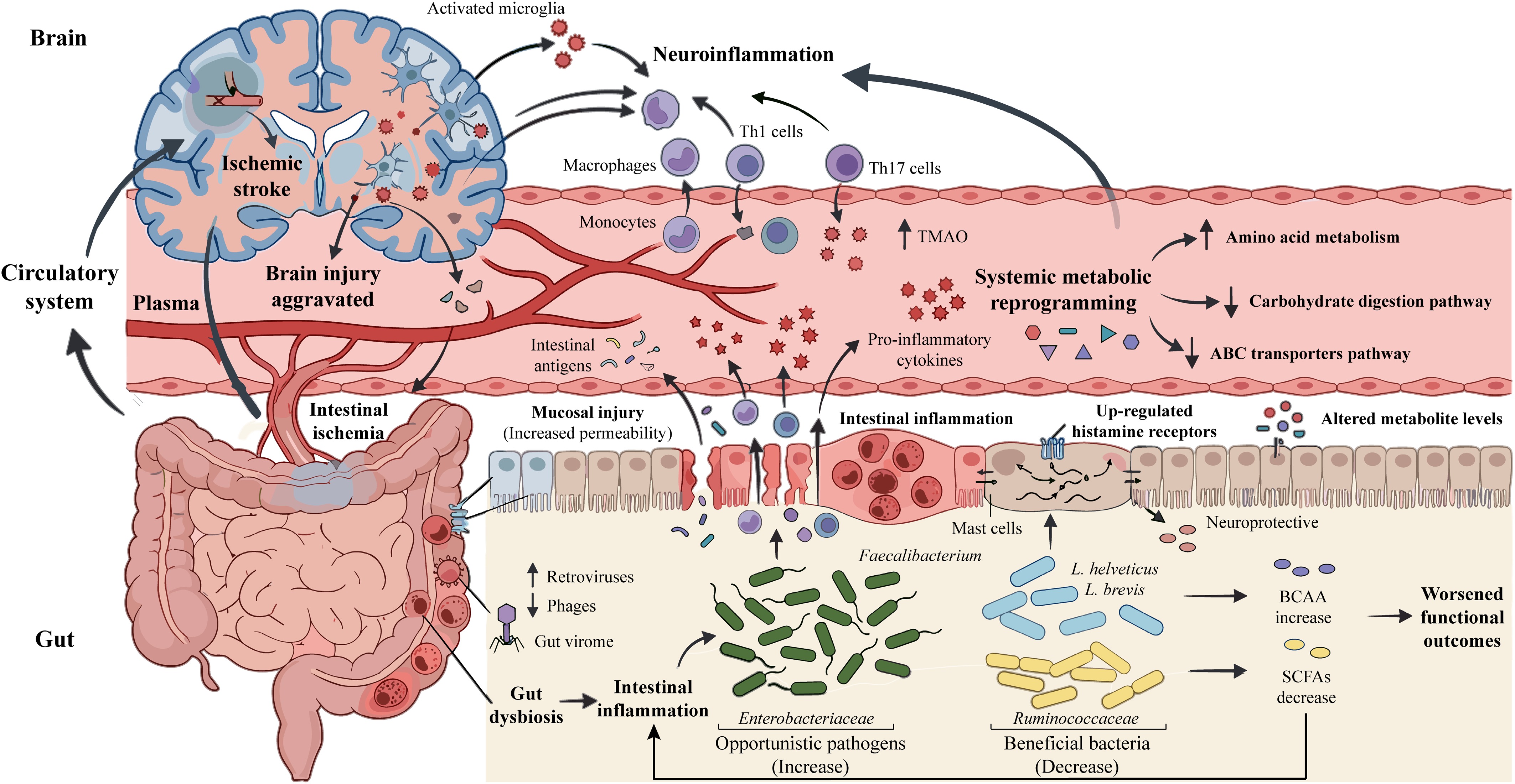
Figure 2.
The brain-gut-microbiota axis in ischemic stroke. Following ischemic stroke, brain injury and released DAMPs contribute to intestinal ischemia, dysbiosis, and mucosal injury. Immune cells, bacterial metabolites, and antigens enter the circulation, forming a feedback loop that amplifies neuroinflammation and secondary brain injury. The figure was independently drawn using Adobe Illustrator. BCAA, branched-chain amino acid; DAMPs, damage-associated molecular patterns; SCFAs, short-chain fatty acids; TMAO, trimethylamine-N-oxide.
The changes in the α-diversity of microorganisms after ischemic stroke in clinical reports are inconsistent. Some studies report that the diversity has increased[53], while others find no significant change[16]. However, in animal models, the diversity decreases in ischemic stroke rats[54]. Furthermore, the variability in microbial abundance can also be affected by gender, age, and other factors[55]. Male mice have higher levels of colonic antimicrobial protein (AMP) after tMCAO, while female mice show increased abundances of Lactobacillus and Bifidobacterium, which is regulated by estradiol[56]. Nevertheless, some changes at the family level are the same. Enterobacteriaceae (opportunistic pathogens) increase in patients and animal models[55,57,58], while beneficial families such as Ruminococcaceae decrease[55,59,60]. It should be noted that Faecalibacterium (Ruminococcaceae) produces neuroprotective SCFAs[60], and specific Lactobacillus helveticus and L. brevis reduce the accumulation of branched-chain amino acids (BCAA), providing a neuroprotective effect[48]. Reproductive ageing in female rats is associated with decreased butyrate and elevated plasma lipopolysaccharide (LPS) after ischemic stroke[61]. Beyond bacteria, ischemic stroke also alters the gut virome, increasing the number of retroviruses such as Murine leukemia virus, while the number of phages decreases, such as Lactobacillus prophage Lj771[62].
These pathological changes include intestinal damage, microbial disorders, and immune responses, which cause the development of ischemic stroke under metabolic disturbance conditions. In the rat ischemia/reperfusion (I/R) model, intestinal flora disorder aggravated intestinal inflammation due to the accumulation of BCAA[47] and a decrease in SCFAs[63], thus worsening the functional results. Preclinical studies found that microbial imbalance increases trimethylamine-N-oxide (TMAO), inflammatory cytokines, and disordered amino acid metabolism levels, thus aggravating brain damage[64,65]. Metabolomic research on miniature pigs showed that the levels of arginine, proline, and cyanoamino acid metabolism increased, and the plasma ABC transporter and carbohydrate digestion pathways were reduced after acute ischemic stroke[64].
Therefore, ischemic stroke can cause damage to intestinal tissue, destroy microbial and viral communities, stimulate immune responses, and aggravate brain damage through inflammation and metabolic mechanisms. After ischemic stroke, the α-diversity of intestinal microbiota has different results in clinical observations and animal studies, but the same pattern appears at the family level. Therefore, it should accurately take into account factors such as human disease subtypes, pathological dynamic evolution, and complex complications, so as to design more targeted and clinically feasible treatment strategies.
Neuro-immune pathways in the brain-heart axis
-
Ischemic stroke generally causes sympathetic hyperactivation or parasympathetic inhibition[66] (Fig. 3). The hypothalamic paraventricular nucleus (PVN) is an important central regulator of the autonomic nervous system and cardiovascular function (Table 1). In most cases, PVN stimulation increases the output of the sympathetic nerve and causes arrhythmia. Rat studies showed that MCAO surgery and the injection of L-glutamate into the brain caused the expression of Fos protein, which is a marker of PVN neuron activation and also increases the risk of arrhythmia[26]. Conversely, VNS in MCAO/R rats reduced chymase and abnormal angiotensin II levels by inhibiting mast cell activation and the release of pro-inflammatory factors, thus reducing myocardial atrophy[67].
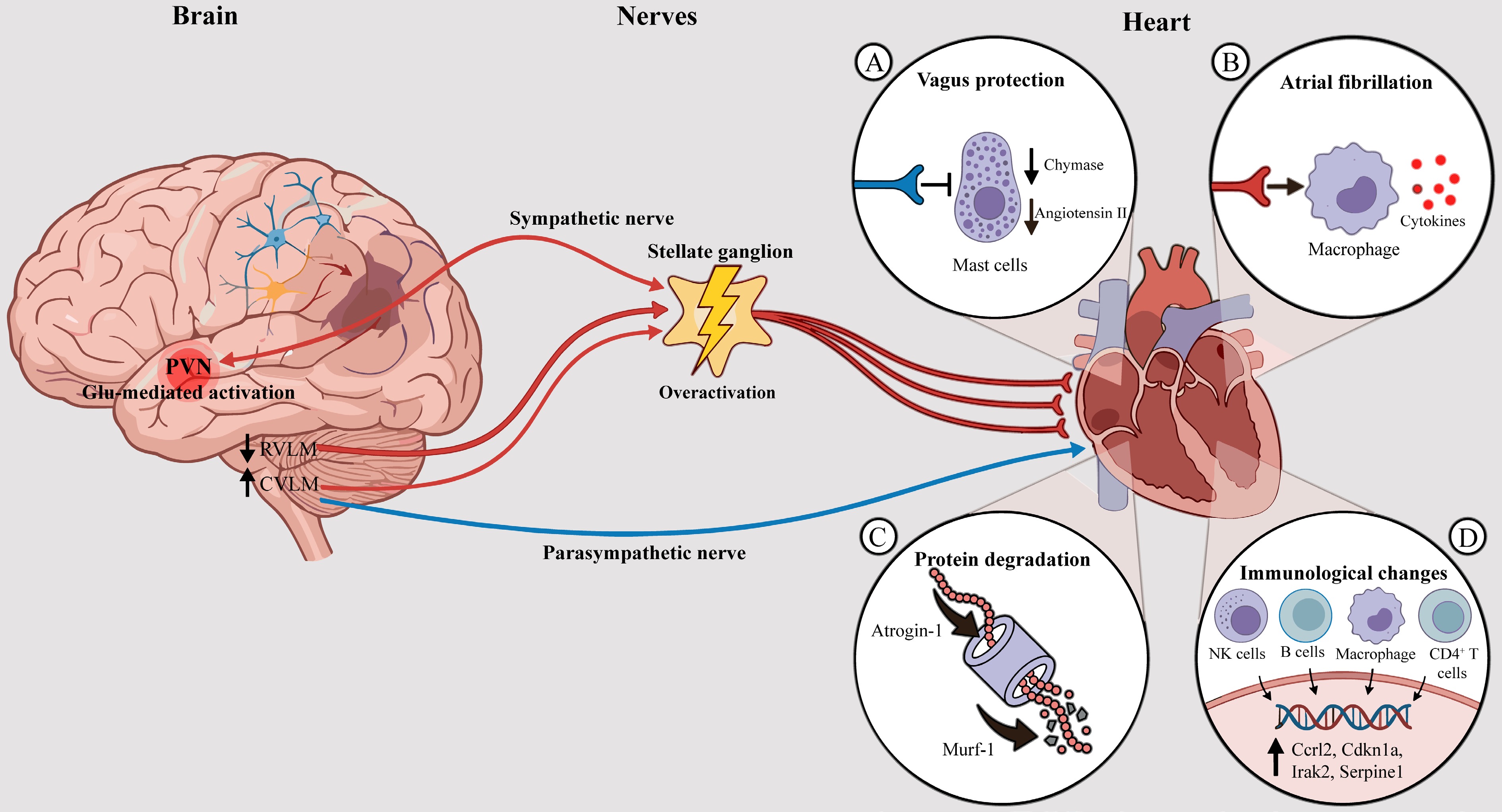
Figure 3.
The brain-heart axis in ischemic stroke. Cerebral ischemia activates the PVN and triggers sympathetic overactivation, increasing the stellate ganglion activity and promoting cardiac inflammation. Meanwhile, vagal inhibition reduces parasympathetic tone, activating mast cells and exacerbating cardiac injury, atrial fibrillation, and other arrhythmias. The figure was independently drawn using Adobe Illustrator. CVLM, caudal ventrolateral medulla; PVN, paraventricular nucleus; RVLM, rostral ventrolateral medulla.
Table 1. Key targets of brain-organ axes in ischemic stroke.
Organ axis Target Molecular mechanism Effects Ref. Brain-heart axis PVN Glutamate activates NMDA receptors on PVN neurons, increasing sympathetic outflow Increased susceptibility to post-stroke cardiac arrhythmias [26] Brain-liver axis TFF3 Liver-derived TFF3 interacts with neuronal LINGO2, activating EGFR/Src signaling, and upregulates Bcl-2 Reduces neuronal apoptosis and alleviates neurological deficits [74] Brain-spleen axis α- and β-adrenergic receptors Norepinephrine activates α- and β-adrenergic receptors, promoting splenic contraction and immune cells mobilization Immune cell mobilization and splenic atrophy after ischemic stroke [76,77] Brain-spleen axis α7nAChR Vagal cholinergic signaling activates α7nAChR on immune cells, inhibiting pro-inflammatory cytokines production through the cholinergic anti-inflammatory pathway Suppresses pro-inflammatory cytokine production and attenuates splenic inflammation after ischemic stroke [78] Brain-spleen axis CD147 CD147 upregulation in the spleen after cerebral ischemia promotes NF-κB signaling and pro-inflammatory mediators release Promotes NF-κB-driven pro-inflammatory responses and splenic immune cell infiltration after ischemic stroke [80] Brain-lung axis α-MSH α-MSH signaling through MC-1R suppresses pulmonary innate antibacterial responses Suppresses innate antibacterial immune defenses, leading to increased pulmonary bacterial load and heightened susceptibility to pneumonia after ischemic stroke [105] Brain-lung axis NLRP3 Cerebral ischemia activates pulmonary NLRP3 inflammasome signaling, promoting caspase-1 activation, IL-1β release, and inflammatory lung injury Caspase-1/IL-1β-mediated inflammation exacerbates pulmonary injury after ischemic stroke [107] Brain-lung axis β-adrenergic receptors Catecholamines-mediated β-adrenergic signaling suppressing pulmonary immune defenses Suppresses innate antibacterial defenses, transforming minor bacterial aspiration into fatal pneumonia, increasing susceptibility by approximately 1,000-fold after ischemic stroke [102] Brain-lung axis α7nAChR Acetylcholine released from hyperactivated parasympathetic nerves binds to α7nAChR on lung immune cells, suppressing pulmonary innate immunity through the cholinergic anti-inflammatory pathway Suppresses pulmonary antibacterial defenses and increases susceptibility to stroke-associated pneumonia [104] Brain-kidney axis HIF1α HIF1α is stabilized in glomerular podocytes and transcriptionally upregulates the ZEB2/TRPC6 axis, leading to TRPC6-mediated calcium influx and aberrant FAK activation Podocyte cytoskeletal rearrangement and foot process effacement disrupt the glomerular filtration barrier, contributing to proteinuria after ischemic stroke [43] Research has found that the neural signaling from the PVN is integrated and relayed by the downstream nuclei, enabling it to exert precise regulation of cardiovascular function. The ventrolateral medulla (VLM) of the brainstem consists of two parts, including the anterior side (RVLM, pressor) and the tail (CVLM, depressor). They regulate cardiovascular reactions and receive input from the PVN. In MCAO rats, the decrease in the expression of neuronal nitric oxide synthase (nNOS) in neurons in the ipsilateral RVLM and the increase in the expression of nNOS in the CVLM are the main reasons for the reduction of cardiovascular response during isometric muscle contraction[27]. The peripheral sympathetic ganglion, especially the stellate ganglion, dominates the heart and transforms the command of the center into the function of heart regulation. In the dog MCAO model, increased stellate ganglion activity, myocardial macrophage infiltration, and elevated pro-inflammatory cytokines aggravate AF during ischemic stroke, whereas stellate ganglion ablation or macrophage consumption inhibits AF[28]. Similarly, left stellate ganglion ablation in canines with acute ischemic stroke can inhibit the polarization and activation of macrophages caused by sympathetic nerves, thereby reducing ventricular arrhythmia[68]. Sympathetic dysfunction also promoted myocardial atrophy in MCAO mice and accelerated the decomposition of proteins in the heart through the upregulation of E3 ubiquitin ligases atrogin-1 and MuRF1[25].
Although the neural mechanisms are the main aspect of cardiac injury after ischemic stroke, the immune response is also closely related. Cardiac atrophy following tMCAO surgery in mice was connected with the differential expression of 383 genes in myocardial tissue, mainly including immune response, inhibition of collagen production, and oxidoreductase, peptidase, and endopeptidase activation, thus causing cardiac dysfunction[69]. Experimental research showed that MCAO mice exhibited higher percentages of activated NK cells, B memory cells, macrophages, and CD4+ naive T cells in cardiac tissue. Many important immune-related genes, including Ccrl2, Cdkn1a, Irak2, and Serpine1, were significantly elevated and related to immune cell infiltration[70].
These findings emphasize that nerve and immune pathways work together in causing cardiovascular complications after ischemic stroke.
Integrated neuro-immune-metabolic dynamics of the brain-liver axis
-
The liver is another important organ involved in the NIMN after ischemic stroke, which has a significant impact on brain damage by triggering immune cells and modulating metabolite synthesis. Parasympathetic nerve inhibition increased vitronectin (VTN) production in the liver of female mice after cerebral stroke, aggravating interleukin-6 (IL-6)-mediated inflammation and brain injury[71]. The liver also releases monocytes, pro-inflammatory cytokines, and hepatotoxic metabolites such as ammonia and lactic acid into the blood[72], destroying the integrity of the BBB and promoting neuroinflammation[73].
In addition to pro-inflammatory effects, the liver also secretes neuroprotective factors. After cerebral ischemia, trefoil factor 3 (TFF3) increased to varying degrees in the liver and blood (Table 1). Delayed revascularization allowed TFF3 to enter the brain through restored blood flow, where it bound to neuronal leucine-rich repeat and immunoglobulin-like domain-containing protein 2 (LINGO2), thereby reducing its inhibitory effect on epidermal growth factor receptor (EGFR). This activated Src kinase and upregulated Bcl-2 expression to reduce neuronal apoptosis and improve neurological outcomes[74]. Hepatectomy reduced the level of TFF3 and eliminated the neuroprotective effect, while supplementing with recombinant TFF3 protein could reverse this effect through the LINGO2/EGFR/Src signaling pathway[74].
Furthermore, the liver serves as a key regulator of metabolic reprogramming after cerebral ischemic injury. Ischemic stroke promotes hepatic gluconeogenesis through multiple pathways to produce hyperglycemia. The expression levels of key gluconeogenic enzymes (FBP1, G6PC, PCK) and their transcriptional regulators (FoxO1, C/EBPs, CREB) were increased, while stress kinases (p38, JNK) inhibited insulin signaling by reducing the phosphorylation of IR, IRS1, Akt, and AMPK[35]. Interestingly, gluconeogenesis, glycolysis, ketogenesis and β-oxidation were decreased within 24 h, but β-oxidation and ketogenesis rebounded, and glycolysis returned to normal by the 5th day[75].
The crosstalk between metabolism and inflammation forms a vicious circle, in which the inflammatory factors from the liver aggravate insulin resistance, which disrupts the immune metabolism of the liver, suggesting that interrupting this feedback loop is crucial for improving the outcome of stroke.
Neuro-immune circuitry of the brain-spleen axis
-
As a central organ of peripheral immunity, the spleen exerts crucial control over immune responses following cerebral ischemia via the neuro-immune pathway (Fig. 4). After cerebral ischemia, sympathetic and parasympathetic nerves participate in the immune regulation of the spleen. Sympathetic nerve excitation triggers the release of norepinephrine, which acts on α- and β-adrenergic receptors on the smooth muscle of the spleen, causing immune cells to enter the circulation and the spleen to atrophy[76,77]. On the contrary, the parasympathetic nerve input is mainly mediated through the vagus nerve, which promotes the release of acetylcholine and activates the α7 nicotinic acetylcholine receptor (α7nAChR) on the spleen macrophage, thus inhibiting the secretion of pro-inflammatory cytokines[78] (Table 1).
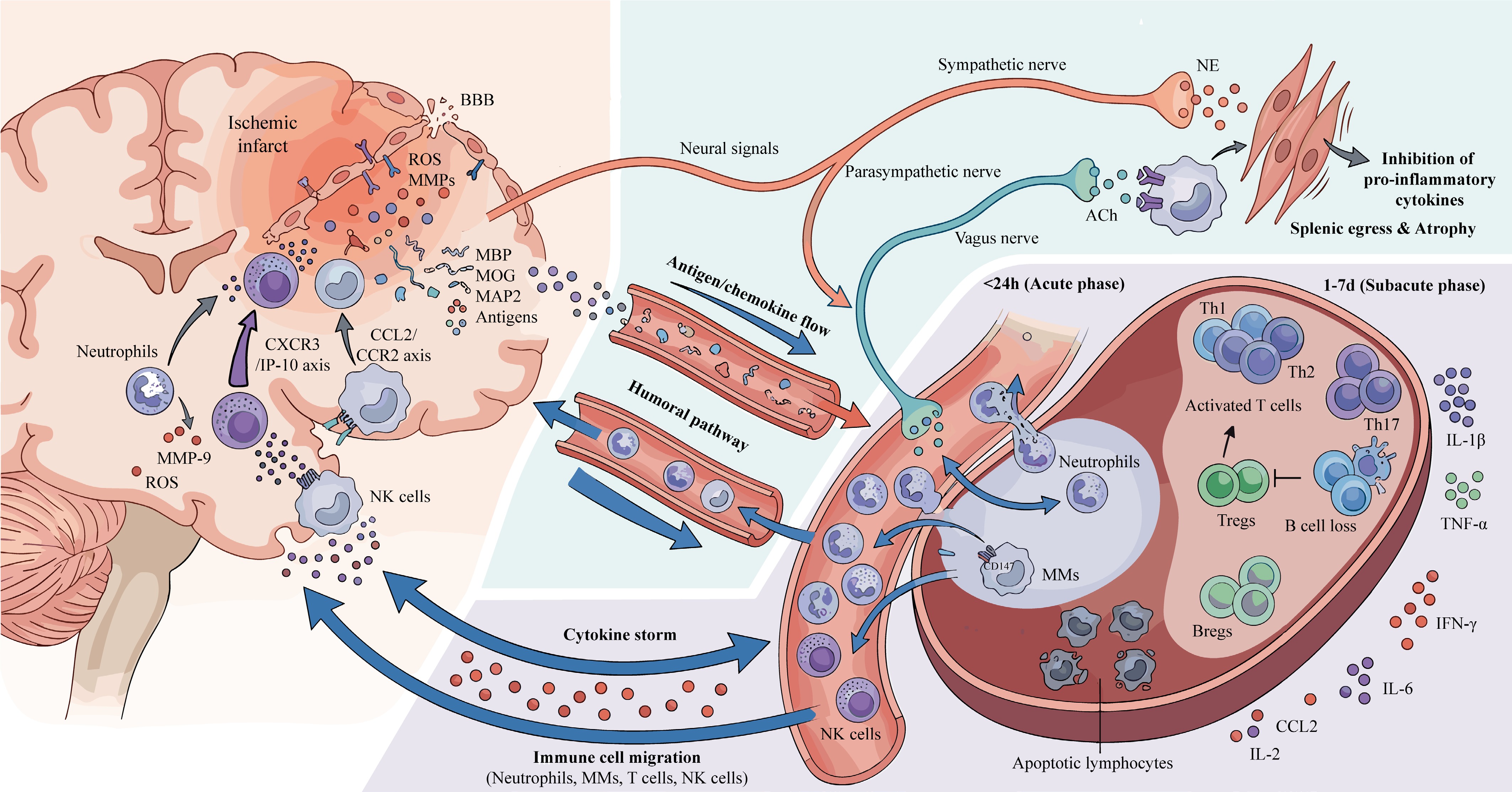
Figure 4.
The brain-spleen axis in ischemic stroke. Cerebral ischemia induces sympathetic activation, leading to immune cell egress and splenic atrophy, and reduces parasympathetic activity to promote inflammation. This results in the release of pro-inflammatory cells and cytokines into the circulation.These cells infiltrate the brain, while cytokines reach the brain and amplify neuroinflammation. The figure was independently drawn using Adobe Illustrator. ACh, acetylcholine; BBB, blood-brain barrier; MAP2, microtubule-associated protein 2; MBP, myelin basic protein; MOG, myelin oligodendrocyte glycoprotein; NE: norepinephrine.
After ischemic injury, the damaged brain tissue releases multiple alarm signals. In response, it triggers a systemic immune response and produces a large number of pro-inflammatory factors[37,79]. In the acute stage, splenic neutrophils, monocytes/macrophages (MMs), and NK cells react quickly and enter the ischemic brain. Neutrophils represent the earliest immune cells to enter the brain, where they rapidly infiltrate and trigger neuroinflammatory responses after ischemic brain injury. Similarly, the spleen MMs are rapidly mobilized, depleted in the spleen, and accumulated in the brain of the MCAO mice[6,80]. Splenectomy before cerebral ischemia inhibited the aggregation of MMs in the brain, indicating that the spleen serves as a primary reservoir of inflammatory monocytes[6]. Macrophage depletion in the tMCAO model alleviated cerebral injury and reduced infarct volume, which once again proved the role of macrophages[81].
As the damage develops into the subacute phase, lymphocytes take on the main role, shifting from immediate defense to targeted immune regulation. Clinically, there were biphasic changes in spleen volume; that is, it decreased in the early stage, reached the lowest point 48 h following ischemic stroke, and then restored to baseline[82]. This volume is inversely associated with the percentage of blood lymphocytes[83]. In the mouse MCAO model, Th1, Th2, and Th17 cells in the spleen were significantly increased[84], and activation markers such as CD25 and CD122 were elevated in the T cell phenotype[85]. Regulatory T cells (Tregs) are also increased in the spleen after ischemic stroke[86]. Depletion of Tregs would strengthen the activation of microglia and T cells, aggravating brain damage and worsening functional outcomes[87], while infusion of Tregs reduced the BBB destruction, inflammation, and peripheral cell infiltration, and provided neuroprotection[88]. B cells are also a major lymphocyte group in the spleen, which decreases 96 h after the occurrence of tMCAO. The mice lacking B cells had increased infarct volumes, thus proving their protective role in ischemic stroke[89]. Splenic regulatory B cells (Bregs) also played a role in inhibiting cerebral I/R damage[89]. However, ischemic stroke causes another systemic immunosuppressive state called stroke-induced immunosuppressive syndrome (SIIS), which is characterized by increased lymphocyte apoptosis and impaired function of the spleen[90].
Splenic immunity is modulated by specific molecular signals in the context of ischemia. Brain-derived antigens, including microtubule-associated protein 2 (MAP2), myelin basic protein (MBP), and myelin oligodendrocyte glycoprotein (MOG), are recognized by immune cells and cause immune activation[91]. Activated splenic immune cells (mononuclear cells and T cells) produce pro-inflammatory factors, mainly TNF-α, interferon-γ (IFN-γ), IL-6, MCP-1, and interleukin-2 (IL-2)[92]. In addition, chemokines and their receptors show increased expression, such as CCL2, C-C motif chemokine ligand 5 (CCL5), C-X-C motif chemokine ligand 2 (CXCL2), and C-C motif chemokine receptor 2 (CCR2) in the brain and spleen, which jointly promote cell transportation to the damaged area[86, 92]. The CCL2-CCR2 axis promotes the infiltration of mononuclear cells into the brain, increasing neuroinflammation[93]. NK cells are attracted via CXCR3-IP-10 and release cytotoxic mediators such as IFN-γ to destroy the integrity of the BBB, thus aggravating cerebral damage[94]. CD147 is a highly expressed glycoprotein in the spleen after cerebral ischemia, which can promote the activation of inflammatory MMs. Using specific antibodies (αCD147) to block CD147 inhibited this reaction and highlighted its therapeutic value[80,95].
After ischemia, the brain produces pro-inflammatory mediators, reactive oxygen species (ROS), and matrix metalloproteinases (MMPs), which destroy the integrity of the BBB. The upregulation of endothelial adhesion molecules such as intercellular adhesion molecule-1 (ICAM-1) and vascular cell adhesion molecule-1 (VCAM-1) causes adhesion and migration of immune cells[96]. Infiltrating neutrophils aggravate BBB damage due to the secretion of MMP-9[97,98] and damage tissues through the release of ROS, cytokines, and proteases[99]. Neutrophil extracellular traps (NETs) could also cause thrombosis and aggravate nerve damage[99,100]. Cytokines from the spleen can increase the levels of TNF-α, IFN-γ, IL-6, MCP-1, and IL-2 in the brain[101].
Therefore, the spleen has become the central link in peripheral immune regulation of ischemic stroke. It relies on autonomic nerves to regulate brain damage and coordinate immune responses. The acute phase involves the mobilization of neutrophils, followed by the subacute phase of lymphocyte regulation. According to the above process, it relies on antigen recognition, cytokine networks, and chemokine signals to affect BBB integrity and tissue repair.
Neuro-immune pathways in the brain-lung axis
-
The lung serves as a key component of the peripheral immune system and exhibits a unique response pattern in immune disorders caused by ischemic stroke. The brain can locally cause pulmonary immunosuppression through excessive activation of the sympathetic nervous system. Mouse experiments induced by MCAO showed that blocking β-adrenergic receptors alleviated immunosuppression and helped prevent lung infections[102]. At the same time, the parasympathetic nerve is also deeply involved in it. tMCAO model studies showed that cerebral ischemia and reperfusion damaged hypoxic pulmonary vasoconstriction (HPV) through excessive activation of the vagus nerve, destroyed the blood-gas barrier, and caused respiratory acidosis; cutting off the vagus nerve could reverse these effects and reduce lung complications, but did not affect the volume of infarction[103]. Consistently, a study found that after MCAO, the parasympathetic activity continued to increase rapidly, and the vagus nerve released acetylcholine and acted on α7nAChR on lung cells, inhibiting innate lung immunity via the cholinergic anti-inflammatory pathway, resulting in stroke-related pneumonia[104]. In addition, the vagus nerve mediates the lung-brain anti-inflammatory reflex via α7nAChR, and the TRPV1+ receptor senses lung pathogens and activates the hypothalamic stress circuit[43]. These mechanisms form the basis of the two-way vicious cycle of the brain-lung axis. Targeting the vagus nerve pathway or α7nAChR is expected to prevent and treat lung complications after ischemic stroke (Table 1).
Following ischemic stroke, the lung shifts to an immunosuppressive state, mainly manifested as reduced lymphocyte count, decreased IFN-γ level, increased IL-4 production in lymphocytes, and increased macrophages IL-10 level[105]. Apoptosis of IFN-γ-producing cells and the decline in IFN-γ level are associated with susceptibility to lung infection following ischemic stroke in mice[106]. The NLRP3 inflammatory pathway can also play a role in pulmonary inflammatory injury. In NLRP3 knockout mice, p65 (NF-κB) phosphorylation decreased, oxidative stress decreased, macrophage and neutrophil infiltration were reduced, and caspase-1 and IL-1β levels decreased in the lung after ischemic stroke[107]. Lung immunosuppression and damage can further cause lung infection. Ischemic stroke can turn nasal bacteria into harmful lung pathogens[102]. In addition, the risk of lung infection can also be amplified by impaired immune function of the spleen. The experimental MCAO model showed that splenic B cell numbers and IgM concentrations declined markedly, a change closely linked to the development of spontaneous bacterial pneumonia[108]. Spleen weight reduction is negatively related to the severity of infection[109], highlighting the coordination failure of spleen and lung immunity, resulting in worsening outcomes.
In addition to the immune mechanism, cerebral ischemia can also directly damage the lung through different molecular pathways. Ischemia can lead to excessive production of glial maturation factor-β (GMFB) in brain astrocytes. After GMFB enters the circulation, it targets pulmonary microvascular endothelial cells (PMVEC), increases intracellular ROS, promotes cell apoptosis, thus causing lung damage[110]. Cerebral ischemia increases vascular endothelial growth factor C (VEGFC) in serum and lung tissue, which is mainly highly expressed in neuronal cytoplasm, pulmonary endothelium, and lung cells. siVEGFC can promote the proliferation and survival of pulmonary epithelial cells (A549) and reduce lung cell apoptosis, thereby alleviating cerebral ischemia-induced pulmonary injury[111]. The expression level of Annexin A5 in lung tissue is higher than that of other major organs, and the content is the lowest in the brain. In the MCAO model, the content of Annexin A5 decreased in the lung tissue and increased in the brain after cerebral ischemia. Exogenous Annexin A5 can cross the blood-brain barrier, reduce infarct volume, and improve neurological outcomes. Conversely, anti-Annexin A5 antibodies aggravate brain damage[112]. These results show that Annexin A5 may act as a protective signaling molecule secreted from the lungs to the brain, mediating endogenous regulation of the brain-lung axis.
After the collapse of the pulmonary immune defense system, it aggravates brain damage to each other. Rat pneumonia increased systemic inflammatory markers and thus aggravated neuronal death[113]. Similarly, mouse respiratory syncytial virus increased BBB permeability and made macromolecular substances easier to enter the brain, thereby promoting the synthesis of nitric oxide and TNF-α in astrocytes[114].
Therefore, ischemic stroke causes immunosuppression of the lung through the neuro-immune pathway, resulting in an abnormal immune response and direct lung damage. The crosstalk between the lung and the spleen will affect susceptibility to infection and aggravate nerve damage. The brain-lung axis provides an important understanding for preventing and treating post-ischemic stroke infection, which is conducive to improving the clinical effect.
Neuro-metabolic dynamics in the brain-kidney axis
-
Kidney dysfunction is tightly associated with the progression of ischemic stroke. In MCAO/R rats, there were significant pathological and molecular changes in the kidney, manifested as a marked increase in serum urea nitrogen (UREA) and creatinine (CREA) levels, suggesting impaired glomerular filtration function[115]. Sympathetic activation after renal ischemia/reperfusion injury causes renal vasoconstriction, which aggravates acute kidney damage[116,117]. In addition, ischemic stroke causes microRNA imbalances to aggravate kidney damage. A total of 138 microRNAs were upregulated in the whole blood, and 19 microRNAs were downregulated in young ischemic stroke patients[118]. In this system, miR-21, miR-29c and miR-200b/c were associated with renal dysfunction[119−121]. Ischemic stroke and its induced hypoxic injury can upregulate the expression of HIF1α in the kidney. In the glomerular podocytes of MCAO rats, the expression of ZEB2 and TRPC6 was elevated, causing calcium overload. This leads to stress fiber rearrangement through FAK activation, which ultimately leads to glomerular podocyte damage and proteinuria[43]. Conversely, CKD also aggravates post-stroke brain damage. In the acute recovery stage, CKD worsened tMCAO-induced brain injury, with increased apoptosis and neuron loss in the ischemic core and penumbra by promoting pro-inflammatory M1-type polarization of microglia/macrophages while suppressing restorative M2-type polarization[122].
On the whole, the six peripheral organ axes of the gut, heart, liver, spleen, lung, and kidney go beyond their traditional role as passive targets for ischemic stroke. On the contrary, a multi-organ network is formed, relying on neuro-immune-metabolic communication to actively amplify ischemic brain injury. By driving systemic inflammation, immune imbalance, metabolic disorders, and barrier destruction, each axis aggravates the secondary cerebral ischemic injury. Therefore, viewing peripheral organs as the central link in ischemic stroke intervention provides new opportunities for treatment.
Notably, these signaling axes converge on multiple shared molecular hubs (Table 1), such as α7nAChR and α1- and β- adrenergic receptors. As a key node of nerve and immunity, α7nAChR can be activated to restrain the release of pro-inflammatory mediators in peripheral macrophages through the cholinergic anti-inflammatory pathway, thus reducing the risk of immunosuppression and infection after ischemic stroke. In addition, the overactivated sympathetic nervous system after ischemic stroke is transmitted through α1- and β-adrenergic receptors, which regulate the output of splenic immune cells and affect systemic metabolic homeostasis. These interrelated pathways exacerbate the systemic pathophysiological process, forming a vicious cycle between central injury and peripheral organ dysfunction.
-
In view of this, the intervention strategy for multi-organ interaction injury after ischemic stroke needs to go beyond the traditional single neuroprotection concept and turn to systematic regulation with the goal of the NIMN. Based on the pathological mechanisms of the aforementioned peripheral organ axes, as well as upstream regulatory factors and common molecular hubs across these axes, this chapter will focus on how to block the vicious circle between peripheral lesions and central injury. First of all, by targeting the neural circuit, the aim is to regulate the imbalance of the autonomic nerve from the source, so as to restore the central control function of peripheral immune organs. Second, the peripheral-central immune interaction is intervened to regulate cell function and the release of inflammatory mediators in peripheral immune organs, including the spleen and intestine, thereby reducing secondary brain injury. Finally, the microbiome-metabolic homeostasis is remodeled to restore systemic metabolic balance by restoring intestinal microecology and key metabolite levels. These strategies aim to provide a scientific basis for improving the long-term prognosis of patients with ischemic stroke through multi-dimensional and multi-target systematic intervention.
Neuro-modulatory therapeutics for ischemic stroke: targeting neural circuits
-
Based on the mechanistic understanding of neurological, immune, and metabolic imbalances, priority should be given to the neural pathway, which serves as the basic bridge between brain injury and systemic response. Therefore, starting from neural pathways is conducive to the development of potential drugs for treating ischemic stroke (Table 2). The sympathetic nervous system is the main transmission route of central stress commands, which coordinates peripheral immunity and metabolic reactions in two main ways: direct synaptic nerve transmission and systemic catecholamine release from the activated adrenal medulla. The excessive activation of the sympathetic nerve is closely related to spleen atrophy, peripheral immunosuppression, and the intensification of systemic inflammatory reactions, suggesting that the signaling pathway is a potential intervention target. Carvedilol is a non-selective adrenergic receptor blocker, which antagonizes the effect of blood-derived catecholamines released by blocking α1 and β adrenergic receptors, thereby relieving spleen atrophy after pMCAO[123]. As a specific β-adrenergic receptor blocker, propranolol can also reverse immunosuppression and spleen volume reduction caused by sympathetic hyperactivation after MCAO[124]. This evidence supports that excessive activation of sympathetic nerves has a potential causal effect on peripheral immune dysfunction. However, these two drugs are mainly indicated for treating cardiovascular diseases in clinical practice, not for stroke-related immune regulation. Astragaloside IV can prevent splenic atrophy, preserve splenic cellularity and inhibit splenocyte apoptosis in MCAO mice, which is linked to inhibiting the activation of the HPA axis and reducing glucocorticoid levels[125]. These changes are correlated with alleviated peripheral immunosuppression and improved prognosis of ischemic stroke.
Table 2. Neuro-targeted, immune-targeted, and metabolic-targeted drugs and their effects on ischemic stroke.
Organ axis Intervention Model Therapeutic effects/mechanisms Ref. Brain-spleen axis Carvedilol pMCAO in rats Blocks α1 and β-adrenergic receptors and attenuates catecholamine-induced splenic atrophy [123] Brain-spleen axis Propranolol MCAO in rats Reduces catecholamine signaling, restores splenic lymphoid tissue, and normalizes cytokine profiles [124] Brain-spleen axis Astragaloside IV MCAO in mice Inhibits HPA axis overactivation; prevents splenic atrophy and preserves NK, T, and B cells [125] Brain-spleen axis IL-33 Ischemic stroke established by 30-min MCAO in mice Modulates splenic Th1/Treg responses; reduces ischemic cerebral injury [127] Brain-spleen axis Resveratrol Cerebral I/R in rats Enhances splenic Treg numbers and function; attenuates post-stroke inflammation [128] Brain-spleen axis Recombinant T-cell receptor ligands Reversible MCAO in mice Modulates immune activation, limits leukocyte recruitment to the brain, and reduces splenic T-cell and neutrophil expansion [129] Brain-spleen axis Prazosin pMCAO in rats Reduces splenic TNF-α levels and ameliorates splenic atrophy [123] Brain-spleen axis Simvastatin tMCAO in mice Inhibits splenocyte apoptosis and prevents splenic atrophy by modulating
Bcl-2 and Bax expression[141] Brain-lung axis Agouti MCAO in mice Reduces pulmonary bacterial load and susceptibility to post-stroke pneumonia [105] Brain-lung axis Clenbuterol MCAO in mice Activates β2-adrenergic signaling to attenuate bacterial burden and pulmonary inflammation [126] Brain-lung axis GM-CSF MCAO in mice Improves post-stroke outcomes by enhancing neutrophil responses, reducing BALF bacterial load, and improving bacterial challenge [130] Brain-lung axis Low doses of IgM-enriched intravenous immunoglobulin tMCAO in mice Enhances spontaneous pulmonary bacterial clearance by improving pathogen opsonization and macrophage-mediated clearance [131] Brain-lung axis Aprepitant Intraluminal monofilament model of MCAO in mice Attenuates post-stroke pneumonia by reducing pulmonary infiltration of neutrophils and macrophages and decreasing the expression of IL-6, IL-1β, TNF-α, and other inflammatory cytokines in pulmonary tissue [133] Brain-lung axis Ginsenoside Rb1 MCAO/R in mice Reduces lung and brain injury by activating PPARγ, suppressing NF-κB-mediated inflammation [142] Brain-liver axis Bethanechol MCAO in mice Reduces VTN transcription and protein release in hepatocytes, and stroke-induced plasma VTN levels [71] Brain-liver axis IL-13 pMCAO in rats Mitigates hyperglycemia and infarct size by modulating STAT3/STAT6 signaling pathways and reducing insulin resistance [5] Brain-gut-microbiota axis Cromolyn tMCAO in aged mice (18–20 months) Improves neurological function by reducing mast cell migration to the brain and decreasing plasma histamine and IL-6 levels [134] Brain-gut-microbiota axis Combination of β-asarone and paeonol MCAO in rats Upregulates cholecystokinin in intestinal mucosa and brain, modulates intestinal/brain NF-κB pathway, reduces peripheral IL-1β/TNF-α, and reduces intestinal inflammation and associated CNS inflammatory responses [135] Brain-gut-microbiota axis Lactulose Photothrombotic stroke in mice Improves metabolic and functional outcomes after stroke by correcting gut dysbiosis and restoring intestinal barrier integrity, and increasing anti-inflammatory factors in the gut [136] Brain-gut-microbiota axis Resveratrol Transient focal cerebral ischemia model in mice Attenuates cerebral infarction and alleviates neurological impairment by modulating the gut microbiome to promote an anti-inflammatory T-cell profile in the small intestine [137] Brain-gut-microbiota axis Indole-3-propionic acid Acute MCAO in mice Reduces neuroinflammation and infarct size by enhancing beneficial bacteria, strengthening intestinal barrier integrity, and modulating intestinal Treg/Th17 balance [138] Brain-gut-microbiota axis Broad-spectrum antibiotics Ischemia induced by endothelin-1 in rats Modulates gut microbiota composition, alters inflammatory signaling, and influences neurological outcomes [139] Brain-gut-microbiota axis Shuanglu Tongnao compound Ischemic stroke in rats established by the Longa's wire bolus method Attenuates neuroinflammation and infarct volume by modulating gut microbiota composition, upregulating tight junction protein expression, and suppressing intestinal NF-κB activation [140] Brain-gut-microbiota axis Escin MCAO rats and LPS-induced Caco-2 cell model Reinforces intestinal tight junctions, reduces endotoxin leakage, and suppresses neuroinflammation through the LPS/TLR4/NF-κB pathway [143] Brain-gut-microbiota axis Fermented soybean (Chungkookjang) Transient forebrain ischemia induction in gerbils Reduces neuronal death and cerebral dysfunction by enriching beneficial cecal microbiota, suppressing LPS biosynthesis and deleterious fatty acid metabolism, and increasing propionate and butyrate levels [152] Brain-gut-microbiota axis Dengzhan shengmai Cerebral ischemia in rats Improves cognitive outcomes by upregulating monocarboxylic acid transporters to facilitate SCFA delivery to the brain, restoring intestinal barrier integrity, limiting LPS translocation, and dampening neuroinflammatory responses by the PI3K/AKT/caspase-3 pathway [153] Brain-gut-microbiota axis Huangqi-Honghua combination MCAO/R in Sprague-Dawley rats Mitigates neuroinflammation by remodeling gut microbial communities and activating the bile acid receptor FXR signaling pathway [154] Brain-gut-microbiota axis Angong Niuhuang Pill Mice with acute ischemic stroke induced by MCAO Decreases infarct size and restores neurological dysfunction by modulating specific microbiota taxa, elevating neuroprotective uridine levels, and suppressing pro-inflammatory prostaglandin I2 [155] Brain-heart axis Shuxuening injection tMCAO in mice Protects the heart and brain against ischemia/reperfusion injury through Tnfrsf12a-related inflammatory signaling [144] Brain-heart axis Resveratrol MCAO in resveratrol-pretreated rat Supports hemodynamic stability and improves cerebral perfusion by preserving mitochondrial function and inhibiting apoptosis in both the brain and heart [145] With sympathetic nervous system activation, it also triggers downstream effector factors, such as α-melanocyte-stimulating hormone (α-MSH), a molecule essential for regulating inflammation and immunity. The naturally occurring MC-1R antagonist agouti blocks α-MSH, significantly reduces the lung bacterial load within 72 h, and improves the antibacterial defense of the lungs after ischemia[105]. This suggests that α-MSH could contribute to regulating sympathetic nerve-mediated post-ischemic lung infection, but the causal regulatory pathway is not clear, and more mechanistic research is still needed for confirmation. Although β-adrenergic blockers can prevent lung infection, the use of the selective β2-adrenergic receptor agonist clenbuterol treatment is proven to reduce bacterial burden and lung inflammation after ischemic stroke[126]. This apparent contradiction suggests complex β-adrenergic signaling in post-ischemic lung immunity, warranting further study. The clinical application value of the above intervention methods still needs to be evaluated based on more solid mechanistic evidence and translational research.
The parasympathetic nervous system uses cholinergic signals to balance the stress response and maintain system stability. Bethanechol, an M1/M3 agonist, reduced VTN transcription and release, as well as plasma VTN levels[71]. Given that VTN is closely related to systemic inflammation, these results indicate that there may be a potential mechanism between vagus nerve tension, liver metabolism, and immune regulation.
Targeting key transmission pathways, especially sympathetic nerves and their effective factors, has been shown to improve spleen atrophy, reduce bacterial load, and reduce systemic inflammation. On the contrary, strengthening the activity of the parasympathetic nerves more fully demonstrates the multiple purposes of nerve intervention. Therefore, it has great potential to enhance post-ischemic stroke functional outcomes from the perspective of coordinating nerve and immune mechanisms.
However, it must be clearly recognized that the current promising neuro-immune comprehensive treatment model relies mostly on evidence from preclinical research. For intervention methods based on updated and more precise goals, such as α-MSH, α7nAChR, and α1- and β-adrenergic receptors, their clinical translation is still lacking. There are many reasons for this translational dilemma. A primary obstacle is the poor CNS bioavailability and a narrow therapeutic window of receptor-targeting agents, leaving the ideal timing, dosage, and route of administration still to be clarified. Moreover, most preclinical data come from young animal models that fail to capture the advanced age, comorbidity status, and individual differences of human patients. The complex interactions and compensatory effects within the NIMN may also weaken the effectiveness of single-target intervention. This translational gap reveals an important direction for future studies; there is an urgent need to transform the mechanistic insights obtained through animal experiments into therapies that can safely and effectively treat patients.
Immune-regulatory strategies for ischemic stroke: targeting peripheral-central immune crosstalk
-
Given that cellular immune and inflammatory reactions are involved in post-ischemic stroke complications, targeting peripheral immunity is a promising strategy for treating ischemic stroke (Table 2). In the acute stage of ischemic stroke, immune activation occurs in the spleen and is accompanied by systemic inflammation, suggesting that interfering with splenic immune reactions can relieve stroke damage. For example, interleukin-33 (IL-33), a member of the IL-1 cytokine family, reduces ischemic brain injury by suppressing the response of Th1 cells and promoting the response of Tregs in the spleen[127]. Resveratrol can increase the number and enhance the function of spleen Tregs to slow down inflammation after ischemic stroke[128]. Recombinant T-cell receptor ligands (RTLs) inhibit immune activation and reduce the proliferation of T cells and neutrophils in the spleen, thus limiting the recruitment of cerebral white blood cells after experimental ischemic stroke[129]. These studies suggest that there is a correlation between the dynamic changes of spleen immune cells and ischemic stroke, but it remains to be clarified whether the changes in these cells occur with the therapeutic effect or only as a companion phenomenon of other systemic or brain changes.
In addition to triggering spleen activation, the immunosuppression caused by ischemic stroke also makes patients more prone to lung infections. Granulocyte-macrophage colony-stimulating factor (GM-CSF) administration increased the lung neutrophil count, decreased the bacterial counts in bronchoalveolar lavage fluid (BALF), and improved the response to bacterial challenges, thereby improving the outcomes in mice with ischemic stroke[130]. Low doses of IgM-enriched intravenous immunoglobulin (IgM-IVIg) enhanced lung bacterial clearance after tMCAO by promoting opsonophagocytosis and reducing pulmonary inflammation via immunomodulation[131]. Rosiglitazone is a PPARγ agonist, which was related to the transformation of neutrophil phenotype in the pMCAO model, which tended to be an anti-inflammatory N2 phenotype and brought a corresponding neuroprotective effect[132]. Although these strategies show a correlation with the improvement of experimental outcomes, the evidence is mainly based on preclinical research, and further human studies are needed to evaluate their applicability and safety in different ischemic stroke populations.
Following the discharge of pro-inflammatory cytokines, this process further intensifies systemic inflammation, which is another important intervention point. Prazosin is an α1-adrenergic receptor antagonist, which decreases splenic TNF-α expression and reduces splenic atrophy in the pMCAO model[123]. Aprepitant is a neurokinin-1 receptor antagonist, which can decrease neutrophil and macrophage infiltration, lower IL-6, IL-1β, TNF-α, and other expression of inflammatory mediators in the lungs, so as to reduce post-ischemic stroke pneumonia[133]. Cromolyn is a mast cell stabilizer that can reduce the number of mast cells from intestinal tissue to the brain, reduce the content of histamine and IL-6 in plasma, and improve neurological function after tMCAO[134]. In addition, β-asarone combined with paeonol can regulate the intestinal/brain NF-κB pathway, reduce peripheral IL-1β/TNF-α expression, and prevent intestinal inflammation from spreading to the CNS in MCAO rats[135]. These studies all support that targeted peripheral inflammation can favorably affect the prognosis of ischemic stroke.
In addition to direct immune regulation, regulating the intestinal microbiota could provide effective treatment. Lactulose can restore the diversity of intestinal microbiota, repair intestinal barrier integrity, increase anti-inflammatory factor expression in the intestine, improve metabolic disorders, and enhance neurological recovery after ischemic stroke[136]. Resveratrol regulates the intestinal microbiota to balance the anti-inflammatory T cells of the small intestine (Th2 exceeds Th1 and Treg exceeds Th17), thus reducing the infarction volume and improving neurological function[137]. Indole-3-propionic acid increases beneficial bacteria proliferation, improves intestinal barrier integrity, and regulates the activity of intestinal Tregs and Th17 cells, thus reducing the infarction volume and neuroinflammation after ischemic stroke[138]. Broad-spectrum antibiotic treatment changes the structure of the intestinal microbial community, increases the number of SCFA producers, inhibits NF-κB and TLR signals, reduces the inflammatory response of the system and brain, therefore reducing brain damage and improving behavior scores in MCAO rats[139]. Similarly, Shuanglu Tongnao compound can restore the composition of intestinal microbiota, increase tight-junction protein expression such as ZO-1, occludin, and claudin-5, and inhibit the activation of intestinal NF-κB, thus reducing the amount of infarction and neuroinflammation[140]. These microbiota-based strategies are closely related to reducing infarction volume and neuroinflammation, which reveals a very promising research direction.
In order to treat more accurately, the system-specific molecular pathway provides an attractive target for intervention in ischemic stroke. Simvastatin inhibits splenic cell apoptosis and prevents spleen atrophy by regulating the expression of Bcl-2 and Bax following MCAO[141]. Ginsenoside Rb1 can activate PPARγ and inhibit the activity of NF-κB. Additionally, it can promote Claudin-5 and VE-Cadherin expression to strengthen BBB integrity, thus reducing lung and brain damage in MCAO/R mice[142]. Analogously, escin reduces intestinal permeability and the leakage of endotoxin, and regulates the LPS/TLR4/NF-κB pathway to inhibit neuroinflammation[143]. Shuxuening injection regulates atherosclerosis and inflammatory response by tissue-specific methods, reduces the inflammatory markers of the heart, and upregulates the neuroprotective gene in the brain, so as to safeguard of heart and brain against I/R injury[144]. Resveratrol can maintain mitochondrial function and inhibit cell apoptosis in the brain and heart, contribute to multi-organ protection, thus supporting hemodynamic stability[145]. IL-13 can regulate STAT3/STAT6-dependent pathways and reduce insulin resistance, to reduce hyperglycemia and infarction area[5].
However, there are many difficulties from preclinical research to successful human trials. Providing effective interventions to animal models administered after ischemic stroke may not be effective in clinical studies due to clinical heterogeneity and the delayed manifestations in human patients[146]. In addition, the ischemic stroke population is very diverse in terms of cause, complications, and baseline immune status, which is not fully reflected in standardized animal models. Extensive and non-stratified clinical trials cannot show beneficial effects that are only available in specific subgroups[147]. Meanwhile, the traditional animal model cannot replicate the main characteristics of human ischemic stroke, common complications including the elderly immune system and atherosclerosis, or the common clinical conditions of reperfusion treatment[148]. Therefore, the intervention strategy should be adapted to the dynamic stages of the post-ischemic stroke immune reaction. Through real-time immune monitoring, the acute inflammatory stage in the ultra-acute phase and the subsequent immunosuppressive stage can be determined. This means that from the large number of recruited participants, targeted treatment is given to patients with specific immune characteristics, such as a high Treg/Th17 ratio or special bone marrow activation, to predict treatment responses[149]. Meanwhile, the development of novel systems such as bionic nanocarriers that can transport targeted drugs, or finding advanced agents such as engineered extracellular vesicles, can improve the overall efficacy and safety[150,151].
Microbiota-metabolism axis modulation for ischemic stroke: restoring systemic homeostasis
-
Metabolic dysregulation is a critical pathological mechanism after ischemic stroke and also represents a promising therapeutic direction that remains to be explored. Although acute reperfusion therapy addresses the fundamental issue of vascular blockage, the therapeutic strategies for restoring metabolic homeostasis in peripheral and brain tissues offer hope for the prognostic management of ischemic stroke. The preclinical evidence shows that the metabolic pathways influenced by intestinal microbiota and their metabolites, as a continuous approach for regulating the post-ischemic stroke microenvironment and promoting recovery, have a strong research foundation (Table 2).
Regulating the microbial ecology, metabolite composition, and host signaling pathways has become an effective approach for intervening in ischemic stroke. In ischemic stroke rodent models, fermented soybeans (chungkookjang) increased the abundance of Lactobacillus, Bacillus, and Akkermansia in the feces, inhibited LPS biosynthesis and harmful fatty acid metabolism, and increased propionate and butyrate levels. They also regulated inflammation and insulin resistance, promoting reduction of nerve cell death and the recovery of brain function[152]. The herbal formula Dengzhan Shengmai changed intestinal microbial composition and increased the content of SCFAs to restore gut barrier function; at the same time, the increase of intestinal SCFAs promoted the expression of monocarboxylate transporters, which accelerated the transport of intestinal-derived SCFAs to brain tissue, and ultimately inhibited the apoptosis of nerve cells through the regulation of the PI3K/AKT/caspase-3 signaling axis in the ischemic rat model[153]. Huangqi–Honghua combinations can maintain bile acid homeostasis by reconstructing the intestinal microbial community and activating the bile acid receptor FXR, thereby reducing the proportion of Th17 cells and expanding the population of Tregs, ultimately alleviating neuroinflammation and improving ischemic damage[154]. Angong Niuhuang Pill (ANP) can improve the imbalance of microbiota by regulating the types of specific intestinal microbial species. At the same time, it regulates microbial metabolites involved in inflammation and neuroprotection, including prostaglandin I2 and uridine. These changes are associated with a reduced cerebral infarct volume and improved neurological function in the acute ischemic stroke mouse model[155]. The above studies suggest the regulatory effect of intestinal microbiota and their metabolites on ischemic stroke, but the direct relationship between intervention and treatment effect still needs more preclinical verification.
The imbalance of intestinal microbiota represents an important link between peripheral organ dysfunction and systemic complications. Supplementing specific symbiotic bacteria is known to provide a protective effect in distal organs. For example, Parabacteroides distasonis can alleviate inflammatory arthritis[156], Bacteroides fragilis can improve renal fibrosis[157], and Barnesiella intestinihominis can help reduce hyperglycemia and liver metabolic disorders[158]. These findings indicate that targeting intestinal microbiota could represent a potential strategy for intervening in systemic microenvironmental disorders after ischemic stroke. In elderly mice after ischemic stroke, transplantation of SCFA producers, including Bifidobacterium longum, Clostridium symbiosum, Faecalibacterium prausnitzii, and Lactobacillus fermentum, can relieve neurological dysfunction and neuroinflammation[159,160]. Microbiota transplantation from female mice to male mice can reduce inflammation, promote the production of beneficial metabolites, reduce the infarct volume, and improve behavioral results and survival rate in the MCAO model[161]. It is worth noting that transplanting fecal microbiota from young healthy mice to elderly ischemic stroke mice can significantly decrease the volume of infarction and enhance the functional score, while transplanting the elderly fecal microbiota to young mice will aggravate the cerebral damage[162]. This suggests that the effects of microbiota intervention are modulated by factors like environment, age, and gender, offering a key reference for precise intervention strategies, though the underlying mechanisms remain to be elucidated.
In summary, targeting the intestine, microbial community, and metabolites is a therapeutic strategy to combat metabolic dysregulation after ischemic stroke. Although the positive effects of these treatments cannot be fully reflected in neurological deficit scales, such as the NIHSS, they may promote long-term recovery, quality of life, or rehabilitation response[163]. Against this background, the NIMN framework provides a systematic theoretical foundation for individualized treatment for ischemic stroke. In view of the significant individual differences in immune status, metabolic characteristics, and intestinal microbial composition, precise intervention should be guided according to the NIMN characteristics. For example, patients with sympathetic hyperactivation, spleen atrophy, or immunosuppression may be more suitable for adrenergic receptor blocking or α7nAChR treatment, while patients with gut microbiota disorders, metabolic disorders, or significant intestinal-brain inflammatory crosstalk may need to target microorganisms or metabolic pathways. At the same time, we should explore reasonable combined treatment plans that combine metabolic intervention with other neurological function recovery methods, and systematically evaluate their synergistic effects[164]. Classifying patients according to the immune phenotype, metabolic characteristics, and microbial composition can promote the personalization and precise intervention, so as to maximize the treatment effect and reduce the heterogeneity of the treatment response.
-
The revelation of the brain-peripheral organ axis has brought a different perspective for treating ischemic stroke. This section reviews the research progress from four aspects: neuromodulation technology regulates immune-mediated inflammation through peripheral nerve pathways; bioengineering and regenerative medicine strategies regulate peripheral immune organs by means of cell therapy or physical intervention; digital and computational medicine tools improve systemic blood flow and achieve precise regulation through physical means; and human simulation systems provide a predictive platform for the study of multi-organ interaction and drug screening.
Neuromodulation technology
-
VNS acts on the NIMN, which is a promising method for post-ischemic stroke functional recovery (Table 3). These strategies can be roughly divided into invasive and non-invasive modes based on their participation in peripheral nerves. The vagus nerve represents a critical route to regulating the inflammatory response, and its specific mechanism includes modulating acetylcholine release and activating the acetylcholine receptor on immune cells, thereby inhibiting inflammatory mediator expression and inflammatory cascades. VNS effectively reduced the infarct volume and improved the prognosis of neurological function, and exerted anti-inflammatory effects by activating the α7nAChR/JAK2 pathway in a rat MCAO model[165].
The non-invasive VNS has become a research hotspot because of its relatively safe operation and easy implementation[166]. It is typically separated into transcutaneous cervical vagus nerve stimulation (tcVNS) and transcutaneous auricular vagus nerve stimulation (taVNS). taVNS treatment boosted acetylcholine levels in the brain and inhibited the release of pro-inflammatory factors such as TNF-α, IL-6, and IL-1β, thereby ameliorating neurological deficits in MCAO models[167]. In addition, taVNS promoted axonal plasticity by activating the BDNF/cAMP/PKA signaling pathway after cerebral I/R injury[168]. For acute MCAO, treatment with taVNS significantly diminished infarct volume by 28% within 24 h and ameliorated neurological recovery[169]. Existing clinical observations have shown that taVNS can promote upper limb motor function restoration in patients with subacute ischemic stroke, and no significant adverse reactions have been observed during the treatment[170,171]. tcVNS can protect the BBB and inhibit neuronal apoptosis by decreasing matrix metalloproteinase-2/9 (MMP-2/9) expression[168]. Therefore, the neuromodulation landscape of ischemic stroke is increasingly inclined toward non-invasive ways. taVNS and related percutaneous methods provide people with a combination of mechanistic effects, including regulating inflammation, enhancing neuroplasticity, and safety, which provides a feasible and effective way for clinical translation.
Biological engineering and regenerative medicine strategies
-
Systemic immunity and inflammatory responses have a major impact on the development of ischemic stroke, and cellular therapy has emerged as a key strategy to relieve neuroinflammation and improve neurological outcomes. Studies have shown that transplantation of neural stem/precursor cells (NSPCs) combined with mesenchymal stromal cells (MSCs) effectively improved the NIHSS score of patients with ischemic stroke[172]. A clinical application of bone marrow mononuclear cell (BMMNC) transplantation in patients with subacute ischemic stroke showed that the operation was feasible at the technical level, the safety data were acceptable, and some subjects were observed to improve neurological function during follow-up[173]. In the rat model of pMCAO, it was found that when bone marrow stromal cells (BMSCs) were directly transplanted into the brain, the cells showed a tendency to migrate to the periphery of the infarct, and the expression of nerve cell markers was detected in the brain. In contrast, after intravenous administration, this direct migration was not observed[174]. Other studies demonstrated that intravenously injected BMSCs could exert therapeutic effects through peripheral mechanisms. In adult rats with ischemic stroke, intravenously injected BMSCs concentrated mainly in the spleen, where they reduced local inflammation, thereby indirectly lowering brain inflammation and infarct volume[175]. Intravascularly implanted BMSCs existed transiently in the brain during the early stage, which promoted the reduction of ischemic lesions by activating microglia in the MCAO rat model[176]. In the cell therapy of ischemic stroke, the administration method and cell type are important factors that must be repeatedly weighed when optimizing the treatment plan.
Cell therapy in ischemic stroke has developed from the laboratory to clinical trials (Table 3). A meta-analysis showed that stem cell treatment significantly enhanced the functional results (reducing mRS score and increasing FMA score) with a safety outcome similar to the control[177]. The phase II MASTERS trial further confirmed that intravenous injection of multipotent adult progenitor cells within 18–36 h after the onset of ischemic stroke was safe and feasible with durable functional recovery and reduced mortality[178]. However, the consistency of therapeutic effects is still challenging. The phase II/III TREASURE trial lacked evidence to show significant short-term efficacy, and the meta-analysis also revealed that the benefits across different functional scales were inconsistent, revealing an obvious translational gap that arose from multiple unresolved issues[179,180]. Key obstacles include ethical issues, allogeneic immune rejection, and high heterogeneity of cell types, doses, and administration routes.
Remote ischemic postconditioning (RIPostC) has a strong immunomodulatory effect. In the mouse MCAO model, it reversed T cell reduction in the spleen and lymph node, inhibited NK cell expansion within the spleen, and decreased pro-inflammatory cytokines in peripheral blood, thus reducing the systemic-central inflammatory cascade[181]. Remote liver ischemic preconditioning (RLIPC) targeted the AKT pathway to reduce I/R injury in the rat MCAO model[182]. After RIPostC treatment, the expression of C-X-C motif chemokine receptor 4 (CXCR4) in brain tissue increased, and the level of stromal cell-derived factor-1α (SDF-1α) in peripheral blood also showed an upward trend. Furthermore, RIPostC was found to exert its neuroprotective effects through the SDF-1α/CXCR4 signaling axis, which may be associated with enhanced neurogenesis and angiogenesis[183]. In addition, remote ischemic preconditioning promoted the production of mitochondrial-derived vesicles and reduced the nuclear displacement of AIF and EndoG to inhibit cell apoptosis, thus alleviating I/R injury after ischemic stroke[184].
The use of injectable hydrogels offers a promising approach for repairing and regenerating nerves following ischemic stroke. Hydrogels can accommodate a large number of substances in their structures, promote cell, drug, and protein transport, and facilitate nerve repair. The development of injectable hydrogels modifies the post-ischemic stroke microenvironment to promote endogenous neuroregeneration[185]. The induced pluripotent stem cell-derived neural precursor cells were encapsulated in a hyaluronic acid hydrogel matrix and introduced into the infarcted brain area of the cortical photothrombotic stroke mouse, where they can effectively differentiate into neuroblasts[186].
Digital and computational medicine tools
-
As a non-invasive physical therapy, enhanced external counterpulsation (EECP) mainly improves systemic and cerebral blood perfusion. An important development is its combination with the computing model, which transforms the application of experience into personalized and accurate processing. Using the combination of EECP and the brain multi-autoregulation (MA) model, researchers can adjust the treatment parameters according to the condition of personal cerebral blood vessels, especially neurogenic, myogenic, and shear stress mechanisms[187]. Using the MA model can simulate different degrees of cerebral vascular stenosis (such as 50%−80%) and determine the ideal time length for EECP pressurization. It can be seen from the simulation of the 0D/3D geometric multi-scale hemodynamic model that at 70%−80% narrowing, the EECP pressure release was set to 0.7 Tc (Tc represents the cardiac cycle), which maintained the flow rate of the narrow branch more effectively than 0.5 Tc. In the case of 80% stenosis, when 0.7 Tc was replaced by 0.6 Tc, the flow rate only decreased by 0.1 mL·s−1[187]. In addition, this method controlled the narrow distal time-averaged wall shear stress (TAWSS) within the safe endothelial range of 4 Pa to 7 Pa, which prevented vascular damage[187]. The strategy led by this model can accurately and safely alleviate cerebral ischemia caused by vascular stenosis and can be defined as an advanced digital treatment tool for ischemic stroke rehabilitation.
Non-invasive wearable biosensors can monitor inflammation-related immune status via cytokines, heart rate variability (HRV), metabolic indicators like glucose, and peripheral physiological biomarkers such as respiratory rate (RR) and skin temperature[188,189]. They can serve as digital biomarkers, providing continuous and objective physiological feedback for real-time adjustment of neuromodulation parameters. Meanwhile, these sensors can serve as critical components of the non-invasive closed-loop neuromodulation system. Based on data from sensors, it is possible to dynamically adjust the parameters of neuromodulation approaches such as VNS.
Human mimetic system for predictive screening
-
In order to test and screen out complex therapies that affect the interaction between multiple organs, a multi-functional research platform that simulates systemic human physiology is indispensable. Multi-organ chip and neurovascular unit-on-a-Chip (NVU-on-a-Chip) technologies have become revolutionary technologies in this field. Multi-organ chips use microfluid channels to connect the tissue niches of organs such as the heart, liver, and brain to each other, recreating a physiologically relevant 'vascular-flow-linked' environment[190]. These systems replicate human pharmacokinetic and pharmacodynamic (PK/PD) profiles to provide a basis for the efficacy and safety evaluation of candidate drugs, such as detecting biomarkers of cardiac toxicity during doxorubicin treatment. This increases the predictability of clinical translation and reduces the risk of drug research and development[190].
In comparison, the NVU-on-a-Chip aims to model the BBB as a central interface[191]. Adopting a triculture design, a micro-neurovascular unit was reconstructed to simulate the damage after ischemia, including the destruction of the BBB, and the permeability to 20 kDa FITC-dextran increased 35-fold under the experimental conditions[191]. In addition, the use of human neural progenitor or stem cells (hNPCs/hNSCs) on the platform has been shown to promote angiogenesis and neural regeneration in the ischemic model through immunomodulation and strengthening cell-cell junctions[192]. Therefore, NVU-on-a-Chip is not only a powerful tool to analyze the local pathological mechanisms of brain regions, but also a core screening platform to evaluate whether candidate drugs or interventions can penetrate the BBB and accurately target the CNS.
This section summarizes several new strategies that affect the interaction between peripheral organs and the brain. These include neuromodulation, bioengineering, regenerative medicine, and chip-based models, which are moving toward integrated and personalized medicine. Clinical studies show that these strategies can improve neurological function and motor recovery in ischemic stroke with acceptable safety (Table 3). Future research should explore the crosstalk mechanisms of organ specificity and improve combination strategies, and use multi-group and artificial intelligence methods to establish a targeted patient multi-organ network atlas for personalized treatment, laying the foundation for ischemic stroke repair therapy.
Table 3. Clinical research data of representative intervention strategies in ischemic stroke.
Therapy Intervention Outcomes Adverse events Ref VNS Implanted VNS device paired with 6-week rehabilitation, followed by continued rehabilitation through day 90 Upper extremity Fugl-Meyer score increased by 9.2 points, with improvements observed in the Box and Block Test, Nine-Hole Peg Test, and Stroke Impact Scale No severe adverse events associated with VNS treatment were observed during follow-up [193] taVNS 60 subjects (18–80 years) randomized to taVNS or sham stimulation plus rehabilitation Improved motor and sensory function and emotional responses No significant adverse reactions or discomfort were observed; transient skin redness occurred in two taVNS participants and resolved after current adjustment [194] taVNS 36 subjects with acute ischemic stroke randomized to taVNS or sham stimulation during and after mechanical thrombectomy No significant difference in systolic blood pressure variability within 24 h after mechanical thrombectomy No serious adverse events were recorded [195] BMMNC 77 patients aged 18−80 years with middle cerebral artery ischemic stroke received autologous BMMNC transplantation or control treatment Intra-arterial BMMNC transplantation was well tolerated in patients with acute ischemic stroke, but it did not significantly improve the mRS score at 180 d No dose-related differences were observed in adverse events; two cases of inguinal hematoma occurred in the low-dose group [196] MSC Five patients received intravenous infusion of autologous MSCs, whereas 25 control subjects received no cell therapy Intravenous infusion of autologous MSC was feasible and safe for patients with severe cerebral infarction, suggesting potential functional benefits; however, the small sample size requires further validation No cellular, serological, or imaging-detected adverse reactions [197] MSC Patients aged 18–70 years with moderate-to-severe subacute ischemic stroke within 2 weeks of onset received intravenous autologous MSC infusion MSCs enhanced motor recovery, potentially through sensorimotor neuroplasticity, which was manifested as a significant increase in motor-NIHSS, motor-Fugl-Meyer scores, and fMRI task-related activities Intravenous infusion of autologous MSCs was safe and feasible, and no unexpected serious adverse events occurred [198] CTX-DP 11 patients with ischemic stroke (NIHSS ≥ 6) received a single stereotactic injection of CTX-DP cells into the ipsilateral putamen, followed by follow-up for 2 years to collect clinical and brain imaging data After 2 years of follow-up, the median NIHSS improvement was 2 points, with a mean improvement of approximately 5 points No immune- or cell-related adverse events were observed [199] RIC 1,893 patients with acute moderate ischemic stroke within 48 h of onset were randomized to RIC plus standard care or standard care alone RIC increased the proportion of patients achieving excellent neurological function at 90 days compared with conventional care RIC and control groups showed adverse event incidences of 6.8% and 5.6%, respectively [200] -
This review emphasizes the change in understanding of ischemic stroke, shifting the focus from simple cerebrovascular events to systemic NIMN disorders. The dynamic communication between the brain and the peripheral organs (gut, heart, liver, spleen, lung, and kidney) aggravates secondary brain damage, thereby causing systemic complications through autonomic disorders, immune hyperactivation, and metabolic disorders. α7nAChR is a key interface between vagus nerve signaling and immunosuppression; its activation inhibits the excessive inflammatory response of the heart, lungs, spleen, and gut, thus providing a target for usable drugs for multisystem complications after ischemic stroke.
It follows that the treatment strategy based on NIMN has achieved a paradigm shift from single-target neuroprotection to central-peripheral synergistic therapy, covering nerve regulation, immune regulation, microbiome reprogramming, and regenerative medicine. Emerging tools such as transcutaneous VNS, stem cell transplantation, remote ischemic conditioning, and multi-organ chips provide technical support for accurate and personalized intervention. Future research should use spatial multiomics and single-cell technology to describe the spatial and temporal dynamics of organ crosstalk, develop multimodal combination schemes to balance nerve, immune, and metabolic branches, and build patient-specific NIMN models with the help of artificial intelligence.
This project was supported by the National Natural Science Foundation of China (No. 82474110), the Natural Science Foundation of Jiangsu Province (No. BK20231484), and the China Association of Chinese Medicine Youth Talent Support Project (No. CACM-2023-QNRC2-B07).
-
Not applicable.
-
The authors confirm their contributions to the paper as follows: writing original draft: Li J; writing, review & editing: Li J, Yao J, Zhao J, Lai Y, Zhou W, Gao W, Li Y; supervision: Zhou W, Gao W, Li Y; project administration and funding acquisition: Li Y. All authors reviewed the results and approved the final version of the manuscript.
-
Data sharing is not applicable to this review as no datasets were generated or analyzed.
-
The authors have no conflict of interest to declare.
- Copyright: © 2026 by the author(s). Published by Maximum Academic Press on behalf of China Pharmaceutical University. This article is an open access article distributed under Creative Commons Attribution License (CC BY 4.0), visit https://creativecommons.org/licenses/by/4.0/.
-
About this article
Cite this article
Li J, Yao J, Zhao J, Lai Y, Zhou W, et al. 2026. The neuro-immune-metabolic network in ischemic stroke: mechanisms and therapeutic targeting of brain-peripheral organ crosstalk. Targetome 2(5): e047 doi: 10.48130/targetome-0026-0045 

The neuro-immune-metabolic network in ischemic stroke: mechanisms and therapeutic targeting of brain-peripheral organ crosstalk
- Received: 11 March 2026
- Revised: 12 May 2026
- Accepted: 20 May 2026
- Published online: 20 September 2026
Abstract: Ischemic stroke is traditionally regarded as a focal brain injury, but recent studies have shown that it is essentially a disease of the neuro-immune-metabolic network involving multiple organs. This article reviews the two-way dialogue between the brain and the gut, heart, liver, spleen, lung, and kidney through autonomic nerves, immune-mediated inflammation, and metabolic pathways. Building on this framework, we delve into the dysregulation of brain-peripheral organ axes following ischemic stroke, focusing on key molecular hubs that drive the systemic pathological process. We then discuss the promising current treatment strategies and the clinical translation of emerging technologies. In summary, an integrated neuro-immune-metabolic framework is proposed to understand ischemic stroke as a systemic disease and to guide combined therapies that target the central nervous system and the peripheral organs to realize the transformation from local intervention to systemic treatment.