


HTML
-
Tomato (Lycopersicum esculentum L.) belonging to the family Solanaceae, is an important vegetable crop all over the world. It is one of the most popular and indispensable ingredients of the human diet. The crops were originated in Western South America and Central America (Wamache 2005). Although it is widely adapted with variable climatic conditions, tomatoes are still vulnerable to a variety of diseases and infections during production, harvesting, transportation and storage. Most of the diseases are a result of fungal infections.
Tomatoes are the most consumed non starchy vegetable and are the most significant source of dietary lycopene and ascorbic acid. Tomato contains antioxidants lycopene, ascorbic acid and phenols (George et al. 2004). Lycopene is the carotenoid pigment principally responsible for the characteristic deep-red color of ripe tomato fruits and tomato products. It has attracted attention due to its biological and physicochemical properties, especially related to its effects as a natural antioxidant (Shi 2008). Lycopene being an efficient quencher of singlet oxygen and free radicals provides protection against a broad range of epithelial cancers (Mascio et al. 1989). The consumption of tomatoes also reduces the risk of cardiovascular disease, osteoporosis, and ultraviolet light-induced skin damage e.g. sun burn and cognitive dysfunction. Tomatoes are a great source of vitamin-carbohydrates, proteins, fats and potassium (Talvas et al. 2010).
Tomatoes are infected by several species of fungi (Alternaria alternata, Colletotrichum dematium, Phytophthora infestans, Pythium aphanidermatum, Geotrichum candidum, Fusarium oxysporum, Curvularia tetramera, Cladosporium sp., Penicillium notatum, Mucor mucedo, Botrytis cinerea, etc.), causing different diseases with distinct symptoms. They hinder the production of tomatoes leading to severe economic losses. Moreover, they can lead to serious health problem if consumed. Tomato contaminated with Fusarium species is dangerous for human health, because they produce mycotoxins (Jofee 1986, Nelson et al. 1990). Alternaria rot caused by Alternaria solani has been considered as the most prevalent disease and causes huge losses to tomato thus making tomatoes unfit for consumption (Douglas 1922). Alternaria rot caused by Alternaria solani is the main decay causing organism of postharvest tomato fruit (Agrawal et al. 1950). Rhizopus rot caused by Rhizopus stolonifer causes significant postharvest losses (Hahn et al. 2004). Fusarium rot caused by Fusarium oxysporum is reported as the most destructive on ripened tomato in the U.S. (Banyal et al. 2008). F. oxysporum can produce mycotoxin that is carcinogenic. Phytophthora rot is caused by Phytophthora infestans (Mills 1940). These postharvest losses are more severe in developing than in developed countries (Enyiukwu et al. 2014).
-
The study area was Kathmandu valley that lies at an altitude of approximately 1400 m above the sea level. It has a pleasant climate with average summer temperature of 25℃-35℃ and 2℃-12℃ in winter. The average annual temperature is 18.1℃. About 1505 mm of precipitation falls annually (Climate Data 2020). Fruit and vegetable markets of Kalimati, Balkhu and Lagankhel of Kathmandu valley were selected as field sites due to their heavy trade and transit.
Collection of samples
-
The infected tomatoes were collected from three different study sites - Lagankhel fruits and vegetable market (Site A), Balkhu agriculture and fruits market (Site B) and Kalimati fruits and vegetable market (Site C) of Kathmandu valley. The material was collected within December, 2019 to March, 2020.
Laboratory analysis
Isolation
-
A total of 90 samples were collected from three different study sites. 30 samples were collected from each sites in 3 visits. The photographs were taken on the spot and collected samples were placed in sterilized plastic bags and brought to the Central Department of Botany, Tribhuvan University laboratory. The collected samples were deposited in the laboratory refrigerator at 4℃. The Transverse Section (T.S.) of the infected part of the tomato was prepared and examined under the microscope for identification. For confirmation, the pathogens were separated from its host and grown in sterile culture medium. Plant pathogenic fungi were isolated by planting surface sterilized bits of the infected plants tissue on sterilized media. For surface sterilization, four sterilized Petri plates were arranged in a row near the Bunsen flame, under sterile condition of laminar air flow cabinet. In the first petridish, ethyl alcohol 75% was filled and the rest three dishes were filled with sterilized water. Small bits of infected tomato tissue were sterilized individually by placing them in ethyl alcohol for 1-2 minutes, and then transferred to sterile water in the next dish after 1-2 min. Then, they were moved to the next dish of sterile water. Thus, surface sterilized tissue was aseptically transferred on the sterilized solidified PDA medium in Petri plates with the help of sterilized forceps.
Culturing
-
The Petri plates containing surface sterilized materiel on sterilized medium were incubated at 25 ± 2℃ for 7 days.
Subculture
-
After seven days, the Petri plates with fungal cultures were taken out from the incubator and photographed. Then, each 7 day old fungal culture was sub-cultured into three fresh PDA plates using the mycelial discs obtained from a cork borer. The Petri plates were sealed with parafilm tapes and kept for incubation at 25 ± 2℃ for 3-5 days. The pure cultures obtained by sub-culturing were photographed.
After the separation of inoculums, rest of the culture was used to identify the pathogen.
Identification
-
The fungi were carefully transferred on to cello tape and mounted on the slide containing a mixture of lactophenol and cotton blue and then examined under an Olympus microscope Model No. CX22 Japan. The photographs were taken under immersion oil. The morphological characteristics of the fungi were studied under high power (10x × 40x). The pathogens were identified with the help of diagnostic morphological characteristics seen under the microscope as well as by concerning standard literature, (Bessey 1950, Barnett 1960, Ellis 1971, Ainsworth et al. 1972, Arx 1974, Gilman 1998), expertise and web surfing on online data bases (such as Index Fungorum, Mycobank.org).
Study area
-
During the investigation, 16 species of pathogenic fungi belonging to 15 genera (Fig. 1) with distinct characteristics (Table 1) were identified from three study sites of A, B and C. The fungal pathogens associated with the tomato diseases varied in each site. (Table 2). Eight species (Alternaria alternata, Alternaria solani, Aspergillus niger, Colletotrichum truncatum, Fusarium oxysporum, Geotrichum candidum, Mucor mucedo and Rhizopus stolonifer) were obtained in all three sites of A, B and C and are dominant (D), 12 species (Alternaria alternata, Alternaria solani, Aspergillus niger, Botrytis cinerea, Cladosporium fulvum., Colletotrichum truncatum, Fusarium oxysporum, Geotrichum candidum, Mucor mucedo, Pythium aphanidermatum, Phytophthora infestans and Rhizopus stolonifer) were common to site A and B. Eight species (Alternaria alternata, Alternaria solani, Aspergillus niger, Colletotrichum truncatum, Fusarium oxysporum, Geotrichum candidum, Mucor mucedo and Rhizopus stolonifer) were common to site A and C. And ten species (Alternaria alternata, Alternaria solani, Aspergillus niger, Colletotrichum truncatum, Curvularia tetramera, Fusarium oxysporum, Geotrichum candidum, Mucor mucedo, Penicillium notatum, Rhizopus stolonifer) were common to site B and C. Similarly, on the basis of frequency of occurrence, the species are categorized as eight dominant (D), six moderate (M) (Botrytis cinerea, Cladosporium fulvum, Curvularia tetramera, Penicillium notatum, Pythium aphanidermatum and Phytophthora infestans) and two rare (R) (Phoma exigua, Pullularia Pullulans) were determined (Table 2).
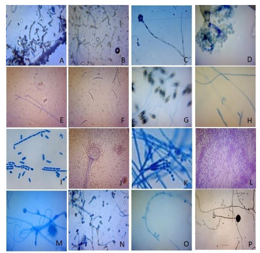
Figure 1. a Alternaria alternata. b Alternaria solani. c Aspergillus niger. d Botrytis cineria. e Cladosporium fulvum. f Colletotrichum truncatum. g Curvularia tetramera. h Fusarium oxysporum. i Geotrichum candidum. j Mucor meucedo. k Penicillium notatum. l Phoma exigua. m Pythium aphanidermatum. n Phytophthora infestans. o Pullularia pullulans. p Rhizopus stolonifer. Photos identified by Pathology Unit, Central Department of Botany, T.U.
Table 1. List of obtained species with their characteristics.
S.N. Name of the obtained species Characteristics 1. Alternaria alternata Conidiophores single or in small groups, simple or branched, straight or flexuous, olivaceous or golden brown, smooth, with conidial scars. Conidia formed in long chain 5-8, obclavate, obpyriform or ellipsoidal, with short, beak, pale or golden brown, smooth or verruculose with up to 8 transverse and usually several longitudinal or oblique septa. Beak pale. 2. Alternaria solani Conidiophores single or in small groups, simple or branched, straight or flexuous, septated, pale brown or olivaceous brown. Conidia usually solitary, or in a group of 2-3, straight or slightly flexuous, oblong or ellipsoidal tapering to a long beak, pale golden or olivaceous brown, smooth with 9-11 transverse and 0 or few longitudinal or oblique septa, beak flexuous, pale tapering gradually. 3. Aspergillus niger Conidiophore erect, straight or flexuous. Colorless or with the upper part brown, swollen at the apex into the spherical vesicle covered by closely packed more or less clavate branches. Flask shaped phialides present in a group at the apex of branches. Conidia catenate, dry usually globose brown verruculose or echinulate conidial heads at first globose, blackish brown to black, in age splitting into several loose columns. 4. Botrytis cinerea Conidiophores erect, unbranched or seldom branched, septate, wall blackish-brown, toward the tip almost hyline with several projections from which the conidia are formed singly on the very fine warts. Conidia ovate or elliptical to almost globose, finely apiculate at the base, with almost hyline, slightly brownish wall. 5. Cladosporium fulvum Conidiophore macronematous, mononematous, caespitose, unbranched or occasionally branched, straight or flexuous; narrow near the base broadening at the unilateral nodes, pale brown or olevaceous brown, smooth. Conidia catenated, frequently branched cylindrical with rounded ends or ellipsoidal, 0-3 septed, hilum slightly protuberant. 6. Colletotrichum truncatum Conidia non-septate, hyline, falcate, truncate, uninucleate with oil drops in the cytoplasm. Setae present in acervulus, telomorph present i.e. Glomerella truncata sp. nov. 7. Curvularia tetramera Mycelium branched septated sub-hyline or brown, conidiophores dark olivaceous brown or very irregular; simple or compound. Conidia produced at irregular distance from the base, chiefly 4 celled, born in cluster of 2-3 to 50 or more; dark olivaceous to brown, rather symmetrical shape tapering towards the rounded ends. Conidia with basal hyline cell, produced on branched conidiophore. 8. Fusarium oxysporum var. aurantiacum Macroconida large, curved, sickle-shaped/lunate in sporodochia and pionnotes, usually 4-5 septed Presence of extended sclerotial bodies, deep purple violet colored stroma. The fungus without aromatic odor. 9. Geotrichum candidum Mycelium hyline, prostate forming turf, turf cushion like somewhat powdery white. Hyphae prostate with few septa. Conidiophores short and erect or ascending, septed producing conidia in chains at their apex. Conidia short cylindrical, truncate at both ends hyline. 10. Mucor mucedo Simple, unsepted mycelium, bearing erect, silvery grey shingling unbranched, sporangiophore that produces non-apophysate, brownish black, cylindrical or companulate spherical sporangia with red-orange content. Elliptic or sub-cylindrical variable sized spores with smooth wall tardily yellow or colorless. Zygospore spherical with black thick varicose hard and fragile exine, enclosed colorless in tine. 11. Penicillium notatum Well developed, copiously branched, septed hyphae. Conidiophores usually arise from submerged mycelium, sometimes branched, produce long heads; fructifications in two stages metulae and phialides on which globose to oval, long chains of conidia are produced successively. 12. Phoma exigua Pycnidia globose to sub-globose or irregular with a non-papillate ostioles glabrous solitary or confluent; membranous to leathery or almost carbonous black. Conidiophore inside pycnidia insignificant. Conidia, sub globose, ellipsoidal to oblong or allantoid, usually with guttules mainly aseptate, maybe singly septed. 13. Pythium aphanidermatum Coenocytic, aseptate, cylindrical and branched mycelium; septa formed at the tip of hyphae to set sex organ; oogonia terminal, globose and smooth. Antheridia mostly intercalary, sometimes broadly shaped, 2 per oogonium, monoclinous or diclinous. Thick-walled, aplerotic oospores and lobed sporangia that contain kidney shaped, biflagellate zoospores. 14. Phytophthora infestans Heterothallic and bisexual. Coenocytic, branched mycelium; septa formed at the tip of hyphae to set sex organ; opaque, white lemon shaped sporangia with papilla at distal end are borne singly on the branch tips of alternatively branched sporangiophores that contains zoospores. 15. Pullularia pullulans Hyphae dark color with age composed of chains of dark, thick-walled cells, connected by strands of lighter colored cells. Conidia as oval to elongated cells, thick-walled and darkly pigmented budding from brown, branching and septate mycelial threads, both terminally and laterally, may continue to multiply by budding and abstriction. Mycelial cells later divided into a number of isodiametric cells with rounded sides and thick double wall, filled with oil drops. 16. Rhizopus stolonifer Simple, non-septed mycelium produces stolon which forms tufts of sporangiophores; well-developed rhizoids attached at the point where stolon are produced; sporangiophores bear large, spherical sporangia with flattened base having well-developed hemi-spherical columella. Spores, round or oval, angular, colorless or brown with circularized wall. Zygospore naked. Source: (Ellis 1971, Gilman 1998) Table 2. Fungal species, the sites from which they were isolated, the category based on frequency of isolation and their corresponding diseases.
S.N. Name of Fungi Site A Site B Site C Category Diseases 1. Alternaria alternata + + + D Back mold rot 2. Alternaria solani + + + D Alternaria rot 3. Aspergillus niger + + + D Back mold rot 4. Botrytis cinerea + + - M Grey mole rot 5. Cladosporium fulvum + + - M Scab/Cladosporium rot 6. Colletotrichum truncatum + + + D Anthracnose rot 7. Curvularia tetramera - + + M Drechslera mold rot 8. Fusarium oxysporum + + + D Fusarium rot 9. Geotrichum candidum + + + D Sour rot 10. Mucor mucedo + + + D Mucor rot 11. Penicillium notatum - + + M Penicillium rot 12. Phoma exigua - + - R Phoma rot 13. Pythium aphanidermatum + + - M Pythium rot 14. Phytophthora infestans + + - M Phytophthora rot 15. Pullularia pullulans + - - R Russet 16. Rhizopus stolonifer + + + D Rhizopus rot Total numbers of species 13 15 10 Numbers of species in percentage 81.25% 93.75% 62.5% It is found that of 16 species, except Curvularia tetramera, Penicillium notatum and Phoma exigua all were recorded from site A, and except Pullularia pullulans all were recorded from site B, whereas 10 species (Alternaria alternata, Alternaria solani, Aspergillus niger, Colletotrichum truncatum, Curvularia tetramera, Fusarium oxysporum, Geotrichum candidum, Mucor mucedo, Penicillium notatum and Rhizopus stolonifer were obtained from site C.
Thus it is observed that, the maximum numbers of fungi were recorded from site B, 15 species (93.75%), followed by site A, 13 species (81.25%) and from site C, has least in number i.e. 10 species (62.5%) (Table 2).
-
Tomatoes were infected by different fungal pathogens, in the field, due to unmanaged market and fungal bio-aerosols in the market environment. Occurrence of pathogens on tomato basically depends upon the sources of tomato and the environment where they are exposed to, and also depends upon the suitable temperature and moisture to grow. Availability of fungal species are varied in different study sites. Among the 16 reported species eight species are most common and dominant (Table 2). Similar results have been reported by Rodrigues & Kakde (2019). They mentioned that A. niger, A. flavus, A. alternata, Colletotrichum sp., Rhizopus sp. and F. oxysporum were the most common and frequently isolated. While Botrytis cinerea, Penicillium digitatum, P. notatum, Phoma sp., Cladosporium sp., Geotrichum candidum were isolated in the least frequency during investigation which is almost similar to the present investigation. And a similar report has also been given by Kakde & Kakde (2012). They reported that the fungi Aspergillus, Penicillium, Cladosporium, Fusarium and Alternaria were the most frequently isolated fungi from vegetables and fruits. These fungi were the most prevalent in the commercial market and also found to be responsible for most of the decay of vegetables and fruits during storage. Hence, there may be a relationship between the prevailing fungal bio-aerosols and the spoilage diseases.
16 reported species of this investigation are responsible for 15 different corresponding diseases (Table 2). Wani (2011) reported nine fungal rot diseases on postharvest tomato. Among them seven were also seen in the present investigation. They are Alternaria rot, Anthracnose rot, Mucor rot, Blue mold rot, Phytophthora rot, Phomopsis blight, Fusarium rot caused by respective pathogen of Alternaria solani, Colletotrichum truncatum, Mucor Mucedo, Penicillium sp., Phytophthora infestans, Phoma destructiva and Fusarium oxysporum respectively. Similar to this investigation, Massoud (2013) also isolated Aspergillus, Acremonium, Alternaria, Fusarium, and Penicillium in Lycopersicum esculentum from Aswan, Egypt. Chigoziri et al. (2018) also reported Aspergillus flavus, Colletotrichum capcisi and Pythium sp. from Nigeria.
Sajad et al. (2017) studied fungi associated with the spoilage of postharvest tomato fruit in different markets of Jabalpur, Madhya Pradesh, India. They reported Sour rot, Rhizopus rot, Black mold rot, Fusarium rot, and Alternaria rot caused by respective pathogen of Geotrichum candidum, Rhizopus stolonifer, Alternaria alternata, Fusarium sp. and Alternaria solani respectively. All of these are similar to the results found in the present investigation.
Bartz et al. (2017) described 11 post-harvest fungal diseases along with bacterial postharvest diseases found in Florida. Among them, nine of the fungal rot diseases namely Fusarium, Phoma, Anthracnose, Cladosporium, Grey mold, Phytophthora, Rhizopus, Black mold and Sour rot were also found in the present investigation.
Thus, 11 fungal rot diseases of Alternaria, Anthracnose, Black mold, Fusarium, Mucor, Penicillium (Blue mold rot), Phytophthora, Pythium, Phomopsis blight, Rhizopus and sour rot were described more or less by different researchers from other countries. Kohl et al. (2015) only reported scab epidemic in Dabrowice on Cortland Apple.
The tomatoes in site C had been imported from outside the country, as well as obtained from local areas like Bhaktapur, Naubisae etc. They were supplied to site B and A. In site B the tomatoes were also collected from Dhading, Bajrabarahi. Likewise, in site A, they were also brought from surrounding areas like Chapagaun, Shankhamul etc. Therefore, some similarities of pathogenic fungi were seen in the dominant species in all three study sites, as they were transmitted from fields to vegetable market through wholesalers with suitable temperature for their growth. Some dissimilarity was also observed that must be due to market environment where opportunistic fungi grow.
Adhikari & Manandhar (1997) mentioned 8 different species of fungi found on tomato bits. Similarly, Manandhar et al. (1997) reported Tolypocladium cylindrosporum including 7 more pathogenic fungi from the leaves of L. esculentum in Dhapasi of Kathmandu, Nepal. Manandhar et.al. (2017), reported 15 species of fungal pathogens from 342 tomato crop sample, along with other solanaceous crop. The sample were collected by Plant Pathological Division (PPD), National Agricultural Research Council (NARC) from fiscal year 2067/068 BS to 2071/072 BS from the different part of the country, Nepal. However, no work has been done in the field of fungal pathogens on the postharvest tomato in Nepal. And it is the first time that an opportunist fungus Pullularia pullulans (Fig. 1o) has been mentioned which is responsible for Russet disease on tomatoes. Thus, it is the first investigation that gave a list of fungal pathogens and postharvest fungal disease of storage tomato in Nepal.
-
Most of the pathogens associated with diseases were soil fungi that are transmitted from field or during harvesting, transportation and storage. Fungal pathogens directly or indirectly, infected the fruits and vegetables sold by traders. Therefore, consumers should be careful of fungal diseases and have knowledge about the symptoms of diseases. Well-managed vegetable markets should be facilitated by the local government and control measures must be applied by farmers and traders.
This study provides awareness of fungal disease of tomato to the public and will be helpful to build up a concrete strategy for management of postharvest fungal disease of tomato. As the study of diseases on tomatoes is important and concerned with public health, it is necessary to carry out such research works on tomato diseases. So, this investigation can be the base for further research in the future.
-
The authors would like to acknowledge Dean's Office of Institute of Science and Technology, Tribhuwan University for providing the research grant to conduct this study. The authors are obliged to the Central Department of Botany, Tribhuvan University for providing laboratory facilities. The Natural History Museum, Swayambhu, Tribhuvan University is thanked for facilitating the administrative asset. Sincere thanks are extended to the local traders of the study area for providing valuable information and materials for the experiment.
- Copyright: © 2021 by the author(s). This article is an open access article distributed under Creative Commons Attribution License (CC BY 4.0), visit https://creativecommons.org/licenses/by/4.0/.
B Shakya, HP Aryal. 2021. A study of commonly occurring fungal diseases on stored tomatoes of Kathmandu Valley. Studies in Fungi 6(1):159−167 doi: 10.5943/sif/6/1/9


|