


-
Blueberry (Vaccinium spp.) fruits are distinguished by their sweet-sour flavor and delicate texture, as well as their abundance of antioxidant compounds, which confer significant nutritional and economic value[1]. However, postharvest blueberries are prone to rapid softening and decay, limiting their storage and transport potential. Consequently, processed blueberry products represent a vital strategy for extending the industrial chain[2]. Among these, blueberry juice stands out as one of the most popular consumer products, as it preserves most of the nutritional components of fresh berries while delivering a rich flavor profile. Nevertheless, during processing and storage, environmental factors such as temperature, pH, oxygen, and light can accelerate the degradation of bioactive compounds of blueberry juice, resulting in nutrient loss and color fading. This challenge constitutes a major constraint for the development of blueberry juice beverages[3]. Blueberries are notably rich in anthocyanins, whose physiological functions including antioxidant, anti-inflammatory, and gut microbiota regulation, have been extensively confirmed and recognized[4]. However, the degradation of anthocyanins during storage not only compromises their potential health benefits, but also affects the product quality in the beverage industry. Therefore, enhancing the stability of anthocyanins has emerged as a key research priority in recent years.
Steviol glycosides, predominantly found in the leaves of perennial herbs, share a common sweet-tasting backbone structure, ent-kaurenoic acid. Among these compounds, stevioside, and rebaudioside-A (Reb-A) are the two most abundant glycosides[5]. As a natural sweetener, steviol glycosides are characterized by a high sweetness intensity and a low caloric content, in addition to diverse biological functions such as antioxidant activity, blood glucose regulation, and gastrointestinal health improvement[6,7]. In food processing, substituting sucrose with steviol glycosides can effectively overcome drawbacks associated with artificial sweeteners, such as metabolic disorders like obesity and diabetes, thereby aligning with contemporary health-conscious trends. As a result, steviol glycosides are widely used in beverages to partially or fully replace sucrose, demonstrating considerable potential for application across the beverage industry[6,8].
In real juice systems, the addition of steviol glycosides as a low-calorie natural sweetener may substantially influence the stability of anthocyanins by modifying the physicochemical environment or facilitating intermolecular interactions. Previous studies have shown that steviol glycosides exert protective effects on anthocyanins, color, and antioxidant activity in sour cherry puree over a 6-month storage period[9]. Comparable protective effects were reported in lingonberry jam, where steviol glycosides proved the most effective in preserving anthocyanin antioxidant capacity[10]. Chokeberry juices with added steviol glycosides had a higher content of analyzed bioactive compounds in comparison with juice samples sweetened with sucrose, and bioactive compounds, and antioxidant capacity was not significantly reduced during the storage period[11]. Furthermore, researchers have indicated that citrus-maqui juice supplemented with steviol glycosides maintains notable color stability, retaining its red hue and natural appearance throughout the shelf life[12]. Similarly, adding steviol glycosides lead to the color of strawberry jam being substantially brighter[13]. Degradation kinetic models of anthocyanins in citrus-maqui juice further suggest that molecular interactions between anthocyanins and steviol glycosides contribute to anthocyanin stabilization[14]. However, most of these studies remain largely confined to the validation of macroscopic effects, with extremely limited investigation into the specific binding modes and interaction mechanisms at the molecular level. Although the application of spectroscopic techniques, molecular docking, and molecular dynamics simulations can elucidate the fundamental nature of this interaction, relevant studies have rarely been reported in the context of anthocyanin and steviol glycoside systems.
The objective of this study was to examine the influence of steviol glycosides on the color, stability, and degradation kinetics of total anthocyanin and antioxidant capacity of blueberry juice under different storage conditions. Additionally, this work explores the mechanism of non-covalent molecular interactions between steviol glycosides and anthocyanins to elucidate the underlying stabilizing mechanism. The findings are expected to provide a theoretical foundation for enhancing the nutritional quality of blueberry juice and support further advances in juice processing technology.
-
Blueberry fruits of the 'Northland' cultivar were sourced from a local farm in Shenyang, Liaoning Province, China. Pectinase (30,000 U/g), pepsin (1:3,000), porcine trypsin (1:250), and porcine bile salts were procured from Shanghai Macklin Biochemical Co., Ltd. (Shanghai, China). Assay kits for determining DPPH radical scavenging capacity, ABTS radical scavenging capacity, and hydroxyl radical (•OH) content were supplied by Suzhou Grace Biotechnology Co., Ltd. (Suzhou, China). All other chemicals used were of analytical grade (purity ≥ 98%). The sweeteners employed in this study were commercially obtained from a health food retailer.
Samples preparation
-
Blueberry fruit (1 kg) were homogenized with 1 L of deionized water using a crusher. Subsequently, 2 g of pectinase (30,000 U/g) was added to the pulp, and the mixture was stirred for 1 h to facilitate enzymatic hydrolysis, yielding a crude blueberry extract. The crude extract was then centrifuged at 3,500 r/min for 20 min, and the supernatant was collected. Aliquots (100 mL) of the juice were supplemented with sweeteners; specifically, steviol glycosides (at 2, 4, 8, 12, 16, or 20 mg), sucralose (4 mg), or sucrose (7.5 g), along with 150 mg/L of benzoic acid. After thorough mixing, the samples were centrifuged and filtered. The pH of the resulting solution was adjusted to 3.5 using 1 mol/L HCl or NaOH, thus obtaining the final blueberry juice (BBJ) solution. A total of nine distinct juice formulations were prepared, with each type replicated three times.
The concentrations of sucralose and sucrose were selected based on consumer sweetness preference. In contrast, the 4 mg dose of steviol glycosides was determined from literature data to achieve an equivalent sweetness intensity[12,15]. The different doses of steviol glycosides were administered to investigate the dose-dependent protective effect of steviol glycosides on anthocyanins. The Brix and pH of the BBJ samples have been provided in (Supplementary Tables S1 and S2). All BBJ samples were stored under dark conditions at 4 and 25 °C for 90 d. Analyses were conducted immediately after preparation (day 0) and at 10-d intervals throughout the storage period.
Color measurements
-
The chromaticity parameters (L*, a*, b*) of the samples were measured using a colorimeter (NR60CP+, Shenzhen Threenh Technology Co., Ltd., China). The L* value represents lightness, while a* and b* denote the degree of redness/greenness and yellowness/blueness, respectively. The chroma (C*ab) and total color difference (ΔE*ab) were calculated according to Eqs. (1) and (2), as follows:
$ \mathrm{C}_{\text{ab}}^{\mathrm{*}}=\sqrt{{\left( {\mathrm{a}}^{\mathrm{*}}\right) }^{2}{+\left( {\mathrm{b}}^{\mathrm{*}}\right) }^{2}} $ (1) $ \Delta \mathrm{E}=\sqrt{{\left( {\Delta \mathrm{L}}^{\mathrm{*}}\right) }^{2}+{\left( {\Delta \mathrm{a}}^{\mathrm{*}}\right) }^{2}{+\left( {\Delta \mathrm{b}}^{\mathrm{*}}\right) }^{2}} $ (2) In this equation, ΔL*, Δa*, and Δb* represent the differences in L*, a*, and b* between the treatment group and the control group, respectively.
Determination of total anthocyanins content (TAC)
-
The total anthocyanin content (TAC) was determined using the pH differential method[16]. Briefly, 1 mL of blueberry juice was diluted with pH 1.0 and 4.5 buffer solutions to a final volume of 20 mL, respectively. After thorough mixing, solutions were incubated in the dark for 60 min, and the absorbance of each solution was measured at 520 and 700 nm. The absorbance difference was calculated using Eq. (3), and the TAC was subsequently determined according to Eq. (4).
$ \rm A={\left({{A}}_{{520}}-{{A}}_{{700}}\right)}_{{pH1.0}}-{\left({{A}}_{{520}}-{{A}}_{{700}}\right)}_{{pH4.5}} $ (3) $ \mathrm{TAC}=\dfrac{\left( \mathrm{A}\times \text{MW}\times \text{DF}\times 1,000\right) }{\varepsilon \times 1} $ (4) In this equation, MW is the relative molecular weight of anthocyanins in the sample (449.2 g/mol); DF is the dilution factor of the sample; ε is the molar absorptivity of anthocyanins in the sample (26,900).
Thermodynamic parameters measurements
-
The thermodynamic parameters were determined following the method described by Cao et al.[17]. Briefly, BBJ samples were incubated at 333, 343, 353, and 363 K for 5 h, with aliquots collected hourly for anthocyanin content analysis. The degradation rate constant (k), Gibbs free energy change (ΔG), enthalpy change (ΔH), and entropy change (ΔS) were calculated based on first-order kinetic equations and the Arrhenius equation, as established by Brouillard et al. and Lambert et al.[18,19].
Degradation rate (k) was calculated using Eq. (5).
First-order kinetic equation:
$ \ln \left({\mathrm{C}}_{\mathrm{t}}-{\mathrm{C}}_{0}\right)=-\text{kt} $ (5) In this equation, C0 is the initial concentration of anthocyanin; Ct is the concentration of anthocyanin after heating time; t is the heating time (h) ; k is the rate constant.
Anthocyanin thermal degradation half-life (t1/2) was calculated using Eq. (6).
$ {\mathrm{t}}_{{1}/{2}}=-\dfrac{\ln \left( 0.5\right) }{\mathrm{k}} $ (6) Activation energy (Ea) was calculated using the Arrhenius equation (Eq. [7]).
$ \mathrm{lnk}=\ln\mathrm{K}_0-\left(\mathrm{E}_{\mathrm{a}}/\text{RT}\right) $ (7) In this equation, Ea is the activation energy (kJ/mol) ; R is the gas constant (8.314 J/mol·K); T is the absolute temperature (K); K0 is the frequency factor (min−1) .
Enthalpy change (ΔH), Gibbs free energy (ΔG), and entropy change (ΔS) were calculated using Eqs. (8)–(10).
$ \Delta \mathrm{H}={\mathrm{E}}_{\mathrm{a}}-\text{RT} $ (8) $ \Delta \mathrm{G}=-\text{RTln}\left(\dfrac{\text{kh}}{{\mathrm{k}}_{\mathrm{B}}\mathrm{T}}\right) $ (9) $ \Delta \mathrm{S}=\dfrac{\Delta \mathrm{H}-\Delta \mathrm{G}}{\mathrm{T}} $ (10) In Eq. (8), h is Planck's constant (6.6262 × 10⁻³⁴ J·s); kB is the Boltzmann constant (1.3806 × 10⁻²³ J/K).
Antioxidant capacity
-
The hydroxyl radical (•OH) and ABTS radical scavenging capacities of the BBJ samples were determined using commercial assay kits (Suzhou Grace Biotechnology Co., Ltd., Suzhou, China) in accordance with the manufacturer's instructions.
Hydroxyl radical scavenging capacity
-
A 50 μL aliquot of BBJ sample was mixed with the assay reagents. The absorbance was measured at 510 nm using a microplate reader, and the hydroxyl radical scavenging activity was calculated as described in the kit protocol.
ABTS radical scavenging capacity
-
A 10 μL aliquot of BBJ sample was combined with the assay reagents. The absorbance was recorded at 734 nm using a microplate reader, and the ABTS radical scavenging activity was calculated according to the manufacturer's instructions.
In vitro simulated digestion
-
In vitro simulated digestion was performed according to a previously described method with minor modifications[20].
Simulated gastric digestion: a 1 mL aliquot of BBJ sample was accurately pipetted and diluted 20-fold with saline. The diluted sample was incubated in a water bath at 37 °C, and the pH was adjusted to 2.0 using 1 mol/L HCl. Then, 4 mL of simulated gastric juice was added. The mixture was placed in a refrigerated constant-temperature shaker and digested at 37 °C for 2 h. Aliquots were collected at 0, 1, and 2 h, centrifuged at 4 °C and 8,000 r/min for 15 min, and the supernatant was collected for TAC analysis.
Simulated intestinal digestion: after gastric digestion, the pH of the sample was adjusted to 7.0 using 1 mol/L NaHCO3 solution. Then, 4 mL of trypsin–bile salt mixture was added. The sample was further digested in a refrigerated constant-temperature shaker at 37 °C for 2 h. Aliquots were taken at 0, 1, and 2 h, centrifuged under the same conditions as above, and the supernatant was used for TAC determination.
$ \mathrm{Retention\;rate\;({\text{%}})}=\rm C_{ \mathrm{digested}} \mathrm{/C}_{ \mathrm{undigested}} \times \mathrm{ 100} $ (11) In this equation, Cdigested is the TAC after gastric and intestinal digestion; Cundigested is the TAC without digestion.
Simulation of the interaction between anthocyanins and steviol glycosides
-
The three-dimensional (3D) structures of the small molecules were obtained from the PubChem database and energy-minimized using Discovery Studio 2019 before being saved in PDB format. The resulting files were imported into AutoDock Tools, where hydrogen atoms were added, and then water removal was performed. Steviol glycosides and anthocyanins were designated as the receptors and ligands, respectively, and both were converted into PDBQT format for molecular docking. Docking simulations were carried out using AutoDock Vina to estimate binding affinities. Finally, the docking poses were visualized and analyzed using PyMOL 3.1.
Statistical analysis
-
Data are expressed as mean ± standard deviation (SD) based on three replicates. Differences between the two groups were assessed using Student's t-tests. The results were considered statistically significant at p < 0.05. Data were analyzed using GraphPad Prism 8.0 (Graph Pad Software, San Diego, CA, USA) and SPSS 17.0 (IBM Corporation, Armonk, NY, USA).
-
The color changes of BBJ during storage are summarized in Tables 1 and 2. Images of BBJ samples during storage are displayed as Supplementary Fig. S1. CIELAB parameters were employed to further evaluate the chromatic properties of BBJ supplemented with different sweeteners. A consistent increase in the L* value was observed with all sweeteners and storage conditions throughout the shelf-life testing. After 90 d of storage at 4 °C, the L* values of the Ste4, Ste8, Ste16, and Ste20 groups were significantly lower (p < 0.05) than those of the sucralose and sucrose groups, whereas the control and Ste2 groups exhibited significantly higher L* values (p < 0.05). At 25 °C, the control group showed the highest L* value, while the sucrose group had the lowest, however, the Ste20 group displayed a significantly higher L* value (p < 0.05) than the other sweetener supplemented groups.
Table 1. Colorimetric parameters changes of BBJ during storage at 4 °C.
Parameter Days Control Ste2 Ste4 Ste8 Ste12 Ste16 Ste20 Sucralose Sucrose L* 0 20.63 ± 0.12a 11.97 ± 0.14g 18.51 ± 0.06c 15.44 ± 0.15e 16.78 ± 0.10d 15.50 ± 0.08e 19.04 ± 0.09b 13.38 ± 0.17f 18.38 ± 0.38c 30 25.12 ± 0.07d 21.51 ± 0.02f 24.51 ± 0.14e 26.78 ± 0.09a 24.64 ± 0.03e 25.58 ± 0.02c 25.53 ± 0.54c 25.78 ± 0.26c 26.24 ± 0.11a 60 24.73 ± 0.19d 24.19 ± 0.29e 24.32 ± 0.02e 24.59 ± 0.03d 23.75 ± 0.02f 25.55 ± 0.06c 26.19 ± 0.03b 27.19 ± 0.03a 26.00 ± 0.08d 90 30.74 ± 0.08a 30.22 ± 0.01b 28.63 ± 0.04g 28.78 ± 0.03e 28.36 ± 0.02i 28.49 ± 0.04h 28.72 ± 0.02f 29.49 ± 0.02c 29.16 ± 0.02d a* 0 9.29 ± 0.14f 13.18 ± 0.06b 9.69 ± 0.18f 11.56 ± 0.07c 10.69 ± 0.22d 11.09 ± 0.03d 10.53 ± 0.18de 14.52 ± 0.12a 10.56 ± 0.69de 30 7.02 ± 0.06a 7.06 ± 0.04a 7.23 ± 0.16a 5.50 ± 0.16c 7.04 ± 0.02a 6.80 ± 0.09ab 6.71 ± 0.56ab 6.89 ± 0.22a 6.96 ± 0.03a 60 6.42 ± 0.31ab 6.52 ± 0.35ab 7.03 ± 0.07a 6.89 ± 0.06a 6.62 ± 0.28a 6.46 ± 0.12ab 6.18 ± 0.25ab 6.89 ± 0.04a 5.94 ± 0.36b 90 4.33 ± 0.12e 3.90 ± 0.02f 3.80 ± 0.07g 4.83 ± 0.04b 4.44 ± 0.02d 4.75 ± 0.04b 5.31 ± 0.01a 4.52 ± 0.03d 4.63 ± 0.05c b* 0 −3.17 ± 0.11b −6.39 ± 0.15f −4.11 ± 0.12d −2.79 ± 0.17a −3.29 ± 0.13b −4.31 ± 0.29d −2.58 ± 0.18a −4.48 ± 0.14de −3.48 ± 0.13bc 30 0.41 ± 0.05de −1.83 ± 0.05g 0.47 ± 0.05d 0.62 ± 0.03c 0.43 ± 0.03de 0.53 ± 0.05d 0.88 ± 0.03b 1.27 ± 0.09a 0.16 ± 0.01f 60 −0.06 ± 0.01b −0.28 ± 0.02d 0.22 ± 0.04a 0.24 ± 0.02a −0.19 ± 0.01c −0.22 ± 0.03c −0.34 ± 0.01e −0.07 ± 0.01b −0.51 ± 0.04f 90 −1.12 ± 0.03e −1.02 ± 0.10d −0.62 ± 0.04b −0.38 ± 0.02a −0.70 ± 0.05c −0.59 ± 0.03b −0.59 ± 0.05b −0.96 ± 0.04d −0.61 ± 0.02b C*ab 0 9.82 ± 0.17f 14.65 ± 0.01b 10.53 ± 0.13de 11.90 ± 0.10c 11.18 ± 0.20d 11.90 ± 0.13c 10.84 ± 0.13d 15.19 ± 0.07a 11.13 ± 0.62d 30 7.03 ± 0.06a 7.29 ± 0.03a 7.24 ± 0.16a 5.54 ± 0.15c 7.05 ± 0.02a 6.82 ± 0.08ab 6.77 ± 0.55ab 7.01 ± 0.21a 6.97 ± 0.03a 60 6.42 ± 0.31ab 6.53 ± 0.35ab 7.03 ± 0.07a 6.89 ± 0.06a 6.62 ± 0.28a 6.46 ± 0.12ab 6.19 ± 0.25b 6.89 ± 0.04a 5.97 ± 0.36b 90 4.47 ± 0.11d 4.04 ± 0.00e 3.85 ± 0.08f 4.85 ± 0.04b 4.50 ± 0.03d 4.78 ± 0.04b 5.34 ± 0.01a 4.62 ± 0.03c 4.67 ± 0.05c ΔE*ab 0 0.00 ± 0.00 10.03 ± 0.12a 2.36 ± 0.03e 5.68 ± 0.12c 4.10 ± 0.08d 5.56 ± 0.10c 2.11 ± 0.17ef 9.04 ± 0.19b 2.63 ± 0.66e 30 6.18 ± 0.01e 2.75 ± 0.01g 5.71± 0.11f 8.16 ± 0.14a 5.84 ± 0.03f 6.66 ± 0.05d 6.88 ± 0.51d 7.21 ± 0.30b 6.93 ± 0.08c 60 5.90 ± 0.02d 5.36 ± 0.36e 5.50 ± 0.01e 5.75 ± 0.05d 5.08 ± 0.15f 6.40 ± 0.02c 6.98 ± 0.08b 7.64 ± 0.02a 6.87 ± 0.11b 90 11.45 ± 0.02a 11.21 ± 0.03b 10.04 ± 0.03d 9.70 ± 0.02e 9.45 ± 0.03f 9.44 ± 0.01f 9.38 ± 0.02g 10.30 ± 0.01c 10.06 ± 0.04d Different superscript letters indicate significant differences among groups at the same time point (p < 0.05). Table 2. Colorimetric parameters changes of BBJ during storage at 25 °C.
Parameter Days Control Ste2 Ste4 Ste8 Ste12 Ste16 Ste20 Sucralose Sucrose L* 0 20.63 ± 0.12a 11.97 ± 0.14g 18.51 ± 0.06c 15.44 ± 0.15e 16.78 ± 0.10d 15.50 ± 0.08e 19.04 ± 0.09b 13.38 ± 0.17f 18.38 ± 0.38c 30 16.15 ± 0.47b 10.85 ± 0.08f 16.03 ± 0.33b 12.20 ± 0.09e 12.21 ± 0.15e 15.35 ± 0.23c 18.83 ± 0.12a 13.67 ± 0.32d 18.68 ± 0.46a 60 22.84 ± 0.04a 17.34 ± 0.17h 19.31 ± 0.03f 22.06 ± 0.26d 20.94 ± 0.08b 20.20 ± 0.13c 19.72 ± 0.29e 18.39 ± 0.22g 19.78 ± 0.13e 90 31.27 ± 0.04a 29.37 ± 0.02g 29.65 ± 0.03f 28.72 ± 0.02h 30.00 ± 0.01d 29.84 ± 0.01e 31.21 ± 0.03b 30.44 ± 0.02c 28.06 ± 0.02i a* 0 9.29 ± 0.14f 13.18 ± 0.06b 9.69 ± 0.18f 11.56 ± 0.07c 10.69 ± 0.22d 11.09 ± 0.03d 10.53 ± 0.18de 14.52 ± 0.12a 10.56 ± 0.69 30 10.25 ± 0.22c 12.67 ± 0.58a 9.54 ± 0.21d 11.56 ± 0.33b 9.56 ± 0.22d 10.46 ± 0.09c 7.53 ± 0.05f 10.67 ± 0.34c 8.20 ± 0.57e 60 5.02 ± 0.05de 7.28 ± 0.10a 6.92 ± 0.05b 5.26 ± 0.10d 5.39 ± 0.26d 5.41 ± 0.19d 5.28 ± 0.07d 6.19 ± 0.36c 6.31 ± 0.16c 90 2.64 ± 0.15c 2.50 ± 0.06d 2.65 ± 0.04c 1.89 ± 0.08g 1.62 ± 0.03h 2.84 ± 0.03b 2.06 ± 0.04f 3.13 ± 0.05a 2.42 ± 0.03e b* 0 −3.17 ± 0.11a −6.39 ± 0.15c −4.11 ± 0.12b −2.79 ± 0.17a −3.29 ± 0.13a −4.31 ± 0.29b −2.58 ± 0.18a −4.48 ± 0.14b −3.48 ± 0.13a 30 −2.00 ± 0.11b −3.29 ± 0.22e −2.03 ± 0.11b −3.02 ± 0.03d −2.43 ± 0.19c −2.39 ± 0.16c −2.04 ± 0.11b −2.16 ± 0.07b −1.23 ± 0.08a 60 3.99 ± 0.22c 5.24 ± 0.22a 4.94 ± 0.12ab 5.37 ± 0.27a 3.70 ± 0.14c 4.77 ± 0.12b 4.65 ± 036b 4.85 ± 0.10b 3.48 ± 0.10cd 90 −0.93 ± 0.02bc −1.00 ± 0.03c −1.62 ± 0.05e −1.82 ± 0.02f −0.80 ± 0.02b −1.22 ± 0.05d −0.33 ± 0.03a −1.28 ± 0.10d −0.86 ± 0.05b C*ab 0 9.82 ± 0.24f 14.65 ± 0.01b 10.53 ± 0.13e 11.90 ± 0.10c 11.18 ± 0.20d 11.90 ± 0.13c 10.84 ± 0.13de 15.19 ± 0.07a 11.13 ± 0.62d 30 10.44 ± 0.06c 13.10 ± 0.53a 9.75 ± 0.18d 11.95 ± 0.32b 9.86 ± 0.16d 10.73 ± 0.07c 7.80 ± 0.02e 10.88 ± 0.32c 8.29 ± 0.55e 60 6.41 ± 0.17f 8.97 ± 0.12a 8.51 ± 0.11b 7.52 ± 0.12d 6.54 ± 0.25f 7.22 ± 0.08d 7.04 ± 0.27de 7.86 ± 0.34c 7.20 ± 0.18d 90 2.80 ± 0.05c 2.69 ± 0.05d 3.10 ± 0.05b 2.62 ± 0.04d 1.81 ± 0.02g 3.09 ± 0.05b 2.08 ± 0.04f 3.38 ± 0.08a 2.57 ± 0.03de ΔE*ab 0 0.00 ± 0.00 10.03 ± 0.12a 2.36 ± 0.03e 5.68 ± 0.12c 4.10 ± 0.08d 5.56 ± 0.10c 2.11 ± 0.17ef 9.04 ± 0.19b 2.63 ± 0.66e 30 4.74 ± 0.43e 10.36 ± 0.25a 4.75 ± 0.31e 8.73 ± 0.11b 8.46 ± 0.17b 5.47 ± 0.25d 2.77 ± 0.08f 7.17 ± 0.38c 3.01 ± 0.20f 60 8.63 ± 0.14b 9.25 ± 0.27a 8.55 ± 0.10bc 9.56 ± 0.25a 7.91 ± 0.15d 8.85 ± 0.18b 8.84 ± 0.29b 8.90 ± 0.08b 7.34 ± 0.04e 90 12.74 ± 0.01b 11.28 ± 0.02f 11.31 ± 0.01f 11.05 ± 0.05g 12.33 ± 0.02c 11.41 ± 0.03e 13.13 ± 0.05a 11.74 ± 0.03d 10.38 ± 0.01h Different superscript letters indicate significant differences among groups at the same time point (p < 0.05). The a* value exhibited a continuous decline over the storage period. After 90 d at 4 °C, the highest a* value was observed in the Ste20 group, and the lowest in the Ste4 group. At 25 °C, the sucralose group showed the highest a* value, while the Ste12 group had the lowest. At the beginning of storage (day 0), all b* values were negative. These values shifted to positive during storage but returned to negative by the end of the 90-d period. After 90 d at 4 °C, the Ste8 group exhibited the highest b* value, and the control group was the lowest. At 25 °C, the highest b* value was observed in the Ste20 group, and the lowest in the Ste8 group.
Chroma (C*ab), defined as a quantitative measure of color saturation or intensity, decreased in all juice samples from day 0 to day 90 under both storage conditions, indicating a fading and graying of BBJ color during storage. At 4 °C, the Ste20 group showed the highest C*ab value, while at 25 °C, the sucralose group had the highest. The total color difference (ΔE*ab) was calculated for all samples throughout storage relative to the baseline control group on day 0. After 90 d at 4 °C, the control group exhibited the highest ΔE*ab value, and the Ste20 group was the lowest. At 25 °C, the highest ΔE*ab value was observed in the Ste20 group, and the lowest in the sucrose group.
Effects of steviol glycosides on anthocyanins of BBJ during storage
-
The effects of different sweeteners on TAC of BBJ are presented in Fig. 1. The addition of steviol glycosides was found to enhance the stability of anthocyanins in BBJ during storage. Specifically, the TAC in the Ste4, Ste8, and Ste16 groups was significantly higher than that in the control group (p < 0.05). After 90 d, the retention rate of anthocyanins in the Ste4, Ste8, and Ste16 groups was higher than those in the control, sucralose, and sucrose groups.
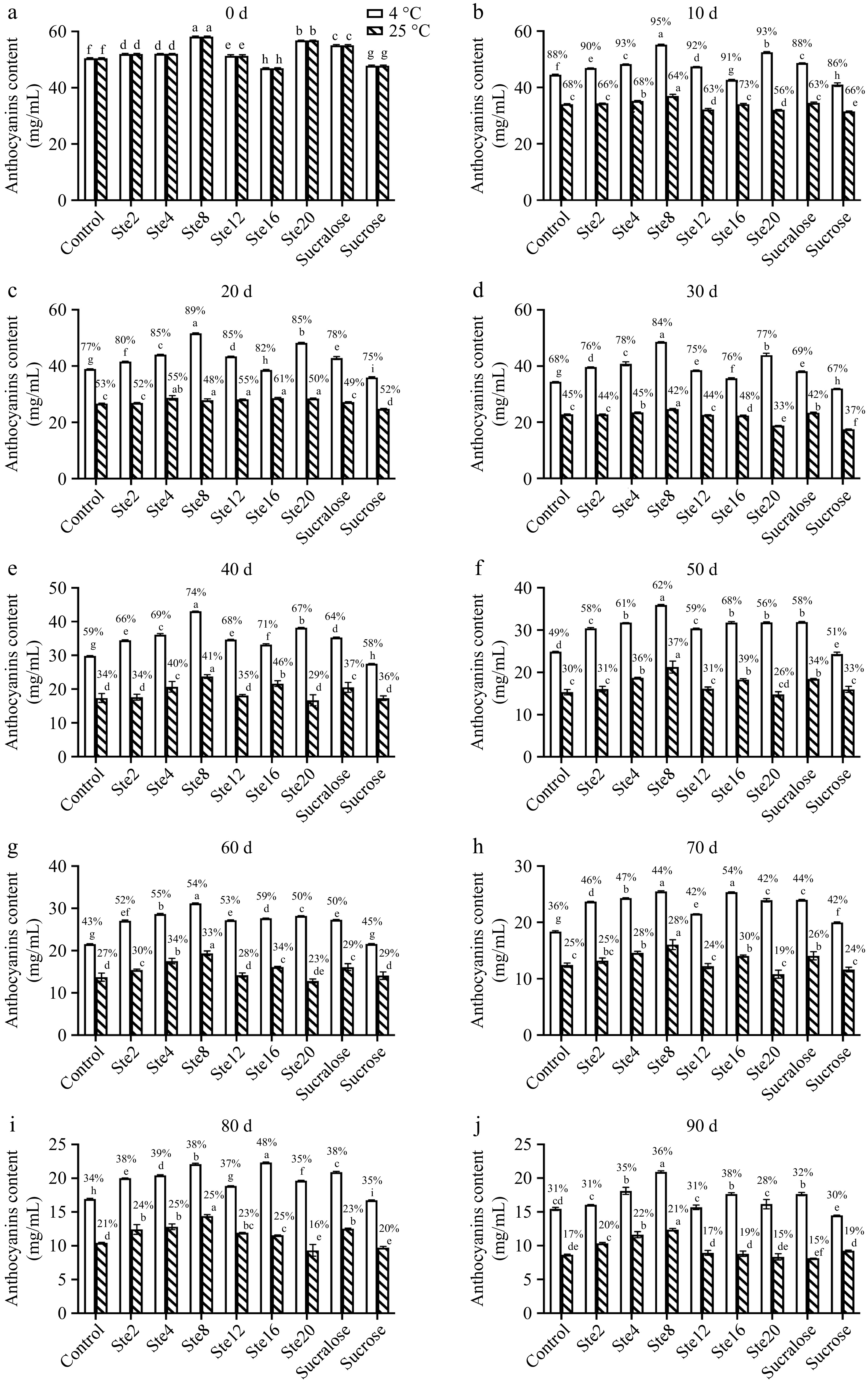
Figure 1.
Changes of TAC of BBJ during storage. The percentages on the graph indicate the retention rate of anthocyanins in BBJ samples during storage relative to day 0. n = 3 per group. Results are expressed as the mean ± SD. Different letters indicate significant differences among groups under the same storage condition (p < 0.05).
As summarized in Table 3, under storage at 4 °C, the degradation rate constant (k) of anthocyanins decreased significantly (p < 0.05) with the addition of steviol glycosides, sucralose, or sucrose. At 25 °C, a significant (p < 0.05) reduction in k was observed only in samples containing steviol glycosides. Furthermore, the addition of steviol glycosides significantly (p < 0.05) extended the half-life (t1/2) of anthocyanins. The retention rate (R) was highest in the Ste16 group at 4 °C and in the Ste4 group at 25 °C, both being significantly greater (p < 0.05) than those of other groups. These findings collectively indicate that steviol glycosides effectively improve the stability of anthocyanins in BBJ during storage.
Table 3. The rate constant k, degradation half-life t1/2, and retention rate R of anthocyanins of BBJ during storage.
Samples k/d−1 t1/2/d R/% 4 °C 25 °C 4 °C 25 °C 4 °C 25 °C Control 0.0137 ± 0.0000a 0.0212 ± 0.0002b 50.59 ± 0.000g 32.65 ± 0.321de 30.60 ± 0.391e 17.17 ± 0.146e Ste2 0.0117 ± 0.0001e 0.0201 ± 0.0000d 59.41 ± 0.295c 34.48 ± 0.000c 30.89 ± 0.120e 19.92 ± 0.213c Ste4 0.0109 ± 0.0001f 0.0186 ± 0.0003e 63.60 ± 0.584b 37.33 ± 0.508a 34.80 ± 0.937c 22.35 ± 0.875a Ste8 0.0108 ± 0.0001f 0.0189 ± 0.0003e 63.99 ± 0.686b 36.75 ± 0.592ab 35.98 ± 0.446b 21.29 ± 0.388b Ste12 0.0119 ± 0.0002d 0.0208 ± 0.0001bc 58.09 ± 0.741d 33.32 ± 0.160d 30.59 ± 0.874e 17.43 ± 0.603e Ste16 0.0094 ± 0.0001g 0.0184 ± 0.0003ef 73.49 ± 0.906a 37.68 ± 0.538a 37.63 ± 0.530a 18.76 ± 0.913cd Ste20 0.0125 ± 0.0001c 0.0243 ± 0.0004a 55.31 ± 0.512e 28.57 ± 0.414f 28.44 ± 1.166f 14.71 ± 0.837f Sucralose 0.0120 ± 0.0001d 0.0210 ± 0.0004b 57.61 ± 0.556d 33.01 ± 0.571d 32.05 ± 0.586d 14.78 ± 0.134f Sucrose 0.0132 ± 0.0001b 0.0207 ± 0.0001c 52.64 ± 0.231f 33.43 ± 0.093d 30.32 ± 0.249e 19.37 ± 0.259c Different superscript letters indicate significant differences among groups under the same storage condition (p < 0.05). Thermodynamic parameters of anthocyanins of BBJ with the addition of steviol glycosides
-
The thermal degradation kinetics of anthocyanins in the presence of different sweeteners were evaluated, and the corresponding thermodynamic parameters are summarized in Table 4. At 333 K, the Ste16 group exhibited the most pronounced improvement in the thermal stability of anthocyanins, as indicated by a significant (p < 0.05) reduction in the degradation rate constant (k) and an extension of the half-life (t1/2) compared to the control group. Specifically, the addition of 16 mg/100 mL steviol glycosides resulted in an approximately 30% decrease in k, and a 4 h prolongation of t1/2. At higher temperatures (343, 353, and 363 K), the inclusion of steviol glycosides, sucralose, or sucrose did not significantly enhance the thermal stability of anthocyanins.
Table 4. Thermodynamic parameters of anthocyanins of BBJ with addition of steviol glycosides.
Samples T (K) k/h−1 t1/2/h Eₐ (kJ/mol) ΔH (kJ/mol) ΔG (kJ/mol) ΔS (J/mol·K) Control 333 0.0628 ± 0.0003g 11.0375 ± 0.0527b 33.16 ± 0.162d 30.39 ± 0.162d 89.52 ± 0.013b −177.57 ± 0.502d 343 0.0792 ± 0.0005g 8.7558 ± 0.0556a 30.31 ± 0.162d 91.64 ± 0.018a −178.80 ± 0.420e 353 0.1141 ± 0.0009g 6.0769 ± 0.0484a 30.23 ± 0.162d 93.32 ± 0.023b −178.74 ± 0.479d 363 0.1676 ± 0.0008g 4.1358 ± 0.0202b 30.14 ± 0.162d 94.89 ± 0.015b −178.36 ± 0.485d Ste2 333 0.0819 ± 0.0011d 8.4608 ± 0.1084d 19.16 ± 0.296i 16.39 ± 0.296i 88.79 ± 0.035e −217.42 ± 0.790i 343 0.1148 ± 0.0015e 6.0368 ± 0.0800c 16.30 ± 0.296i 90.58 ± 0.038d −216.53 ± 0.755h 353 0.1311 ± 0.0019e 5.2879 ± 0.0756c 16.22 ± 0.296i 92.91 ± 0.042c −217.25 ± 0.738h 363 0.1474 ± 0.0005h 4.7025 ± 0.0166a 16.14 ± 0.296i 95.27 ± 0.011a −218.01 ± 0.798h Ste4 333 0.0767 ± 0.0009f 9.0379 ± 0.1009c 35.75 ± 0.160c 32.98 ± 0.160c 88.97 ± 0.031c −168.15 ± 0.392c 343 0.1464 ± 0.0008c 4.7358 ± 0.0262e 32.89 ± 0.160c 89.88 ± 0.016f −166.15 ± 0.429c 353 0.1712 ± 0.0011d 4.0497 ± 0.0265d 32.81 ± 0.160c 92.13 ± 0.019d −168.04 ± 0.484c 363 0.2369 ± 0.0022b 2.9261 ± 0.0272f 32.73 ± 0.160c 93.84 ± 0.028g −168.36 ± 0.372c Ste8 333 0.0789 ± 0.0006e 8.7892 ± 0.0655d 38.78 ± 0.265b 36.01 ± 0.265b 88.89 ± 0.021d −158.81 ± 0.735b 343 0.1711 ± 0.0015a 4.0505 ± 0.0346f 35.93 ± 0.265b 89.44 ± 0.024h −156.01 ± 0.717b 353 0.2011± 0.0013b 3.4463 ± 0.0222f 35.84 ± 0.265b 91.66 ± 0.019f −158.11 ± 0.699b 363 0.2681 ± 0.0010a 2.5858 ± 0.0099g 35.76 ± 0.265b 93.47 ± 0.012h −158.98 ± 0.742b Ste12 333 0.0767 ± 0.0025f 9.0397 ± 0.2938c 26.81 ± 0.580g 24.04 ± 0.580g 88.97 ± 0.091c −194.97 ± 1.472g 343 0.1669 ± 0.0016b 4.1525 ± 0.0397f 23.96 ± 0.580g 89.51 ± 0.027g −191.10 ± 1.634f 353 0.1728 ± 0.0038d 4.0118 ± 0.0881d 23.88 ± 0.580g 92.10 ± 0.065d −193.27 ± 1.462f 363 0.1820 ± 0.0016e 3.8080 ± 0.0327c 23.79 ± 0.580g 94.64 ± 0.026d −195.16 ± 1.526f Ste16 333 0.0455 ± 0.0006h 15.2247 ± 0.2035a 47.31± 0.209a 44.54 ± 0.209a 90.41 ± 0.037a −137.76 ± 0.547a 343 0.0798 ± 0.0013g 8.6912 ± 0.1413a 44.46 ± 0.209a 91.61 ± 0.046a −137.49 ± 0.477a 353 0.1139 ± 0.0011g 6.0842 ± 0.0585a 44.37 ± 0.209a 93.32 ± 0.028b −138.67 ± 0.563a 363 0.1942 ± 0.0019d 3.5695 ± 0.0348d 44.29 ± 0.209a 94.44 ± 0.029e −138.16 ± 0.519a Ste20 333 0.1006 ± 0.0009a 6.8883 ± 0.0644f 27.64 ± 0.165f 24.87 ± 0.165f 88.22 ± 0.026h −190.23 ± 0.423f 343 0.1398 ± 0.0010d 4.9583 ± 0.0338d 24.79 ± 0.165f 90.01 ± 0.019e −190.16 ± 0.463f 353 0.1771 ± 0.0005c 3.9139 ± 0.0101e 24.71 ± 0.165f 92.03 ± 0.008e −190.72 ± 0.463e 363 0.2325 ± 0.0011c 2.9817 ± 0.0138e 24.62 ± 0.165f 93.90 ± 0.014f −190.85 ± 0.420e Sucralose 333 0.0844 ± 0.0016c 8.2114 ± 0.1580d 23.48 ± 0.654h 20.71 ± 0.654h 88.70 ± 0.053f −204.19 ± 1.803h 343 0.1025 ± 0.0016f 6.7635 ± 0.1065b 20.63 ± 0.654h 90.90 ± 0.045c −204.88 ± 1.775g 353 0.1216 ± 0.0012f 5.7006 ± 0.0555b 20.54 ± 0.654h 93.13 ± 0.029a −205.63 ± 1.771g 363 0.1744 ± 0.0001f 3.9752 ± 0.0026b 20.46 ± 0.654h 94.77 ± 0.002c −204.70 ± 1.805g Sucrose 333 0.0906 ± 0.0009b 7.6539 ± 0.0719e 31.57 ± 0.056e 28.80 ± 0.056e 88.51 ± 0.026g −179.30 ± 0.112e 343 0.1724 ± 0.0010a 4.0214 ± 0.0235fg 28.72 ± 0.056e 89.42 ± 0.017h −176.96 ± 0.129d 353 0.2130 ± 0.0015a 3.2548 ± 0.0224g 28.64 ± 0.056e 91.49 ± 0.020g −178.05 ± 0.121d 363 0.2382 ± 0.0018b 2.9105 ± 0.0223f 28.55 ± 0.056e 93.83 ± 0.023g −179.82 ± 0.114d Different superscript letters indicate significant differences among groups under the same temperature (p < 0.05). The activation energy (Ea), representing the minimum energy required for a molecule to undergo a chemical reaction, was significantly increased (p < 0.05) in the Ste4, Ste8, and Ste16 groups compared to the control. A higher Ea indicates enhanced resistance to degradation, reflecting greater structural stability[16]. Similarly, the enthalpy change (ΔH) was significantly elevated (p < 0.05) in these groups. The positive ΔH values confirm that anthocyanin degradation is an endothermic process. The increased ΔH observed in the presence of steviol glycosides suggests a higher energy barrier for degradation, implying that stronger intermolecular interactions must be disrupted[21]. Moreover, the decline in ΔH at elevated temperatures indicates accelerated degradation under more severe thermal conditions.
Thermodynamic analysis further revealed that the degradation of anthocyanins under heating is an endergonic reaction, as supported by the positive Gibbs free energy (ΔG) values, which reflect the spontaneity of the process[16,21]. A negative entropy change (ΔS) suggests that the system becomes more ordered during degradation of anthocyanins. The decreased absolute ΔS values in the Ste4, Ste8 and Ste16 groups indicate increased entropy within the juice matrix, resulting in a more disordered system. Conversely, the elevated absolute ΔS values in the Ste2, Ste20, sucralose, and sucrose groups imply a reduction in entropy, corresponding to a more ordered state. Collectively, these results demonstrate that the addition of steviol glycosides enhances the thermal stability of anthocyanins in BBJ.
Effects of steviol glycosides on antioxidant capacity of BBJ during storage
-
The antioxidant activity of BBJ during storage was evaluated by hydroxyl radical (•OH) and ABTS radical scavenging assays (Fig. 2). A general decline in antioxidant capacity was observed across all samples under both evaluation methods. Regarding hydroxyl radical scavenging capacity, the Ste12 group exhibited a significantly higher retention rate (p < 0.05) than the other groups under 4 °C storage. No statistically significant differences (p > 0.05) were detected among the remaining groups at the same time point. Similarly, for ABTS radical scavenging, the Ste16 group demonstrated a significantly higher retention rate (p < 0.05) on day 90 at 25 °C, while no significant differences (p > 0.05) were observed among the other groups throughout the storage period.
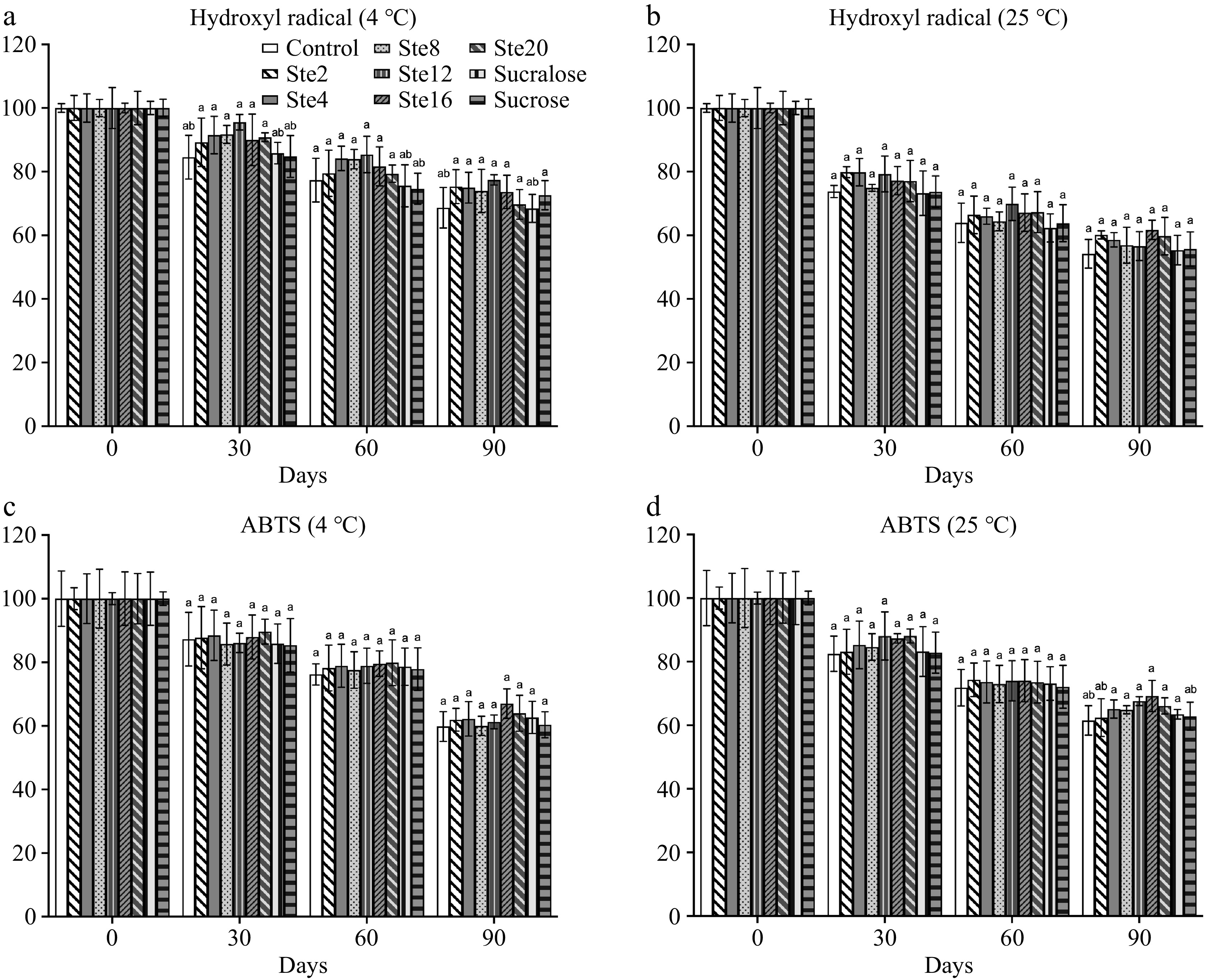
Figure 2.
Antioxidant capacity changes of antioxidants by (a), (b) hydroxyl radical scavenging activity, and (c), (d) ABTS assays of BBJ during storage. Antioxidant capacity determined at day 0 is set as 100%. n = 3 per group. Results are expressed as the mean ± SD. Different letters indicate significant differences among groups at the same time point (p < 0.05).
In vitro simulated digestion of anthocyanin of BBJ, with the addition of steviol glycosides
-
The stability of anthocyanins during the in vitro simulated digestion of BBJ was assessed to evaluate the impact of different sweeteners (Fig. 3). Under gastric conditions, the Ste4 group exhibited the highest retention rate (71.77%), whereas the Ste12 group showed the lowest (33.88%). The addition of sucralose and sucrose reduced the retention rate after gastric digestion to 50.72% and 48.48%, respectively. Upon transition to the intestinal phase, a marked decline in retention rate was observed. The Ste4 group maintained the highest retention rate after intestinal digestion (47.57%), in contrast to the sucrose group, which displayed the lowest (14.60%). Furthermore, the addition of sucralose resulted in a reduction of intestinal retention rate to 25%. These findings demonstrate that the choice of sweetener significantly influences the stability of anthocyanins in BBJ during in vitro gastrointestinal digestion.
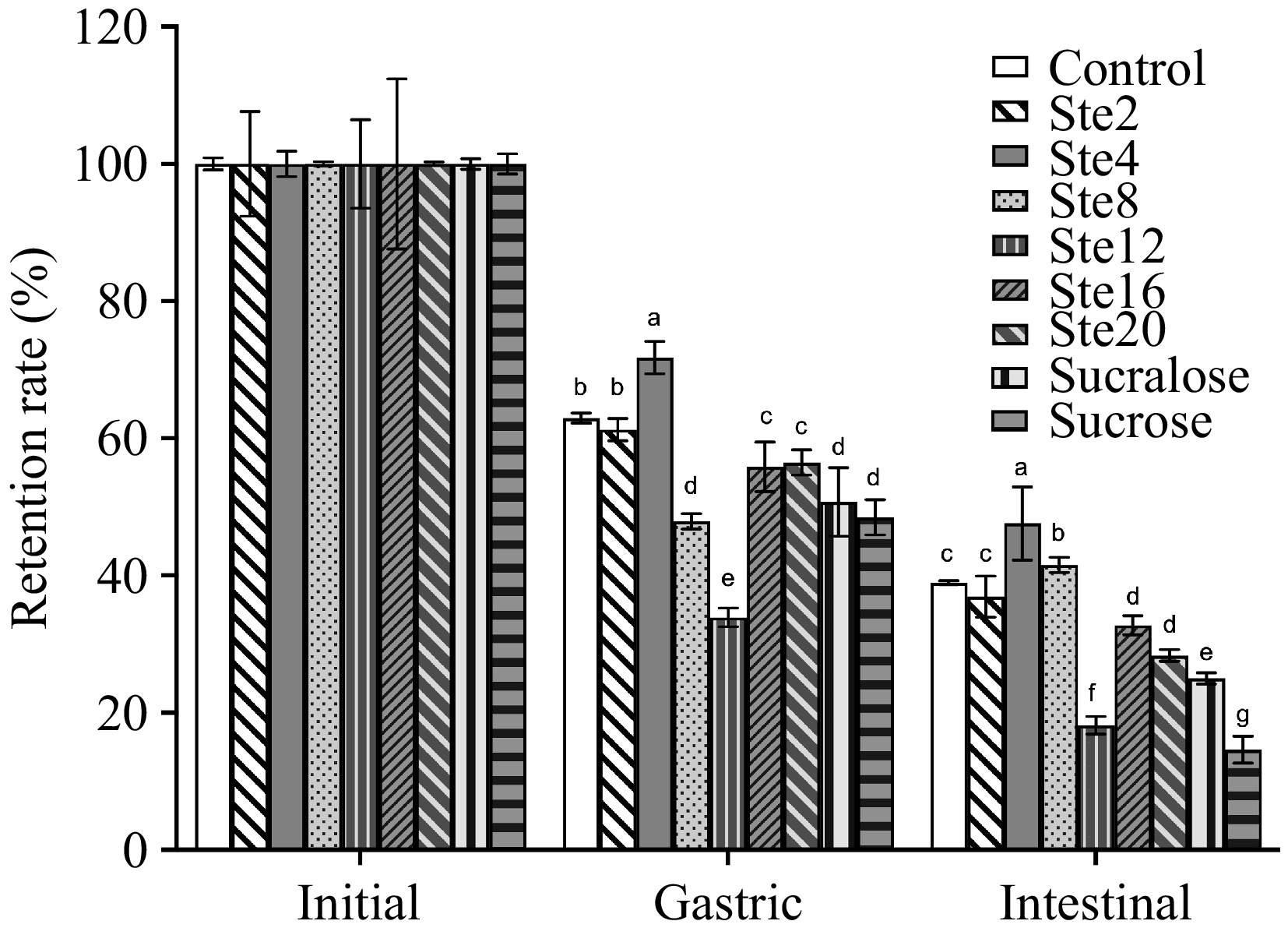
Figure 3.
Changes in retention rate (%) of total anthocyanins during in vitro simulated digestion of BBJ. The initial TAC determined in non-digested BBJ is set as 100%. n = 3 per group. Results are expressed as the mean ± SD. Different letters indicate significant differences among groups at the same time point (p < 0.05).
Analysis of the interaction mechanism between anthocyanin and stevioside
-
Molecular docking simulations were conducted to investigate the interaction between stevioside and the key anthocyanins, cyanidin-3-O-glucoside (C3G) and malvidin-3-O-galactoside (M3G). The analyses of the binding modes (Fig. 4) suggest that hydrophobic interaction and potential hydrogen bonds were formed between stevioside and C3G, as well as M3G. The highest observed binding affinities for the stevioside-C3G and stevioside-M3G complexes were −4.6 and −4.3 kcal/mol, respectively.
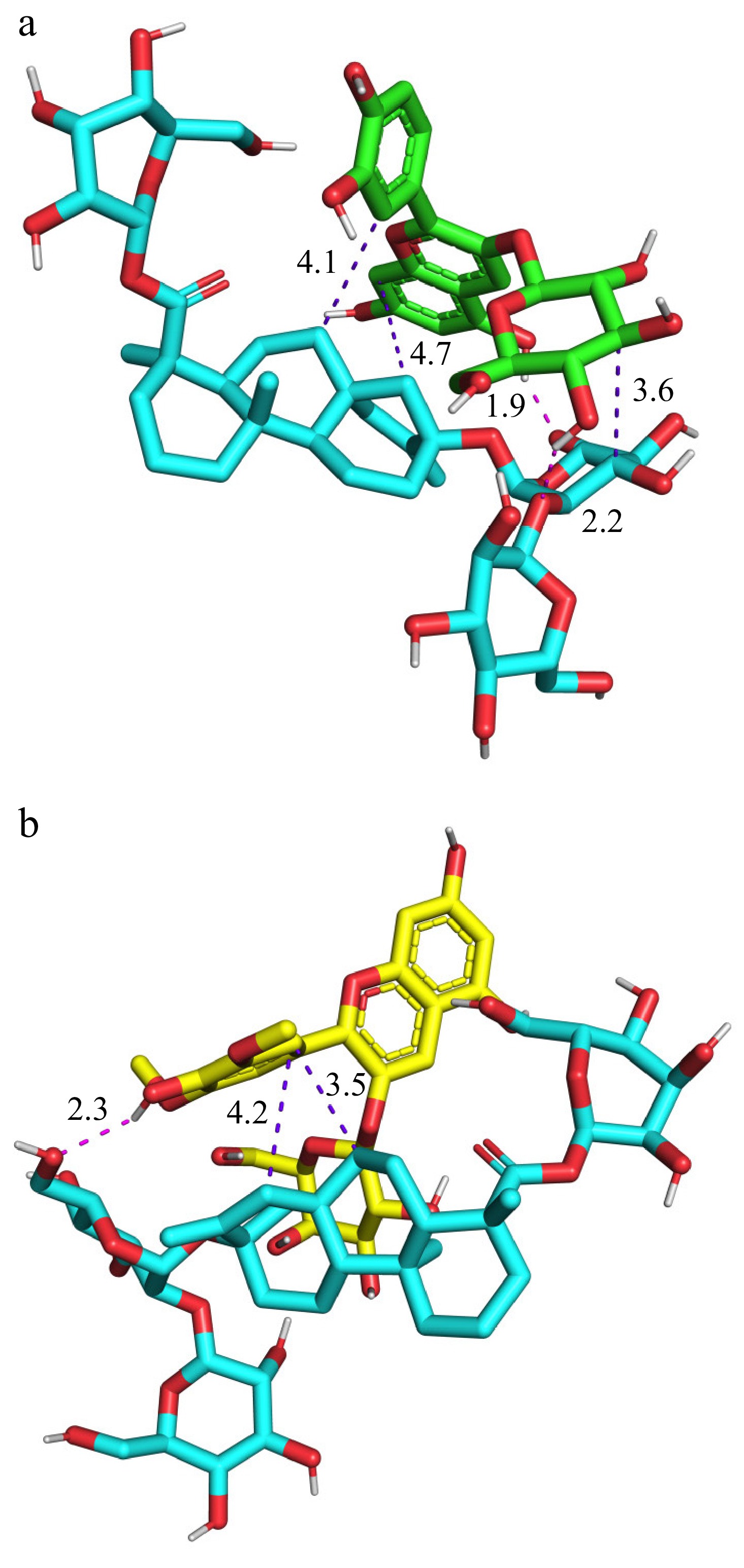
Figure 4.
The molecular docking simulation results of (a) stevioside with C3G, as well as (b) stevioside with M3G. Hydrogen bonds are represented by red dashed lines, and hydrophobic bonds are represented by purple dashed lines.
-
This study aimed to systematically evaluate the protective effect of steviol glycosides on anthocyanins in BBJ from a multi-scale perspective. First, the macroscopic manifestations were investigated, including color changes and degradation kinetics of anthocyanins during storage. Subsequently, the thermodynamic behavior and the in vitro simulated digestion were explored to understand the stability of anthocyanins. Finally, molecular docking was employed to probe the potential molecular-scale interactions, aiming to reveal the mechanism underlying the observed stabilization.
The susceptibility of anthocyanins to light, heat, and oxygen presents a major challenge for their application in the food industry. Under varying environmental conditions such as pH, temperature, and light, the structures of anthocyanins undergo a transition from the stable red flavylium cation, to lightly colored or nearly colorless forms, including methanol pseudobase, chalcone, and quinoidal base, under equilibrium conditions[22]. Current strategies to enhance the stability of anthocyanins primarily fall into two categories: one focuses on modulating extrinsic factors such as environmental conditions, while the other aims at structural stabilization through intrinsic modifications to preserve color and reduce degradation rate[17,23,24]. To date, research on environmental influences is relatively well-established, providing a solid theoretical basis for optimizing BBJ processing and storage. Effective approaches such as copigmentation, acylation, and biosynthesis have been shown to improve the stability and color retention of anthocyanins, thereby facilitating their broader application in the food and beverage processing[25,26].
Anthocyanins are highly susceptible to nucleophilic attack by water molecules in aqueous solutions. The flavylium cation undergoes hydroxylation at the C-2 position of its C-ring, forming a hemiketal structure. This hemiketal and the corresponding chalcone glycoside exist in equilibrium via tautomerization. After deglycosylation, the chalcone degrades under specific conditions, ultimately leading to the decoloration of anthocyanins. Anthocyanins-copigment complexes form conjugated systems through non-covalent interactions, adopting a sandwich-like configuration that shields the central anthocyanin from nucleophilic attack by water molecules. This structural arrangement inhibits hydration of the flavylium cation chromophore, thereby enhancing both stability and color integrity. These complexes are primarily stabilized by van der Waals forces, hydrophobic interactions, or ionic bonds, with common copigments including phenolic acids, amino acids, polysaccharides, flavonoids, proteins, and metal ions[27]. The observed color fading in all BBJ samples, characterized by an increase in L* value, and a decrease in a*, b*, and C*ab values can be attributed to two primary chemical processes: the degradation of anthocyanins, and the progression of non-enzymatic browning reactions during storage. Based on the ΔE*ab values, almost all BBJ samples exhibited visible color changes on day 30 (ΔE*ab > 5)[28]. Notably, the significantly smaller total color difference (ΔE*ab) in specific steviol glycoside groups, particularly Ste20 at 4 °C and Ste4 at 25 °C, indicate steviol glycosides have better color retention, and could effectively decelerate these underlying chemical pathways leading to color deterioration. The underlying protective mechanism may be attributed to the ability of steviol glycosides to form non-covalent complexes with anthocyanins in the juice. This interaction reduces the accessibility of the C-2 position on the C-ring of anthocyanin flavylium cation to nucleophilic attack by water molecules, thereby preventing the degradation of anthocyanins; this hypothesis is further supported by molecular docking results. From day 0 to 90, the content of anthocyanins has decreased by 62.37%~71.56% at 4 °C and 77.65%~85.29% at 25 °C. The stability of anthocyanins and their degradation is influenced by a number of factors such as pH, light, temperature, co-pigmentation, sulfites, ascorbic acid, oxygen, and enzymes[29]. The effect of color stabilization provides visual evidence for the subsequent kinetic data, which quantitatively confirms a slower degradation rate (k) and an extended half-life (t1/2) of anthocyanins in steviol glycosides-treated groups, a 23-d extension of half-life (t1/2) compared to the control at 4 °C. Although these colorimetric trends align with established literature, one study reported no statistically significant differences in color between stevia and sucrose-sweetened beverages[12]. Woźniak et al. reported that steviol glycosides did not influence the degradation kinetics of anthocyanins, and found that a combination of sucrose and steviol glycosides provided optimal anthocyanin stabilization[15].
Thermodynamic analysis revealed that steviol glycosides significantly enhanced the thermal stability of anthocyanins in BBJ, particularly at moderate temperatures. At 333 K, the Ste16 group showed markedly improved stability, with a 30% reduction in the degradation rate constant (k) and a 4 h extension of half-life (t1/2) compared to the control. Although this protective effect diminished at higher temperatures (343–363 K), key thermodynamic parameters indicated fundamental stabilization mechanisms. Significant increases in activation energy (Ea) and enthalpy change (ΔH) for the Ste4, Ste8 and Ste16 groups suggested higher energy barriers for degradation and stronger intermolecular bonding. The observed increase in Ea and ΔH in steviol glycosides-treated groups is a hallmark of copigmentation phenomena[30]. In a typical copigmentation complex, the anthocyanins-copigment complexes require additional energy to dissociate before anthocyanin degradation. Positive ΔG values confirmed the endergonic nature of anthocyanin degradation, while reduced entropy (ΔS) in steviol glycosides-treated groups implied enhanced molecular ordering within the system, possibly due to the stacking and reduced mobility of anthocyanin molecules within a protective matrix formed by steviol glycosides, which aligns with the proposed 'sandwich' structure in copigmentation theory[21,31,32]. These comprehensive thermodynamic analyses affirm that steviol glycosides, particularly at optimal concentrations, stabilize the structure of anthocyanins through a combination of elevated energy barriers and entropic modulation. In line with this, Fan et al. reported that copigmentation induced molecular reordering of anthocyanins toward thermodynamic equilibrium underlies their structural stabilization[16].
The relationship between BBJ color, anthocyanin content, and antioxidant capacity is well demonstrated[33]. The antioxidant activity of anthocyanins stems from their molecular geometry, which facilitates hydrogen atom donation from aromatic hydroxyl groups, and is further influenced by specific hydroxylation and methoxylation patterns[34]. Notably, the preservation of antioxidant activity in our study followed a selective pattern. Although overall antioxidant capacity declined during storage, specific steviol glycoside concentrations conferred targeted protection; Ste12 uniquely maintained hydroxyl radical scavenging capacity at 4 °C, whereas Ste16 retained superior ABTS radical scavenging at 25 °C on day 90. This differential effect suggests that steviol glycosides may stabilize specific anthocyanin subclasses with distinct antioxidant functions, rather than providing broad-spectrum protection. The improved kinetic stability and selective antioxidant preservation indicate that steviol glycosides act as multifunctional stabilizers in BBJ, offering both structural protection to anthocyanins and maintaining key antioxidant pathways. These results highlight the potential of steviol glycosides as a natural additive for extending the shelf life and functional quality of anthocyanin-rich beverages. Consistent with our findings, Nowicka & Wojdylo also reported the protective effects of steviol glycosides on polyphenol content, particularly anthocyanins, and consequently on color and antioxidant activity[9].
Gastric digestion had no significant effect on anthocyanins, and intestinal digestion substantially decreased anthocyanins[35]. Our study shows that steviol glycosides significantly influence the gastrointestinal stability of anthocyanins in BBJ. Under gastric conditions, the Ste4 group exhibited the highest retention rate, exceeding that of sucralose and sucrose, whereas Ste12 showed the lowest. This trend continued during intestinal digestion, with Ste4 maintaining the highest retention rate, significantly greater than that of sucralose and sucrose. These results suggest that lower concentrations of steviol glycosides (e.g., Ste4) enhance the stability of anthocyanin throughout digestion, whereas higher concentrations (e.g., Ste12) may have inhibitory effects. Steviol glycosides are superior to artificial and traditional sweeteners in maintaining the stability of anthocyanins during in vitro simulated digestion. This highlights their potential as functional additives for improving the nutritional value of anthocyanins-rich beverages. Previous studies have indicated that the effects of steviol glycosides on stability, bioactivity, and bioavailability of anthocyanins are mediated through molecular interactions[36,37].
To explore the mechanism by which steviol glycosides enhance the stability of anthocyanins in BBJ, we hypothesized that non-covalent interactions between them may play a crucial role. The most abundant steviol glycoside, stevioside, and the predominant anthocyanins in BBJ, C3G, and M3G, were selected for molecular docking studies. However, the docking results indicated that although the ligand occupies the binding pocket, the interactions are predominantly hydrophobic, and lack the extensive hydrogen-bonding network typically associated with high-affinity binding. This observation aligns with the low binding affinity observed, suggesting that stevioside acts as a weak binder under the simulated conditions. While the molecular docking results indicated only weak binding affinity between stevioside and individual anthocyanins (C3G, M3G), the consistent protective effects observed across color, kinetic, thermodynamic, and bioavailability assays strongly suggest the existence of an alternative or supplementary stabilization mechanism. The stabilization may not rely solely on high affinity, specific binding between single molecules; it could be governed by weak, cumulative, and non-specific interactions within a complex juice matrix. These interactions, such as hydrogen bonding, van der Waals forces, and hydrophobic effects, might collectively create a molecular environment that shields anthocyanins from water, oxygen, and nucleophilic attack. Therefore, future research should focus on alternative analytical techniques, such as isothermal titration calorimetry (ITC) to directly measure the binding stoichiometry and affinity in solution, or nuclear magnetic resonance (NMR) spectroscopy to detect weak intermolecular interactions in a model system that more closely mimics the complex juice matrix.
-
In conclusion, this study confirms the effect of steviol glycosides as multi-functional stabilizers in blueberry juice. Firstly, specific concentrations of stevia glycosides were proven to preserve color integrity and significantly decelerate the kinetic degradation of anthocyanins during storage. Furthermore, specific concentrations of stevia glycosides enhanced the thermodynamic stability and antioxidant activity of anthocyanins, and increased their stability in in vitro simulated digestion. Ultimately, these findings establish a solid scientific basis for the application of steviol glycosides in the food industry, not only for improving the nutritional profile of fruit juice, but also for guiding the development of novel stabilization strategies in beverage processing.
The authors gratefully acknowledge financial support by The Provincial Doctoral Research Start up Fund of Shenyang Agricultural University (8804/880420024).
-
The authors confirm their contributions to the paper as follows: study conception and design: Zhang C, Jiao X; data collection: Zhang C, Zhang L; analysis and interpretation of results: Zhang C, Zhang L, Yun X; draft manuscript preparation: Zhang C, Wang Y, Tan H, Ashour AA, Jiao X. All authors reviewed the results and approved the final version of the manuscript.
-
The datasets generated during and/or analyzed during the current study are available from the corresponding author upon reasonable request.
-
The authors declare that they have no conflict of interest.
-
accompanies this paper online at: https://doi.org/10.48130/fia-0026-0019.
- Supplementary Table S1 The Brix of BBJ samples during storage.
- Supplementary Table S2 The pH value of BBJ samples during storage.
- Supplementary Fig. S1 Images of BBJ samples during storage under conditions maintained at (A) 4°C and (B) 25°C.
- Copyright: © 2026 by the author(s). Published by Maximum Academic Press on behalf of China Agricultural University, Zhejiang University and Shenyang Agricultural University. This article is an open access article distributed under Creative Commons Attribution License (CC BY 4.0), visit https://creativecommons.org/licenses/by/4.0/.
-
About this article
Cite this article
Zhang C, Zhang L, Yun X, Ashour AA, Tan H, et al. 2026. Influence of steviol glycosides on the stability of anthocyanins during storage of blueberry juice. Food Innovation and Advances 5(2): 219−229 doi: 10.48130/fia-0026-0019 

Influence of steviol glycosides on the stability of anthocyanins during storage of blueberry juice
- Received: 26 November 2025
- Revised: 02 February 2026
- Accepted: 01 March 2026
- Published online: 13 May 2026
Abstract: Blueberry juice maintains the nutritional value of fresh berries while providing their complex flavor profile. Anthocyanins contribute to the vibrant color of blueberry juice and exhibit physiological functions. Steviol glycosides are widely utilized as a natural sweetener in fruit beverages, and investigating their regulatory role in the degradation of anthocyanins within juice systems is of considerable importance. This study aimed to examine the effects of steviol glycosides on the color, anthocyanin content, antioxidant capacity, and in vitro simulated digestion stability of anthocyanins in blueberry juice during storage. The juice was stored for 90 d at 4 and 25 °C in dark conditions. Throughout storage, the juice exhibited color fading, accompanied by a loss of its original color intensity. Notably, the sample supplemented with 20 mg/100 mL steviol glycosides showed the least color change when stored at 4 °C. The addition of steviol glycosides resulted in a reduced degradation rate constant (k) and an extended half-life (t1/2) of anthocyanins. Thermodynamic analysis indicated that steviol glycosides enhanced the thermal stability of anthocyanins in blueberry juice. Furthermore, blueberry juice containing 12 and 16 mg/100 mL steviol glycosides demonstrated the highest hydroxyl radical scavenging capacity at 4 °C, and the strongest ABTS radical scavenging activity at 25 °C, respectively. During in vitro simulated digestion, the sample with 4 mg/100 mL steviol glycosides exhibited the highest retention rate of total anthocyanins. Molecular docking analysis revealed the formation of hydrophobic interactions and hydrogen bonds between stevioside and cyanidin-3-O-glucoside, as well as malvidin-3-O-galactoside. The findings of this study provide a solid foundation for advancing fruit juice processing technologies.
-
Key words:
- Blueberry juice /
- Steviol glycosides /
- Anthocyanins /
- Stability