













-
The tea plant (Camellia sinensis) is one of the most important non-alcoholic beverage crops worldwide and is widely cultivated in tropical and subtropical regions. However, the warm and humid environments favorable for tea growth also facilitate the occurrence and spread of fungal diseases. In China, more than 100 diseases have been reported in tea-growing areas, among which over 30 are commonly observed[1]. Leaf diseases, including leaf spot, gray blight, brown blight, and anthracnose, severely affect leaf production, plant vigor, and tea quality, thereby causing substantial economic losses[2−5].
Tea gray blight disease, caused by fungi of the genus Pseudopestalotiopsis, is a destructive disease that can infect leaves and stems at different developmental stages[6]. Severe infection may lead to leaf blight, defoliation, and even plant death, ultimately reducing both yield and quality[7]. Therefore, identification of key genes involved in tea plant resistance to gray blight disease is of great importance for understanding defense mechanisms and for breeding resistant cultivars.
Calcium ion (Ca2+) is a universal second messenger in eukaryotic cells and participates in a broad range of plant physiological and stress-responsive processes. In plants, Ca2+ signaling plays a pivotal role in innate immunity, including transcriptional activation, stomatal defense, and local, as well as systemic signal transduction[8,9]. To decode Ca2+ signals, plants employ several classes of Ca2+ sensors, including calmodulins (CaMs), calcineurin B-like proteins (CBLs), calmodulin-dependent protein kinases (CaMKs), and calcium-dependent protein kinases (CDPKs/CPKs)[10].
CDPKs belong to the serine/threonine protein kinase family and are found primarily in plants and some protozoans[11,12]. A typical CDPK protein contains an N-terminal variable region, a serine/threonine kinase domain, an autoinhibitory/junction domain, and a calmodulin-like domain containing EF-hand motifs[13]. Upon changes in intracellular Ca2+ concentration, Ca2+ binds to the EF-hand motifs and induces conformational changes that activate CDPK kinase activity, thereby transducing Ca2+ signals to downstream targets[14−16]. Owing to this unique structure, CDPKs can function both as Ca2+ sensors and as signal transducers.
CDPK gene families have been identified in many plant species. For example, 34 CDPK genes have been reported in Arabidopsis thaliana[17], 31 in rice[18,19], 20 in wheat[20], and 39 in soybean[21]. Increasing evidence indicates that CDPKs are key regulators of plant responses to both abiotic and biotic stresses. In abiotic stress responses, LpCDPK27 improves salt tolerance in Arabidopsis[22], StCDPK32 positively regulates salt tolerance and photosynthesis in potato[23], and TaCDPK25-U enhances drought tolerance in Arabidopsis[24]. In addition, PpCDPK7 may mediate the crosstalk between Ca2+ and ROS signaling during cold storage in peach fruit[25].
In plant immunity, CDPKs are also important signaling components. For instance, in Brassica rapa, multiple CDPKs interact with respiratory burst oxidase homologs (RBOHs) and participate in defense against Pseudomonas syringae pv. tomato DC3000 by regulating H2O2 accumulation and stomatal closure[26]. In strawberry, FvCDPKs respond to pathogen-induced Ca2+ fluctuations and are involved in signaling triggered by viral and fungal pathogens[27]. Moreover, some CDPKs are closely linked to phytohormone-mediated immunity. For example, AtCPK5 acts as a positive regulator of innate immunity[28], AtCPK6 promotes MeJA-induced stomatal closure[29], and AtCPK1 enhances SA accumulation and SA-dependent defense gene expression[30].
Despite increasing evidence for CDPK-mediated stress responses in many plant species, a systematic understanding of the CDPK family in tea plants, especially in relation to gray blight disease resistance, is still lacking. Previously, Ding et al.[31] reported 26 CsCPK members in tea plant based on early transcriptome data, with a primary focus on cold stress responses; the exploration of biotic stress, particularly pathogen-induced CDPK expression, was not addressed in their study. In the present study, using an improved tea plant genome assembly, we performed a more comprehensive genome-wide identification and specifically focused on the roles of CsCDPKs in defense against gray blight disease. In addition, we examined their expression profiles under pathogen infection and hormone treatments, measured endogenous SA and MeJA levels after pathogen challenge, and functionally evaluated selected candidate genes using AsODN-mediated silencing. Our study provides new insights into the potential roles of CsCDPK genes in tea plant defense against gray blight disease.
-
One-year-old tea seedlings of the national clonal cultivar 'Zhuyeqi' were used in this study. Seedlings were planted in pots at a spacing of 7 cm and maintained in a controlled growth chamber under the following conditions: 25 ± 2 °C, 16 h light/8 h dark photoperiod, and 65% ± 5% relative humidity. After 1 month of normal growth, uniformly growing and disease-free seedlings were selected for subsequent experiments.
Pathogen culture and inoculation
-
The culture and inoculation procedure for tea gray blight disease was modified from a previously reported method[32]. Potato dextrose agar (PDA) was prepared following the protocol of the reported method[33]. Briefly, mycelia of the gray blight pathogen were transferred to the center of PDA plates and incubated in the dark at 28 °C for 5 d. Fresh mycelial plugs (8 mm in diameter) were then cut from the actively growing edge of the colony and used for inoculation. The fully expanded second, third, and fourth leaves of tea seedlings were surface-sterilized with 75% ethanol, rinsed with sterile water, and air-dried. Before inoculation, each leaf was gently wounded using a sterile 1 mL syringe needle, with three punctures made on each side of the midrib while avoiding the major veins. The prepared mycelial plug was then placed on the wounded area with the mycelial side in contact with the leaf surface. Infected leaves were collected 3 d after inoculation, immediately frozen in liquid nitrogen, and stored at −80 °C until further use.
Determination of endogenous hormone contents
-
Uniform and disease-free 'Zhuyeqi' tea seedlings were inoculated with the gray blight pathogen. To ensure that samples from all time points could be collected simultaneously, a staggered treatment schedule was applied: inoculations were performed at different time points so that sampling for all time points could be carried out at the same time. Leaf samples were collected at 12, 24, 48, and 72 h after inoculation; the 0 h samples served as the uninoculated control. All samples were immediately frozen in liquid nitrogen and stored at −80 °C. Three biological replicates were included for each time point. Subsequently, endogenous hormone levels were measured in the pathogen-inoculated tea leaves.
Endogenous SA, JA, and MeJA contents were determined according to the method of Pan et al.[34]. Frozen leaf tissue (0.5–1 g) was ground and extracted with 5 mL of extraction solvent at 4 °C in the dark for 30 min. Subsequently, 5 mL of dichloromethane was added, shaken for another 30 min, and centrifuged at 13,000 r/min for 5 min at 4 °C. The lower organic phase was collected, dried over anhydrous magnesium sulfate, and concentrated to dryness using a centrifugal concentrator (< 30 °C). The residue was reconstituted in 0.2 mL of 0.1% formic acid in methanol, filtered (0.22 μm), and analyzed by HPLC-MS/MS. Separation was performed on a Poroshell 120 SB-C18 column (2.1 mm × 150 mm, 2.7 μm) at 30 °C with gradient elution (injection volume: 3 μL). Mass spectrometric detection was carried out in multiple reaction monitoring (MRM) mode using both positive and negative ion scanning. Hormone contents were calculated as ng/g = C × V/M, where C is the concentration in the test solution (ng/mL), V is the extraction volume (mL), and M is the sample mass (g).
Exogenous hormone treatments
-
Tea seedlings were sprayed evenly with 100 μM SA or MeJA solution. Leaves were sampled at 0 (control), 12, 24, 48, and 72 h after treatment. Three biological replicates were collected at each time point. The treatment conditions were the same as the growth conditions described above. Samples were immediately frozen in liquid nitrogen and stored at −80 °C until analysis.
Identification of CsCDPK family members
-
The genome sequence, genome annotation file, and protein sequence file of the tea cultivar 'Tieguanyin' were downloaded from the Tea Plant Information Archive (TPIA2.0;
https://tpia.teaplants.cn/index.html )[35]. Arabidopsis CDPK protein sequences annotated as calcium-dependent protein kinases were downloaded from TAIR2.0 (www.arabidopsis.org ), yielding 34 reference proteins.The hidden Markov model (HMM) profiles of the CDPK-related conserved domains, including the protein kinase domain (PF00069) and the EF-hand-related domain (PF13499), were obtained from the Pfam database (
https://pfam-legacy.xfam.org ). Candidate CsCDPK proteins were screened using HMMER 3.0. Protein sequences identified from the two domain searches were intersected using TBtools. Candidate proteins were then further verified using Pfam and the NCBI Conserved Domain Database (CDD;www.ncbi.nlm.nih.gov/Structure/bwrpsb/bwrpsb.cgi ). After removal of redundant or incomplete sequences, 32 non-redundant CsCDPK genes were obtained.Physicochemical characterization and subcellular localization prediction
-
The physicochemical properties of CsCDPK proteins, including amino acid length, molecular weight, theoretical isoelectric point, instability index, aliphatic index, and grand average of hydropathicity (GRAVY) were analyzed using the Protein Parameter Calc function in TBtools. Subcellular localization was predicted using the WoLF PSORT online server (
https://wolfpsort.hgc.jp ).Phylogenetic analysis, gene structure, and conserved motif analysis
-
The full-length amino acid sequence of CDPK proteins from tea plant and Arabidopsis were aligned and used to construct a phylogenetic tree in MEGA 11.0 using the neighbor-joining method with 1,000 bootstrap replicates. The exon–intron structures of CsCDPK genes were visualized in TBtools based on the annotation file of the tea genome. Conserved motifs in CsCDPK proteins were identified using the MEME suite (
https://meme-suite.org/meme/tools/meme )[36], with the maximum number of motifs set to 10 and other parameters kept at default values. Motif distributions were subsequently visualized using TBtools.Chromosomal localization and promoter cis-element analysis
-
The chromosomal positions of the 32 CsCDPK genes were obtained by importing gene IDs and the GFF annotation file into the Gene Location Visualize from the GFF function in TBtools. For promoter analysis, the 2,000 bp genomic sequence upstream of each CsCDPK gene was extracted and submitted to PlantCARE (
https://bioinformatics.psb.ugent.be/webtools/plantcare/html ) for prediction of cis-acting regulatory elements[37]. The resulting annotations were summarized and visualized using TBtools.Synteny analysis and Ka/Ks calculation
-
Intra-genomic and inter-genomic synteny analyses were performed using TBtools. For intra-species analysis, the tea plant genome sequence and annotation files were used to identify collinear gene pairs involving CsCDPKs. For inter-species analysis, the genomes and annotation files of tea plant and Arabidopsis were compared to identify homologous syntenic gene pairs. Ka/Ks values of duplicated gene pairs were calculated using the Simple Ka/Ks Calculator (NG) in TBtools.
Expression profile analysis using public transcriptomic data
-
Publicly available RNA-seq data of the tea cultivar 'Tieguanyin' were downloaded from TPIA2.0. Expression data of CsCDPK genes in eight organs, including apical bud, young leaf, mature leaf, old leaf, stem, root, flower, and fruit, were extracted. Expression data under gray blight disease infection and exogenous MeJA treatment were also obtained and analyzed.
RNA extraction and RT-qPCR analysis
-
Total RNA was extracted from treated tea leaves using the FastPure Universal Plant Total RNA Isolation Kit (Vazyme, Nanjing, China). First-strand cDNA was synthesized using the HiScript IV All-in-One Ultra RT SuperMix for qPCR kit (Vazyme). Quantitative real-time PCR was performed using ChamQ Blue Universal SYBR qPCR Master Mix (Vazyme). Three biological replicates were used for each sample. The tea Actin gene was used as the internal control, and relative expression levels were calculated using the 2−ΔΔCᴛ method. Data visualization was performed using GraphPad Prism 8. Primer sequences are listed in (Supplementary Table S1) .
AsODN-mediated gene silencing assay
-
Antisense oligodeoxynucleotide (AsODN) sequences targeting selected CsCDPK genes were designed using the Soligo online tool (
https://sfold.wadsworth.org/cgi-bin/soligo.pl ) and synthesized by Sangon Biotech (Shanghai, China). One bud with two leaves was excised from tea shoots and immersed in 1 mL of 10 μM gene-specific AsODN solution[38]. Distilled water was used as the control treatment. After silencing treatment, leaves were inoculated with the gray blight pathogen as described above. Lesion diameters were measured 4 d after inoculation using Photoshop 2018. Samples were photographed and immediately stored at −80 °C. Primer sequences are listed in (Supplementary Table S1) .Statistical analysis
-
All data analyses and graph generation were performed using GraphPad Prism 8. Image processing was conducted using Photoshop 2018. Statistical significance was evaluated based on the experimental design described in the corresponding figure legends.
-
A total of 32 CsCDPK genes were identified in the tea plant genome and named CsCDPK1 to CsCDPK32 according to their positions on the chromosomes. These genes were unevenly distributed across 14 chromosomes (Fig. 1a). Chromosome 13 contained the largest number of CsCDPK genes (five), followed by Chromosome 6 (four), whereas no CsCDPK gene was found on chromosome 3.
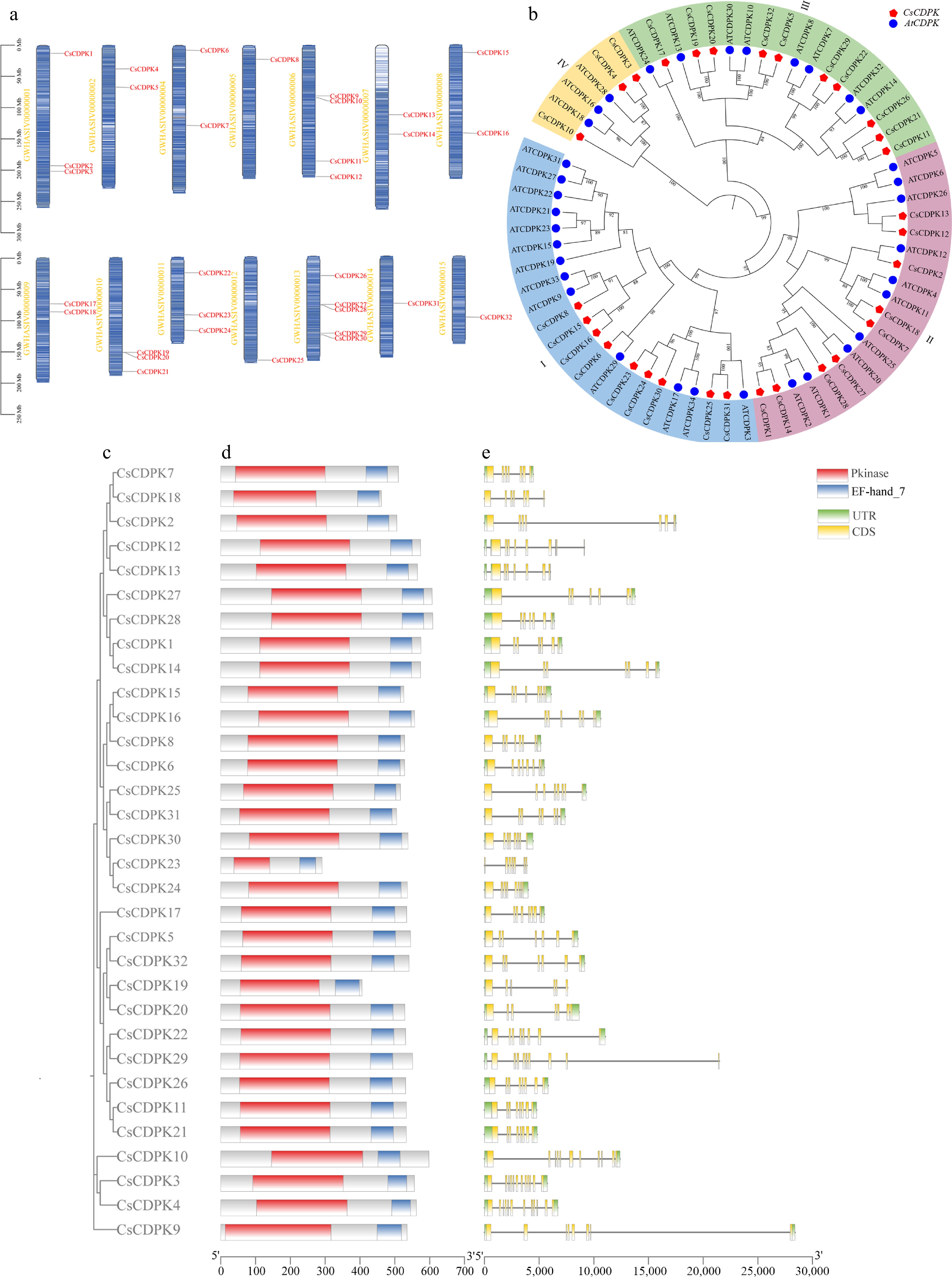
Figure 1.
Chromosomal localization of CsCDPK family members, phylogenetic tree of CDPK genes from tea plant and Arabidopsis, and analyses of conserved motifs, conserved domains, and gene structures of CsCDPK. (a) Chromosomal localization of CsCDPKs in the tea plant genome. (b) Phylogenetic relationships of CDPK proteins from tea plant and Arabidopsis. Red pentagons represent CsCDPKs, and blue circles represent AtCDPKs. The tree was constructed using the neighbor-joining method with 1,000 bootstrap replicates. (c) Phylogenetic tree of CDPKs from tea plant. (d) Distribution of conserved domains in CsCDPKs. (e) Gene structure. Yellow boxes represent CDS (coding sequences), green boxes represent UTRs (untranslated regions), and horizontal lines represent introns. CDS and UTR together constitute the exon regions.
The predicted CsCDPK proteins ranged from 291 to 609 amino acids in length and from 32.30 to 68.24 kDa in molecular weight. The theoretical isoelectric points varied from 5.19 to 9.21. Most members were predicted to be acidic proteins, whereas CsCDPK3, CsCDPK4, and CsCDPK10 were basic. All CsCDPK proteins showed negative GRAVY values, indicating that they are hydrophilic proteins. The instability index ranged from 32.28 to 49.93, suggesting that 13 proteins are stable and the others are unstable. In addition, the aliphatic index ranged from 74.58 to 93.81. Subcellular localization prediction indicated that most CsCDPK proteins were localized in the cytoplasm. Specifically, 19 out of 32 members (59.38%) were predicted to localize mainly to the cytoplasm, whereas the remaining proteins were predicted to localize to the chloroplast, nucleus, plasma membrane, endoplasmic reticulum, or peroxisome (Table 1) .
Table 1. Physicochemical properties and predicted subcellular localization of CsCDPK proteins.
Gene name Amino acids/aa Molecular weight/kDa Theoretical pI Instability index Aliphatic index Hydropathicity Subcellular localization CsCDPK1 575 64.08 5.47 46.51 80.03 −0.39 Chloroplast CsCDPK2 506 56.51 5.19 43.60 85.55 −0.33 Cytoplasm CsCDPK3 556 62.90 8.63 42.56 79.08 −0.61 Chloroplast CsCDPK4 562 63.72 9.21 42.60 76.53 −0.66 Chloroplast CsCDPK5 545 62.11 6.25 33.93 83.87 −0.45 Endoplasmic reticulum CsCDPK6 528 59.31 5.58 45.96 83.30 −0.46 Cytoplasm CsCDPK7 510 57.47 5.26 36.65 82.06 −0.38 Chloroplast CsCDPK8 528 59.12 6.24 45.73 80.17 −0.45 Cytoplasm CsCDPK9 535 58.91 6.11 46.28 91.48 −0.19 Cytoplasm CsCDPK10 598 66.65 8.98 45.26 81.77 −0.40 Cytoplasm CsCDPK11 533 60.62 6.19 34.14 81.56 −0.54 Cytoplasm CsCDPK12 574 64.20 5.55 45.83 81.88 −0.41 Chloroplast CsCDPK13 565 63.59 5.58 37.94 83.88 −0.38 Cytoplasm CsCDPK14 574 64.13 5.38 42.13 79.46 −0.38 Chloroplast CsCDPK15 526 59.13 5.87 37.43 77.66 −0.47 Cytoplasm CsCDPK16 557 62.97 6.59 45.13 74.58 −0.59 Cytoplasm CsCDPK17 534 60.96 5.73 35.57 83.95 −0.45 Cytoplasm CsCDPK18 461 51.97 5.45 42.24 87.57 −0.34 Cytoplasm CsCDPK19 406 45.58 6.54 40.19 85.00 −0.30 Cytoplasm CsCDPK20 529 59.54 5.60 40.93 86.43 −0.37 Cytoplasm CsCDPK21 533 60.55 6.32 33.28 81.91 −0.53 Cytoplasm CsCDPK22 531 59.77 6.24 34.97 84.48 −0.43 Cytoplasm CsCDPK23 291 33.30 6.72 45.36 93.81 −0.35 Cytoplasm CsCDPK24 536 59.84 5.48 37.62 77.72 −0.52 Cytoplasm CsCDPK25 516 58.11 5.73 43.13 76.90 −0.50 Chloroplast CsCDPK26 531 60.01 6.38 36.47 82.58 −0.50 Cytoplasm CsCDPK27 607 66.88 5.30 42.03 84.96 −0.35 Cytoplasm CsCDPK28 609 68.24 5.45 49.93 83.55 −0.36 Cytoplasm CsCDPK29 551 62.30 6.63 43.88 83.72 −0.43 Cytoplasm CsCDPK30 538 59.83 5.45 34.10 77.04 −0.50 Cytoplasm CsCDPK31 505 57.19 5.85 36.88 79.15 −0.46 Peroxisome CsCDPK32 541 61.54 6.16 33.52 85.19 −0.42 Cytoplasm Phylogenetic relationships, conserved motifs, and gene structures of CsCDPKs
-
To investigate the evolutionary relationships of the CDPK family in tea plant, a phylogenetic tree was constructed using the full-length amino acid sequences of CsCDPKs and AtCDPKs. The 32 CsCDPK proteins were classified into four groups, designated I, II, III, and IV, containing 9, 9, 10, and 3 members, respectively (Fig. 1b). All 32 CsCDPK members contain conserved Pkinase and EF-hand-7 domains (Fig. 1d). Gene structure analysis (Fig. 1e) shows that all members have CDS; UTRs are mainly at both ends, except one member without a UTR, and another with a UTR at only one end.
To further assess the conservation and divergence of this family, conserved motifs were analyzed. A total of 10 conserved motifs were identified and designated Motif1 to Motif10 (Supplementary Fig. S1). Motif1, Motif3, and Motif10 were present in all CsCDPK proteins, indicating that these motifs represent core conserved elements of the family. Most family members contained all 10 motifs, whereas CsCDPK9, CsCDPK10, CsCDPK18, CsCDPK19, and CsCDPK23 showed partial motif loss. Notably, CsCDPK23 contained only five motifs and encoded a much shorter protein than the other family members, suggesting a relatively divergent structure.
Multiple sequence alignment further showed that the kinase domain and EF-hand-containing calmodulin-like region were highly conserved among most CsCDPK proteins, whereas sequence divergence was mainly observed in the N-terminal variable region (Supplementary Fig. S2) . The much shorter protein sequence of CsCDPK23 was also evident from the alignment, consistent with its reduced motif composition.
Synteny analysis of the CsCDPK gene family
-
To explore the evolutionary expansion of the CsCDPK family, intra-genomic synteny analysis was performed within the tea genome. Thirteen duplicated gene pairs were identified among the CsCDPK genes (Fig. 2a), indicating that segmental duplication has played an important role in the expansion of this family in tea plant. To further examine their evolutionary conservation, comparative synteny analysis was conducted between tea plant and Arabidopsis. A total of 38 homologous gene pairs were identified between the two species (Fig. 2c). Some tea genes corresponded to multiple Arabidopsis homologs, and vice versa, suggesting complex evolutionary relationships.
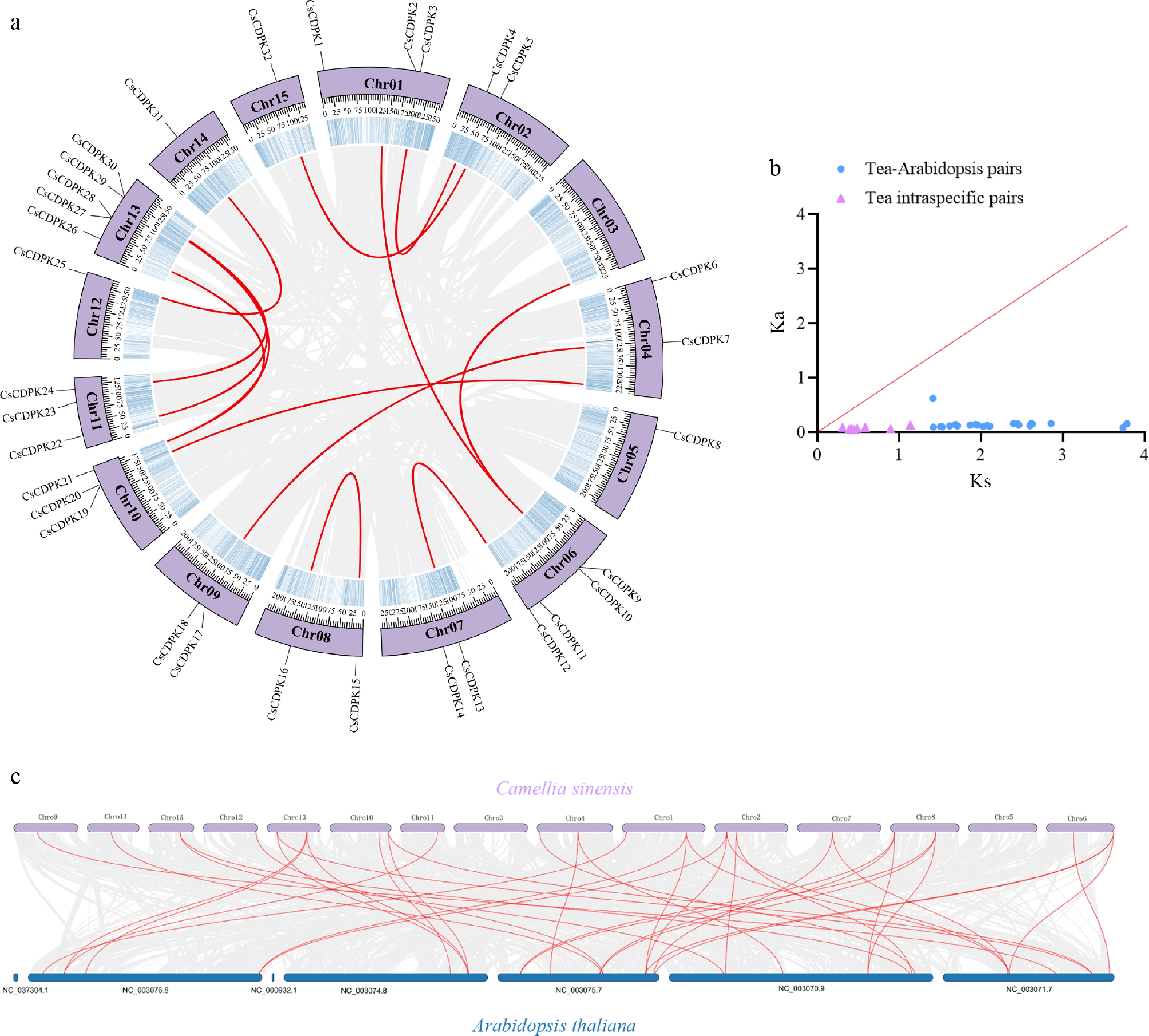
Figure 2.
Synteny analysis of CsCDPK genes. (a) Intraspecific synteny analysis within the tea plant genome. Gray lines indicate all syntenic blocks in the tea genome, and red lines indicate syntenic gene pairs involving CsCDPK genes. (b) Distribution of Ka/Ks ratios. The red line indicates Ka/Ks = 1. Points below the red line represent Ka/Ks < 1 (purifying selection), and points above the red line represent Ka/Ks > 1 (positive selection). (c) Interspecific synteny analysis between tea plant and Arabidopsis. Gray lines indicate all syntenic gene pairs between the Arabidopsis and tea plant genomes, and red lines indicate syntenic gene pairs of CDPK genes between Arabidopsis and tea plants.
All duplicated gene pairs identified within the tea genome (intraspecific pairs) exhibited Ka/Ks ratios of less than 1, indicating that the CsCDPK family has undergone strong purifying selection during evolution. For the interspecific gene pairs between tea plant and Arabidopsis, the majority also showed Ka/Ks values of less than 1 (Fig. 2b). However, a small fraction of these pairs lacked a valid Ka/Ks ratio due to saturated synonymous substitutions (resulting in Ks values of 'NA'), likely reflecting the high sequence divergence between these two species. These results suggest that while the CsCDPK family has expanded in tea plant, its core functions have remained evolutionarily conserved.
Cis-acting element analysis of CsCDPK promoters
-
To infer the potential regulatory mechanisms of CsCDPK genes, the 2-kb upstream promoter regions were analyzed for cis-acting elements. The results showed that the promoters of CsCDPK genes contain numerous cis-elements associated with light responsiveness, hormone responses, stress responses, growth and development, and anaerobic induction (Fig. 3).
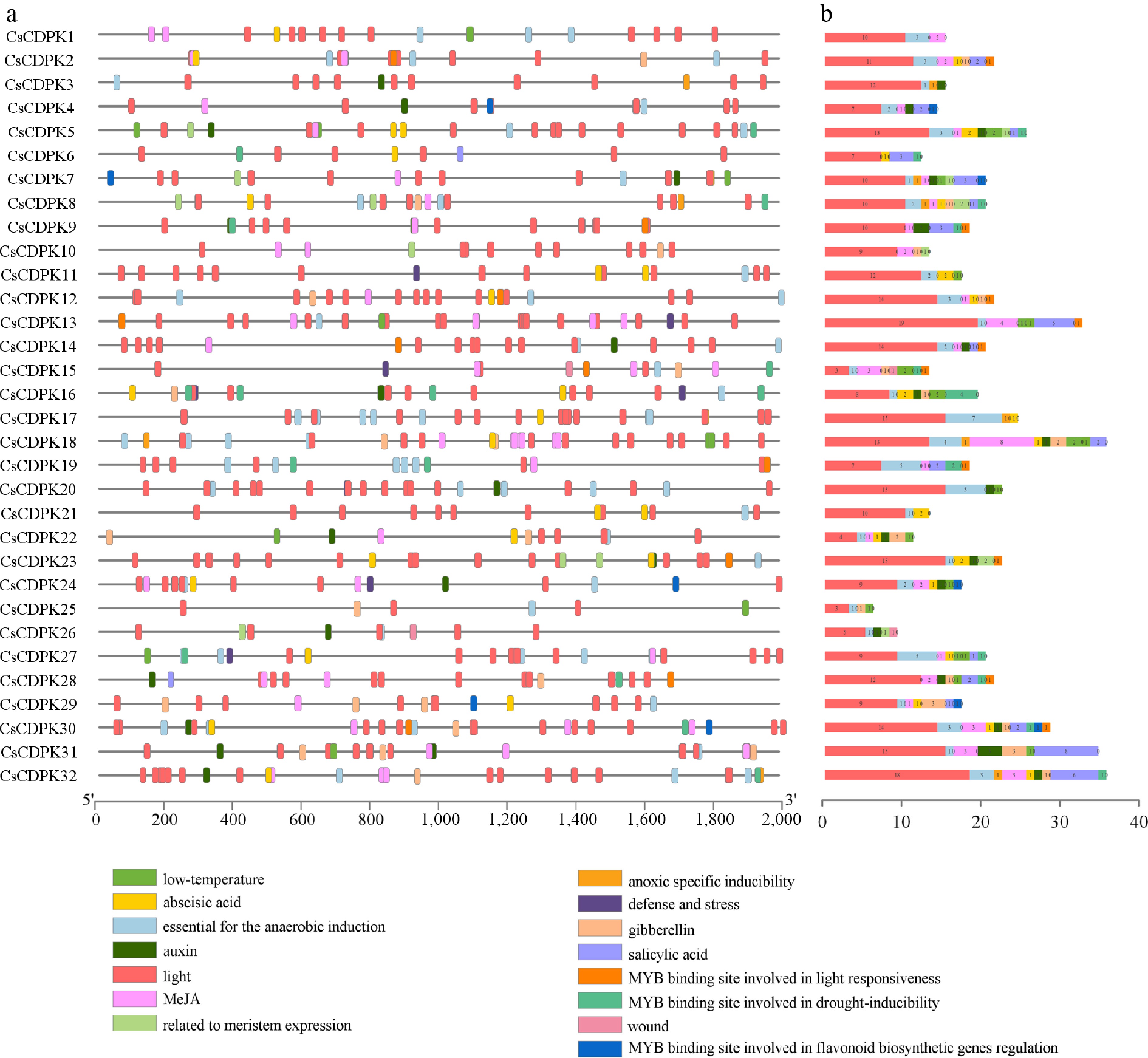
Figure 3.
Cis-acting regulatory elements in the promoter regions of CsCDPK genes. (a) Distribution of cis-acting elements in the promoters of CsCDPK genes; different colors represent different elements. (b) Stacked bar chart showing the number of cis-acting elements in CsCDPK promoters.
Among these elements, stress-responsive elements were identified in nine genes, SA-responsive elements in 18 genes, MeJA-responsive elements in 22 genes, and ABA-responsive elements in 17 genes. In addition, anaerobic induction-related elements were present in 30 of the 32 promoters. These findings suggest that the expression of CsCDPK genes may be regulated by multiple environmental and endogenous signals, especially hormone-mediated and stress-related pathways.
Expression profiles of CsCDPK genes in different tissues and under stress conditions
-
To gain insight into the potential biological functions of CsCDPK genes, their expression profiles in different tea tissues were analyzed using public transcriptomic datasets. Distinct spatial expression patterns were observed among family members. CsCDPK12 displayed relatively high expression in roots, stems, buds, and flowers, whereas CsCDPK26 was highly expressed in young leaves, mature leaves, and old leaves (Fig. 4a, b). These patterns suggest possible functional differentiation among CsCDPK members in tea plant growth and development.
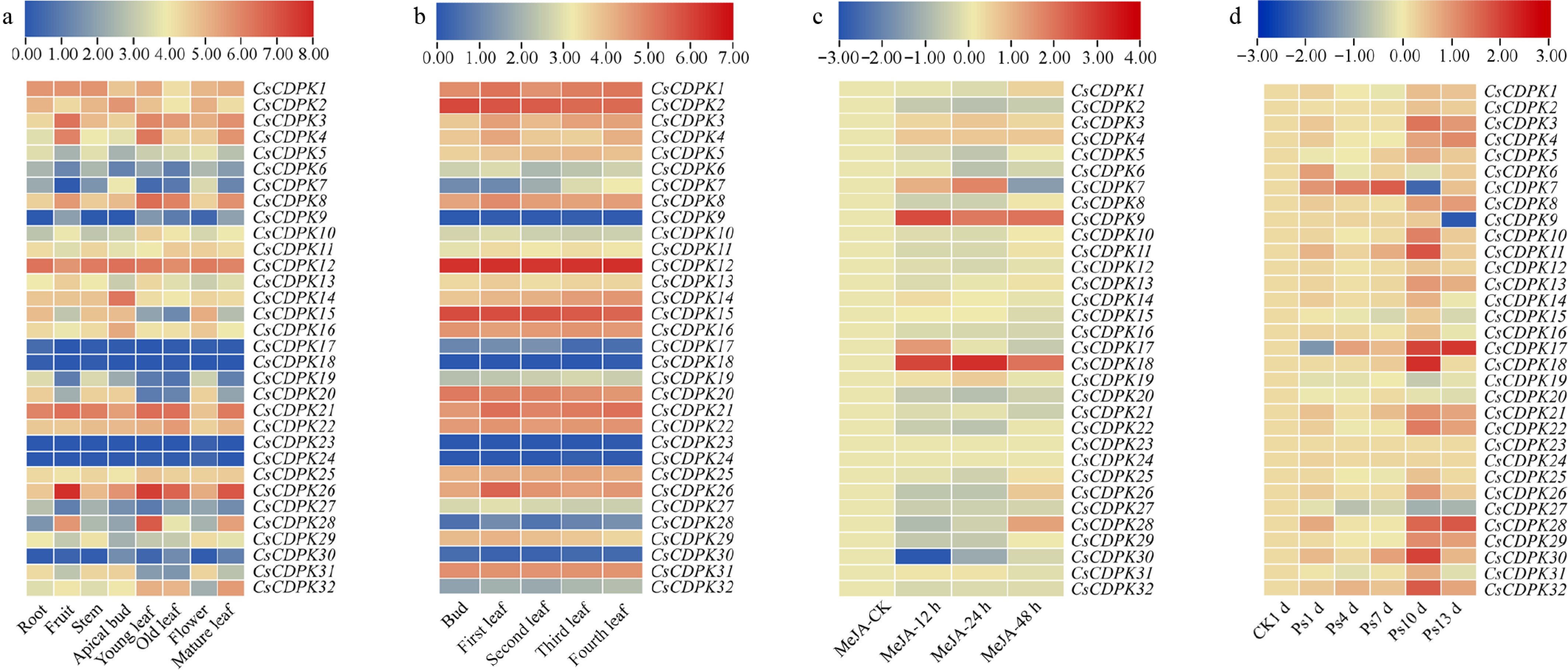
Figure 4.
Expression profiles of CsCDPK genes in different tissues and under stress-related treatments. (a) Expression patterns in tea plant tissues. The heatmap was generated using log2(TPM + 1) without row normalization. Color intensity directly reflects the absolute expression level of each gene across different tissues. (b) Expression patterns in bud and leaf tissues. Data processing and visualization are the same as in (a). (c) Expression dynamics after exogenous MeJA treatment. The heatmap was generated using log2([TPM_treatment + 1]/[TPM_0 h + 1]). Red indicates upregulation relative to 0 h, and blue indicates downregulation. (d) Expression dynamics after gray blight pathogen infection. Data processing and visualization are the same as in (c). The color scale is shown above the heatmap; the specific range is indicated in the figure. Expression data were obtained from the TPIA2.0 database.
Expression profiles under MeJA treatment showed that several CsCDPK genes were responsive to exogenous hormone stimulation (Fig. 4c). For example, CsCDPK3 and CsCDPK4 showed a gradual increase in expression within 48 h of treatment. CsCDPK14, CsCDPK15, and CsCDPK31 were upregulated at earlier time points but declined by 48 h, whereas CsCDPK1 and CsCDPK26 reached peak expression at 48 h.
Under gray blight pathogen infection (Fig. 4d), many CsCDPK genes exhibited pathogen-responsive expression patterns. Among them, CsCDPK12 and CsCDPK21 were strongly induced at the early stage of infection. CsCDPK11 remained highly expressed at 1, 4, 7, 10, and 13 d after inoculation, reaching its maximum at day 10. Several other genes, including CsCDPK22, CsCDPK26, CsCDPK28, and CsCDPK32, also showed obvious upregulation during the infection period. In contrast, CsCDPK19 and CsCDPK27 displayed relatively low expression and a decreasing trend after infection. These data indicate that different CsCDPK genes may play distinct roles during the tea plant's response to gray blight disease.
Dynamic changes in endogenous SA, JA, and MeJA contents after pathogen infection
-
To investigate whether hormone signaling is involved in the tea plant response to gray blight disease, endogenous SA, JA, and MeJA contents were measured after pathogen inoculation. Both SA and MeJA accumulated rapidly and reached their highest levels at 12 h after infection (Fig. 5a). Their levels then declined, followed by a secondary increase at 48 h, and a final decrease again by 72 h. JA showed a different accumulation pattern, reaching its peak at 48 h after infection. These dynamic changes observed relative to the 0 h baseline indicate that both SA and jasmonate-related signaling pathways are rapidly mobilized in tea leaves following pathogen inoculation.
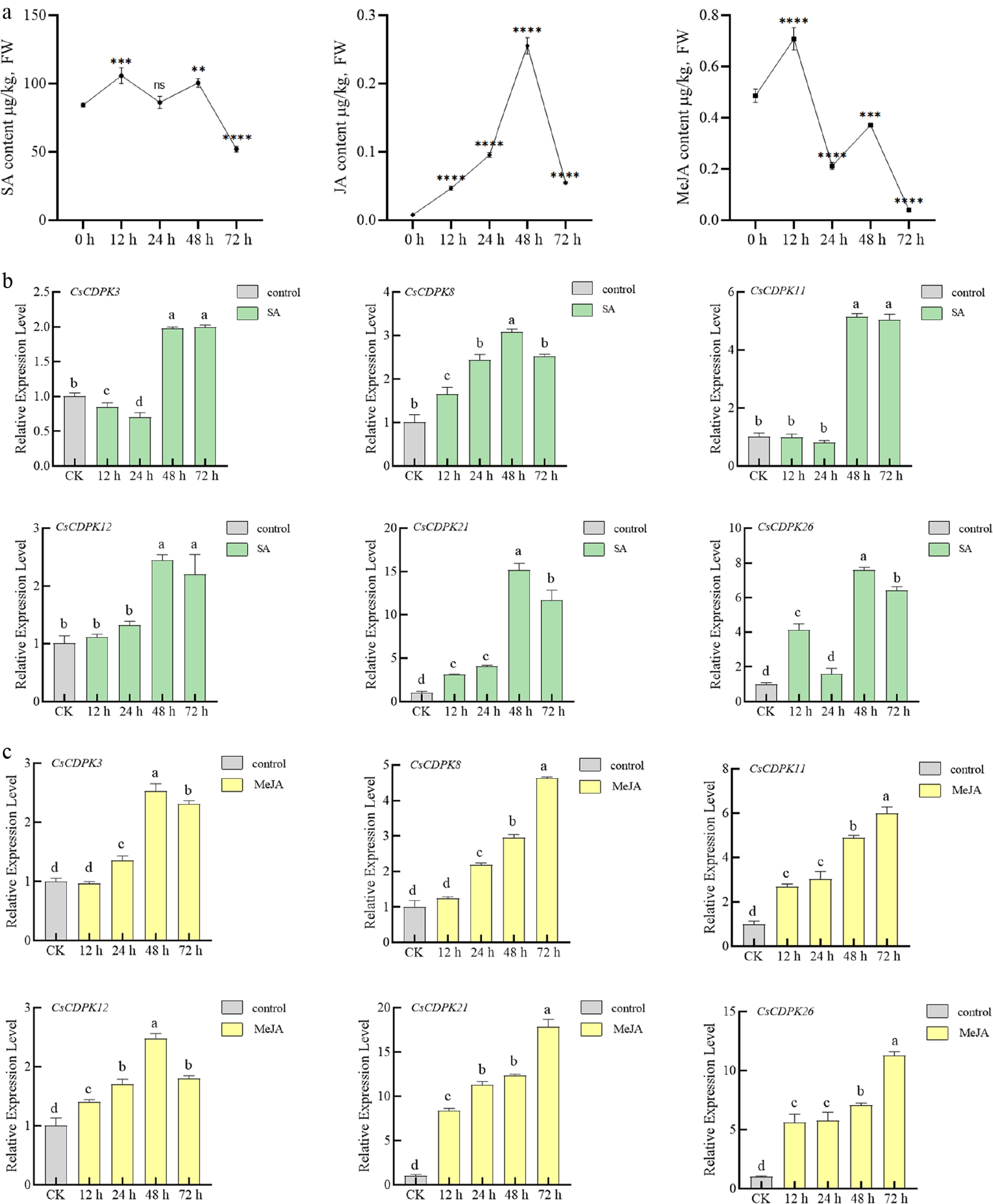
Figure 5.
Dynamic changes in endogenous hormone contents in tea plants after pathogen infection and expression responses of CsCDPK genes to exogenous hormone treatments. (a) Dynamic changes of endogenous SA, JA, and MeJA in tea plant leaves after inoculation with Pseudocercospora camelliae. Each post-inoculation time point was compared with the 0 h control using one-way ANOVA followed by Dunnett's multiple comparison test. Comparisons among non-zero time points were not performed. ns, not significant; ** p < 0.01, *** p ≤ 0.001, **** p < 0.0001. (b) Expression of six CsCDPK genes in response to SA. (c) Expression of six CsCDPK genes in response to MeJA. Different letters indicate significant differences among groups (p < 0.05; one-way ANOVA with Tukey's post hoc test). Groups sharing the same letter are not significantly different.
Expression patterns of selected CsCDPK genes in response to exogenous SA and MeJA
-
Based on the transcriptome data and pathogen-responsive expression patterns, six candidate genes, CsCDPK3, CsCDPK8, CsCDPK11, CsCDPK12, CsCDPK21, and CsCDPK26, were selected for RT-qPCR analysis after exogenous SA and MeJA treatments.
Following SA treatment, all six genes exhibited an overall upregulation trend relative to the 0 h baseline, and most of them reached their highest expression levels at 48 h (Fig. 5b). CsCDPK3, CsCDPK11, and CsCDPK12 showed similar expression patterns, with little change in the first 24 h, followed by a clear increase at 48 h. CsCDPK8 and CsCDPK21 increased continuously within 48 h and then declined thereafter.
Following MeJA treatment, CsCDPK8, CsCDPK11, CsCDPK21, and CsCDPK26 displayed continuous induction relative to the 0 h baseline and reached peak expression at 72 h (Fig. 5c). By contrast, CsCDPK3 and CsCDPK12 showed an increase followed by a decrease, with peak expression at 48 h. These results indicate that under the present experimental conditions, the six selected CsCDPK genes responded to both SA and MeJA treatments, although their induction kinetics differ.
Functional analysis of selected CsCDPK genes by AsODN-mediated silencing
-
To investigate whether the selected CsCDPK genes are involved in tea plant defense against gray blight disease, AsODN-mediated gene silencing was performed. RT-qPCR analysis confirmed that the candidate genes were effectively silenced at different time points relative to the 0 h baseline (Fig. 6).
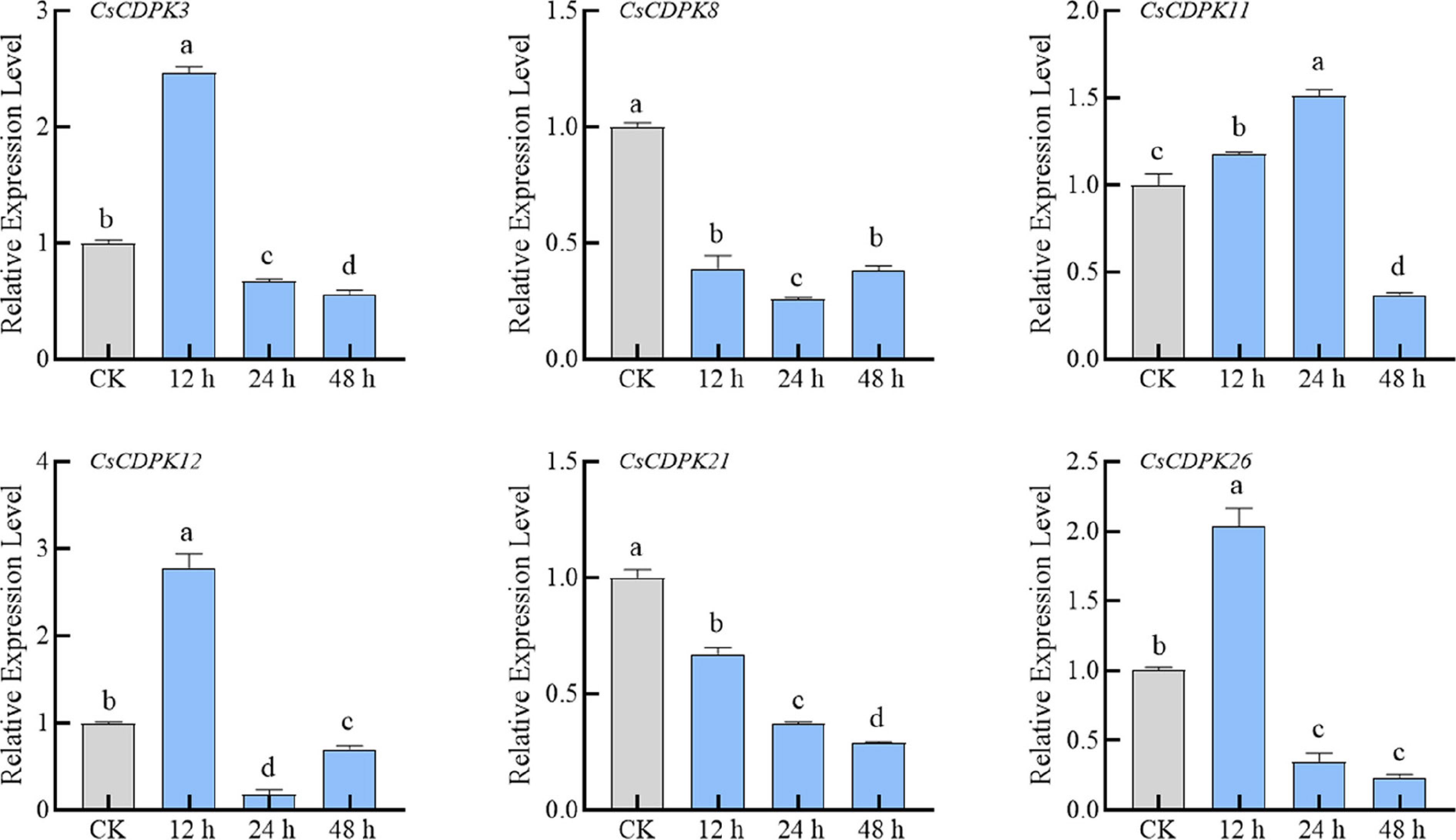
Figure 6.
Expression analysis of six selected CsCDPK genes after AsODN treatment. Different letters indicate significant differences among groups (p < 0.05; one-way ANOVA with Tukey's post hoc test). Groups sharing the same letter are not significantly different.
Subsequently, healthy control plants (CK), healthy plants inoculated with the gray blight pathogen (CK-T), and silenced plants inoculated with the gray blight pathogen (AS-T) were used to evaluate the effects of pathogen infection on CsCDPKs expression and the impact of gene silencing on disease resistance. At 3 d post-inoculation, the expression levels of CsCDPKs in CK-T plants were significantly higher than those in CK plants, while the expression levels in AS-T plants were lower than those in CK-T plants, indicating that gene silencing was effective. Among the six genes, CsCDPK11 showed no significant difference between groups (Fig. 7a). Phenotypic observations showed that silencing of any of the six CsCDPK genes resulted in more severe disease symptoms than in the control leaves (Fig. 7c). Statistical analysis revealed that lesion diameters in the silenced leaves were significantly larger than those in the control group (Fig. 7b), indicating reduced resistance to gray blight disease. Among the six genes, silencing of CsCDPK26 produced the most obvious increase in lesion size, suggesting that this gene may play a particularly important role in tea plant defense. Collectively, these data demonstrate that the selected CsCDPK genes basal defenses regulate resistance to gray blight disease under the present experimental conditions.
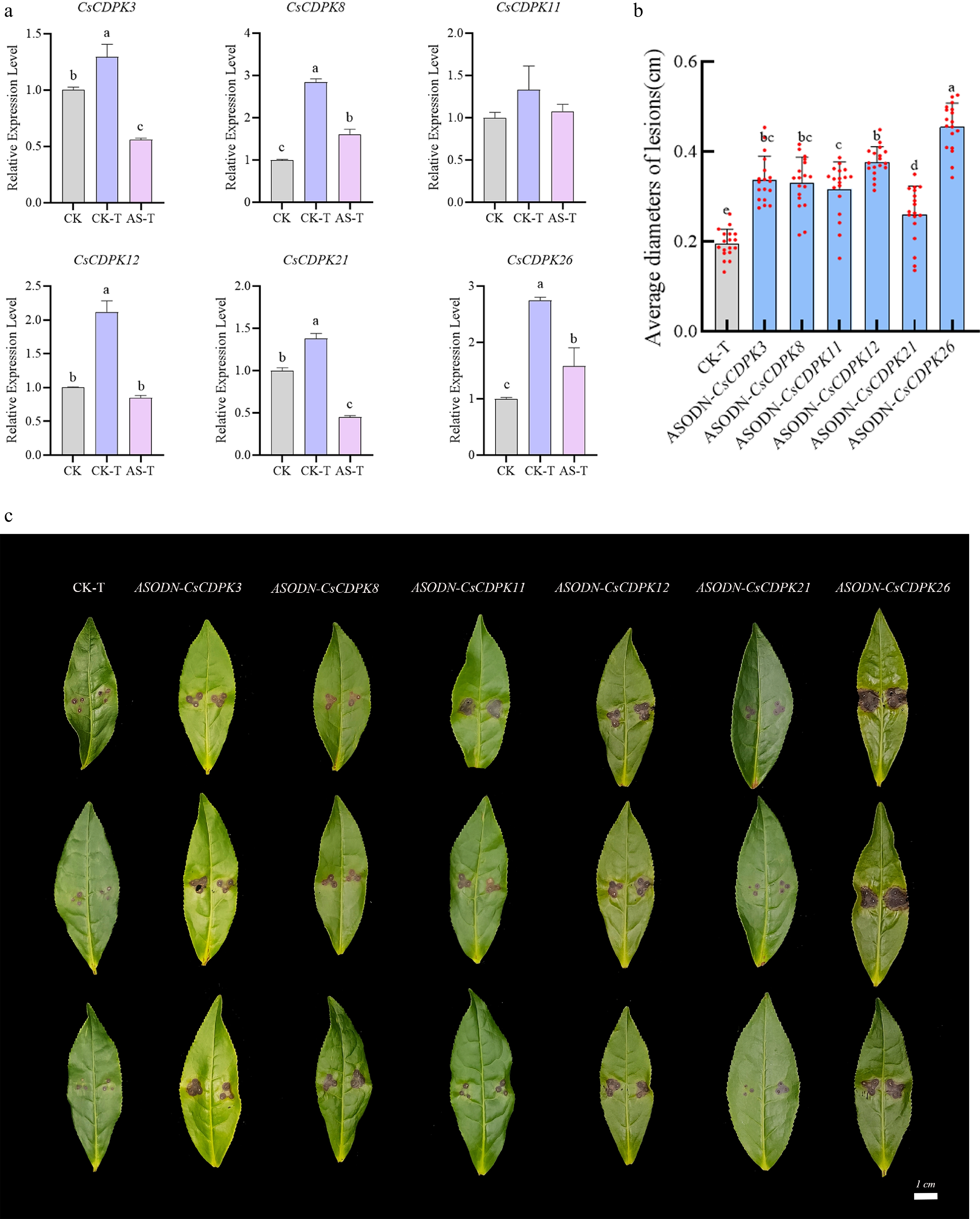
Figure 7.
Analysis of the effects of silencing six selected CsCDPK genes on disease resistance in tea plants. (a) Expression of six CsCDPK genes in silenced leaves after pathogen inoculation, CK: healthy control plants, CK-T: healthy plants inoculated with gray blight treatment, AS-T: silenced plants inoculated with gray blight treatment. (b) Statistical analysis of lesion diameters. Each gene included 18 biological replicates. (c) Representative disease lesions on tea leaves at 3 d after pathogen inoculation. Data are presented as mean ± SD. Different lowercase letters indicate significant differences (p < 0.05), while the same letter indicates no significant difference. No letters indicate no significant differences.
-
In this study, a total of 32 CsCDPK genes were identified in the tea plant genome, whereas Ding et al.[31,39] reported 26 CsCPK members based on transcriptome data. This discrepancy may be explained by the following factors. First, the genome assembly used in this study (TPIA2.0) represents an improved version with more comprehensive gene annotation compared with the earlier version (TPIA1.0) used by Ding et al. Second, the HMM-based genome-wide identification approach adopted in this study is less dependent on transcript abundance and assembly quality, which may account for the identification of additional members. Moreover, Ding et al. focused primarily on cold stress responses, whereas the present study examined the association between the CsCDPK family and biotic stress caused by the gray blight pathogen. Furthermore, we combined this analysis with ASODN-based gene silencing experiments to preliminarily validate the functions of these genes in disease defense. Collectively, building upon previous work, this study expands our understanding of the functional diversity of the CsCDPK family in tea plants and addresses a gap in research regarding their roles in biotic stress.
Evolutionary conservation and structural characteristics of the CsCDPK family
-
Their encoded proteins shared the typical structural characteristics of plant CDPKs, including a highly conserved kinase domain and an EF-hand-containing calmodulin-like domain. Most family members also exhibited similar motif organization and gene structure, indicating that the CsCDPK family is evolutionarily conserved. At the same time, some differences in motif composition, protein length, and gene structure were observed, suggesting that certain members may have undergone functional divergence during evolution. The hydrophilic nature of all CsCDPK proteins is consistent with their role as calcium-responsive signaling proteins. Ca2+ functions as a critical intracellular second messenger. Upon an increase in intracellular Ca2+ concentration, Ca2+ binds to the C-terminal EF-hand motifs of CDPK, inducing conformational changes that activate their kinase activity[40,41]. Since Ca2+ acts as an intracellular second messenger, the ability of CDPK proteins to interact efficiently with Ca2+ is closely related to their structural characteristics. The predominance of cytoplasmic localization predicted for CsCDPKs further supports their potential involvement in intracellular signal perception and transduction.
CDPK proteins typically contain an N-terminal variable domain (NTD), a calmodulin-like domain (CaM-LD), a protein kinase domain (PKD), and an autoinhibitory junction domain (JD)[42]. In this study, CsCDPK family members display both high conservation and some divergence in their protein sequences. Phylogenetic analysis divided the CsCDPK family into four groups, in agreement with the classification pattern reported for CDPK families in other plant species. Moreover, synteny analysis revealed that segmental duplication contributed substantially to family expansion in tea plants. Similar duplication-driven expansion has been reported for CDPK families in apple, jujube, and rapeseed[43−45]. Ka/Ks analysis was performed on both interspecific (tea vs. Arabidopsis) and intraspecific (within-tea) duplicated gene pairs. Most calculable interspecific pairs exhibited Ka/Ks values below 1, suggesting purifying selection since species divergence. Some interspecific pairs lacked valid Ka/Ks ratios due to saturated synonymous substitutions, reflecting the distant relationship between tea and Arabidopsis. In contrast, all intraspecific pairs yielded calculable Ka/Ks ratios below 1, providing direct evidence for functional conservation of the CsCDPK family. Together, both intra- and interspecific data support that CsCDPK genes have been maintained under strong purifying selection during evolution.
Synergistic accumulation pattern of SA, JA, and MeJA in tea plants under gray blight infection
-
Exogenous SA and MeJA have been reported to enhance disease resistance in various plant species. For example, in kiwifruit, exogenous MeJA enhances resistance to gray mold by increasing antioxidant enzyme activity and activating phenylpropanoid metabolism and hormone signal transduction pathways[46]. In tea plants, exogenous SA treatment activates the lignin metabolic pathway and promotes lignin accumulation, thereby enhancing defense responses[39]. In banana, exogenous SA and MeJA treatment significantly reduces the disease index and lesion diameter of anthracnose, and induces the expression of PR genes and WRKY transcription factors[47]. These findings indicate that exogenous SA and MeJA treatments can enhance disease resistance in multiple plant species, suggesting that SA- and MeJA-mediated defense mechanisms may have positive regulatory potential in plants.
Given that both exogenous SA and MeJA individually promote disease resistance, we further examined the dynamic accumulation patterns of endogenous hormones under infection by the gray blight pathogen, in order to investigate the relationship between the SA and JA pathways during the defense response in tea plants. In plant–pathogen interactions, SA and JA generally exhibit an antagonistic relationship, with SA primarily involved in defense against biotrophic pathogens and JA mainly involved in defense against necrotrophic pathogens[48,49]. In recent years, however, reports of SA-JA synergistic interactions have been increasing. Increasing evidence suggests that the SA-JA relationship is context-dependent and may vary according to plant species, pathogen lifestyle, infection stage, and tissue type[50]. For example, in poplar, the SA and JA signaling pathways display a positive synergistic pattern, co-enhancing resistance to leaf rust[51]; in plum trees infected by Apiosporina morbosa, endogenous SA and JA accumulation also shows a synergistic trend[52]. In the present study, upon infection by the gray blight pathogen, endogenous SA and MeJA both peaked at 24 h post-inoculation, displaying a highly synchronized 'rise-then-fall' accumulation pattern. This pattern is consistent with the SA-JA synergistic accumulation observed in poplar and plum described above, and differs from the classical antagonistic model, which typically features a temporal separation with SA accumulation preceding JA. For example, in resistant wheat infected by Fusarium graminearum, SA accumulated as early as 6 h, while JA levels began to rise at approximately 12 h[53], indicating a time lag with SA preceding JA. Similarly, a temporal difference between SA and JA was also observed in this study: SA peaked at 12 h, whereas JA peaked at 48 h. However, no apparent time lag was observed between SA and MeJA accumulation, and MeJA and JA were not completely synchronized in this system—JA peaked at 48 h rather than 12 h. One possible explanation is that JA methylation is rapidly induced during the early defense response, allowing MeJA to accumulate preferentially and function cooperatively with SA in early signaling, while free JA participates in the regulation of later signaling events. That said, direct evidence for this hypothesis is currently lacking, and future studies examining the expression and activity of jasmonic acid carboxyl methyltransferase (JMT), the key enzyme responsible for converting JA to MeJA, during the early stages of infection will be necessary to further clarify this phenomenon.
It should be noted that JA-Ile, rather than MeJA, is generally regarded as the primary bioactive jasmonate in plant defense signaling. In this study, JA served as the primary indicator of jasmonate pathway activation, while MeJA was measured as auxiliary information. However, JA-Ile was not quantified, which limits a complete interpretation of jasmonate-mediated defense signaling. Future studies incorporating JA-Ile measurements will be necessary to fully resolve the jasmonate dynamics in this pathosystem.
In summary, both the disease resistance-promoting effects of exogenous SA/MeJA and the synergistic accumulation of endogenous SA and MeJA collectively suggest that tea plants may simultaneously mobilize both the SA and MeJA pathways in defense against gray blight disease. Further experiments, including SA/JA pathway inhibition, exogenous hormone pretreatment followed by pathogen challenge, and downstream defense marker gene analysis, will be necessary to determine the functional relationship between SA and JA signaling during tea plant defense against gray blight disease.
Potential roles of CsCDPK genes in tea plant defense against gray blight disease
-
All CsCDPKs identified in this study contain the conserved EF-hand-7 domain, which provides the structural basis for sensing changes in cytosolic Ca2+ concentration[8,54]. Although we did not directly measure cytosolic Ca2+ concentration, Ca2+-dependent kinase activity, or downstream phosphorylation targets following pathogen infection, this structural feature suggests that CsCDPK activation may be regulated by upstream Ca2+ signals. The causal relationship between Ca2+ signaling and CsCDPK-mediated defense responses remains to be validated through future studies.
Given that CDPKs typically function as decoders of Ca2+ signals, converting upstream changes in cytosolic Ca2+ concentration into downstream phosphorylation events, they may execute defense functions in tea plants by regulating ROS production. Accumulating evidence indicates that CDPKs play important roles in plant defense through multiple pathways, including gene regulation, ROS production, and metabolic regulation[55,56]. In Arabidopsis, AtCPK5/AtCPK6 phosphorylate the transcription factor AtWRKY33 at Thr-229, enhancing its DNA-binding activity and activating the biosynthesis of the antimicrobial compound camalexin[57]. StCDPK7 has been shown to specifically phosphorylate phenylalanine ammonia-lyase (PAL), thereby enhancing plant defense[58]. StCDPK5 in potato phosphorylates StRBOH, inducing ROS burst and enhances resistance against biotrophic pathogens[59]. In Medicago truncatula, MtCDPK5 directly phosphorylates MtRBOHB/C/D to activate plant immune responses[60]. Given that CDPK-mediated ROS production is a common mechanism in plant immunity, and that silencing of CsCDPKs resulted in enhanced susceptibility in this study, it would be interesting to investigate in the future whether CsCDPKs contribute to disease resistance by modulating ROS accumulation.
In this study, silencing each of the six CsCDPK genes via AsODN followed by pathogen inoculation significantly increased lesion diameters, indicating that these genes play positive regulatory roles in the basal immunity of tea plants. Among them, CsCDPK26 showed high expression in leaves and caused the most pronounced susceptibility phenotype upon silencing, suggesting that it may play an important role in leaf defense against gray blight disease. It should be noted that mock-inoculated controls for the silenced plants were not included in this study, and this methodological limitation should be taken into consideration when interpreting the silencing phenotypes.
-
A total of 32 CsCDPK genes were identified in the tea plant genome and classified into four phylogenetic groups. Comprehensive analyses of their chromosomal distribution, protein properties, conserved motifs, gene structures, promoter elements, and syntenic relationships demonstrated that the CsCDPK family is evolutionarily conserved while also showing some degree of divergence. Expression profiling indicated that several CsCDPK genes respond strongly to gray blight pathogen infection and to exogenous SA and MeJA treatments. Functional assays further showed that CsCDPK3, CsCDPK8, CsCDPK11, CsCDPK12, CsCDPK21, and CsCDPK26 positively regulate resistance to gray blight disease in tea plants. These findings provide a useful framework for understanding the molecular basis of tea plant disease resistance and offer candidate genes for future genetic improvement.
These results suggest that CsCDPK-mediated defense in tea plants may be associated with SA- and MeJA-related pathways. Further investigation of downstream phosphorylation targets and signaling networks will help elucidate the precise molecular mechanisms.
-
The authors confirm their contributions to the paper as follows: study conception and design, and manuscript editing and revision: Hao L, Chen L; data analysis: Hao L, Liu S, Chen Z; figures and manuscript construction: Tian N, Wu T; performing experiments, manuscript writing: Hao L; materials provision and facility testing: Liu Z, Huang J. All authors reviewed the results and approved the final version of the manuscript.
-
The raw RNA-seq data used in this study are publicly available from the TPIA2.0 database (https://tpia.teaplants.cn). The qPCR data generated and analyzed during this study are available from the corresponding author upon reasonable request.
-
This work was financially supported by the Yuelushan Laboratory Breeding Program (YLS-2025-ZY02020), the National Key Research and Development Program of China (2021YFD1200200), the National Natural Science Foundation of China (U22A20500, 32172629), the Hunan Agriculture Research System (HARS-10), and the National Tea Tree Breeding Joint Research Project (GJCSYZLHGG-12).
-
The authors declare that they have no conflict of interest.
-
accompanies this paper online at: https://doi.org/10.48130/bpr-0026-0019.
- Supplementary Table S1 Primers used for RT-qPCR and AsODN sequences in this study.
- Supplementary Fig. S1 Conserved motifs identified in CsCDPK family members.
- Supplementary Fig. S2 Multiple sequence alignment of CsCDPK proteins.
- Copyright: © 2026 by the author(s). Published by Maximum Academic Press, Fayetteville, GA. This article is an open access article distributed under Creative Commons Attribution License (CC BY 4.0), visit https://creativecommons.org/licenses/by/4.0/.
-
About this article
Cite this article
Hao L, Chen L, Chen Z, Wu T, Huang J, et al. 2026. Genome-wide characterization of the calcium-dependent protein kinase gene family in tea plant and its potential involvement in resistance to gray blight disease. Beverage Plant Research 6: e025 doi: 10.48130/bpr-0026-0019 

Genome-wide characterization of the calcium-dependent protein kinase gene family in tea plant and its potential involvement in resistance to gray blight disease
- Received: 16 March 2026
- Revised: 27 April 2026
- Accepted: 08 May 2026
- Published online: 30 June 2026
Abstract: Calcium-dependent protein kinases (CDPKs) are important Ca2+ sensors that play crucial roles in plant responses to environmental stresses. However, the functions of CDPK genes in the tea plant (Camellia sinensis), especially in disease resistance, remain poorly understood. In this study, 32 CsCDPK genes were identified from the tea plant genome and classified into four phylogenetic groups. These genes were unevenly distributed across 14 chromosomes. Analyses of gene structure, conserved motifs, physicochemical properties, promoter cis-elements, and syntenic relationships indicated that the CsCDPK family is highly conserved but also shows some evolutionary divergence. Expression analyses based on transcriptomic data and RT-qPCR revealed that several CsCDPK genes were strongly induced by infection with the gray blight pathogen Pseudopestalotiopsis spp. and by exogenous salicylic acid (SA) and methyl jasmonate (MeJA) treatments. Endogenous SA, jasmonic acid (JA), and MeJA levels also increased rapidly after pathogen infection. Six genes, CsCDPK3, CsCDPK8, CsCDPK11, CsCDPK12, CsCDPK21, and CsCDPK26, were further functionally analyzed using antisense oligodeoxynucleotide-mediated silencing. Silencing of these genes significantly increased lesion size after pathogen inoculation, indicating reduced resistance. These findings suggest that the selected CsCDPK genes are likely involved in tea plant defense against gray blight disease, providing candidate genes for further functional characterization and potential resistance improvement.