













-
Soil salinity is a major abiotic stress factor affecting horticultural crops, inducing ion toxicity, osmotic stress, nutrient imbalance, and other detrimental effects on plants[1,2]. Approximately 20% of cultivated land worldwide is threatened by salinization, severely restricting plant growth, development, and biomass accumulation. This stress triggers morphological, physiological, biochemical, and molecular alterations in crops[3,4]. Due to improper irrigation, fertilization, and industrial pollution, salinized land continues to expand by over 1 million ha annually. By 2050, it is projected that more than half of the world's cultivated land will be at risk from salinization[5,6]. Plants exhibit varying sensitivity to salinity depending on their developmental stage, with seed germination and early seedling growth being the most vulnerable phases. During these stages, osmotic stress and ion toxicity severely restrict growth and development[7−9].
Peaches are cultivated in over 80 countries, covering a global planting area of 1.5 million ha by 2020, with China accounting for 52% of this total[10]. Peach (Prunus persica [L.] Batsch) seeds are essential for seedling propagation and germplasm screening in breeding programs[11]. Seed germination, the first and most critical stage of plant morphogenesis, determines seedling quality and vigor but is particularly sensitive to abiotic stress, especially salinity[12,13]. Salt-induced inhibition of germination arises mainly from osmotic stress, excessive reactive oxygen species (ROS) accumulation, and cellular structural damage, which collectively reduce germination rates and delay germination time[14]. Salt stress delays germination in tomato, inhibits cottonseed germination and endogenous melatonin accumulation, decreases α-amylase and β-galactosidase activity in cotton, and reduces germination rates in Suaeda salsa[15−17]. Similarly, in peach, salt stress during germination can lead to abnormal seedling development.
Hydrogen (H2), a colorless, odorless, and low-density gas with strong reducing capacity, has recently emerged as a novel bioactive molecule influencing various aspects of plant growth and development, including seed germination[18], seedling growth[19], adventitious root formation[20], and fruit ripening[21]. Hydrogen-rich water (HRW) has been shown to promote adventitious root formation in cucumber[22] and enhance tolerance to multiple stresses, including heavy metal toxicity[23], heat[24], and ultraviolet radiation[25]. Under lead (Pb) stress, HRW enhances rice root growth and mitigates Pb toxicity by strengthening antioxidant defense[26]. In salt-stressed fragrant rice, HRW increases antioxidant enzyme activity and decreases malondialdehyde (MDA) content[27]. In maize, HRW upregulates salt-tolerance genes, enhances H+ transport, maintains Na+/K+ homeostasis, reduces oxidative damage, and promotes root growth, thereby improving salt tolerance[28]. These findings indicate that H2 functions as both a signaling regulator in plant development and a synergistic molecule in stress responses. Nevertheless, the potential role of HRW in regulating seed germination, especially in fruit tree species, has not been well documented.
Flavonoids are a class of phenylpropanoid compounds with a C6-C3-C6 backbone that act as antioxidants to mitigate oxidative damage under salt stress[29−31]. Their biosynthesis begins with phenylalanine and proceeds via 4-coumaroyl-CoA, catalyzed by enzymes including chalcone isomerase (CHI), flavonol synthase (FLS), flavone synthase (FNS), flavanone 3-hydroxylase (F3H), flavonoid 3'-hydroxylase (F3'H), UDP-glucosyltransferase (UGT), and isoflavone synthase (IFS)[32]. Studies indicate that salt stress upregulates the expression of several genes encoding these enzymes[33,34]. For instance, 1.2% NaCl enhances the expression of PAL, C4H, 4CL, CHS, CHI, and ANR in Sophora japonica roots[34]. In transgenic tobacco, NtCHS1 RNAi suppression reduces flavonoid accumulation and impairs ROS scavenging under salt stress, whereas overexpression of NtCHS1 enhances salt tolerance[35]. Therefore, genes involved in flavonoid biosynthesis play critical roles in enhancing plant salt tolerance.
In this study, integrated transcriptomic and metabolomic analyses were employed to elucidate physiological and molecular mechanisms regulating peach seed germination under salt stress and hydrogen-rich water treatment. Additionally, qRT-PCR was conducted to validate transcriptional changes in key flavonoid biosynthesis genes and quantify corresponding enzyme activity. This investigation provides valuable insights into the relationships between key genes and metabolites during salt stress and hydrogen treatment in peach seeds, enhancing understanding of how hydrogen-rich water mitigates salt-induced physiological and molecular disturbances.
-
Stratified Prunus davidiana seeds were obtained from the experimental station of Shandong Agricultural University. Hydrogen-rich water (HRW, 0.27 mM) was prepared by dissolving a 15 mg magnesium hydride tablet in 0.5 L of water, referred to as 50% HRW.
NaCl concentration screening
-
Healthy peach seeds were surface-sterilized with 3% NaClO for 15 min, rinsed thoroughly, and placed in Petri dishes (30 seeds per dish) lined with filter paper. Treatments included 0 (control), 50, 100, 150, 200, 250, and 300 mmol/L NaCl, with three replicates per treatment. The dishes were incubated at 25 ± 1 °C and 60% relative humidity. Germination was defined as radicle emergence. Germination rates and treatment solutions were recorded daily until no new germination occurred for three consecutive days.
Experimental treatments
-
Based on pre-experimental results, 200 mmol/L NaCl was selected to simulate salt stress. Sterile water served as the control, while treatment groups included 200 mmol/L NaCl and 200 mmol/L NaCl + 50% HRW, designated as CK, Na, and Na + H2, respectively. Seeds were treated as described above. Filter paper in each Petri dish was soaked with 4 mL of treatment solution, and 25 peach seeds were placed per dish. Each treatment was repeated six times. On a daily basis, an appropriate amount of peach seeds (cotyledons and embryos) was harvested, then chopped, mixed, and sampled. Each treatment was repeated six times, with daily sampling, liquid nitrogen freezing, and storage at −80 °C (three biological replicates). After the germination ends (7 d), radicle lengths of 15 randomly selected seeds were measured using a vernier caliper.
Assays of oxidative stress and osmotic adjustment indicators
-
MDA content was measured using thiobarbituric acid colorimetry[36]. H2O2 (G0112W) and O2− (G0116W) levels were quantified using microplate kits (Geruisi Biotech, Suzhou, China) with three technical replicates per sample. The SOD activity was measured using the nitrogen blue tetrazolium (NBT) method[37]. The POD activity was determined using the guaiacol method, while CAT activity was measured by the UV spectrophotometric method[38]. Proline, soluble sugars, and soluble proteins were measured according to Bates et al.[39], Buysse & Merckx[40], and Bradford[41], respectively.
RNA extraction and transcriptome sequencing
-
Peach seeds were sampled before treatment (0 d) and three days after treatment from the control, Na treatment, and Na + H2 treatment groups. Each group had three biological replicates. Thus, a total of 12 samples were collected: three pre-treatment samples (named CK0d), three control samples at 3 d (CK3d), three Na treatment samples at 3 d (Na3d), and three Na + H2 treatment samples at 3 d (H3d). Samples were stored in liquid nitrogen and sent to Annoroad Gene Technology (Beijing, China). Total RNA was extracted from each biological replicate individually using the Plant Total RNA Extraction Reagent (BSC55M1; Beijing Baiyi Xinchuang Technology Co., Ltd). RNA purity was assessed using a NanoPho-rhhzhyphentometer® spectrophotometer (IMPLEN, CA, USA). Qualified RNA samples were used for library construction and high-throughput sequencing. No technical replicates were performed for RNA extraction or sequencing.
The Prunus persica reference genome [Whole Genome Assembly v2.0 & Annotation v2.1 (v2.0.a1)] was downloaded from the Rosaceae Genome Database (
www.rosaceae.org ), (Interna-rhhzhyphentional Peach Genome Initiative et al.; Verde et al.)[42,43] for transcriptome analysis. Differential gene expression was analyzed using DESeq2. Screening criteria were set as fold change ≥ 2, p-value ≤ 0.05, and padj ≤ 0.05. Functional enrichment of differentially expressed genes (DEGs) was performed using the ClusterProfilersoftware for Gene Ontology (GO) and Kyoto Encyclopedia of Genes and Genomes (KEGG) analyses.Quantitative Real-Time PCR (qPCR)
-
Peach seeds from the three treatment groups were sampled three days post-treatment. Total RNA was extracted from 12 samples using a FastPure Plant Total RNA Isolation Kit and reverse-transcribed into cDNA with HiScript® III RT SuperMix for qPCR (+gDNA wiper). Quantitative real-time PCR was performed using UltraSYBR Mixture reagents (Conwin Biotech) on a CFX Connect™ Real-Time PCR System. Relative gene expression was calculated using the 2−ΔΔCᴛ method. Primer sequences are listed in the Supplementary Table S1.
Metabolite extraction
-
Metabolomic analysis used the same samples as the transcriptomic analysis. Extraction and sequencing were performed by Annoroad Gene Technology. The methods were described in our previous study[44]. Twelve samples were lyophilized in a vacuum freeze dryer (Scientz-100F) and ground into fine powder at 30 Hz for 1.5 min using a ball mill (MM 400, Retsch). Fifty milligrams of the powdered sample were extracted with 1,200 μL of pre-cooled (−20 °C) 70% methanol containing internal standards. After centrifugation for 3 min, the supernatant was retrieved and filtered for subsequent UPLC-MS analysis. The UPLC conditions were as follows: a High Strength Silica T3 column (Waters Company), a column temperature of 40 °C, a flow velocity of 0.40 mL/min, and a total injection volume of 4 μL. The solvent was composed of water (0.1 % formic acid) and acetonitrile (0.1 % formic acid).
Untargeted metabolomics data analysis
-
Raw mass spectrometry data were converted to mzXML format using ProteoWizard and subjected to peak extraction with the XCMS program. Principal component analysis (PCA) was conducted using the prcomp function in R (
www.r-project.org ). Differentially abundant metabolites were identified based on fold change (FC) and variable importance in projection (VIP) values derived from the orthogonal partial least squares-discriminant analysis (OPLS-DA) model. Screening thresholds were FC ≥ 2 or ≤ 0.5 and VIP > 1. Cluster heatmaps of differentially expressed metabolites were generated using the ComplexHeatmap package in R. The KEGG database was used for functional annotation of metabolites.Integrated analysis of untargeted metabolomics and transcriptomics
-
To obtain a comprehensive understanding of molecular functions and regulatory networks, integrated transcriptomic and metabolomic analyses were conducted at Wuhan Metware Biotechnology Co., Ltd, China. Correlation analysis between gene expression and metabolite abundance across all samples was performed using the Pearson correlation coefficient calculated by the cor function in R. Pairs with |Pearson coefficient| > 0.8 and p-value < 0.05 were selected, and differential expression patterns (FC) were visualized using a nine-quadrant plot.
Data processing
-
All experimental data were statistically analyzed using SPSS 24.0 software, with appropriate tests applied to determine significance.
-
Preliminary experiments using different NaCl concentrations revealed a dose-dependent decline in peach seed germination. Compared with the control, germination rates under 50–300 mmol/L NaCl decreased by 3.33%, 6.67%, 13.33%, 16.67%, 23.33%, and 63.33%, respectively (Supplementary Fig. S1). Germination was significantly lower than the control at 200–300 mmol/L NaCl throughout the experiment, while other concentrations showed no significant difference on day 3. Given its moderate inhibitory effect (without causing extensive mortality), 200 mmol/L NaCl was selected for subsequent analyses. From days 1 to 3, the control group exhibited significantly higher germination rates than both the Na and Na + H2 groups. Although Na + H2 group seeds showed higher germination than the Na group, the difference was not statistically significant. No significant differences were observed among treatments from days 4 to 7 (Fig. 1a). Radicle length, a key indicator of seedling establishment, was significantly reduced under salt stress compared with the control, while the Na + H2 group markedly promoted radicle elongation relative to salt stress (Fig. 1c, b).
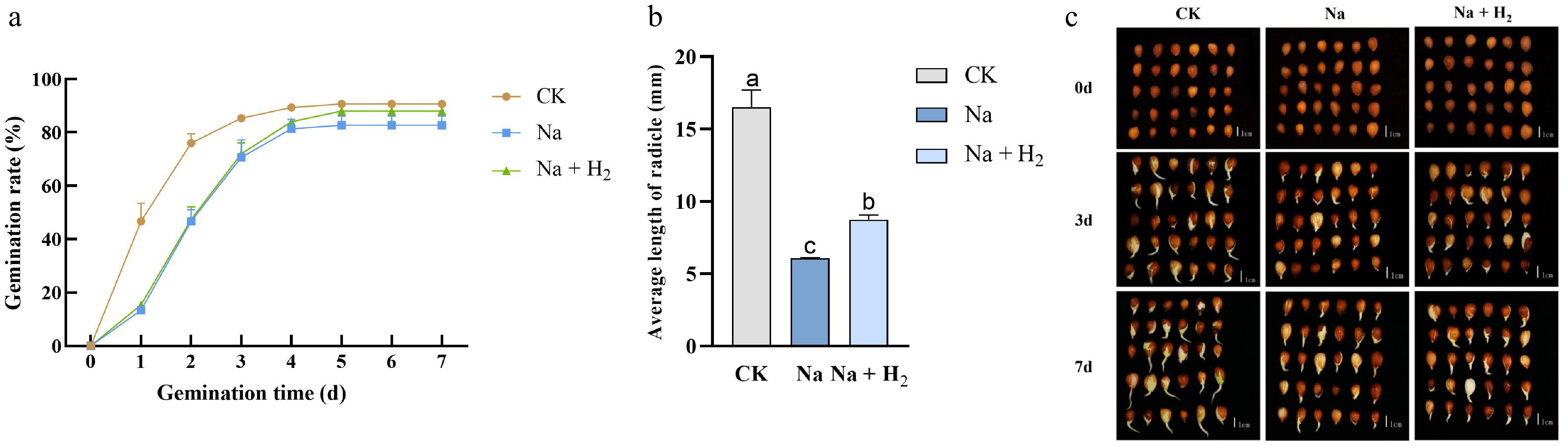
Figure 1.
Effects of different treatments on peach seed germination and radicle growth. (a) Germination rates under various treatments. (b) Radicle length on day 7 (mm). (c) Germination status on days 3 and 7. CK, Na, and Na + H2 denote 0 (control), 200 mmol/L NaCl, and 200 mmol/L NaCl + 50% HRW, respectively. Each experiment was replicated at least three times. Values are mean ± standard deviation. Significant differences are indicated by different lowercase letters (p < 0.05).
HRW improves the physiological status of peach seeds under salt stress.
HRW reduces lipid peroxidation and ROS accumulation under salt stress
-
MDA content, an indicator of lipid peroxidation, increased under salt stress but was reduced in the Na + H2 group treatment by 15.72%, 34.08%, and 28.38% compared with salt stress alone on days 1, 3, and 4, respectively (Fig. 2a). These results indicate that HRW mitigates membrane lipid oxidation. Dynamic monitoring of H2O2 and O2− levels revealed increased ROS accumulation in treated groups compared with the control. Na + H2 group seeds exhibited lower (though not significant) H2O2 levels than Na group ones (Fig. 2b). Notably, O2− levels in salt-stressed seeds were significantly higher than in the control throughout germination and exceeded Na + H2 group seeds by 16.09%–50.98% on days 2 to 4 (Fig. 2c). By days 3 to 4, O2− levels in Na + H2 group seeds returned to control values, demonstrating HRW's capacity to alleviate salt-induced oxidative stress.
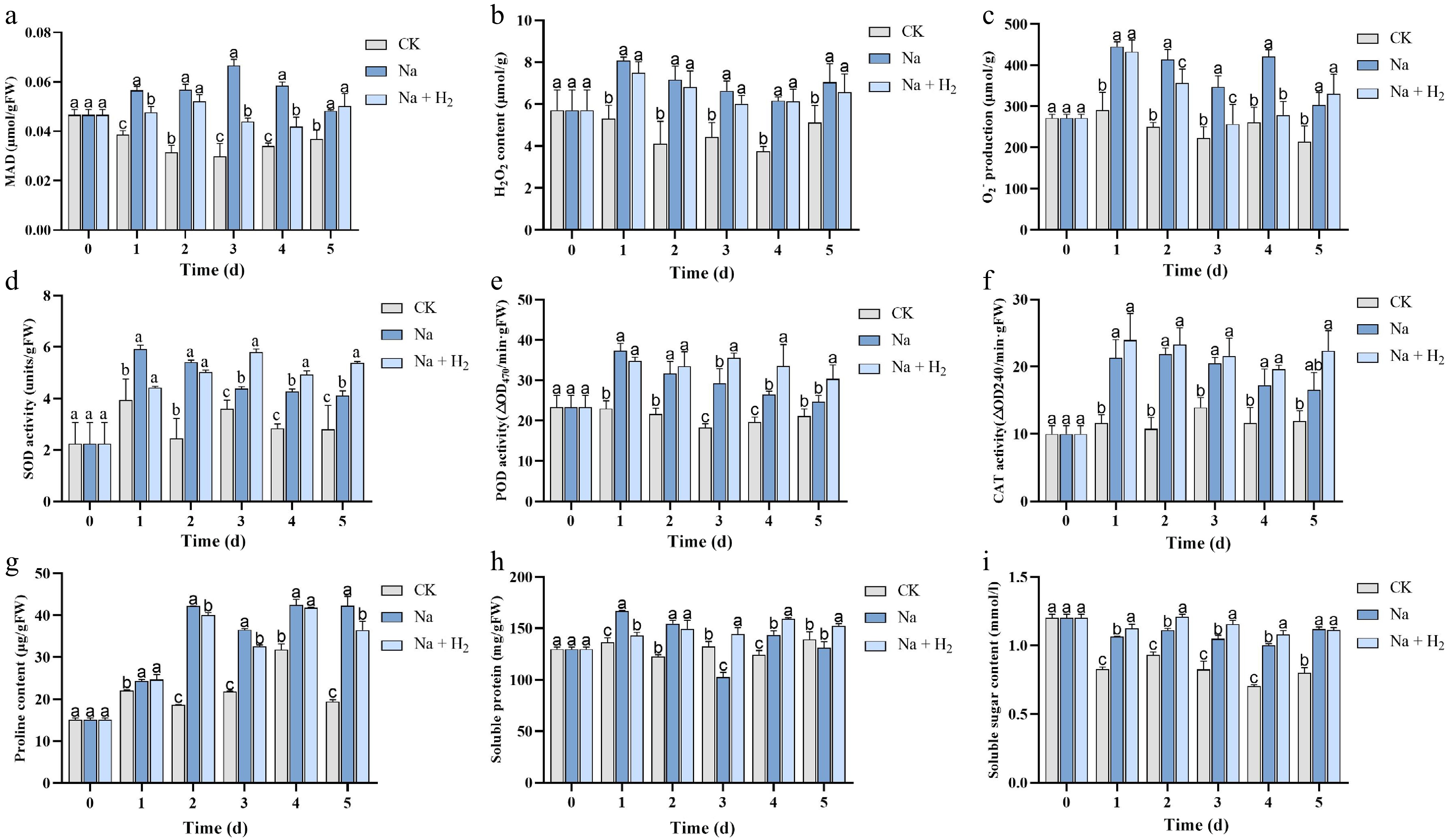
Figure 2.
Changes in physiological indicators of peach seeds under different treatments. (a) MDA content. (b) Hydrogen peroxide content. (c) Superoxide anion generation rate. (d) SOD. (e) POD. (f) CAT. (g) Proline. (h) Soluble proteins. (i) Soluble sugars. Values are mean ± standard deviation. At each time point, one-way ANOVA was used to assess intergroup differences among the control, Na+, and Na+ + H2 groups. Significant differences are indicated by different lowercase letters (p < 0.05).
HRW enhances antioxidant enzyme activities under salt stress
-
During germination, antioxidant enzymes (SOD, POD, CAT) accumulated in response to salt stress, with activities in both the Na group and the Na + H2 group seeds generally increasing before declining (Fig. 2d–f). SOD activity in treated groups exceeded that of the control throughout germination, with Na + H2 group seeds showing 32.42%, 14.95%, and 30.58% higher SOD activity than salt-stressed seeds on days 3, 4, and 5, respectively (Fig. 2d). POD activity in treated groups was higher than the control on days 1 and 2 (no significant inter-treatment difference), but Na + H2 group seeds exhibited 1.21–1.27-fold higher POD activity than Na seed ones on days 3 to 5 (Fig. 2e). CAT activity in treated groups surpassed the control through day 4, with no significant difference between the Na and the Na + H2 group treatments (Fig. 2f). These results indicate that HRW protects the antioxidant system.
HRW modulates osmotic adjustment substances under salt stress
-
Proline content increased in both treated groups compared with the control, with Na group seeds showing 1.05–1.12 times higher proline levels than Na + H2 group seeds on days 2 to 3 (Fig. 2g). Soluble protein content was consistently higher under stress conditions, with Na + H2 group seeds showing 9.3%–10.92% greater levels than Na group ones on days 3 to 4 (Fig. 2h). Soluble sugar levels in Na + H2 group seeds were also 7%–10% higher than in Na group seeds from days 1 to 4 (Fig. 2i). Collectively, these findings suggest that HRW enhances osmotic adjustment in salt-stressed peach seeds by promoting soluble protein and sugar accumulation.
Despite the absence of a statistically significant disparity in the germination rate between the Na group and the Na + H2 group on the third day, the most substantial variations were observed in the physiological indicators (MDA and SOD) at this stage. This finding suggests that the third day of seed germination represents the peak period of salt stress response, after which seed stress tolerance gradually declines. Consequently, samples collected for 3 d post-treatment were selected for transcriptomic and metabolomic sequencing. This approach aims to dissect the gene expression regulatory networks and metabolite changes in peach seeds during the peak of salt stress, thereby elucidating the molecular mechanisms and metabolic pathways underlying hydrogen gas-mediated salt stress alleviation.
Transcriptomic analysis and identification of differentially expressed genes (DEGs)
-
Transcriptomic sequencing of peach seeds from CK3d, Na3d, and H3d treatments generated a total of 400,214,468 high-quality reads (ranging from 39,975,466 to 47,196,876 per sample) (Supplementary Table S2). Mapping rates exceeded 93% for all samples, confirming the reliability of the reference genome (Supplementary Table S2). The sequencing data were of high quality, with Q30 values and GC contents ranging from 95.20%–95.80% and 45.20%–45.92%, respectively (Supplementary Table S3). DEG analysis identified 2,517 DEGs (1,082 upregulated and 1,435 downregulated) in Na3d vs CK3d, 332 DEGs (44 upregulated and 288 downregulated) in H3d vs Na3d, and 2,067 DEGs (869 upregulated and 1,198 downregulated) in H3d vs CK3d (Supplementary Fig. S2a–S2c; Supplementary Table S4). Hierarchical clustering of normalized DEGs was conducted for all comparisons, producing detailed heatmaps that visualized gene expression patterns among groups (Supplementary Fig. S2d–S2f).
GO and KEGG enrichment analyses were performed to elucidate the biological pathways involved in the Na group and the Na + H2 group. As shown in Fig. 3, DEGs in the Na3d vs CK3d comparison were enriched in pathways such as 'plant-type primary cell wall biogenesis', with five upregulated and 10 downregulated genes (Supplementary Table S5). These findings suggest that the Na group and the Na + H2 group primarily affect processes related to cell wall biogenesis, organization, and the abscisic acid-activated signaling pathway. In the Na3d vs H3d comparison, DEGs were enriched in pathways including 'cellular response to hypoxia', with 2 upregulated and 13 downregulated genes (Supplementary Table S6). In the H3d vs CK3d comparison, DEGs were enriched in 'response to abscisic acid', comprising 35 upregulated and 24 downregulated genes (Supplementary Table S7).
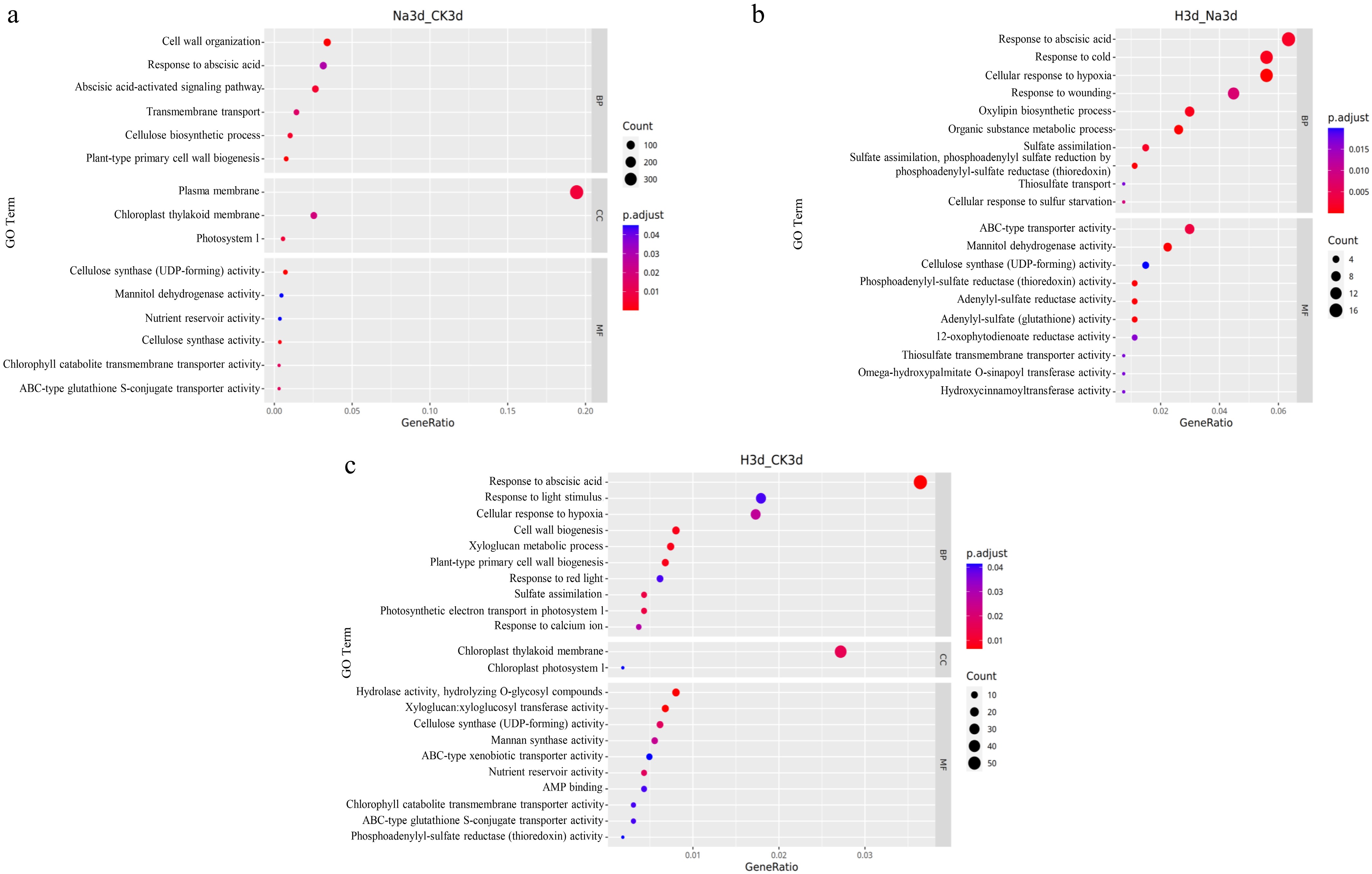
Figure 3.
GO enrichment analysis of differentially expressed genes in various pairwise comparisons under the Na group and the Na + H2 group.
As shown in Fig. 4, KEGG pathway enrichment provided further insight into DEG-associated metabolic processes. In the Na3d vs CK3d comparison, 'Photosynthesis' was significantly enriched, with ten genes downregulated and none upregulated (Supplementary Table S8). In the H3d vs Na3d comparison, 'Sulfur metabolism' was enriched, where four genes were downregulated and none upregulated (Supplementary Table S9). Similarly, in the H3d vs CK3d comparison, 'Sulfur metabolism' was enriched, with one upregulated and eight downregulated genes (Supplementary Table S10).
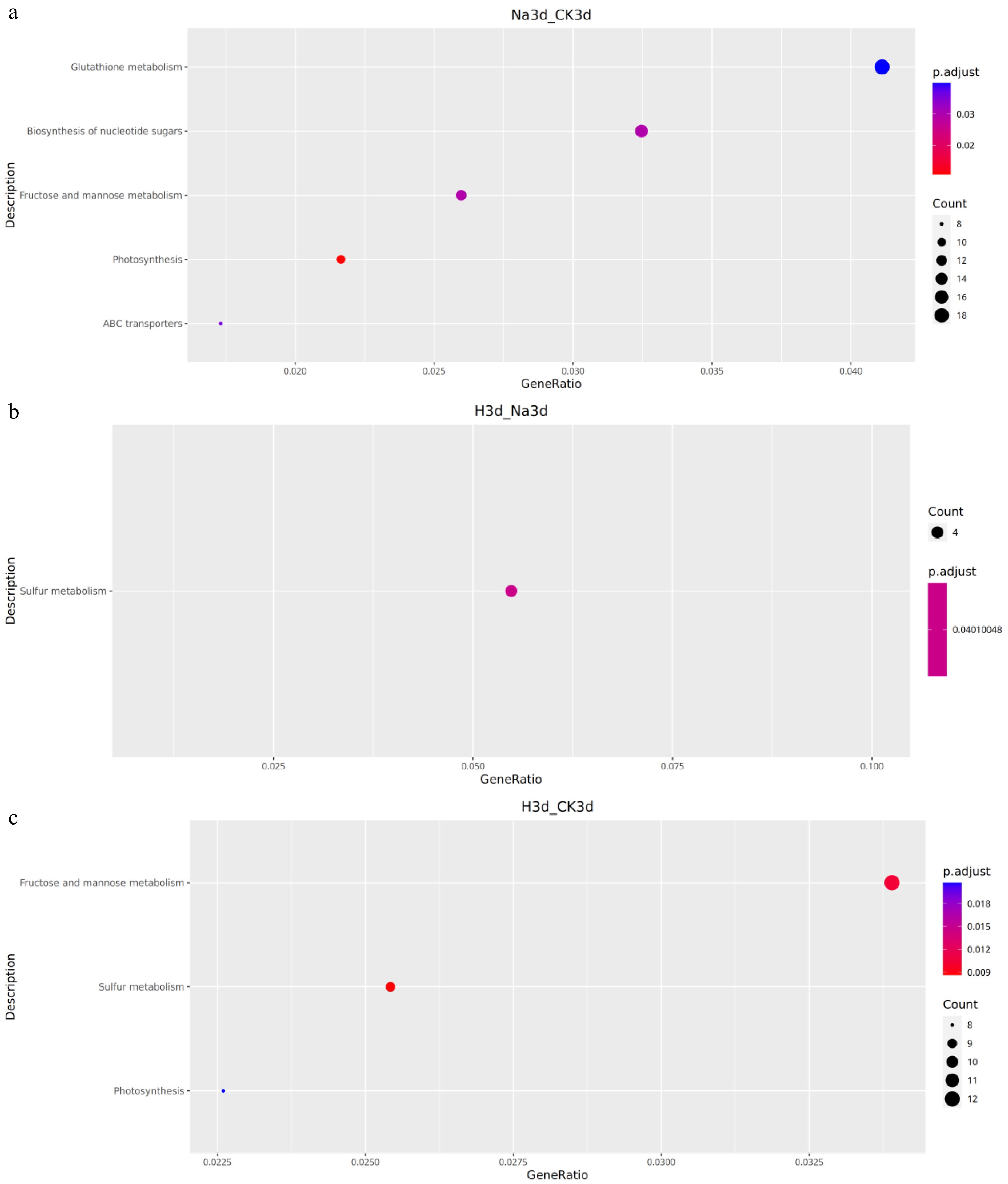
Figure 4.
KEGG pathway enrichment analysis of differentially expressed genes across pairwise comparisons under salt stress and H2 exposure.
Metabolomic data analysis
Metabolomic data quality assessment
-
Non-targeted metabolomics (LC–MS) was used to analyze differentially accumulated metabolites (DAMs) in peach seeds subjected to the Na group and the Na + H2 group. A total of 1,615 metabolites were identified (Supplementary Table S11). Hierarchical clustering heatmaps revealed distinct metabolite accumulation patterns across treatments (Supplementary Fig. S3a–S3c), indicating substantial differences in seed metabolic profiles under the Na group and the Na + H2 group, thereby demonstrating their strong influence on metabolic pathways.
Metabolite content
-
Across all samples, 873 DAMs were identified, including 344 in H3d vs CK3d (156 upregulated and 188 downregulated), 335 in Na3d vs CK3d (104 upregulated and 231 downregulated), and 194 in H3d vs Na3d (144 upregulated and 50 downregulated) (Supplementary Table S12). The 20 DAMs with the most significant fold changes were identified for Na3d vs CK3d and H3d vs Na3d (Fig. 5a–d).
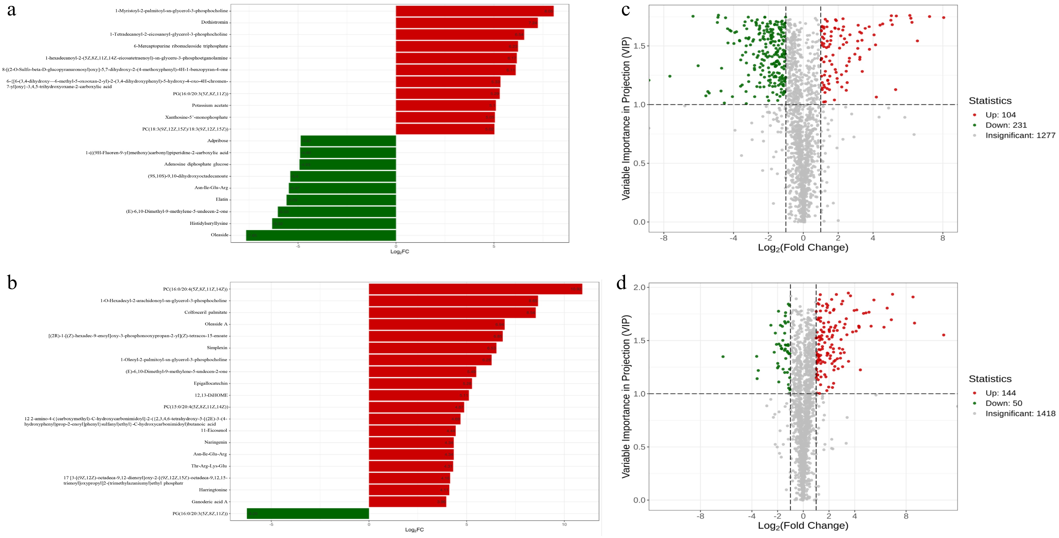
Figure 5.
Up- and downregulation of DAMs in different metabolite classes. (a) Top 20 DAMs with the largest fold changes in Na3d vs CK3d. (b) Volcano plot of up- and downregulated DAMs in Na3d vs CK3d. (c) Top 20 DAMs with the largest fold changes in H3d vs Na3d. (d) Volcano plot of up and downregulated DAMs in H3d vs Na3d.
As shown in Fig. 5 and Supplementary Table S13, several metabolite classes exhibited distinct regulatory patterns. The accumulation of steroids (e.g., Oleaside A) decreased by 99.50% in Na3d vs CK3d but increased by 12,220% in H3d vs Na3d, with no significant difference between H3d vs CK3d. Flavonoids (e.g., Naringenin) decreased by 91.68% in Na3d vs CK3d, but increased by 1,927% in H3d vs Na3d, showing no significant change in H3d vs CK3d. Alkaloids (e.g., Harringtonine) decreased by 88.71% in Na3d vs CK3d but increased by 1,629% in H3d vs Na3d, with no significant difference between H3d vs CK3d. Ketones (e.g., Cucurbitacin O) decreased by 88.68% in Na3d vs CK3d and increased by 423% in H3d vs Na3d, without significant differences between H3d vs CK3d. Benzene and substituted derivatives (e.g., Leiocarposide) decreased by 85.58% in Na3d vs CK3d but increased by 1,332% in H3d vs Na3d and by 107% in H3d vs CK3d. Phenolic acids (e.g., Chlorogenic acid) decreased by 80.18% in Na3d vs CK3d, increased by 150% in H3d vs Na3d, and decreased by 50.43% in H3d vs CK3d. Saccharides (e.g., Gluconic acid) decreased by 71.28% in Na3d vs CK3d and increased by 141% in H3d vs Na3d, with no significant difference in H3d vs CK3d. Terpenoids (e.g., Ganoderic acid Md) decreased by 70.44% in Na3d vs CK3d and increased by 118% in H3d vs Na3d, while organic acids (e.g., 3-Furoic acid) decreased by 67.36% in Na3d vs CK3d and increased by 107% in H3d vs Na3d, both showing no significant change between H3d vs CK3d.
In summary, the metabolite composition of peach seeds was strongly influenced by the Na group and the Na + H2 group, indicating that hydrogen-rich water alleviates salt-induced stress by modulating metabolite accumulation.
Integrated multi-omics analysis of the transcriptomic and metabolomic profiles
Correlation analyses between genes and metabolites and key pathway identification
-
To explore the relationship between DEGs and DAMs in peach seeds under the Na group and the Na + H2 group, a correlation analysis was conducted between these two datasets. The results revealed that numerous DEGs were strongly correlated with DAMs in both Na3d vs CK3d and H3d vs CK3d comparisons (Fig. 6a–d), indicating that these DEGs may regulate the accumulation of specific metabolites. KEGG enrichment analysis identified four significantly enriched pathways in the Na3d vs CK3d comparison (Fig. 6e; Supplementary Table S14). Integrating transcriptomic and metabolomic data revealed that 'Fructose and mannose metabolism', 'Biosynthesis of nucleotide sugars', 'ABC transporters', and 'Glutathione metabolism' were key salt-responsive pathways in peach seeds.
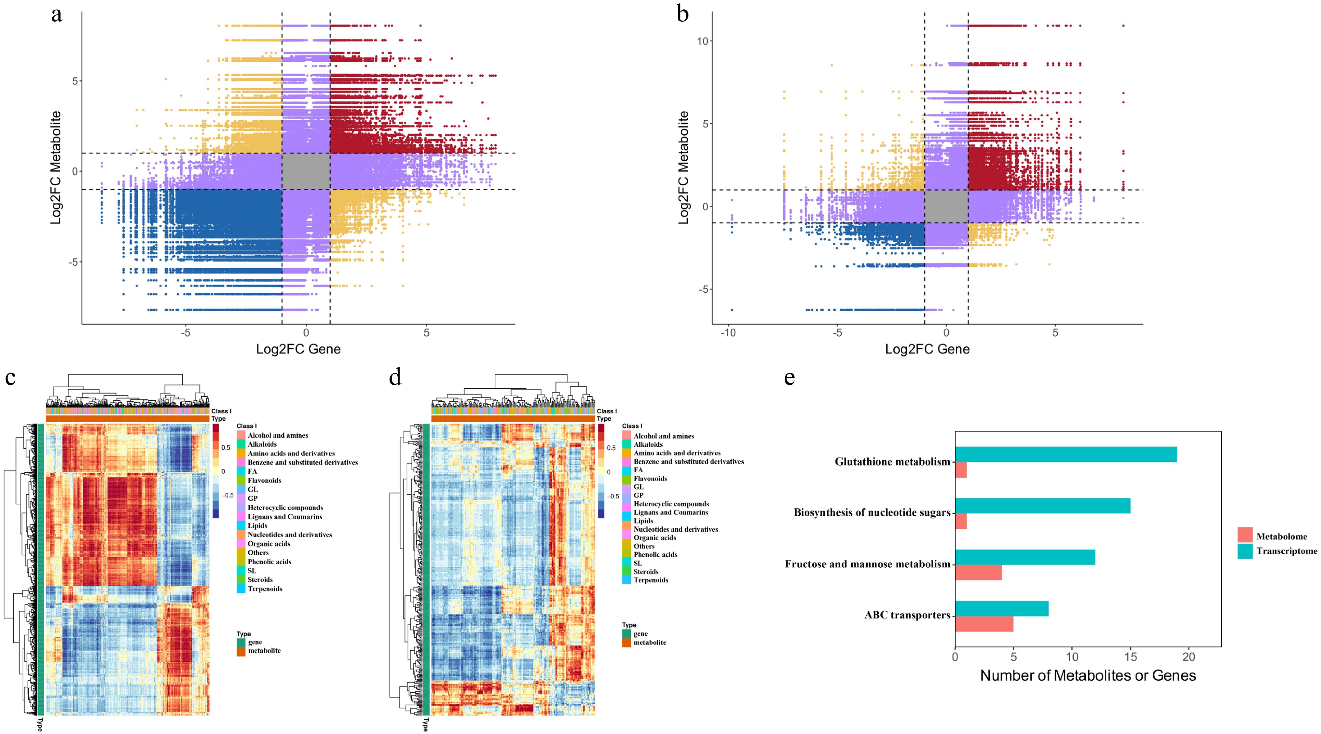
Figure 6.
Correlation analysis of transcriptomic and metabolomic data in peach seeds under the Na group and the Na + H2 group. (a), (b) Nine-quadrant plots showing correlations between genes and metabolites in Na3d (left) and H3d (right). (c), (d) Heatmap displaying cluster analysis results for DEGs (blue) and DAMs (red). (e) KEGG enrichment analysis of DEGs (blue) and DAMs (red) enriched in the same pathways.
Analysis of DEGs related to the flavonoid biosynthesis pathway and qRT-PCR validation
-
Further examination of DEGs in the phenylpropanoid and flavonoid biosynthesis pathways identified eight significantly regulated genes. To elucidate the molecular mechanisms underlying flavonoid accumulation in peach seeds under salt stress, the phenylpropanoid and flavonoid biosynthetic pathways were reconstructed (Fig. 7a). Compared with CK, PAL, F3'H, HCT, C4H, and FOMT were significantly downregulated under both Na group and Na + H2 group treatments, with stronger suppression in the Na group. Conversely, 4CL, CHI, and CHS were significantly upregulated compared with CK, with CHI showing the highest expression level in the Na + H2 group.
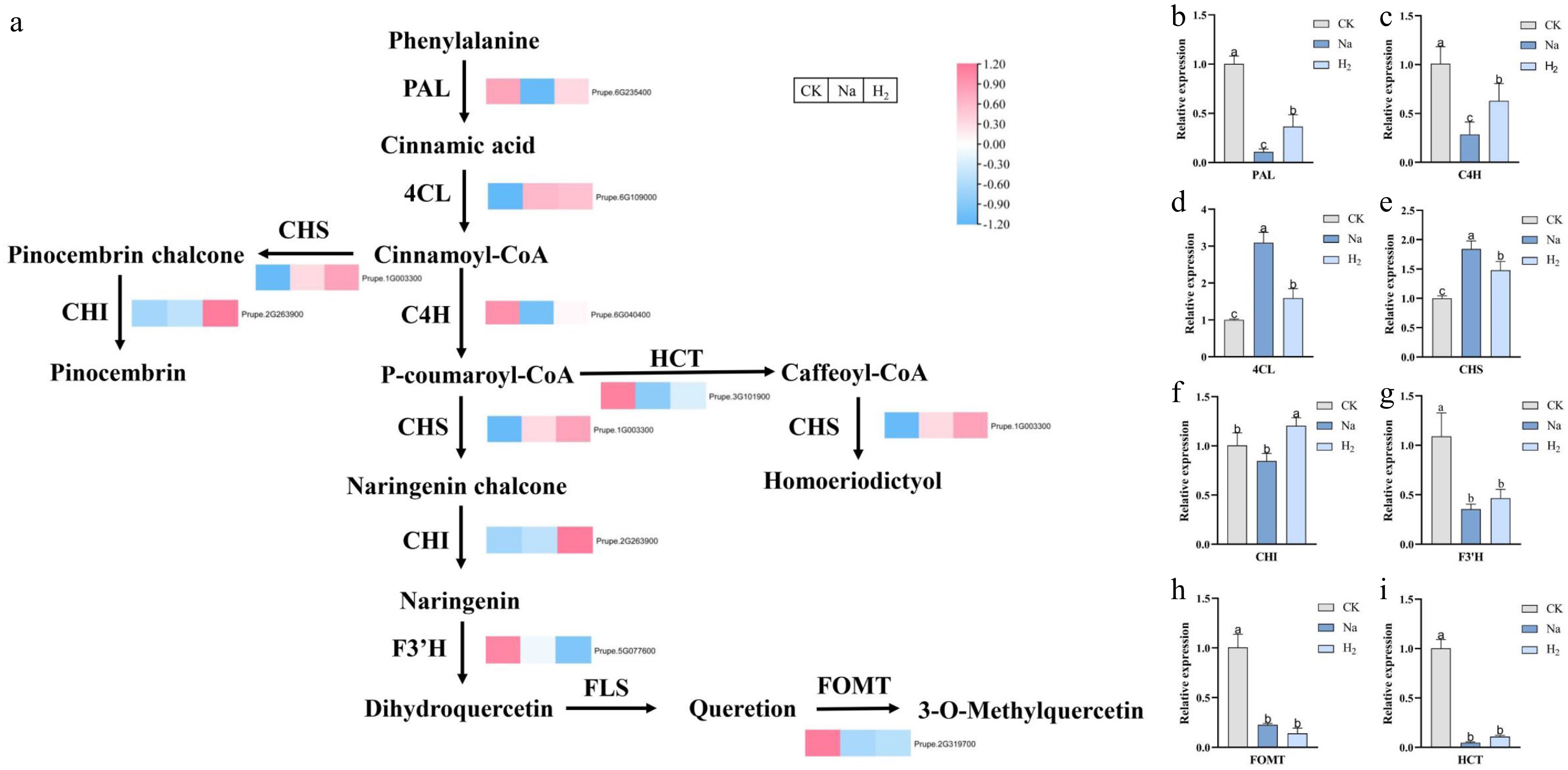
Figure 7.
Analysis of DEGs related to the phenylpropanoid and flavonoid biosynthesis pathways and qRT-PCR validation. (a) Phenylpropanoid and flavonoid biosynthetic pathways reconstructed based on the KEGG database. Gene expression levels are shown as heatmaps of log2(FPKM) values, where red indicates high expression and blue indicates low expression. (b)–(i) qRT-PCR validation of eight genes in peach seeds under different treatments. Values are mean ± standard deviation. Significant differences are indicated by different lowercase letters (p < 0.05).
To verify the reliability of the RNA-seq results, qRT-PCR analysis was performed on these eight flavonoid biosynthesis genes. The expression of PAL and C4H was significantly lower in the Na group compared with the Na + H2 group, whereas 4CL and CHS were significantly higher in the Na group than in the Na + H2 group. CHI exhibited the highest expression level under Na + H2 group treatment (Fig. 7b–i). The qRT-PCR results were consistent with the RNA-seq data, confirming the accuracy of the transcriptomic findings.
-
Salt stress markedly inhibits seed germination and radicle growth[13]. It has been shown that hydrogen gas can alleviate salt stress-induced inhibition of rice seed germination by enhancing antioxidant defense[18], and our results are consistent with this finding. HRW treatment reduced MDA and O2− levels while increasing SOD and POD activities—similar to findings in rice, cabbage, and cucumber[18,45,46]—suggesting that HRW mitigates oxidative stress in peach seeds through conserved antioxidant mechanisms. In terms of osmotic regulation, salt stress promoted proline accumulation, whereas HRW preferentially increased soluble sugar and protein levels, consistent with previous studies in rice and cucumber[18,24]. This suggests that HRW may favor the synthesis of energy-rich osmolytes over proline to maintain osmotic balance under salt stress. Collectively, these physiological data demonstrate that HRW systematically enhances antioxidant defense, reduces oxidative damage, and restores osmotic balance in salt-stressed peach seeds.
A growing body of evidence has established that flavonoids and other polyphenols function as potent ROS scavengers in plants under abiotic stress, directly neutralizing superoxide anions and hydrogen peroxide while contributing to membrane stabilization[47,48]. Consistent with this, our metabolomic data revealed that HRW treatment markedly induced the accumulation of several flavonoids and phenolic compounds, most notably naringenin and chlorogenic acid (Supplementary Table S13). Naringenin, in particular, has been demonstrated to alleviate salt-induced oxidative stress by enhancing antioxidant enzyme activities and reducing ROS levels in bean plants[49]. The strong correlation between flavonoid accumulation and the attenuation of oxidative damage in our study therefore suggests that HRW-mediated ROS mitigation is largely attributable to the restoration of flavonoid biosynthesis.
HRW potentiates ABA signaling to reactivate the flavonoid biosynthesis pathway
-
To identify the pathways underlying this metabolic response, we examined the GO enrichment results from our transcriptomic comparisons. In plants, salt stress rapidly triggers plasma membrane (PM) depolarization and activates receptor kinases, initiating early stress perception[50]. Our transcriptomic data were consistent with this: PM-related GO terms were most significantly enriched in Na3d vs CK3d (Fig. 3; Supplementary Table S5). In the H3d vs Na3d comparison, however, the enrichment pattern shifted markedly—the ABA-activated signaling pathway emerged as the most prominent category (Supplementary Table S6), suggesting that HRW potentiates ABA signaling under salt stress. Notably, ABA signaling has been shown to directly regulate flavonoid biosynthesis through transcription factors such as HY5 and ABI5, and exogenous ABA application enhances salt tolerance in rice primarily by upregulating flavonoid biosynthesis[51,52]. This sequential enrichment pattern suggests that HRW does not act at the membrane level to block stress perception but rather potentiates ABA signaling to relay the defense signal toward downstream effector pathways, particularly flavonoid biosynthesis.
Specifically, the expression of key phenylpropanoid pathway genes, including the rate-limiting enzyme gene PAL, as well as C4H and HCT, was suppressed under salt stress but was restored or even upregulated in the Na + H2 group (Supplementary Table S13), indicating that HRW activated this upstream pathway. This activation provides precursors for downstream flavonoid synthesis. Crucially, the upregulation of genes such as CHI, which catalyzes the formation of the core flavonoid skeleton, corresponded with the accumulation trend of related metabolites like naringenin, isoscutellarein, and chlorogenic acid in the metabolomics data. These findings collectively suggest that HRW is likely to promote flavonoid biosynthesis by systematically upregulating the gene network from PAL to CHI, thereby enhancing the antioxidant capacity of peach seeds to mitigate salt stress damage.
Previous studies have shown that under salt stress, PAL, C4H, 4CL, CHS, and CHI expression levels increase in the roots of Sophora japonica and seeds of Morus alba[34,53]. However, in our study, PAL and C4H were downregulated, possibly due to distinct regulatory mechanisms in peach seeds. Overexpression of NtCHS1 in tobacco enhances salt tolerance[35], while overexpression of GmCHI4 in soybean hairy roots increases isoflavone accumulation and salt tolerance[54]. This further supports that the specific activation of these key flavonoid biosynthetic genes by HRW constitutes an important molecular basis for salt stress alleviation.
Integration of multi-level data reveals a signaling-to-metabolite cascade
-
Taken together, our results support a hierarchical model for HRW-mediated salt stress alleviation in peach seeds. Salt stress initially impairs plasma membrane function and triggers ROS accumulation. Rather than directly restoring membrane integrity, HRW potentiates ABA signaling, which acts as a signal relay to reactivate the flavonoid biosynthetic pathway. The resulting accumulation of flavonoids—such as naringenin and chlorogenic acid—enhances ROS scavenging and membrane protection, ultimately improving germination and seedling growth. This signaling-to-metabolite cascade aligns with the growing recognition that ABA-mediated stress tolerance is largely executed through the reprogramming of secondary metabolism[51].
Several key questions, however, remain open. The molecular link between HRW perception and ABA signal enhancement has yet to be identified, and the transcription factors that bridge ABA signaling with flavonoid gene activation in peach seeds are unknown. Moreover, direct functional evidence—achieved through overexpression or knockout of key flavonoid pathway genes—is still required to establish causality. Addressing these gaps will not only deepen our mechanistic understanding of HRW action but also inform the development of hydrogen-based seed priming strategies for improving stress tolerance in fruit tree species.
-
This study provides an integrative view of how HRW alleviates salt stress in peach seeds. Our integrated transcriptomic and metabolomic analyses revealed that salt stress initially impaired plasma membrane function, whereas HRW treatment potentiated ABA signaling, which in turn reactivated the flavonoid biosynthetic pathway by restoring the expression of PAL, C4H, HCT, and CHI. The resulting accumulation of flavonoids such as naringenin and chlorogenic acid enhanced ROS scavenging, reduced MDA levels, and favored soluble sugar and protein accumulation for osmotic adjustment, ultimately improving germination rate and radicle growth (Fig. 8).
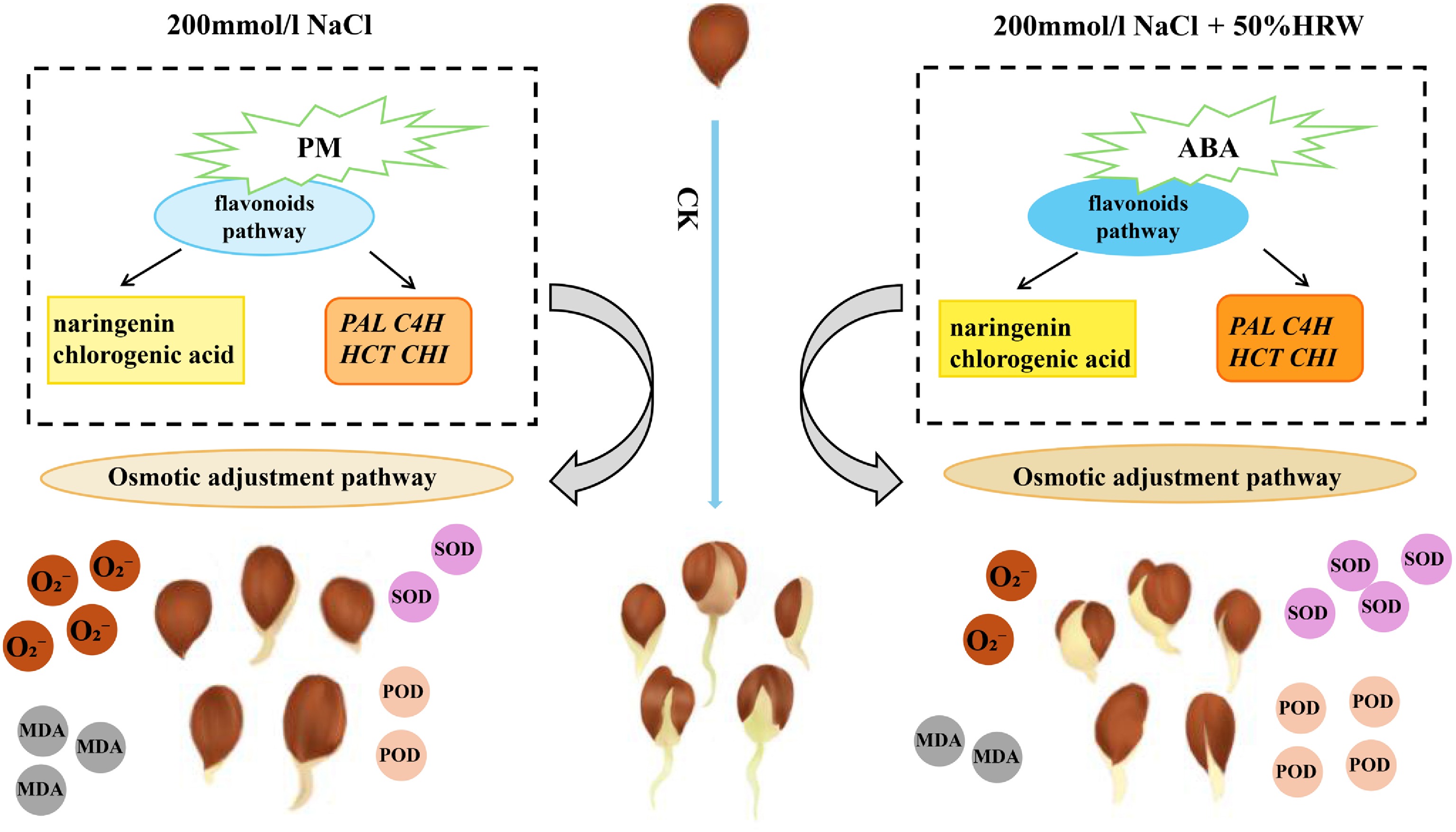
Figure 8.
A model of hydrogen-rich water alleviating salt stress in peach seeds. Darker color indicates higher content, while lighter color indicates lower content.
These findings demonstrate that HRW alleviates salt stress through an ABA–flavonoid signaling-to-metabolite cascade, offering new insights into the role of flavonoid biosynthesis in seed salt tolerance. Future studies on the functional validation of key flavonoid pathway genes and the upstream regulators linking HRW perception to ABA signaling will be essential.
-
Not applicable.
-
The authors confirm contributions to the paper as follows: study conception and design: Luo T, Tan Q, Xiao W, Li L; data collection: Luo T, Tan Q, He D, Yang L, Niu X; analysis and interpretation of results: Luo T, Tan Q; draft manuscript preparation: Luo T, Tan Q, Xiao W, Li L. All authors reviewed the results and approved the final version of the manuscript.
-
The raw sequence data of transcriptome of Prunus davidiana seeds was deposited in the NCBI Sequence Read Archive under accession number PRJNA1353577. Supplementary materials may be found in the online DOI. And related data will be made available on request.
-
This research was supported by the Shandong Fruit Industry Technology System Project (SDAIT-06-04), the Xinjiang Uygur Autonomous Region 'Tianchi Elite' Project, and the Shandong Modern Facility Fruit Tree Technology Innovation Center Support Project (2022CXPT017).
-
The authors declare that they have no known competing financial interests or personal relationships that could have appeared to influence the work reported in this paper.
-
accompanies this paper online at: https://doi.org/10.48130/frures-0026-0020.
-
# Authors contributed equally: Tongqiu Luo, Qiuping Tan
- Supplementary Table S1 The primers sequences used in this study.
- Supplementary Table S2 Statistics of RNA-seq reads and mapping rate among different samples.
- Supplementary Table S3 Statistics of RNA-seq reads among different samples.
- Supplementary Table S4 Differentially expressed genes among different samples.
- Supplementary Table S5 GO enrichment analyses in the Na3d vs CK3d comparison.
- Supplementary Table S6 GO enrichment analyses in the Na3d vs H3d comparison.
- Supplementary Table S7 GO enrichment analyses in the H3d vs CK3d comparison.
- Supplementary Table S8 KEGG enrichment analyses in the Na3d vs CK3d comparison.
- Supplementary Table S9 KEGG enrichment analyses in the H3d vs Na3d comparison.
- Supplementary Table S10 KEGG enrichment analyses in the H3d vs CK3d comparison.
- Supplementary Table S11 Number of metabolites among different samples.
- Supplementary Table S12 Differential metabolites among different samples.
- Supplementary Table S13 Variation of metabolite abundance among different samples.
- Supplementary Table S14 Changes in expression levels of key genes among different samples.
- Supplementary Fig. S1 Germination rates of peach seeds at different NaCl concentrations.
- Supplementary Fig. S2 Identification of DEGs under salt stress and H₂ exposure.
- Supplementary Fig. S3 Quality assessment of metabolomic data and variance in DAM accumulation under salt stress and H2 exposure.
- Copyright: © 2026 by the author(s). Published by Maximum Academic Press, Fayetteville, GA. This article is an open access article distributed under Creative Commons Attribution License (CC BY 4.0), visit https://creativecommons.org/licenses/by/4.0/.
-
About this article
Cite this article
Luo T, Tan Q, He D, Yang L, Niu X, et al. 2026. Integrated transcriptome and metabolome analysis reveals the mechanisms of hydrogen-rich water in mitigating salt stress in peach seeds (Prunus davidiana). Fruit Research 6: e029 doi: 10.48130/frures-0026-0020 

Integrated transcriptome and metabolome analysis reveals the mechanisms of hydrogen-rich water in mitigating salt stress in peach seeds (Prunus davidiana)
- Received: 06 November 2025
- Revised: 05 May 2026
- Accepted: 26 May 2026
- Published online: 23 July 2026
Abstract: Salt stress is a major abiotic stress that impairs plant growth and development, affecting gene transcription, metabolite profiles, and overall phenotypes. However, the underlying molecular response mechanisms, especially those mediated by hydrogen-rich water (HRW), remain unclear. In this study, we integrated physiological, transcriptomic, and metabolomic analyses to explore the effects of HRW on salt stress tolerance in Prunus davidiana seeds. The results showed that, compared with salt stress alone, exogenous hydrogen-rich water (HRW) treatment significantly reduced oxidative damage in peach seeds. Specifically, HRW decreased the accumulation of superoxide anion (O2−) and the content of malondialdehyde (MDA), while increasing the activities of superoxide dismutase (SOD) and peroxidase (POD). Integrated transcriptomic and metabolomic analyses revealed significant reprogramming of genes and metabolites, particularly those involved in the phenylpropanoid and flavonoid biosynthesis pathways. Salt stress markedly suppressed the biosynthesis and accumulation of secondary metabolites such as flavonoids, alkaloids, and chlorogenic acid by downregulating key rate-limiting genes (PAL, C4H, HCT). Exogenous HRW treatment effectively reversed the repression of these genes and restored related metabolite levels to nearly those of the control group. In summary, hydrogen-rich water acts as an alleviator of salt stress, and the identified metabolites and key rate-limiting genes offer potential targets for improving salt tolerance in mountain peach seeds.
-
Key words:
- Hydrogen-rich water /
- Salt stress /
- Prunus davidiana /
- Peach seeds /
- Transcriptomics /
- Metabolomics