










-
Leucoagaricus and Leucocoprinus sensu lato (s.l.) are highly diverse groups of Lepiotaceous fungi within Agaricaceae s.l. Both are widely distributed saprobic fungi commonly found singly or scattered in the forest humus, soil, grasslands, flowerpots, and compost heaps[1]. Certain Leucocoprinus species have been confirmed to participate in symbiotic relationships with leaf-cutter ants[2,3]. However, the presence of poisonous[4] and inedible species within both genera highlights the practical significance of accurate species identification within this group.
In classical taxonomy, Leucocoprinus was established by Patouillard[5] to segregate white-spored species from Lepiota (Pers.) Gray that exhibit Coprinus-like characteristics and, crucially, possess a distinct germ pore. Locquin[6] subsequently divided the genus into four subgenera. Building upon this, Singer[7] established the tribe Leucocoprineae, which is defined by key features, such as a striate pileus margin, an often double and movable annulus, and spores that are thick-walled, dextrinoid, have a germ pore, and are metachromatic in Cresyl Blue. In the same year, Singer[8] elevated Locquin's[6] Leucocoprinus subgen. Leucoagaricus to the generic rank Leucoagaricus, and initially divided it into sect. Macrospori and sect. Rubrotincti based on spore size and pileus coloration. Singer & Cleḿençon[9] later expanded Leucoagaricus by establishing two new sections, resulting in four recognized sections at that time: sect. Sphaerocystophori, sect. Sculpturati, sect. Piloselli, and sect. Annulati. By 1986, Singer, in his definitive work[10], further refined the classification by re-including sect. Leucoagaricus and sect. Rubrotincti, leading to six recognized sections of Leucoagaricus. Concurrently, the tribe Leucocoprineae was expanded to include seven genera, including Clarkeinda Kunze[11], Chlorophyllum Massee[12], Macrolepiota Singer[7], Volvolepiota Singer[13], and Sericeomyces Heinem.[14]. Subsequent works by scholars such as Candusso et al.[15] and Akers[16] largely maintained Singer's framework, albeit with minor revisions at the sectional level (replacing sect. Sphaerocystophori with sect. Pulverulenti Bon) (Table 1).
Table 1. Chronological summary of key taxonomic revisions and framework shifts in Leucoagaricus and Leucocoprinus s.l.
Author (year) Methodological basis Key contribution and diagnostic features Patouillard (1888)[5] Morphology Established Leucocoprinus to segregate white-spored species from Lepiota (Pers.) Gray that exhibit Coprinus-like characteristics and possess a distinct germ pore. Locquin (1945)[6] Morphology Subdivided Leucocoprinus into four subgenera: subgen. Eu-Leucocoprinus (Pat.) emend., subgen. Leucoagaricus Locq., subgen. Hiatula (Fr.) emend., and subgen. Leucobolbitius (Lange) emend. Singer (1948a)[8] Macro- and micromorphology Elevated Leucocoprinus subgen. Leucoagaricus (Locq.) to the generic rank: Leucoagaricus. Characterized by small, colorless, dextrinoid, and metachromatic spores with a germ pore, and the presence of an annulus (Type: La. macrorhizus [Locquin] Singer= Lc. barssii [Zeller] Migl. & Donato). Divided into two sections: – sect. Macrospori: spores 12–15 µm (Type: La. excoriatus = Macrolepiota excoriata [Schaeff.] Wasser). – sect. Rubrotincti: spores < 12 µm, with brightly colored pileus squamules (Type: La. rubrotinctus [Peck] Singer). Singer et al. (1972)[9] Macro- and micromorphology Further expanded the infrageneric classification of Leucoagaricus by establishing four sections: – sect. Sphaerocystophori: pileipellis composed of sphaerocysts (Type: La. exannulatus Singer). – sect. Sculpturati: similar to sect. Rubrotincti, but with ornamented spores (Type: La. rubrosquamosus [Rick] Singer). – sect. Piloselli (Kühn.) Singer: Lamellae becoming pinkish at maturity; context often turning red/darkening; spores smooth, lacking a germ pore (Type: La. georginae [W.G. Sm.] Singer= Lc. georginae [W.G. Sm.] M. Asif, Saba & Vellinga). – Sect. Annulati (Fr.) Singer: Species with smooth, dextrinoid spores possessing a germ pore; often robust basidiomata (Type: La. naucinus [Fr.] Singer = Lc. leucothites [Vittad.] Redhead). Singer (1986)[10] Macro- and micromorphology The tribe Leucocoprineae Sing. was defined to contain seven genera: Clarkeinda, Chlorophyllum, Macrolepiota, Volvolepiota, Sericeomyces, Leucoagaricus, and Leucocoprinus.
Re-evaluated Leucoagaricus and Leucocoprinus. Expanded Leucoagaricus to six sections by adding sect. Leucoagaricus (Type: La. macrorhizus Locq. ex Singer = Lc. barssii [Zeller] Migl. & Donato) and sect. Rubrotincti Singer, which includes species with non-brightly colored, fibrous, scaly, or tomentose pileus coverings (often colorless or sparsely pigmented), and smooth spores.Candusso & Lanzoni (1990)[15] Macro- and micromorphology Reviewed the infrageneric classification. Removed subgen. Hiatula and subgen. Leucobolbitius from Leucocoprinus, restricting it to two sections: – sect. Leucocoprinus: slender basidiomata, thin pileus scales, small, elliptical basidiospores (Type: Lc. cepistipes [Sowerby] Pat.). – sect. Denudali Herink: pileus scales with sphaerocysts; spores with inconspicuous or immature germ pores (Type: Lc. denudatus [Sacc.] Singer = Lc. straminellus [Bagl.] Narducci & Caroti).
Maintained six sections in Leucoagaricus, largely consistent with Singer[10], but replaced sect. Sphaerocystophori with sect. Pulverulenti Bon.Akers (1997)[16] Macro- and micromorphology Recognizing four sections within Leucocoprinus: – sect. Denudali Herink. – Sect. Velutipedes Heinem.: basidiospores with a distinct apical pore; stipe base with scales; pileipellis lacking sphaerocysts and non-hymeniform. – Sect. Leucocoprinus (Pat.) Heinem. – Sect. Pepinispori Heinem.: basidiospores flattened-amygdaliform; distinct apical pore absent or incomplete.
Maintained the sectional views of Candusso & Lanzoni[15] for Leucoagaricus.Migliozzi & Testoni (2000)[35] Macro- and micromorphology Established the new sect. Cystidiosi Migliozzi et Testoni (Type: La. pleurocystidius). Characterized by pleurocystidia (present in the subhymenium, but lacking clamp connections). Vellinga (2004)[17] ITS and nrLSU phylogeny Demonstrated that Leucoagaricus, Leucocoprinus, and Sericeomyces together form a single, large monophyletic clade, revealing that these genera are not monophyletic individually under traditional morphological concepts. Vellinga et al. (2011)[18] Multi-locus phylogeny (ITS, nrLSU, rpb2, tef1) Revealed that tef1 sequences resolve the Leucoagaricus/Leucocoprinus complex into distinct clades corresponding to traditional sections (e.g., La. sect. Rubrotincti, La. sect. Piloselli). Furthermore, demonstrated that the La. americanus clade is phylogenetically distant from the core complex and allied with Micropsalliota Redhead (2023)[22] Nomenclatural reassessment Proposed several new combinations by transferring species from related genera into Leucocoprinus to address taxonomic issues. Asif et al. (2024)[23] ITS and nrLSU phylogeny Proposed 64 new combinations within Leucoagaricus and Leucocoprinus. Migliozzi et al. (2024)[24] Nomenclatural reassessment Transferred sect. Pilosellae and sect. Rubrotincti from Leucoagaricus to Leucocoprinus; proposed 34 new combinations. Yang et al. (2024)[25] Multi-locus phylogeny (ITS, nrLSU, rpb2, tef1) Erected Candelolepiota and Macropsalliota; redefined Leucoagaricus s.s. to include only original sect. Leucoagaricus and Sericeomyces. Li et al. (2025)[34] Multi-locus phylogeny and comprehensive morphological examination Expanded species-level diversity by describing 10 new taxa. Highlighted that the circumscription of the revised Leucocoprinus s.l. remained unsettled and identified a distinct 'Clade Cretaceous' separate from the core Leucocoprinus. Radnóti et al. (2025)[26] Multi-locus phylogeny (ITS, nrLSU, rpb2, tef1) Erected Mystagaricus to accommodate La. brunneolilacinus, positioning it outside the core Leucoagaricus and Leucocoprinus s.l. group. Li et al. (2025)[29] Phylogenomics (ITS, nrLSU, rpb2, tef1) Erected the new genus Conioexocarpus and the new subfamily Leucocoprinoideae to accommodate Leucoagaricus, Leucocoprinus, Conioexocarpus, Candelolepiota, Macropsalliota, and Micropsalliota. Yang et al. (2026)[30] Multi-locus phylogenetic analyses (ITS, nrLSU, rpb2, tef1-α), coalescent-based species tree estimation (ASTRAL), and comprehensive microstructural investigations Restructured the generic framework of subfam. Leucocoprinoideae. Established the new genera Pulchrolepiota and Tristolepiota to resolve the paraphyly within Leucocoprinus s.l. Defined Leucocoprinoideae as comprising five genera (Conioexocarpus, Leucoagaricus, Leucocoprinus s.s., Pulchrolepiota, and Tristolepiota) based on pileus center microstructures (trichoderm, conioderm, or cutis) and the presence/absence of hymenial physalides. Additionally established subfam. Micropsalliotoideae to accommodate Mesopsalliota (gen. nov.), Macropsalliota, and Candelolepiota. Vellinga et al. (2026)[31] Nomenclatural reassessment Facilitated a broadly defined Leucocoprinus concept by proposing 10 new combinations for North American taxa. Emphasized that generic delimitation should integrate practical taxonomic utility and broader criteria, rather than relying exclusively on phylogenetic topological parameters. Despite the substantial contributions made by early mycologists based on traditional morphology, elucidating the phylogenetic relationships between Leucoagaricus and Leucocoprinus has proven increasingly challenging in the molecular era. Early molecular phylogenetic studies, such as those by Vellinga[17,18] and Ge[19], demonstrated that the tribe Leucocoprineae is not a monophyletic group. Instead, it was shown to be broadly paraphyletic, with some lineages intermingling with the type genus of the tribe Agariceae, Agaricus L.[20], and its relative Micropsalliota Höhn[21].
Extensive systematic revisions have been undertaken to resolve between Leucoagaricus and Leucocoprinus s.l.; taxonomists have undertaken active and extensive revisions. This reorganization was initiated by Redhead[22], whose nomenclatural note served as the foundation for these systematic changes. Based on a combined ITS and nrLSU phylogeny, Asif et al.[23] proposed 64 new combinations, primarily transferring taxa into Leucocoprinus following Redhead's[22] approach. Similarly, Migliozzi & Donato[24] subsequently relocated two sections (Pilosellae and Rubrotincti) from Leucoagaricus to Leucocoprinus, encompassing 34 novel combinations. Further refining this framework through four-locus phylogenetic analyses, Yang et al.[25] restricted Leucoagaricus to species from the original sect. Leucoagaricus (formerly sect. Rubrotincti) and Sericeomyces, allocating the remaining taxa to Leucocoprinus. In parallel, they erected the new genera Candelolepiota and Macropsalliota to accommodate the guttating species La. lacrymans and the La. americanus clade, respectively. Continuing this trend of generic reassessment, Radnóti et al.[26] transferred La. brunneolilacinus Babos—a species characterized by a purplish-lilac universal veil and an orange-brown context discoloration—into the novel genus Mystagaricus. This placement grouped it with Pseudolepiota Z.W. Ge[27] and Xanthagaricus (Heinem.) Little Flower, Hosag. & T.K. Abraham[28], effectively excluding this taxon from the core Leucoagaricus and Leucocoprinus s.l. lineages. The higher-level classification of this complex was later addressed by Li et al.[29] who utilized an integrative phylogenomic approach to establish the subfamily Leucocoprinoideae and the novel genus Conioexocarpus. Extending this phylogenomic framework, Yang et al.[30] proposed a multi-generic system by introducing the subfamily Micropsalliotoideae (encompassing Micropsalliota, Candelolepiota, Macropsalliota, and the novel Mesopsalliota). Within Leucocoprinoideae, they further segregated Leucocoprinus s.l. into narrower genera, such as Pulchrolepiota and Tristolepiota, based on pileus covering microstructures and the presence or absence of hymenial physalides. However, this multi-generic classification has yet to reach a broad consensus. Providing an alternative perspective, Vellinga et al.[31] refrained from adopting these newly proposed genera, advocating instead for a two-genus taxonomic arrangement that maintains a broadly defined Leucocoprinus alongside a narrowly defined Leucoagaricus. In the present study, we align with this more conservative and pragmatic framework, applying the broadly circumscribed Leucocoprinus concept throughout our taxonomic treatments. (Table 1).
In addition to the ongoing debates concerning generic boundaries, aligning infrageneric classifications with stable phylogenetic clades presents a parallel challenge. The numerous novel sections recently established by Yang et al.[30] within Leucocoprinus s.l. offer a useful foundational framework. However, the phylogenetic stability and broad taxonomic applicability of these infrageneric units require further testing through expanded global sampling and the integration of more comprehensive datasets. Furthermore, beyond these supraspecific unresolved issues, the extensive species-level diversity within this complex continually complicates taxonomic treatments. Currently, Index Fungorum (accessed Jun 25, 2026) contains 388 nomenclatural records for Leucoagaricus and 393 for Leucocoprinus. Additionally, the database includes several dozen records cataloged under recently proposed segregates—such as Pulchrolepiota (47 records) and Tristolepiota (27)—both of which are treated as part of the broadly circumscribed Leucocoprinus in the present study. Records also exist for other closely related genera within this complex, including Macropsalliota (9), Mesopsalliota (2), Conioexocarpus (5), and Candelolepiota (1). Moreover, the widespread morphological similarity among distinct species significantly complicates traditional taxonomy, frequently resulting in a discordance between macroscopic morphology and genetic divergence. To resolve these complexities and stabilize the topologies of these essential clades, the continuous discovery and inclusion of previously undescribed taxa are critical. In this study, we employed an integrative taxonomic strategy building upon existing phylogenetic frameworks[23,25,26,29,32−34]. To effectively evaluate and clarify species boundaries, we incorporated a robust dataset of validated sequences alongside rigorous verification of the original literature. By synthesizing these multiple lines of evidence, this study seeks to provide a reliable reference framework for the taxonomic revision of this hyper-diverse group.
Given the rapid and continuous taxonomic revisions of Leucocoprinus s.l. in recent years, its generic circumscription has undergone significant changes. For clarity, the term 'Leucocoprinus s.l.' in the present study refers to the traditional, broad generic concept prior to the most recent segregations, encompassing Leucocoprinus s.s., Pulchrolepiota, and Tristolepiota. Regarding the infrageneric classification, we maintain a broader framework based on major evolutionary clades (as detailed in the Discussion) to accommodate this diversity. To facilitate referencing historical taxa and distinct phylogenetic lineages throughout the text, the following generic abbreviations are used: Cl. = Candelolepiota, Co. = Conioexocarpus, La. = Leucoagaricus, Lc. = Leucocoprinus, Map. = Macropsalliota, and Mep. = Mesopsalliota.
-
In this study, a total of 560 specimens collected from diverse geographical regions across China between 1997 and June 2026 were examined. Macromorphological descriptions were formulated based on detailed field notes and photographs of fresh basidiomata, with colour notations standardized using hexadecimal colour codes (Supplementary File 1). Micromorphological structures were examined using an Olympus CX33 light microscope, and digital measurements were acquired via EPview v1.4.0. The protocols for specimen preparation, morphological terminology, and microchemical evaluations of basidiospores followed the standardized methodologies established in previous studies[10,36,37].
DNA extraction, PCR amplification, and sequencing
-
Total genomic DNA was extracted from fresh or dried specimens using the NuClean PlantGen DNA Kit (CWBIO, Beijing, China). PCR amplification of the ITS, nrLSU, rpb2, and tef1-α regions was conducted using the primer pairs ITS1F/ITS4[38,39], LR0R/LR5[40], 6F/RPB2-7.1R[41], and EF1-983F/EF1-1567R[42], respectively. Thermal cycling conditions followed previously described procedures[37,43]. All PCR amplicons were purified and sequenced by Sangon Biotech (Shanghai, China). The newly generated sequences were deposited in GenBank.
Phylogenetic analyses
-
To comprehensively evaluate phylogenetic relationships and species boundaries, we reconstructed a multi-locus phylogeny based on established taxonomic frameworks[25,26,29,30,32−34], utilizing reliable sequences from species within Leucoagaricus and Leucocoprinus s.l. The dataset comprised 288 ITS, 167 nrLSU, 117 rpb2, and 117 tef1-α sequences (Supplementary File 2).
Sequences for each locus were aligned independently using MAFFT v7.110[44], manually inspected, and further optimized in MEGA v7.0.26[45]. Unreliable alignment regions and gaps were removed using the "-automated1" command in trimAl[46]. Following previous studies, intronic regions within the tef1-α sequences were excluded[29,30]. The aligned loci were then concatenated using the "Concatenate Sequence" function in PhyloSuite v2[47], generating a combined alignment of 2,518 base pairs (bp). The sequence partitions were defined as follows: ITS (1–629 bp), nrLSU (630–1,476 bp), rpb2 (1,477–2,103 bp), and tef1-α (2,104–2,518 bp), with gaps treated as missing data. The final concatenated alignment used for phylogenetic inference is also available in Supplementary File 2.
Phylogenetic reconstructions were performed using Maximum Likelihood (ML) and Bayesian Inference (BI) methods. To address evolutionary heterogeneity across different loci, partitioned analyses were applied to the combined dataset. For the ML analysis, ModelFinder v3.0.1[48] was used to select the best-fit substitution models under the Akaike Information Criterion (AIC). The ML tree was inferred using IQ-TREE v3.0.1[49] with an edge-linked partition model. The selected optimal models were GTR + F + R7 for ITS, GTR + F + I + R4 for nrLSU and rpb2, and GTR + R6 for tef1-α. Branch support was assessed using 1,000 ultrafast bootstrap replicates[50] and the Shimodaira–Hasegawa approximate likelihood-ratio test (SH-aLRT)[51].
For the BI analysis, ModelFinder v3.0.1[48] was employed to determine the optimal partition models based on the Bayesian Information Criterion (BIC), resulting in GTR + F + I + G4 for ITS and nrLSU, GTR + F + G4 for rpb2, and HKY + I + G4 for tef1-α. The BI analysis was conducted in MrBayes v3.2.7a[52], running two parallel chains for 26,611,000 generations. The analysis was terminated when the average standard deviation of split frequencies (ASDSF) dropped below 0.01. Run convergence was subsequently evaluated via Tracer v1.7.2[53] to confirm that all Effective Sample Size (ESS) values remained well above 200. The initial 22% of the sampled trees were discarded as burn-in. Finally, the consensus topologies from both analyses were visualized in FigTree v1.4.3[54] and graphically refined using tvBOT v2.6.1[55] and Adobe Photoshop 2020 (Adobe Inc., San Jose, CA, USA) to precisely integrate branch support values.
-
The final concatenated multi-locus dataset (ITS, nrLSU, rpb2, and tef1-α) comprised 285 terminal taxa, representing eight genera: Candelolepiota (5 taxa), Conioexocarpus (5 taxa), Leucoagaricus (97 species and 5 provisional taxa), Leucocoprinus s.l. (168 species and 4 provisional taxa), Macropsalliota (2 species and 1 affine taxon), Mesopsalliota (2 taxa), Micropsalliota (2 taxa), and Macrolepiota (2 taxa). Both maximum likelihood (ML) and Bayesian inference (BI) analyses yielded congruent topologies with no supported conflicts at major nodes. Consequently, the BI consensus tree is presented as the representative topology (Figs 1−3), with statistical support values from both analyses (UFBoot ≥ 80% and BPP ≥ 0.90) annotated at the respective nodes.
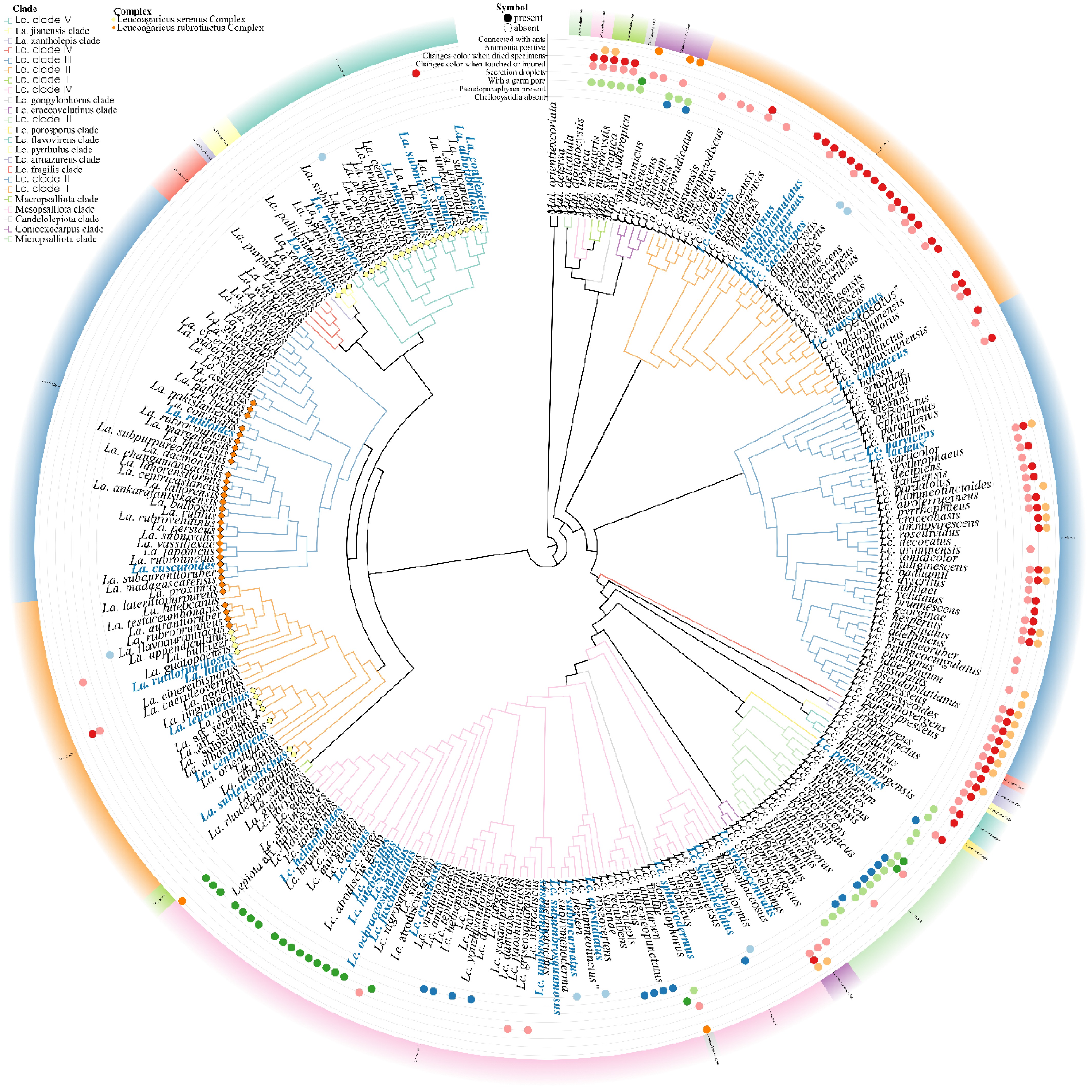
Figure 1.
Bayesian Inference (BI) phylogeny of Leucoagaricus and Leucocoprinus s.l. inferred from the concatenated ITS-nrLSU-rpb2-tef1-α dataset. Branch lengths are not drawn to scale for visual clarity. Selected macro- and micro-morphological diagnostic characters are mapped alongside the topology.
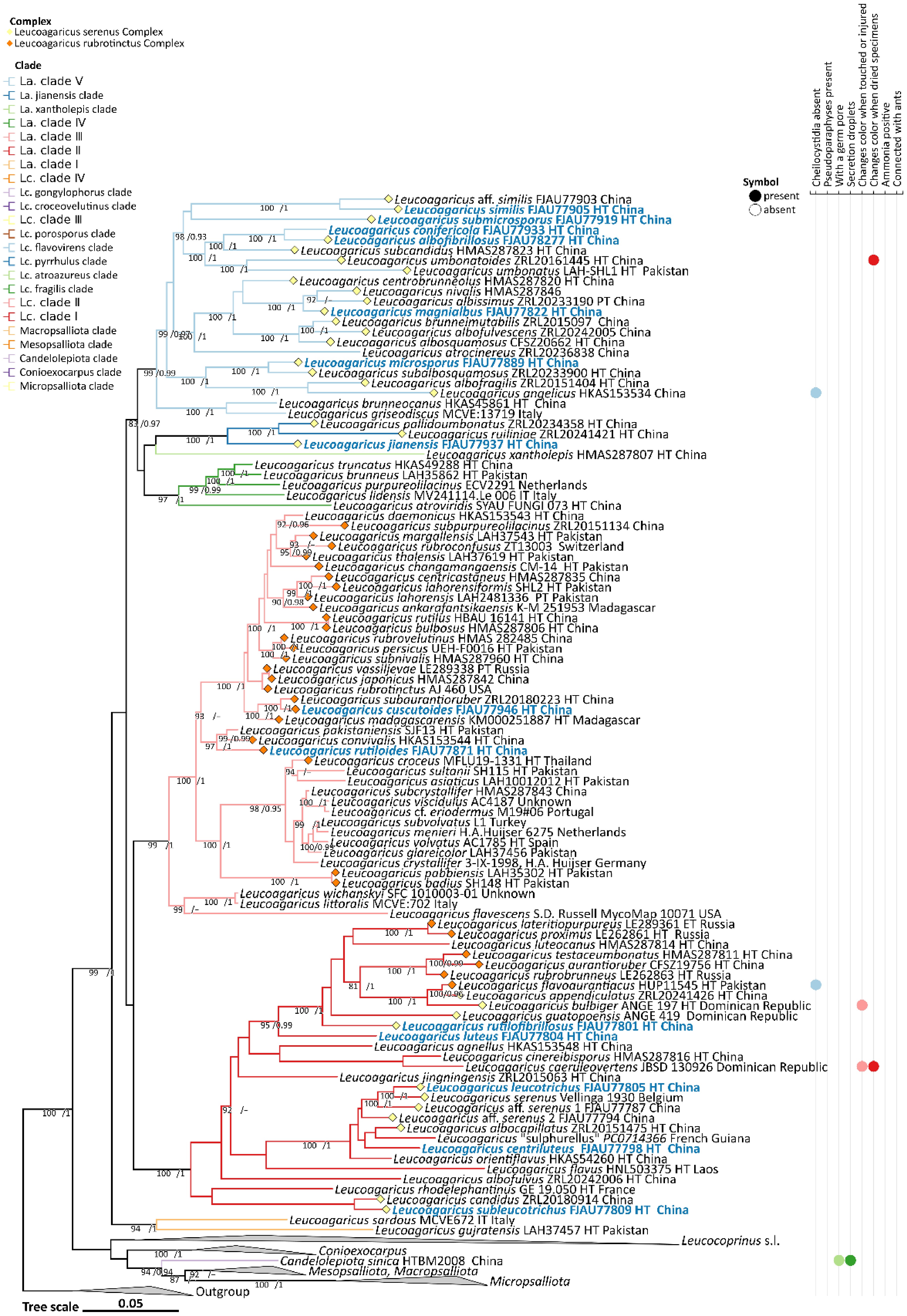
Figure 2.
Partial view of the Bayesian Inference (BI) phylogeny of Leucoagaricus and Leucocoprinus s.l. inferred from the concatenated ITS-nrLSU-rpb2-tef1-α dataset, focusing on Leucoagaricus to illustrate its internal phylogenetic structure and morphological congruence. Due to its size, the comprehensive phylogenetic tree is provided in Supplementary File 2. Selected macro- and micro-morphological diagnostic characters are mapped alongside the topology.
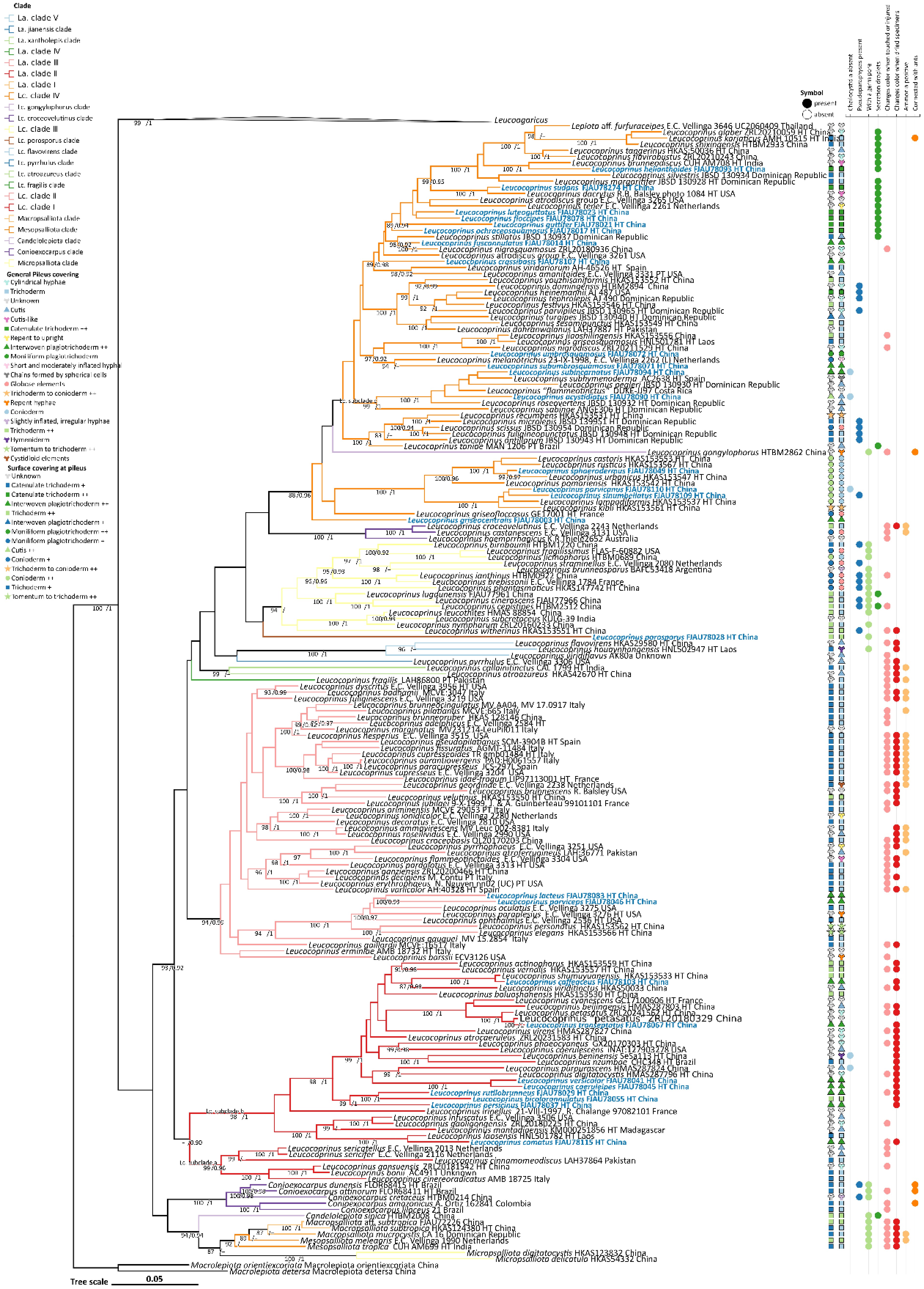
Figure 3.
Partial view of the Bayesian Inference (BI) phylogeny of Leucoagaricus and Leucocoprinus s.l. inferred from the concatenated ITS-nrLSU-rpb2- tef1-α dataset, focusing on Leucocoprinus s.l. to illustrate its internal phylogenetic structure and morphological congruence. Due to its size, the comprehensive phylogenetic tree is provided in Supplementary File 2. Selected macro- and micro-morphological diagnostic characters are mapped alongside the topology. For the pileus centre covering columns, data in the 'General pileus covering' column are based on original protologues and reliable literature; under 'Surface covering at pileus', '++' indicates explicitly observed microstructures, while '+' indicates character states cautiously inferred from original descriptions.
At the generic level, the multi-locus phylogeny recovered a well-resolved backbone for the core lepiotaceous lineage. Within the core lineage, the respective genera—Micropsalliota, Mesopsalliota, Macropsalliota, Candelolepiota, Conioexocarpus, Leucocoprinus s.l., and Leucoagaricus—diverged sequentially. This generic topology is highly consistent with recent classification frameworks.
Within this framework, recently proposed narrow segregates (e.g., Pulchrolepiota, Tristolepiota) were nested deeply within the Leucocoprinus s.l. clade, often lacking robust independent support when sampled extensively. Consequently, in this study, we treat all these segregates as synonyms of a broadly circumscribed Leucocoprinus. Furthermore, although our phylogeny resolves several distinct internal groups within this broad genus, assigning these clades to formal infrageneric ranks (e.g., subgenera or sections) requires a strict concordance with diagnostic morphological traits, a condition that remains partially unresolved due to extensive morphological homoplasy. Therefore, to objectively present the topological structure of our expanded dataset without introducing premature sectional names, we utilize neutral lineage designations (e.g., clade I, clade II) to describe the evolutionary positions of both historical and novel species. A comprehensive taxonomic evaluation of these phylogenetic clades is provided in the Discussion.
Leucoagaricus was recovered as a strongly supported monophyletic group (99/1.0; Figs 1, 2). Within this generic clade, our phylogeny resolved five major, well-supported infrageneric lineages (designated here as La. clades I–V) along with two unstable lineages (the La. jianensis and La. xantholepis clades). The early-diverging La. clade I (94/1.0) comprises La. sardous (Zecchin & Migl.) Consiglio & Contu, and La. gujratensis A. Rehman, Usman, Afshan & Khalid, separating from the core Leucoagaricus taxa. La. clade II (100/1.0) topologically corresponds to the traditional Leucoagaricus subgen. Sericeomyces. It forms a sister relationship with La. clade III (99/1.0)—which primarily contains species formerly assigned to sect. Rubrotincti—although the backbone connecting them lacks robust support. La. clade IV (97/1.0) consists of five species represented by La. purpureolilacinus Huijsman. Meanwhile, La. clade V (99/0.99) clustered with strong internal support and clustered together with La. clade IV, the La. jianensis clade and the La. xantholepis clade, forming a broader, moderately supported group (83/0.97). The phylogenetic analyses support the recognition of the 14 novel Leucoagaricus species proposed herein. Specifically, five novel species, two new geographical records for China, and two undetermined affine taxa nested within La. clade II. Two novel species fell into La. clade III, one into the La. jianensis clade, and the remaining six novel species plus one affine taxon clustered in La. clade V. Morphological groups defined a priori—the La. rubrotinctus and La. serenus complexes—were not recovered as monophyletic. Species belonging to the La. rubrotinctus complex were distributed across La. clades II and III, whereas members of the La. serenus complex were scattered across La. clades II, V, and the La. jianensis clade. This non-monophyletic distribution highlights the widespread morphological similarity among distinct genetic lineages within this highly diverse genus.
Within Leucocoprinus s.l., the multi-locus phylogeny resolved four major clades alongside several isolated lineages (93/0.92; Figs 1, 3). Lc. clade I (–/0.90), corresponding topologically to the recently proposed genus Tristolepiota, lacked robust backbone support but further bifurcated into two strongly supported subclades: Lc. subclade a (99/0.96) and Lc. subclade b (98/1). Lc. clade II (94/0.99) structurally corresponds to the segregated genus Pulchrolepiota. Lc. clade III (94/–) and Lc. clade IV (88/0.96) collectively represent the core Leucocoprinus s.s. Interspersed among these major clades are seven phylogenetically isolated or poorly supported lineages (Lc. gongylophorus, Lc. croceovelutinus, Lc. porosporus, Lc. flavovirens, Lc. pyrrhulus, Lc. atroazureus, and Lc. fragilis clades). The analyses support the recognition of 27 novel Leucocoprinus s.l. species. Their phylogenetic placements are distributed as follows: eight novel species were nested within Lc. subclade b; two within Lc. clade II; and the majority, comprising 16 novel species, were concentrated within Lc. clade IV. The single remaining novel species—introduced herein as Leucocoprinus porosporus—formed a highly distinct, long-branched independent lineage. The precise systematic position of this isolated lineage remains unresolved and warrants further investigation with expanded sampling.
Taxonomy
Notes on the micro-morphological terminology of the pileus covering
-
Prior to presenting the formal taxonomic descriptions of the novel and recorded taxa, we outline the refined 'six-type' morphological framework for the surface covering at the pileus centre employed in this study (Figs 4−6). To standardize taxonomic descriptions and avoid ambiguity, we build upon previous broad frameworks by adopting classical morphological terminology[36,56] to categorize these microstructures into three main categories, each comprising two sub-types. This refined terminological system is explicitly designed to better capture and document the phenotypic complexity and evolutionary continuity observed within the Leucoagaricus and Leucocoprinus s.l.
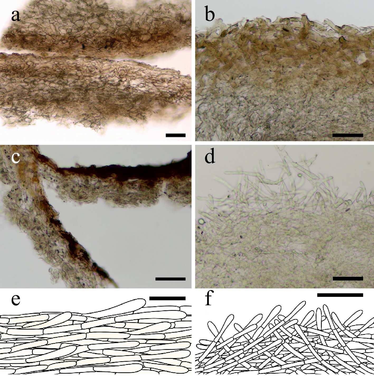
Figure 4.
Representative microstructures of Type I at the pileus center. Type I-A cutis (a), (e) Leucoagaricus rutiloides (FJAU77871); (c) Leucocoprinus acystidiatus (FJAU78090). Type I-B interwoven plagiotrichoderm (b) Leucocoprinus fuscannulatus (FJAU78013); (d), (f) Leucocoprinus parviceps (FJAU78046). Scale bars = 50 μm.
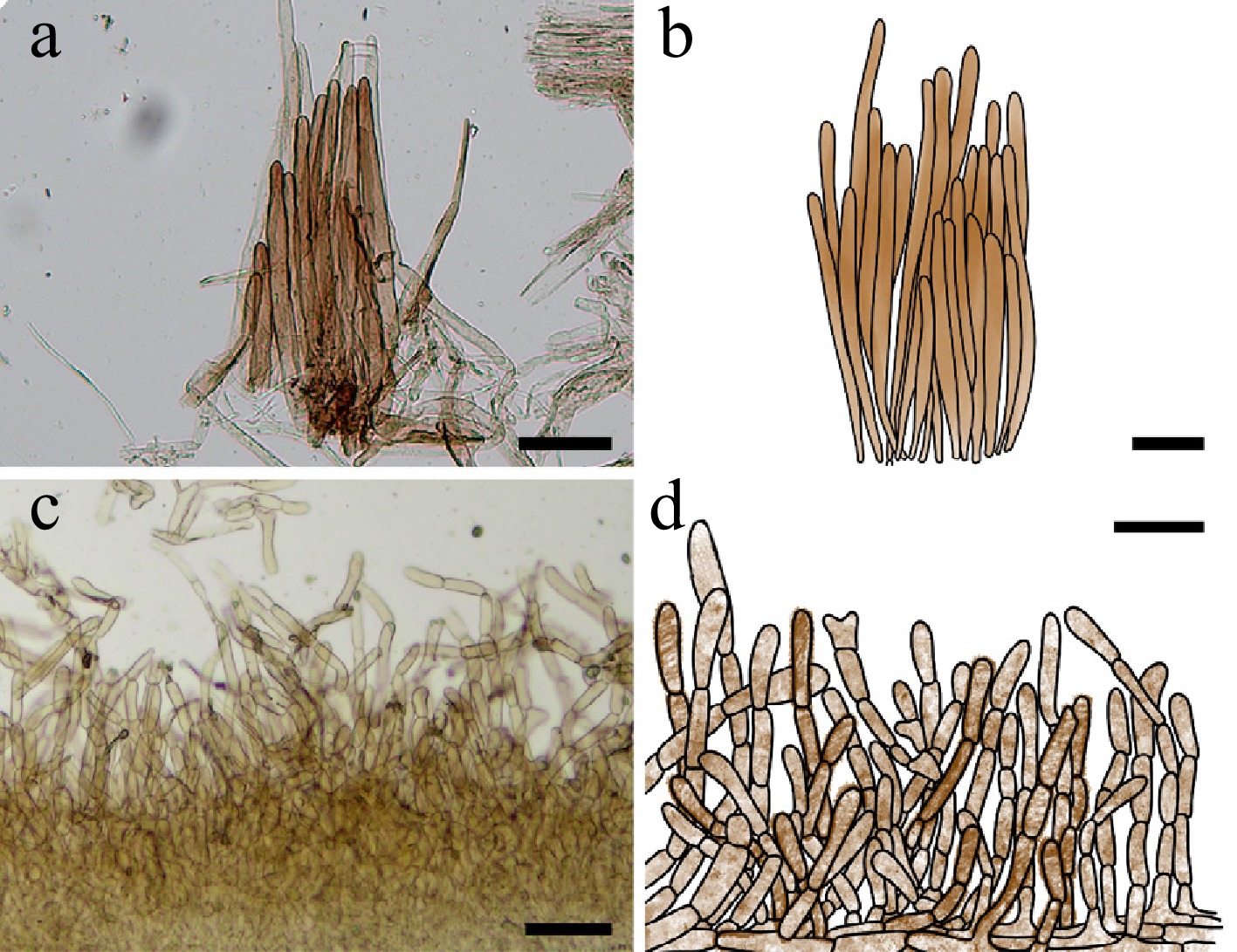
Figure 5.
Representative microstructures of Type II at the pileus center. (a), (b) Type II-A trichoderm. Leucocoprinus bicolorannulatus (FJAU78055); (c), (d) Type II-B catenulate trichoderm. Leucocoprinus guttifer (FJAU78018). Scale bars = 50 μm.
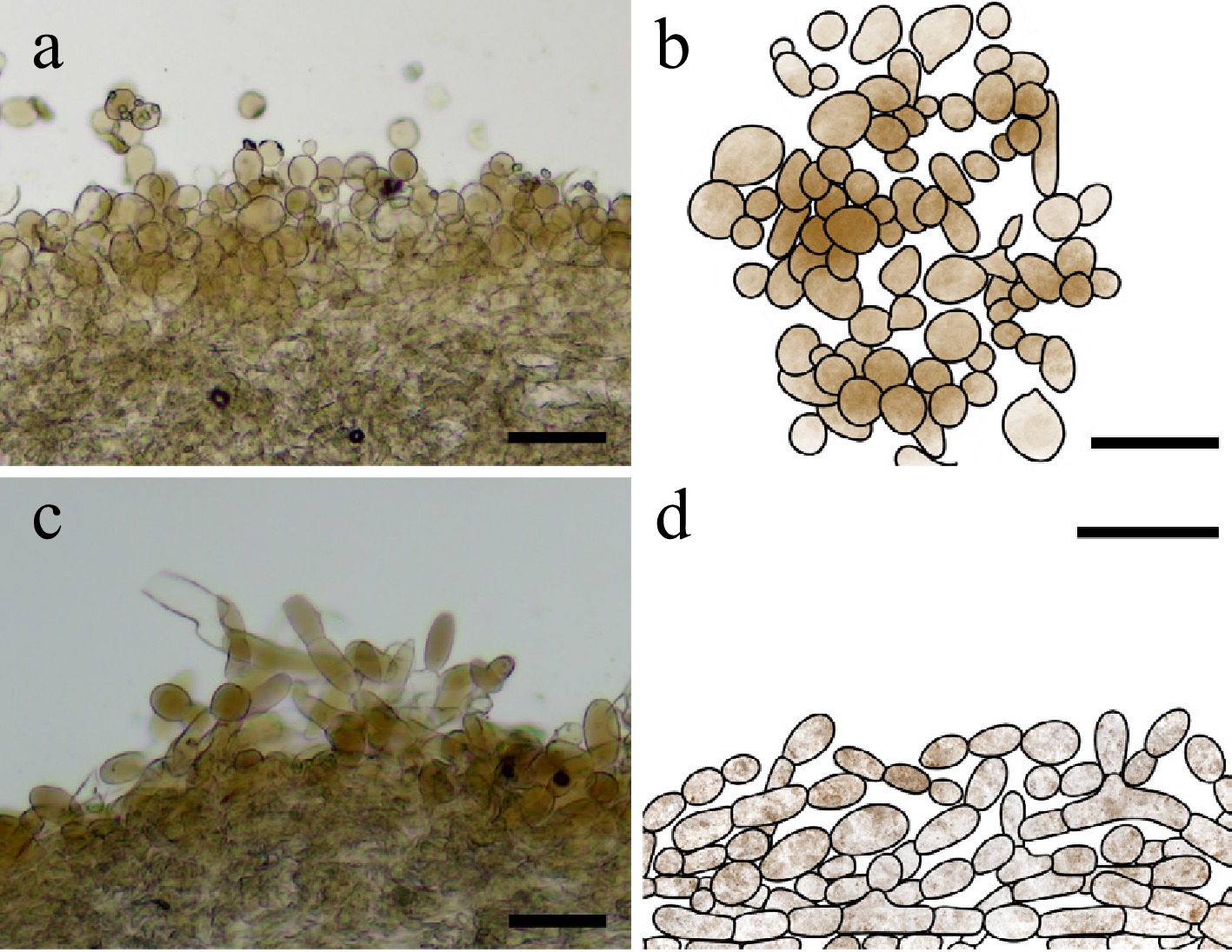
Figure 6.
Representative microstructures of Type III at the pileus center. Type III-A conioderm. (a), (b) Leucocoprinus parvicanus (FJAU78110); (c), (d) Type III-B moniliform plagiotrichoderm. Leucocoprinus umbrosquamosus (FJAU78072). Scale bars = 50 μm.
Leucoagaricus Locq. ex Singer, Sydowia 2(1-6): 35 (1948)
Type species – Leucoagaricus rubrotinctus (Peck) Singer, Sydowia 2(1-6): 36 (1948)
Notes – Integrating molecular phylogeny (Figs 1, 2) and morphological evidence, this study confirms that the Leucoagaricus comprises five distinct molecular clades observed in our phylogenetic analysis. These clades correspond to the former subgen. Sericeomyces (one clade) and the former subgen. Leucoagaricus (represented by the remaining three clades, including the formally recognized sect. Leucoagaricus[57]). A detailed discussion concerning the incongruence between the traditional infrageneric classification and the current molecular topology is presented in the Discussion section.
Leucoagaricus rubrotinctus Complex Figs 7, 8
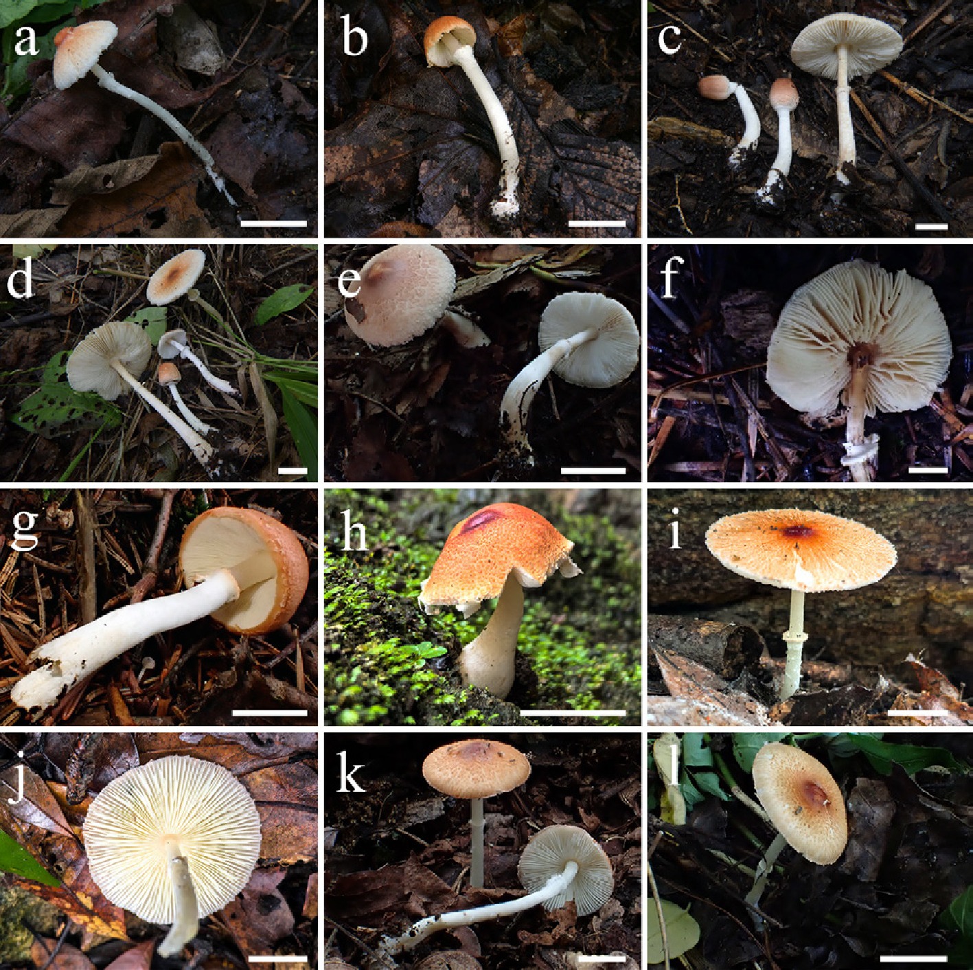
Figure 7.
Basidiomata of Leucoagaricus rubrotinctus complex. La. lateritiopurpureus (a) FJAU77772, (b) FJAU77771. La. rutiloides (c) FJAU77870, (d) FJAU77871, (e) FJAU77869. La. rubrovelutinus (f) FJAU77847, (g) FJAU77846. La. cuscutoides (h) FJAU77946, (i), (j) FJAU77945. La. rutilofbrillosus (k) FJAU77801, (l) FJAU77802. Scale bars = 1 cm. These pictures are copyright of the collectors of specimens.
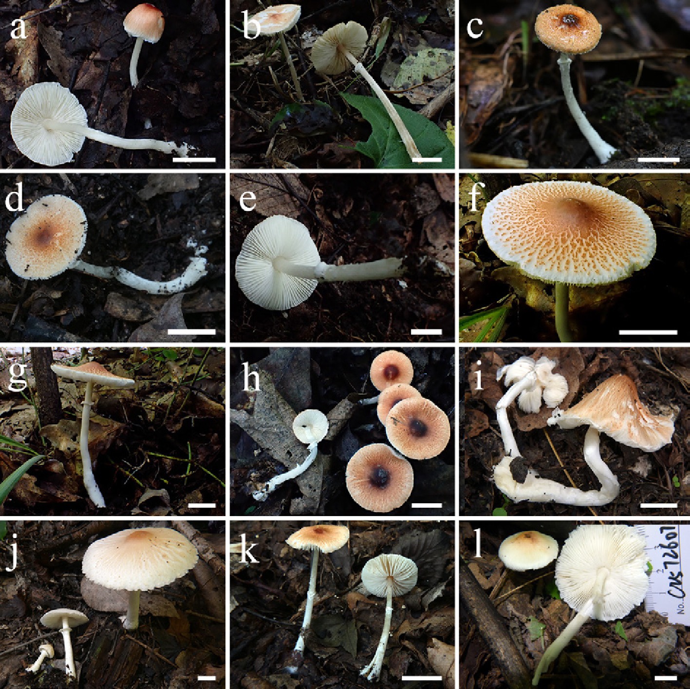
Figure 8.
Basidiomata of Leucoagaricus rubrotinctus complex. La. proximus (a) FJAU77767, (b) FJAU77765. La. rubrobrunneus (c) FJAU77780, (d) FJAU77784, (e) FJAU77783. (f), (g) La. vassiljevae (FJAU77843). (h) La. aurantioruber (FJAU77776). (i) La. subpurpureolilacinus (FJAU77943). (j) La. subnivalis (FJAU77858). (k) La. testaceumbonatus (FJAU77913). (l) La. bulbosus (FJAU77942). Scale bars = 1 cm. These pictures are copyright of the collectors of specimens.
Notes – The species currently known as Leucoagaricus rubrotinctus was originally introduced by Peck as Agaricus rubrotinctus Peck.[58]. However, this name was an illegitimate later homonym because of the pre-existing Agaricus rubrotinctus Berk.[59], (which represents a Mycena [Pers.] Roussel species). Consequently, when Peck subsequently published Lepiota rubrotincta, he effectively created a nomen novum for his illegitimate Agaricus name. Singer[8] later transferred the species to Leucoagaricus. While Singer designated La. macrorhizus as the generic type (Table 1), Redhead[57] deemed La. macrorhizus an invalid name due to the lack of a Latin description. Given that La. rubrotinctus was the only species (among Singer's two type species, La. excoriatus and La. rubrotinctus) consistently retained within Leucoagaricus in phylogenetic analyses (La. excoriatus belongs to Macrolepiota), Redhead[57] subsequently designated La. rubrotinctus as the lectotype of the genus.
Historically, the lack of molecular data from the type specimen has led to numerous taxa with similar orange-red pilei being misidentified as La. rubrotinctus in public databases such as GenBank. However, based on collections from the type locality and following the taxonomic concepts of Ge et al.[60], the authentic La. rubrotinctus is now considered to be represented by sequences such as MN483036 and KP300876[60,61]. In our phylogenetic analyses, MN483036 is robustly nested within La. clade III. Currently, the 31 phylogenetically recognized species within this complex are primarily distributed across two major lineages (La. clade II and La. clade III) and exhibit extreme macroscopic similarities. Given this high degree of morphological similarity, molecular sequence data presently provide the most reliable method for accurate species-level identification within this complex.
The species within this complex typically share the following macroscopic features: basidiomata small. Pileus plano-convex to applanate; background white, densely overlaid with reddish to orange-brown, silky-fibrillose, radially arranged squamules torn towards the margin; squamules at disc dark reddish to dark brown, persistent, entire; marginal veil remnants variable. Lamellae free, moderately close to crowded, white to cream, sometimes pale yellow or pale brown; lamellulae interspersed. Stipe subcylindrical, white; surface smooth or silky; base slightly to distinctly bulbous or spherical, occasionally volvate. Annulus superior to median, white, membranous, ascending, fugacious or persistent.
Leucoagaricus cuscutoides T. Bau, X.Y. Zhou & C. Cheng, sp. nov. Figs 7h–j, 9
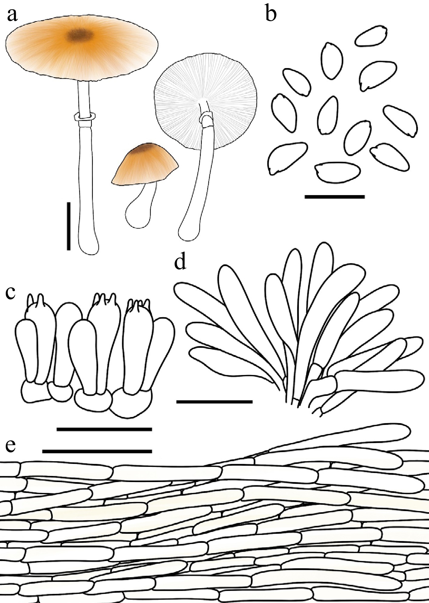
Figure 9.
Leucoagaricus cuscutoides. (a) Basidiomata. (b) Basidiospores. (c) Basidia. (d) Cheilocystidia. (e) Pileus covering. Scale bars: (a) = 1 cm; (b) = 10 μm; (c), (d) = 20 μm; (e) = 50 μm.
Chinese name: 菟丝子白环蘑 (Pīn yīn: tù sī zǐ bái huán mó)
Index Fungorum number: IF904632; Facesoffungi number: FoF18918
Etymology – 'cuscutoides' is formed by combining the genus name Cuscuta (dodder, a parasitic plant with thread-like stems) and the Greek suffix -oides (ὁειδής, meaning 'resembling'). It refers to the orange-red, fibrillose squamules on the pileus, which resemble the tangled stems of the dodder plant.
Type – China. Guangdong: Shenzhen City, Wutong Mountain, 15 June 2025, coll. C. Cheng, FJAU77946.
Diagnosis – Morphologically belonging to the Leucoagaricus rubrotinctus complex, but characterized by non-dextrinoid basidiospores and narrowly cylindrical to narrowly fusiform cheilocystidia.
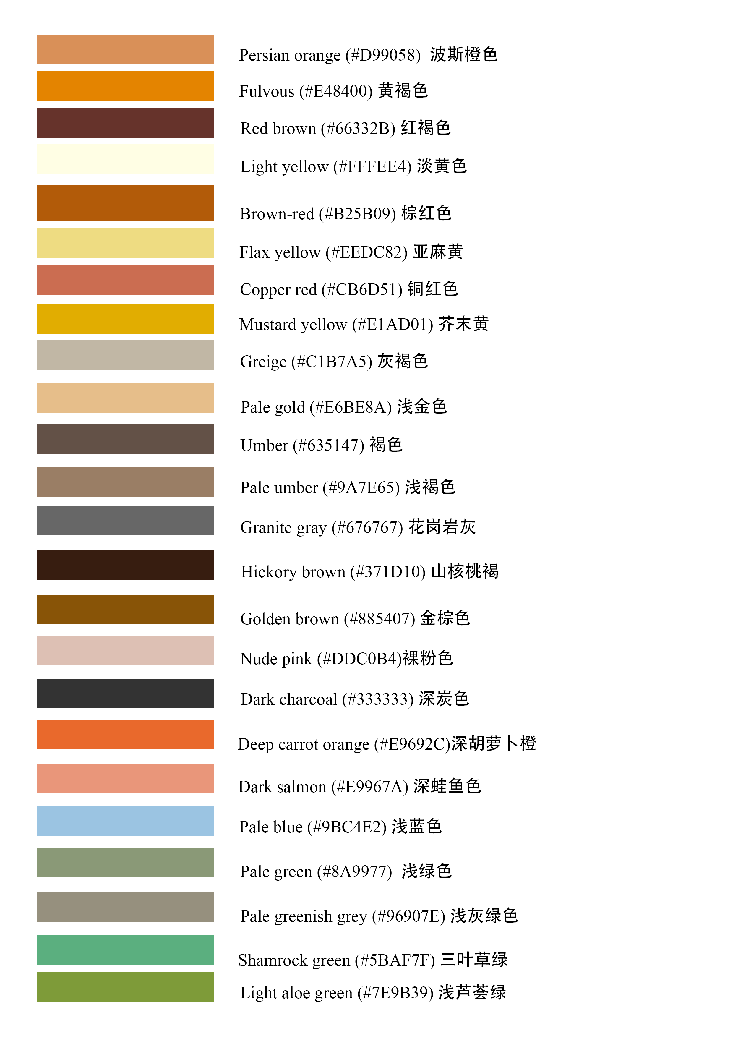
Basidiomata small. Pileus 1.6–3.0 cm in diam., initially hemispherical, expanding to plano-convex or applanate; umbo obtuse; background white to cream; centre with red-brown (#66332B) squamules; squamules around the centre Persian orange (#D99058) to fulvous (#E48400), silky-fibrillose, loosely attached. Lamellae free, close, white to cream; edge concolorous; lamellulae in 0–2 tiers. Stipe 3.6–6.2 cm × 0.3–0.5 cm, subcylindrical, hollow, slightly thickened towards the slightly bulbous base, white. Annulus median to superior, ascending, white with a Persian orange (#D99058) margin, membranous. Context thin, white, unchanging when bruised. Odour and taste not recorded.
Basidiospores (40/2/2) 6.0–7.6 × 2.8–4.0 μm, avl × avw = 6.6–6.9 × 3.4–3.5 μm, Q = 1.61–2.21, avQ = 1.88–2.02, amygdaliform or oblong to cylindrical, hyaline, slightly thick-walled, smooth, non-dextrinoid, cyanophilous, congophilous, metachromatic in Cresyl Blue. Basidia 16–25 × 6–8 μm, clavate, predominantly 4-spored, occasionally 2-spored. Pleurocystidia absent. Cheilocystidia 25–46 × 5–12 μm, narrowly cylindrical to narrowly fusiform, hyaline, thin-walled. Surface covering at pileus centre a cutis consisting of cylindrical hyphae; hyphae repent, 6–14 μm wide, slightly thick-walled, with Persian orange intracellular pigment. Stipe covering a cutis consisting of cylindrical hyphae; hyphae repent, 4–11 μm wide, thin-walled, hyaline. Clamp connections not observed.
Habit and habitat – solitary to scattered in leaf litter of broadleaf forests.
Known distribution – Central-Southern China.
Additional specimens examined – China. Guangdong: Shenzhen City, Wutong Mountain, 19 May 2025, coll. C. Cheng, FJAU77947; 15 June 2025, coll. C. Cheng, FJAU77945; 5 July 2025, coll. C. Cheng, FJAU77944.
Notes – Leucoagaricus cuscutoides is a member of the La. rubrotinctus complex. However, its non-dextrinoid basidiospores easily differentiate it from the other species within the La. rubrotinctus complex. Phylogenetically, La. cuscutoides is nested within La. clade III, showing a close affinity to La. madagascarensis Ralaiv., Liimat. & Niskanen and La. subaurantioruber R.L. Zhao & J.X. Li. Morphologically, however, La. madagascarensis differs by possessing dextrinoid basidiospores and broader cheilocystidia (22–50.5 × 10⁻²⁰ µm)[62], whereas La. cuscutoides is characterized by non-dextrinoid spores and narrower cheilocystidia (25–46 × 5–12 µm). Furthermore, La. subaurantioruber can be distinguished from the new species by its broadly clavate, sub-cylindrical, or obpyriform cheilocystidia[29], which contrast sharply with the narrowly cylindrical to narrowly fusiform elements of La. cuscutoides.
Leucoagaricus rutilofibrillosus T. Bau & X.Y. Zhou, sp. nov. Figs 7k, l, and 10
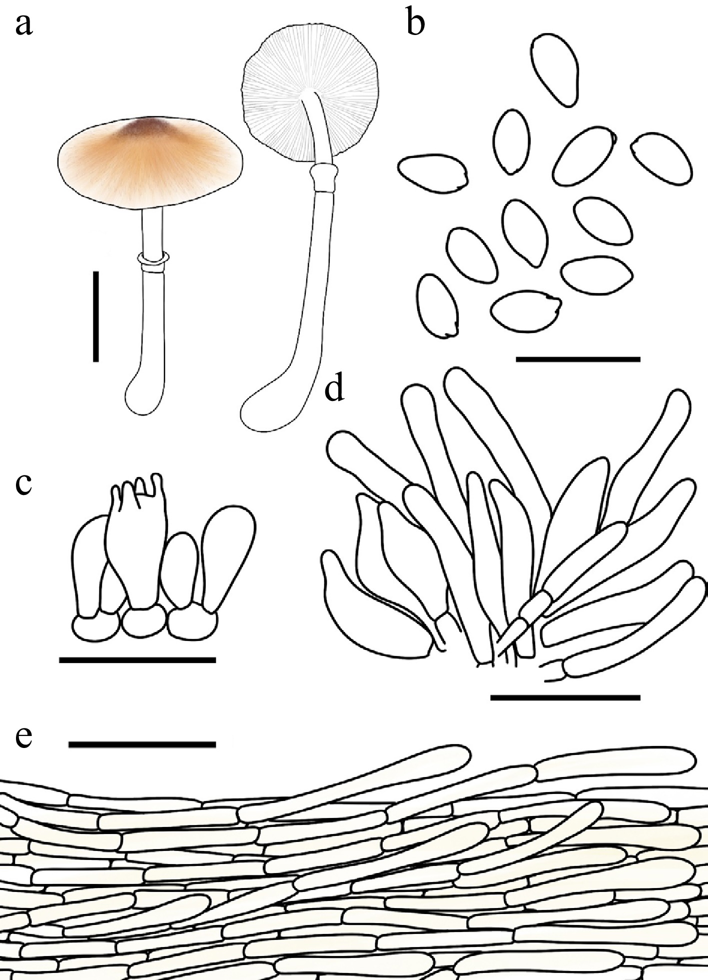
Figure 10.
Leucoagaricus rutilofibrillosus. (a) Basidiomata. (b) Basidiospores. (c) Basidia. (d) Cheilocystidia. (e) Pileus covering. Scale bars: (a) = 1 cm; (b) = 10 μm; (c), (d) = 20 μm; (e) = 50 μm.
Chinese name: 橙鳞白环蘑 (Pīn yīn: chéng lín bái huán mó)
Index Fungorum number: IF904633; Facesoffungi number: FoF18962
Etymology – 'rutilofibrillosus' is a compound adjective derived from the Latin rutilus (reddish-orange) and fibrillosus (fibrillose), referring to the characteristic reddish-orange, silky-fibrillose squamules on the pileus.
Type – China. Jilin: Ji'an City, Yushan Park, 2 August 2023, coll. Q.R. Liu, FJAU77801.
Diagnosis – Morphologically belonging to the Leucoagaricus rubrotinctus complex, but characterized by its narrowly fusiform to fusiform cheilocystidia with papillate or obtuse apices.
Basidiomata small. Pileus 1.5–2.1 cm in diam., plano-convex to applanate when mature; umbo obtuse; background white; centre with red-brown (#66332B) felted squamules; squamules around the centre Persian orange (#D99058) to fulvous (#E48400), silky-fibrillose, readily detachable. Lamellae free, crowded, white; edge concolorous; lamellulae interspersed in two tiers. Stipe 2.8–5.3 cm × 0.2–0.3 cm, subcylindrical, hollow, with a distinctly bulbous base, white; surface smooth. Annulus median to superior, ascending, white, membranous. Context thin, white, unchanging when bruised. Odour and taste not recorded.
Basidiospores (40/2/2) 5.3–6.6 × 2.7–4.4 μm, avl × avw = 5.7–5.9 × 3.1–3.2 μm, Q = 1.28–2.14, avQ = 1.67–1.87, amygdaliform or ellipsoid, oblong to cylindrical, hyaline, slightly thick-walled, smooth, dextrinoid, cyanophilous, congophilous, metachromatic in Cresyl Blue. Basidia 13–22 × 6–9 μm, clavate, predominantly 4-spored, occasionally 2-spored, thin-walled. Pleurocystidia absent. Cheilocystidia 22–41 × 5–12 μm, narrowly fusiform to fusiform, with papillate or obtuse apices, hyaline, thin-walled. Surface covering at pileus centre a cutis consisting of cylindrical hyphae; hyphae repent, 7–14 μm wide, thin-walled, with light-yellow (#FFFEE4) intracellular pigment. Stipe covering a cutis consisting of cylindrical hyphae; hyphae repent, 5–11 μm wide, thin-walled, hyaline. Clamp connections not observed.
Habit and habitat – solitary to scattered in leaf litter of broadleaf forests.
Known distribution – currently known only from Northeast China.
Additional specimens examined – China. Jilin: Ji'an City, Yushan Park, 26 July 2025, coll. L.H. Li, FJAU77802.
Notes – Unlike the majority of the La. rubrotinctus complex, La. rutilofibrillosus is phylogenetically nested within La. clade II. Among the members of this complex located in the same clade, La. rutilofibrillosus is primarily characterized by its basidiospores measuring 5.3–6.6 × 2.7–4.4 μm and narrowly fusiform to fusiform cheilocystidia (22–41 × 5–12 μm) with papillate or obtuse apices. It can be readily distinguished from its allied species by a combination of macroscopic and microscopic features. Specifically, La. guatopoensis (Dennis) Justo, Bizzi & Angelini and La. bulbiger Justo, Bizzi & Angelini both possess larger basidiospores (5.5–8.0 × 3.5–5.0 µm and 7.5–11.0 × 4.0–5.5 µm, respectively); additionally, the former features crystalline apical encrustations on its cheilocystidia, while the latter is distinguished by a white stipe that stains orange-ochraceous when touched or rubbed[61]. Furthermore, La. rutilofibrillosus is distinctly separated from the remaining clade II members based on cheilocystidial morphology: La. flavoaurantiacus (M. Ishaq, Fiaz & Khalid) Kun L. Yang & Jia Y. Lin lacks distinct cheilocystidia or possesses elements indistinguishable from basidioles[30]; La. appendiculatus R.L. Zhao & J.X. Li produces smaller, clavate to subcylindrical cheilocystidia (13.2–19.8 × 6.2–9.3 μm)[29]; La. rubrobrunneus E.F. Malysheva, Svetash. & E.M. Bulakh has lageniform cheilocystidia with a short neck and obtuse apex, or broadly fusiform elements[63]; and La. aurantioruber Y.R. Ma, Z.W. Ge & T.Z. Liu bears cheilocystidia typically equipped with a terminal appendage and tiny apical crystals[64].
Leucoagricus rutiloides T. Bau & X.Y. Zhou, sp. nov Figs 7c–e, 11
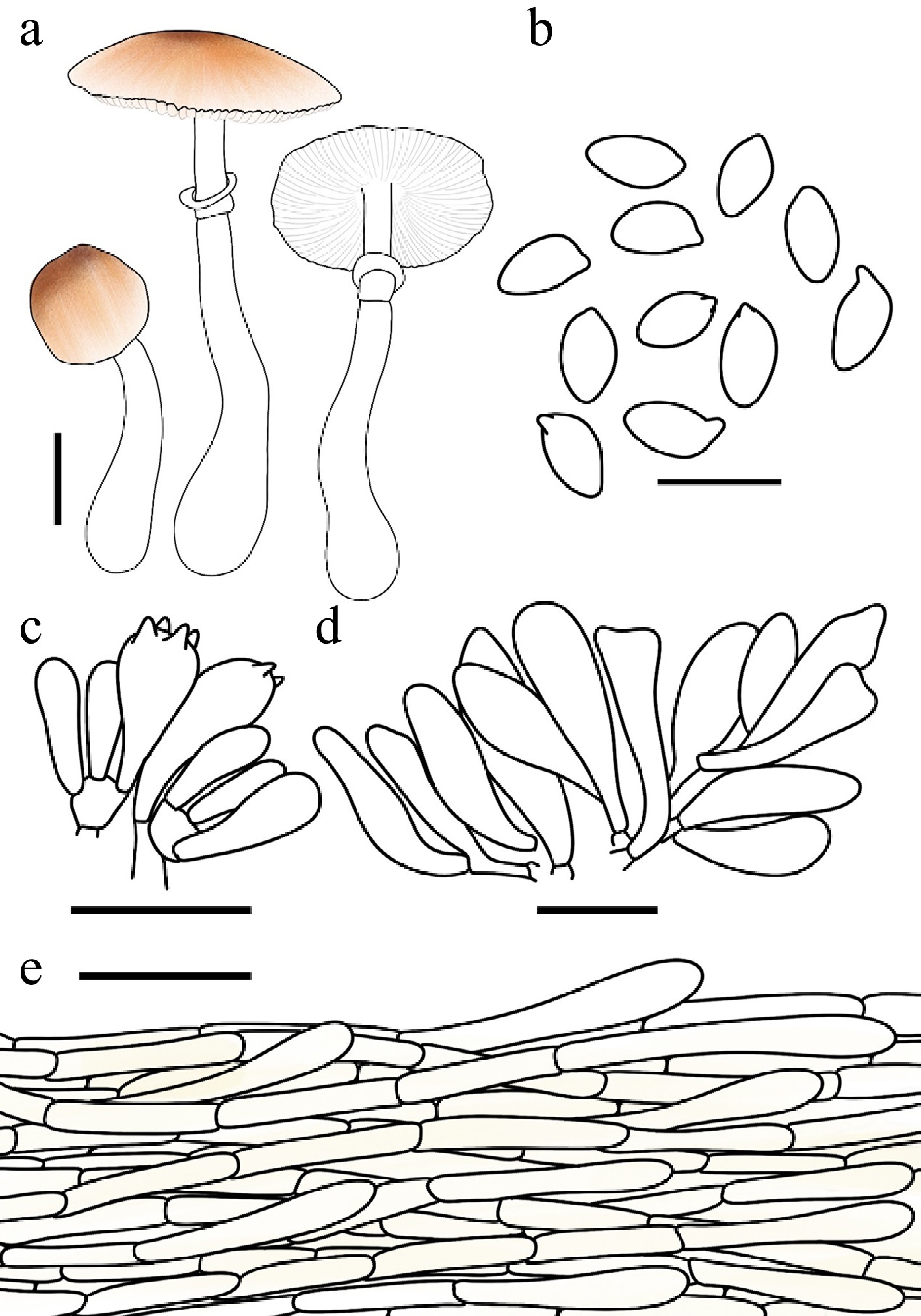
Figure 11.
Leucoagaricus rutiloides. (a) Basidiomata. (b) Basidiospores. (c) Basidia. (d) Cheilocystidia. (e) Pileus covering. Scale bars: (a) = 1 cm; (b) = 10 μm; (c), (d) = 20 μm; (e) = 50 μm.
Chinese name: 拟橙红白环蘑 (Pīn yīn: nǐ chéng hóng bái huán mó)
Index Fungorum number: IF904634; Facesoffungi number: FoF18919
Etymology – 'rutiloides' is derived from the Latin rutilus (reddish-orange, golden-red) and the Greek suffix -oides (resembling), referring to the species' close morphological similarity to certain reddish-orange fungi.
Type – China. Inner Mongolia: Tongliao City, Daqinggou National Nature Reserve, 31 July 2025, coll. X.Y. Zhou, FJAU77871.
Diagnosis – morphologically belonging to the Leucoagaricus rubrotinctus complex, but characterized by its fusiform to narrowly utriform cheilocystidia with occasionally branched apices.
Basidiomata small to medium-sized. Pileus 0.8–4.6 cm in diam., initially ovate, expanding to plano-convex or applanate; umbo obtuse; background white; centre with brown-red (#B25B09) felted squamules; squamules around the centre Persian orange (#D99058) to fulvous (#E48400), silky-fibrillose, readily detachable. Lamellae free, close, white to cream; edge concolorous; lamellulae interspersed in two tiers. Stipe 6–7.8 cm × 0.3–0.7 cm, subcylindrical, hollow, slightly thickened downwards, with a distinctly bulbous base, white; surface smooth or covered with silky fibrils. Annulus median to superior, ascending, white, membranous. Context thin, white, unchanging when bruised. Odour and taste not recorded.
Basidiospores (40/2/2) 6.7–8.0 × 3.9–4.7 μm, avl × avw = 7.2–7.4 × 4.2–4.4 μm, Q = 1.56–1.89, avQ = 1.69–1.75, amygdaliform to oblong, hyaline, slightly thick-walled, smooth, dextrinoid, cyanophilous, congophilous, metachromatic in Cresyl Blue. Basidia 15–24 × 6–10 μm, clavate, predominantly 4-spored, occasionally 2-spored, thin-walled. Pleurocystidia absent. Cheilocystidia 22–50 × 6–12 μm, fusiform to narrowly utriform, with occasionally branched apices, hyaline, thin-walled. Surface covering at pileus centre a cutis consisting of cylindrical hyphae; hyphae repent, 4–13 μm wide, thin-walled, with light yellow (#FFFEE4) intracellular pigment. Stipe covering a cutis consisting of cylindrical hyphae; hyphae repent, 3–7 μm wide, thin-walled, hyaline. Clamp connections not observed.
Habit and habitat – solitary to scattered on humus in deciduous forests.
Known distribution – so far recorded only from China.
Additional specimens examined – China. Inner Mongolia: Tongliao City, Daqinggou National Nature Reserve, 6 August 2017, coll. T. Bau, FJAU77868; 31 July 2025, coll. X.Y. Zhou & H. Cheng, FJAU77870, FJAU77869; Jilin: Jilin City, Long Tan Shan Park, 20 July 2023, coll. S.E. Wang, FJAU77867; Changchun City, Nanhu Park, 24 July 2023, coll. X.Y. Zhou, FJAU77872.
Notes – Phylogenetically, Leucoagaricus rutiloides is nested within La. clade III, showing a close affinity to La. pakistaniensis Jabeen & Khalid and La. convivalis Kun L. Yang & Jia Y. Lin. Morphologically, however, La. pakistaniensis differs by lacking distinct orange-red hues at the pileus center and possessing larger basidiospores (7.5–8.0 × 4.5–5.0 µm) compared to those of La. rutiloides (6.7–8.0 × 3.9–4.7 µm[65]). Furthermore, La. convivalis can be distinguished from the new species by its clavate cheilocystidia equipped with apical crystals[30], which contrast sharply with the fusiform to narrowly utriform cheilocystidia of La. rutiloides.
New record in China
-
Leucoagaricus proximus E.F. Malysheva, Svetash. & E.M. Bulakh, Mikol. Fitopatol. 47(3): 174 (2013) Figs 8a, b, 12
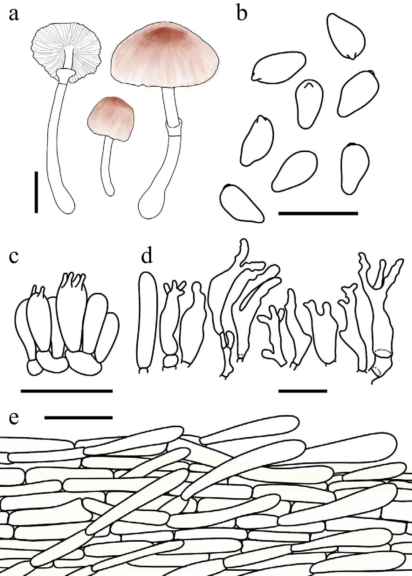
Figure 12.
Leucoagaricus proximus. (a) Basidiomata. (b) Basidiospores. (c) Basidia. (d) Cheilocystidia. (e) Pileus covering. Scale bars: (a) = 1 cm; (b) = 10 μm; (c), (d) = 20 μm; (e )= 50 μm.
Basidiomata small. Pileus 1.2–2.9 cm in diam., initially ovate, expanding to plano-convex or applanate; umbo low, obtuse; background white; centre with brown-red (#B25B09) felted squamules; squamules around the centre Persian orange (#D99058) to fulvous (#E48400), silky-fibrillose, readily detachable. Lamellae free, crowded, white to cream; edge concolorous; lamellulae 0–3 between each pair of lamellae. Stipe 2.7–5.9 cm × 0.2–0.4 cm, subcylindrical, hollow, slightly thickened towards the slightly bulbous base, white; surface smooth. Annulus median to superior, ascending, white, membranous. Context thin, white, unchanging when bruised. Odour and taste not recorded.
Basidiospores (40/2/2) 5.3–6.8 (–7.4) × 2.8–3.8 μm, avl × avw = 6.1–6.3 × 3.3–3.5 μm, Q = 1.63–1.99, avQ = 1.78–1.82, amygdaliform to oblong, hyaline, slightly thick-walled, smooth, dextrinoid, cyanophilous, congophilous, metachromatic in Cresyl Blue. Basidia 11–22 × 6–10 μm, clavate, predominantly 4-spored, occasionally 2-spored, thin-walled. Pleurocystidia absent. Cheilocystidia 16–41 × 7–20 μm, clavate to subfusiform, polymorphic, frequently branched or with excrescences at the apex, hyaline, thin-walled. Surface covering at pileus centre a cutis consisting of cylindrical hyphae; hyphae repent, 4–15 μm wide, thin-walled, with light yellow (#FFFEE4) intracellular pigment. Stipe covering a cutis consisting of cylindrical hyphae; hyphae repent, 4–11 μm wide, thin-walled, hyaline. Clamp connections not observed.
Habit and habitat – solitary on humus in mixed coniferous and broadleaf forests.
Known distribution – China, Russian Far East.
Additional specimens examined – China. Jilin: Changchun City, Jingyuetan National Forest Park, 16 August 2022, coll. W.N. Hou, FJAU77762; 12 July 2023, coll. S.E. Wang, FJAU77767; 28 July 2025, coll. X.Y. Zhou, FJAU77764; Baishan City, Lushuihe National Forest Park, 10 August 2024, coll. J.L. Wei, FJAU77760; Jilin City, Zhuque Mountain, 1 August 2025, coll. Y. Wang, FJAU77763; Heilongjiang: Yichun City, Xing'an National Forest Park, 25 July 2023, coll. H.B. Song, FJAU77761, FJAU78139; Inner Mongolia: Tongliao City, Daqinggou National Nature Reserve, 31 July 2025, coll. X.Y. Zhou and L.H. Li, FJAU77766; FJAU77765.
Notes – Leucoagaricus proximus, a new record for China originating from the Russian Far East, is a member of the La. rubrotinctus complex. However, the unique morphology of its cheilocystidia is sufficiently distinct to separate it from all other species within this complex.
Leucoagaricus rubrobrunneus E.F. Malysheva, Svetash. & E.M. Bulakh, Mikol. Fitopatol. 47(3): 175 (2013) Figs 8c–e, 13
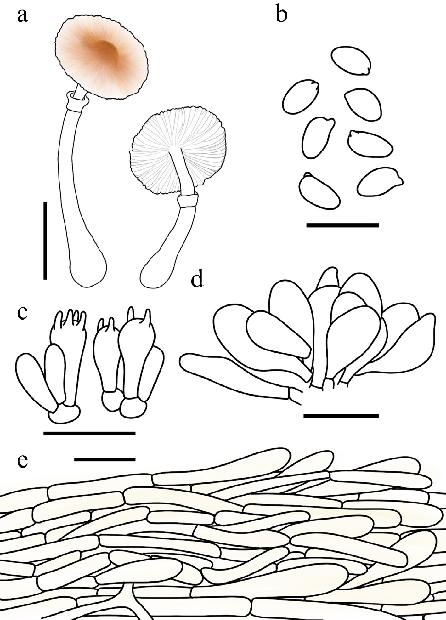
Figure 13.
Leucoagaricus rubrobrunneus. (a) Basidiomata. (b) Basidiospores. (c) Basidia. (d) Cheilocystidia. (e) Pileus covering. Scale bars: (a) = 1 cm; (b) = 10 μm; (c), (d) = 20 μm; (e) = 50 μm.
Basidiomata small. Pileus 1.0–2.5 cm in diam., initially hemispherical, expanding to plano-convex or applanate; umbo low, obtuse; background white; centre with brown-red (#B25B09) felted squamules; squamules around the centre Persian orange (#D99058) to fulvous (#E48400), silky-fibrillose, readily detachable. Lamellae free, crowded, white to cream; edge concolorous; lamellulae 0–2 between each pair of lamellae. Stipe 2.2–4.7 cm × 0.2–0.4 cm, subcylindrical, hollow, with a distinctly bulbous base, white; surface covered with silky fibrils. Annulus median to superior, ascending, white, membranous. Context thin, white, unchanging when bruised. Odour and taste not recorded.
Basidiospores (40/2/2) 5.2–6.6 × 3.0–4.4 μm, avl × avw = 5.7–5.8 × 3.4–3.5 μm, Q = 1.47–1.90, avQ = 1.67–1.69, amygdaliform or ellipsoid to oblong, hyaline, slightly thick-walled, smooth, dextrinoid, cyanophilous, congophilous, metachromatic in Cresyl Bluet. Basidia 15–28 × 8–12 μm, clavate, predominantly 4-spored, occasionally 2-spored, thin-walled. Pleurocystidia absent. Cheilocystidia 23–36 × 9–12 μm, broadly fusiform, hyaline, thin-walled. Surface covering at pileus centre a cutis consisting of cylindrical hyphae; hyphae repent, 3–16 μm wide, thin-walled, with light yellow (#FFFEE4) intracellular pigment. Stipe covering a cutis consisting of cylindrical hyphae; hyphae repent, 4–12 μm wide, thin-walled, hyaline. Clamp connections not observed.
Habit and habitat – solitary on humus-rich soil in mixed coniferous and broadleaf forests.
Known distribution – China, Russian Far East.
Additional specimens examined – China. Heilongjiang: Hulin City, 5 September, 2003, coll. T. Bau, FJAU3047; Jilin: Jiaohe City, Shansongling,24 July 2022, coll. L.M. Song, FJAU77785; Dunhua City, State-Owned Forest Farm, 27 July 2022, coll. X. Wang, FJAU77781; Laobaishan Primitive Ecological Area, 22 July 2025, coll. X.Y. Zhou, FJAU77784; Huadian City, Hongshi National Forest Park, 16 August 2024, coll. R.H. Lin, FJAU77780; 30 July 2025, coll. Y.F. Han, FJAU77782; Ji'an City, Wunufeng National Forest Park, 25 July 2025, coll. H. Cheng, FJAU77783.
Notes – Phylogenetically, Leucoagaricus rubrobrunneus is nested within La. clade II, where it clusters together with La. aurantioruber Y.R. Ma, Z.W. Ge & T.Z. Liu and La. testaceumbonatus R.L. Zhao & J.X. Li. Morphologically, however, these closely related species can be readily distinguished based on cheilocystidial features. Specifically, La. aurantioruber differs by possessing tiny crystals on the apex of its cheilocystidia[64], while La. testaceumbonatus is characterized by clavate, subclavate, to narrowly clavate cheilocystidia with capitate to subcapitate apices[34]. These morphological traits contrast sharply with those of La. rubrobrunneus, which possesses broadly fusiform cheilocystidia completely lacking apical crystals.
Leucoagaricus serenus complex Figs 14−17
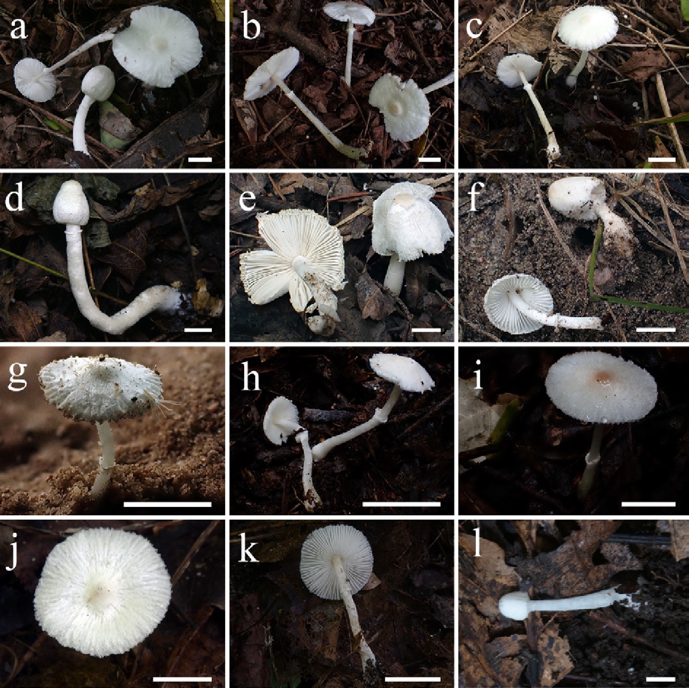
Figure 14.
Basidiomata of Leucoagaricus serenus complex. La. albissimus (a) FJAU77928, (b) FJAU78127. La. brunneimutabilis (c) FJAU77891, (d) FJAU77894. La. albofulvescens (e) FJAU77861, (f) FJAU77863, (g) FJAU77897. La. albofibrillosus (h) FJAU78277, (i) FJAU78276. La. leucotrichus (j), (k) FJAU77805, (l) FJAU77807. Scale bars = l cm. These pictures are copyright of the collectors of specimens.
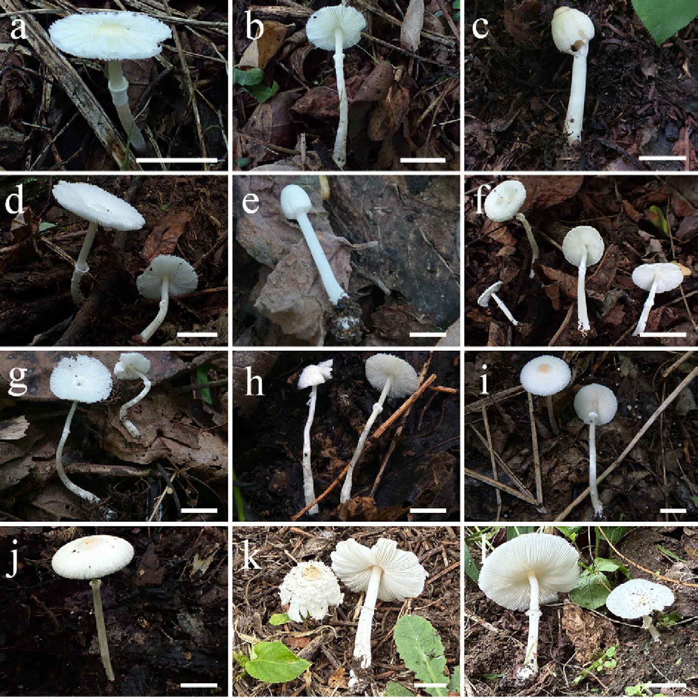
Figure 15.
Basidiomata of Leucoagaricus serenus complex. La. subleucotrichus (a), (b) FJAU78135, (c) FJAU77808. La. microsporus (d) FJAU77889, (e) FJAU77887, (f) FJAU77885. La. submicrosporus (g) FJAU77919, (h) FJAU77920. La. centrobrunneolus (i) FJAU77909, (j) FJAU77908. La. albosquamosus (k) FJAU78148, (l) FJAU78140. Scale bars = 1 cm. These pictures are copyright of the collectors of specimens.
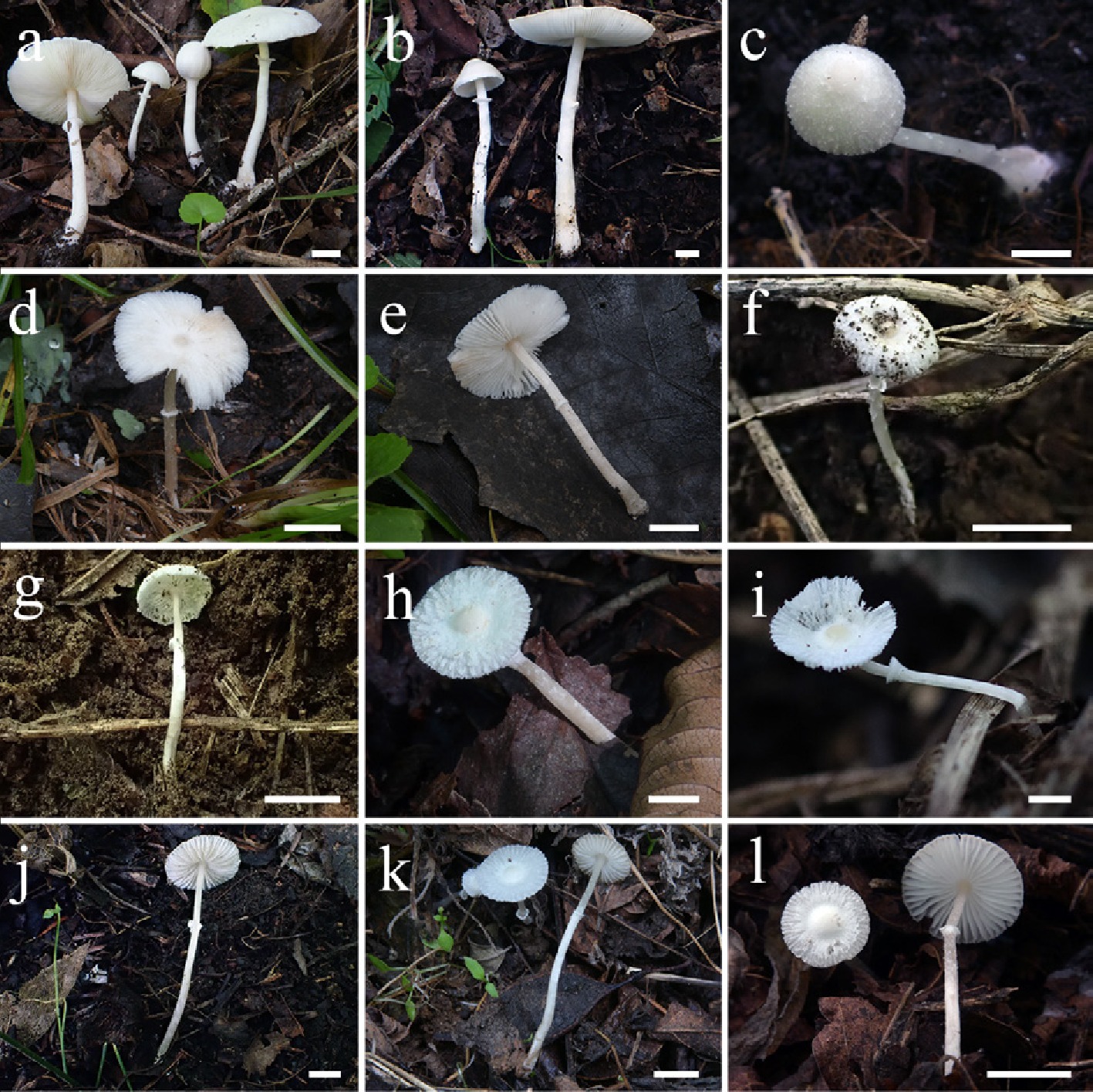
Figure 16.
Basidiomata of the Leucoagaricus serenus complex. La. magnialbus (a) FJAU77822, (b) FJAU77820. La. similis (c) FJAU77905, (d), (e) FJAU78099. (f), (g) La. aff. similis (FJAU77903). La. pallidoumbonatus (h) FJAU77878, (i) FJAU77876, (j) FJAU77875. La. jianensis (k) FJAU77937, (l) FJAU77936. Scale bars = 1 cm. These pictures are copyright of the collectors of specimens.
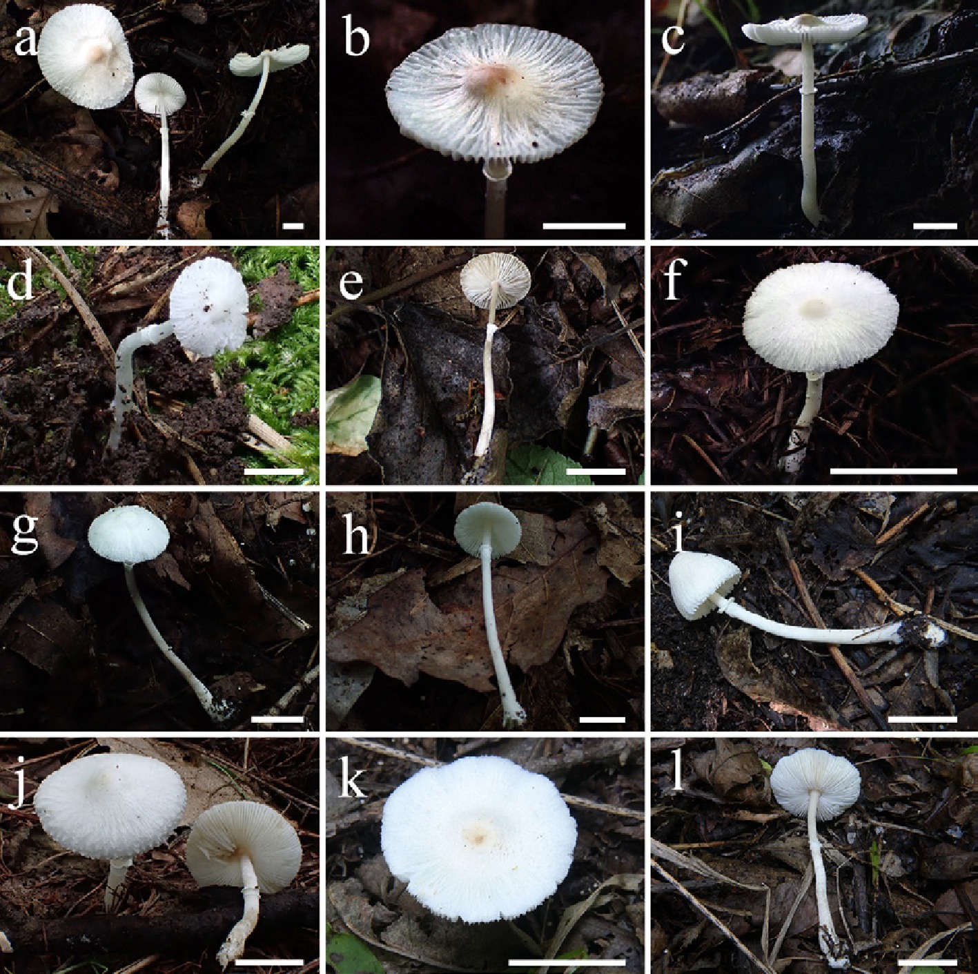
Figure 17.
Basidiomata of the Leucoagaricus serenus complex. La. ruiliniae (a) FJAU77884, (b), (c) FJAU77880. La. aff. serenus 1 (d) FJAU77789, (e) FJAU77790, (f) FJAU77787. La. aff. serenus 2 (g) FJAU77794, (h) FJAU77792. La. candidus (i) FJAU77754, (j) FJAU77753. (k), (l) La. subcandidus (FJAU77940). Scale bars = 1 cm. These pictures are copyright of the collectors of specimens.
Notes – Historically, the taxonomic concept of Leucoagaricus serenus (Fr.) Bon & Boiffard was subject to prolonged confusion. Fries[66] originally described the taxon 'serenus' as an elegant, pure white agaric but provided no micromorphological characters. Subsequently, Lange[67] described Lepiota serena as a species with striking, lageniform cheilocystidia. However, Kühner[68] disagreed with Lange's[67] interpretation, applying the name 'serenus' to taxa possessing broadly clavate cheilocystidia. Pearson[69] erroneously applied 'serenus' to a species he later[70,71] described as L. pinguipes A. Pearson, which is characterized by robust basidiomata, ellipsoid basidiospores, and a close affinity to Lc. leucothites (Vittad.) Wasser.
Thereafter, a fourth taxonomic delimitation for 'serenus' was proposed by Bon[71], Rodríguez Armas et al.[72], and several other mycologists[73−75], who characterized the species by narrowly clavate cheilocystidia with apical crystals. Although Rodríguez Armas et al.[72], citing Malençon, questioned whether their specimens represented the true Sericeomyces serenus, Bon maintained this identification. Furthermore, the descriptions provided by Candusso[75], MBreitenbach[73], and Breitenbach & Kranzlin[73] featured considerably broader cheilocystidia than typical, suggesting that their studied materials were likely mixed with other taxa (e.g., L. menieri [Sacc.] Sing.). Moser[77] even conflated different taxonomic concepts, broadly stating that the 'sterile hairs' are clavate or cylindrical.
Given the severe nomenclatural and conceptual chaos mentioned above, Vellinga[78] provided a systematic clarification. She segregated the taxa with narrow, crystal-bearing cheilocystidia and formally described them as a novel species, Leucoagaricus crystallifer Vellinga. Concurrently, based on the taxonomic concepts of Kühner[68] and Huijsman[79], Vellinga designated a neotype for Leucoagaricus serenus (Fr.) Bon & Boiffard and provided a comprehensive modern description, thereby establishing a reliable taxonomic foundation for this species.
Based on this clear taxonomic delimitation, the La. serenus complex proposed in this study largely corresponds to the traditional concept of Leucoagaricus subgen. Sericeomyces (Heinem.) M. Bon, which is typified by La. serenus. In our phylogenetic framework, this primarily corresponds to La. clade II. Furthermore, the present study reveals that the majority of species within La. clade V also exhibit macroscopic morphologies highly similar to La. serenus. As noted by Vellinga[78], members of this complex typically produce elegant, predominantly white basidiomata, primarily characterized by a dry or slightly viscid pileus with a pileipellis composed of adnate to ascending, radially arranged hyphae.
Species within this complex exhibit extreme morphological uniformity. As historically emphasized, they 'look disconcertingly similar in the field'[78]. Although classical taxonomy heavily relied on the micromorphology of cheilocystidia and pileipellis elements in attempts to differentiate these taxa, their basidiospores are known to be highly variable in both shape and size. Therefore, species delimitation within this group based solely on traditional morphological traits is highly unreliable. We treat this complex as a group exhibiting remarkable morphological similarity, and objectively state that accurate species-level identification and delimitation presently necessitate the use of molecular data.
The species within this complex typically share the following macroscopic features: basidiomata small to medium-sized; pileus initially ovate, expanding to plano-convex or plane, with a white surface densely decorated with concolorous, radially arranged, silky-fibrillose squamules; disc yellowish-brown to brownish; marginal veil remnants often sparse. Lamellae free, moderately close to crowded, interspersed with lamellulae, white to cream. Stipe subcylindrical, white to cream, smooth or silky, slightly tapering basally. Annulus superior to median, white, membranous, and ascending. Context white, thin to slightly thick.
Leucoagaricus albofibrillosus T. Bau & X.Y. Zhou, sp. nov. Figs 14h, i, and 18
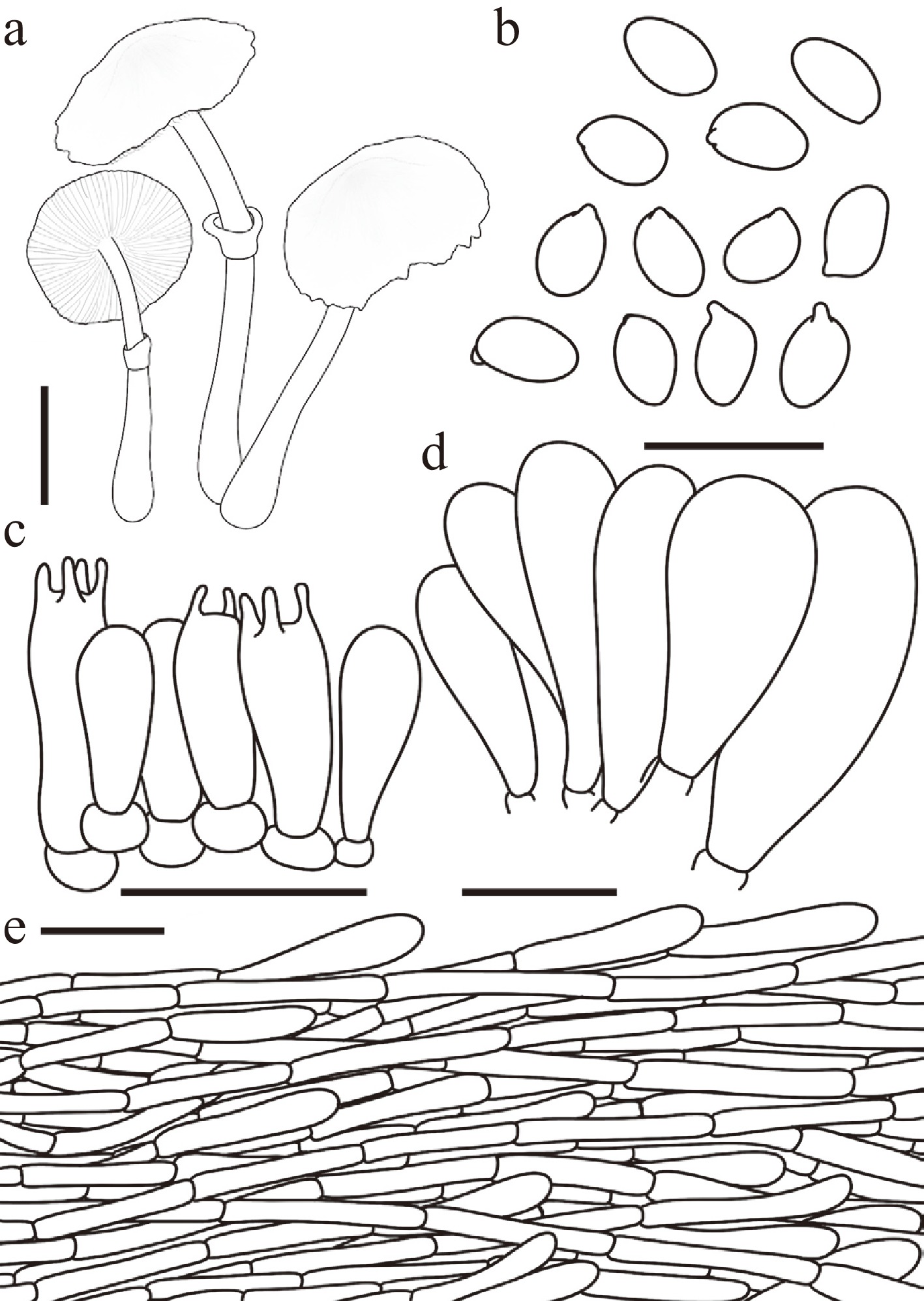
Figure 18.
Leucoagaricus albofibrillosus. (a) Basidiomata. (b) Basidiospores. (c) Basidia. (d) Cheilocystidia. (e) Pileus covering. Scale bars: (a) = 1 cm; (b) = 10 μm; (c), (d) = 20 μm; (e) = 50 μm.
Chinese name: 白纤白环蘑 (Pīn yīn: bái xiān bái huán mó)
Index Fungorum number: IF905149; Facesoffungi number: FoF19599
Etymology – 'albofibrillosus' is derived from the Latin albus (white) and fibrillosus (fibrillose), referring to the characteristic white fibrillose squamules covering the stipe and parts of the basidiomata.
Type – China. Zhejiang: Lishui City, 9 November 2025, coll. X.Y. Zhou, FJAU78277.
Diagnosis – characterized by its small basidiomata, a stipe covered with prominent white fibrillose squamules, subamygdaliform basidiospores (4.7–5.9 × 2.7–3.7 μm), and narrowly fusiform to oblong cheilocystidia.
Basidiomata small. Pileus 1.3–1.7 cm in diam., plano-convex to applanate at maturity; umbo indistinct, obtuse; background white; centre flax yellow (#EEDC82) to copper red (#CB6D51); squamules around the centre white, densely fibrillose. Lamellae free, crowded, white to cream; edge concolorous; lamellulae interspersed in two tiers. Stipe 3.6–4.3 × 0.2–0.3 cm, subcylindrical, slightly broader towards the base, white; surface covered with concolorous fibrillose squamules. Annulus medium to superior, ascending, white, membranous. Context thin, white, unchanging when bruised. Odour and taste not recorded.
Basidiospores (40/2/2) 4.7–5.9 × 2.7–3.7 μm, avl × avw = 5.2–5.4 × 3.1–3.2 μm, Q = 1.34–1.98, avQ = 1.62–1.75, subamygdaliform in side view, ellipsoid to oblong in frontal view, hyaline, thin-walled, dextrinoid, cyanophilous, congophilous, metachromatic in Cresyl Blue. Basidia 12–21 × 6–10 μm, clavate, predominantly 4-spored, occasionally 2-spored, thin-walled. Pleurocystidia absent. Cheilocystidia 22–34 × 7–20 μm, narrowly fusiform to oblong or ellipsoid, hyaline, thin-walled. Surface covering at pileus centre a cutis consisting of cylindrical hyphae; hyphae repent, 4–12 μm wide, thin-walled, with light yellow intracellular pigment. Stipe covering a cutis consisting of cylindrical hyphae; hyphae repent, 4–10 μm wide, thin-walled, hyaline. Clamp connections not observed.
Habit and habitat – solitary to scattered on the humus layer in forests.
Known distribution – currently known from Zhejiang, China.
Additional specimens examined – China. Zhejiang: Lishui City, 9 November 2025, coll. H.Y. Zhou, FJAU78276.
Notes – Phylogenetically, Leucoagaricus albofibrillosus forms a sister clade with La. conifericola (La. clade V). However, the latter can be distinguished by its relatively larger basidiomata (pileus 2.3–2.9 cm in diam.) with a coppery red to reddish-brown disc, as well as distinct ecological and geographical preferences. Currently known collections of La. conifericola inhabit the humus layer of coniferous-dominated forests in Northeast China, whereas La. albofibrillosus occurs in mixed coniferous and broad-leaved forests in East China.
Furthermore, La. albofibrillosus clusters within a broader clade representing the La. serenus complex, alongside La. umbonatus S. Hussain, H. Ahmad & Afshan, La. umbonatoides R.L. Zhao & J.X. Li, and La. subcandidus R.L. Zhao & J.X. Li. Morphologically, La. albofibrillosus [basidiospores 4.7–5.9 × 2.7–3.7 µm; cheilocystidia narrowly fusiform to oblong or ellipsoid] is readily separated from La. umbonatus and La. umbonatoides, both of which possess larger basidiospores (5.5–6.5 × 3.5–4.0 μm and 5.1–6.8 × 3.1–5.0 μm, respectively)[29,65]. Additionally, La. umbonatus features mostly ventricose to fusiform cheilocystidia[29], while La. umbonatoides has clavate to subclavate ones[29]. Conversely, La. subcandidus differs from La. albofibrillosus by having relatively smaller basidiospores (4.2–4.9 × 2.9–3.4 μm) and predominantly narrowly clavate cheilocystidia[34].
Leucoagaricus jianensis T. Bau & X.Y. Zhou, sp. nov Figs 16k, l, 19
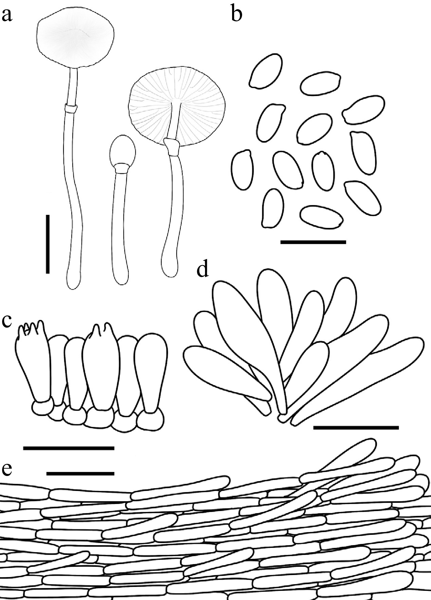
Figure 19.
Leucoagaricus jianensis. (a) Basidiomata. (b) Basidiospores. (c) Basidia. (d) Cheilocystidia. (e) Pileus covering. Scale bars: (a) = 1 cm; (b) = 10 μm; (c), (d) = 20 μm; (e) = 50 μm.
Chinese name: 集安白环蘑 (Pīn yīn: jí ān bái huán mó)
Index Fungorum number: IF905150; Facesoffungi number: FoF19600
Etymology – 'jianensis' refers to Ji'an City (Jilin Province, China), the locality where the holotype specimen was collected.
Type – China. Jilin: Ji'an City, Yushan Park, 24 July 2025, coll. X.Y. Zhou, FJAU77937.
Diagnosis – Leucoagaricus jianensis is mainly characterized by its very small, white basidiomata (pileus 0.4–1.5 cm in diam.) covered with concolorous fibrillose squamules, basidiospores measuring 6.0–7.1 × 3.1–3.7 μm, and oblong to fusiform cheilocystidia.
Basidiomata small. Pileus 0.4–1.5 cm in diam., initially ovate, expanding to plano-convex or applanate; umbo slightly obtuse; background white; centre with white to fulvous (#E48400) squamules; squamules around the centre white, silky-fibrillose, readily detachable. Lamellae free, moderately crowded, white; edge concolorous; lamellulae 0–1 between each pair of lamellae. Stipe 1.8–3.4 cm × 0.1–0.2 cm, subcylindrical, hollow, white; surface covered with concolorous, silky-fibrillose squamules. Annulus medium to superior, ascending, white, membranous. Context thin, white, unchanging when bruised. Odour and taste not recorded.
Basidiospores (40/2/2) 6.0–7.1 × 3.1–3.7 μm, avl × avw = 6.4–6.5 × 3.4 μm, Q = 1.68–2.21, avQ = 1.89–1.93, oblong to cylindrical, hyaline, thin-walled, smooth, dextrinoid, cyanophilous, congophilous, metachromatic in Cresyl Blue. Basidia 14–24 × 6–10 μm, clavate, predominantly 4-spored, occasionally 2-spored, thin-walled. Pleurocystidia absent. Cheilocystidia 25–40 × 7–19 μm, oblong to fusiform, hyaline, thin-walled. Surface covering at pileus centre a cutis consisting of cylindrical hyphae; hyphae repent, 4–14 μm wide, slightly thick-walled, with light yellow intracellular pigment in the central squamules. Stipe covering a cutis consisting of cylindrical hyphae; hyphae repent, 3–11 μm wide, thin-walled, hyaline. Clamp connections not observed.
Habit and habitat – solitary to scattered in the humus layer of broadleaf forests.
Known distribution – Northeast China.
Additional specimens examined – China. Jilin Province: Ji'an City, Yushan Park, 24 July 2025, coll. X.Y. Zhou, FJAU77936, FJAU77938; Inner Mongolia: Tongliao City, Daqinggou National Nature Reserve, 31 July 2025, coll. X.Y. Zhou, FJAU77939.
Notes – Morphologically, Leucoagaricus jianensis belongs to the La. serenus complex. However, it can be readily distinguished from most other species within this group, including La. magnialbus, by its exceptionally small basidiomata with a pileus measuring only 0.4 to 1.5 cm in diameter.
Phylogenetically, La. jianensis clusters with La. pallidoumbonatus R.L. Zhao & J.X. Li and La. ruiliniae J.X. Li to form the strongly supported La. jianensis clade, a distinct lineage currently accommodating only these three taxa. Despite this robust internal support, the exact phylogenetic position of this clade within the entire genus Leucoagaricus remains unstable. Morphologically, however, these closely related species are clearly separated. Specifically, La. pallidoumbonatus differs by producing relatively larger basidiomata with a pileus diameter of 3.0 to 4.0 cm, alongside smaller, clavate to subcylindrical cheilocystidia measuring 12.0–18.2 × 6.0–7.9 µm[29]. Similarly, La. ruiliniae can be separated from the new species by its larger basidiomata reaching 1.8 to 3.2 cm across the pileus, as well as its ellipsoid to broadly clavate cheilocystidia measuring 13.0–50.0 × 8.0–26.0 µm[29]. These features contrast sharply with the narrowly utriform to fusiform cheilocystidia of La. jianensis, which measure 25.0–40.0 × 7.0–19.0 µm.
Leucoagaricus leucotrichus T. Bau & X.Y. Zhou, sp. nov Figs 14j–l, 20
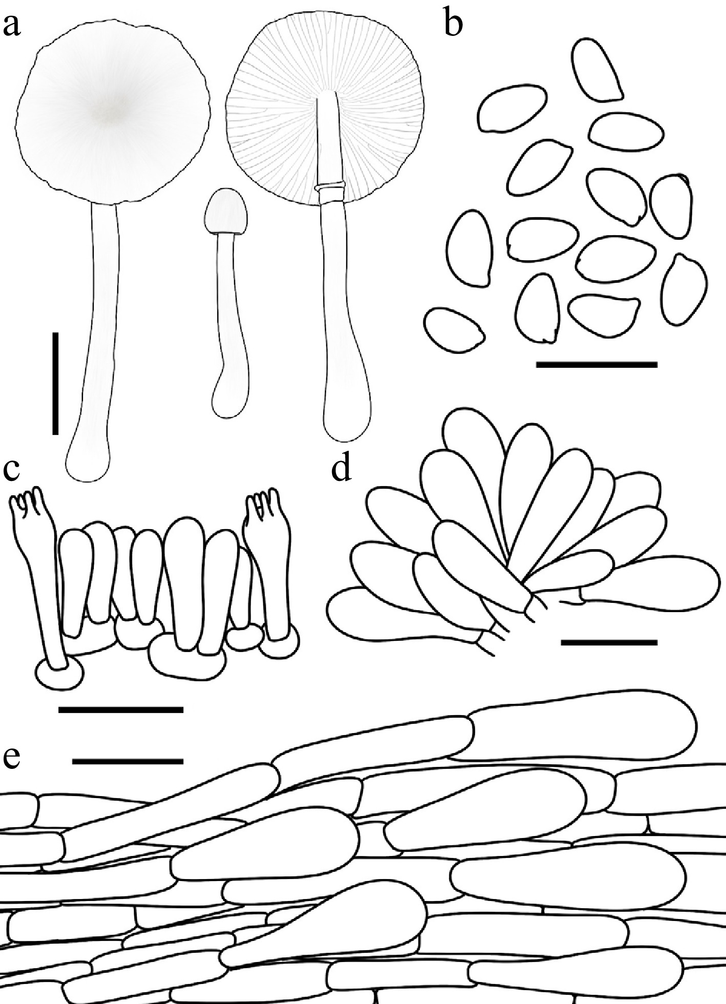
Figure 20.
Leucoagaricus leucotrichus. (a) Basidiomata. (b) Basidiospores. (c) Basidia. (d) Cheilocystidia. (e) Pileus covering. Scale bars: (a) = 1 cm; (b) = 10 μm; (c)–(e) = 20 μm.
Chinese name: 白毛白环蘑 (Pīn yīn: bái máo bái huán mó)
Index Fungorum number: IF904636; Facesoffungi number: FoF18927
Etymology – 'leucotrichus' is derived from the Greek leuko- (white) and trichos (hair), referring to the characteristic white, fibrillose squamules on the pileus.
Type – China. Inner Mongolia: Tongliao City, Daqinggou National Nature Reserve, 31 July 2025, coll. X.Y. Zhou, FJAU77805.
Diagnosis – characterized by its white basidiomata covered with concolorous, silky- to fibrillose squamules, and non-dextrinoid to weakly dextrinoid basidiospores.
Basidiomata small. Pileus 0.4–2.3 cm in diam., initially ovate, expanding to plano-convex or applanate; umbo obtuse; background white; squamules concolorous, silky-fibrillose, readily detachable. Lamellae free, moderately crowded, white; edge concolorous; lamellulae interspersed in 0–2 tiers. Stipe 2.0–3.9 cm × 0.2–0.3 cm, subcylindrical, widening downwards to a slightly bulbous base, hollow, white; surface covered with concolorous, silky-fibrillose squamules. Annulus medium to superior, ascending, white, membranous. Context thin, white, unchanging when bruised. Odour and taste not recorded.
Basidiospores (40/2/2) 5.1–6.4 × 2.8–3.8 μm, avl × avw = 5.7–5.9 × 3.3–3.4 μm, Q = 1.52–1.95, avQ = 1.72–1.74, ellipsoid to oblong, hyaline, thin-walled, smooth, non-dextrinoid to weakly dextrinoid, cyanophilous, congophilous, metachromatic in Cresyl Blue. Basidia 18–28 × 6–8 μm, clavate, predominantly 4-spored, occasionally 2-spored, thin-walled. Pleurocystidia absent. Cheilocystidia 26–46 × 8–14 μm, clavate to broadly fusiform, with obtuse apices, hyaline, thin-walled. Surface covering at pileus centre a cutis consisting of cylindrical hyphae; hyphae repent, 4–15 μm wide, slightly thick-walled, hyaline. Stipe covering a cutis consisting of cylindrical hyphae; hyphae repent, 5–12 μm wide, thin-walled, hyaline. Clamp connections not observed.
Habit and habitat – solitary on humus in broadleaf forests.
Known distribution – so far recorded only from Northeast China.
Additional specimens examined – China. Jilin: Changchun City, Jingyuetan National Forest Park, 28 July 2025, coll. X.Y. Zhou, FJAU77806, FJAU77807.
Notes – Morphologically, Leucoagaricus leucotrichus belongs to the Leucoagaricus serenus complex, from which its separation based on macroscopic features alone is often difficult. Phylogenetically, La. leucotrichus is nested within Leucoagaricus subgenus Sericeomyces, where most species are white and characterized by a dry or slightly viscid pileus covering composed of adnate to ascending, radially arranged hyphae. Within this subgenus, La. leucotrichus is one of four species—along with La. candidus Y.R. Ma, Z.W. Ge & T.Z. Liu, La. subleucotrichus T. Bau & X.Y. Zhou, and La. serenus—that exhibit white basidiomata, making field identification challenging. However, differentiation is possible using the dextrinoid reaction of the basidiospores: La. leucotrichus and La. subleucotrichus possess non-dextrinoid to weakly dextrinoid basidiospores, while La. candidus and La. serenus have dextrinoid basidiospores[64,78]. Furthermore, these taxa are distinguishable by variations in basidiospore dimensions and cheilocystidia morphology.
Leucoagaricus magnialbus T. Bau & X.Y. Zhou, sp. nov Figs 16a, b, 21
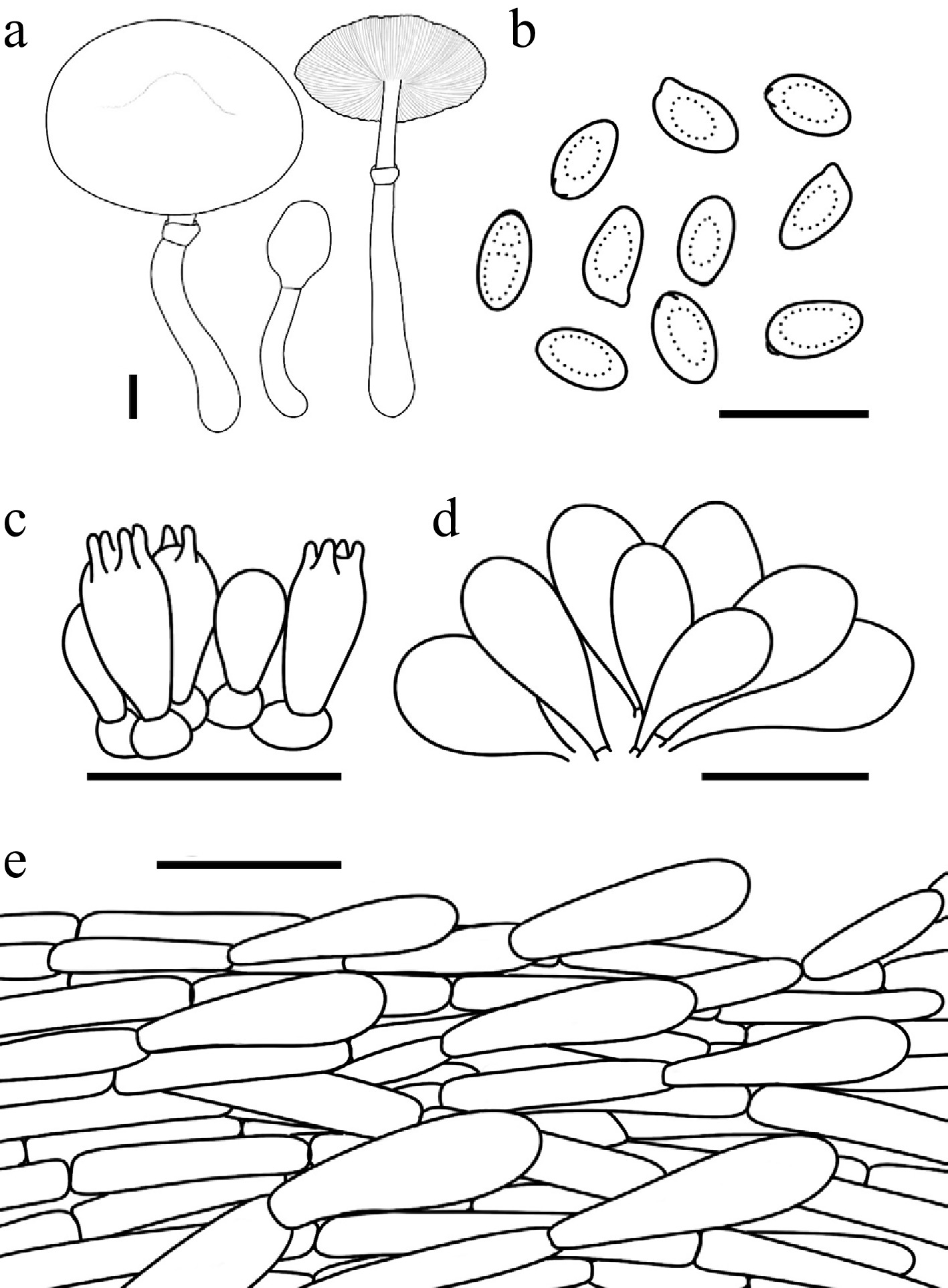
Figure 21.
Leucoagaricus magnialbus. (a) Basidiomata. (b) Basidiospores. (c) Basidia. (d) Cheilocystidia. (e) Pileus covering. Scale bars: (a) = 1 cm; (b) = 10 μm; (c), (d) = 20 μm; (e) = 50 μm.
Chinese name: 大白环蘑 (Pīn yīn: dà bái huán mó)
Index Fungorum number: IF904642; Facesoffungi number: FoF18934
Etymology – 'magnialbus' is a compound Latin adjective derived from magnus (large, great) and albus (white), referring to the characteristically large and white basidiomata.
Type – China. Inner Mongolia: Tongliao City, Daqinggou National Nature Reserve, coll. X.Y. Zhou, FJAU77822.
Diagnosis – characterized by its medium- to large-sized white basidiomata covered with concolorous, silky- to fibrillose squamules, and clavate to broadly clavate cheilocystidia.
Basidiomata medium to large-sized. Pileus 1.5–7.0 cm in diam., initially ovate, expanding to obtusely conical to plano-convex; umbo low, obtuse; background white; squamules concolorous, silky-fibrillose, readily detachable. Lamellae free, close, white; edge concolorous; lamellulae in 2 tiers. Stipe 4.5–13.0 cm × 0.4–1.0 cm, subcylindrical, hollow, lightly thickened towards the slightly bulbous base, white; surface covered with concolorous fibrillose squamules. Annulus medium to superior, ascending, white, membranous. Context thick, white, unchanging when bruised. Odour and taste not recorded.
Basidiospores (80/4/4) 5.8–6.9 × 3.2–3.9 μm, avl × avw = 6.2–6.3 × 3.5–3.6 μm, Q = 1.54–2.12, avQ = 1.72–1.80, ellipsoid to oblong or cylindrical, hyaline, thin-walled, smooth, dextrinoid, cyanophilous, congophilous, metachromatic in Cresyl Blue. Basidia 8–16 × 5–8 μm, clavate, predominantly 4-spored, occasionally 2-spored, thin-walled. Pleurocystidia absent. Cheilocystidia 20–44 × 9–17 μm, clavate to broadly clavate, hyaline, thin-walled. Surface covering at pileus centre a cutis consisting of cylindrical hyphae; hyphae repent, 4–15 μm wide, slightly thick-walled, hyaline, lacking distinct pigment. Stipe covering a cutis consisting of cylindrical hyphae; hyphae repent, 4–12 μm wide, thin-walled, hyaline. Clamp connections not observed.
Habit and habitat – solitary to scattered on humus in broadleaf forests.
Known distribution – so far recorded only from China.
Additional specimens examined – China. Hebei, Chengde City, Fengning Bashang Grassland, 22 August 2000, coll. T. Bau, FJAU0805; Beijing, Huairou reservoir, 28 August 2000, coll. T. Bau, FJAU0624; Jilin: Jiaohe City, Shengli Forest Farm, 5 August 2016, coll. T. Bau, FJAU77828; Lianhua Mountain, 19 September 2024, coll. J.H. Zu, FJAU77820; Ji'an City, Wunufeng National Forest Park, 25 July 2025, coll. X.Y. Zhou and Y. Wang, FJAU77826, FJAU77827; Changchun City, Jingyuetan National Forest Park, 28 July 2025, coll. X.Y. Zhou, FJAU77823, FJAU77824; Inner Mongolia: Tongliao City, Daqinggou National Nature Reserve, coll. W.N. Hou and X.Y. Zhou, FJAU72220, FJAU77821; Liaoning: Benxi City, Tanggou Greenstone Valley National Forest Park, coll. X.Y. Zhou, FJAU77825.
Notes – Historically, sequences representing the lineage now described as Leucoagaricus magnialbus were frequently misidentified or referred to as La. purpureolilacinus Huijsman in public databases and certain preliminary phylogenetic works. However, a critical re-evaluation of the specimens within this clade demonstrates that they do not align with the original description of La. purpureolilacinus. According to Vellinga[80], La. purpureolilacinus is characterized by a pileus with purplish brown, dark purplish red-brown, or dark vinaceous brown hues at the center, whereas the basidiomata of La. magnialbus are consistently purely white or only slightly off-white. Furthermore, La. purpureolilacinus possesses significantly larger basidiospores measuring on average 8.7–10.3 × 4.2–5.3 μm, and its cheilocystidia bear crystals only on the upper part or the apex. These critical morphological differences, combined with the robust phylogenetic separation, confirm that the species in this clade is distinct from La. purpureolilacinus and warrants recognition as a novel taxon.
Leucoagaricus microsporus T. Bau & X.Y. Zhou, sp. nov Figs 15d–f, 22
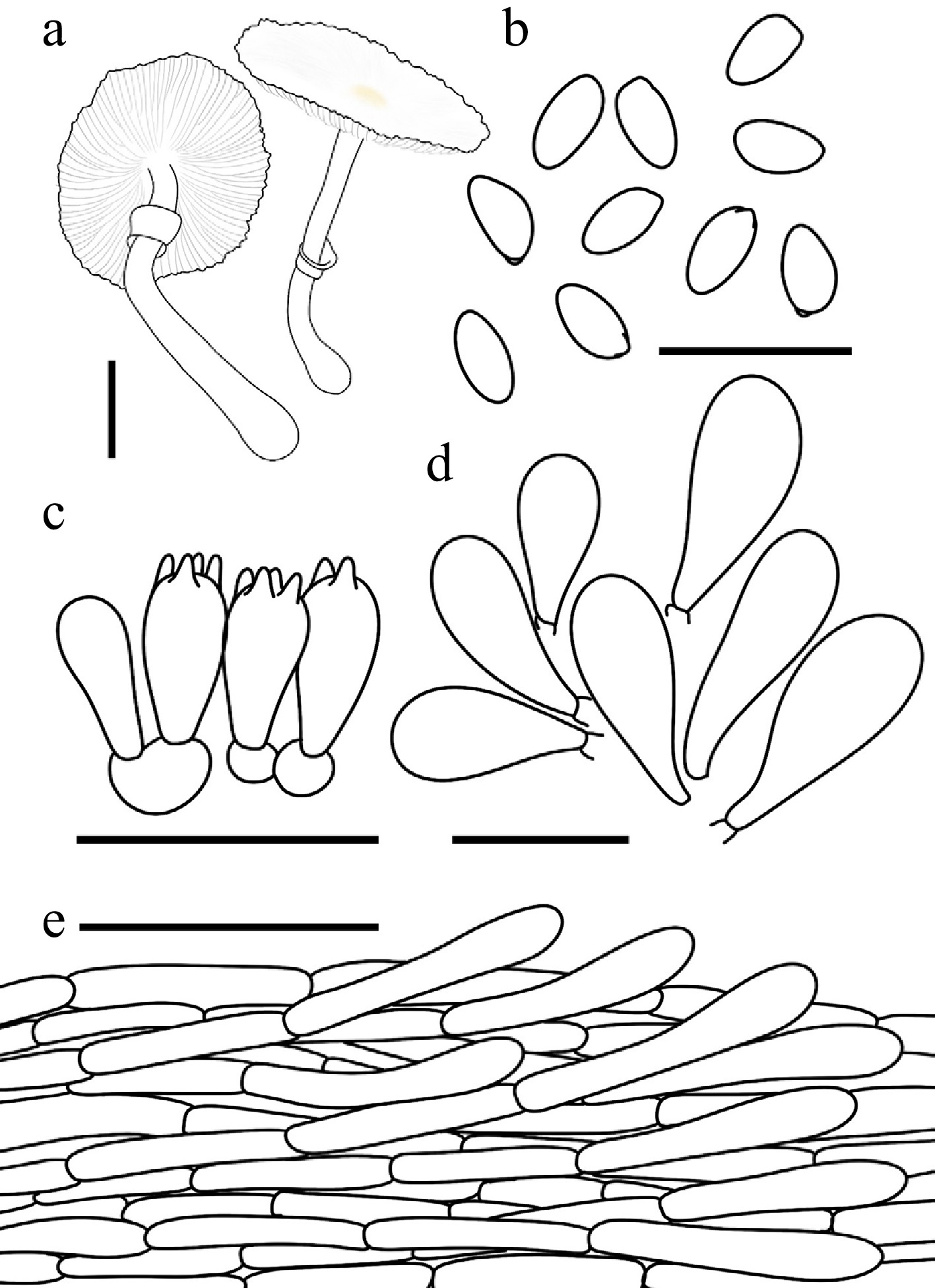
Figure 22.
Leucoagaricus microsporus. (a) Basidiomata. (b) Basidiospores. (c) Basidia. (d) Cheilocystidia. (e) Pileus covering. Scale bars: (a) = 1 cm; (b) = 10 μm; (c), (d) = 20 μm; (e) = 50 μm.
Chinese name: 小孢白环蘑 (Pīn yīn: xiǎo bāo bái huán mó)
Index Fungorum number: IF904638; Facesoffungi number: FoF 18926
Etymology – 'microsporus' is derived from the Greek mikros- (small) and sporos (spore), referring to the characteristically small-sized basidiospores.
Type – China. Jilin: Ji'an City, Yushan Park, 2 August 2023, coll. Q.R. Liu, FJAU77889.
Diagnosis – characterized by its white basidiomata with concolorous, silky-fibrillose squamules, minute basidiospores, and clavate to broadly clavate cheilocystidia
Basidiomata small. Pileus 0.8–2.5 cm in diam., initially ovate, expanding to plano-convex or applanate; umbo low, obtuse; background white; squamules concolorous, silky-fibrillose, readily detachable. Lamellae free, moderately crowded, white; edge concolorous; lamellulae interspersed in 2 tiers. Stipe 2.5–5.1 cm × 0.1–0.2 cm, subcylindrical, hollow, slightly thickened downwards with a slightly bulbous base, white; surface smooth or covered with silky fibrils. Annulus median to subapical, ascending, white, membranous. Context thin, white, unchanging when bruised. Odour and taste not recorded.
Basidiospores (40/2/2) 4.3–5.5 × 2.6–3.2 μm, avl × avw = 4.7–4.8 × 2.7–2.8 μm, Q = 1.49–2.08, avQ = 1.70–1.71, ellipsoid to oblong, hyaline, slightly thick-walled, smooth, dextrinoid, cyanophilous, congophilous, metachromatic in Cresyl Blue. Basidia 9–15 × 3–6 μm, clavate, predominantly 4-spored, occasionally 2-spored, thin-walled. Pleurocystidia absent. Cheilocystidia 16–37 × 8–18 μm, clavate to broadly clavate, hyaline, thin-walled. Surface covering at pileus centre a cutis consisting of cylindrical hyphae; hyphae repent, 4–10 μm wide, slightly thick-walled, hyaline. Stipe covering a cutis consisting of cylindrical hyphae; hyphae repent, 4–9 μm wide, thin-walled, hyaline. Clamp connections not observed.
Habit and habitat – solitary to scattered on humus-rich soil in mixed forests dominated by Acer and Ulmus.
Known distribution – so far recorded only from Northeast China.
Additional specimens examined – China. Jilin: Ji'an City, Yushan Park, 2 August 2023, coll. Q.R. Liu, FJAU77885; 24 July 2025, coll. X.Y. Zhou & H. Cheng, FJAU77887, FJAU77888, FJAU77886.
Notes – Morphologically, Leucoagaricus microsporus is a member of the La. serenus complex. However, it can be readily distinguished from most other members of this group by its relatively smaller basidiospores.
Phylogenetically, La. microsporus is nested within La. clade V, clustering together with La. subalbosquamosus R.L. Zhao & J.X. Li, La. albofragilis R.L. Zhao & J.X. Li, and La. angelicus Kun L. Yang & Jia Y. Lin. Morphologically, however, these closely related species are clearly separated. Specifically, La. subalbosquamosus differs in the dimensions of its cheilocystidia, which measure 13.3–26.8 × 7.0–14.2 µm[29], separating it from La. microsporus whose cheilocystidia measure 16.0–37.0 × 8.0–18.0 µm. Furthermore, La. albofragilis possesses slightly longer basidiospores measuring 5.1–6.3 × 2.7–3.6 µm, compared to those of the new species which measure only 4.3–5.5 × 2.6–3.2 µm[29]. Similarly, La. angelicus can be distinguished by producing distinctly larger basidiospores measuring 5.5–7.0 × 3.0–4.0 µm, as well as by lacking distinct cheilocystidia or possessing elements that are nearly indistinguishable from basidioles[30].
Leucoagaricus similis T. Bau & X.Y. Zhou, sp. nov Figs 16c–e, 23
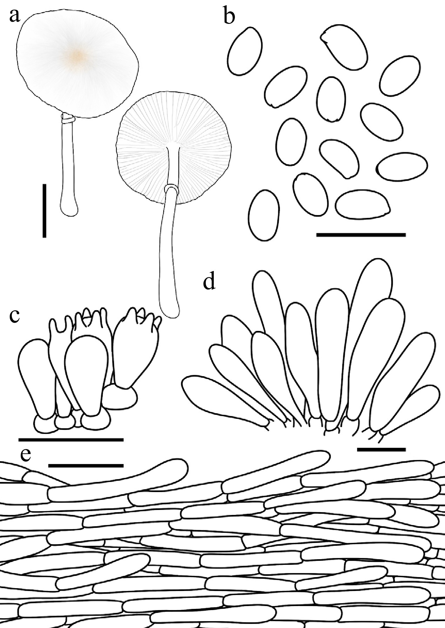
Figure 23.
Leucoagaricus similis. (a) Basidiomata. (b) Basidiospores. (c) Basidia. (d) Cheilocystidia. (e) Pileus covering. Scale bars: (a) = 1 cm; (b) = 10 μm; (c), (d) = 20 μm; (e) = 50 μm.
Chinese name: 相似白环蘑 (Pīn yīn: xiāng sì bái huán mó)
Index Fungorum number: IF904643; Facesoffungi number: FoF 18935
Etymology – 'similis' is derived from the Latin similis (similar), referring to the great morphological similarity of this species to other taxa within the morphologically defined Leucoagaricus serenus complex.
Type – China. Inner Mongolia: Tongliao City, Daqinggou National Nature Reserve, 31 July 2025, coll. X.Y. Zhou, FJAU77905.
Diagnosis – characterized by its white basidiomata covered with concolorous, silky-fibrillose squamules, non-dextrinoid basidiospores, and fusiform to clavate cheilocystidia
Basidiomata small. Pileus 1.1–2.3 cm in diam., plano-convex to applanate at maturity; umbo slightly obtuse; background white; squamules concolorous, silky-fibrillose, readily detachable. Lamellae free, moderately crowded, white; edge concolorous; lamellulae in 2 tiers. Stipe 1.9–5.5 cm × 0.1–0.2 cm, subcylindrical, widening downwards to a slightly bulbous base, hollow, white; surface smooth or covered with silky fibrils. Annulus superior, ascending, white, membranous. Context thin, white, unchanging when bruised. Odour and taste not recorded.
Basidiospores (40/2/2) 4.9–6.2 × 2.9–3.7 μm, avl × avw = 5.3–5.4 × 3.3–3.4 μm, Q = 1.43–1.98, avQ = 1.58–1.65, ellipsoid to oblong, hyaline, thin-walled, smooth, non-dextrinoid, cyanophilous, congophilous, metachromatic in Cresyl Blue. Basidia 12–20 × 6–9 μm, clavate, predominantly 4-spored, occasionally 2-spored, thin-walled. Pleurocystidia absent. Cheilocystidia 26–50 × 5–17 μm, oblong, fusiform to clavate, hyaline, thin-walled. Surface covering at pileus centre a cutis consisting of cylindrical hyphae; hyphae repent, 6–16 μm wide, slightly thick-walled, hyaline. Stipe covering a cutis consisting of cylindrical hyphae; hyphae repent, 4–12 μm wide, thin-walled, hyaline. Clamp connections not observed.
Habit and habitat – solitary on humus-rich soil in broadleaf forests dominated by Populus.
Known distribution – so far recorded only from Northeast China.
Additional specimens examined – China. Jilin: Changchun City, Jilin Agricultural University, 23 September 2021, coll. L.Y. Zhu, FJAU77904; 29 August 2025, coll. X.Y. Zhou, FJAU78099, FJAU78101, FJAU78126; Changchun City, Jingyuetan National Forest Park, 23 August 2025, coll. X. Wang, FJAU77906.
Notes – Phylogenetically, Leucoagaricus similis is nested within La. clade V, where La. aff. similis and La. submicrosporus cluster together to form a tightly related sister lineage. Morphologically, however, La. submicrosporus differs from the new species by possessing smaller basidiospores measuring 4.2–5.6 × 2.8–3.3 µm, compared to those of La. similis, which measure 4.9–6.2 × 2.9–3.7 µm. Additionally, the cheilocystidia of La. submicrosporus are predominantly clavate, oblong, or ellipsoid, which differ distinctly from the oblong, fusiform to clavate cheilocystidia observed in La. similis. Furthermore, La. aff. similis represents an independent evolutionary lineage uncovered in the present study. Although this lineage exhibits a significant genetic distance from La. similis in the phylogeny, no clear morphological diagnostic features have yet been identified to unambiguously separate the two taxa. Therefore, for the sake of taxonomic rigor, we provisionally designate this lineage as La. aff. similis.
Leucoagaricus subleucotrichus T. Bau & X.Y. Zhou, sp. nov Figs 15a–c, 24
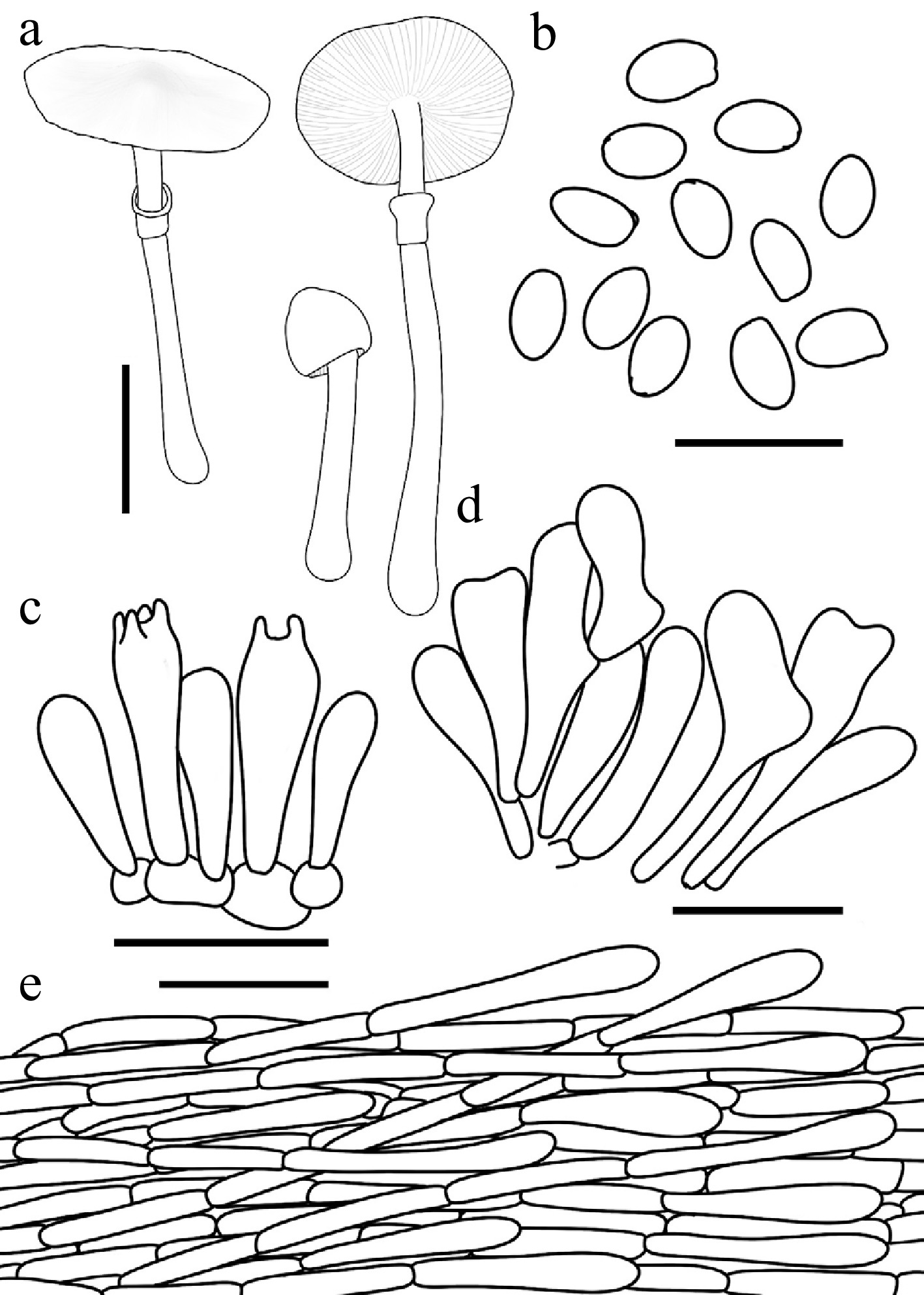
Figure 24.
Leucoagaricus subleucotrichus. (a) Basidiomata. (b) Basidiospores. (c) Basidia. (d) Cheilocystidia. (e) Pileus covering. Scale bars: (a) = 1 cm; (b) = 10 μm; (c), (d) = 20 μm; (e) = 50 μm.
Chinese name: 近白毛白环蘑 (Pīn yīn: jìn bái máo bái huán mó)
Index Fungorum number: IF904637; Facesoffungi number: FoF18929
Etymology – 'subleucotrichus' is derived from the Latin prefix sub- (implying resemblance) and the specific epithet leucotrichus, referring to its close morphological similarity to Leucoagaricus leucotrichus.
Type – China. Jilin: Ji'an City, Yushan Park, 24 July 2025, coll. X.Y. Zhou, FJAU77809.
Diagnosis – characterized by its white basidiomata covered with white to light yellow silky-fibrillose squamules, and non-dextrinoid basidiospores
Basidiomata small. Pileus 0.8–1.5 cm in diam., initially campanulate, expanding to plano-convex or applanate; umbo obtuse; background white; centre with light yellow (#FFFEE4) felted squamules; squamules around the centre light yellow to white, silky-fibrillose, readily detachable. Lamellae free, moderately crowded, white; edge concolorous; lamellulae interspersed in 1–3 tiers. Stipe 2.7–3.6 cm × 0.2–0.3 cm, subcylindrical, hollow, slightly thickened downwards with a slightly bulbous base, white surface smooth or covered with silky fibrils. Annulus medium to superior, ascending, white, membranous. Context thin, white, unchanging when bruised. Odour and taste not recorded.
Basidiospores (60/3/3)(4.0–) 4.3–5.5 × 2.6–3.6 μm, avl × avw = 4.6–5.0 × 2.9–3.2 μm, Q = 1.34–1.73, avQ = 1.52–1.57, ellipsoid to oblong, hyaline, thin-walled, smooth, non-dextrinoid, cyanophilous, congophilous, metachromatic in Cresyl Blue. Basidia 13–22 × 5–10 μm, clavate, predominantly 4-spored, occasionally 2-spored, thin-walled. Pleurocystidia absent. Cheilocystidia 20–38 × 4–11 μm, clavate to broadly fusiform, occasionally with branched apices, hyaline, thin-walled. Surface covering at pileus centre a cutis consisting of cylindrical hyphae; hyphae repent, 5–14 μm wide, thin-walled, hyaline. Stipe covering a cutis consisting of cylindrical hyphae; hyphae repent, 4–12 μm wide, thin-walled, hyaline. Clamp connections not observed.
Habit and habitat – solitary on humus-rich soil in broadleaf dominant forests.
Known distribution – so far recorded only from Northeast China.
Additional specimens examined – China. Jilin: Ji'an City, Yushan Park, 24 July 2025, coll. X.Y. Zhou & Y. Wang, FJAU77808, FJAU77810, FJAU77811.
Notes – Phylogenetically, Leucoagaricus subleucotrichus and La. candidus are positioned at the base of La. clade II. Morphologically, however, La. candidus differs by possessing distinctly larger basidiospores measuring 6.0–7.5 × 4.5–5.0 µm, compared to those of La. subleucotrichus which measure only 4.3–5.5 × 2.6–3.6 µm.
Within La. clade II, there are also several members belonging to the La. serenus complex, including La. albocapillatus, La. serenus, La. aff. serenus 1, and La. aff. serenus 2. Similarly, both La. albocapillatus R.L. Zhao & J.X. Li and La. serenus can be distinguished from La. subleucotrichus by their larger basidiospores, which measure 5.9–7.6 × 3.4–4.6 µm[29,78] and 6.7–9.1 × 3.6–4.3 µm, respectively.
Furthermore, La. aff. serenus 1 and La. aff. serenus 2 represent two independent evolutionary lineages uncovered in the present study. Although they exhibit significant genetic distances from the authentic La. serenus, no reliable morphological diagnostic features have been found to unambiguously separate them. Therefore, for the sake of taxonomic rigor, we provisionally designate these two lineages as La. aff. serenus 1 and La. aff. serenus 2 (Figs 25, 26).
Leucoagaricus submicrosporus T. Bau & X.Y. Zhou, sp. nov Figs 15g, h, 27
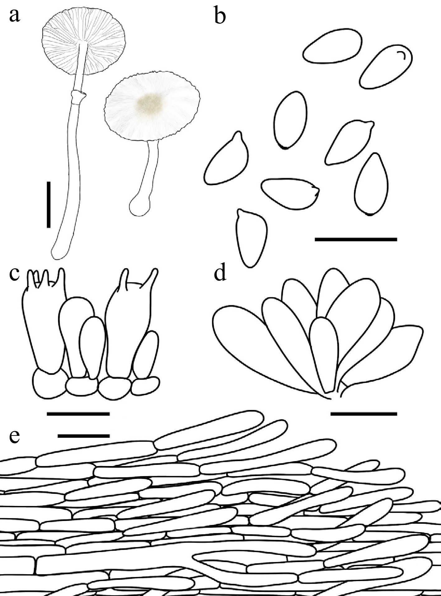
Figure 25.
Leucoagaricus aff. serenus 1. (a) Basidiomata. (b) Basidiospores. (c) Basidia. (d) Cheilocystidia. (e) Pileus covering. Scale bars: (a) = 1 cm; (b) = 10 μm; (c), (d) = 20 μm; (e) = 50 μm.
Chinese name: 近小孢白环蘑 (Pīn yīn: jìn xiǎo bāo bái huán mó)
Index Fungorum number: IF904639; Facesoffungi number: FoF18930
Etymology – 'submicrosporus' is derived from the prefix 'sub-' (meaning near, approaching) and 'microspores', referring to its close morphological similarity to the related species Leucoagaricus microsporus, particularly in the shared character of small basidiospores.
Type – China. Jilin: Ji'an City, Wunufeng National Forest Park, 25 July 2025, coll. X.Y. Zhou, FJAU77919.
Diagnosis – characterized by its white basidiomata covered with concolorous, silky- to fibrillose squamules, exceptionally small basidiospores, and clavate to ellipsoid cheilocystidia.
Basidiomata small. Pileus 0.7–1.5 cm in diam., initially hemispherical, expanding to plano-convex or applanate; umbo slightly obtuse; background white; squamules concolorous, silky-fibrillose, readily detachable. Lamellae free, moderately crowded, white; edge concolorous; lamellulae interspersed in 2–3 tiers. Stipe 2.6–3.9 cm × 0.1–0.2 cm, subcylindrical, hollow, widening downwards to a slightly bulbous base, white; surface covered with concolorous squamules. Annulus median to superior, ascending, white, membranous. Context thin, white, unchanging when bruised. Odour and taste not recorded.
Basidiospores (40/2/2) 4.2–5.6 × 2.8–3.3 μm, avl × avw = 4.6–4.7 × 3.0 μm, Q = 1.41–1.77, avQ = 1.53–1.59, ellipsoid to oblong, hyaline, thin-walled, smooth, non-dextrinoid to weakly dextrinoid, cyanophilous, congophilous, metachromatic in Cresyl Blue. Basidia 10–18 × 5–13 μm, clavate, predominantly 4-spored, occasionally 2-spored, thin-walled. Pleurocystidia absent. Cheilocystidia 19–44 × 8–17 μm, clavate to oblong or ellipsoid, hyaline, thin-walled. Surface covering at pileus centre a cutis consisting of cylindrical hyphae; hyphae repent, 6–16 μm wide, slightly thick-walled, hyaline. Stipe covering a cutis consisting of cylindrical hyphae; hyphae repent, 4–14 μm wide, thin-walled, hyaline. Clamp connections not observed.
Habit and habitat – solitary to scattered in sandy soil of mixed coniferous and broadleaf forests.
Known distribution – so far recorded only from Northeast China.
Additional specimens examined – China. Jilin: Ji'an City, Wunufeng National Forest Park, 25 July 2025, coll. W. Sun, FJAU77918; Inner Mongolia: Tongliao City, Daqinggou National Nature Reserve, 31 July 2025, coll. X.Y. Zhou & L.H. Li, FJAU77921, FJAU77920, FJAU77917.
Notes – Morphologically, Leucoagaricus submicrosporus is a member of the La. serenus complex. However, it can be readily distinguished from most other members of this group by its relatively small basidiospores.
Phylogenetically, La. submicrosporus clusters together with La.similis and La. aff. similis (Figs 2, 28).
Morphologically, however, these closely related taxa exhibit clear differences. Specifically, La. submicrosporus differs from La. similis by possessing smaller basidiospores measuring 4.2–5.6 × 2.8–3.3 µm, compared to the slightly larger spores of the latter, which measure 4.9–6.2 × 2.9–3.7 µm. Furthermore, the cheilocystidia of La. submicrosporus are predominantly clavate, oblong, or ellipsoid, which differ from the oblong, fusiform, or clavate cheilocystidia observed in La. similis.
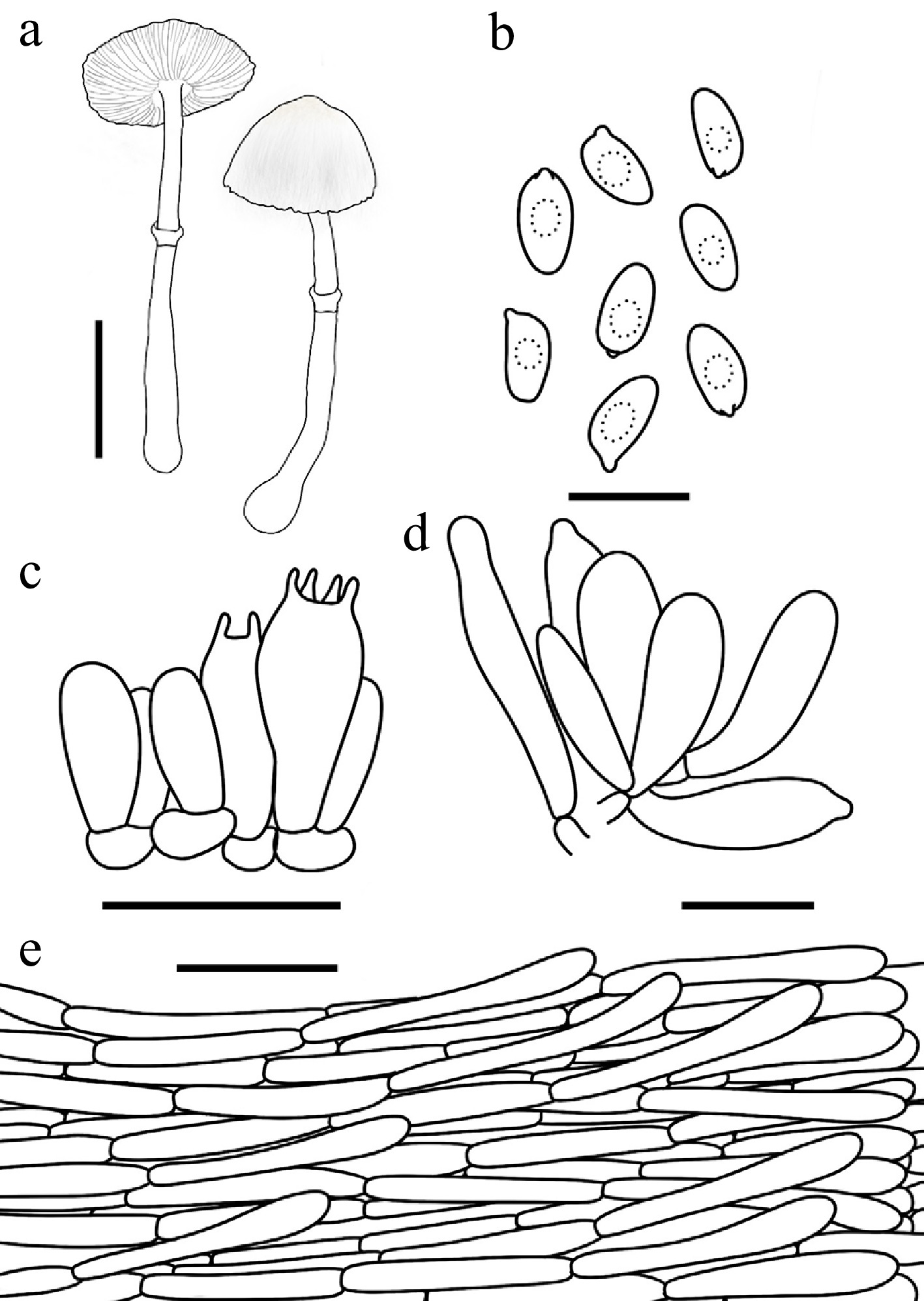
Figure 26.
Leucoagaricus aff. serenus 2. (a) Basidiomata. (b) Basidiospores. (c) Basidia. (d) Cheilocystidia. (e) Pileus covering. Scale bars: (a) = 1 cm; (b) = 10 μm; (c), (d) = 20 μm; (e) = 50 μm.
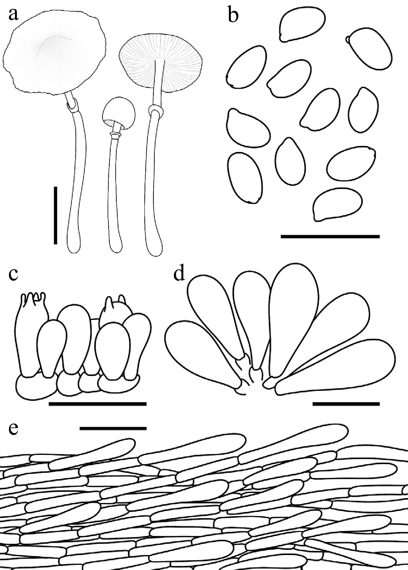
Figure 27.
Leucoagaricus submicrosporus. (a) Basidiomata. (b) Basidiospores. (c) Basidia. (d) Cheilocystidia. (e) Pileus covering. Scale bars: (a) = 1 cm; (b) = 10 μm; (c), (d) = 20 μm; (e) = 50 μm.
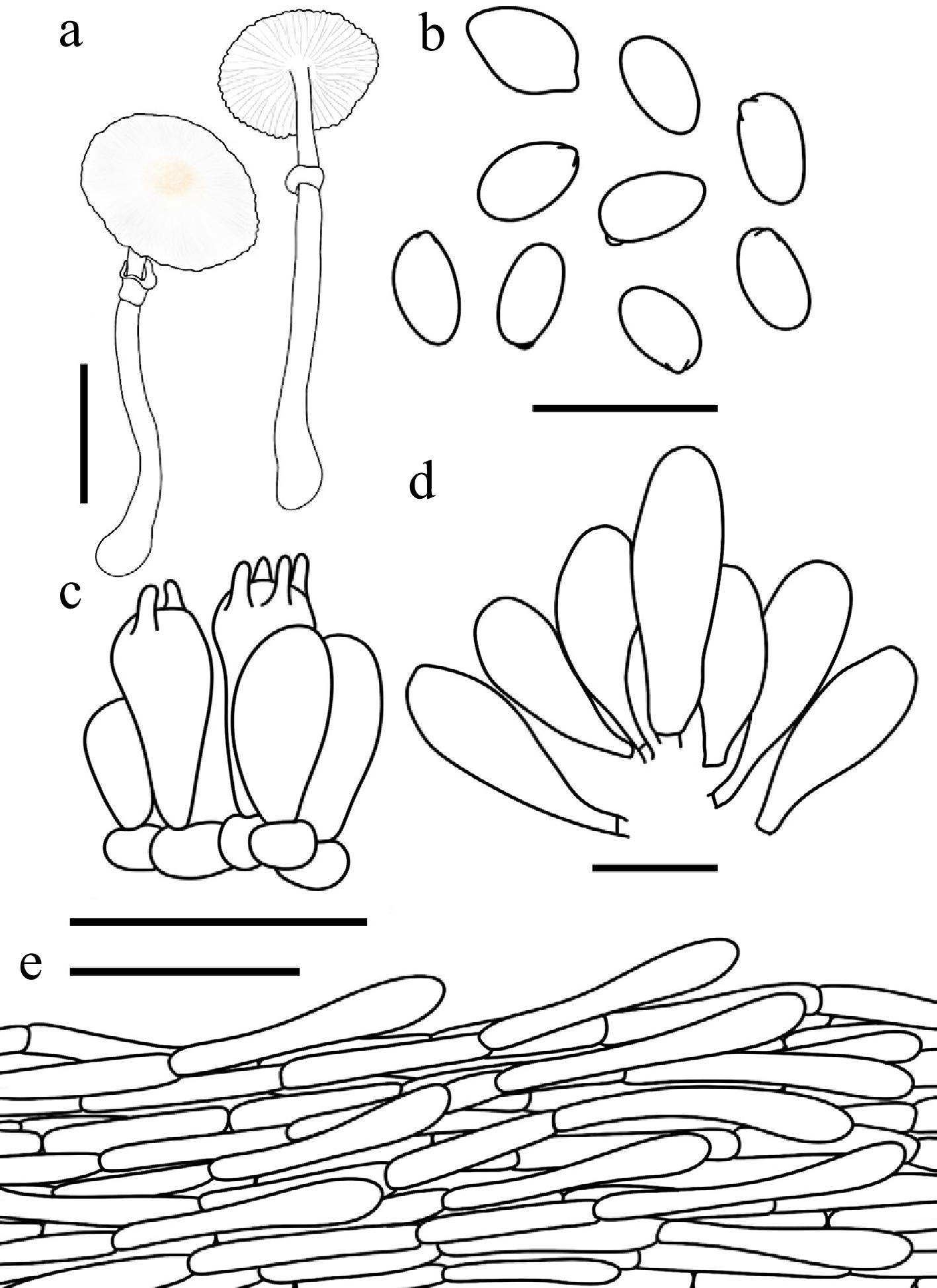
Figure 28.
Leucoagaricus aff. similis. (a) Basidiomata. (b) Basidiospores. (c) Basidia. (d) Cheilocystidia. (e) Pileus covering. Scale bars: (a) = 1 cm; (b) = 10 μm; (c), (d) = 20 μm; (e) = 50 μm.
Other Leucoagaricus species Fig. 29
Leucoagaricus centriluteus T. Bau & X.Y. Zhou, sp. nov Figs 29c–e, 30
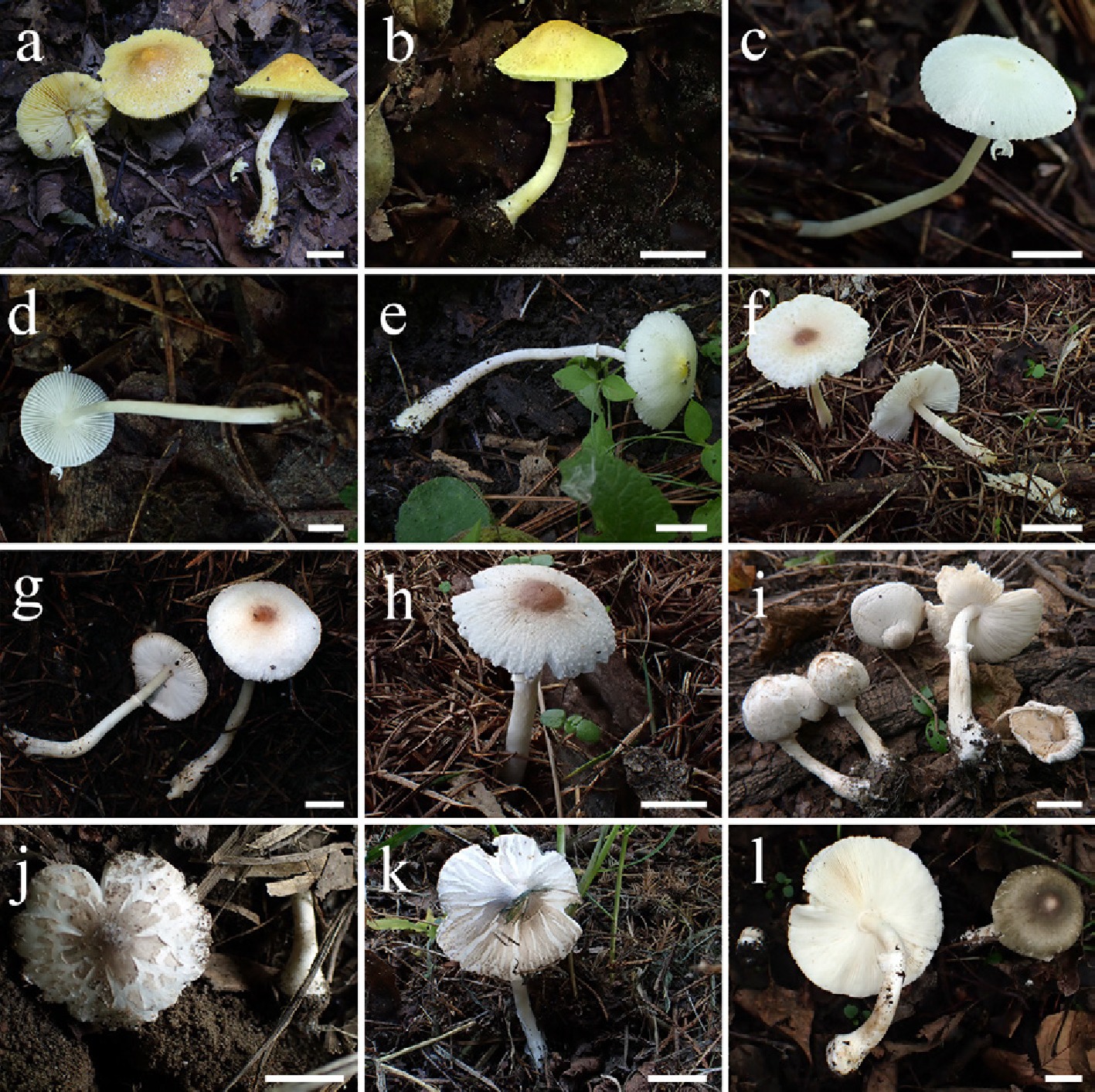
Figure 29.
Basidiomata of Leucoagaricus species. La. luteus (a) FJAU77804, (b) FJAU77803. La. centriluteus (c), (d) FJAU77798, (e) FJAU77797. La. conifericola (f) FJAU77933, (g) FJAU77932, (h) FJAU77935). La. subcrystallifer (i) FJAU77834, (j) FJAU77831). La. atroviridis (k) FJAU77840, (l) FJAU77842. Scale bars = 1 cm. These pictures are copyright of the collectors of specimens.
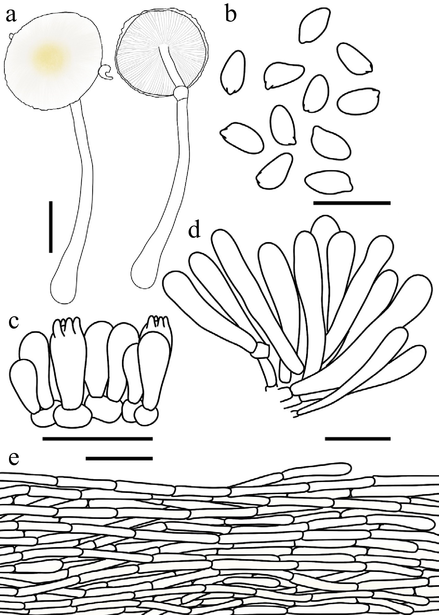
Figure 30.
Leucoagaricus centriluteus. (a) Basidiomata. (b) Basidiospores. (c) Basidia. (d) Cheilocystidia. (e) Pileus covering. Scale bars: (a) = 1 cm; (b) = 10 μm; (c), (d) = 20 μm; (e) = 50 μm.
Chinese name: 黄顶白环蘑 (Pīn yīn: huáng ding bái huán mó)
Index Fungorum number: IF905533; Facesoffungi number: FoF 19997
Etymology – 'centriluteus' is a compound adjective derived from the Latin centrum (center) and luteus (yellow), referring to the distinctive flax-to-pale-yellow coloration at the center of the pileus.
Type – China. Jilin: Dunhua City, Laobaishan Primitive Ecological Area, 22 July 2025, coll. H. Cheng, FJAU77798.
Diagnosis – Phylogenetically closely allied to Leucoagaricus serenus (Fr.) Bon & Boiffard and La. albocapillatus R.L. Zhao & J.X. Li, but can be readily distinguished macroscopically by its brighter, pale-yellow to flax-yellow pileus center (rather than yellowish-white to creamish), and microscopically by its distinctly smaller basidiospores (4.5–5.8 × 2.7–3.4 μm).
Basidiomata small. Pileus 1.2–2.7 cm in diam., plano-convex to applanate when mature, with a slightly obtuse umbo; background white; centre flax yellow (#EEDC82); squamules around the centre flax yellow to light yellow (#FFFEE4), silky-fibrillose, readily detachable. Lamellae free, moderately crowded, ventricose, white; edge concolorous; lamellulae in 2 tiers. Stipe 2.9–7.2 cm × 0.1–0.3 cm, subcylindrical, hollow, slightly thickened towards the slightly bulbous base, white to cream; surface covered with concolorous, fibrillose squamules. Annulus medium to superior, ascending, white, membranous, ephemeral. Context thin, white, unchanging when bruised. Odour and taste not recorded.
Basidiospores (40/2/2) 4.5–5.8 × 2.7–3.4 μm, avl × avw = 5.1–5.2 × 3.0–3.1 μm, Q = 1.54–2.0, avQ = 1.65–1.77, amygdaliform to oblong, hyaline, slightly thick-walled, smooth, dextrinoid, cyanophilous, congophilous, metachromatic in Cresyl Blue. Basidia 12–20 × 5–8 μm, clavate, predominantly 4-spored, occasionally 2-spored. Pleurocystidia absent. Cheilocystidia 27–45 × 6–11 μm, narrowly cylindrical to narrowly clavate, hyaline, thin-walled. Surface covering at pileus centre a cutis consisting of cylindrical hyphae; hyphae repent, 6–11 μm wide, slightly thick-walled, with light yellow (#FFFEE4) intracellular pigment. Stipe covering a cutis consisting of cylindrical hyphae; hyphae repent, 4–10 μm wide, thin-walled, hyaline. Clamp connections not observed.
Habit and habitat – solitarily in humus under broadleaf trees.
Known distribution – so far recorded only from China.
Additional specimens examined – China. Jilin: Jiaohe City, Qianjin Forest Farm, 25 August 2023, coll. H. Cheng, FJAU77796; Dunhua City, Laobaishan Primitive Ecological Area, 22 July 2025, coll. X.Y. Zhou, FJAU77797.
Notes – Although Leucoagaricus centriluteus and La. orientiflavus Z.W. Ge both possess yellowish tints at the pileus center, the latter is readily distinguished by the presence of yellowish hues on the stipe, annulus, and context, as well as its larger basidiospores ([6.0–]6.5–7.5 × [3.0–]3.5–4.0 µm)[81]. Phylogenetically, La. centriluteus is nested within La. clade II, showing a close affinity to La. serenus and La. albocapillatus. Morphologically, however, La. centriluteus is characterized by a brighter pale yellow to flaxen pileus center, whereas the latter two exhibit yellowish-white to creamish tones. Furthermore, the basidiospores of La. centriluteus (4.5–5.8 × 2.7–3.4 µm) are conspicuously smaller than those of La. serenus (6.7–9.1 × 3.6–4.3 µm) and La. albocapillatus (5.3–7.7 × 3.4–4.6 µm)[29,78].
Notably, our phylogenetic analysis shows that a sequence labeled as "La. sulphurellus (Pegler) B.P. Akers" (voucher PC0714366, GenBank MN994687) clusters closely with La. centriluteus; however, this specimen lacks a formal morphological description[82]. According to the literature, the authentic La. sulphurellus possesses basidiospores with a germ pore and context that stains blue to green when bruised[83]. These features are exceptionally rare within Leucoagaricus and are more typically associated with Lc. clade II or the Lc. atroazureus clade. Given that these diagnostic characters of La. sulphurellus are entirely incongruent with the defining features of La. clade II, we conclude that sequence MN994687 is likely a misidentification and does not represent the true La. sulphurellus.
Leucoagaricus conifericola T. Bau & X.Y. Zhou, sp. nov Figs 29f–h, 31
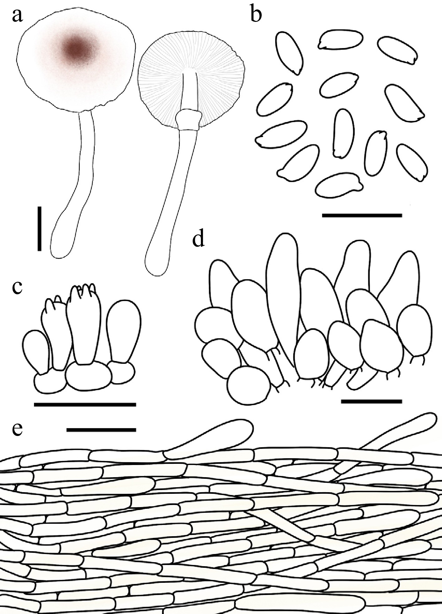
Figure 31.
Leucoagaricus conifericola. (a) Basidiomata. (b) Basidiospores. (c) Basidia. (d) Cheilocystidia. (e) Pileus covering. Scale bars: (a) = 1 cm; (b) = 10 μm; (c), (d) = 20 μm; (e) = 50 μm.
Chinese name: 针生白环蘑 (Pīn yīn: zhēn sheng bái huán mó)
Index Fungorum number: IF904630; Facesoffungi number: FoF18923
Etymology – 'conifericola' is formed by combining the Latin conifer (cone-bearing tree) and the suffix -cola (dweller), referring to its typical habitat in coniferous forests.
Type – China. Jilin: Changchun City, Jingyuetan National Forest Park, 8 August 2025, coll. L.H. Li, FJAU77933.
Diagnosis – Leucoagaricus conifericola is distinguished by the copper-red to red-brown squamules at the pileus center, elongate basidiospores (5.0–6.4 × 2.2–2.9 µm), and oblong to ellipsoid cheilocystidia.
Basidiomata small. Pileus 2.3–2.9 cm in diam., plano-convex to applanate when mature, with a low, obtuse umbo; background white; centre with copper-red (#CB6D51) to red-brown (#66332B) felted squamules; squamules around the centre white to light yellow (#FFFEE4), silky-fibrillose, loosely attached to the surface. Lamellae free, moderately crowded, white; edge concolorous; lamellulae 0–3 between each pair of lamellae. Stipe 2.5–5.7 cm × 0.2–0.4 cm, subcylindrical, slightly thickened downwards, white; surface covered with concolorous, silky-fibrillose squamules. Annulus medium to superior, ascending, white, membranous. Context thin, white, unchanging when bruised. Odour and taste not recorded.
Basidiospores (60/3/3) 5.0–6.4 × 2.2–2.9 μm, avl × avw = 5.3–5.5 × 2.5–2.7 μm, Q = 1.85–2.67, avQ = 2.02–2.16, oblong to cydlindrical, hyaline, slightly thick-walled, smooth, dextrinoid, cyanophilous, congophilous, metachromatic in Cresyl Blue. Basidia 10–16 × 5–8 μm, clavate, predominantly 4-spored, occasionally 2-spored. Pleurocystidia absent. Cheilocystidia 22–46 × 10–18 μm, oblong, ellipsoid to broadly ellipsoid, hyaline, thin-walled. Surface covering at pileus centre a cutis consisting of cylindrical hyphae; hyphae repent, 6–12 μm wide, slightly thick-walled, with copper red intracellular pigment. Stipe covering a cutis consisting of cylindrical hyphae; hyphae repent, 5–11 μm wide, thin-walled, hyaline. Clamp connections not observed.
Habit and habitat – solitary to scattered on humus in coniferous forests.
Known distribution – so far recorded only from Northeast China.
Additional specimens examined – China. Jilin: Changchun City, Jingyuetan National Forest Park, 8 August 2025, coll. L.H. Li, W. Sun, Y. Wang, & T.Y. Zhang, FJAU77934, FJAU77932, FJAU77935, FJAU77931.
Notes – Macroscopically, Leucoagaricus conifericola is quite similar to species within the Leucoagaricus serenus complex. However, taxa in that complex generally produce entirely white basidiomata. Phylogenetically, La. conifericola is closely related to La. albofibrillosus and La. subcandidus R.L. Zhao & J.X. Li. Nevertheless, La. subcandidus possesses smaller basidiospores (4.2–4.9 × 2.9–3.4 μm) and smaller cheilocystidia (17.8–27.1 × 5.6–7.4 μm) that are predominantly narrowly clavate[34]. Moreover, the pileus disc of La. albofibrillosus lacks the distinct copper-red to reddish-brown obtuse umbo observed in La. conifericola. Furthermore, La. albofibrillosus is currently only known from East China, whereas La. conifericola is restricted to Northeast China.
Leucoagaricus luteus T. Bau & X.Y. Zhou, sp. nov. Figs 29a, b, 32
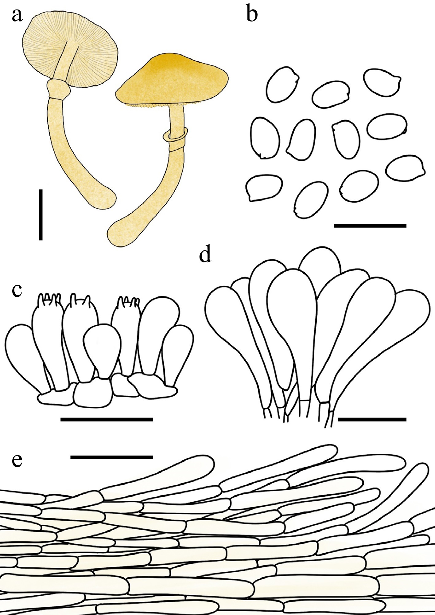
Figure 32.
Leucoagaricus luteus. (a) Basidiomata. (b) Basidiospores. (c) Basidia. (d) Cheilocystidia. (e) Pileus covering. Scale bars: (a) = 1 cm; (b) = 10 μm; (c), (d) = 20 μm; (e) = 50 μm.
Chinese name: 黄白环蘑 (Pīn yīn: huáng bái huán mó)
Index Fungorum number: IF904631; Facesoffungi number: FoF18924
Etymology – 'luteus' (Lat., yellow) refers to the yellow colouration of the basidiomata.
Type – China. Inner Mongolia: Tongliao City, Daqinggou National Nature Reserve, 31 July 2025, coll. X.Y. Zhou, FJAU77804.
Diagnosis – Leucoagaricus luteus is primarily characterized by its pale-yellow to flax-yellow basidioma and its very small, weakly dextrinoid basidiospores.
Basidiomata small. Pileus 1.9–3.3 cm in diam., obtusely conical to plano-convex or applanate when mature; background light yellow (#FFFEE4); centre with mustard-yellow (#E1AD01) felted squamules; squamules around the centre small, light yellow to flax yellow (#EEDC82), finely squamulose. Lamellae free, crowded, light yellow to flax yellow; edge concolorous; lamellulae in 2 tiers. Stipe 3.8–5.3 cm × 0.3–0.5 cm, subcylindrical, slightly thickened downwards, with a distinctly bulbous base, light yellow to flax yellow; surface covered with concolorous, silky-fibrillose squamules. Annulus medium to superior, ascending, light yellow, membranous, friable. Context thin, light yellow, unchanging when bruised. Odour and taste not recorded.
Basidiospores (40/2/2) 4.6–5.6 × 2.9–3.8 μm, avl × avw = 4.9 × 3.1–3.3 μm, Q = 1.33–1.85, avQ = 1.50–1.58, ellipsoid to oblong, hyaline, smooth, weakly dextrinoid, cyanophilous, congophilous, metachromatic in Cresyl Blue, thin-walled. Basidia 13–20 × 6–9 μm, clavate, predominantly 4-spored, occasionally 2-spored, thin-walled. Cheilocystidia 24–44 × 7–15 μm, clavate to broadly clavate, hyaline, thin-walled. Pleurocystidia absent. Surface covering at pileus centre a cutis consisting of cylindrical hyphae; hyphae repent, 4–12 μm wide, thin-walled, with light yellow intracellular pigment. Stipe covering a cutis consisting of cylindrical hyphae; hyphae repent, 4–11 μm wide, thin-walled. Clamp connections absent.
Habit and habitat – solitary to scattered in sandy soil of broadleaf forests.
Known distribution – currently known only from China.
Additional specimens examined – China. Inner Mongolia: Tongliao City, Daqinggou National Nature Reserve, 31 July 2025, coll. T. Bau & H. Cheng, FJAU77803. Jilin: Changchun City, Jilin Agricultural University, 24 June 2026, coll. T. Bau, FJAU79629.
Notes – Leucoagaricus luteus exhibits a distinctly yellow basidioma, a feature that readily separates it from the vast majority of species within the Leucoagaricus–Leucocoprinus s.l. Macroscopically, La. luteus resembles Lepiota xanthophylla P.D. Orton due to their shared yellow basidiomata. However, the latter is easily distinguished by its larger basidiospores (6.0–7.5 × 3.0–4.0 μm) and the presence of clamp connections on the hyphal septa[84]. Phylogenetically, La. luteus is nested within La. clade II, showing a close affinity to La. agnellus Kun L. Yang & Jia Y. Lin and La. rutilofibrillosus. Morphologically, however, these phylogenetically related species differ remarkably in overall coloration: La. agnellus produces white basidiomata[30], and La. rutilofibrillosus displays orange-red hues on the pileus, both contrasting sharply with the notably yellow basidiomata of La. luteus.
Notes on recently described Leucoagaricus species
-
In our phylogenetic analyses (Figs 1, 2; Supplementary File 3), we observed that Leucoagaricus bailangshanensis J.Z. Xu and La. albissimus R.L. Zhao & J.X. Li cluster together to form a single lineage. A pairwise comparison of their holotype sequences (HMJU 9098 and HMAS 282471, respectively) reveals an exceptionally high ITS identity of 99.77%. According to their original protologues, La. bailangshanensis produces basidiospores measuring 4.7–6.4 × 3.1–4.5 µm with an average of 5.6 × 3.9 µm, alongside cheilocystidia measuring 23.4–37.4 × 9.0–13.7 µm. Similarly, La. albissimus possesses basidiospores measuring 5.3–7.7 × 3.4–4.6 µm with an average of 6.5 × 4.0 µm, and cheilocystidia measuring 22.1–45.4 × 10.3–15.3 µm. Interestingly, our newly collected specimens belonging to this lineage exhibit intermediate micromorphological features, possessing basidiospores measuring 4.8–6.0 × 3.1–3.7 µm with an average of 5.3 × 3.4 µm, and cheilocystidia measuring 17.0–41.0 × 8.0–19.0 µm. These continuous overlapping variations perfectly bridge the minor morphological gaps between the two described taxa. Given that La. bailangshanensis was formally published on November 14, 2025[85], predating the valid publication of La. albissimus on December 23, 2025[29], the latter should be regarded as a later synonym of the former according to the principle of priority.
A parallel nomenclatural issue is observed between La. rutilus G.J. Li and La. bulbosus R.L. Zhao & J.X. Li. Their holotype sequences (HBAU 16141 and HMAS 287806, respectively) cluster tightly with an absolute ITS identity of 100%. Nomenclaturally, La. bulbosus holds priority as it was formally published on February 18, 2025[34], earlier than the publication of La. rutilus on May 29, 2025[86]. However, a significant discrepancy exists in their reported basidiospore dimensions. While La. rutilus was described as having smaller spores measuring 4.6–5.8 × 3.5–4.8 µm with an average of 5.3 × 4.2 µm, La. bulbosus was reported to possess distinctly larger spores measuring 7.8–8.4 × 4.5–5.5 µm. Despite the identical ITS sequences strongly indicating conspecificity, this apparent morphological divergence warrants further rigorous investigation to determine whether it stems from measurement bias in the original studies or the existence of morphologically similar taxa unresolved by the ITS marker.
Leucocoprinus Pat., J. Bot., Paris 2: 16 (1888) Figs 33−37
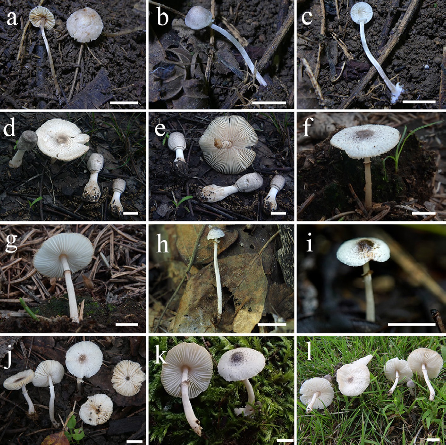
Figure 33.
Basidiomata of Leucocoprinus species. Lc. acystidiatus (a) FJAU78091, (b), (c) FJAU78090. (d), (e) Lc. crassibasis (FJAU78107). Lc. fuscannulatus (f), (g) FJAU78014, (h) FJAU78010, (i) FJAU78009. Lc. griseocentralis (j) FJAU78118, (k) FJAU78006, (l) FJAU78003. Scale bars = 1 cm. These pictures are copyright of the collectors of specimens.
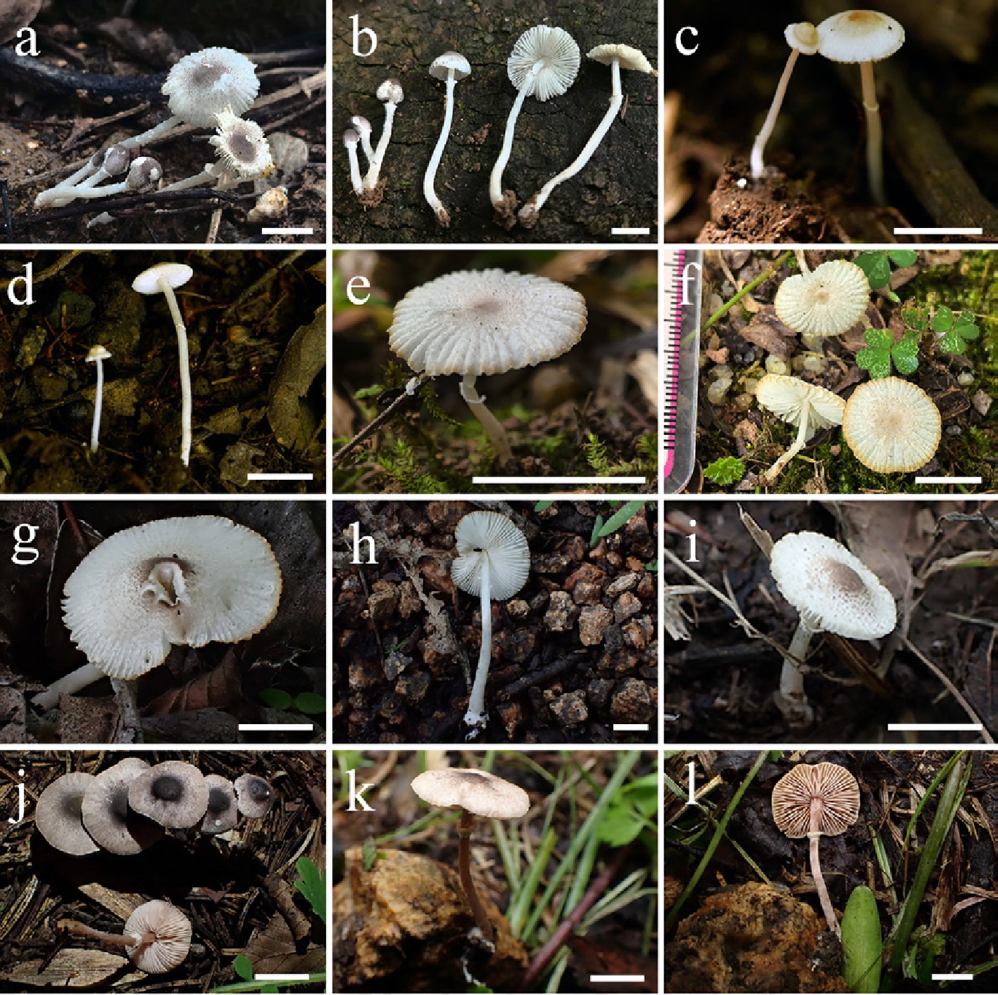
Figure 34.
Basidiomata of Leucocoprinus species. (a), (b) Lc. parvicanus (FJAU78110). (c), (d) Lc. porosporus (FJAU78028). Lc. sinumbellatus (e) FJAU77949, (f) FJAU78109). Lc. sphaerodermus (g), (h) FJAU78049, (i) FJAU78050. Lc. subincarnatus (j) FJAU78134, (k), (l) FJAU78095. Scale bars = 1 cm. These pictures are copyright of the collectors of specimens.
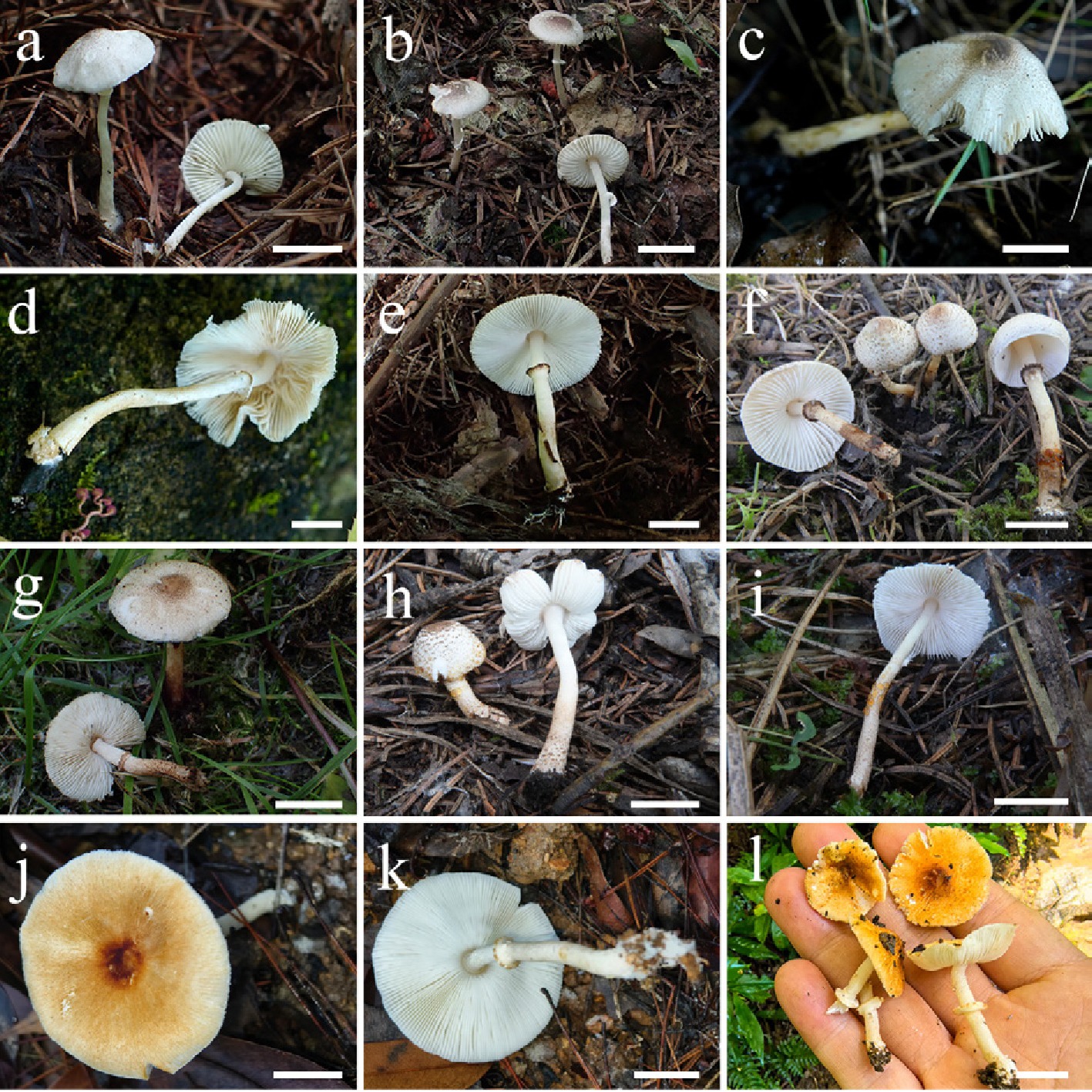
Figure 35.
Basidiomata of Leucocoprinus species. (a) Lc. subumbrosquamosus (FJAU78071). (b) Lc. umbrosquamosus (FJAU78072). Lc. guttifer (c), (d) FJAU78021, (e) FJAU78018. Lc. floccipes (f) FJAU78078, (g) FJAU78073, (h) FJAU78075, (i) FJAU78074). Lc. helianthoides (j), (k) FJAU78092, (l) FJAU78093. Scale bars = 1 cm. These pictures are copyright of the collectors of specimens.
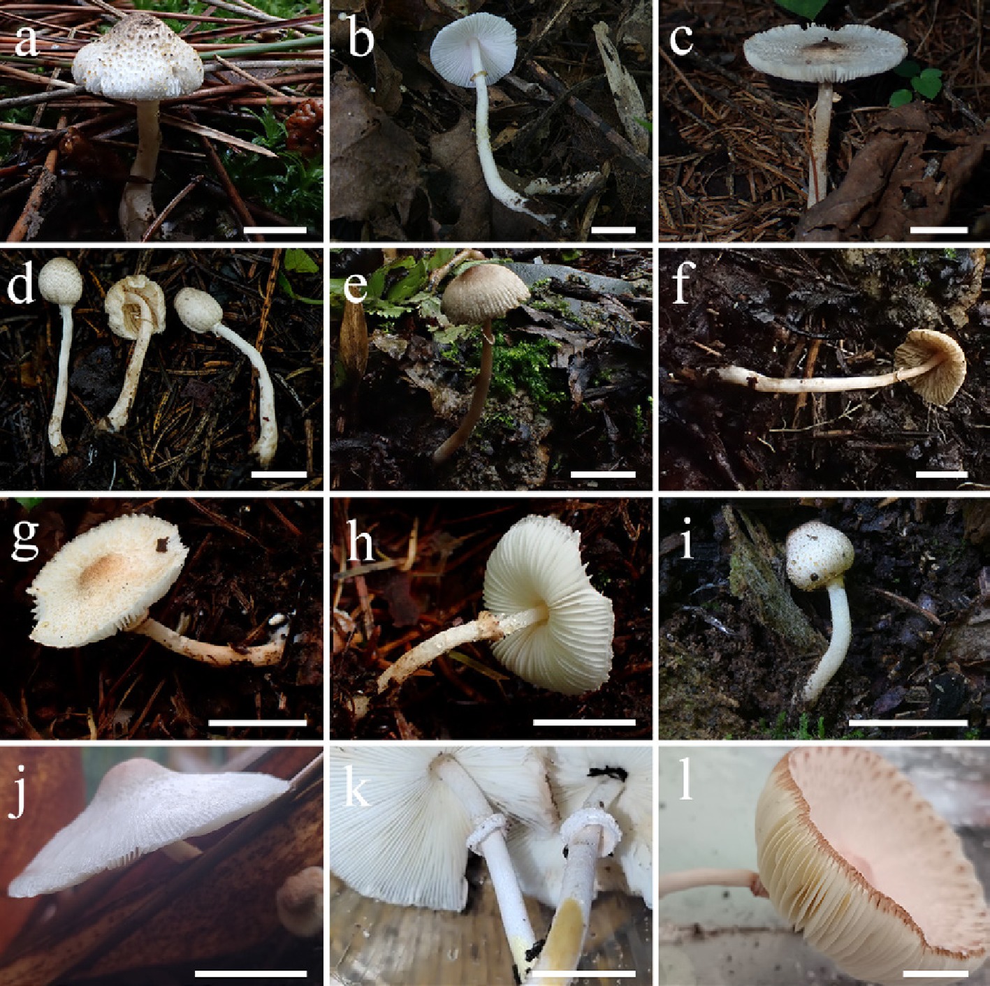
Figure 36.
Basidiomata of Leucocoprinus and Macropsalliota species. Lc. luteoguttatus (a) FJAU78022, (b) FJAU78023, (c) FJAU78026). Lc. ochraceosquamosus (d) FJAU78017, (e), (f) FJAU78016. Lc. sudans (g), (h) FJAU78274, (i) FJAU78106). (j), (k) Map. aff. subtropica (FJAU72225). (l) Map. subtropica. (FJAU77730). Scale bars = 1 cm. These pictures are copyright of the collectors of specimens.
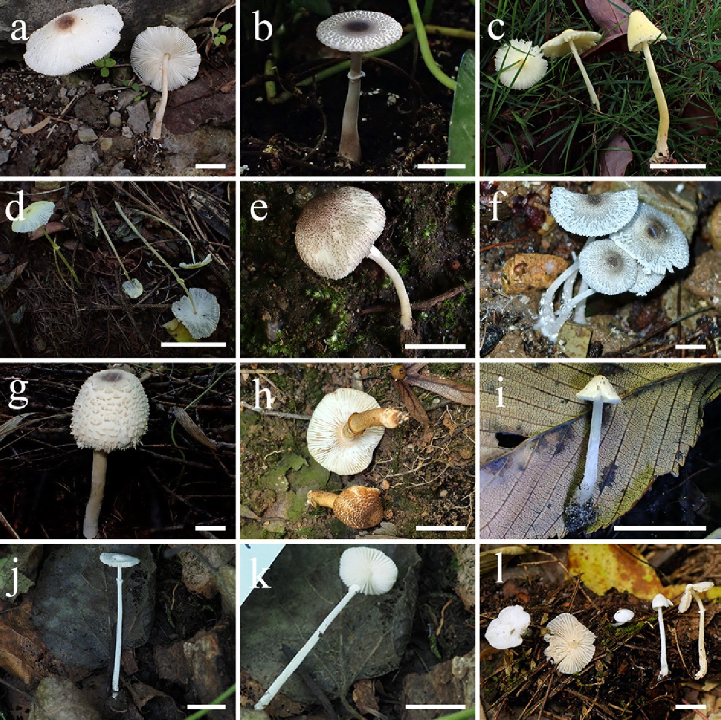
Figure 37.
Basidiomata of Leucocoprinus species. Lc. cinerascens (a) FJAU77966, (b) FJAU77965. (c) Lc. birnbaumii (FJAU79140). (d) Lc. licmophorus (FJAU79141). Lc. lugdunensis (e) FJAU77961, (f) FJAU77962. (g) Lc. nympharum (i) FJAU77969). (h) Lc. tangerinus (FJAU78137). Lc. parviceps (i) FJAU78047, (j), (k) FJAU78046. (l) Lc. lacteus (FJAU78083). Scale bars = 1 cm. These pictures are copyright of the collectors of specimens.
Type species – Leucocoprinus cepistipes (Sowerby) Pat. (as 'cepaestipes'), J. Bot., Paris 3: 336 (1889)
= Pulchrolepiota K.L. Yang & Jia Y. Lin, Phytotaxa 746 (1): 52 (2026).
= Tristolepiota K.L. Yang & Jia Y. Lin, Phytotaxa 746 (1): 64 (2026).
Diagnosis: Compared to the morphologically conserved Leucoagaricus, Leucocoprinus s.l. exhibits a highly diverse and feature-rich phenotypic profile. The macroscopic pileus covering is variable, ranging from fibrillose to floccose, punctate, or tomentose. Microscopically, the pileus covering structures are equally diverse, encompassing a typical cutis, trichoderm, or conioderm, alongside various intermediate transitional configurations. Fresh basidiomata occasionally exude droplets or exhibit a positive reaction to ammonia; furthermore, distinct tissue discoloration upon bruising or drying is frequently observed. Basidiospores are typically hyaline (occasionally tinged pale yellowish-brown in dried specimens due to darkened lamellae), with or without a germ pore. Hymenial pseudoparaphyses may be present or absent. Cheilocystidia are generally present, though occasionally lacking. Ecologically, certain lineages are notably associated with ants.
Notes: Aligning with the perspective of Vellinga et al.[31], who emphasized the practical consequences of generic fragmentation, we adopt a broadly circumscribed Leucocoprinus in the present study. Within this generic framework, our multi-locus phylogeny resolves four major clades alongside seven phylogenetically unstable or isolated lineages within this generic framework. Recently, Yang et al.[30] proposed an alternative multi-generic system for this complex, erecting several narrowly defined genera. Topologically, their newly proposed genera Tristolepiota and Pulchrolepiota correspond to Lc. clade I and Lc. clade II in our phylogeny, respectively, whereas the core Leucocoprinus s.s. corresponds to Lc. clade III and Lc. clade IV. Furthermore, Yang et al.[30] introduced numerous subgeneric and sectional names within these taxa. A comprehensive evaluation of these infrageneric classifications is provided in the Discussion.
Herein, we describe 27 novel Leucocoprinus species. To objectively present the topological structure without introducing premature formal sectional names, the following species descriptions and nomenclatural acts are explicitly organized by neutral lineage designations (i.e., Lc. clades).
Nomenclatural changes
-
In accordance with the broadly circumscribed generic concept adopted herein, we treat the recently erected genera Pulchrolepiota and Tristolepiota as synonyms of Leucocoprinus. Consequently, we reinstate the original Leucocoprinus combinations for the 60 species that were recently transferred to these narrow segregates. Additionally, eight species recently described as new to science within Pulchrolepiota and Tristolepiota are formally recombined into Leucocoprinus.
Reinstated names in Leucocoprinus
Leucocoprinus adelphicus (Vellinga) Redhead, Index Fungorum 551: 1 (2023) [MB#900877]
Basionym: Leucoagaricus adelphicus Vellinga, Mycotaxon 112: 410 (2010) [MB#515363]
Synonym: Pulchrolepiota adelphica (Vellinga) K.L. Yang & Jia Y. Lin, Phytotaxa 746 (1): 57 (2026) [MB#573373]
Leucocoprinus ammovirescens (Bon) Migl. & Donato, Errotari 21: 60 (2024) [MB#902347]
Basionym: Leucoagaricus marriageae var. ammovirescens Bon, Doc. Mycol. 22 (88): 31 (1993) [MB#542953]
Synonym: Pulchrolepiota ammovirescens (Bon) K.L. Yang & Jia Y. Lin, Phytotaxa 746 (1): 62 (2026) [MB#573395]
Leucocoprinus ariminensis (Dovana, Angeli, Contu & Brandi) Asif, Saba & Vellinga, Mycologia 116 (4): 613 (2024) [MB#901193]
Basionym: Leucoagaricus ariminensis Dovana, Angeli, Contu & Brandi, Mycotaxon 132 (1): 209 (2017) [MB#552049]
Synonym: Pulchrolepiota ariminensis (Dovana, Angeli, Contu & Brandi) K.L. Yang & Jia Y. Lin, Phytotaxa 746 (1): 62 (2026) [MB#573396]
Leucocoprinus atroazureus (J.F. Liang, Zhu L. Yang & J. Xu) Asif, Saba & Vellinga, Mycologia 116 (4): 613 (2024) [MB#901195]
Basionym: Leucoagaricus atroazureus J.F. Liang, Zhu L. Yang & J. Xu, Mycologia 102 (5): 1144 (2010) [MB#515456]
Synonym: Pulchrolepiota atroazurea (J.F. Liang, Zhu L. Yang & J. Xu) K.L. Yang & Jia Y. Lin, Phytotaxa 746 (1): 59 (2026) [MB#573381]
Leucocoprinus atrocaeruleus R.L. Zhao & J.Xin Li, Fungal Diversity 135: 815 (2025) [MB#572565]
Synonym: Tristolepiota atrocaerulea (R.L. Zhao & J.Xin Li) K.L. Yang & Jia Y. Lin, Phytotaxa 746 (1): 65 (2026) [MB#573411]
Leucocoprinus atroferrugineus Ishaq, Fiaz & Khalid, Phytotaxa 711 (2): 125 (2025). [MB#852066]
Synonym: Pulchrolepiota atroferruginea (Ishaq, Fiaz & Khalid) K.L. Yang & Jia Y. Lin, Phytotaxa 746 (1): 56 (2026). [MB#573364]
Leucocoprinus aurantiovergens (A. Gennari & Mig.) Mig. & Donato, Errotari 21: 61 (2024) [MB#902339]
Basionym: Leucoagaricus aurantiovergens A. Gennari & Migl., Rivista Micol. 41 (4): 291 (1999) [MB#450128]
Synonym: Pulchrolepiota aurantiovergens (A. Gennari & Migl.) K.L. Yang & Jia Y. Lin, Phytotaxa 746 (1): 60 (2026) [MB#573387]
Isonym: Leucocoprinus aurantiovergens (A. Gennari & Migl.) K.L. Yang, Jia Y. Lin & Zhu L. Yang, Phytotaxa 676 (3): 234 (2024) [MB#572145]
Leucocoprinus badhamii (Berk. & Broome) Locq., Bull. mens. Soc. linn. Lyon: 15 (1943) [MB#316814]
Basionym: Agaricus badhamii Berk. & Broome, Ann. Mag. Nat. Hist. 13: 397 (1854) [MB#375110]
Synonym: Pulchrolepiota badhamii (Bon & Boiffard) K.L. Yang & Jia Y. Lin, Phytotaxa 746 (1): 63 (2026) [MB#573400]
Leucocoprinus barssii (Zeller) Migl. & Donato, Errotari 21: 61 (2024). [MB#902340]
Basionym: Lepiota barssii Zeller, Mycologia 26 (3): 211 (1934). [MB#258218]
Synonym: Leucoagaricus barssii (Zeller) Vellinga, Mycotaxon 76: 431 (2000). [MB#466260]
Synonym: Pulchrolepiota barssii (Zeller) K.L. Yang & Jia Y. Lin, Phytotaxa 746 (1): 63 (2026). [MB#573401]
Isonym: Leucocoprinus barssii (Zeller) K.L. Yang, Jia Y. Lin & Zhu L. Yang, Phytotaxa 676 (3): 234 (2024). [MB#572146]
Leucocoprinus beijingensis R.L. Zhao & J.Xin Li, Mycology 16 (4): 1567 (2025) [MB#571949]
Synonym: Tristolepiota beijingensis (R.L. Zhao & J.Xin Li) K.L. Yang & Jia Y. Lin, Phytotaxa 746 (1): 65 (2026) [MB#573412]
Leucocoprinus bonii (A. Caball.) K.L. Yang, Jia Y. Lin & Zhu L. Yang, Phytotaxa 676 (3): 234 (2024) [MB#572147]
Basionym: Leucoagaricus bonii A. Caball., Doc. Mycol. 27 (106): 40 (1997) [MB#443217]
Synonym: Tristolepiota bonii (A. Caball.) K.L. Yang & Jia Y. Lin, Phytotaxa 746 (1): 72 (2026) [MB#573443]
Leucocoprinus brunneocingulatus (P.D. Orton) Migl. & Donato, Errotari 21: 61 (2024) [MB#902341]
Basionym: Lepiota brunneocingulata P.D. Orton, Trans. Brit. Mycol. Soc. 43 (2): 282 (1960) [MB#333114]
Synonym: Pulchrolepiota brunneocingulata (P.D. Orton) K.L. Yang & Jia Y. Lin, Phytotaxa 746 (1): 57 (2026) [MB#573374]
Isonym: Leucocoprinus brunneocingulatus (P.D. Orton) K.L. Yang, Jia Y. Lin & Zhu L. Yang, Phytotaxa 676 (3): 234 (2024) [MB#572148]
Leucocoprinus brunneoruber (Jin M. Zhang & X.T. Zhu) R.L. Zhao & J. Xin Li, Mycology 16 (4): 1571 (2025) [MB#572311]
Basionym: Leucoagaricus brunneoruber Jin M. Zhang & X.T. Zhu, Phytotaxa 642 (1): 88 (2024) [MB#663900]
Synonym: Pulchrolepiota brunneorubra (Jin M. Zhang & X.T. Zhu) K.L. Yang & Jia Y. Lin, Phytotaxa 746 (1): 58 (2026) [MB#573375]
Leucocoprinus brunnescens (Peck) Locq., Bull. Mens. Soc. Linn. Lyon 14: 93 (1945) [MB#440966]
Basionym: Lepiota brunnescens Peck, Bull. Torrey Bot. Club 31: 177 (1904) [MB#247398]
Synonym: Pulchrolepiota brunnescens (Peck) K.L. Yang & Jia Y. Lin, Phytotaxa 746 (1): 60 (2026) [MB#573382]
Leucocoprinus callainitinctus (K.P.D. Latha, K.N.A. Raj & Manim.) Asif, Saba & Vellinga, Mycologia 116 (4): 614 (2024) [MB#901219]
Basionym: Leucoagaricus callainitinctus K.P.D. Latha, K.N.A. Raj & Manim., Phytotaxa 442 (2): 113 (2020) [MB#834942]
Synonym: Pulchrolepiota callainitincta (K.P.D. Latha, K.N.A. Raj & Manim.) K.L. Yang & Jia Y. Lin, Phytotaxa 746 (1): 60 (2026) [MB#573383]
Leucocoprinus cinereoradicatus (Boisselet & Migl.) Migl. & Donato, Errotari 21: 62 (2024) [MB#902345]
Basionym: Leucoagaricus cinereoradicatus Boisselet & Migl., Boll. Gruppo Micol. G. Bresadola 45 (3): 20 (2002) [MB#372907]
Synonym: Tristolepiota cinereoradicata (Boisselet & Migl.) K.L. Yang & Jia Y. Lin, Phytotaxa 746 (1): 72 (2026) [MB#573444]
Isonym: Leucocoprinus cinereoradicatus (Boisselet & Migl.) K.L. Yang, Jia Y. Lin & Zhu L. Yang, Phytotaxa 676 (3): 235 (2024) [MB#572151]
Leucocoprinus cinnamomeodiscus Asif, Saba & Raza, Mycologia 116 (4): 605 (2024) [MB#849107]
Synonym: Tristolepiota cinnamomeodisca (Asif, Saba & Raza) K.L. Yang & Jia Y. Lin, Phytotaxa 746 (1): 72 (2026) [MB#573445]
Leucocoprinus coerulescens (Peck) K.L. Yang, Jia Y. Lin & Zhu L. Yang, Phytotaxa 676 (3): 235 (2024) [MB#572152]
Basionym: Lepiota coerulescens Peck, Bull. Torrey Bot. Club 26: 63 (1899) [MB#547046]
Synonym: Tristolepiota coerulescens (Peck) K.L. Yang & Jia Y. Lin, Phytotaxa 746 (1): 66 (2026) [MB#573414]
Leucocoprinus croceobasis (G. Muñoz, A. Caball., Contu & Vizzini) Asif, Saba & Vellinga, Mycologia 116 (4): 614 (2024) [MB#901222]
Basionym: Leucoagaricus croceobasis G. Muñoz, A. Caball., Contu & Vizzini, Mycol. Progr. 13: 652 (2013) [MB#807027]
Synonym: Pulchrolepiota croceobasis (G. Muñoz, A. Caball., Contu & Vizzini) K.L. Yang & Jia Y. Lin, Phytotaxa 746 (1): 63 (2026) [MB#573402]
Leucocoprinus cupresseoides (Migl. & Forin) Migl. & Donato, Errotari 21: 63 (2024) [MB#902350]
Basionym: Leucoagaricus cupresseoides Migl. & Forin, Phytotaxa 536 (2): 129 (2022) [MB#841188]
Synonym: Pulchrolepiota cupresseoides (Migl. & Forin) K.L. Yang & Jia Y. Lin, Phytotaxa 746 (1): 61 (2026) [MB#573389]
Isonym: Leucocoprinus cupresseoides (Migl. & Forin) K.L. Yang, Jia Y. Lin & Zhu L. Yang, Phytotaxa 676 (3): 235 (2024) [MB#572153]
Leucocoprinus cupresseus (Burl.) Asif, Saba & Vellinga, Mycologia 116 (4): 614 (2024) [MB#901223]
Basionym: Lepiota cupressea Burl., Mycologia 37 (1): 53 (1945) [MB#287583]
Synonym: Pulchrolepiota cupressea (Burl.) K.L. Yang & Jia Y. Lin, Phytotaxa 746 (1): 60 (2026) [MB#573388]
Leucocoprinus cyanescens (Corriol & Chalange) K.L. Yang, Jia Y. Lin & Zhu L. Yang, Phytotaxa 676 (3): 236 (2024) [MB#572154]
Basionym: Leucoagaricus cyanescens Corriol & Chalange, Bull. Soc. Mycol. France 133 (1-2): 2 (2020) [MB#830187]
Synonym: Tristolepiota cyanescens (Corriol & Chalange) K.L. Yang & Jia Y. Lin, Phytotaxa 746 (1): 66 (2026) [MB#573415]
Leucocoprinus decipiens (Contu, Vizzini & Vellinga) Asif, Saba & Vellinga, Mycologia 116 (4): 614 (2024) [MB#901224]
Basionym: Leucoagaricus decipiens Contu, Vizzini & Vellinga, Mycologia 102 (2): 448 (2010) [MB#513553]
Synonym: Pulchrolepiota decipiens (Contu, Vizzini & Vellinga) K.L. Yang & Jia Y. Lin, Phytotaxa 746 (1): 56 (2026) [MB#573365]
Leucocoprinus digitatocystis R.L. Zhao & J.Xin Li, Mycology 16 (4): 1563 (2025) [MB#571948]
Synonym: Tristolepiota digitatocystis (R.L. Zhao & J.Xin Li) K.L. Yang & Jia Y. Lin, Phytotaxa 746 (1): 66 (2026) [MB#573426]
Leucocoprinus dyscritus (Vellinga) Asif, Saba & Vellinga, Mycologia 116 (4): 614 (2024) [MB#901225]
Basionym: Leucoagaricus dyscritus Vellinga, Mycotaxon 112: 416 (2010) [MB#515365]
Synonym: Pulchrolepiota dyscrita (Vellinga) K.L. Yang & Jia Y. Lin, Phytotaxa 746 (1): 63 (2026) [MB#573403]
Leucocoprinus erminiae (Cons., Setti & Vizzini) K.L. Yang, Jia Y. Lin & Zhu L. Yang, Phytotaxa 676 (3): 236 (2024) [MB#572156]
Basionym: Leucoagaricus erminiae Cons., Setti & Vizzini, Micol. Veg. Medit. 36 (1-2): 204 (2021) [MB#841276]
Synonym: Pulchrolepiota erminiae (Cons., Setti & Vizzini) K.L. Yang & Jia Y. Lin, Phytotaxa 746 (1): 63 (2026) [MB#573404]
Leucocoprinus erythrophaeus (Vellinga) Redhead, Index Fungorum 551: 1 (2023) [MB#900878]
Basionym: Leucoagaricus erythrophaeus Vellinga, Mycologia 102 (2): 450 (2010) [MB#513552]
Synonym: Pulchrolepiota erythrophaea (Vellinga) K.L. Yang & Jia Y. Lin, Phytotaxa 746 (1): 56 (2026) [MB#573366]
Leucocoprinus fissuratus (Morosini, M. Bianchi & Vizzini) Migl. & Donato, Errotari 22: 80 (2025) [MB#903306]
Basionym: Leucoagaricus fissuratus Morosini, M. Bianchi & Vizzini, Micologia Toscana 6: 71 (2024) [MB#855942]
Synonym: Pulchrolepiota fissurata (Morosini, M. Bianchi & Vizzini) K.L. Yang & Jia Y. Lin, Phytotaxa 746 (1): 61 (2026) [MB#573390]
Leucocoprinus flammeotinctoides (Vellinga) Redhead, Index Fungorum 551: 1 (2023) [MB#900879]
Basionym: Leucoagaricus flammeotinctoides Vellinga, Mycotaxon 112: 429 (2010) [MB#515367]
Synonym: Pulchrolepiota flammeotinctoides (Vellinga) K.L. Yang & Jia Y. Lin, Phytotaxa 746 (1): 56 (2026) [MB#573367]
Leucocoprinus fragilis (Asif, Niazi, Izhar & Khalid) Asif, Saba & Vellinga, Mycologia 116 (4): 614 (2024) [MB#901226]
Basionym: Leucoagaricus fragilis Asif, Niazi, Izhar & Khalid, Phytotaxa 501 (1): 144 (2021) [MB#839737]
Synonym: Pulchrolepiota fragilis (Asif, Niazi, Izhar & Khalid) K.L. Yang & Jia Y. Lin, Phytotaxa 746 (1): 63 (2026) [MB#573405]
Leucocoprinus gaillardii (Bon & Boiffard) Asif, Saba & Vellinga, Mycologia 116 (4): 615 (2024) [MB#901278]
Basionym: Leucoagaricus gaillardii Bon & Boiffard, Bull. Soc. Mycol. France 90 (4): 295 (1974) [MB#316783]
Synonym: Pulchrolepiota gaillardii (Bon & Boiffard) K.L. Yang & Jia Y. Lin, Phytotaxa 746 (1): 64 (2026) [MB#573407]
Leucocoprinus gansuensis R.L. Zhao & J.Xin Li, Fungal Diversity 135: 819 (2025) [MB#572598]
Synonym: Tristolepiota gansuensis (R.L. Zhao & J.Xin Li) K.L. Yang & Jia Y. Lin, Phytotaxa 746 (1): 72 (2026) [MB#573446]
Leucocoprinus ganziensis R.L. Zhao & J.X. Li, Fungal Diversity 135: 821 (2025) [MB#572584]
Synonym: Pulchrolepiota ganziensis (R.L. Zhao & J.X. Li) K.L. Yang & Jia Y. Lin, Phytotaxa 746 (1): 56 (2026) [MB#573368]
Leucocoprinus gaoligongensis R.L. Zhao & J.Xin Li, Fungal Diversity 135: 823 (2025) [MB#572563]
Synonym: Tristolepiota gaoligongensis (R.L. Zhao & J.Xin Li) K.L. Yang & Jia Y. Lin, Phytotaxa 746 (1): 68 (2026) [MB#573428]
Leucocoprinus gauguei (Bon & Boiffard) Migl. & Donato, Errotari 21: 63 (2024) [MB#902354]
Basionym: Leucoagaricus gauguei Bon & Boiffard, Bull. Soc. Mycol. France 90 (4): 290 (1975) [MB#316784]
Synonym: Pulchrolepiota gauguei (Bon & Boiffard) K.L. Yang & Jia Y. Lin, Phytotaxa 746 (1): 62 (2026) [MB#573358]
Isonym: Leucocoprinus gauguei (Bon & Boiffard) K.L. Yang, Jia Y. Lin & Zhu L. Yang, Phytotaxa 676 (3): 236 (2024). [MB#572159]
Leucocoprinus georginae (W.G. Sm.) M.M. Moser, Kleine Kryptogamenflora von Mitteleuropa - Die Blätter- und Baupilze (Agaricales und Gastromycetes) IIb: 132 (1955) [MB#135249]
Basionym: Agaricus georginae W.G. Sm., J. Bot., London: 1 (1871) [MB#372122]
Synonym: Pulchrolepiota georginae (W.G. Sm.) K.L. Yang & Jia Y. Lin, Phytotaxa 746 (1): 60 (2026) [MB#573384]
Leucocoprinus hesperius (Vellinga) Asif, Saba & Vellinga, Mycologia 116 (4): 615 (2024) [MB#901231]
Basionym: Leucoagaricus hesperius Vellinga, Mycotaxon 112: 413 (2010) [MB#515366]
Synonym: Pulchrolepiota hesperia (Vellinga) K.L. Yang & Jia Y. Lin, Phytotaxa 746 (1): 58 (2026) [MB#573376]
Leucocoprinus idae-fragum (Guinb., Boisselet & G. Dupuy) Asif, Saba & Vellinga, Mycologia 116 (4): 615 (2024) [MB#901232]
Basionym: Leucoagaricus idae-fragum Guinb., Boisselet & G. Dupuy, Bull. Soc. Mycol. France 114 (3): 14 (1998) [MB#446559]
Synonym: Pulchrolepiota idae-fragum (Guinb., Boisselet & G. Dupuy) K.L. Yang & Jia Y. Lin, Phytotaxa 746 (1): 61 (2026) [MB#573391]
Leucocoprinus infuscatus (Vellinga) K.L. Yang, Jia Y. Lin & Zhu L. Yang, Phytotaxa 676 (3): 237 (2024) [MB#572162]
Basionym: Leucoagaricus infuscatus Vellinga, Mycotaxon 102: 275 (2007) [MB#511150]
Synonym: Tristolepiota infuscata (Vellinga) K.L. Yang & Jia Y. Lin, Phytotaxa 746 (1): 68 (2026) [MB#573429]
Leucocoprinus ionidicolor (Bellù & Lanzoni) Migl. & Donato, Errotari 21: 64 (2024). [MB#902356]
Basionym: Leucoagaricus ionidicolor Bellù & Lanzoni, Rivista Micol. 31 (3-4): 107 (1988). [MB#134692]
Synonym: Pulchrolepiota ionidicolor (Bellù & Lanzoni) K.L. Yang & Jia Y. Lin, Phytotaxa 746 (1): 62 (2026). [MB#571180] (as 'ionidicolora')
Isonym: Leucocoprinus ionidicolor (Bellù & Lanzoni) K.L. Yang, Jia Y. Lin & Zhu L. Yang, Phytotaxa 676 (3): 237 (2024). [MB#572163]
Leucocoprinus irinellus (Chalange) K.L. Yang, Jia Y. Lin & Zhu L. Yang, Phytotaxa 676 (3): 237 (2024) [MB#572164]
Basionym: Leucoagaricus irinellus Chalange, Bull. Soc. Mycol. France 115 (4): 385 (1999) [MB#460910]
Synonym: Tristolepiota irinella (Chalange) K.L. Yang & Jia Y. Lin, Phytotaxa 746 (1): 68 (2026) [MB#573430]
Leucocoprinus laosensis (Sysouph.) K.L. Yang, Jia Y. Lin & Zhu L. Yang, Phytotaxa 676 (3): 237 (2024) [MB#572165]
Basionym: Leucoagaricus laosensis Sysouph., Mycosphere 13 (1): 531 (2022) [MB#841760]
Synonym: Tristolepiota laosensis (Sysouph.) K.L. Yang & Jia Y. Lin, Phytotaxa 746 (1): 68 (2026) [MB#573431]
Leucocoprinus mantadiaensis Ralaiv., Liimat. & Niskanen, Fungal Diversity 130: 153 (2024) [MB#902404]
Synonym: Tristolepiota mantadiaensis (Ralaiv., Liimat. & Niskanen) K.L. Yang & Jia Y. Lin, Phytotaxa 746 (1): 68 (2026) [MB#573433]
Leucocoprinus marginatus (Burl.) Migl. & Donato, Errotari 21: 64 (2024) [MB#902358]
Basionym: Lepiota marginata Burl., Mycologia 37 (1): 55 (1945) [MB#287596]
Synonym: Pulchrolepiota marginata (Burl.) K.L. Yang & Jia Y. Lin, Phytotaxa 746 (1): 58 (2026) [MB#573377]
Isonym: Leucocoprinus marginatus (Burl.) K.L. Yang, Jia Y. Lin & Zhu L. Yang, Phytotaxa 676 (3): 237 (2024) [MB#572167]
Leucocoprinus nzumbae (C. Heisecke, A.A. Carvalho & M.A. Neves) K.L. Yang, Jia Y. Lin & Zhu L. Yang, Phytotaxa 676 (3): 238 (2024) [MB#572169]
Basionym: Leucoagaricus nzumbae C. Heisecke, A.A. Carvalho & M.A. Neves, Phytotaxa 494 (1): 52 (2021) [MB#835923]
Synonym: Tristolepiota nzumbae (C. Heisecke, A.A. Carvalho & M.A. Neves) K.L. Yang & Jia Y. Lin, Phytotaxa 746 (1): 68 (2026) [MB#573434]
Leucocoprinus ophthalmus (Vellinga) K.L. Yang, Jia Y. Lin & Zhu L. Yang, Phytotaxa 676 (3): 238 (2024) [MB#572170]
Basionym: Leucoagaricus ophthalmus Vellinga, Mycotaxon 102: 275 (2007) [MB#511149]
Synonym: Pulchrolepiota ophthalma (J.E. Lange & Zeller) K.L. Yang & Jia Y. Lin, Phytotaxa 746 (1): 55 (2026) [MB#573360]
Leucocoprinus paracupresseus (Salom, Siquier, Planas & Espinosa) K.L. Yang, Jia Y. Lin & Zhu L. Yang, Phytotaxa 676 (3): 238 (2024) [MB#572172]
Basionym: Leucoagaricus paracupresseus Salom, Siquier, Planas & Espinosa, Index Fungorum 485: 1 (2021) [MB#558417]
Synonym: Pulchrolepiota paracupressea (Salom, Siquier, Planas & Espinosa) K.L. Yang & Jia Y. Lin, Phytotaxa 746 (1): 61 (2026) [MB#573392]
Leucocoprinus paraplesius (Vellinga) K.L. Yang, Jia Y. Lin & Zhu L. Yang, Phytotaxa 676 (3): 238 (2024) [MB#572171]
Basionym: Leucoagaricus paraplesius Vellinga, Mycotaxon 102: 271 (2007) [MB#511148]
Synonym: Pulchrolepiota paraplesia (Vellinga) K.L. Yang & Jia Y. Lin, Phytotaxa 746 (1): 56 (2026) [MB#573362]
Leucocoprinus pardalotus (Vellinga) Redhead, Index Fungorum 551: 1 (2023) [MB#900882]
Basionym: Leucoagaricus pardalotus Vellinga, Mycotaxon 112: 420 (2010) [MB#515364]
Synonym: Pulchrolepiota pardalota (Vellinga) K.L. Yang & Jia Y. Lin, Phytotaxa 746 (1): 57 (2026) [MB#573369]
Leucocoprinus petasatus R.L. Zhao & J.Xin Li, Fungal Diversity 135: 829 (2025) [MB#572564]
Synonym: Tristolepiota petasata (R.L. Zhao & J.Xin Li) K.L. Yang & Jia Y. Lin, Phytotaxa 746 (1): 68 (2026) [MB#573435]
Leucocoprinus phaeocyaneus R.L. Zhao & J.Xin Li, Fungal Diversity 135: 831 (2025) [MB#572562]
Synonym: Tristolepiota phaeocyanea (R.L. Zhao & J.Xin Li) K.L. Yang & Jia Y. Lin, Phytotaxa 746 (1): 69 (2026) [MB#573436]
Leucocoprinus pilatianus (Demoulin) M.M. Moser, Kleine Krypt.-Fl. 2b (2): 186 (1967). [MB#114851]
Basionym: Lepiota pilatiana Demoulin, Lejeunia: 11 (1966). [MB#333136]
Synonym: Pulchrolepiota pilatiana (Demoulin) K.L. Yang & Jia Y. Lin, Phytotaxa 746 (1): 58 (2026). [MB#573378]
Leucocoprinus pseudopilatianus (Migl., Rocabruna & Tabarés) Migl. & Donato, Errotari 21: 66 (2024) [MB#902366]
Basionym: Leucoagaricus pseudopilatianus Migl., Rocabruna & Tabarés, Revista Catal. Micol. 23: 67 (2001) [MB#479389]
Synonym: Pulchrolepiota pseudopilatiana (Migl., Rocabruna & Tabarés) K.L. Yang & Jia Y. Lin, Phytotaxa 746 (1): 61 (2026) [MB#573393]
Isonym: Leucocoprinus pseudopilatianus (Migl., Rocabruna & Tabarés) K.L. Yang, Jia Y. Lin & Zhu L. Yang, Phytotaxa 676 (3): 238 (2024) [MB#572173]
Leucocoprinus purpurascens (T. Guo & Z.W. Ge) K.L. Yang, Jia Y. Lin & Zhu L. Yang, Phytotaxa 676 (3): 239 (2024) [MB#572174]
Basionym: Leucoagaricus purpurascens T. Guo & Z.W. Ge, Phytotaxa 584 (3): 202 (2023) [MB#845096]
Synonym: Tristolepiota purpurascens (T. Guo & Z.W. Ge) K.L. Yang & Jia Y. Lin, Phytotaxa 746 (1): 69 (2026) [MB#573437]
Leucocoprinus pyrrhophaeus (Vellinga) K.L. Yang, Jia Y. Lin & Zhu L. Yang, Phytotaxa 676 (3): 239 (2024) [MB#572175]
Basionym: Leucoagaricus pyrrhophaeus Vellinga, Mycotaxon 112: 433 (2010) [MB#515369]
Synonym: Pulchrolepiota pyrrhophaea (Vellinga) K.L. Yang & Jia Y. Lin, Phytotaxa 746 (1): 57 (2026) [MB#573370]
Leucocoprinus pyrrhulus (Vellinga) Asif, Saba & Vellinga, Mycologia 116 (4): 616 (2024) [MB#901255]
Basionym: Leucoagaricus pyrrhulus Vellinga, Mycotaxon 112: 435 (2010) [MB#515368]
Synonym: Pulchrolepiota pyrrhula (Vellinga) K.L. Yang & Jia Y. Lin, Phytotaxa 746 (1): 57 (2026) [MB#573371]
Leucocoprinus sericifer (Locq.) Asif, Saba & Vellinga, Mycologia 116 (4): 616 (2024) [MB#901260]
Basionym: Pseudobaeospora sericifera Locq., Bull. Soc. Mycol. France 68: 169 (1952) [MB#304341]
Synonym: Tristolepiota sericifera (Locq.) K.L. Yang & Jia Y. Lin, Phytotaxa 746 (1): 72 (2026) [MB#573448]
Leucocoprinus variicolor (G. Muñoz, A. Caball., Contu & Vizzini) Asif, Saba & Vellinga, Mycologia 116 (4): 617 (2024). [MB#901273]
Basionym: Leucoagaricus variicolor G. Muñoz, A. Caball., Contu & Vizzini, IMA Fungus 3 (2): 119 (2012). [MB#801565]
Synonym: Pulchrolepiota variicolor (G. Muñoz, A. Caball., Contu & Vizzini) K.L. Yang & Jia Y. Lin, Phytotaxa 746 (1): 64 (2026). [MB#571179] (as 'variicolora')
Leucocoprinus virens (Y.R. Ma, Z.W. Ge & T.Z. Liu) Asif, Saba & Vellinga, Mycologia 116 (4): 617 (2024) [MB#901275]
Basionym: Leucoagaricus virens Y.R. Ma, Z.W. Ge & T.Z. Liu, Diversity 14 (5, no. 314): 14 (2022) [MB#843378]
Synonym: Tristolepiota virens (Y.R. Ma, Z.W. Ge & T.Z. Liu) K.L. Yang & Jia Y. Lin, Phytotaxa 746 (1): 71 (2026) [MB#573438]
Leucocoprinus viriditinctus (Berk. & Broome) Asif, Saba & Vellinga, Mycologia 116 (4): 617 (2024) [MB#901276]
Basionym: Agaricus viriditinctus Berk. & Broome, Bot. J. Linn. Soc. 11: 503 (1871) [MB#477618]
Synonym: Tristolepiota viriditincta (Berk. & Broome) K.L. Yang & Jia Y. Lin, Phytotaxa 746 (1): 71 (2026) [MB#573439]
New combinations in Leucocoprinus
Leucocoprinus actinophorus (K.L. Yang, Jia Y. Lin & Z.Chao Liu) T. Bau, comb. nov. [MB#864326]
Synonym: Tristolepiota actinophora K.L. Yang, Jia Y. Lin & Z.Chao Liu, Phytotaxa 746 (1): 64 (2026) [MB#573410]
Leucocoprinus boluoshanensis (K.L. Yang, Jia Y. Lin & Z.Chao Liu) T. Bau, comb. nov. [MB#864327]
Synonym: Tristolepiota boluoshanensis K.L. Yang, Jia Y. Lin & Z.Chao Liu, Phytotaxa 746 (1): 66 (2026) [MB#573413]
Leucocoprinus elegans (K.L. Yang, Jia Y. Lin & Z.Chao Liu) T. Bau, comb. nov. [MB#864328]
Synonym: Pulchrolepiota elegans K.L. Yang, Jia Y. Lin & Z.Chao Liu, Phytotaxa 746 (1): 53 (2026) [MB#573356]
Leucocoprinus personatus (K.L. Yang & Jia Y. Lin) T. Bau, comb. nov. [MB#864329]
Synonym: Pulchrolepiota personata K.L. Yang & Jia Y. Lin, Phytotaxa 746 (1): 54 (2026) [MB#573357]
Leucocoprinus sericatellus (Malençon) T. Bau, comb. nov. [MB#864330]
Basionym: Lepiota sericatella Malençon, Trav. Inst. Sci. Chérifien, Sér. Bot. Biol. Vég. 33: 152 (1970). [MB#316523]
Synonym: Leucoagaricus sericatellus (Malençon) Bon, Doc. Mycol. 9 (33): 40 (1979). [MB#316805]
Synonym: Tristolepiota sericatella (Malençon) K.L. Yang & Jia Y. Lin, Phytotaxa 746 (1): 72 (2026). [MB#573447]
Leucocoprinus shumuyuanensis (K.L. Yang, Jia Y. Lin & Z.Chao Liu) T. Bau, comb. nov. [MB#864331]
Synonym: Tristolepiota shumuyuanensis K.L. Yang, Jia Y. Lin & Z.Chao Liu, Phytotaxa 746 (1): 69 (2026) [MB#573440]
Leucocoprinus velutinus (K.L. Yang & Jia Y. Lin) T. Bau, comb. nov. [MB#864332]
Synonym: Pulchrolepiota velutina K.L. Yang & Jia Y. Lin, Phytotaxa 746 (1): 58 (2026) [MB#573380]
Leucocoprinus vernalis (K.L. Yang & Jia Y. Lin) T. Bau, comb. nov. [MB#864333]
Synonym: Tristolepiota vernalis K.L. Yang & Jia Y. Lin, Phytotaxa 746 (1): 70 (2026) [MB#573441]
Leucocoprinus acystidiatus T. Bau & X.Y. Zhou, sp. nov Figs 33a−c, 38
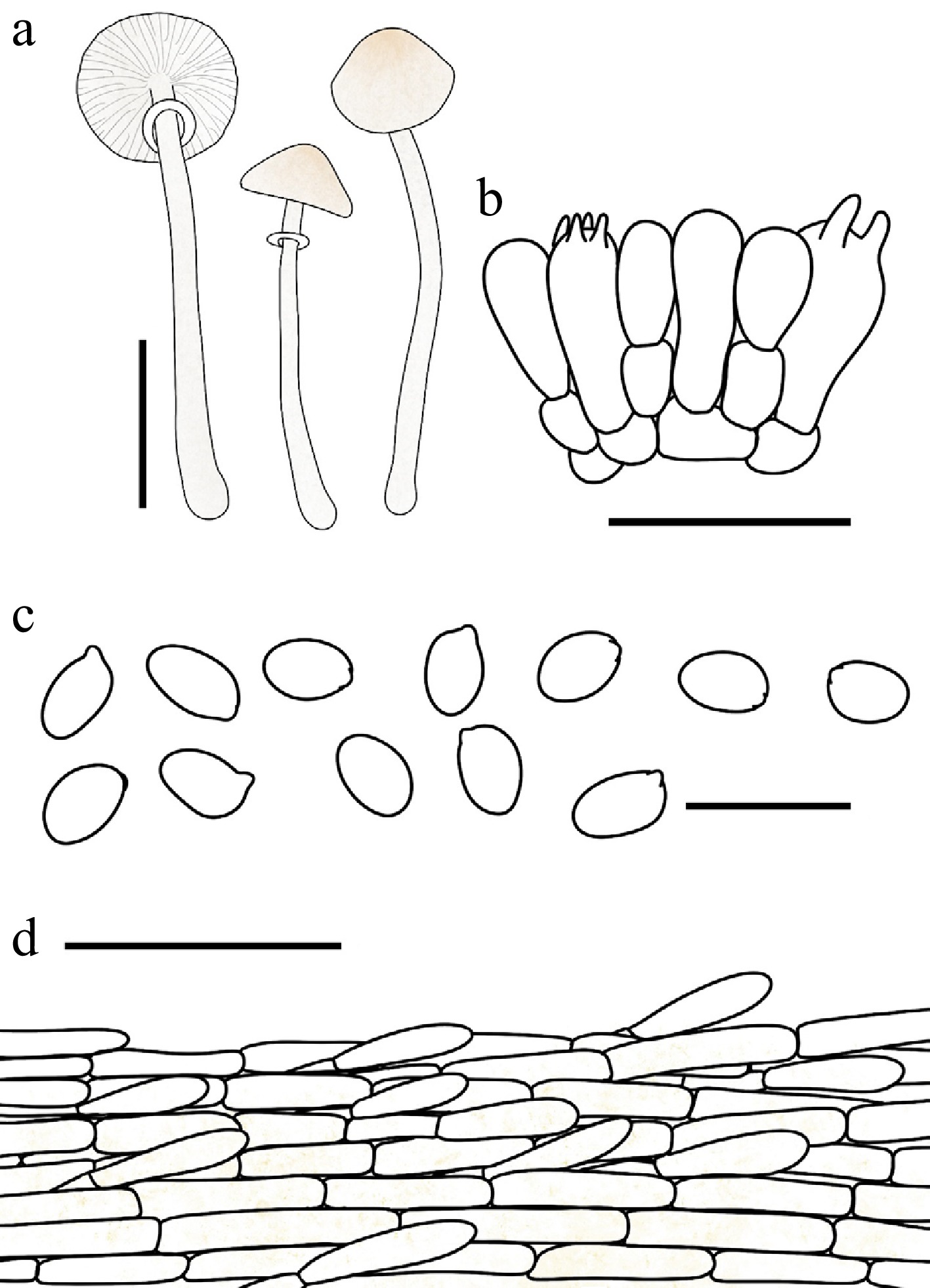
Figure 38.
Leucocoprinus acystidiatus. (a) Basidiomata. (b) Basidia. (c) Basidiospores. (d) Surface covering at pileus centre. Scale bars: (a) = 1 cm; (b) = 10 μm; (c), (d) = 20 μm; (e) = 50 μm.
Chinese name: 无囊白鬼伞 (Pīn yīn: wú náng bái guǐ sǎn)
Index Fungorum number: IF904656; Facesoffungi number: FoF18947
Etymology – 'acystidiatus' is formed from the Greek prefix a- (without) and the New Latin cystidium, referring to the taxonomically significant absence of cheilocystidia in this species.
Type – China. Jilin: Ji'an City, Yushan Park, 26 July 2025, coll. X.Y. Zhou, FJAU78090.
Diagnosis – Characterized by its white pileus with greige to pale-gold fibrillose squamules and the absence of cheilocystidia.
Basidiomata small. Pileus 0.6–1 cm in diam., obtusely conical to plano-convex or applanate when mature; umbo slightly obtuse; background white; squamules greige (#C1B7A5) to pale gold (#E6BE8A), fibrillose. Lamellae free, moderately crowded, white to cream; edge concolorous; lamellulae in 1–3 tiers. Stipe 1.7–3 cm × 0.05–0.1 cm, subcylindrical, hollow, white to greige (#C1B7A5); surface covered with white fibrillose squamules; base with white mycelia. Annulus superior, ascending, white, membranous, fragile. Context thin, white, unchanging when bruised. Odour and taste not recorded.
Basidiospores (60/3/3) 4.8–6.2 × 3.2–4.0 μm, avl × avw = 5.3–5.5 × 3.4–3.6 μm, Q = 1.31–1.71, avQ = 1.42–1.55, ellipsoid to oblong, hyaline, slightly thick-walled, smooth, non-dextrinoid, cyanophilous, congophilous, metachromatic in Cresyl Blue; germ pore absent. Basidia 13–24 × 5–8 μm, clavate, predominantly 4-spored, occasionally 2-spored, thin-walled. Pseudoparaphyses absent. Cheilocystidia and pleurocystidia absent. Surface covering at pileus centre a cutis consisting of cylindrical hyphae; hyphae repent, 5–13 μm wide, thin-walled, with pale-gold intracellular pigment. Stipe covering a cutis consisting of cylindrical hyphae; hyphae repent, 3–12 μm wide, thin-walled, hyaline to light yellow. Clamp connections not observed.
Habit and habitat – solitary to scattered in humus-rich soil in mixed coniferous and broadleaf forests.
Known distribution – so far recorded only from Northeast China.
Additional specimens examined – China. Jilin: Dunhua City, Beishan Park, 17 August 2024, coll. X.Y. Zhou, FJAU78088; Ji'an City, Yushan Park, 26 July 2025, coll. X.Y. Zhou, FJAU78089, FJAU78091.
Notes – Morphologically, Leucocoprinus acystidiatus closely resembles Lc. pegleri (Justo, Bizzi & Angelini) M. Asif, Saba & Vellinga, a species originally described from the Dominican Republic. However, Lc. pegleri can be readily distinguished by its yellowish lamellae and distinctly dextrinoid basidiospores[61], which contrast sharply with the white to cream lamellae and non-dextrinoid basidiospores of the new species.
Phylogenetically, Lc. acystidiatus is nested within Lc. clade IV, specifically grouping into Lc. subclade c alongside Lc. subhymenoderma (Bon & A. Caball.) Kun L. Yang, Jia Y. Lin & Zhu L. Yang, Lc. pegleri, Lc. roseovertens (Justo, Bizzi & Angelini) M. Asif, Saba & Vellinga, Lc. sabinae (Angelini, Justo & Vizzini) Kun L. Yang, Jia Y. Lin & Zhu L. Yang, and Lc. subincarnatus T. Bau & X.Y. Zhou. According to their original protologues[61,87,88], the cheilocystidia of these previously described taxa are remarkably similar in size and shape to their basidia (Table 2). Furthermore, our microscopic examinations of Lc. acystidiatus and Lc. subincarnatus confirmed the absence of any specialized terminal hyphae protruding beyond the basidia at the lamellar edge. Based on these congruent observations, we hypothesize that the presence of poorly developed cheilocystidia—or elements nearly indistinguishable from basidia—may represent a shared characteristic for species within Lc. subclade c.
Table 2. Leucocoprinus species possessing poorly developed cheilocystidia.
Taxon Basidia Cheilocystidia Lc. sabinae[88] Basidia tetra-sterigmate, clavate, or cylindrical with a square base, 18–30 × 6.5–10 µm. Cheilocystidia 13.5–21 × 3–6 µm, cylindrical-capitulated, some with flexuous outline, infrequent and unevenly distributed along the lamella edge. Lc. subhymenoderma[87] Basidia 20–30 × 7–9 μm, (1–2–) 4-spored. Cheilocystidia 25–35 × 7–12 μm, clavate to ± ventricose, or sometimes constricted. Lc. pegleri[61] Basidia 18–23 × 6–8 μm, clavate, tetrasporic. Cheilocystidia 12–23 × 7–11 μm, clavate, basidia-like, not very visible on the lamellar edge Lc. roseovertens[61] Basidia 14.5–24.5 × 6–9 μm, clavate, tetrasporic. Cheilocystidia 12–23 × 7–13 μm, ventricose, clavate, hyaline, emerging in little tufts from the lamellar edge. Lc. acystidiatus (this study) Basidia 13–24 × 5–8 μm, clavate, 4(–2)-spored. No specialized terminal hyphal cells observed. Lc. subincarnatus (this study) Basidia 16–22 × 6–9 μm, clavate, 4(–2)-spored. No specialized terminal hyphal cells observed. Phylogenetically, it is worth noting that a sequence labeled as Leucocoprinus flammeotinctus (Kauffman) Redhead (DUKE-JJ97, GenBank U85331) is also nested within Lc. subclade c. However, according to its morphological description, Lc. flammeotinctus exhibits an orange-red discoloration on the pileus upon bruising, and both the pileus and stipe turn dark brown upon drying. This combination of macroscopic features aligns much better with the diagnostic characters of Lc. clade II or Lc. clade III. Most crucially, authentic sequences of Lc. flammeotinctus explicitly validated by Vellinga[89] are robustly placed within Lc. subclade b of Lc. clade II. Therefore, we conclude that the sequence U85331 derived from the collection DUKE-JJ97 is highly likely to represent a misidentification.
Leucocoprinus crassibasis T. Bau & X.Y. Zhou, sp. nov Figs 33d, e, 39
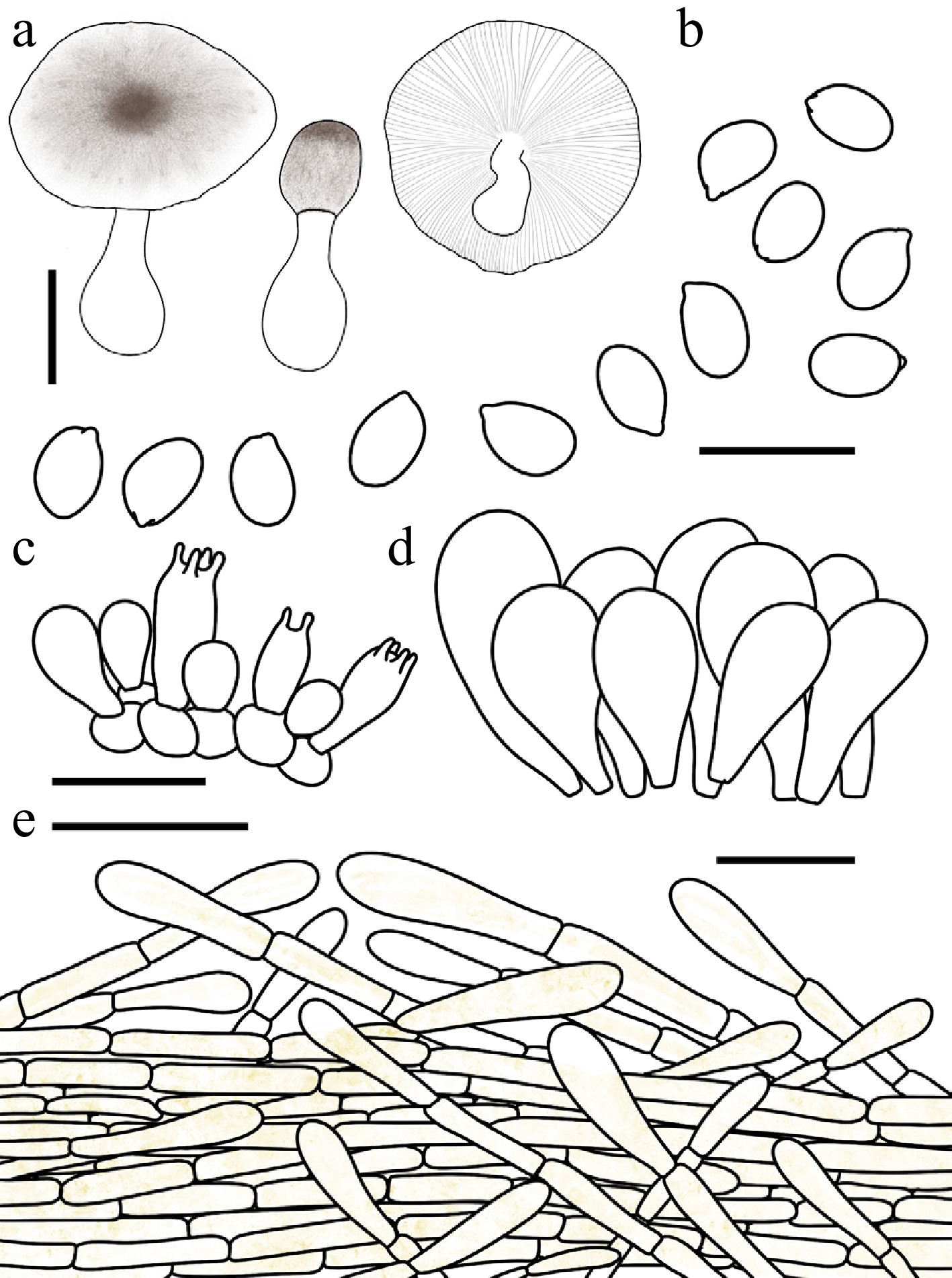
Figure 39.
Leucocoprinus crassibasis. (a) Basidiomata. (b) Basidiospores. (c) Basidia. (d) Cheilocystidia. (e) Surface covering at pileus centre. Scale bars: (a) = 1 cm; (b) = 10 μm; (c), (d) = 20 μm; (e) = 50 μm.
Chinese name: 粗脚白鬼伞 (Pīn yīn: cū jiǎo bái guǐ sǎn)
Index Fungorum number: IF905534; Facesoffungi number: FoF 19998
Etymology – 'crassibasis' is a compound adjective derived from the Latin crassus (thick) and basis (base), referring to the distinctly thickened base of the stipe.
Type – China. Jiangsu: Nanjing Botanical Garden Memorial Sun Yat-Sen, 11 June 2025, coll. H.Y. Zhou, FJAU78107,
Diagnosis – characterized by its relatively robust basidiomata compared to other Leucocoprinus species, brownish pileal squamules, and a distinctly bulbous stipe base.
Basidiomata small to medium-sized. Pileus 0.7–4.0 cm in diam., initially ovate, expanding to applanate; background white to cream; centre with umber (#635147) felted squamules; squamules around the centre pale-umber (#9A7E65) to umber, finely squamulose; margin costate-plicate up to 1/3 of the radius or up to the umbo, easily radially splitting. Lamellae free, moderately crowded, white to cream; edge concolorous; lamellulae interspersed in 3 tiers. Stipe 2.1–5.2 cm × 0.3–0.6 cm, subcylindrical, hollow, with a distinctly bulbous base, white; surface smooth or covered with fine fibrils. Annulus median to superior, ascending, white with an umber margin, membranous. Context thick, white, unchanging when bruised. Odour and taste not recorded.
Basidiospores (40/2/2) 5.0–6.5 × 3.5–4.3 μm, avl × avw = 5.7–5.8 × 3.9–4.0 μm, Q = 1.34–1.62, avQ = 1.43–1.45, ellipsoid to oblong, hyaline, slightly thick-walled, smooth, non-dextrinoid, cyanophilous, congophilous, metachromatic in Cresyl Blue; germ pore absent. Basidia 14–20 × 7–10 μm, clavate, predominantly 4-spored, occasionally 2-spored, thin-walled. Pseudoparaphyses absent. Pleurocystidia absent. Cheilocystidia 23–42 × 9–17 μm, broadly clavate, hyaline, slightly thick-walled. Surface covering at pileus centre an interwoven plagiotrichoderm consisting of cylindrical hyphae; hyphae densely entangled, repent to slightly ascending, 7–17 μm wide, slightly thick-walled, with light yellow intracellular pigment. Stipe covering a cutis consisting of cylindrical hyphae; hyphae repent, 5–14 μm wide, thin-walled, hyaline. Clamp connections not observed.
Habit and habitat – scattered on soil in chestnut forests.
Known distribution –so far recorded only from China.
Additional specimens examined – China. Jiangsu: Nanjing Botanical Garden Memorial Sun Yat-Sen, 11 June 2025, coll. N. Li, FJAU78125.
Notes – Macroscopically, Leucocoprinus crassibasis shares relatively stout basidiomata and brownish pileus squamules with Lc. amanitoides (R.M. Davis & Vellinga) Kun L. Yang, Jia Y. Lin & Zhu L. Yang. However, the latter can be readily distinguished by its significantly larger basidiospores and narrower cheilocystidia[90].
Although the genus Leucocoprinus accommodates several other taxa characterized by stout stipes or distinctly bulbous bases—such as Lc. cepistipes (Sowerby) Pat. and Lc. bulbipes (Mont.) Raithelh.—these species are clearly separable from the new taxon. Specifically, Lc. cepistipes is fundamentally separated by producing basidiospores with a distinct germ pore[80]. Regarding Lc. bulbipes, a historical taxon originally described in 1856 and subsequently transferred to Leucocoprinus by Raithelhuber in 1987, its differentiation from the new species relies primarily on macroscopic features due to the sparse nature of its original protologue. Nevertheless, Lc. bulbipes is distinctly characterized by its reddish-brown pileus squamules[91,92], which contrast sharply with the umber coloration observed in Lc. crassibasis.
Leucocoprinus fuscannulatus T. Bau & X.Y. Zhou, sp. nov Figs 33f−i, 40
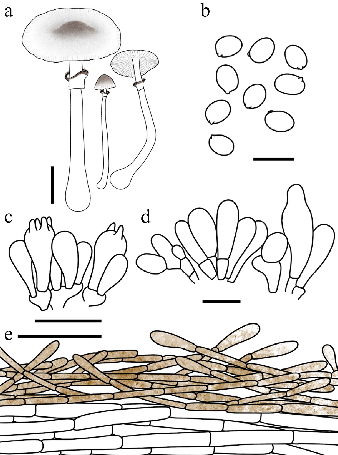
Figure 40.
Leucocoprinus fuscannulatus. (a) Basidiomata. (b) Basidiospores. (c) Basidia. (d) Cheilocystidia. (e) Surface covering at pileus centre. Scale bars: (a) = 1 cm; (b) = 10 μm; (c), (d) = 20 μm; (e) = 50 μm.
Chinese name: 褐环白鬼伞 (Pīn yīn: hè huán bái guǐ sǎn)
Index Fungorum number: IF904664; Facesoffungi number: FoF18955
Etymology – 'fuscannulatus' is a compound adjective derived from the Latin fuscus (brown, dusky) and annulatus (with a ring), referring to the distinctive golden-brown to hickory-brown outer surface of the bicolorous annulus.
Type – China. Heilongjiang: Nenjiang City, Gaofeng National Forest Park, 13 August 2025, coll. X.Y. Zhou, FJAU78014.
Diagnosis – characterized by its pileus adorned with grayish to brownish cottony squamules, a bicolorous annulus featuring a golden brown to hickory brown outer surface and a white inner surface, and septate cheilocystidia.
Basidiomata small. Pileus 0.5–2.9 cm in diam., initially obtusely conical, expanding to plano-convex or applanate; umbo obtuse; background white; centre with granite-gray (#676767) to hickory-brown (#371D10) finely floccoso-felted squamules; squamules around the centre fading radially toward the margin; margin with inconspicuous short striations. Lamellae free, moderately crowded, white; edge concolorous; lamellulae interspersed in 1–3 tiers. Stipe 2.8–5.2 × 0.1–0.3 cm, subcylindrical, hollow, widening downwards to a slightly bulbous base, white; surface smooth or covered with fine fibrils. Annulus superior, ascending, outer surface golden brown (#885407) to hickory brown, inner surface white, membranous. Context thin, white, unchanging when bruised. Odour and taste not recorded.
Basidiospores (60/3/3) 5.8–6.9 × 4.1–4.9 μm, avl × avw = 6.0–6.2 × 4.4–4.5 μm, Q = 1.27–1.58, avQ = 1.34–1.42, ellipsoid, hyaline, slightly thick-walled, smooth, dextrinoid, cyanophilous, congophilous, metachromatic in Cresyl Blue; germ pore absent. Basidia 16–25 × 7–10 μm, clavate, predominantly 4-spored, occasionally 2-spored, thin-walled. Pseudoparaphyses absent. Pleurocystidia absent. Cheilocystidia 22–53 × 6–17 μm, clavate to narrowly utriform, often septate, hyaline, thin-walled. Surface covering at pileus centre an interwoven plagiotrichoderm consisting of cylindrical hyphae; hyphae densely entangled, repent to slightly ascending, 3–10 μm wide, slightly thick-walled, with golden-brown to hickory-brown intracellular and parietal pigments. Stipe covering a cutis consisting of cylindrical hyphae; hyphae repent, 3–11 μm wide, thin-walled, hyaline to light yellow. Clamp connections not observed.
Habit and habitat – solitary in humus-rich soil in mixed coniferous and broadleaf forests.
Known distribution – so far recorded only from Northeast China.
Additional specimens examined – China. Inner Mongolia Autonomous Region: Hulunbeier City, Genheyuan National Wetland Park, 8 August 2020, coll. T. Bau, FJAU78013; Jilin: Longjing City, Tianfozhishan National Nature Reserve, 23 August 2020, coll. T. Bau, FJAU78011; Jiaohe City, Qianjin Forest Farm, 24 July 2022, coll. L.Y. Zhu, FJAU78009; Jiaohe City, Shansongling, 24 July 2022, coll. X. Wang, FJAU78012; Baishan City, Lushuihe National Forest Park, 8 August 2024, coll. W.N. Hou, FJAU78010.
Notes – Leucocoprinus fuscannulatus is similar to Lepiota atrodisca Zeller and Lc. ochraceosquamosus T. Bau & X.Y. Zhou. However, L. atrodisca Zeller is distinguished by its yellowish-white stipe that turns yellowish-brown upon handling, and its basidiospores are non-metachromatic in Cresyl Blue[93]. Lc. ochraceosquamosus, on the other hand, has smaller basidiospores (5.1–6.1 × 3.5–4.6 μm, avl × avw = 5.6 × 4.2 μm) and narrowly clavate cheilocystidia.
Phylogenetically, Leucocoprinus fuscannulatus and Lc. nigrosquamosus R.L. Zhao & J.X. Li are resolved as sister taxa. However, Lc. nigrosquamosus can be distinguished by its relatively smaller basidiospores (5.3–6.0 × 3.6–4.5 μm) and wavy, clavate to narrowly clavate cheilocystidia. Furthermore, Lc. nigrosquamosus is currently known only from Southwestern (Xizang) and Northwestern (Gansu) China[29]. In contrast, the cheilocystidia of Lc. fuscannulatus are mostly clavate to narrowly utriform and often septate. Additionally, its known distribution is currently restricted to Northeast China (Inner Mongolia, Heilongjiang, and Jilin).
Leucocoprinus griseocentralis T. Bau & X.Y. Zhou, sp. nov. Figs 33j−l, 41
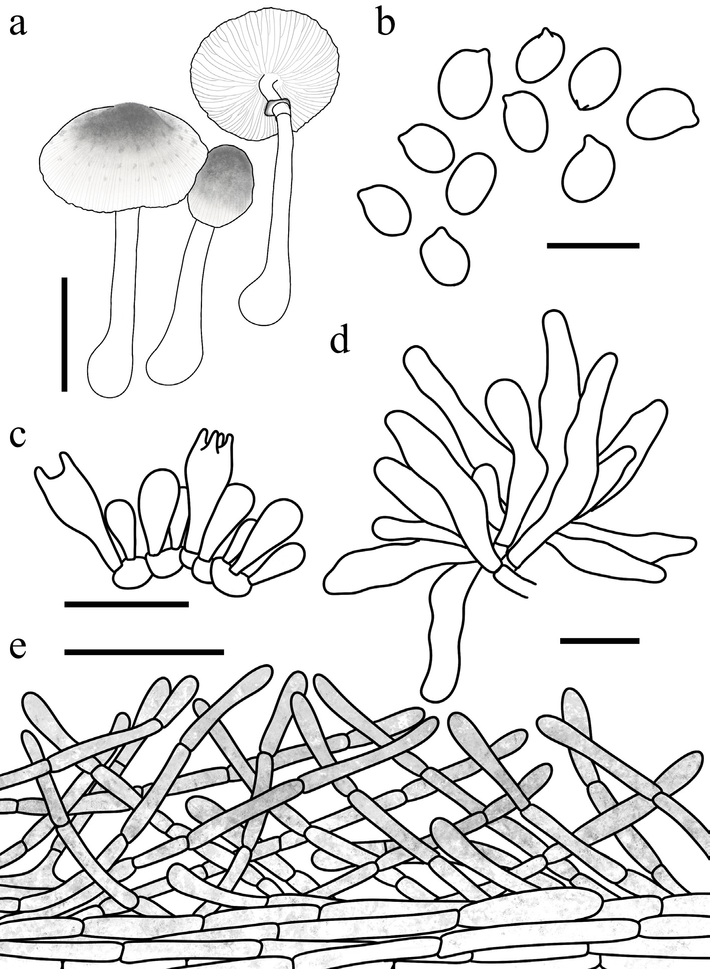
Figure 41.
Leucocoprinus griseocentralis. (a) Basidiomata. (b) Basidiospores. (c) Basidia. (d) Cheilocystidia. (e) Surface covering at pileus centre. Scale bars: (a) = 1 cm; (b) = 10 μm; (c), (d) = 20 μm; (e) = 50 μm.
Chinese name: 灰顶白鬼伞 (Pīn yīn: huī ding bái guǐ sǎn)
Index Fungorum number: IF904660; Facesoffungi number: FoF18951
Etymology – 'griseocentralis' is derived from the Latin griseus (grey) and centralis (central), referring to the distinctive grey disc at the centre of the pileus.
Type – China. Heilongjiang: Daxinganling area, Guqi Valley National Wetland Park, 14 August 2025, coll. X.Y. Zhou, FJAU78003.
Diagnosis – characterized by its white pileus with felted, granite gray squamules, a white annulus with a granite gray margin, and predominantly cylindrical cheilocystidia.
Basidiomata small to medium-sized. Pileus 0.6–3.8 cm in diam., initially ovate, expanding to plano-convex or applanate; umbo obtuse; background white; centre with greige (#C1B7A5) to granite gray (#676767) finely lumpy-squamulose, felted squamules; squamules around the centre fading radially toward the margin; margin with inconspicuous short striations, easily radially splitting. Lamellae free, close, white to cream; edge concolorous; lamellulae in 2 tiers. Stipe 1.9–3.9 cm × 0.1–0.3 cm, subcylindrical, hollow, slightly thickened towards the slightly bulbous base, white; surface smooth or covered with fine fibrils. Annulus superior, ascending, white with a granite gray margin, membranous, ephemeral. Context thin to slightly thick, white, unchanging when bruised. Odour and taste not recorded.
Basidiospores (60/3/3) 5.7–7.0(–7.4) × 3.5–5.0 μm, avl × avw = 6.0–6.2 × 4.2–4.6 μm, Q = 1.30–1.66, avQ = 1.42–1.50, ellipsoid to oblong, hyaline, slightly thick-walled, smooth, dextrinoid, cyanophilous, congophilous, metachromatic in Cresyl Blue; germ pore absent. Basidia 16–24 × 6–9 μm, clavate, predominantly 4-spored, occasionally 2-spored, thin-walled. Pseudoparaphyses absent. Pleurocystidia absent. Cheilocystidia 24–63 × 5–12 μm, predominantly cylindrical, sometimes narrowly utriform, flexuose, hyaline, thin-walled. Surface covering at pileus centre an interwoven plagiotrichoderm consisting of cylindrical hyphae; hyphae densely entangled, repent to slightly ascending, 5–17 μm wide, slightly thick-walled, with granite-gray intracellular pigment, sometimes with parietal pigment. Stipe covering a cutis consisting of cylindrical hyphae; hyphae repent, 3–13 μm wide, thin-walled, hyaline to light yellow. Clamp connections not observed.
Habit and habitat – solitary to scattered on humic soil in forests or meadows, sometimes growing among mosses.
Known distribution – Northeast China.
Additional specimens examined – China. Heilongjiang: Daxinganling area, Guqi Valley National Wetland Park, 25 July 2024, coll. W.N. Hou & C. Cheng, FJAU78001, FJAU77997, 14 August 2025, coll. X.Y. Zhou, H. Cheng & L.H. Li, FJAU78002, FJAU78006, FJAU78004, FJAU78005; Qixia Mountain Botanical Garden, 14 August 2025, coll. W.N. Hou, FJAU78007; Daxinganling area, Mohe City, 26 July 2024, coll. W.N. Hou, FJAU77999; Jilin: Baishan City, Lushuihe National Forest Park, 28 July 2022, coll. H.B. Song, FJAU78000; Yanbian Korean Autonomous Prefecture, Chinese Merganser Park, 1 August 2022, coll. X. Wang, FJAU78008; Dunhua City, Beishan Park, 17 August 2024, coll. X.Y. Zhou, FJAU77998; Changchun City, Jilin Agricultural University, 27 August 2025, coll. X.Y. Zhou, FJAU78119, FJAU78120, FJAU78121; 29 August 2025, coll. X.Y. Zhou, FJAU78116, FJAU78117; 30 August 2025, coll. T. Bau & X.Y. Zhou, FJAU78118.
Notes – Leucocoprinus griseocentralis shares morphological similarities—specifically a pale-greyish pileus and indistinct marginal striations—with members of the Lc. heinemannii complex (Lc. heinemannii Migl., Lc. domingensis Justo, Bizzi, Angelini & Vizzini, Lc. tephrolepis Justo, Bizzi, Angelini & Vizzini, and Lc. parvipileus Justo, Bizzi, Angelini & Vizzini) as well as with phylogenetically related species (Lc. parvicanus T. Bau & X.Y. Zhou, Lc. sphaerodermus T. Bau & X.Y. Zhou, and Lc. griseofloccosus Lagardère & Eyssart.). However, these lookalikes can be distinguished by micromorphology: species in the Lc. heinemannii complex possess pseudoparaphyses surrounding the basidia, whereas Lc. parvicanus Lc. sphaerodermus, and Lc. griseofloccosus are characterized by pileal squamules composed of sphaerocysts[94,95].
Leucocoprinus parvicanus T. Bau, X.Y. Zhou & C. Cheng, sp. nov. Figs 34a, b, 42
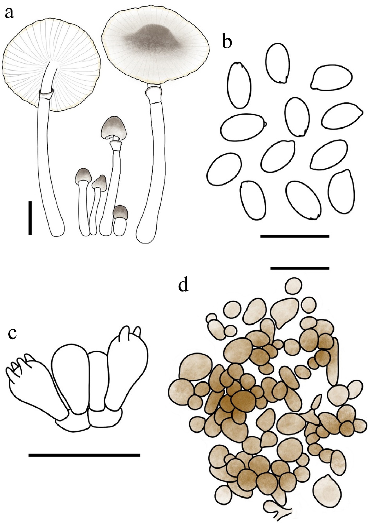
Figure 42.
Leucocoprinus parvicanus. (a) Basidiomata. (b) Basidiospores. (c) Basidia. (d) Surface covering at pileus centre. Scale bars: (a) = 1 cm; (b) = 10 μm; (c), (d) = 20 μm; (e) = 50 μm.
Chinese name: 小灰白鬼伞 (Pīn yīn: xiǎo huī bái guǐ sǎn)
Index Fungorum number: IF904657; Facesoffungi number: FoF18948
Etymology – 'parvicanus' is a compound Latin adjective derived from parvus (small) and canus (hoary, grey-white), referring to the small size and greyish-white colour of the basidiomata.
Type – China. Guangdong: Shenzhen City, Wutong Mountain, 7 May 2025, coll. C. Cheng, FJAU78110.
Diagnosis – characterized by its pileus with greige to umber squamules composed of globose to ellipsoid elements, non-dextrinoid basidiospores, and the absence of cheilocystidia.
Basidiomata small. Pileus 0.4–1.9 cm in diam., initially ovate, expanding convex to plano-convex or applanate; umbo obtuse; background white; centre with umber (#635147) fine, fibrillose and powdery squamules; squamules around the centre greige (#C1B7A5) to umber (#635147), finely squamulose; margin costate-plicate up to 1/3 of the radius or up to the umbo, becoming mustard yellow (#E1AD01) at the utmost margin with age. Lamellae free, distant, white; edge concolorous; lamellulae interspersed in 1–3 tiers. Stipe 1.4–5.3 cm × 0.1–0.2 cm, subcylindrical, white; surface covered with concolorous fibrillose squamules. Annulus superior, ascending, white, membranous. Context thin, white, unchanging when bruised. Odour and taste not recorded.
Basidiospores (40/2/2) 5.4–6.9 × 3.0–3.9 μm, avl × avw = 6.0–6.1 × 3.4–3.5 μm, Q = 1.55–2.12, avQ = 1.76–1.82, ellipsoid to oblong or cylindrical, hyaline, slightly thick-walled, smooth, non-dextrinoid, cyanophilous, congophilous, metachromatic in Cresyl Blue; germ pore absent. Basidia 12–19 × 6–9 μm, clavate, predominantly 4-spored, occasionally 2-spored, thin-walled. Pseudoparaphyses absent. Pleurocystidia absent. Cheilocystidia absent. Surface covering at pileus centre a conioderm consisting of loosely and irregularly arranged cells; cells predominantly subglobose, globose, or slightly ellipsoid, with golden-brown (#885407) intracellular pigment. Stipe covering a cutis consisting of cylindrical hyphae; hyphae repent, 4–13 μm wide, thin-walled, hyaline to light yellow. Clamp connections not observed.
Habit and habitat – solitary to scattered on humus in broadleaf forests.
Known distribution – so far recorded only from Central-Southern China.
Additional specimens examined – China. Guangdong: Shenzhen City, Wutong Mountain, 3 May 2025, coll. C. Cheng, FJAU78102.
Notes – Macroscopically, Leucocoprinus parvicanus shares similar grayish-brown pileus squamules with Lc. lampadiformis Kun L. Yang & Jia Y. Lin and Lc. kibii Kun L. Yang & Jia Y. Lin. Microscopically, the pileus coverings at the centre of all three species are uniformly composed of a conioderm[30]. However, Lc. parvicanus can be readily distinguished from the latter two species by the complete absence of cheilocystidia.
Phylogenetically, Lc. parvicanus clusters closely with Lc. lampadiformis, Lc. kibii, and Lc. sinumbellatus T. Bau & X.Y. Zhou to form a distinct lineage. Morphologically, all members within this clade share a similar pileus covering structure composed of conioderm cells. Nonetheless, Lc. parvicanus stands out within this group due to its lack of cheilocystidia, whereas the other three taxa all possess well-developed cheilocystidia. Furthermore, Lc. sinumbellatus can be easily separated from Lc. parvicanus, Lc. lampadiformis, and Lc. kibii by its light golden to pale-pinkish pileus squamules, which contrast sharply with the grayish-brown coloration observed in the other three species.
Leucocoprinus porosporus T. Bau & X.Y. Zhou, sp. nov Figs 34c, d, 43
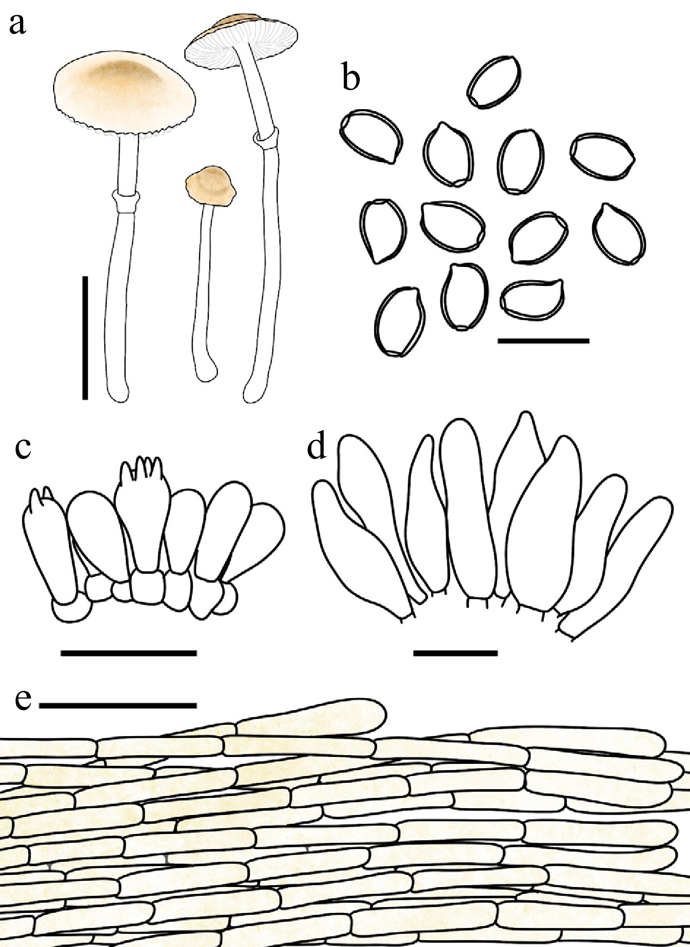
Figure 43.
Leucocoprinus porosporus. (a) Basidiomata. (b) Basidiospores. (c) Basidia. (d) Cheilocystidia. (e) Surface covering at pileus centre. Scale bars: (a) = 1 cm; (b) = 10 μm; (c), (d) = 20 μm; (e) = 50 μm.
Chinese name: 孔孢白鬼伞 (Pīn yīn: kǒng bāo bái guǐ sǎn)
Index Fungorum number: IF904670; Facesoffungi number: FoF18961
Etymology – 'porosporus' is derived from the Greek poros (pore) and sporos (spore), referring to the presence of a distinct germ pore on the basidiospores.
Type – China Anhui: Chizhou City, Guniujiang National Nature Reserve, 15 June 2022, coll. L.Y. Zhu, FJAU78028.
Diagnosis – Leucocoprinus porosporus is distinguished by the presence of a germ pore in the basidiospores and a pileus decorated with yellow-brown to red-brown scales.
Basidiomata small. Pileus 0.5–1.1 cm in diam., convex to plano-convex or applanate when mature; background white; squamules fulvous (#E48400) to brown-red (#B25B09), fading gradually toward the margin. Lamellae free, moderately crowded, white; edge concolorous; lamellulae interspersed in two tiers. Stipe 1.8–3.6 cm × 0.1–0.2 cm, subcylindrical, hollow, white; surface smooth or covered with fine fibrils. Annulus median to superior, ascending, white, membranous, readily detachable. Context thin, white, unchanging when bruised. Odour and taste not recorded.
Basidiospores (40/2/2) 5.9–7.0 × 3.4–4.2 µm, avl × avw = 6.3–6.4 × 3.7–3.9 µm, Q = 1.48–1.87, avQ = 1.63–1.70, ellipsoid to oblong, hyaline, slightly thick-walled, smooth, dextrinoid, cyanophilous, congophilous, metachromatic in Cresyl Blue; germ pore present. Basidia 13–19 × 6–10 µm, clavate, predominantly 4-spored, occasionally 2-spored, thin-walled. Pseudoparaphyses absent. Pleurocystidia absent. Cheilocystidia 32–55 × 6–14 µm, narrowly utriform to utriform, occasionally with apical crystals, hyaline, thin-walled. Surface covering at pileus centre a cutis consisting of cylindrical hyphae; hyphae strictly horizontal, parallel, 4–14 µm wide, thin-walled, with flax-yellow intracellular pigment. Stipe covering a cutis consisting of cylindrical hyphae; hyphae repent, 3–10 μm wide, thin-walled, hyaline to light yellow. Clamp connections not observed.
Habit and habitat – solitary on humus or sand in broadleaf forests dominated by Acer.
Known distribution – so far recorded only from East China.
Additional specimens examined – China. Anhui: Chizhou City, Guniujiang National Nature Reserve, 15 June 2022, coll. H. Cheng, FJAU78027.
Notes – Macroscopically, Leucocoprinus porosporus shares a resemblance with Lc. atroazureus, as both exhibit yellowish-brown, grey-orange, to reddish-brown pileus squamules. However, Lc. atroazureus is immediately distinguishable by its basidiomata turning dark blue upon injury or drying, and by its basidiospores lacking a germ pore[96].
Phylogenetically, Lc. porosporus occupies an unusual and highly isolated position (Figs 1, 3). It forms a remarkably long, independent branch nested between Lc. clade III and Lc. clade IV. Consequently, we provisionally designate this unique lineage as the Lc. porosporus clade. Although its exact topological placement between clades III and IV fluctuates across different phylogenetic analyses, it consistently remains within this intermediate zone and never clusters with Lc. clades I or II.
Interestingly, this phylogenetic isolation is mirrored by an atypical combination of microscopic features. In the genus Leucocoprinus, species possessing basidiospores with a distinct germ pore typically also feature pseudoparaphyses in the hymenium. However, Lc. porosporus represents a striking exception, as it completely lacks pseudoparaphyses. The underlying evolutionary drivers for this peculiar character combination and its long-branch isolation remain unclear. Future studies with expanded taxon sampling are essential to fully elucidate the evolutionary history and precise systematic position of this unique lineage.
Leucocoprinus sinumbellatus T. Bau, X.Y. Zhou & C. Cheng, sp. nov Figs 34e, f, 44
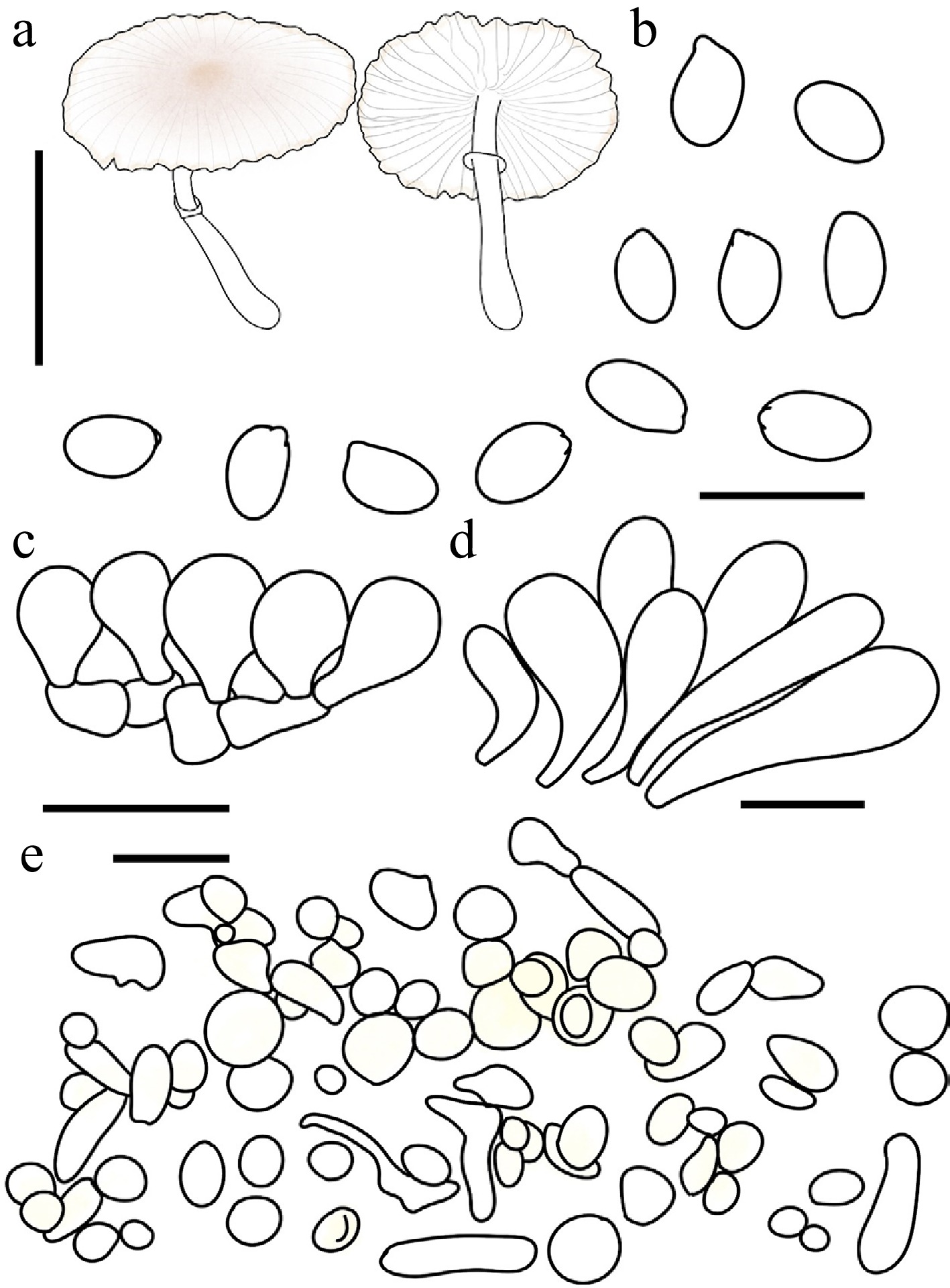
Figure 44.
Leucocoprinus sinumbellatus. (a) Basidiomata. (b) Basidiospores. (c) Basidia. (d) Cheilocystidia. (e) Surface covering at pileus centre. Scale bars: (a) = 1 cm; (b) = 10 μm; (c), (d) = 20 μm; (e) = 50 μm.
Chinese name: 油纸伞白鬼伞 (Pīn yīn: yóu zhǐ sǎn bái guǐ sǎn)
Index Fungorum number: IF904658; Facesoffungi number: FoF18949
Etymology – 'sinumbellatus' from Sinae (China) and umbellatus (umbrella-like), denoting the pileus that resembles a traditional Chinese oil-paper umbrella.
Type – China. Guangdong: Shenzhen City, Wutong Mountain, 19 June 2025, FJAU78109.
Diagnosis – characterized by its white pileus with a costate-plicate and pale-gold margin, non-dextrinoid basidiospores, and the pileal squamules composed of globose to ellipsoid cells.
Basidiomata small. Pileus 1.0–1.5 cm in diam., plano-convex to applanate when mature; background white to cream; centre with pale-gold (#E6BE8A) fine, fibrillose squamules; squamules around the centre nude pink (#DDC0B4) to pale gold (#E6BE8A), finely squamulose; margin costate-plicate up to 2/3 of the radius or up to the umbo, becoming pale gold at the utmost margin with age. Lamellae free, distant, white; edge concolorous; lamellulae interspersed in 1–3 tiers. Stipe 1.4–2.1 cm × 0.1–0.2 cm, subcylindrical, hollow, white; surface smooth. Annulus superior, ascending, white, membranous, ephemeral. Context thin, white, unchanging when bruised. Odour and taste not recorded.
Basidiospores (40/2/2) 5.6–6.7 × 3.3–4.6 μm, avl × avw = 6.1 × 3.6–3.7 μm, Q = 1.45–1.85, avQ = 1.66–1.68, ellipsoid to oblong, hyaline, slightly thick-walled, smooth, non-dextrinoid, cyanophilous, congophilous, metachromatic in Cresyl Blue; germ pore absent. Basidia 14–20 × 8–11 μm, broadly clavate to spheropedunculate; sterigmata not observed. Pseudoparaphyses present, spheropedunculate, dimensions similar to basidia. Pleurocystidia absent. Cheilocystidia 28–54 × 8–20 μm, oblong to clavate, hyaline, thin-walled. Surface covering at pileus centre a conioderm consisting of loosely and irregularly arranged cells; cells predominantly subglobose, globose, or slightly ellipsoid, 11–43 μm wide, some possessing broad connecting outgrowths to cylindrical elements, thin-walled, with light-yellow intracellular pigment. Stipe covering a cutis consisting of cylindrical hyphae; hyphae repent, 4–12 μm wide, thin-walled, hyaline. Clamp connections not observed.
Habit and habitat – solitary to scattered among mosses.
Known distribution – so far recorded only from Central-Southern China.
Additional specimens examined – China. Guangdong: Shenzhen City, Wutong Mountain, 15 June 2025, coll. C. Cheng, FJAU77949.
Notes – Phylogenetically, Leucocoprinus sinumbellatus forms a distinct lineage that clusters with Lc. parvicanus, Lc. lampadiformis, and Lc. kibii. However, these three phylogenetically allied taxa can be readily distinguished by their grayish-brown pileus squamules[30], which contrast sharply with the light-golden to pale-pinkish squamules of the new species. Furthermore, microscopically, Lc. sinumbellatus possesses pseudoparaphyses in the hymenium, a feature completely absent in the other three closely related species.
Macroscopically, Leucocoprinus sinumbellatus also shares a resemblance with Lc. parviceps T. Bau & X.Y. Zhou and Lc. lacteus T. Bau & X.Y. Zhou, particularly in the overall white coloration of the basidiomata. Nevertheless, the latter two species are easily differentiated by possessing distinctly dextrinoid basidiospores, well-developed cheilocystidia, and a pileipellis structured as a cutis.
Microscopic examinations of the two available collections of Lc. sinumbellatus revealed no sterigmata on the basidia. Instead, the hymenium was predominantly occupied by broadly clavate to spheropedunculate pseudoparaphyses. It remains unclear whether this absence of sterigmata is an artifact of the developmental state of the collected basidiomata, or if it represents an inherent species-specific trait. Therefore, additional collections and further investigations are required to definitively verify this morphological feature.
Leucocoprinus sphaerodermus T. Bau & X.Y. Zhou, sp. nov Figs 34g−i, 45
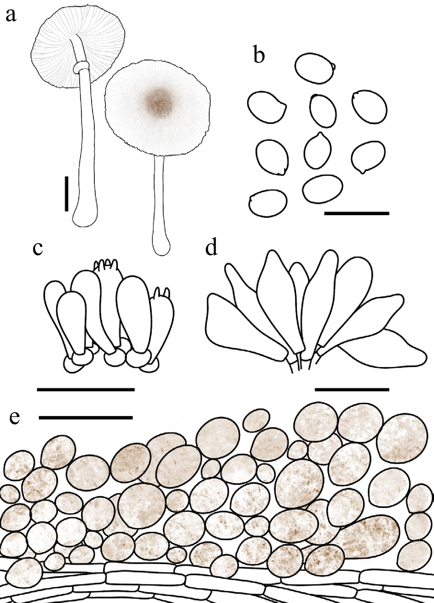
Figure 45.
Leucocoprinus sphaerodermus. (a) Basidiomata. (b) Basidiospores. (c) Basidia. (d) Cheilocystidia. (e) Surface covering at pileus centre. Scale bars: (a) = 1 cm; (b) = 10 μm; (c), (d) = 20 μm; (e) = 50 μm.
Chinese name: 球鳞白鬼伞 (Pīn yīn: qiú lín bái guǐ sǎn)
Index Fungorum number: IF904659; Facesoffungi number: FoF18950
Etymology – 'sphaerodermus' is derived from the Greek sphaira (sphere, alluding to the sphaerocysts) and derma (skin, pileus covered), in reference to the pileus cover, which is composed of sphaerocysts that form the surface squamules.
Type – China. Jilin: Jilin City, Beishan Park, 2 August 2025, coll. Y. Wang, FJAU78049.
Diagnosis – characterized by its white pileus decorated with gingerbread- to hickory-brown granular to punctiform squamules, dextrinoid basidiospores, and narrowly utriform to fusiform cheilocystidia.
Basidiomata small. Pileus 0.9–3.1 cm in diam., plano-convex to plane when mature; umbo low, obtuse; background white; centre with red-brown (#66332B) to hickory-brown (#371D10) finely lumpy-squamulose, felted squamules; squamules around the centre granular to punctiform, becoming lighter in colour towards the margin; margin with indistinct striations. Lamellae free, moderately crowded, white to cream; edge concolorous; lamellulae in 1–3 tiers. Stipe 1.7–5.4 cm × 0.1–0.3 cm, subcylindrical, hollow, white; base with white mycelial tomentum; surface smooth or covered with fine fibrils. Annulus superior, ascending, white, membranous, readily detachable. Context thin, white, unchanging when bruised. Odour and taste not recorded.
Basidiospores (60/3/3) 4.9–5.8 × 3.2–3.7 μm, avl × avw = 5.0–5.2 × 3.4–3.7 μm, Q = 1.23–1.60, avQ = 1.37–1.49, broadly ellipsoid to ellipsoid, hyaline, slightly thick-walled, smooth, dextrinoid, cyanophilous, congophilous, metachromatic in Cresyl Blue; germ pore absent. Basidia 13–19 × 5–9 μm, clavate, predominantly 4-spored, occasionally 2-spored, thin-walled. Pseudoparaphyses absent. Pleurocystidia absent. Cheilocystidia 24–48 × 5–21 μm, narrowly utriform to fusiform, hyaline, thin-walled. Surface covering at pileus centre a conioderm consisting of loosely and irregularly arranged cells; cells predominantly subglobose, globose, or slightly ellipsoid, 11–39 μm wide, occasionally exhibiting broad connecting outgrowths to cylindrical elements, thin to slightly thick-walled, with red-brown intracellular pigment. Stipe covering a cutis consisting of cylindrical hyphae; hyphae repent, 3–8 μm wide, thin-walled, hyaline to light yellow. Clamp connections absent.
Habit and habitat – solitary on humus-rich soil in broadleaf forests.
Known distribution – currently known only from Northeast China.
Additional specimens examined – China. Jilin: Changchun City, Nanhu Park, 11 July 2021, coll. X. Wang, FJAU78048; Jilin Agricultural University, 5 July 2022, coll. W.N. Hou, FJAU78050.
Notes – Morphologically, Leucocoprinus sphaerodermus is similar to Lc. parvicanus and Lc. griseofloccosus[95]. However, Lc. parvicanus is readily distinguished by its non-dextrinoid basidiospores and the complete absence of cheilocystidia. Meanwhile, Lc. griseofloccosus can be separated by its clavate to lageniform cheilocystidia and its specific habitat on decaying Alnus glutinosa.
Phylogenetically, Lc. sphaerodermus is nested within Lc. clade IV, where it clusters together with Lc. castoris Kun L. Yang & Jia Y. Lin, Lc. rusticus Kun L. Yang, Jia Y. Lin & Z.Chao Liu, Lc. urbanicus Kun L. Yang & Jia Y. Lin, and Lc. pomariensis Kun L. Yang & Jia Y. Li to form a distinct subclade. A shared morphological characteristic of the species within this subclade is that their pileus squamules are uniformly composed of conioderm cells.
Despite this structural similarity, Lc. sphaerodermus can be clearly differentiated from its phylogenetically allied taxa. Compared to Lc. castoris, it possesses distinct brownish squamules on the pileus, whereas Lc. castoris lacks such brownish tones and produces slightly longer basidiospores measuring 5.5–6.5 × 3.0–3.5 µm, as opposed to the spores of Lc. sphaerodermus, which measure 4.9–5.8 × 3.2–3.7 µm. Furthermore, Lc. sphaerodermus is characterized by its narrowly utriform to fusiform cheilocystidia measuring 24.0–48.0 × 5.0–21.0 µm. This prominent microscopic feature effectively separates it from the remaining three species in the subclade, which typically possess mostly clavate cheilocystidia. Specifically, Lc. rusticus produces variably clavate cheilocystidia measuring 6.0–28.0 × 8.0–12.0 µm alongside noticeably larger basidiospores (6.0–7.0 × 3.0–3.5 µm); Lc. urbanicus is characterized by predominantly clavate cheilocystidia and slightly longer basidiospores (5.5–6.5 × 3.0–3.5 µm); and Lc. pomonalis features generally clavate and comparatively smaller cheilocystidia measuring 20.0–32.0 × 7.5–13.0 µm.
Leucocoprinus subincarnatus T. Bau & X.Y. Zhou, sp. nov Figs 34j−l, 46
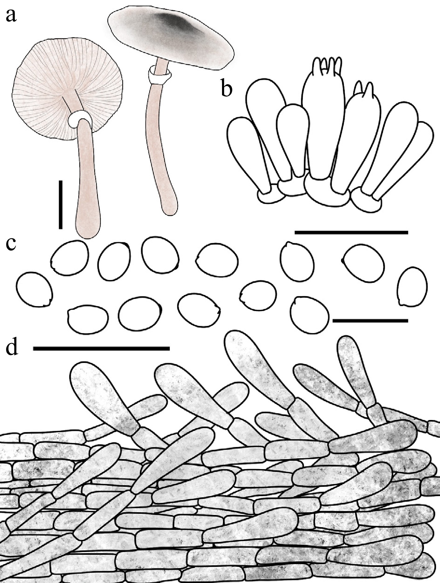
Figure 46.
Leucocoprinus subincarnatus. (a) Basidiomata. (b) Basidia. (c) Basidiospores. (d) Surface covering at pileus centre. Scale bars: (a) = 1 cm; (b) = 10 μm; (c), (d) = 20 μm; (e) = 50 μm.
Chinese name: 近粉褶白鬼伞 (Pīn yīn: jìn fěn zhě bái guǐ sǎn)
Index Fungorum number: IF904655; Facesoffungi number: FoF 18946
Etymology – 'subincarnatus' is derived from the Latin prefix sub- (pale, somewhat) and incarnatus (flesh-coloured), referring to the distinctive nude pink tint of the lamellae.
Type – China. Jilin: Jiaohe City, Qianjin Forest Farm, 24 July 2022, coll. W.N. Hou, FJAU78095.
Diagnosis – characterized by its greige pileus with a prominent dark charcoal disc, white to nude-pink lamellae, and the absence of cheilocystidia.
Basidiomata small. Pileus 1.1–2.1 cm in diam., plano-convex to plane; umbo obtuse; background greige (#C1B7A5); centre with dark charcoal (#333333) fine, floccose-felted squamules; squamules around the centre umber (#635147) to dark charcoal, finely squamulose. Lamellae free to slightly adnexed, moderately crowded, cream to nude pink (#DDC0B4); edge concolorous; lamellulae in 1–3 tiers. Stipe 2.2–3.8 cm × 0.2–0.3 cm, subcylindrical, hollow, nude pink; surface covered with white fibrillose squamules. Annulus superior, ascending, white, membranous. Context thin, white to nude pink, unchanging when bruised. Odour and taste not recorded.
Basidiospores (40/2/2) 4.9–5.9(–6.3) × 3.6–4.9 μm, avl × avw = 5.4–5.5 × 4.2–4.3 μm, Q = 1.13–1.57, avQ = 1.27–1.33, broadly ellipsoid to ellipsoid, hyaline to very pale yellowish brown, slightly thick-walled, smooth, weakly dextrinoid, cyanophilous, congophilous, metachromatic in Cresyl Blue; germ pore absent. Basidia 16–22 × 6–9 μm, clavate, predominantly 4-spored, occasionally 2-spored, thin-walled. Pseudoparaphyses absent. Cheilocystidia and pleurocystidia absent. Surface covering at pileus centre an interwoven plagiotrichoderm consisting of cylindrical hyphae; hyphae densely entangled, repent to slightly ascending, 7–14 μm wide, slightly thick-walled, with hickory-brown (#371D10) to dark-charcoal intracellular and parietal pigment. Stipe covering a cutis consisting of cylindrical hyphae; hyphae repent, 5–8 μm wide, thin-walled, hyaline to pale yellowish brown. Clamp connections not observed.
Habit and habitat – solitary to scattered on the forest floor.
Known distribution – so far recorded only from Northeast China
Additional specimens examined – China, Jilin: Jiaohe City, Qianjin Forest Farm, 23 July 2023, coll. L.Y. Zhu, FJAU78275; 24 August 2023, coll. H. Cheng, FJAU78134.
Notes – Macroscopically, Leucocoprinus subincarnatus and Lc. subhymenoderma (Bon & A. Caball.) Kun L. Yang, Jia Ying Lin & Zhu L. Yang (described from Spain) both exhibit a pileus with grey to greyish-brown dark tones; furthermore, the lamellae in both species are slightly tinted with colour. However, Lc. subhymenoderma is distinguished by its stipe being vinaceous-red in the lower section and the presence of cheilocystidia. Microscopically, the pileus covering of the latter species is composed of a catenulate structure of ellipsoid to globose cells, a structure similar to the pileus covering observed in Echinoderma (Locq. ex Bon) Bon[87].
Leucocoprinus subumbrosquamosus T. Bau & X.Y. Zhou, sp. nov Figs 35a, 47
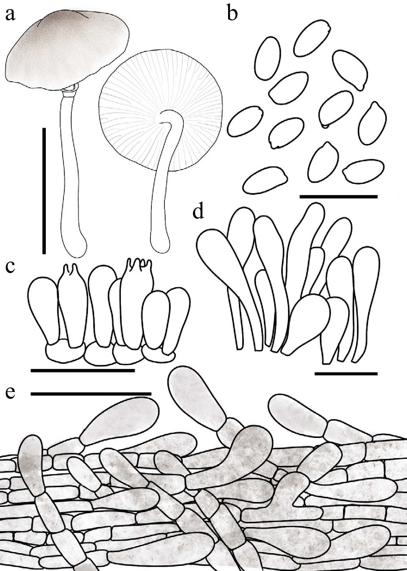
Figure 47.
Leucocoprinus subumbrosquamosus. (a) Basidiomata. (b) Basidiospores. (c) Basidia. (d) Cheilocystidia. (e) Surface covering at pileus centre. Scale bars: (a) = 1 cm; (b) = 10 μm; (c), (d) = 20 μm; (e) = 50 μm.
Chinese name: 近褐鳞白鬼伞 (Pīn yīn: jìn hè lín bái guǐ sǎn)
Index Fungorum number: IF904662; Facesoffungi number: FoF18953
Etymology – 'subumbrosquamosus' (from the Latin prefix sub-, implying resemblance, and umbrosquamosus) denotes its close morphological similarity to Leucocoprinus umbrosquamosus, particularly in the umber-coloured squamules on the pileus.
Type – China. Jilin: Ji'an City, Wunufeng National Forest Park, 14 July 2024, coll. J.L. Wei, FJAU78071.
Diagnosis – characterized by its white pileus covered with brownish floccose squamules, non-dextrinoid and weakly cyanophilous basidiospores, and greige cheilocystidia.
Basidiomata small. Pileus 1.2–1.8 cm in diam., convex to plano-convex or applanate; umbo obtuse; background white; centre with umber (#635147) floccose squamules; squamules around the centre pale umber (#9A7E65) to umber, finely squamulose. Lamellae free, moderately crowded, white; edge concolorous; lamellulae in 1–3 tiers. Stipe 2.1–3.0 cm × 0.1–0.2 cm, subcylindrical, hollow, white; surface smooth to covered with fine fibrils; base with white mycelium. Annulus median, ascending, white, membranous, friable. Context thin, white, unchanging when bruised. Odour and taste not recorded.
Basidiospores (40/2/2) 5.0–6.2 × 2.4–3.4 μm, avl × avw = 5.6–5.7 × 2.9 μm, Q = 1.74–2.30, avQ = 1.93–1.97, oblong to cylindrical, hyaline, slightly thick-walled, smooth, non-dextrinoid, weakly cyanophilous, congophilous, metachromatic in Cresyl Blue; germ pore absent. Basidia 13–18 × 5–7 μm, clavate, predominantly 4-spored, occasionally 2-spored, thin-walled. Pseudoparaphyses absent. Pleurocystidia absent. Cheilocystidia 25–40 × 8–13 μm, clavate to narrowly fusiform, with an obtuse apex, hyaline to greige, thin-walled. Surface covering at pileus centre an interwoven plagiotrichoderm consisting of repent to slightly ascending hyphae; hyphae densely entangled, elements predominantly cylindrical to oblong-ellipsoid, 4–13 μm wide, slightly thick-walled, with umber intracellular and parietal pigments. Stipe covering a cutis consisting of cylindrical hyphae; hyphae repent, 4–10 μm wide, thin-walled, hyaline to pale yellowish brown. Clamp connections not observed.
Habit and habitat – solitary to scattered on humus-rich soil in coniferous forests.
Known distribution – currently known only from Northeast China.
Additional specimens examined – China. Jilin: Ji'an City, Wunufeng National Forest Park, 14 July 2024, coll. X.Y. Zhou, FJAU78124.
Notes – Phylogenetically, Leucocoprinus subumbrosquamosus is nested within Lc. clade IV, where it clusters tightly with Lc. umbrosquamosus T. Bau & X.Y. Zhou and Lc. melanotrichus (Malençon & Bertault) Migl. & Donato to form a distinct subclade. A notable shared characteristic among the taxa within this subclade is their extremely weak reaction to Melzer's reagent[80].
Morphologically, Lc. subumbrosquamosus closely resembles its sister taxon, Lc. umbrosquamosus, as both exhibit brownish pileus squamules and non-dextrinoid basidiospores. However, Lc. umbrosquamosus can be readily distinguished by possessing significantly more elongated basidiospores with a Q value of 1.74–2.30, compared to the relatively broader spores of the new species, which exhibit a Q value of 1.29–1.65. Furthermore, the pileus covering of Lc. umbrosquamosus is a typical moniliform plagiotrichoderm, which differs from the interwoven plagiotrichoderm observed in Lc. subumbrosquamosus.
Microscopically, the pileus squamules of the Lc. subumbrosquamosus present a structural configuration. The hyphae are composed of catenulate, cylindrical to elongated-ellipsoid cells. While this chain-like arrangement superficially resembles a moniliform plagiotrichoderm (Type III-B), it fundamentally lacks the intimately intermixed subglobose to globose cells that define this specific type. Consequently, we tentatively classify its pileipellis as an interwoven plagiotrichoderm (Type I-B), interpreting it as a transitional morphological state between an interwoven plagiotrichoderm and a moniliform plagiotrichoderm. However, we cannot entirely rule out the possibility that the absence of globose cells might be an artifact related to the specific development of the two available collections. Further sampling is thus required to definitively confirm the precise composition of its pileus covering.
Leucocoprinus umbrosquamosus T. Bau & X.Y. Zhou, sp. nov Figs 35b, 48
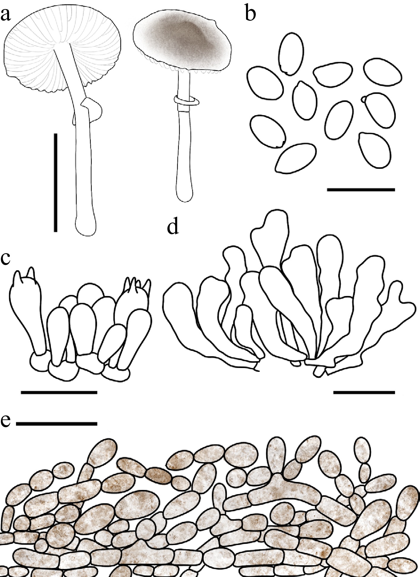
Figure 48.
Leucocoprinus umbrosquamosus. (a) Basidiomata. (b) Basidiospores. (c) Basidia. (d) Cheilocystidia. (e) Surface covering at pileus centre. Scale bars: (a) = 1 cm; (b) = 10 μm; (c), (d) = 20 μm; (e) = 50 μm.
Chinese name: 褐鳞白鬼伞 (Pīn yīn: hè lín bái guǐ sǎn)
Index Fungorum number: IF904661; Facesoffungi number: FoF18952
Etymology – 'umbrosquamosus' is derived from the Latin umbra (shadow, referring to the umber colour) and squamosus (scaly), in reference to the umber-coloured squamules covering the pileus.
Type – China. Jilin: Dunhua City, Beishan Park, 17 August 2024, coll. X.Y. Zhou, FJAU78072.
Diagnosis – characterized by its umber squamulose pileus, non-dextrinoid and weakly cyanophilous basidiospores, and cylindrical to clavate cheilocystidia.
Basidiomata small. Pileus 1.1–1.6 cm in diam., plano-convex to applanate; umbo low, obtuse; background white; centre with umber (#635147) finely felted squamules; squamules around the centre greige (#C1B7A5) to umber (#635147), small, fine. Lamellae free, moderately crowded, white; edge concolorous; lamellulae in 1–3 tiers. Stipe 2.2–3.2 × 0.1–0.2 cm, subcylindrical, hollow, white; surface smooth to covered with fine fibrils. Annulus median, ascending, white, membranous, ephemeral. Context thin, white, unchanging when bruised. Odour and taste not recorded.
Basidiospores (40/2/2) 5.0–6.2 × 3.5–4.5 μm, avl × avw = 5.5–5.7 × 3.7–3.9 μm, Q = 1.29–1.65, avQ = 1.40–1.48, broadly ellipsoid to ellipsoid or oblong, hyaline, thin-walled, smooth, non-dextrinoid, weakly cyanophilous, congophilous, metachromatic in Cresyl Blue; germ pore absent. Basidia 16–23 × 7–9 μm, clavate, predominantly 4-spored, occasionally 2-spored, thin-walled. Pseudoparaphyses absent. Pleurocystidia absent. Cheilocystidia 28–44 × 6–13 μm, cylindrical to clavate, flexuose, occasionally with a branched apex, hyaline, thin-walled. Surface covering at pileus centre a moniliform plagiotrichoderm consisting of repent to slightly ascending hyphae in moniliform chains; cells intermixed with cylindrical, ellipsoid, and subglobose elements, 12–33 μm wide, some with broad connecting excrescences, thin-walled to slightly thick-walled, with greige (#C1B7A5) to umber intracellular pigment. Stipe covering a cutis consisting of cylindrical hyphae; hyphae repent, 3–8 μm wide, thin-walled, hyaline. Clamp connections not observed.
Habit and habitat – solitary to scattered in humus-rich soil in mixed coniferous and broadleaf forests.
Known distribution – so far recorded only from the type locality in Beishan Park, Northeast China.
Additional specimens examined – China. Jilin: Dunhua City, Beishan Park, 17 August 2024, coll. X.Y. Zhou, FJAU78123.
Notes – Macroscopically, Leucocoprinus umbrosquamosus, Lc. subumbrosquamosus, and Lc. turgipes (Justo, Bizzi & Angelini) M. Asif, Saba & Vellinga share a strong morphological resemblance, as all three species possess brownish squamules on the pileus. However, they can be reliably distinguished based on a combination of microscopic structures and lamellar features.
Lc. turgipes is readily segregated from the other two taxa by its distinctly dextrinoid basidiospores and its lamellae, which are initially white but transition to beige-hazel or even ocher-brownish in very mature specimens[61]. In contrast, both Lc. umbrosquamosus and Lc. subumbrosquamosus produce non-dextrinoid basidiospores and retain white to cream lamellae throughout their development.
Furthermore, the structure of the pileus covering provides an additional crucial diagnostic characteristic. Microscopically, the pileipellis of Lc. subumbrosquamosus and Lc. turgipes is composed of an interwoven plagiotrichoderm, whereas Lc. umbrosquamosus is uniquely characterized by a moniliform plagiotrichoderm.
Droplet-exuding species of Leucocoprinus and Candelolepiota
-
Notes: The present study encompasses 21 droplet-exuding species, comprising 20 species within Leucocoprinus and one within Candelolepiota. Currently, 13 of these taxa are known to occur in China. This includes six novel species described herein, alongside seven previously established taxa: Lc. glaber R.L. Zhao & J.X. Li, Lc. shixingensis (Z.S. Bi & T.H. Li) Kun L. Yang, Jia Y. Lin & Zhu L. Yang, Lc. tangerinus, (Y. Yuan & Jun F. Liang) Kun L. Yang, Jia Y. Lin & Zhu L. Yang, Lc. flavirobustus R.L. Zhao & J.X. Li, Lc. lugdunensis S. Basso & N. Schwab, Lc. cepistipes, and Candelolepiota sinica (J.Z. Ying) Kun L. Yang, Jia Y. Lin & Zhu L. Yang[29,80,97,98].
Phylogenetically, Lc. lugdunensis and Lc. cepistipes are nested within Lc. clade III, whereas the six novel species and the remaining droplet-exuding taxa belong to Lc. clade IV. Among them, Lc. taniae is easily segregated from allied taxa by its distinctive sand-dwelling habitat. Strikingly, 17 of these species cluster tightly to form the terminal crown subclade of Lc. clade IV. Within this terminal lineage, all taxa—with the sole exception of Lc. silvestris (Justo, Bizzi & Angelini) M. Asif, Saba & Vellinga—consistently exhibit the macroscopic trait of droplet exudation when fresh. Furthermore, this group includes the ecologically unique Lc. karjaticus (P.B. Patil, N.P. Patil, Chahar & S. Maurya), R.L. Zhao & J.X. Li, which is reportedly associated with termite mounds.
To facilitate morphological identification, the following key is provided for the species of Leucocoprinus and Candelolepiota characterized by distinct macroscopic exudation of droplets. Because these exudates are strictly associated with fresh basidiomata and are invariably lost upon desiccation or in herbarium specimens, the diagnostic features utilized in this key rely heavily on in vivo observations of fresh materials. The key treats a total of 20 species, encompassing both the newly described taxa from the present study and previously established species sourced from comprehensive literature reviews.
It should be noted that a taxon designated as the Leucocoprinus atrodiscus group (voucher: E.C. Vellinga 3265, GenBank GU903302) has been reported in previous literature to exhibit droplet exudation[99]. However, due to the complete absence of a formal and comprehensive morphological description for this collection, it has been excluded from the present identification key, which relies strictly on well-documented macro- and microscopic diagnostic features. Similarly, although Leucocoprinus cinerascens was reported by Yang to exude colourless droplets during its immature stages[30], our observations of two fresh collections—consistent with historical literature[15]—failed to corroborate this guttation phenomenon. Given this phenotypic uncertainty, Lc. cinerascens has likewise been excluded from the present key.
Key 1 Key to the droplet-exuding species of Leucocoprinus and Candelolepiota treated in this study.
1 Habitat restricted to sandy soils; basidiomata almost entirely buried in sand Lc. taniae 1' Habitat not sandy soils; basidiomata epigeous 2 2 Habitat strictly linked to attine ants Lc. karjaticus 2' Habitat not linked to attine ants 3 3 Growing on decaying wood Lc. dacrytus 3' Growing on humus or soil 4 4 Basidiospores possessing a distinct germ pore 5 4' Basidiospores lacking a germ pore 7 5 Pseudoparaphyses absent Candelolepiota sinica 5 Pseudoparaphyses present 6 6 Cheilocystidia 35–48 × 12–22 μm, clavate to broadly fusiform, with papillate or capitate apices Lc. lugdunensis 6 Cheilocystidia 25–60 × 8–15 μm, clavate, with lageniform apices Lc. cepistipes 7 Droplets secreted from the entire basidioma when fresh 8 7 Droplets secreted exclusively from the stipe when fresh 15 8 Currently known from Europe or the Americas 9 8 Currently known only from Asia (China) 11 9 Distributed in Europe; lamellae slightly greyish-white, sometimes with a pinkish sheen; cheilocystidia frequently with peculiar apical appendages Lc. tener 9 Distributed in the Americas; lamellae white to yellowish; cheilocystidia without peculiar apical appendages 10 10 Cheilocystidia 22–58 × 7–20 μm, clavate, lacking mucronate, lageniform, or bifurcated apices Lc. stillatus 10 Cheilocystidia 24–74 × 8–30 μm, clavate, mostly mucronate, lageniform, or bifurcated Lc. margaritifer 11 Stipe surface lacking conspicuous squamules 12 11 Stipe surface covered with squamules 13 12 Basidiospores 6.1–7.1 × 3.6–4.2 μm (avl × avw = 6.4 × 3.8 μm); cheilocystidia narrowly utriform to clavate Lc. luteoguttatus 12 Basidiospores generally smaller, 5.2–6.6 × 2.9–3.4 μm (avl × avw = 5.8 × 3.2 μm); cheilocystidia fusiform to oblong Lc. sudans 13 Cheilocystidia subfusiform to clavate, with distinct apical appendages Lc. shixingensis 13 Cheilocystidia narrowly cylindrical, narrowly fusiform, or clavate, lacking apical appendages 14 14 Stipe surface below the annulus covered from the mid-region to the base with golden-brown to gingerbread-brown floccose squamules Lc. floccipes 14 Stipe surface below the annulus off-white, yellowish-white, or brownish-yellow with fine concolorous squamules Lc. flavirobustus 15 Pileus surface glabrous to finely fibrillose, lacking distinct squamules Lc. glaber 15 Pileus surface with distinct coloured squamules 16 16 Pileus squamules pale gold, golden-brown, or greyish-brown 17 16 Pileus squamules pale orange, brownish-yellow, reddish-brown, or orange-red 18 17 Mature pileus relatively large, 2.7–3.5 cm in diam Lc. guttifer 17 Mature pileus relatively small, 1.1–1.5 cm in diam Lc. ochraceosquamosus 18 Currently known only from India Lc. brunneodiscus 18 Currently known only from China 19 19 Basidiospores 5.2–6.3 × 3.2–4.1 μm Lc. helianthoides 19 Basidiospores 6.5–7.0 × 4.0–4.5 μm Lc. tangerinus The descriptive terms used in Key 1 is based on the original species descriptions[25,29,61,80,99−101], as well as on the present study.
Leucocoprinus floccipes T. Bau & X.Y. Zhou, sp. nov Figs 35f−i, 49
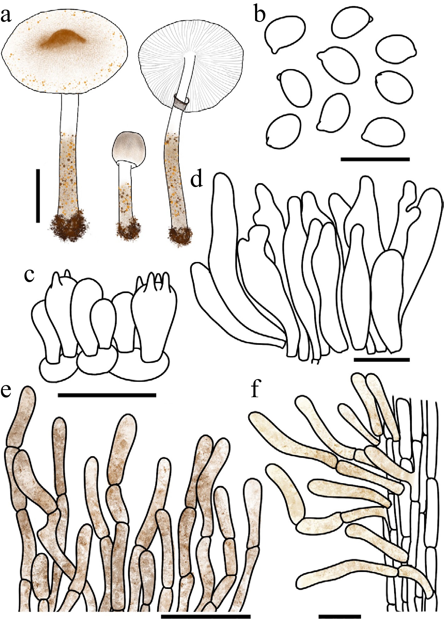
Figure 49.
Leucocoprinus floccipes. (a) Basidiomata. (b) Basidiospores. (c) Basidia. (d) Cheilocystidia. (e) Surface covering at pileus centre. (f) Stipe covering. Scale bars: (a) = 1 cm; (b) = 10 μm; (c), (d), (f) = 20 μm; (e) = 50 μm.
Chinese name: 絮柄白鬼伞 (Pīn yīn: xù bǐng bái guǐ sǎn)
Index Fungorum number: IF904669; Facesoffungi number: FoF18959
Etymology – 'floccipes' is derived from the Latin flocci- (floccose) and -pes (foot, stipe), referring to the distinctly floccose-squamose stipe.
Type – China. Heilongjiang: Daxinganling area, Guqi Valley National Wetland Park, 14 August 2025, coll. X.Y. Zhou, FJAU78078.
Diagnosis – characterized by its basidiomata exuding yellowish-brown to brownish droplets, a pileus with nude pink to brown squamules, the lower half of the stipe covered with golden-brown to gingerbread-brown floccose squamules, and the absence of pleurocystidia.
Basidiomata small, exuding fulvous (#E48400) to red-brown (#66332B) droplet-like secretions when fresh. Pileus 0.8–2.5 cm in diam., initially hemispherical, expanding to plano-convex or applanate; umbo obtuse; background white to cream; squamules nude pink (#DDC0B4), pale gold (#E6BE8A) to brown-red (#B25B09), floccose. Lamellae free, crowded, white; edge concolorous; lamellulae in 1–3 tiers. Stipe 1.3–4.3 × 0.2–0.4 cm, subcylindrical, widening downwards to a slightly bulbous base, hollow, white to cream; surface covered from the mid-region to the base with golden-brown (#885407) to red-brown (#66332B) floccose squamules, more crowded toward the base. Annulus superior, ascending, outer surface golden brown to hickory brown, inner surface white, membranous, ephemeral. Context thin, white, unchanging when bruised. Odour and taste not recorded.
Basidiospores (40/2/2) 5.5–6.5 × 3.7–4.5 µm, avl × avw = 5.7–6.0 × 4.0–4.2 µm, Q = 1.30–1.55, avQ = 1.40–1.44, ellipsoid, hyaline, slightly thick-walled, smooth, dextrinoid, cyanophilous, congophilous, metachromatic in Cresyl Blue; germ pore absent. Basidia 13–24 × 7–10 µm, clavate, predominantly 4-spored, occasionally 2-spored, thin-walled. Pseudoparaphyses absent. Pleurocystidia absent. Cheilocystidia 28–61 × 6–13 µm, narrowly cylindrical to narrowly fusiform or clavate, frequently branched at the apex, hyaline, thin-walled. Surface covering at pileus centre a catenulate trichoderm consisting of erect, frequently septate hyphae appearing as long chains; terminal cells 41–126 × 5–11 µm, subcylindrical and flexuous, slightly thick-walled, sometimes encrusted, with brown-red to red brown intracellular and parietal pigments. Stipe covering a cutis consisting of cylindrical hyphae; hyphae repent; terminal cells 12–64 × 4–8 µm, clavate, with flax yellow to brown-red intracellular and parietal pigment, slightly thick-walled. Clamp connections not observed.
Habit and habitat – solitary to scattered on humus in coniferous forests or in grassy areas.
Known distribution – so far recorded only from Northeast China.
Additional specimens examined – China. Heilongjiang: Daxinganling area, Mohe City, 26 July 2024, coll. H.B. Song, FJAU78073; Nenjiang City, Gaofeng National Forest Park, 13 August 2025, coll. X.Y. Zhou, FJAU78074, Daxinganling area, Guqi Valley National Wetland Park, 14 August 2025, coll. X.Y. Zhou & W.N. Hou, FJAU78075, FJAU78076, FJAU78077, FJAU780810, FJAU78080; Gagdach City, Dongfeng Forest Farm, 15 August 2025, coll. X.Y. Zhou, FJAU78079.
Notes – Macroscopically, Leucocoprinus floccipes and Lc. tener (P.D. Orton) M. Asif, Saba & Vellinga share notable similarities, specifically the exudation of droplets from the fresh stipe and the presence of conspicuous dark brownish squamules on the stipe surface. However, Lc. tener is readily distinguished by its cream-coloured stipe that darkens upon bruising, as well as its larger basidiospores measuring 6.5–8.0 × (3.5–)4.0–4.5(–5.0) μm[80].
Phylogenetically, Lc. floccipes is nested within Lc. clade IV, where it forms a sister branch to Lc. luteoguttatus T. Bau, X.Y. Zhou. Despite their close evolutionary affinity, the latter species can be easily separated morphologically; Lc. luteoguttatus possesses a relatively glabrous stipe, completely lacking the distinct gingerbread-brown floccose squamules characteristic of the new species.
Leucocoprinus guttifer T. Bau & X.Y. Zhou, sp. nov Figs 35c−e, 50
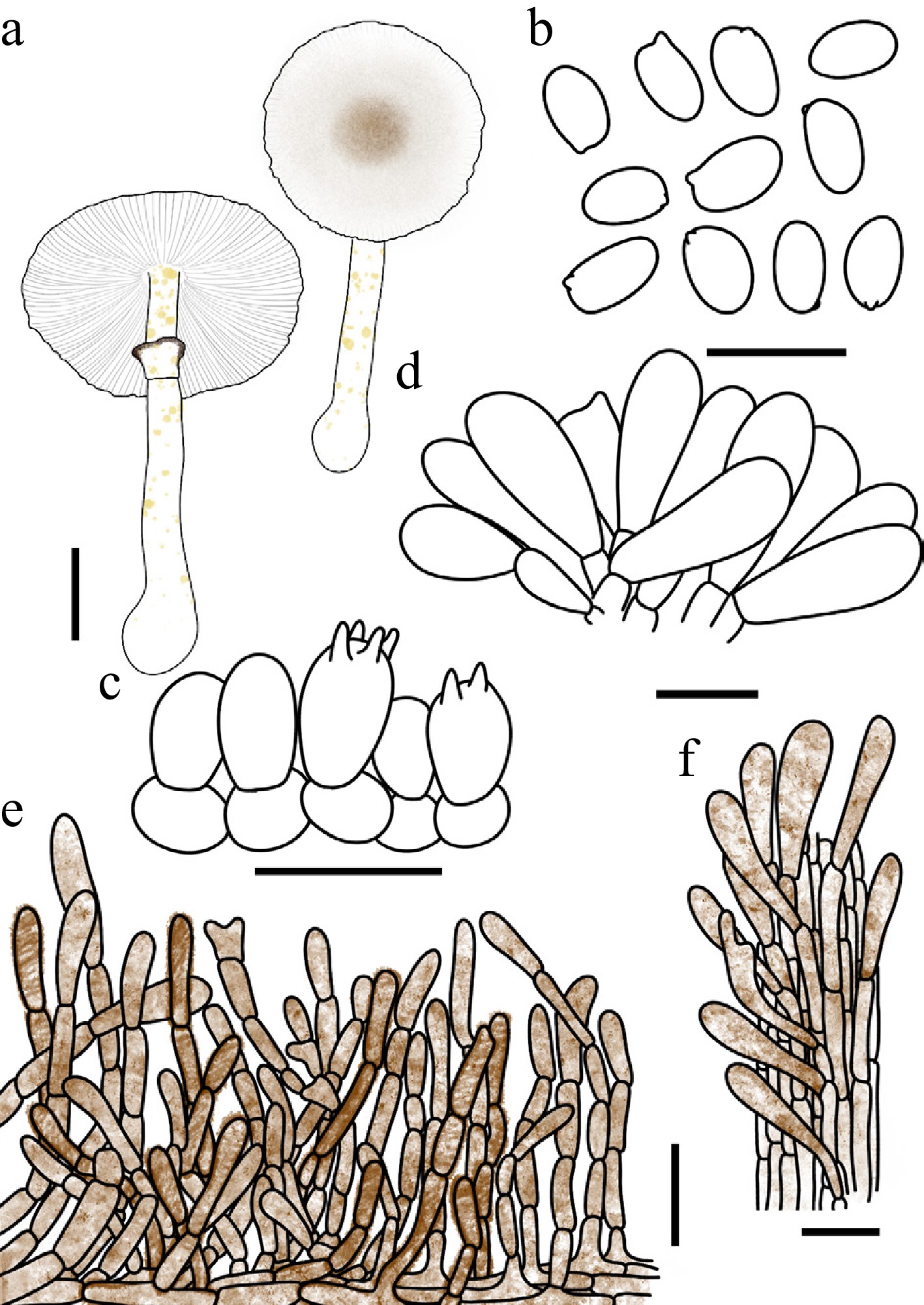
Figure 50.
Leucocoprinus guttifer. (a) Basidiomata. (b) Basidiospores. (c) Basidia. (d) Cheilocystidia. (e) Surface covering at pileus centre. (f) Stipe covering. Scale bars: (a) = 1 cm; (b) = 10 μm; (c), (d), (f) = 20 μm; (e) = 50 μm.
Chinese name: 具液滴白鬼伞 (Pīn yīn: jù yè dī bái guǐ sǎn)
Index Fungorum number: IF905535; Facesoffungi number: FoF 19999
Etymology – 'guttifer' is derived from the Latin 'gutta' (drop or droplet) and the suffix '-fer' (bearing or carrying), referring to the characteristic droplets exuded by fresh basidiomata.
Type – China. Anhui: Chizhou City, Guniujiang National Nature Reserve, 15 June 2022, coll. L.Y. Zhu, FJAU78021.
Diagnosis – characterized by its pileus with brownish cottony squamules, flax yellow droplet-like secretions on the lower half of the stipe, and clavate, septate cheilocystidia.
Basidiomata small. Exuding flax yellow (#EEDC82) droplets from the lower half of the stipe when fresh. Pileus 2.7–3.5 cm in diam., plano-convex to plane when mature; umbo low, obtuse; background white to cream; centre with greige (#C1B7A5) to golden-brown (#885407) or red-brown (#66332B) floccose squamules; squamules around the centre gradually fading in colour towards the margin. Lamellae free, crowded, white to cream; edge concolorous; lamellulae in 2–3 tiers. Stipe 4.5–5.2 × 0.2–0.4 cm, subcylindrical, hollow, white; surface covered with droplets as described above. Annulus superior, ascending, white with a golden brown to hickory brown margin, membranous, persistent. Context thin, white, unchanging when bruised. Odour and taste not recorded.
Basidiospores (40/2/2) 5.2–6.3 × 3.4–4.3 μm, avl × avw = 5.7–6.1 × 3.8–3.9 μm, Q = 1.32–1.70, avQ = 1.46–1.57, ellipsoid to oblong, hyaline, slightly thick-walled, smooth, non-dextrinoid to weakly dextrinoid, cyanophilous, congophilous, metachromatic in Cresyl Blue; germ pore absent. Basidia 14–21 × 6–10 μm, clavate, predominantly 4-spored, occasionally 2-spored, thin-walled. Pseudoparaphyses absent. Pleurocystidia absent. Cheilocystidia 25–50 × 7–20 µm, clavate, frequently septate, hyaline, thin-walled. Surface covering at pileus centre a catenulate trichoderm consisting of erect, frequently septate hyphae appearing as long chains, 70–179 × 8–12µm, slightly thick-walled, with pale gold to red-brown intracellular and parietal pigment. Stipe covering a cutis consisting of repent hyphae; terminal cells 23–71 × 4–8 µm, cylindrical to clavate, with flax yellow to red-brown intracellular and parietal pigment. Clamp connections not observed.
Habit and habitat – solitary to scattered on the forest floor.
Known distribution – East, Northeast, and Southwest China.
Additional specimens examined – China.; Jilin: Changchun City, Jingyuetan National Forest Park, 11 August 2023, coll. J.L. Wei, FJAU78018; Sichuan: Mianyang City, Southwest University of Science and Technology, 14 October 2024, coll. Enthusiasts, FJAU78019; FJAU78020.
Notes – Microscopic examinations of the four available collections of Leucocoprinus guttifer revealed notable intraspecific variation in basidiospore morphology. Specifically, the two collections from Sichuan Province (FJAU78019 and FJAU78020) possess relatively larger, strictly non-dextrinoid basidiospores measuring 6.5–7.7 × 4.2–5.0 μm (avl × avw = 7.0–7.1 × 4.3–4.5 μm, Q = 1.41–1.79, avQ = 1.51–1.58). In contrast, the specimens from Anhui (FJAU78021) and Jilin (FJAU78018) produce slightly smaller basidiospores measuring 5.2–6.3 × 3.4–4.3 μm (avl × avw = 5.7–6.1 × 3.8–3.9 μm, Q = 1.32–1.70, avQ = 1.46–1.57) that exhibit a weakly dextrinoid to dextrinoid reaction. Despite these microscopic discrepancies, all four collections cluster tightly together into a single, well-supported lineage in our phylogeny. Consequently, we interpret these differences as continuous intraspecific variation. Furthermore, since the Sichuan collections lack in situ photographs of the fresh basidiomata, the formal morphological description of this taxon relies primarily on the fully documented collections FJAU78021 and FJAU78018.
Macroscopically, Lc. guttifer and Lc. ochraceosquamosus T. Bau & X.Y. Zhou share several similarities, as both possess pale golden to grayish-brown squamules on the pileus and secrete droplets from the stipe when fresh. However, Lc. ochraceosquamosus can be distinguished by the generally colorless nature of its exudates, whereas the droplets of Lc. guttifer are characteristically flaxen yellow. Microscopically, Lc. guttifer is characterized by frequently septate cheilocystidia, a structural feature that is completely absent in Lc. ochraceosquamosus.
Leucocoprinus helianthoides T. Bau, X.Y. Zhou & C. Cheng, sp. nov Figs 35j−l, 51
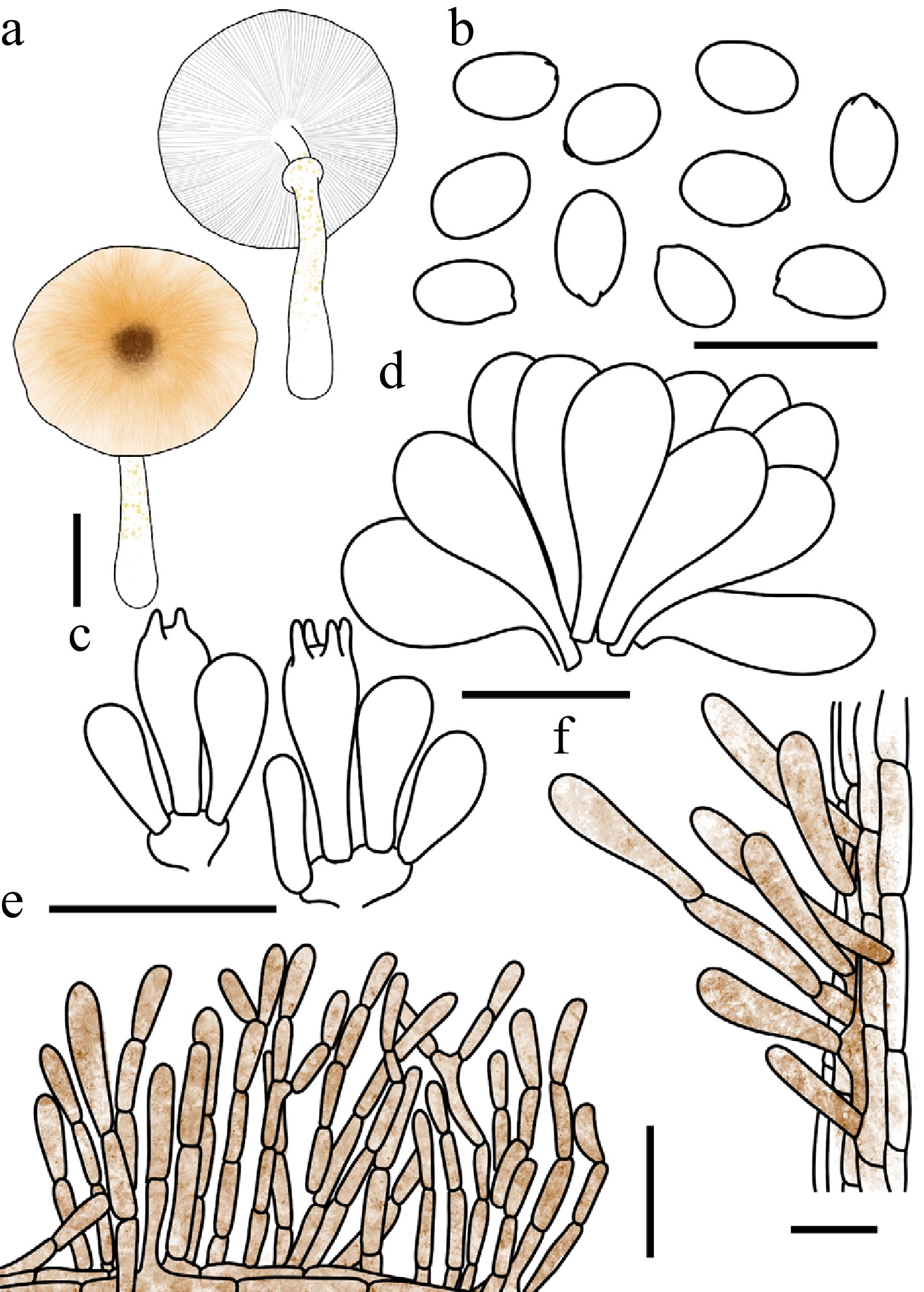
Figure 51.
Leucocoprinus helianthoides. (a) Basidiomata. (b) Basidiospores. (c) Basidia. (d) Cheilocystidia. (e) Surface covering at pileus centre. (f) Stipe covering. Scale bars: (a) = 1 cm; (b) = 10 μm; (c), (d), (f) = 20 μm; (e) = 50 μm.
Chinese name: 向日葵白鬼伞 (Pīn yīn: xiàng rì kuí bái guǐ sǎn)
Index Fungorum number: IF904668; Facesoffungi number: FoF18960
Etymology – 'helianthoides' from the Greek hēlios (sun) and anthos (flower), with the suffix -oides (resembling). The name refers to the sunflower-like appearance of the pileus, which features a gingerbread-brown disc at the center surrounded by a fulvous margin.
Type – China. Guangdong, Shenzhen City, Wutong Mountain, 22 June 2025, coll. C. Cheng, FJAU78093.
Diagnosis – characterized by its basidiomata exuding flax-yellow droplets, a pileus with yellowish-brown to brownish silky-fibrillose squamules, and clavate to broadly clavate cheilocystidia.
Basidiomata small, exuding flax-yellow (#EEDC82) droplet-like secretions from the stipe when fresh. Pileus 0.8–3.0 cm in diam., convex to plano-convex or plane expanding; umbo slightly obtuse; background white; centre with pale gold (#E6BE8A), fulvous (#E48400) to red-brown (#66332B) silky-fibrillose squamules; squamules around the centre fading gradually in colour toward the margin. Lamellae free, crowded, white; edge concolorous; lamellulae 0–3 between each pair of lamellae. Stipe 2.0–4.0 × 0.2–0.4 cm, subcylindrical, widening downwards to a slightly bulbous base, hollow, white; surface covered with secretions as described above. Annulus superior, ascending, white with a fulvous margin, membranous. Context thin, white, unchanging when bruised. Odour and taste not recorded.
Basidiospores (40/2/2) 5.2–6.3 × 3.2–4.1 μm, avl × avw = 5.5–5.6 × 3.5–3.7 μm, Q = 1.38–1.79, avQ = 1.56–1.61, ellipsoid to oblong, hyaline, thin-walled, smooth, non-dextrinoid to weakly dextrinoid, cyanophilous, congophilous, metachromatic in Cresyl Blue; germ pore absent. Basidia 14–20 × 6–9 μm, clavate, predominantly 4-spored, occasionally 2-spored, thin-walled. Pseudoparaphyses absent. Pleurocystidia absent. Cheilocystidia 17–37 × 9–15 μm, clavate to broadly clavate, hyaline, thin-walled. Surface covering at pileus centre a catenulate trichoderm consisting of erect, frequently septate hyphae appearing as long chains; terminal cells 38–150 × 4–15 µm, cylindrical, slightly thick-walled, with flax yellow brown-red intracellular pigment. Stipe covering a cutis consisting of cylindrical hyphae; hyphae repent; terminal cells 28–66 × 4–13 μm, clavate, with flax yellow to brown-red intracellular pigment, thin-walled. Clamp connections not observed.
Habit and habitat – solitary to scattered on humus in broadleaf forests or grasslands.
Known distribution – so far recorded only from China.
Additional specimens examined – China. Guangxi: Nanning City, 20 May 2025, coll. D.D. Jiang, FJAU78092.
Notes – Both morphologically and phylogenetically, Leucocoprinus helianthoides is most closely related to Lc. brunneodiscus (A.K. Dutta & K. Acharya) Kun L. Yang, Jia Y. Lin & Zhu L. Yang, a species originally described from West Bengal. However, the latter can be readily distinguished macroscopically by its yellowish-orange stipe that slightly darkens upon bruising. Microscopically, Lc. brunneodiscus is further separated by producing comparatively larger basidiospores measuring 7.1–7.8 × 4.3–4.8 μm, as opposed to the smaller spores (5.2–6.3 × 3.2–4.1 μm) of Lc. helianthoides. Additionally, it features noticeably smaller cheilocystidia measuring 20–23 × 11–12 μm, contrasting with the more variable and generally larger cheilocystidia (17–37 × 9–15 μm) observed in the new species[100].
Leucocoprinus luteoguttatus T. Bau & X.Y. Zhou, sp. nov Figs 36a−c, 52
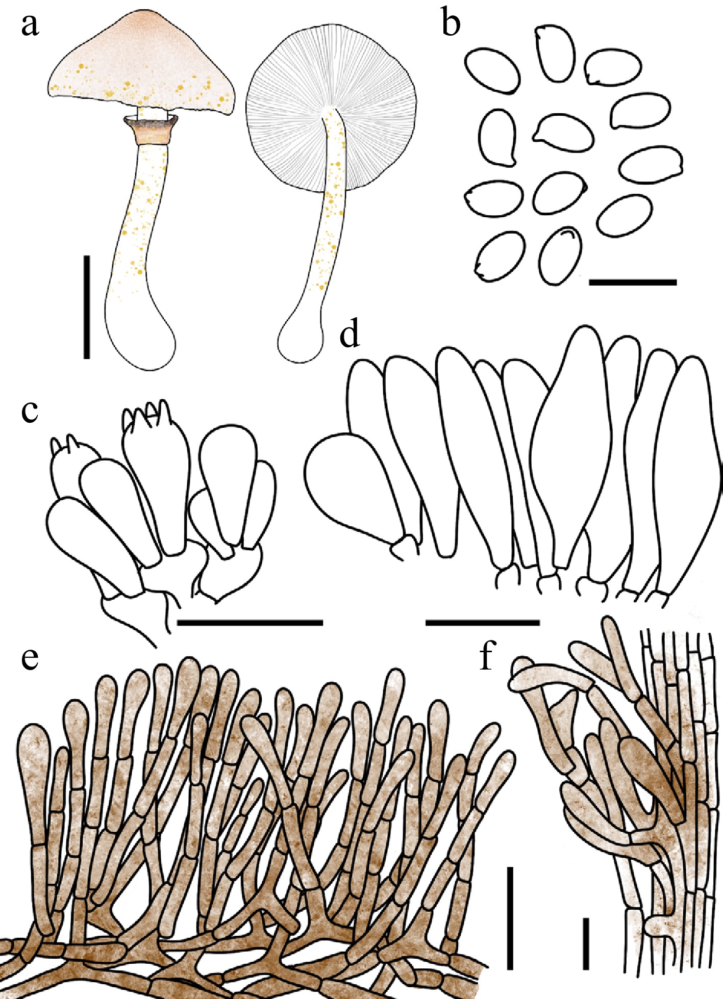
Figure 52.
Leucocoprinus luteoguttatus. (a) Basidiomata. (b) Basidiospores. (c) Basidia. (d) Cheilocystidia. (e) Surface covering at pileus centre. (f) Stipe covering. Scale bars: (a) = 1 cm; (b) = 10 μm; (c), (d), (f) = 20 μm; (e) = 50 μm.
Chinese name: 具黄滴白鬼伞 (Pīn yīn: jù huáng dī bái guǐ sǎn)
Index Fungorum number: IF904667; Facesoffungi number: FoF18958
Etymology – 'luteoguttatus' is derived from the Latin luteus (yellow) and guttatus (with drops), referring to the characteristic secretion of flax-yellow droplets by the basidiomata.
Type – China. Jilin: Ji'an City, Wunufeng National Forest Park, 25 July 2025, coll. X.Y. Zhou, FJAU78023.
Diagnosis – characterized by its basidiomata exuding flax-yellow to mustard-yellow droplets, a pileus with nude pink to Persian orange squamules, and narrowly utriform to clavate cheilocystidia.
Basidiomata small, exuding flax-yellow (#EEDC82) to mustard-yellow (#E1AD01) droplet-like secretions when fresh. Pileus 1.7–3.2 cm in diam., initially campanulate, expanding to plano-convex or applanate; umbo obtuse; background white; centre with nude pink (#DDC0B4) to Persian orange (#D99058) floccose squamules; squamules around the centre gradually fading in colour toward the margin; margin with inconspicuous short striations. Lamellae free, crowded, white to cream; edge concolorous; lamellulae in 2 tiers. Stipe 2.1–4.7 × 0.2–0.4 cm, subcylindrical, hollow, white; surface covered with secretions as described above. Annulus superior, ascending, white with a Persian orange to hickory-brown margin, membranous, persistent. Context thin, white, unchanging when bruised. Odour and taste not recorded.
Basidiospores (40/2/2) 6.1–7.1 × 3.6–4.2 μm, avl × avw = 6.4–6.5 × 3.8–4.0 μm, Q = (1.51–)1.55–1.81, avQ = 1.60–1.66, ellipsoid to oblong, hyaline, slightly thick-walled, smooth, non-dextrinoid to weakly dextrinoid, cyanophilous, congophilous, metachromatic in Cresyl Blue; germ pore absent. Basidia 19–28 × 8–11 μm, clavate, predominantly 4-spored, occasionally 2-spored, thin-walled. Pseudoparaphyses absent. Pleurocystidia absent. Cheilocystidia 33–51 × 6–11 μm, narrowly utriform to clavate, occasionally broadly clavate, hyaline, thin-walled. Surface covering at pileus centre a catenulate trichoderm consisting of erect, frequently septate hyphae appearing as long chains; terminal cells 77–125 × 4–13 µm, cylindrical, slightly thick-walled, with brown-red (#B25B09) to red-brown (#66332B) intracellular and parietal pigment. Stipe covering a cutis consisting of cylindrical hyphae; hyphae repent; terminal cells 22–58 × 5–13 μm, clavate, with flax-yellow to brown-red intracellular pigment, thin-walled. Clamp connections not observed.
Habit and habitat – solitary in humus-rich soil in mixed coniferous and broadleaf forests.
Known distribution – so far recorded known only from Northeast China.
Additional specimens examined – China. Jilin: Changchun City, Jingyuetan National Forest Park, 19 July 2022, coll. W.N. Hou, FJAU78022; Ji'an City, Mirror Groove, 7 August 2023, coll. Q.R. Liu, FJAU78026; Wunufeng National Forest Park, 25 July 2025, coll. X.Y. Zhou & W. Sun, FJAU78024; FJAU78025.
Notes – Macroscopically, Leucocoprinus luteoguttatus, Lc. dacrytus, and Lc. margaritifer share a remarkable feature that distinguishes them from allied taxa such as Lc. guttifer: they secrete distinct droplets from the entire basidioma, rather than exclusively from the stipe. However, Lc. luteoguttatus can be reliably separated from Lc. dacrytus and Lc. margaritifer by a combination of macro- and microscopic characters. Specifically, the new species possesses pale pinkish to Persian orange pileus squamules and cheilocystidia that completely lack apical appendages. In contrast, both Lc. dacrytus and Lc. margaritifer are characterized by brown-rosy to dark brownish pileus squamules, as well as cheilocystidia adorned with distinct apical appendages[61,99].
Phylogenetically, Lc. luteoguttatus is nested within Lc. clade IV, where it forms a well-supported sister branch to Lc. floccipes. Despite their close evolutionary affinity, these two species exhibit stark morphological differences. Notably, Lc. floccipes features conspicuous golden brown to gingerbread-brown floccose squamules on its stipe, whereas the stipe of Lc. luteoguttatus is comparatively glabrous.
Leucocoprinus ochraceosquamosus T. Bau & X.Y. Zhou, sp. nov Figs 36d−f, 53
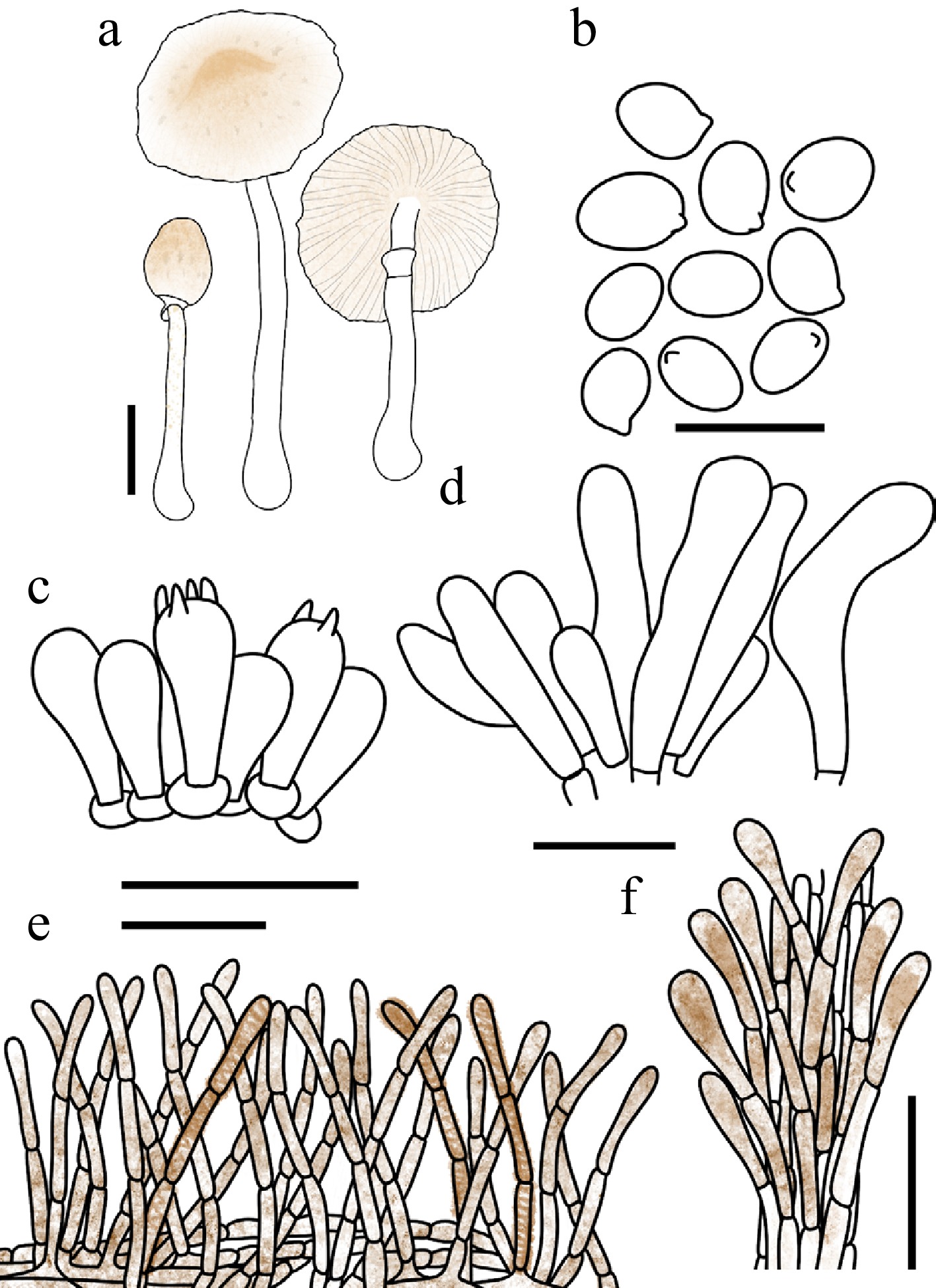
Figure 53.
Leucocoprinus ochraceosquamosus. (a) Basidiomata. (b) Basidiospores. (c) Basidia. (d) Cheilocystidia. (e) Surface covering at pileus centre. Scale bars: (a) = 1 cm; (b) = 10 μm; (c), (d) = 20 μm; (e), (f) = 50 μm.
Chinese name: 赭鳞白鬼伞 (Pīn yīn: zhě lín bái guǐ sǎn)
Index Fungorum number: IF904665; Facesoffungi number: FoF18956
Etymology – 'ochraceosquamosus' is a compound adjective derived from the Latin ochraceus (ochraceous, yellowish-brown) and squamosus (scaly), referring to the greige to pale gold squamules that adorn the pileus surface.
Type – China. Jilin: Jiaohe City, Qianjin Forest Farm, 24 July 2022, coll. L.Y. Zhu, FJAU78017.
Diagnosis – characterized by its basidiomata exuding colourless droplets, a pileus with greige to pale gold squamules, a bicolorous annulus with a hickory-brown margin, and narrowly clavate cheilocystidia.
Basidiomata small, exuding colourless droplet-like secretions from the stipe when fresh. Pileus 1.1–1.5 cm in diam., initially ovate, expanding to plano-convex or plane; umbo low, obtuse; background white to cream; centre with greige (#C1B7A5) to pale gold (#E6BE8A) floccose squamules; squamules around the centre fading gradually in colour toward the margin; margin with inconspicuous short striations. Lamellae free to subadnexed, moderately crowded, cream to pale gold; edge concolorous; lamellulae 1–3 between each pair of lamellae. Stipe 3.6–5.0 × 0.2–0.3 cm, subcylindrical, broadening toward the base, hollow, white to cream; surface covered with secretions as described above. Annulus superior, ascending, white with a hickory-brown margin, membranous, persistent. Context thin, white, unchanging when bruised. Odour and taste not recorded.
Basidiospores (40/2/2) 5.1–6.1 × 3.5–4.6 μm, avl × avw = 5.6–5.7 × 4.0–4.2 μm, Q = 1.22–1.56, avQ = 1.34–1.38, broadly ellipsoid to ellipsoid, hyaline, slightly thick-walled, smooth, dextrinoid, cyanophilous, congophilous, metachromatic in Cresyl Blue; germ pore absent. Basidia 15–23 × 5–9 μm, clavate, predominantly 4-spored, occasionally 2-spored, thin-walled. Pseudoparaphyses absent. Pleurocystidia absent. Cheilocystidia 22–47 × 4–15 μm, narrowly clavate, slightly flexuose, hyaline to pale gold, thin-walled. Surface covering at pileus centre a catenulate trichoderm consisting of erect, frequently septate hyphae appearing as long chains; terminal cells 56–118 × 4–13 µm, cylindrical, slightly thick-walled, with pale gold to red-brown intracellular and parietal pigments. Stipe covering a cutis consisting of cylindrical hyphae; hyphae repent; terminal cells 20–60 × 4–9 µm, cylindrical to narrowly clavate, with pale gold intracellular and parietal pigment, thin-walled. Clamp connections not observed.
Habit and habitat – solitary to scattered in soil in broadleaf forests.
Known distribution – so far recorded only from Qianjin Forest Farm, Northeast China.
Additional specimens examined – China. Jilin: Jiaohe City, Qianjin Forest Farm, 24 July 2022, coll. W.N. Hou, FJAU78016.
Notes – Morphologically, Leucocoprinus ochraceosquamosus is easily confused with Lepiota atrodiscus and Lc. fuscannulatus. However, L. atrodisca can be distinguished by its yellowish-white stipe that turns yellowish-brown upon bruising, and by its basidiospores lacking a metachromatic reaction in Cresyl Blue[93]. Conversely, Lc. fuscannulatus is separated by its noticeably larger basidiospores (5.8–6.9 × 4.1–4.9 μm, avl × avw = 6.0 × 4.5 μm) and septate, clavate to narrowly utriform cheilocystidia.
Regarding its macroscopic features, of the two available collections of Lc. ochraceosquamosus, colorless droplets on the stipe surface were exclusively observed in specimen FJAU78017. The absence of these exudates in collection FJAU78016 is likely an artifact attributable to the suboptimal condition of the basidiomata during observation. Furthermore, it should be noted that guttation in Lc. ochraceosquamosus appears to be generally sparse and considerably less conspicuous when compared to the typical droplet-secreting species within the genus Leucocoprinus.
Leucocoprinus sudans T. Bau & X.Y. Zhou, sp. nov Figs 36g−i, 54
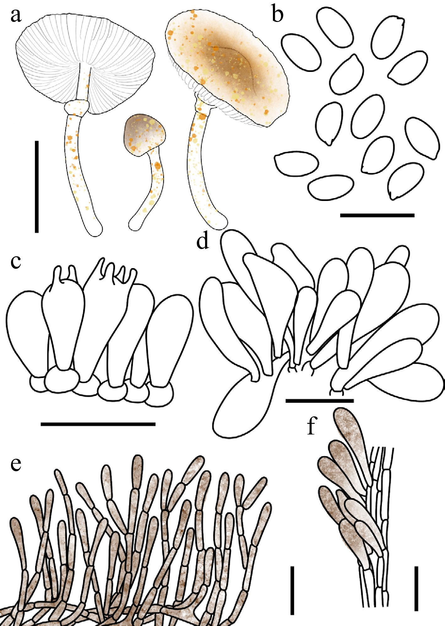
Figure 54.
Leucocoprinus sudans. (a) Basidiomata. (b) Basidiospores. (c) Basidia. (d) Cheilocystidia. (e) Surface covering at pileus centre. (f) Stipe covering. Scale bars: (a) = 1 cm; (b) = 10 μm; (c), (d), (f) = 20 μm; (e) = 50 μm.
Chinese name: 渗液白鬼伞 (Pīn yīn: shèn yè bái guǐ sǎn)
Index Fungorum number: IF905151; Facesoffungi number: FoF19598
Etymology – 'sudans' is derived from the Latin present participle sudans (meaning 'sweating' or 'exuding'), referring to the characteristic hyaline to flax yellow drops exuded by the fresh basidiomata.
Type – China. Heilongjiang: Yichun City, Xing'an National Forest Park, 25 July 2023, coll. W.N. Hou, FJAU78274.
Diagnosis – characterized by its basidiomata exuding hyaline to flax-yellow drops, a pileus with pale-gold to gingerbread-brown squamules, non-dextrinoid basidiospores, and fusiform to oblong cheilocystidia.
Basidiomata small, exuding hyaline to flax-yellow (#EEDC82) droplet-like secretions when fresh. Pileus 0.6–1.6 cm in diam., initially campanulate, expanding to plano-convex; background white; centre with fulvous (#E48400) to red-brown (#66332B) fine squamules; squamules around the centre pale gold (#E6BE8A) to fulvous. Lamellae free, crowded; edge concolorous; lamellulae in 1–3 tiers. Stipe 1.4–2.0 cm × 0.1–0.2 cm, subcylindrical, hollow, white at the apex, pale-gold in the middle and lower parts; surface covered with secretions as described above; base with white mycelium. Annulus median to superior, ascending, upper surface white, lower surface pale gold, membranous. Context thin, white, unchanging when bruised. Odour and taste not recorded.
Basidiospores (40/2/2) 5.2–6.6 × 2.9–3.4 μm, avl × avw = 5.7–5.8 × 3.0–3.2 μm, Q = 1.57–2.00, avQ = 1.72–1.81, subamygdaliform, ellipsoid to oblong, hyaline, thin-walled, smooth, non- to weakly dextrinoid, weakly cyanophilous, congophilous, metachromatic in Cresyl Blue; germ pore absent. Basidia 15–20 × 7–9 μm, clavate, predominantly 4-spored, occasionally 2-spored, thin-walled. Pseudoparaphyses absent. Pleurocystidia absent. Cheilocystidia 26–38 × 6–13 μm, fusiform to oblong, hyaline, thin-walled. Surface covering at pileus centre a catenulate trichoderm consisting of erect, frequently septate hyphae appearing as long chains; terminal cells 71–185 × 5–10 µm, cylindrical, slightly thick-walled, with pale gold to red-brown intracellular and parietal pigments. Stipe covering a cutis consisting of cylindrical hyphae; hyphae repent, terminal cells 35–80 × 5–11 µm, clavate, with golden-brown (#885407) to red-brown intracellular and parietal pigment, thin-walled. Clamp connections absent.
Habit and habitat – solitary in humus-rich soil in mixed coniferous and broadleaf forests.
Known distribution – so far recorded only from Northeast China.
Additional specimens examined – China. Jilin: Dunhua City, Laobaishan Primitive Ecological Area, 22 July 2025, coll. X.Y. Zhou, FJAU78106.
Notes – When fresh, the entire basidiomata of Leucocoprinus sudans exude drops, a feature that easily distinguishes it from species that only exude drops on the stipe, such as Lc. brunneodiscus. Within the genus Leucocoprinus, several other species also exude drops across the entire basidiomata, including Lc. floccipes, Lc. flavirobustus, Lc. luteoguttatus, Lc. dacrytus, and Lc. margaritifer. However, Lc. dacrytus and Lc. margaritifer are currently only known to occur in North America. Among the remaining species, Lc. floccipes can be separated by the presence of distinct golden-brown to hickory-brown squamules on its stipe, whereas the stipes of Lc. flavirobustus, Lc. luteoguttatus, and Lc. sudans are relatively glabrous or possess only fine squamules. Furthermore, these three morphologically similar species can be clearly differentiated by their cheilocystidia: Lc. flavirobustus has clavate to cylindrical to capitate-stipitate cheilocystidia that are regularly catenulate (composed of 3–5 elements with cylindrical ante-terminal elements); Lc. luteoguttatus possesses narrowly utriform to clavate cheilocystidia; whereas the new species, Lc. sudans, is characterized by its fusiform to oblong cheilocystidia.
Species in Lc. clade II Figs 37i−l
Notes: Morphologically, this clade accommodates species that exhibit a distinct macrochemical reaction to ammonia when fresh, as well as taxa that undergo discoloration (or remain unchanged) upon bruising or drying. Topologically, Lc. clade II corresponds to the segregated genus Pulchrolepiota recently proposed by Yang et al.[30]. Two novel species belonging to this clade, both characterized by the absence of discoloration upon bruising or drying, are described below.
Leucocoprinus lacteus T. Bau & X.Y. Zhou, sp. nov Figs 37l, 55
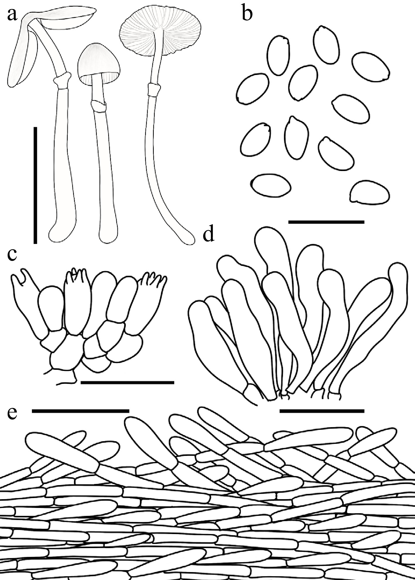
Figure 55.
Leucocoprinus lacteus. (a) Basidiomata. (b) Basidiospores. (c) Basidia. (d) Cheilocystidia. (e) Surface covering at pileus centre. Scale bars: (a) = 1 cm; (b) = 10 μm; (c), (d) = 20 μm; (e) = 50 μm.
Chinese name: 乳白白鬼伞 (Pīn yīn: rǔ bái bái guǐ sǎn)
Index Fungorum number: IF904654 ; Facesoffungi number: FoF 18945
Etymology – 'lacteus' is the feminine form of the Latin adjective 'lacteus' (milky, milk-white), referring to the white coloration of the pileus and its silky-fibrillose squamules.
Type – China. Hubei: Enshi, Wangchengpo, 20 June 2022, coll. L.Y. Zhu FJAU78083.
Diagnosis – characterized by its white pileus covered with concolorous silky-fibrillose squamules, and cylindrical to clavate cheilocystidia.
Basidiomata small. Pileus 0.7–2.5 cm in diam., initially campanulate, expanding to plano-convex or applanate; umbo low, obtuse; background white; squamules concolorous, silky-fibrillose. Lamellae free, moderately crowded, white to cream; edge concolorous; lamellulae interspersed 1–3 between each pair of lamellae. Stipe 0.8–3.1 × 0.1–0.2 cm, subcylindrical, widening downwards to a slightly bulbous base, hollow, white; surface smooth to covered with fine fibrils. Annulus superior, ascending, white, membranous. Context thin, white, unchanging when bruised. Odour and taste not recorded.
Basidiospores (40/2/2) 4.6–5.6 × 2.5–3.3 μm, avl × avw = 5.0–5.1 × 2.9–3.0 μm, Q = 1.44–2.01, avQ = 1.69–1.75, ellipsoid to oblong or cylindrical, hyaline, thin-walled, smooth, dextrinoid, cyanophilous, congophilous, metachromatic in Cresyl Blue; germ pore absent. Basidia 13–22 × 5–9 μm, clavate, predominantly 4-spored, occasionally 2-spored, thin-walled. Pseudoparaphyses absent. Pleurocystidia absent. Cheilocystidia 24–44 × 5–11 μm, cylindrical to clavate, slightly flexuose, hyaline, thin-walled. Surface covering at pileus centre an interwoven plagiotrichoderm consisting of cylindrical hyphae; hyphae densely entangled, repent to slightly ascending, 4–12 µm wide, thin-walled, colourless, lacking distinct pigment. Stipe covering a cutis consisting of cylindrical hyphae; hyphae repent, 3–8 μm wide, thin-walled, colourless. Clamp connections absent.
Habit and habitat – solitary to scattered in humus-rich soil in mixed bamboo and pine forests.
Known distribution – currently known only from the type locality in Enshi City, Central-Southern China.
Additional specimens examined – China. Hubei: Enshi City, Wangchengpo, 20 June 2022, coll. H. Cheng, FJAU78122.
Notes – Both macroscopically and phylogenetically, Leucocoprinus lacteus is most closely related to Lc. parviceps T. Bau & X.Y. Zhou. However, they can be reliably distinguished based on basidioma size, basidiospore shape, and ecological preferences. Morphologically, Lc. lacteus produces larger mature pilei (0.7–2.5 cm vs 0.6–1 cm) and notably more elongated basidiospores (Q = 1.44–2.01, avQ = 1.69–1.75) compared to the less elongated spores (Q = 1.24–1.53, avQ = 1.38–1.41) of Lc. parviceps. Ecologically, Lc. lacteus is currently known only from Central-Southern China (Hubei Province), growing in mixed bamboo and pine forests, whereas Lc. parviceps is restricted to Northeast China (Inner Mongolia and Jilin Province), occurring in broadleaf forests dominated by Ulmus spp., Quercus spp., Juglans mandshurica, and Fraxinus mandshurica.
Phylogenetically, both species are nested within Lc. clade II. The vast majority of species in this clade typically exhibit distinct discolouration upon bruising or desiccation, or display a positive chemical reaction to ammonia. While the ammonia reaction was unfortunately not evaluated for our fresh collections of Lc. lacteus and Lc. parviceps, these two taxa form a well-supported subclade with five other species: Lc. oculatus (J.E. Lange & Zeller) Vellinga, Birkebak & Justo, Lc. paraplesius (Vellinga) Kun L. Yang, Jia Y. Lin & Zhu L. Yang, Lc. paraplesius (Vellinga) Kun L. Yang, Jia Y. Lin & Zhu L. Yang, Lc. personatus (K.L. Yang & Jia Y. Lin) T. Bau, and Lc. elegans (K.L. Yang, Jia Y. Lin & Z.Chao Liu) T. Bau. Notably, there are no documented records of positive ammonia reactions for these five species either[30,102]. Furthermore, none of the seven species within this subclade have ever been reported to discolour upon bruising or drying. Consequently, we hypothesize that this subclade represents an evolutionary exception within Lc. clade II, entirely lacking its typical phenotypic hallmarks.
Microscopically, another intriguing distinction exists within this seven-species subclade. The pileus covering of the other five species is documented as a trichoderm, with Lc. oculatus, Lc. paraplesius, and Lc. paraplesius exhibiting a typical trichoderm (Type II-A) composed of strictly erect hyphae. In contrast, critical microscopic examinations of Lc. lacteus and Lc. parviceps revealed that the hyphae at the pileus centre are repent to ascending and lack distinctly differentiated terminal elements. This structural arrangement appears to be a transitional state between a cutis and a trichoderm (Type Ⅰ-B). Therefore, we characterize the surface covering at the pileus centre of Lc. lacteus and Lc. parviceps as an interwoven plagiotrichoderm, rather than a typical erect trichoderm.
Leucocoprinus parviceps T. Bau & X.Y. Zhou sp. nov Figs 37i−k, 56
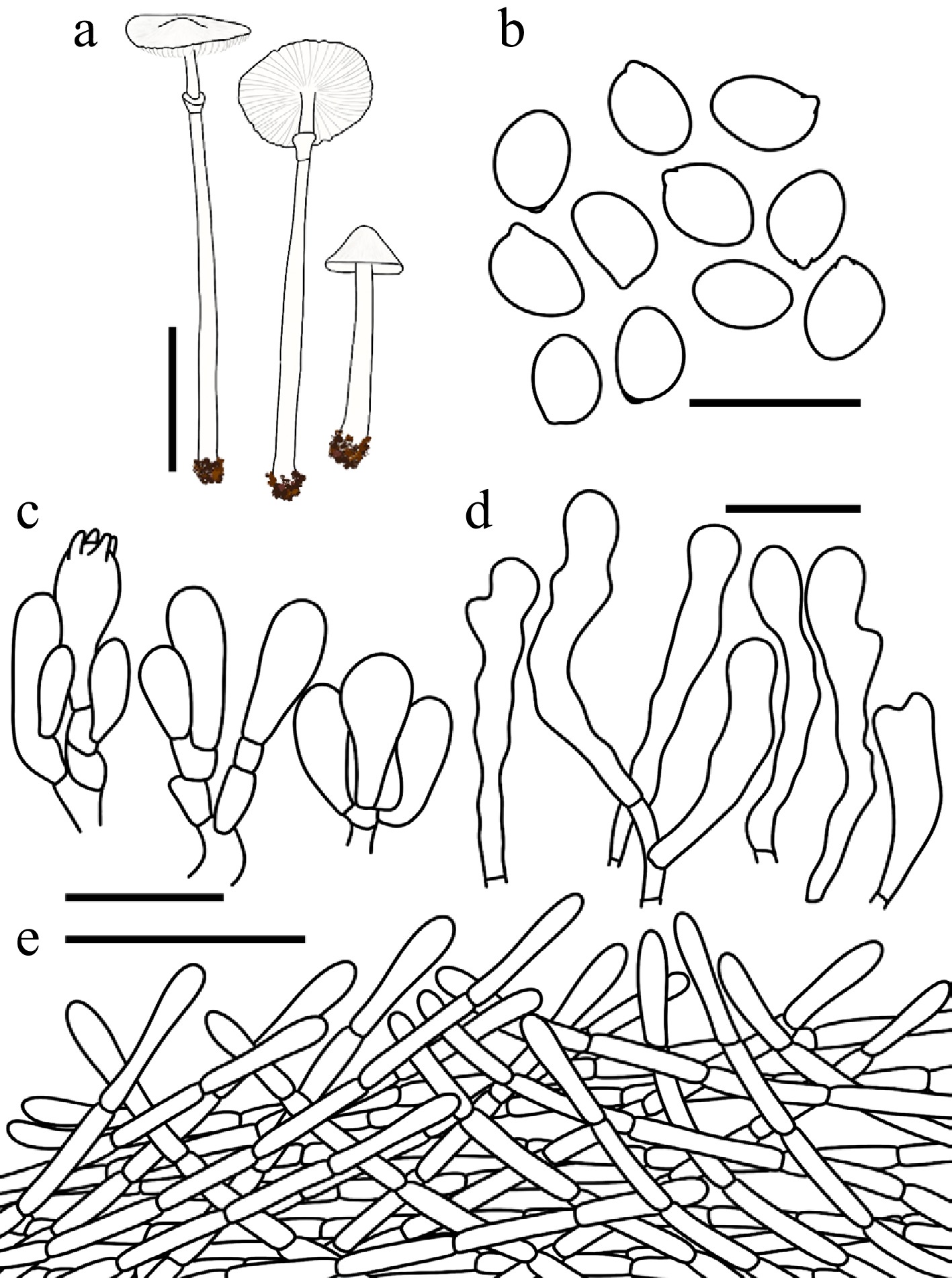
Figure 56.
Leucocoprinus parviceps. (a) Basidiomata. (b) Basidiospores. (c) Basidia. (d) Cheilocystidia. (e) Surface covering at pileus centre. Scale bars: (a) = 1 cm; (b) = 10 μm; (c), (d) = 20 μm; (e) = 50 μm.
Chinese name: 小盖白鬼伞 (Pīn yīn: xiǎo gài bái guǐ sǎn)
Index Fungorum number: IF904653; Facesoffungi number: FoF 18944
Etymology – 'parviceps' is a compound adjective derived from the Latin parvus (small) and -ceps (headed), referring to the characteristically small pileus of this species.
Type – China. Jilin: Baishan City, Lushuihe National Forest Park, 8 August 2024, coll. W.N. Hou, FJAU78046.
Diagnosis – most similar to Leucocoprinus lacteus (Q = 1.44–2.01), but differs by its less elongated basidiospores (Q = 1.24–1.53), and occurring in broadleaf forests dominated by Ulmus and Quercus rather than in mixed bamboo and pine forests.
Basidiomata small. Pileus 0.6–1 cm in diam., initially obtusely conical, expanding to applanate; umbo obtuse; background white; squamules concolorous, silky-fibrillose, readily detachable. Lamellae free, moderately crowded, white; edge concolorous; lamellulae interspersed 1–3 between each pair of lamellae. Stipe 2.2–3.1 cm × 0.1–0.2 cm, subcylindrical, widening downwards to a slightly bulbous base, hollow, white; surface smooth to covered with fine fibrils. Annulus subapical to median, ascending, white, membranous. Context thin, white, unchanging when bruised. Odour and taste not recorded.
Basidiospores (40/2/2) 4.9–6.2 × 3.5–4.5 μm, avl × avw = 5.4–5.5 × 3.9–4.0 μm, Q = 1.24–1.53, avQ = 1.38–1.41, broadly ellipsoid to ellipsoid, hyaline, thin-walled, smooth, dextrinoid, cyanophilous, congophilous, metachromatic in Cresyl Blue; germ pore absent. Basidia 15–24 × 5–9 μm, clavate, predominantly 4-spored, thin-walled. Pseudoparaphyses absent. Pleurocystidia absent. Cheilocystidia 27–53 × 6–11 μm, cylindrical to clavate, flexuose, slightly subcapitate at the apex, hyaline, thin-walled. Surface covering at pileus centre an interwoven plagiotrichoderm consisting of cylindrical hyphae; hyphae densely entangled, repent to slightly ascending, 3–7 μm wide, thin-walled, colourless, lacking distinct pigment. Stipe covering a cutis consisting of cylindrical hyphae; hyphae repent, 4–9 μm wide, thin-walled, colourless. Clamp connections absent.
Habit and habitat – solitary in humus or sandy soil in broadleaf forests dominated by Ulmus, Quercus, Juglans mandshurica Maxim., and Fraxinus mandshurica Rupr.
Known distribution – Northeast China.
Additional specimens examined – China. Inner Mongolia Autonomous Region, Tongliao City, Daqinggou National Nature Reserve, 23 August 2022, coll. W. N. Hou, FJAU78047.
Notes – for a detailed comparative discussion on the morphological similarities and phylogenetic relationships of this taxon, refer to the notes provided for Pulchrolepiota lactea.
Species in Lc. clade I Figs 57−59
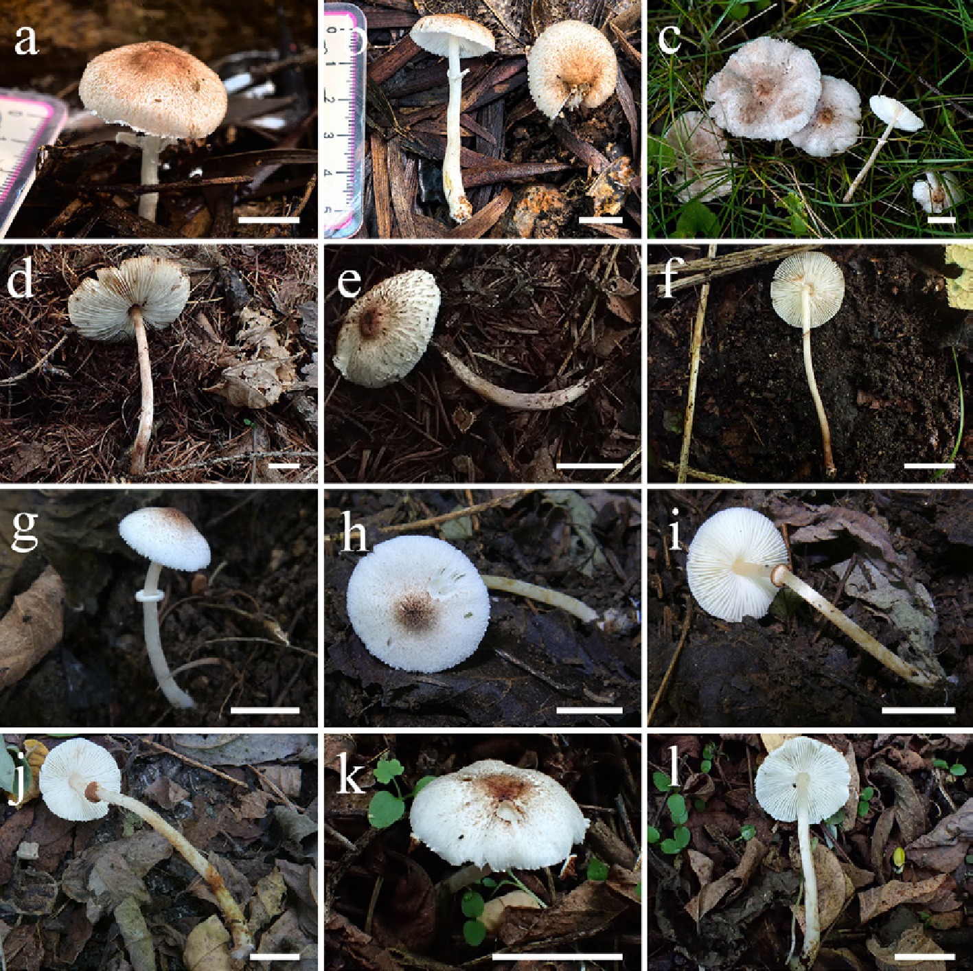
Figure 57.
Basidiomata of Leucocoprinus species. Lc. comatus (a) FJAU78115, (b) FJAU78065. Lc. rutilobrunneus (c) FJAU78029, (d) FJAU78032, (e) FJAU78030, (f) FJAU78036. Lc. bicolorannulatus (g) FJAU78054, (h), (i) FJAU78055, (j) FJAU78053, (k), (l) FJAU78132. Scale bars = 1 cm. These pictures are copyright of the collectors of specimens.
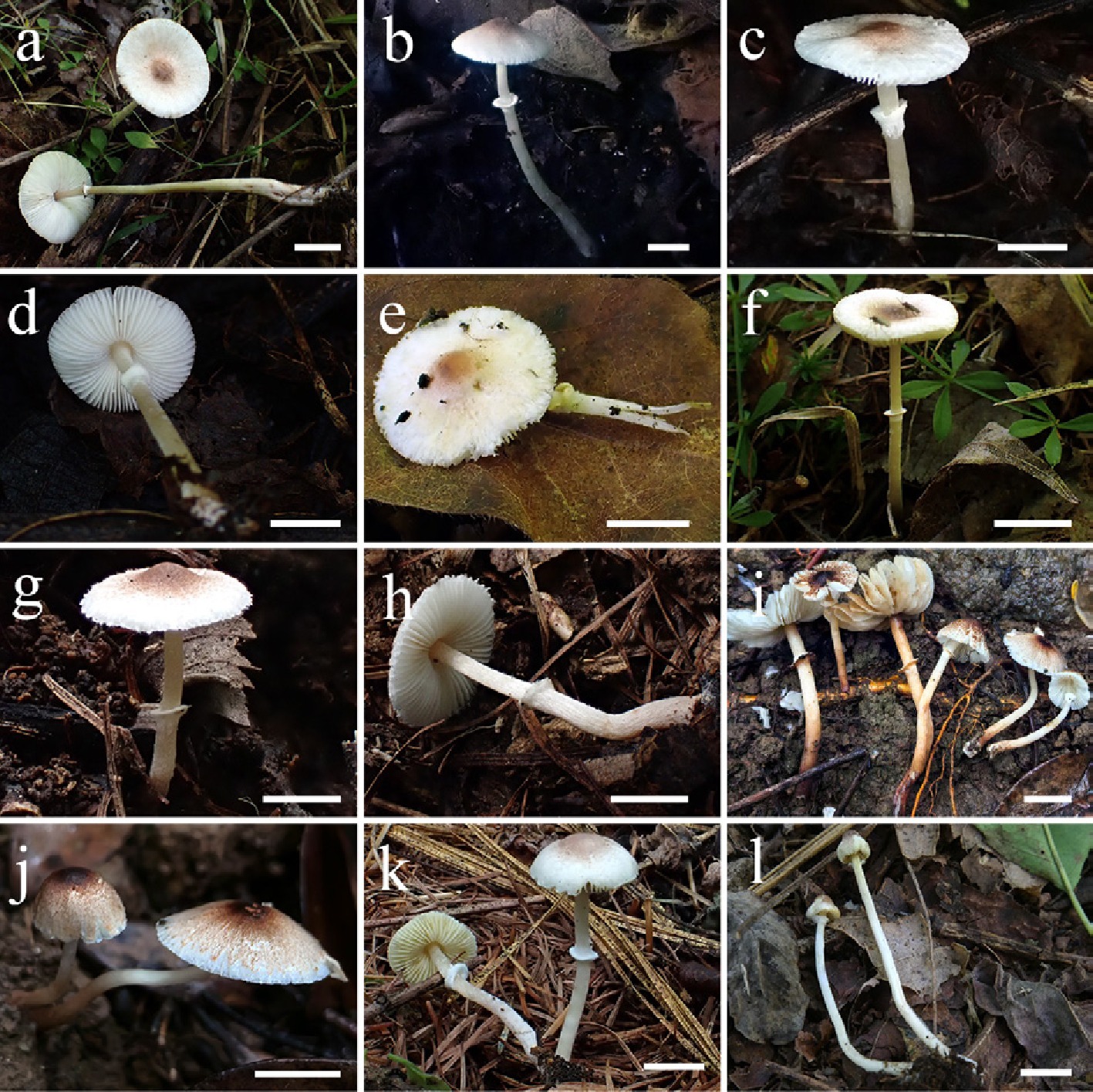
Figure 58.
Basidiomata of Leucocoprinus species. Lc. persicinus (a) FJAU78037, (b) FIAU78039. Lc. caeruleipes (c), (d) FJAU78045, (e) FJAU78044. Lc. versicolor (f) FJAU78041, (g), (h) FJAU78042. Lc. caffeaceus (i) FJAU78103, (j) FJAU78104. Lc. transeptatus (k) FJAU78067, (l) FJAU78069. Scale bars = l cm. These pictures are copyright of the collectors of specimens.
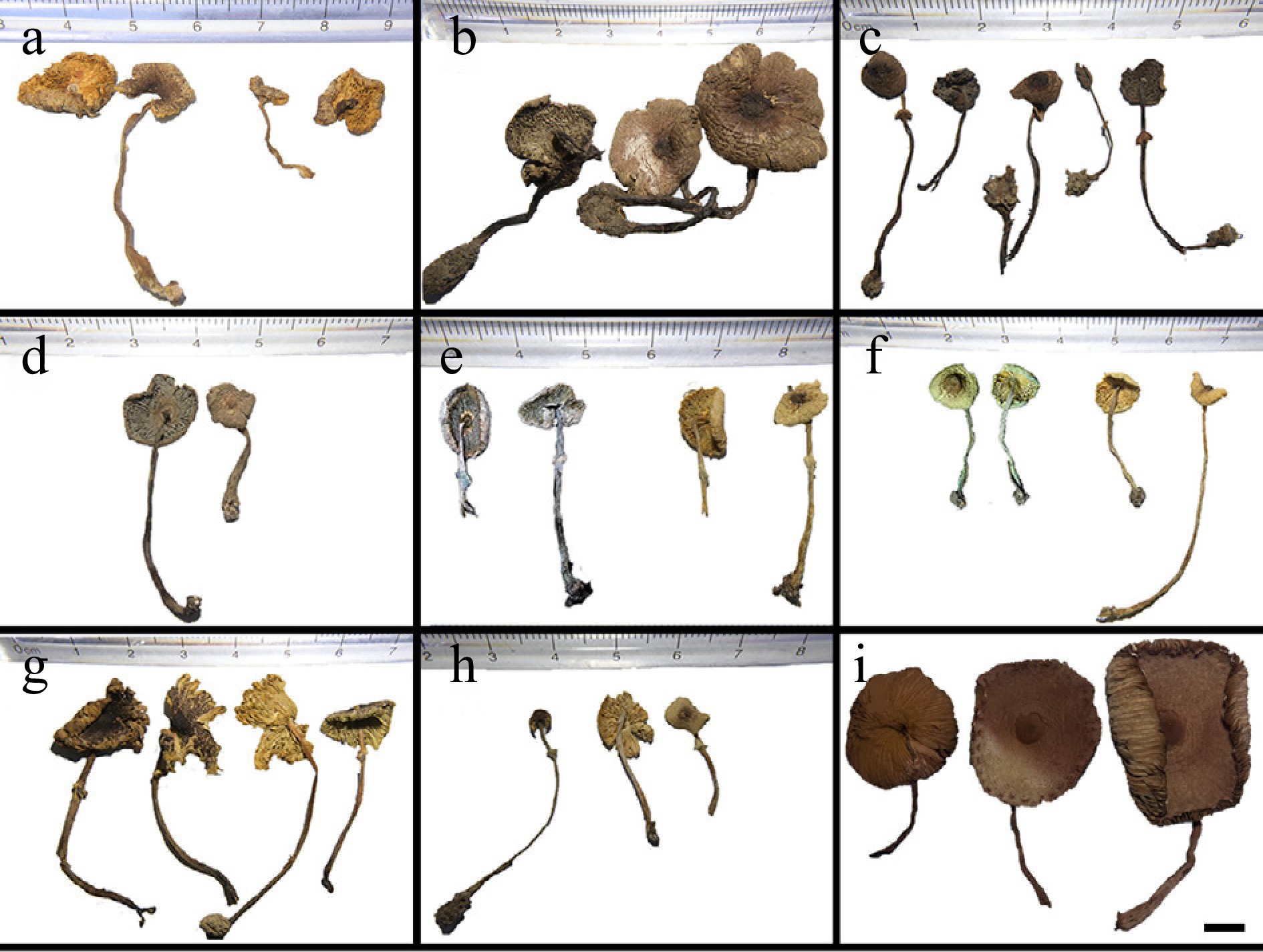
Figure 59.
Dried specimens of Leucocoprinus and Macropsalliota species. (a) Lc. comatus (left two: FJAU78115, right two: FJAU78065). (b) Lc. rutilobrunneus (FJAU78029). (c) Lc. bicolorannulatus (from left to right: FJAU78051, FJAU78052, FJAU78053, FJAU78054, and FJAU78055). (d) Lc. persicinus (FJAU78037). (e) Lc. caeruleipes (left two basidiomata photographed on 24 May 2025, right two photographed on 6 September 2025; first and third: FJAU78044, second and fourth: FJAU78045). (f) Lc. versicolor (left two basidiomata photographed on 24 May 2025, right two photographed on 6 September 2025; left three: FJAU78043, right one: FJAU78041). (g) Lc. caffeaceus (FJAU78103). (h) Lc. transeptatus (left one: FJAU78068, right two: FJAU78067). (i) Map. aff. subtropica (FJAU72226). These pictures are copyright of X.Y. Zhou.
Notes – Morphologically, this clade predominantly comprises species whose basidiomata undergo discoloration upon bruising or drying. Phylogenetically, this lineage further bifurcates into two strongly supported subclades. Topologically, Lc. clade I corresponds to the segregated genus Tristolepiota recently proposed by Yang et al.[30]. Eight novel species belonging to this clade are described below.
Leucocoprinus bicolorannulatus T. Bau & X.Y. Zhou, sp. nov Figs 57g−l, 59c, 60
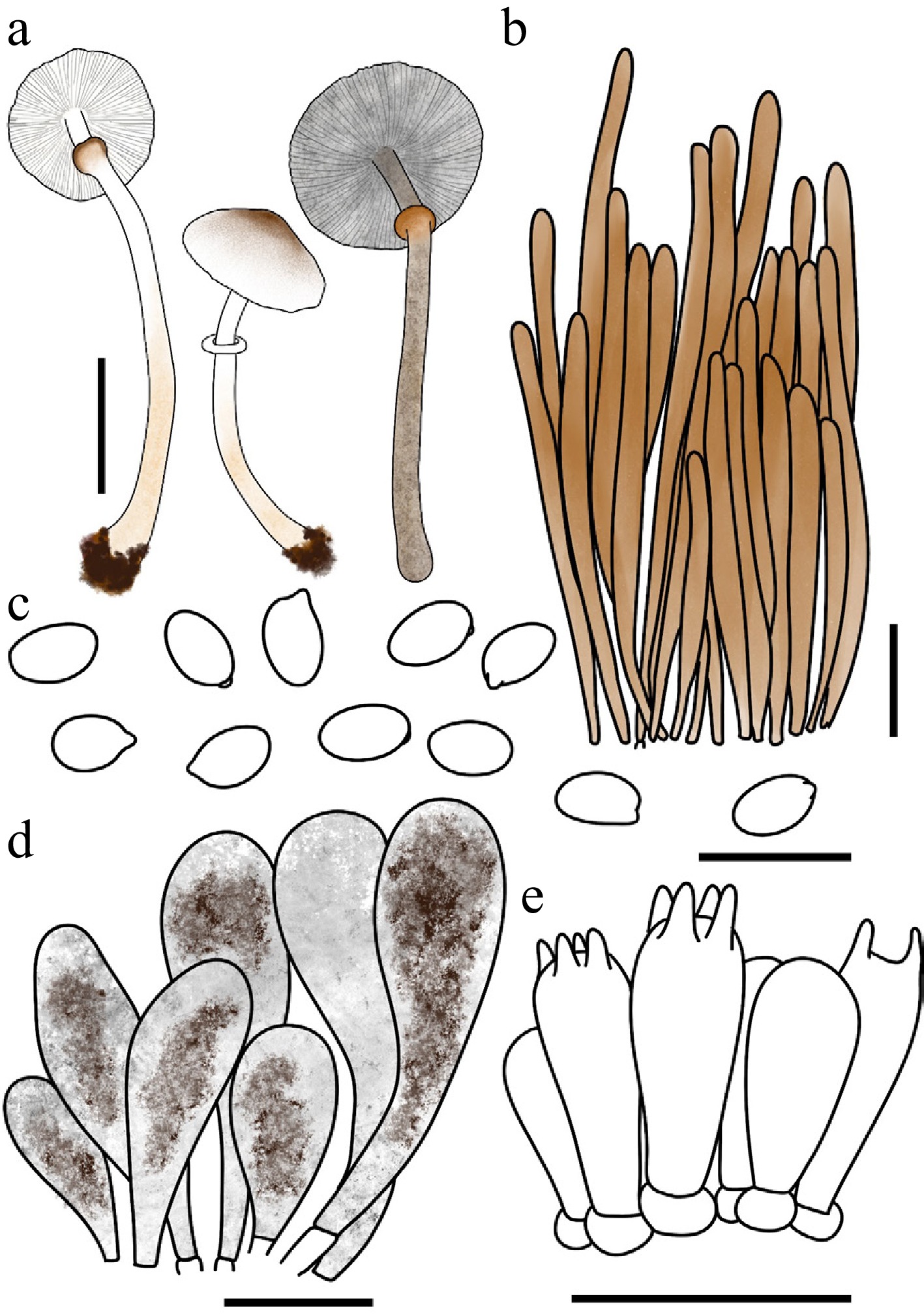
Figure 60.
Leucocoprinus bicolorannulatus. (a) Basidiomata. (b) Surface covering at pileus centre. (c) Basidiospores. (d) Cheilocystidia. (e) Basidia. Scale bars: (a) = 1 cm; (b) = 10 μm; (c), (d) = 20 μm; (e) = 50 μm.
Chinese name: 双色环白鬼伞 (Pīn yīn: shuāng sè huán bái guǐ sǎn)
Index Fungorum number: IF904648; Facesoffungi number: FoF 18936
Etymology – 'bicolorannulatus' is derived from the Latin bi- (two), colour (colour), and annulatus (ringed), referring to the distinctive bicolorous annulus.
Type – China. Jilin: Ji'an City, Yushan Park, 26 July 2025, coll. X.Y. Zhou, FJAU78055.
Diagnosis – characterized by a pileus and lower stipe covered with brownish floccose squamules, a bicolorous annulus, and dried basidiomata that turn golden to hickory-brown with granite-gray to dark charcoal lamellae.
Basidiomata small. Pileus 1–1.7 cm in diam., convex to plano-convex or applanate when mature; umbo obtuse; background white; centre with brown-red (#B25B09) to red-brown (#66332B) floccose squamules; squamules around the centre fading gradually in colour toward the margin. Lamellae free, moderately crowded, white to cream; edge concolorous; lamellulae 1–3 between each pair of lamellae. Stipe 2.4–4.9 × 0.1–0.2 cm, subcylindrical, hollow, white; surface covered from the mid-region to the base with pale gold (#E6BE8A) to fulvous (#E48400) floccose squamules, more crowded toward the base; base with inconspicuous white mycelium. Annulus superior, ascending, outer surface brown-red to gingerbread-brown, inner surface white, membranous, readily detachable. Context thin, white, unchanging when bruised. Odour and taste not recorded.
Dried basidiomata: Pileus, stipe, and context golden brown (#885407) to hickory brown (#371D10); lamellae granite gray (#676767) to dark charcoal (#333333); annulus brown-red to brown-red.
Basidiospores (40/2/2) 5.0–6.6 × 2.9–4.2 μm, avl × avw = 5.7–5.8 × 3.5–3.7 μm, Q = 1.42–1.78, avQ = 1.57–1.65, ellipsoid to oblong, hyaline, slightly thick-walled, smooth, non-dextrinoid to weakly dextrinoid, cyanophilous, congophilous, metachromatic in Cresyl Blue; germ pore absent. Basidia 16–24 × 6–9 μm, clavate, predominantly 4-spored, occasionally 2-spored, thin-walled. Pseudoparaphyses absent. Pleurocystidia absent. Cheilocystidia 30–48 × 8–17 μm, oblong to broadly fusiform with an obtuse apex, thin-walled, with hickory-brown to dark charcoal intracellular pigment. Surface covering at pileus centre a typical trichoderm consisting of erect hyphae arranged in tufts or bundles; terminal cells 99–246 × 8–15 μm, cylindrical, slightly thick-walled, with brown-red intracellular pigment. Stipe covering a cutis consisting of cylindrical hyphae; hyphae repent, 4–12 μm wide, thin-walled, golden brown. Clamp connections not observed.
Habit and habitat – solitary in humus-rich soil in mixed forests dominated by Acer and Ulmus.
Known distribution – so far recorded only from the type locality in Yushan Park, Northeast China
Additional specimens examined – China. Jilin: Ji'an City, Yushan Park, 26 July 2025, coll. X.Y. Zhou, FJAU78051, FJAU78052, FJAU78053, FJAU78054.
Notes – Macroscopically, Leucocoprinus bicolorannulatus shares the presence of brownish squamules at the pileus centre with Lc. comatus T. Bau, X.Y. Zhou & C. Cheng, Lc. actinophorus (K.L. Yang, Jia Y. Lin & Z.Chao Liu) T. Bau, and Lc. boluoshanensis (K.L. Yang, Jia Y. Lin & Z.Chao Liu) T. Bau. However, Lc. comatus can be distinguished by its initially white stipe that acquires a yellowish hue upon bruising, and its entire basidioma turning yellowish to golden brown upon desiccation. In contrast, Lc. bicolorannulatus exhibits no discolouration upon bruising; moreover, upon desiccation, its pileus and stipe become golden brown, the lamellae turn greyish brown, and the annulus distinctly changes to reddish brown. Additionally, Lc. actinophorus is highly distinctive in turning green to blue after drying, whereas Lc. boluoshanensis lacks any obvious colour change upon desiccation[30].
Phylogenetically, Lc. bicolorannulatus is recovered as the sister species to Lc. rutilobrunneus T. Bau & X.Y. Zhou. Despite their close evolutionary affinity, the latter is readily separated by its dramatic oxidation reaction: upon bruising, Lc. rutilobrunneus rapidly discolours to deep carrot orange or red-brown, before swiftly transitioning to golden brown or blackish brown.
Leucocoprinus comatus T. Bau, X.Y. Zhou & C. Cheng, sp. nov Figs 57a, b; 59a; 61
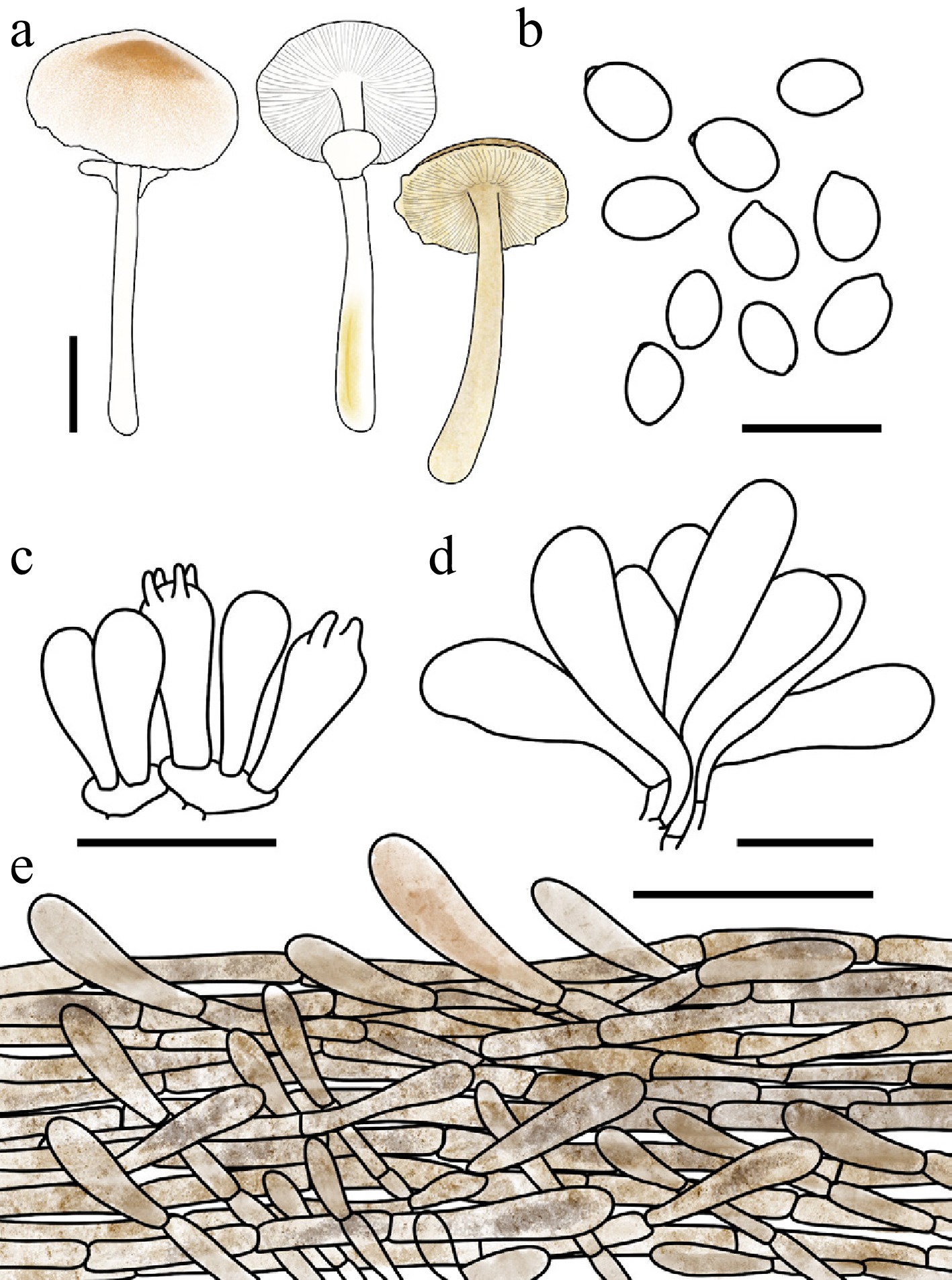
Figure 61.
Leucocoprinus comatus. (a) Basidiomata. (b) Basidiospores. (c) Basidia. (d) Cheilocystidia. (e) Surface covering at pileus centre. Scale bars: (a) = 1 cm; (b) = 10 μm; (c), (d) = 20 μm; (e) = 50 μm.
Chinese name: 毛头白鬼伞 (Pīn yīn: máo tóu bái guǐ sǎn)
Index Fungorum number: IF904671; Facesoffungi number: FoF 18937
Etymology – 'comatus' is derived from the Latin comatus (hairy, shaggy), referring to the shaggy, hair-like squamules on the pileus.
Type – China. Guangdong: Shenzhen City, Wutong Mountain, 2 July 2025, coll. C. Cheng, FJAU78115.
Diagnosis – characterized by its pileus covered with brownish, tufted, hair-like squamules, a white stipe that turns flax yellow to mustard yellow upon bruising, and oblong to clavate cheilocystidia.
Basidiomata small. Pileus 0.8–2.9 cm in diam., plano-convex to applanate when mature; umbo obtuse; background white; centre with brown-red (#B25B09) tufted squamules; squamules around the centre fulvous (#E48400) to brown-red, hair-like to arachnoid, radiating toward the margin. Lamellae free, moderately crowded, white; lamellulae 1–3 between each pair of lamellae. Stipe 1.8–5.9 × 0.2–0.5 cm, subcylindrical, hollow, white; surface covered with concolorous fibrillose squamules, turning flax yellow (#EEDC82) to mustard yellow (#E1AD01) when bruised. Annulus superior, ascending, white, membranous, readily detachable. Context thin, white, bruising reaction not recorded. Odour and taste not recorded.
Dried basidiomata: Basidiomata become mustard yellow to golden brown (#885407).
Basidiospores (40/2/2) 4.9–6.6 × 3.1–4.1 μm, avl × avw = 5.4–5.7 × 3.4–3.5 μm, Q = 1.41–1.91, avQ = 1.59–1.63, ellipsoid to oblong, hyaline, slightly thick-walled, smooth, dextrinoid, cyanophilous, congophilous, metachromatic in Cresyl Blue; germ pore absent. Basidia 13–20 × 6–10 μm, clavate, predominantly 4-spored, thin-walled. Pseudoparaphyses absent. Pleurocystidia absent. Cheilocystidia 30–43 × 8–15 μm, oblong to clavate, hyaline to pale yellowish brown, thin-walled. Surface covering at pileus centre an interwoven plagiotrichoderm consisting of cylindrical hyphae; hyphae densely entangled, repent to slightly ascending; terminal cells 5–13 μm wide, slightly thick-walled, with golden-brown to hickory-brown intracellular and parietal pigment. Stipe covering a cutis consisting of cylindrical hyphae; hyphae repent, 3–10 μm wide, thin-walled, pale yellowish brown. Clamp connections not observed.
Habit and habitat – solitary to scattered in humus-rich soil in bamboo stands.
Known distribution – so far recorded only from Central-Southern China.
Additional specimens examined – China. Guangdong: Shenzhen City, Wutong Mountain, 21 June 2025, coll. C. Cheng, FJAU78065.
Notes – Macroscopically, Leucocoprinus comatus bears a striking resemblance to certain species of Agaricus, sharing similar squamulose textures on the pileus and a stipe that acquires a yellowish hue upon bruising. However, the new species can be readily distinguished by its white lamellae, which do not turn blackish-brown upon desiccation, and by its strictly hyaline basidiospores. Furthermore, while Lc. comatus shares brownish, tufted squamules with Lc. infuscatus (Vellinga) Kun L. Yang, Jia Y. Lin & Zhu L. Yang, the latter is segregated by its stipe that does not undergo any colour change upon bruising, as well as its lamellae that exhibit a faint cream-pinkish tint[102].
Phylogenetically, Lc. comatus is nested within Lc. clade I, where it forms a sister lineage to Lc. laosensis (Sysouph.) Kun L. Yang, Jia Y. Lin & Zhu L. Yang. Despite their close evolutionary affinity, Lc. laosensis is easily separated by its complete lack of colour change upon either bruising or desiccation. Microscopically, Lc. laosensis is further distinguished by producing significantly larger basidiospores measuring 7.0–7.5 × 4.3–5.0 μm, as opposed to the smaller spores (4.9–6.6 × 3.1–4.1 μm) of Lc. comatus[103].
Leucocoprinus persicinus T. Bau & X.Y. Zhou sp. nov Figs 58a, b; 59d; 62
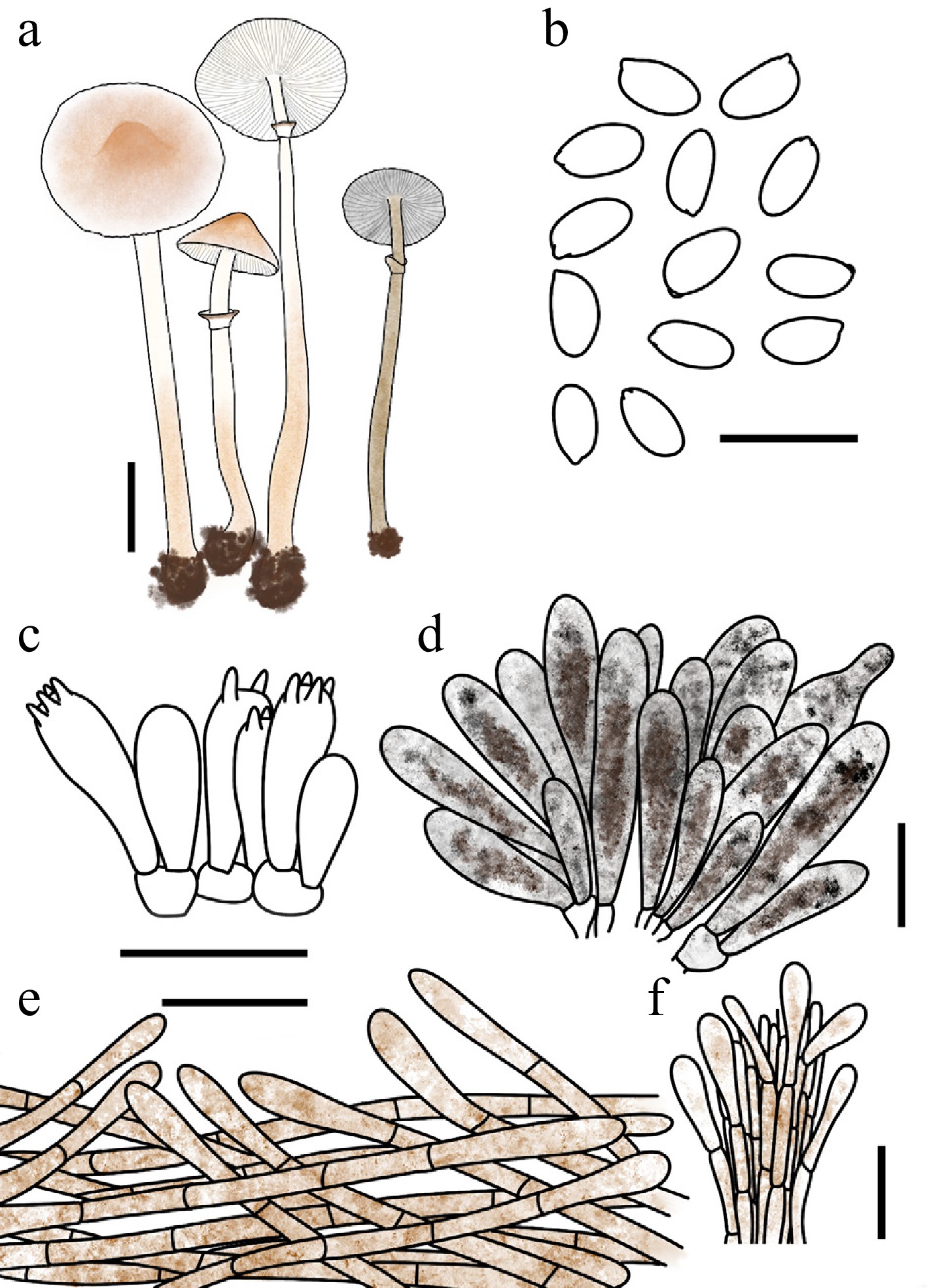
Figure 62.
Leucocoprinus persicinus. (a) Basidiomata. (b) Basidiospores. (c) Basidia. (d) Cheilocystidia. (e) Pileus covering at pileus centre. (f) Stipe covering. Scale bars: (a) = 1 cm; (b) = 10 μm; (c), (f) = 20 μm.
Chinese name: 桃色白鬼伞 (Pīn yīn: táo sè bái guǐ sǎn)
Index Fungorum number: IF904649; Facesoffungi number: FoF 18938
Etymology – 'persicinus' is derived from the Latin persicus (peach), referring to the peach-like coloration (nude pink to Persian orange) of the squamules on the pileus.
Type – China. Jilin: Baishan City, Lushuihe National Forest Park, 18 August 2024, coll. H. Cheng, FJAU78037.
Diagnosis – characterized by a pileus with nude pink to Persian orange squamules, a bicolorous annulus (white with a Persian orange to gingerbread-brown margin), and dried basidiomata that turn greige to golden brown with granite-gray lamellae.
Basidiomata small. Pileus 0.9–2.4 cm in diam., obtusely conical to plano-convex or plane when mature; umbo low, obtuse; background white; centre with nude pink (#DDC0B4) to Persian orange (#D99058) squamules; squamules around the centre fading gradually in colour toward the margin. Lamellae free, crowded, white to cream; lamellulae 0–1 between each pair of lamellae. Stipe 3.5–8.3 cm × 0.2–0.4 cm, subcylindrical, widening downwards, hollow, white; surface covered from the lower third to the base with Persian orange (#D99058) to fulvous (#E48400) squamules. Annulus superior, ascending, white with a Persian orange to gingerbread brown margin, membranous. Context thin, white. Odour and taste not recorded.
Dried basidiomata: Pileus, stipe, and context greige (#C1B7A5) to golden brown (#885407); lamellae granite gray (#676767).
Basidiospores (60/3/3) 5.8–7.1 × 3.1–4.0 μm, avl × avw = 6.2–6.4 × 3.3–3.6 μm, Q = 1.62–2.17, avQ = 1.82–1.90, oblong to cylindrical, hyaline, thin-walled, smooth, dextrinoid, cyanophilous, congophilous, metachromatic in Cresyl Blue; germ pore absent. Basidia 16–29 × 6–10 μm, clavate, predominantly 4-spored, occasionally 2-spored, thin-walled. Pseudoparaphyses absent. Pleurocystidia absent. Cheilocystidia 30–48 × 8–17 μm, oblong to slightly utriform, thin-walled, with hickory-brown (#371D10) to dark charcoal (#333333) intracellular pigment. Surface covering at pileus centre an interwoven plagiotrichoderm consisting of cylindrical hyphae; hyphae densely entangled, repent to slightly ascending; terminal cells 5–16 μm wide, slightly thick-walled, with golden-brown intracellular and parietal pigment. Stipe covering a cutis consisting of cylindrical hyphae; hyphae repent; terminal cells 29–89 × 4–14 μm, clavate to cylindrical, thin-walled, with pale yellowish-brown to golden-brown intracellular pigment. Clamp connections absent.
Habit and habitat – solitary to scattered in humus-rich soil in broadleaf forests dominated by Quercus mongolica and Juglans mandshurica.
Known distribution – so far recorded only from Northeast China.
Additional specimens examined – China. Jilin: Tonghua City, Sanxianjia National Forest Park, 12August 2022, coll. X. Wang & L.M. Song, FJAU78040, FJAU78039; Huadian City, Hongshi National Forest Park, 27 August 2023, coll. Y. Wang, FJAU78038.
Notes – Macroscopically, Leucocoprinus persicinus and Lc. rutilobrunneus T. Bau & X.Y. Zhou share a strong morphological resemblance, as both species possess nude pink squamules on the pileus. However, they can be readily distinguished by their bruising reactions: the basidiomata of Lc. rutilobrunneus undergo a distinct discolouration upon bruising, whereas Lc. persicinus remains completely unchanged.
Phylogenetically, Lc. persicinus is nested within Lc. clade I, where it clusters together with Lc. rutilobrunneus and Lc. bicolorannulatus to form a well-supported subclade. Within this three-taxon lineage, each species possesses unique diagnostic features that facilitate reliable separation. Specifically, Lc. rutilobrunneus is the only species among the three to exhibit a distinct bruising reaction. Microscopically, Lc. bicolorannulatus stands out as the only species featuring a pileus covering composed of a typical erect trichoderm. Conversely, Lc. persicinus is distinctively defined by its stipe covering, which exhibits specialized clavate to cylindrical cells.
Leucocoprinus rutilobrunneus T. Bau & X.Y. Zhou, sp. nov Figs 57c−f; 59b; 63
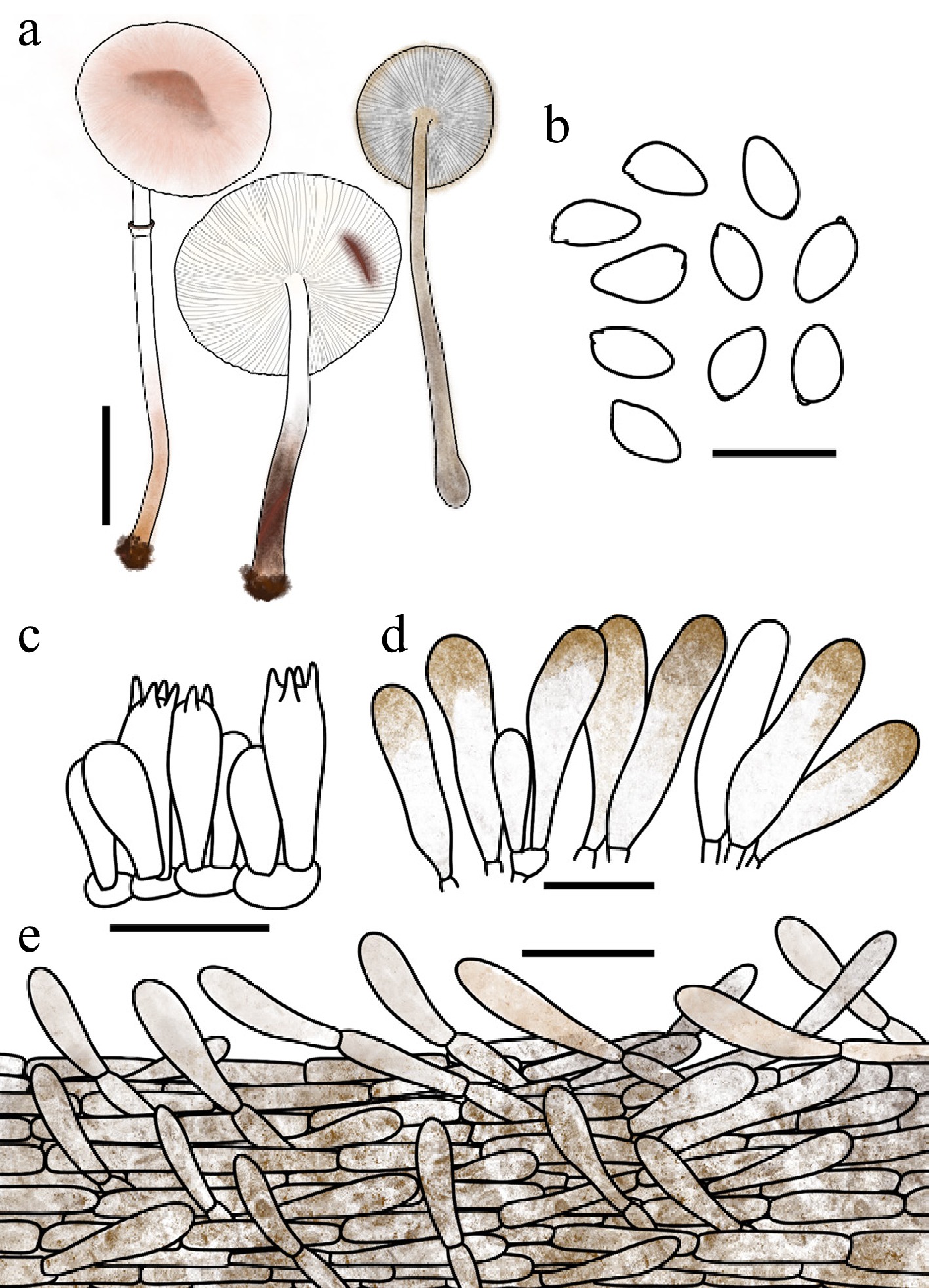
Figure 63.
Leucocoprinus rutilobrunneus. (a) Basidiomata. (b) Basidiospores. (c) Basidia. (d) Cheilocystidia. (e) Surface covering at pileus centre. Scale bars: (a) = 1 cm; (b) = 10 μm; (c), (d) = 20 μm; (e) = 50 μm.
Chinese name: 霞褐白鬼伞 (Pīn yīn: xiá hè bái guǐ sǎn)
Index Fungorum number: IF904647; Facesoffungi number: FoF 18939
Etymology – 'rutilobrunneus' is derived from the Latin rutilus (reddish-orange) and brunneus (brown), referring to the distinctive colour change of the basidiomata, which stain reddish-orange when injured and subsequently turn deep brown.
Type – China. Jilin: Yanbian Korean Autonomous Prefecture, Chinese Merganser Park, 1 August 2022, coll. L.Y. Zhu, FJAU78029.
Diagnosis – characterized by a pileus with pale squamules that darken to brownish-black with age, basidiomata turning reddish-orange then deep brown upon bruising, a bicolorous annulus, and dried basidiomata that turn golden to hickory brown with granite-gray lamellae.
Basidiomata small to medium-sized, initially turning deep carrot orange (#E9692C) to red-brown (#66332B), subsequently darkening to golden brown (#885407) and finally hickory brown (#371D10) when bruised. Pileus 0.6–4.7 cm in diam., plano-convex to applanate when mature; umbo obtuse; background white; squamules radially arranged, silky-fibrillose, initially nude pink (#DDC0B4) to dark salmon (#E9967A), turning hickory brown with age. Lamellae free, crowded, white to cream; lamellulae interspersed 1–3 between each pair of lamellae. Stipe 3.3–5.7 cm × 0.1–0.4 cm, subcylindrical, widening downwards, cream at the apex, becoming copper red (#CB6D51) to brown red (#B25B09) on the middle to lower portion; surface smooth to covered with fine fibrils. Annulus median to superior, ascending, white with a gingerbread brown to hickory brown margin, membranous, friable. Context thin, white, turning reddish orange then brown when bruised. Odour and taste not recorded.
Dried basidiomata: Pileus, stipe, and context golden brown to hickory brown; lamellae granite gray (#676767).
Basidiospores (60/3/3) 6.4–7.8 × 3.6–4.5 μm, avl × avw = 6.8–7.0 × 3.9–4.1 μm, Q = 1.50–2.01, avQ = 1.67–1.74, amygdaliform, ellipsoid to oblong or cylindrical, hyaline to yellowish brown, slightly thick-walled, smooth, dextrinoid, cyanophilous, congophilous, metachromatic in Cresyl Blue; germ pore absent. Basidia 17–27 × 6–10 μm, clavate, predominantly 4-spored, occasionally 2-spored, thin-walled. Pseudoparaphyses absent. Pleurocystidia absent. Cheilocystidia 28–59 × 8–19 μm, oblong to narrowly utriform, with an obtuse apex, thin-walled, with hickory-brown (#371D10) intracellular pigment. Surface covering at pileus centre an interwoven plagiotrichoderm consisting of cylindrical hyphae; hyphae densely entangled, repent to slightly ascending; terminal cells 5–16 μm wide, slightly thick-walled, with golden-brown to hickory-brown intracellular and parietal pigment. Stipe covering a cutis consisting of cylindrical hyphae; hyphae repent, 4–11μm wide; with golden-brown intracellular pigment, thin-walled. Clamp connections not observed.
Habit and habitat – solitary to scattered in humus in forested areas or grassy ground.
Known distribution – China.
Additional specimens examined – China. Inner Mongolia Autonomous Region: Tongliao City, Daqinggou National Nature Reserve, 1999, coll. T. Bau, FJAU1439; 9 August 2021, coll. X. Wang, FJAU78036; Jilin: Longjing City, Tianfozhishan National Nature Reserve, 27 August 2019, coll. T. Bau FJAU57918; Changchun City, Zoo and Botanical Garden, 28 August 2021, coll. L.Y. Zhu, FJAU78031; Jingyuetan National Forest Park, 18 July 2022, coll. W.N. Hou, FJAU78034; 18 August 2022, coll. X. Wang, FJAU78035; 20 August 2023, coll. S.E. Wang, FJAU78030; Dunhua City, State-Owned Forest Farm, 27 July 2022, coll. W.N. Hou, FJAU78033; Jiaohe City, Laoyeling, 28 July 2023, coll. S.E. Wang, FJAU78032.
Notes – Macroscopically, Leucocoprinus rutilobrunneus bears a striking resemblance to Lc. flammeotinctus (Kauffman) Redheadand and Lc. flammeotinctoides (Vellinga) Redhead, as all three taxa exhibit a conspicuous reddish discolouration upon bruising. However, Lc. flammeotinctus can be easily distinguished by its non-staining white lamellae and distinctly cylindrical, wavy-constricted cheilocystidia. While Lc. flammeotinctoides shares the reddish-staining lamellae, it differs microscopically by possessing predominantly narrowly clavate cheilocystidia. Furthermore, Lc. rutilobrunneus is unique in its sequential bruising reaction—progressing rapidly from reddish-orange to ultimately blackish coffee—and its distinct dried state, where in the lamellae turn granite grey[89]. Phylogenetically, both Lc. rutilobrunneus and Lc. flammeotinctus are nested within Lc. clade I, whereas Lc. flammeotinctoides is distantly positioned within Lc. clade II.
Species of Leucocoprinus exhibiting blue, green, or blue-green discolouration
-
Notes: The following dichotomous key is specifically provided to facilitate the morphological identification of Leucocoprinus species characterized by conspicuous blue, green, or blue-green discolouration. These taxa exhibit striking colour transitions to these specific hues either upon bruising of fresh basidiomata or during the desiccation process. It is crucial to note that many other species within the genus Leucocoprinus undergo different macroscopic colour changes (e.g., turning orange-red, brown, or purplish) upon bruising or drying[89,104,105]; those taxa, along with species completely lacking any discolouration[30], have been explicitly excluded from this key. Furthermore, this key is strictly limited to the taxa treated in the present study; additional species exhibiting similar blue/green discolourations may exist but are not covered here. Because these diagnostic blue/green features are highly dynamic and often transient, accurate identification relies heavily on detailed field notes. The key treats a total of 15 species known to be distributed in China, including four novel species described in the present study.
Key 2 Key to the species of Leucocoprinus exhibiting blue, green, or blue-green discolouration in the present study.
1 Basidiomata turning blue, green, blue-green, or brown upon bruising 2 1' Basidiomata not discolouring upon bruising 9 2 2 Basidiomata turning brown or darkening upon bruising 3 2' Basidiomata turning blue or green upon bruising 4 3 Stipe yellow, turning brown upon bruising, and transitioning to dark blue upon desiccation; cheilocystidia 17.2–28.6 × 8.7–11.7 μm Lc. petasatus 3' Basidiomata darkening more or less upon bruising, transitioning to blue-green upon desiccation; cheilocystidia 26–43 × 9.5–22 μm, variably clavate Lc. vernalis 4 Basidiomata turning blue upon bruising 5 4' Basidiomata turning green upon bruising 7 5 Cheilocystidia relatively small, 11.5–17 × 9–10.6 μm, pyriform to sublageniform Lc. phaeocyaneus 5' Cheilocystidia relatively large, 10–64.3 × 7.5–21 μm, clavate, narrowly clavate, broadly clavate, pyriform, or sometimes sphaeropedunculate 6 6 Cheilocystidia 40.5–64.3 × 10.9–16.8 μm, clavate, narrowly clavate, or broadly clavate Lc. atrocaeruleus 6' Cheilocystidia 10–50 × 7.5–21 μm, broadly clavate to pyriform, sometimes sphaeropedunculate, rarely clavate Lc. viriditinctus 7 Basidia characterised by a digitate apical appendage Lc. digitatocystis 7' Basidia lacking a digitate apical appendage 8 8 Cheilocystidia narrowly clavate to clavate, sometimes fusiform Lc. virens 8' Cheilocystidia subcylindrical, broadly clavate to pyriform, rarely clavate Lc. beijingensis 9 Cheilocystidia absent Lc. beninensis 9' Cheilocystidia present 10 10 Cheilocystidia frequently 1- to 2-septate, with the terminal segment distinctly digitate Lc. transeptatus 10' Cheilocystidia llacking a distinctly digitate terminal segment 11 11 Basidiomata turning blue then green upon desiccation, but ultimately not acquiring a yellowish hue Lc. actinophorus 11' Basidiomata turning blue then green upon desiccation, ultimately acquiring a yellowish hue, or exhibiting a highly transient blue phase 12 12 Pileus surface with distinct brown-red to hickory-brown floccose squamules Lc. caffeaceus 12' Pileus surface lacking distinct floccose squamules 13 13 Cheilocystidia 21.5–39 × 10–20 μm, clavate to broadly clavate Lc. coerulescens 13' Cheilocystidia 27–57 × 5–12 μm, narrowly cylindrical to cylindrical, or narrowly clavate 14 14 Stipe transiently developing a pale blue hue upon desiccation, before the entire basidioma acquires a greenish tint and ultimately turns yellow Lc. caeruleipes 14' Stipe completely lacking an initial blue phase upon desiccation; basidiomata directly transitioning to a greenish, and subsequently yellowish, phase Lc. versicolor The descriptive terms used in Key 2 is based on the original species descriptions cited below[29,30,34,64,96], as well as on the present study.
Leucocoprinus caeruleipes T. Bau & X.Y. Zhou, sp. nov Figs 58c−e; 59e; 64
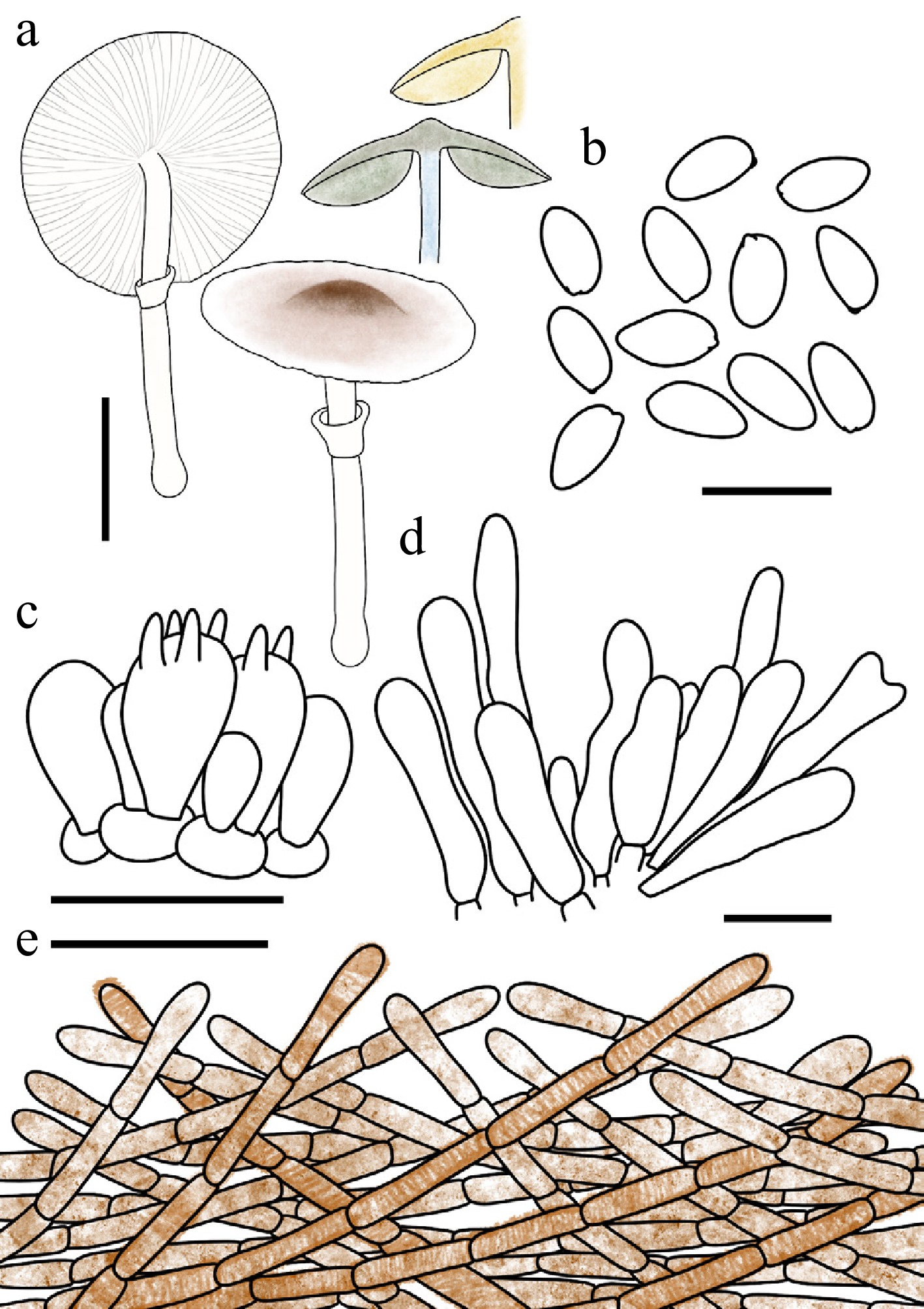
Figure 64.
Leucocoprinus caeruleipes. (a) Basidiomata. (b) Basidiospores. (c) Basidia. (d) Cheilocystidia. (e) Surface covering at pileus centre. Scale bars: (a) = 1 cm; (b) = 10 μm; (c), (d) = 20 μm; (e) = 50 μm.
Chinese name: 蓝柄白鬼伞 (Pīn yīn: lán bǐng bái guǐ sǎn)
Index Fungorum number: IF904650; Facesoffungi number: FoF 18940
Etymology – 'caeruleipes' is a compound adjective derived from the Latin caeruleus (blue) and pes (foot, stipe), referring to the distinctive pale blue coloration that develops on the stipe upon drying.
Type – China. Jilin: Jiaohe City, Lafashan National Forest Park, 5 September 2024, coll. T.Y. Zhang, FJAU78045.
Diagnosis – characterized by a pileus with nude pink to gingerbread-brown squamules, a white stipe that turns pale blue then pale green to light greenish-grey upon drying, and oblong to cylindrical basidiospores.
Basidiomata small. Pileus 1.8–2.2 cm in diam., plano-convex to applanate when mature; umbo obtuse; background white; squamules nude pink (#DDC0B4) to red brown (#66332B), fading gradually in colour toward the margin. Lamellae free, moderately crowded, white; edge concolorous; lamellulae in 3 tiers. Stipe 2.8–3.2 cm × 0.2–0.3 cm, subcylindrical, widening downwards to a slightly bulbous base, hollow, white to cream; surface smooth to covered with fine fibrils. Annulus median to superior, ascending, white, membranous. Context thin, white, unchanging when bruised. Odour and taste not recorded.
Dried basidiomata: Stipe initially turning pale blue (#9BC4E2) but fading rapidly; the entire basidioma subsequently turning pale green (#8A9977) to pale greenish grey (#96907E), and eventually drying flax yellow (#EEDC82) to mustard yellow (#E1AD01).
Basidiospores (40/2/2) 6.4–7.8 × 3.4–4.3 μm, avl × avw = 7.2 × 3.8–3.9 μm, Q = 1.63–2.20, avQ = 1.85–1.88, subamygdaliform, oblong to cylindrical, hyaline, slightly thick-walled, smooth, dextrinoid, cyanophilous, congophilous, metachromatic in Cresyl Blue; germ pore absent. Basidia 12–23 × 7–11 μm, clavate, predominantly 4-spored, thin-walled. Pseudoparaphyses absent. Pleurocystidia absent. Cheilocystidia 30–57 × 5–12 μm, narrowly cylindrical to narrowly clavate, occasionally branched at the apex, hyaline, slightly thick-walled. Surface covering at pileus centre an interwoven plagiotrichoderm consisting of cylindrical hyphae; hyphae densely entangled, repent to slightly ascending; terminal cells 5–11 μm wide, slightly thick-walled, with brown-red to red-brown intracellular pigment and concolorous encrustations. Stipe covering a cutis consisting of cylindrical hyphae; hyphae repent, 5–11 μm wide, with pale yellowish-brown intracellular pigment, thin-walled. Clamp connections not observed.
Habit and habitat – solitary in humus-rich soil in broadleaf forests dominated by Quercus mongolica and Acer.
Known distribution – so far recorded only from Northeast China.
Additional specimens examined – China. Jilin: Huadian City, Huaxi Valley, 15 August 2024, coll. X.Y. Zhou, FJAU78044.
Notes – Both macroscopically and phylogenetically, Leucocoprinus caeruleipes is most closely related to Lc. versicolor T. Bau & X.Y. Zhou. Morphologically, both species share pale pinkish to gingerbread-brown squamules on the pileus surface, and their basidiomata undergo a conspicuous colour transition to greenish and ultimately yellowish hues upon desiccation. Phylogenetically, they are recovered as sister taxa nested within Lc. clade I. However, they can be reliably distinguished by the specific sequence of their desiccation reactions. In Lc. caeruleipes, the stipe initially develops a pale blue hue upon drying; this coloration is highly transient and rapidly fades as the entire basidioma acquires a greenish tint, eventually stabilizing at a yellowish hue. In contrast, the basidiomata of Lc. versicolor transition directly to the greenish—and subsequently yellowish—phases upon desiccation, completely lacking the initial pale-blue stage on the stipe. Furthermore, while the basidiomata of Lc. atroazureus (Jun F. Liang, Zhu L. Yang & J. Xu) M. Asif, Saba & Vellinga and Lc. viriditinctus (Berk. & Broome) M. Asif, Saba & Vellinga also exhibit a blue discoloration upon drying[96], they can be readily segregated from Lc. caeruleipes by their lack of subsequent colour transitions; specifically, they do not proceed to develop the green and final yellow hues that characteristically define the latter species.
Leucocoprinus caffeaceus T. Bau, X.Y. Zhou & C. Cheng, sp. nov Figs 58i, j; 59g; 65
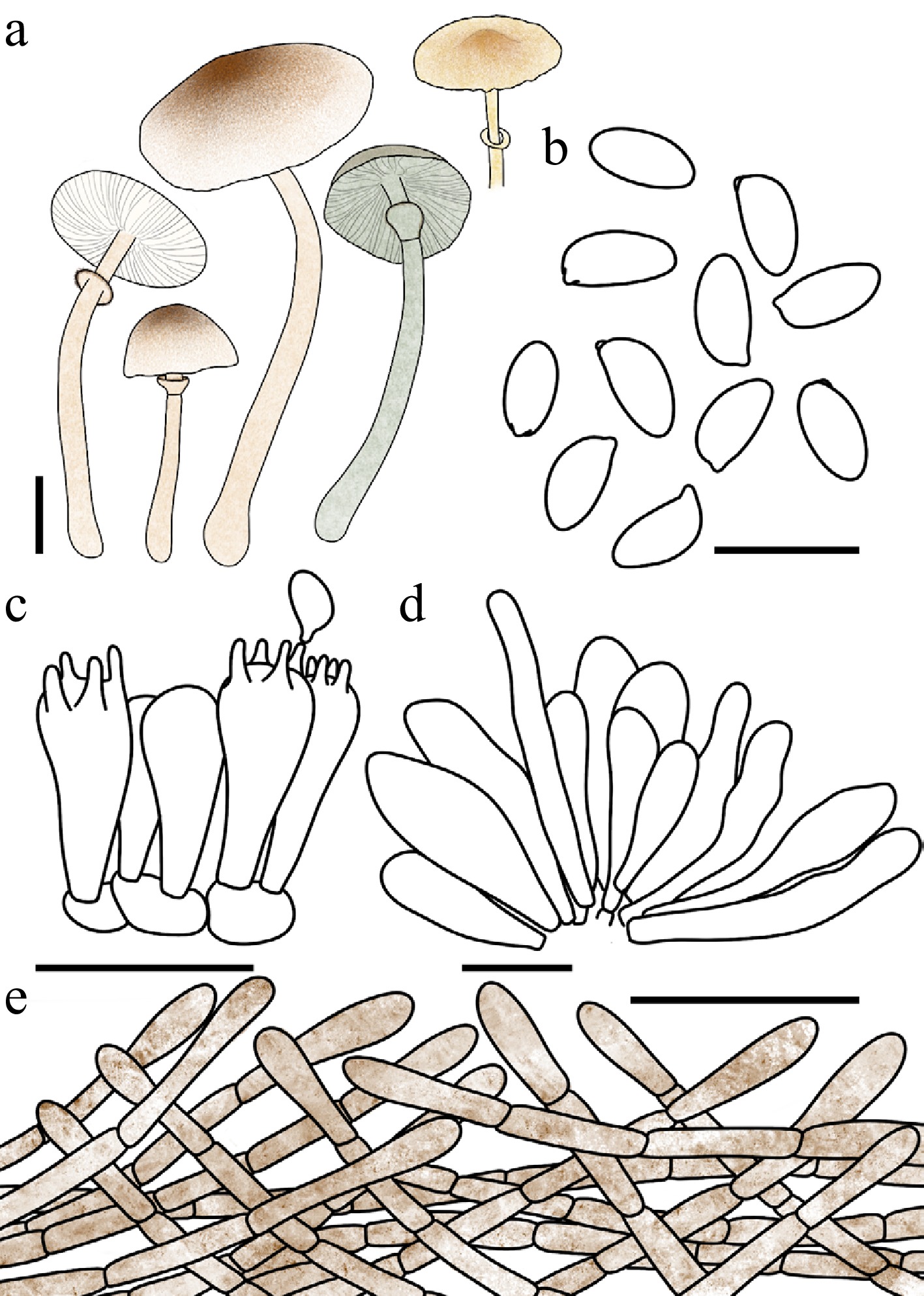
Figure 65.
Leucocoprinus caffeaceus. (a) Basidiomata. (b) Basidiospores. (c) Basidia. (d) Cheilocystidia. (e) Surface covering at pileus centre. Scale bars: (a) = 1 cm; (b) = 10 μm; (c), (d) = 20 μm; (e) = 50 μm.
Chinese name: 咖啡色白鬼伞 (Pīn yīn: kā fēi sè bái guǐ sǎn)
Index Fungorum number: IF904672; Facesoffungi number: FoF 18941
Etymology – 'caffeaceus' (modern Latin, adjective) is derived from caffea (coffee) and the suffix -aceus (resembling), referring to the characteristic coffee-coloured tone of the pileus surface and its squamules.
Type – China. Guangdong: Shenzhen City, Wutong Mountain, 15 June 2025, coll. C. Cheng, FJAU78103.
Diagnosis – characterized by a pileus with brown-red to hickory-brown floccose squamules, and dried basidiomata that initially turn pale green before fading to flax yellow or mustard yellow.
Basidiomata small. Pileus 1.5–2.4 cm in diam., obtusely conical to plano-convex or applanate; umbo low, obtuse; background white; centre with hickory brown (#371D10) floccose squamules; squamules around the centre pale gold (#E6BE8A) to brown-red (#B25B09). Lamellae free, moderately crowded, white to cream; lamellulae 0–2 between each pair of lamellae. Stipe 3.5–4.8 cm × 0.1–0.3 cm, subcylindrical, hollow, cream to pale gold at the apex, gradually turning brown-red to red brown (#66332B) towards the base; surface smooth to covered with fine fibrils. Annulus median to superior, ascending, white to pale gold with a gingerbread brown margin, membranous, ephemeral. Context thin, white, bruising reaction not recorded. Odour and taste not recorded.
Dried basidiomata: basidiomata initially turn pale green (#8A9977), subsequently fading to flax yellow (#EEDC82) to mustard yellow (#E1AD01).
Basidiospores (40/2/2) 6.2–7.8 × 3.3–4.2 μm, avl × avw = 7.0–7.1 × 3.6–3.9 μm, Q = 1.67–2.20, avQ = 1.88–1.97, oblong to cylindrical, hyaline, thin-walled, smooth, dextrinoid, cyanophilous, congophilous, metachromatic in Cresyl Blue; germ pore absent. Basidia 16–23 × 8–11 μm, clavate, predominantly 4-spored, occasionally 2-spored, thin-walled. Pseudoparaphyses absent. Pleurocystidia absent. Cheilocystidia 29–60 × 6–19 μm, narrowly cylindrical to clavate, occasionally slightly fusiform, thin-walled. Surface covering at pileus centre an interwoven plagiotrichoderm consisting of cylindrical hyphae; hyphae densely entangled, repent to slightly ascending; terminal cells 6–16 μm wide, slightly thick-walled, with red-brown to hickory-brown intracellular pigment and conspicuous concolorous encrustations. Stipe covering a cutis consisting of cylindrical hyphae; hyphae repent, 5–13, with pale yellowish-brown intracellular pigment, thin-walled. Clamp connections not observed.
Habit and habitat – scattered in humus-rich soil in forests.
Known distribution – known only from the type locality in Wutong Mountain, Central-Southern China.
Additional specimens examined – China. Guangdong: Shenzhen City, Wutong Mountain, 15 June 2025, coll. C. Cheng, FJAU78104.
Notes – It should be noted that the basidiomata of Leucocoprinus caffeaceus and Lc. transeptatus T. Bau & X.Y. Zhou also undergo a colour transition to greenish and ultimately yellowish hues upon desiccation. However, due to the highly transient nature of this initial green phase, the coloration faded too rapidly to be photographically documented.
Both macroscopically and phylogenetically, Lc. caffeaceus is most closely related to Lc. shumuyuanensis (K.L. Yang, Jia Y. Lin & Z.Chao Liu) T. Bau. However, they can be readily distinguished both macro- and microscopically. Macroscopically, unlike Lc. caffeaceus, the basidiomata of Lc. shumuyuanensis do not exhibit any discolouration upon drying. Microscopically, Lc. shumuyuanensis is entirely devoid of cheilocystidia, whereas Lc. caffeaceus is characterized by distinct narrowly cylindrical to clavate cheilocystidia.
Leucocoprinus transeptatus T. Bau & X.Y. Zhou, sp. nov Figs 58k, l; 59h; 66
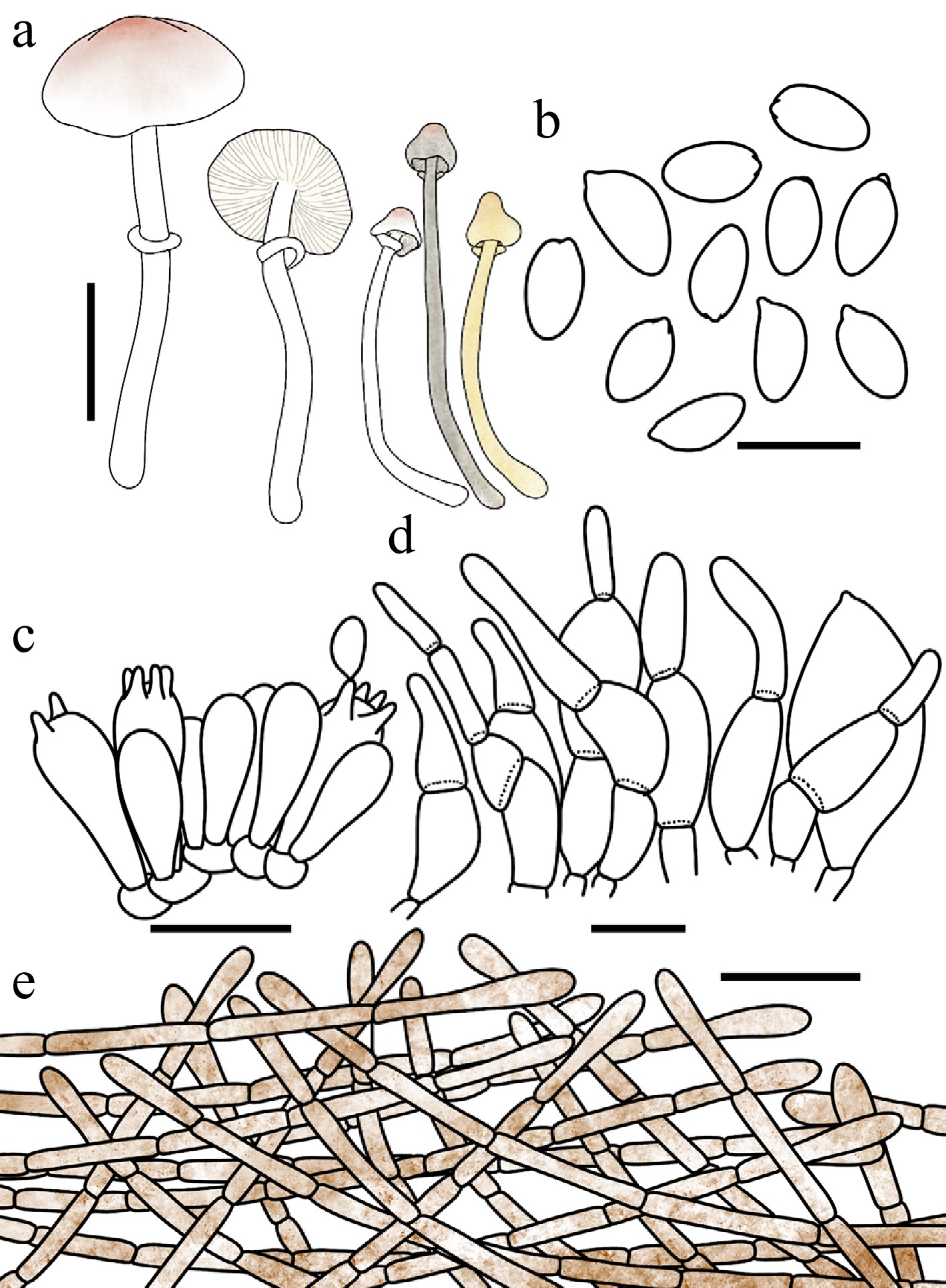
Figure 66.
Leucocoprinus transeptatus. (a) Basidiomata. (b) Basidiospores. (c) Basidia. (d) Cheilocystidia. (e) Surface covering at pileus centre. Scale bars: (a) = 1 cm; (b) = 10 μm; (c), (d) = 20 μm; (e) = 50 μm.
Chinese name: 具横隔白鬼伞 (Pīn yīn: jù héng gé bái guǐ sǎn)
Index Fungorum number: IF904652; Facesoffungi number: FoF 18942
Etymology – 'transeptatus' is derived from the Latin prefix trans- (across) and septatus (septate), referring to the transversely septate apical appendages of the cheilocystidia
Type – China. Jilin: Ji'an City, Mirror groove, 8 August, 2023, coll. Q.R. Liu, FJAU78067.
Diagnosis – characterized by a pileus with nude pink to copper-red squamules, dried basidiomata that initially turn pale greenish grey before fading to flax yellow or mustard yellow, and clavate to broadly fusiform cheilocystidia with digitate, transversely septate apical appendages.
Basidiomata small. Pileus 0.7–1.4 cm in diam., initially campanulate, expanding to plano-convex; umbo obtuse; background white; centre with nude pink (#DDC0B4) to copper-red (#CB6D51) squamules; squamules around the centre fading gradually in colour toward the margin. Lamellae free, moderately crowded, floral white to cream; lamellulae interspersed 1–3 between each pair of lamellae. Stipe 2.8–5.9 cm × 0.1–0.2 cm, subcylindrical, hollow, white; surface covered with concolorous fibrillose squamules. Annulus median to superior, ascending, white, membranous. Context thin, white to cream, bruising reaction not recorded. Odour and taste not recorded.
Dried basidiomata: basidiomata initially turn pale greenish grey (#96907E), subsequently fading to flax yellow (#EEDC82) to mustard yellow (#E1AD01).
Basidiospores (40/2/2) (7.6–)7.9–9.0 × 4.3–5.5(–5.8) μm, avl × avw = 8.3–8.5 × 4.7–4.8 μm, Q = 1.62–2.03, avQ = 1.69–1.81, subamygdaliform, oblong to cylindrical, hyaline, thin-walled, smooth, dextrinoid, cyanophilous, congophilous, metachromatic in Cresyl Blue; germ pore absent. Basidia 16–29 × 8–13 μm, clavate, predominantly 4-spored, occasionally 2-spored, thin-walled. Pseudoparaphyses absent. Pleurocystidia absent. Cheilocystidia 29–72 × 6–19 μm, clavate to broadly fusiform, frequently 1- to 2-septate, with the terminal segment distinctly digitate, hyaline, thin-walled. Surface covering at pileus centre an interwoven plagiotrichoderm consisting of cylindrical hyphae; hyphae densely entangled, repent to slightly ascending; terminal cells 7–14 μm wide, slightly thick-walled, with pale gold to brown-red intracellular and parietal pigment. Stipe covering a cutis consisting of cylindrical hyphae; hyphae repent, 5–15 μm wide; with pale gold intracellular pigment, thin-walled. Clamp connections not observed.
Habit and habitat – solitary to scattered in humus-rich soil in broadleaf forests dominated by Cunninghamia, Acer, and Ulmus.
Known distribution – China.
Additional specimens examined – China. Jilin: Ji'an City, Yushan Park, 26, July, 2025, coll. X.Y. Zhou, FJAU78068, FJAU78069.
Notes – Both macroscopically and phylogenetically, Leucocoprinus transeptatus is most closely related to Lc. petasatus R.L. Zhao & J.Xin Li. Morphologically, both species exhibit reddish-brown squamules on the pileus surface. Phylogenetically, they are recovered as sister taxa nested within Lc. clade I. Despite this striking similarity, including their shared tendency to develop blue-greenish hues, they can be reliably distinguished. In Lc. transeptatus, the dried basidiomata ultimately transition to a yellowish hue, a phenomenon not documented for Lc. petasatus. More decisively, they differ microscopically: the cheilocystidia of Lc. petasatus are clavate to broadly clavate and lack any apical appendages[29], whereas those of Lc. transeptatus are uniquely characterized by a distinct, septate, finger-like apical appendage.
Furthermore, based on our phylogenetic analyses (Figs 1, 3; Supplementary File 3), a publicly available sequence previously identified as 'Leucocoprinus petasatus' (voucher ZRL20180329, GenBank PV470813) clusters tightly with our collections of Lc. transeptatus. Therefore, we formally propose that this sequence represents a misidentification and should be reassigned to Lc. transeptatus.
Leucocoprinus versicolor T. Bau & X.Y. Zhou sp. nov Figs 58f−h, 59f, 67
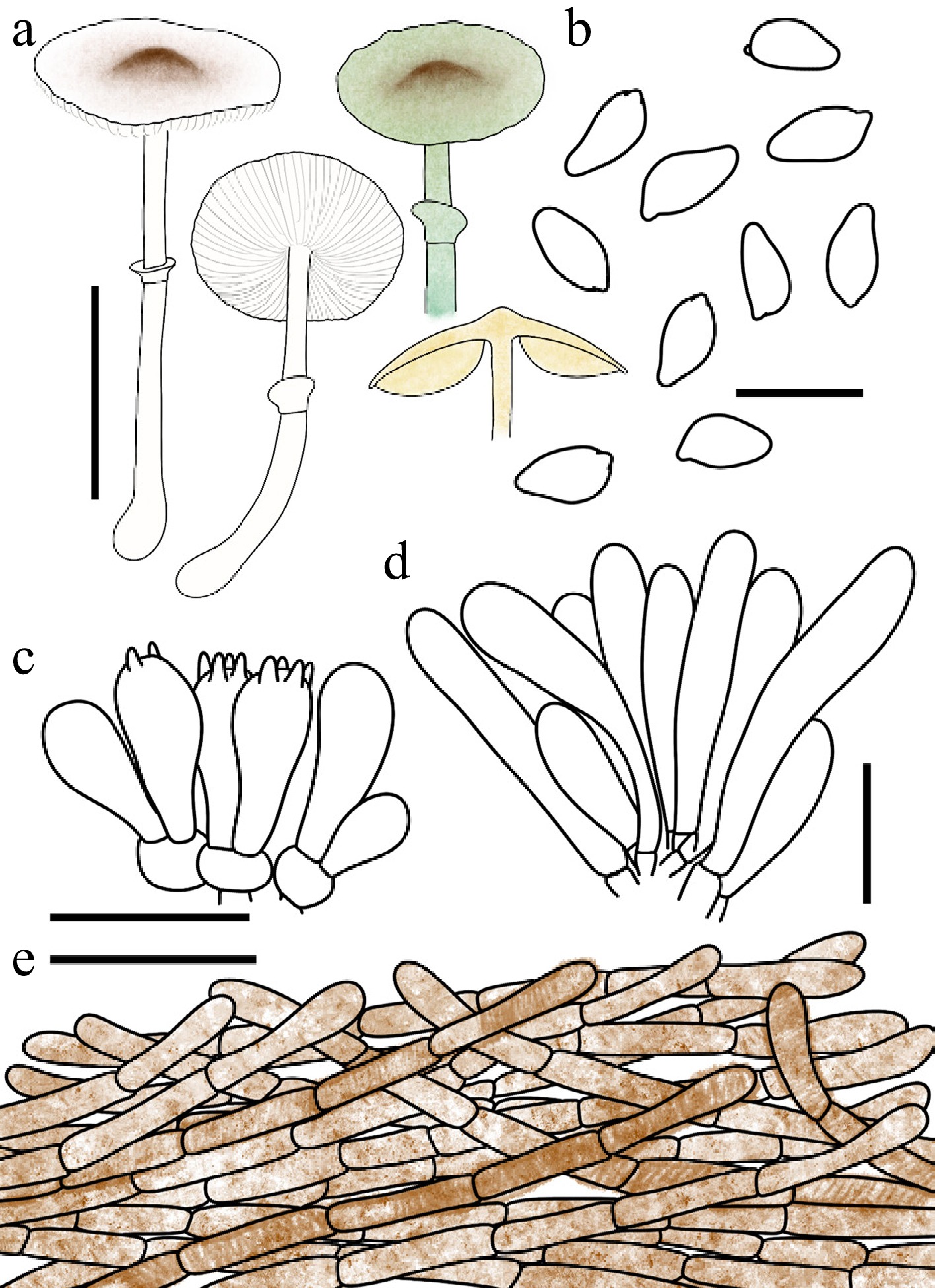
Figure 67.
Leucocoprinus versicolor. (a) Basidiomata. (b) Basidiospores. (c) Basidia. (d) Cheilocystidia. (e) Surface covering at pileus centre. Scale bars: (a) = 1 cm; (b) = 10 μm; (c), (d) = 20 μm; (e) = 50 μm.
Chinese name: 变色白鬼伞 (Pīn yīn: biàn sè bái guǐ sǎn)
Index Fungorum number: IF904651; Facesoffungi number: FoF 18943
Etymology – 'versicolor' is derived from the Latin versare (to change, to turn) and color (colour), referring to the striking colour shift of the basidiomata, which change from nude pink or gingerbread brown when fresh to green and yellow hues upon drying.
Type – China. Jilin: Baishan City, Lushuihe National Forest Park, 8 August 2024, coll. Q. R. Liu, FJAU78041.
Diagnosis – characterized by a pileus with nude pink to gingerbread-brown squamules, and dried basidiomata that initially turn shamrock green to light aloe green before fading to flax yellow or mustard yellow.
Basidiomata small. Pileus 1.0–1.8 cm in diam., plano-convex to applanate when mature; umbo obtuse; background white; squamules nude pink (#DDC0B4) to red brown (#66332B), fading gradually in colour toward the margin. Lamellae free, moderately crowded; lamellulae interspersed in 2 tiers. Stipe 1.4–5.7 cm × 0.1–0.2 cm, subcylindrical, hollow, white to cream; surface covered with concolorous fibrillose squamules; base brown-red (#B25B09). Annulus median, ascending, white to cream, membranous. Context thin, white, unchanging when bruised. Odour and taste not recorded.
Dried basidiomata: basidiomata initially turn shamrock green (#5BAF7F) to light aloe green (#7E9B39), subsequently fading to flax yellow (#EEDC82) to mustard yellow (#E1AD01).
Basidiospores (60/3/3) 6.8–8.2 × 3.4–4.3 μm, avl × avw = 7.1–7.3 × 4.0–4.1 μm, Q = 1.58–1.96, avQ = 1.77–1.96, subamygdaliform, ellipsoid to oblong, hyaline, slightly thick-walled, smooth, dextrinoid, cyanophilous, congophilous, metachromatic in Cresyl Blue; germ pore absent. Basidia 13–23 × 7–10 μm, clavate, predominantly 4-spored, thin-walled. Pseudoparaphyses absent. Pleurocystidia absent. Cheilocystidia 27–52 × 6–11 μm, narrowly cylindrical to cylindrical, hyaline, slightly thick-walled. Surface covering at pileus centre an interwoven plagiotrichoderm consisting of cylindrical hyphae; hyphae densely entangled, repent to slightly ascending; terminal cells 5–14 μm wide, slightly thick-walled, with brown-red (#B25B09) to red-brown (#66332B) intracellular and parietal pigment. Stipe covering a cutis consisting of cylindrical hyphae; hyphae repent, 4–12 μm wide, with pale yellowish-brown intracellular pigment, thin-walled. Clamp connections not observed.
Habit and habitat – solitary in humus-rich soil in mixed coniferous and broadleaf forests.
Known distribution – Northeast China.
Additional specimens examined – China. Jilin: Tonghua City, Luotong Mountain, 11 August, 2022, coll. L.M. Song, FJAU78043; Jiaohe City, Laoyeling, 28 July 2023, coll. X. Wang, FJAU78042.
Notes – Macroscopically, Leucocoprinus versicolor bears a striking morphological resemblance to Lc. beijingensis R.L. Zhao & J.X. Li, Lc. virens (Y.R. Ma, Z.W. Ge & T.Z. Liu) Asif, Saba & Vellinga, and Lc. flavovirens (Jun F. Liang, Zhu L. Yang & J. Xu), Kun L. Yang, Jia Y. Lin & Zhu L. Yang, as all four taxa undergo a distinct transition to blue-greenish hues upon desiccation. However, they can be reliably distinguished based on their bruising reactions. Specifically, while the basidiomata of Lc. beijingensis, Lc. virens, and Lc. flavovirens exhibit a conspicuous greenish discolouration upon bruising[34,64,96], Lc. versicolor completely lacks this greenish bruising reaction.
-
The generic boundaries between Leucoagaricus and Leucocoprinus s.l. have historically been a focal point of debate in Agaricaceae s.l. research due to their similar morphological traits and persistent non-monophyly. Recent attempts to resolve this paraphyly initially favored broadening the generic circumscription, as proposed by Redhead[22] and expanded by Asif et al.[23], who subsumed most Leucoagaricus taxa into Leucocoprinus s.l. However, subsequent multi-locus and phylogenomic data revealed that such broad generic concepts likely mask deep evolutionary divergences. Consequently, systematic frameworks have gradually shifted toward fine-scale delimitation. For instance, Yang et al.[25] segregated distinct lineages into the novel genera Candelolepiota and Macropsalliota, while Li et al.[29] established the subfamily Leucocoprinoideae and introduced the new genus Conioexocarpus. This trend of subdivision continued with Yang et al.[30], who correlated phylogenetic clades with morphological traits to redefine the subfamilies Leucocoprinoideae and Micropsalliotoideae. This framework ultimately dispersed the traditional Leucoagaricus and Leucocoprinus s.l. across eight narrowly circumscribed genera, including the newly segregated Pulchrolepiota, Tristolepiota, and Mesopsalliota. Recently, Vellinga et al.[31] emphasized that taxonomic decisions should integrate robust diagnostic morphological criteria alongside phylogenetic topology[106]. Rather than adopting the newly proposed segregated genera, they explicitly aligned themselves with a two-genus arrangement that maintains a broadly circumscribed Leucocoprinus alongside a narrowly defined Leucoagaricus. To make this framework operational, they provided the requisite formal combinations, thereby facilitating its adoption without dismissing alternative classifications as invalid.
Aligning with this perspective, we adopt the broadly circumscribed Leucocoprinus concept and apply the two-genus framework advocated by Vellinga et al.[31] to ensure taxonomic consistency within this two-genus framework. The multi-generic framework recently proposed by Yang et al.[30] provides a detailed morphological reassessment of this complex. By correlating morphological traits with specific phylogenetic lineages, their proposed narrow segregates offer improved diagnostic resolution for identifying fine-scale morphological trends. While this fine-scale delimitation has practical utility for characterizing restricted clades, our expanded regional sampling and subsequent evaluation of both molecular and morphological evidence lead us to retain Leucocoprinus s.l. to prioritize overall taxonomic stability. From a phylogenetic standpoint, our analysis recovers the broadly defined Leucocoprinus s.l. as a monophyletic group with moderate support (93/0.92). In contrast, the narrowly defined segregates exhibit variable or insufficient support when subjected to our increased taxon sampling. For instance, Lc. clade I (Tristolepiota) receives only weak support (–/0.90), and the narrowly delimited Leucocoprinus s.s. Yang et al. lacks statistical support in our topology[30]. While we recognize that Yang et al.[30] achieved stable monophyly for these segregates using an 825-locus phylogenomic dataset (their Fig. 7), that specific analysis was based on a relatively limited number of species from this complex. Indeed, in their corresponding four-locus phylogeny, which included broader sampling (their Fig. 8), critical segregates such as Leucocoprinus s.s. (ML = 52) and Pulchrolepiota (ML = 54) similarly received only weaky support values. This indicates that the monophyly of these narrow genera is highly sensitive to sampling density, suggesting that a broadly defined genus offers a more stable framework under current sampling. This topological sensitivity is further corroborated by the morphological patterns observed across the complex. Our diagnostic character mapping reveals that key traits used to delimit these narrow genera exhibit multiple independent origins rather than serving as exclusive synapomorphies. For example, discoloration upon bruising or drying occurs independently in both Lc. clade I (Tristolepiota) and Lc. clade II (Pulchrolepiota). Although some species in Lc. clade II are distinguished by a positive reaction to ammonia, taxa sharing these specialized morphological traits are also recovered within phylogenetically unresolved, transitional lineages in our study (e.g., the Lc. croceovelutinus and Lc. atroazureus clades). The recurrence of these features across disparate lineages confirms widespread homoplasy, complicating their utility as primary boundaries for generic delimitation. Consequently, recognizing a single, broadly circumscribed Leucocoprinus provides a practical and stable framework that effectively accommodates both the evolutionary complexity and the transitional lineages inherent to this group.
Phylogenetic resolution of major clades
-
Within this adopted generic framework, our current four-locus phylogenetic analysis resolved the evolutionary relationships across the complex. Our backbone topology recovered Leucoagaricus with strong support (99/1.0), whereas Leucocoprinus s.l. received moderate support (93/0.92). For Leucoagaricus, our phylogenetic framework is highly congruent with previous studies[29,30], revealing five major clades alongside two unstable lineages. Specifically, our results can be reliably mapped onto the subclades identified by Li et al.[29]: their clades B1, B4, B5, and B6 correspond to our La. clade III, La. clade V, La. clade II, and the La. jianensis clade, respectively; while their clades B2 and B3 jointly correspond to our La. clade IV. Notably, a singleton lineage lacking a definitive assignment in their study consistently forms a long, independent branch in our phylogeny, represented by La. xantholepis. Furthermore, our La. clade I, which also lacked a stable position in Li et al.'s analysis, remains independent from the core Leucoagaricus in our study. This overall molecular framework for Leucoagaricus is also in general agreement with that proposed by Yang et al.[30].
Within Leucocoprinus s.l., the phylogenetic branching exhibits greater complexity. Nevertheless, our topology—comprising four major clades and seven unassigned lineages—aligns well with the branching patterns observed by Li et al.[29], despite their lack of formal clade nomenclature for this group. Furthermore, our clades strongly correspond to the generic boundaries recently redefined by Yang et al.[30]. In our framework, Lc. clade I and Lc. clade II precisely represent the newly erected genera Tristolepiota and Pulchrolepiota, respectively, while our Lc. clades III and IV correspond to their restricted concept of Leucocoprinus. However, a topological discrepancy was observed regarding the Lc. pyrrhulus, Lc. atroazureus, and Lc. fragilis clades. In our analysis, these lineages are positioned basally and externally to Lc. clades III and IV without significant statistical support, whereas Yang et al.[30] nested them within Pulchrolepiota (Lc. clade II in this study). Consistent with our generic rationale, this topological instability primarily stems from our dense taxon sampling; the inclusion of numerous newly generated sequences representing phylogenetically transitional taxa likely increased the complexity of phylogenetic inference and reduced deep-node resolution. Despite these persisting molecular uncertainties at the deepest generic boundaries, the combined diagnostic features revealed by morphological and phylogenetic congruence provide a practical and adequate framework for distinguishing Leucoagaricus and Leucocoprinus s.l. Ultimately, achieving robust resolution in these relationships will necessitate large-scale phylogenomic approaches in future studies.
Phylogenetic reassessment of traditional morphology-based sections
-
Prior to the widespread application of molecular phylogenetics, the infrageneric taxonomy of Leucoagaricus and Leucocoprinus relied entirely on macro- and micro-morphological features, as epitomized by the classic monograph of Candusso & Lanzoni[15]. This system detailed the division of these taxa into various subgenera and sections. However, mapping these traditional morphological divisions onto our current multi-locus phylogeny reveals that, while classical morphological features remain highly practical for preliminary field identification, frequent convergent evolution has resulted in many traditional sections being polyphyletic.
Within Leucocoprinus, our analysis indicates that most core members traditionally assigned to Leucocoprinus sect. Leucocoprinus exhibit high morphological and molecular congruence. For example, Lc. birnbaumii, Lc. brebissonii (Godey) Locq., Lc. cepistipes, and Lc. fragilissimus (Ravenel ex Berk. & M.A. Curtis) Pat. cluster stably within Lc. clade III. This grouping suggests that the defining features of this section genuinely reflect a monophyletic origin; however, Lc. cretatus, originally assigned to this group, is a notable exception and has been explicitly segregated into the genus Conioexocarpus[29] .
In contrast, traditional morphology-based groupings within Leucoagaricus show a broader phylogenetic distribution across multiple lineages. For instance, species traditionally assigned to Leucoagaricus subgen. Leucoagaricus sect. Leucoagaricus are now recovered in distinctly different lineages: La. brunneocingulatus (P.D. Orton) Migl. & Donato and La. ionidicolor (Bellù & Lanzoni) Migl. & Donato are firmly nested within Leucocoprinus[30] (Lc. clade II), whereas La. melanotrichus and La. tener fall into Lc. clade IV. Similarly, taxa formerly united in sect. Pilosellae are now understood to represent multiple independent evolutionary origins: La. meleagris forms the independent Mesopsalliota lineage, while most members of this section (e.g., La. badhamii, La. pilatianus [Demoulin] Bon & Boiffard, and La. jubilaei [Joss.] Bon) have been reassigned to Leucocoprinus (Lc. clade II). Additionally, classic members of sect. Annulati (such as La. cinerascens and La. leucothites) consistently cluster with core Leucocoprinus (Lc. clade III). Subgenus Sericeomyces displays a comparable pattern, with its representative species distributed among Leucocoprinus (e.g., La. sericatellus and La. sericeus in Lc. clade I), and multiple Leucoagaricus clades. Regarding sect. Rubrotincti, apart from a few species transferred to Leucocoprinus (e.g., La. gaillardii Bon & Boiffard and La. gauguei Bon & Boiffard), its core taxa (e.g., La. rubrotinctus, La. purpureolilacinus, and La. littoralis [Ménier] Bon & Boiffard) remain within Leucoagaricus but are distributed across La. clades III and IV, indicating a polyphyletic arrangement rather than strict monophyly.
To concisely summarize the status of historical sectional systems: with the notable exception of Leucocoprinus sect. Leucocoprinus, which largely retains its phylogenetic integrity, most other traditional sections across this complex exhibit polyphyletic patterns. These findings highlight the intricate evolutionary reality of this group, providing a deeper molecular perspective that builds upon foundational taxonomic frameworks. Specifically, due to widespread homoplasy, historical sectional boundaries based primarily on macroscopic traits do not always correspond to monophyletic evolutionary lineages. Therefore, while classical sections remain indispensable and highly practical for preliminary morphological sorting, contemporary systematic treatments can be further enriched by integrating these valuable morphological observations into a robust, clade-based phylogenomic framework.
Evaluation of recent infrageneric classification frameworks
-
Yang et al.[30] recently proposed an updated classification framework for Leucoagaricus and Leucocoprinus s.l., establishing new subgenera and sections based on phylogenomic data. While their analysis provides valuable insights into the deep divergences of representative taxa, applying this detailed sectional framework universally remains challenging due to the rapidly expanding species diversity within this complex.
One major challenge is the extensive phenotypic plasticity and homoplasy within these fungi. As taxon sampling increases, we observe that certain sectional diagnostic traits often overlap with the broader phenotypic diversity. For example, the absence of typical hymenial physalides is a key diagnostic character for Lc. subgen. Coprinagaricus sect. Bicolores[30]. However, several species recently assigned to this section (Lc. domingensis Justo, Bizzi, Angelini & Vizzini, Lc. heinemannii Migl., Lc. tephrolepis Justo, Bizzi, Angelini & Vizzini, and Lc. parvipileus Justo, Bizzi, Angelini & Vizzini) are documented to possess pseudoparaphyses[94]. Similarly, in sect. Coniosquamosi (also defined by the lack of typical physalides), the hymenium of our newly described Lc. sinumbellatus is almost entirely composed of pseudoparaphyses, rendering sterigmate basidia completely unobservable. These morphological overlaps suggest that such microscopic traits have evolved in parallel across different lineages, making them difficult to apply consistently as strict diagnostic markers for formal sectional delimitation.
Additionally, the phylogenetic topology of this complex is highly sensitive to taxon sampling. Given the continuous discovery of new species, current phylogenies may not yet capture all transitional lineages. When we incorporated our densely sampled novel species, the resolution of several deeper nodes shifted. For instance, taxa such as Leucocoprinus fragilis (sect. incertae sedis), Lc. atroazureus (sect. Pilosellae), Lc. callainitinctus (sect. Pilosellae), and Lc. pyrrhulus (sect. Flammeae), which were assigned to specific sections in the previous framework[30], emerged as more independent lineages basal to Ⅲ and IV in our expanded analysis. This topological fluidity is consistent with recent findings by Li et al.[29], where Lc. atroazureus and Lc. callainitinctus were resolved at the base of Lc. clade I (their Tristolepiota[30]). The varying placements of these taxa across different studies highlight the dynamic nature of deep-node resolution in this group.
Given the sensitivity of phylogenetic resolution to taxon sampling density, the comprehensive implementation of formal infrageneric ranks is likely susceptible to frequent revisions in the future. To prevent continuous nomenclatural flux and avoid potential discrepancies between morphological diagnoses and molecular boundaries, we currently prefer a cautious approach toward adopting newly established formal infrageneric classifications. Instead, we opt to employ informal 'clades' to structure the phylogenetic and taxonomic discussions herein. This flexible, clade-based strategy better accommodates the continuously discovered morphological diversity within the broadly circumscribed Leucocoprinus complex, while avoiding premature taxonomic inflation and unnecessary fluctuations in the formal nomenclatural system.
Evolutionary continuum of pileus covering microstructures
-
In the Agaricaceae, the micro-morphological structure of the pileus covering has long been recognized as an informative valuable taxonomic marker[36,80,107,108]. Recently, Yang et al.[30] emphasized its critical taxonomic utility, using the hyphal orientation of the surface covering at the pileus centre—specifically distinguishing between horizontally arranged structures (cutis) and erect elements (trichoderm or conioderm)—as the primary diagnostic criterion for generic boundaries within Leucocoprinoideae. This standard has greatly advanced the infrageneric classification system of this subfamily, and we fully agree that these microstructures carry significant phylogenetic information.
Building upon the taxonomic framework established by Yang et al.[30], and supported by our increased sampling density and detailed microscopic observations of new taxa, we found that the micro-evolution of this group exhibits a complex continuum alongside the macroscopic trends noted by previous researchers. The pileus centre covering structure within Leucocoprinus s.l. encompasses not only the well-established states but also evolutionary transitional states between them. To capture this structural diversity as accurately as possible, we categorized and refined the surface covering at the pileus centre of this complex into three main categories and six sub-types (Figs 4−6). Consistent with previous observations, core Leucoagaricus species predominantly exhibit a cutis with horizontal, parallel hyphae (Type I-A), and most core Leucocoprinus s.l. species form a typical erect trichoderm (Type II-A/B) or conioderm (Type III-A). However, our study further identifies three key morphological transitional states:
First, an interwoven plagiotrichoderm (Type I-B; Fig. 4). The pileus centre in this type primarily consists of repent to slightly ascending hyphae; they depart from a strictly horizontal cutis but have not fully developed into erect elements. The interwoven arrangement of these hyphae bridges the morphological gap between a cutis and a trichoderm.
Second, a catenulate trichoderm was observed in Type II-B (Fig. 5). Unlike a typical trichoderm characterized by aseptate terminal cells, this type is composed of erect, frequently septate hyphae forming long chains of short to elongated cylindrical cells. Morphologically, these chains closely resemble the repent hyphae typical of a cutis, but are distinctly arranged as vertically oriented elements.
Third, the moniliform plagiotrichoderm (Type III-B; Fig. 4). For example, in Lc. umbrosquamosus, we observed repent to slightly ascending moniliform hyphal chains in which cylindrical, ellipsoid, and subglobose cells are intimately intermixed. This is morphologically distinct from a typical conioderm (Type III-A), which consists of loosely arranged pure globose cells.
Furthermore, additional complex transitional states were observed in certain species. For instance, the surface covering at the pileus centre in Lc. subumbrosquamosus is composed of inflated cylindrical to slightly ellipsoid hyphae. It lacks the prominently swollen subglobose cells of Type III-B and differs from the narrowly cylindrical hyphae of Type I-B. Because it may represent an intermediate state between the interwoven plagiotrichoderm and the moniliform plagiotrichoderm, we refrain from formally naming this subtype. Notably, this microstructure aligns closely with previous descriptions of 'short and moderately inflated hyphal cells' at the pileus centre of Lc. melanotrichus[30]. Consistently, Lc. subumbrosquamosus and Lc. melanotrichus resolve as sister taxa in our molecular phylogeny, further underscoring the high congruence between micro-morphological transitional states and molecular evolutionary trajectories. Additionally, based on original species descriptions[87], we identified a unique pileus-covering evolutionary state in Lc. subhymenoderma: its pileus surface squamules are composed of globose cells forming catenulate structures. Although morphologically similar to the moniliform plagiotrichoderm (Type III-B), it conspicuously lacks the abundant long cylindrical cells present in the latter. Concurrently, it differs entirely from a typical conioderm (Type III-A), which usually presents loose globose cells without such distinct chain-like arrangements. Interestingly, this specific catenulate globose cell structure is microscopically closer to the typical features of certain Echinoderma species[87,97,107−109]. The discovery of these complex intermediate states in both Lc. subumbrosquamosus and Lc. subhymenoderma unequivocally confirms that phenotypic evolution of the pileus covering is not stepwise, but rather exists as a multidimensional micro-morphological transitional continuum.
The presence of these transitional states highlights the phenotypic complexity of this complex during its evolution. To document and distinguish the microscopic features of these transitional lineages more clearly, this study subdivides the microstructures of the surface covering at the pileus centre into 'six types' of sub-categories, guided by the theoretical framework of Yang et al[30]. This preliminary descriptive classification aims to serve as a practical supplementary tool, better accommodating the widespread morphological continuity in nature and providing a reference for finer species delimitation and morpho-molecular mapping within this complex in future studies.
Widespread homoplasy and ecological adaptation
-
The congruence analysis between morphology and phylogeny within the Leucoagaricus–Leucocoprinus s.l. complex (Figs 1−3) reveals distinctive diagnostic feature combinations for distinct molecular lineages, providing a basis for infrageneric classification. However, the integration of these features also provides strong evidence for widespread morphological homoplasy within this complex. Basal lineages (Conioexocarpus, Candelolepiota, Macropsalliota, and Mesopsalliota) generally display a complex mosaic of ancestral traits. In contrast, the crown group Leucoagaricus is morphologically highly unified, typically lacking hymenial pseudoparaphyses and rarely exhibiting basidioma discolouration. The features of Lc. clades III and IV are notably variable, encompassing the presence or absence of basidiospore germ pores, hymenial pseudoparaphyses, and cheilocystidia, accompanied by occasional droplet exudation or ant associations. Additionally, Lc. clade II and Lc. clade I are primarily characterized by distinct discolouration reactions upon bruising or drying, with Lc. clade II being uniquely distinguished by a positive reaction to ammonia.
Tracing specific diagnostic traits across the phylogenetic tree further highlights their multiple independent origins. Basidioma discolouration upon bruising or drying appears in the near-basal Macropsalliota and Mesopsalliota, extends upward into Lc. clade I and Lc. clade II, but is largely absent during the evolutionary transition toward the Leucoagaricus crown clades. Despite sharing this discolouration trait, each lineage retains exclusive diagnostic markers, such as germ pores in Macropsalliota and a positive ammonia reaction in Lc. clade II. Similarly, basidiospore germ pores have evolved independently multiple times: excluding basal genera, they are only present in Lc. clade III and the Lc. porosporus clade, each accompanied by different complementary features. Hymenial pseudoparaphyses also display a scattered distribution, being reported in Conioexocarpus and evolving independently multiple times within Lc. clades III and IV. Furthermore, the exudation of coloured droplets by fresh basidiomata (known in Candelolepiota) independently appears in scattered species at the base of Lc. clades III and IV, clustering predominantly at the crown of Lc. clade IV. A pileus covering composed of globose cells is primarily concentrated in Lc. clades III and IV; however, Lc. clade III uniquely combines this trait with pseudoparaphyses and germ pores. This scattered, mosaic distribution strongly indicates that these morphological markers arose through convergent evolution within the complex.
Beyond morphological features, convergent evolution is also evident in ecological strategies. For example, mutualistic symbioses with attine ants appear in three distinct phylogenetic lineages: the Lc. gongylophorus lineage, Lc. clade IV (Lc. karjaticus), and Conioexocarpus (Co. dunensis, Co. attinorum). This distribution indicates that ant mutualism independently evolved multiple times within the subfamily. Notably, these symbiotic relationships are accompanied by divergent morphological profiles: for instance, fresh basidiomata of Lc. karjaticus exude droplets (a trait absent in Lc. gongylophorus), while ant-associated Conioexocarpus species are distinguished by the presence of germ pores.
Within Leucoagaricus, macroscopic colour phenotypes (such as orange-red and whitish) show notable parallel evolution, notably spanning La. clades II, III, and V, which form the basis of the two currently recognized species complexes. The orange-red La. rubrotinctus complex exhibits minimal internal genetic differentiation, whereas the Leucoagaricus serenus complex exhibits substantial genetic differentiation. This contrast suggests that colour evolution in this genus may be strongly influenced by specific selective pressures and rapid diversification. Such rapid diversification may have resulted in a limited accumulation of morphological variation, thereby phenotypically maintaining the macroscopic cohesion within these lineages. Furthermore, these evolutionary patterns are closely associated with specific ecological preferences. Numerous Leucoagaricus species demonstrate a strong preference for sandy soils. For instance, the majority of specimens from the La. serenus complex in La. clade V (e.g., La. albofulvescens and La. aff. similis) were collected from sandy environments. Accordingly, literature records indicate that several species in La. clade III (e.g., La. crystallifer, La. littoralis, La. menieri, La. purpureolilacinus, La. rubroconfusus, La. subvolvatus) and La. gujratensis in La. clade I are typically associated with nutrient-poor coastal or sandy habitats[76,78,80,110−112]. This highlights edaphic specialization as a potentially important driver shaping the evolution and speciation of this genus in specific habitats.
Microscopic variation in problematic species
-
Microscopic observations of several problematic specimens revealed taxonomic complexity and potential trait instability.
Mesopsalliota meleagris: Specimen FJAU72223 possessed pseudoparaphyses adjacent to the basidia, a feature not described in the literature[80,97], whereas specimen FJAU72222 lacked this feature. This suggests that the presence of pseudoparaphyses in certain taxa is related to the state of the specimen.
Map. aff. subtropica: Specimens FJAU72224–FJAU72226 (Fig. 36j, k) exhibited a unique combination of morphological features: the stipe turned yellow upon touching, and they possessed pseudoparaphyses and digitate sterigmata (as reported by Li et al.[34] for Lc. digitatocystis). Despite these significant morphological differences, the specimens showed no discernible phylogenetic divergence from Map. subtropica (Yu Yang, X. Luo & Y.P. Xiao) Kun L. Yang, Jia Y. Lin & Zhu L. Yang[113] (Fig. 36l). Therefore, we cautiously assigned this material to an affine species of Map. subtropicus.
Leucocoprinus cinerascens: The two observed specimens (FJAU77965–FJAU77966, Fig. 37a, b) lacked sphaerocystic structures under the erect hyphae of the pileus cover, contradicting the descriptions by Candusso et al.[15] and Akers[16]. Given the absence of molecular data for historical-type materials, an accurate circumscription of Lc. cinerascens remained unclear.
Leucoagaricus serenus complex: In the Leucoagaricus serenus complex, a series of species (La. aff. serenus 1, La. aff. serenus 2, and La. aff. similis) were resolved as independent molecular lineages (Figs 1−2, 16f, g, 17d–h, 25–26, 28). Although these nodes showed distinct molecular divergence, they lacked reliable morphological differentiating features compared to their respective close relatives (La. serenus and La. similis). Given the contradiction between molecular divergence and morphological convergence, we cautiously assigned these specimens to the affine species pending further in-depth investigation.
-
Overall, this study investigated the complex taxonomic boundaries within the Leucoagaricus and Leucocoprinus s.l. based on morphological and multi-locus phylogenetic analyses, and evaluated the applicability of traditional morphological frameworks. By integrating newly generated sequencing data with public sequences, we analyzed the phylogenetic relationships across Leucoagaricus, Leucocoprinus s.l., and related genera. Under this systematic framework, we described 41 novel species and reported two new records for China. In accordance with the broadly circumscribed generic concept adopted herein, we reinstated 60 species names within Leucocoprinus and proposed eight new combinations into this genus. This taxonomic work highlights the rich species diversity of this group in East Asia and provides baseline data and a practical reference for future regional and global taxonomic revisions.
Although this study has improved the resolution of the systematic framework of this complex to some extent, statistical support for certain deep nodes remains insufficient in the current multi-locus phylogeny. Given the rapid evolutionary radiation likely experienced by this group, future studies should incorporate phylogenomic data specifically targeting these unstable transitional lineages. Furthermore, the 'six-type' classification system for the pileus-covering microstructures proposed here is primarily based on current regional observations; future research will be necessary to re-evaluate the micro-morphology of historical taxa to verify the broader applicability of this framework on a global scale. Finally, the widespread convergent evolution observed in this complex suggests that future systematic studies should extend beyond pure molecular phylogenies. Integrating taxonomic revisions with specific ecological traits, substrate preferences, and biogeographical data will be essential for a more comprehensive understanding of the actual mechanisms driving species diversification in this complex.
The authors are deeply grateful to their supervisor and laboratory colleagues for their invaluable guidance, continuous support, and extensive assistance with fieldwork and experiments. We also extend our special thanks to mushroom enthusiasts YM Cheng (C Cheng), DD Jiang, and several anonymous enthusiasts for their significant contributions to the specimen collections. We express our sincere appreciation to the associate editor and the anonymous reviewers for their constructive critiques and insightful suggestions, which substantially improved the quality and rigor of this manuscript. Additionally, we would like to thank Editage for English language editing. This work was supported by the Ministry of Education Innovation Team (No. IRT1134, IRT-15R25), and the National Natural Science Foundation of China (No. 32070010).
-
Not applicable.
-
The authors confirm contribution to the paper as follows: study conception and design: Zhou XY and Bau T; data collection: Zhou XY and Bau T; analysis and interpretation of results: Zhou XY and Bau T; draft manuscript preparation: Zhou XY. All authors reviewed the results and approved the final version of the manuscript.
-
The data that support the findings of this study are openly available in the National Center for Biotechnology Information (NCBI) GenBank database, the Index Fungorum database, and the Faces of Fungi repository. All data generated or analyzed during this study are included in this published article and its supplementary information files.
-
All supplementary materials for this study are deposited in Zenodo (doi: 10.5281/zenodo.17759675), which include the following: Supplementary File 1: Colour chart and corresponding nomenclature used for macroscopic descriptions; Supplementary File 2: Sequence accession information, and original phylogenetic tree files generated in this study.
-
Due to computational limitations regarding Bayesian convergence on massive datasets and for visual clarity, the phylogenies presented in Figs 1–3 are reduced backbone trees prioritizing type and reference sequences. An expanded maximum likelihood (ML) phylogeny based on the concatenated four-locus dataset, encompassing all sequenced accessions and additional unclassified sequences (labeled as "sp."), is provided as Supplementary File 3 (Expanded multi-locus ML phylogeny).
-
The authors declare that they have no conflict of interest.
-
accompanies this paper online at: https://doi.org/10.48130/mycosphere-0026-0013.
- Supplementary File 1 Colour chart and corresponding nomenclature used for macroscopic descriptions.
- Supplementary File 2 Sequence accession information, and original phylogenetic tree files generated in this study.
- Supplementary File 3 Expanded multi-locus ML phylogeny.
- Copyright: © 2026 by the author(s). Published by Maximum Academic Press, Fayetteville, GA. This article is an open access article distributed under Creative Commons Attribution License (CC BY 4.0), visit https://creativecommons.org/licenses/by/4.0/.
-
About this article
Cite this article
Zhou XY, Bau T. 2026. A comprehensive survey of Leucoagaricus and Leucocoprinus s.l. (Agaricaceae, Agaricales): species delimitation and novel taxa. Mycosphere 17: e013 doi: 10.48130/mycosphere-0026-0013 

A comprehensive survey of Leucoagaricus and Leucocoprinus s.l. (Agaricaceae, Agaricales): species delimitation and novel taxa
- Received: 16 March 2026
- Revised: 25 June 2026
- Accepted: 03 July 2026
- Published online: 07 August 2026
Abstract: The generic boundaries and phylogenetic relationships within the Leucoagaricus–Leucocoprinus sensu lato complex have long been a focal point of taxonomic debate. Recently, integrative phylogenomic studies have re-evaluated this group, accommodating it within the newly established subfamily Leucocoprinoideae and recognizing several novel genera. Despite these updated generic frameworks, clarifying species-level boundaries and comprehensively understanding morphological evolutionary patterns within this hyper-diverse complex require further exploration. Based on extensive taxon sampling, multi-locus phylogenetic analyses, and detailed micro-morphological assessments, we investigated the evolutionary relationships within this group. Our analyses demonstrate that Leucoagaricus comprises five major molecular clades encompassing two primary morphological assemblages: the orange-red La. rubrotinctus complex and the whitish La. serenus complex. Concurrently, Leucocoprinus s.l. is resolved into four major clades alongside several transitional lineages. Supported by this comprehensive evidence, we formally describe 41 novel species and report two new geographic records from China. Furthermore, in alignment with the broadly circumscribed generic concept adopted herein, we reinstate the original Leucocoprinus combinations for 60 species and propose eight new combinations into this genus. Mapping specific diagnostic traits onto the phylogeny indicates widespread morphological homoplasy across distantly related lineages. Finally, we provide detailed morphological descriptions and line drawings for all new taxa and records, along with specialized identification keys for species exhibiting fresh guttation or greenish to bluish discoloration upon drying.
-
Key words:
- Leucoagaricus /
- Leucocoprinus /
- Agaricaceae /
- New species /
- Multi-locus phylogeny /
- Species delimitation /
- Taxonomy /
- China