











-
End-stage heart failure is a cardiovascular disorder associated with high mortality worldwide, and heart transplantation remains the only definitive curative therapy currently available[1]. In recent years, European and North American countries have been undergoing a rapid transition from conventional static storage to mechanical perfusion technologies. The application of modalities such as hypothermic oxygenated mechanical perfusion (HOPE)[2] and normothermic in vitro perfusion using the Organ Care System (OCS)[3] has extended donor heart preservation times to 8–9 h while maintaining post-transplant survival outcomes comparable to those achieved with short-term static preservation[2,3]. Concurrently, these regions have also implemented donation after circulatory death (DCD) as a routine clinical practice, further expanding the donor pool[4]. In contrast, conventional static cold storage (SCS) remains the predominant method of donor heart preservation in China. Unlike the well-established dedicated organ transportation networks in Western countries, donor heart transportation in China still relies largely on public transportation systems, including high-speed rail and commercial air travel. Given the stringent requirement that cold ischemia time must be restricted to 4–6 h[5], this logistical reality makes every donor heart transfer a race against time. According to the 2024 Report on the Development of Organ Donation and Transplantation in China, 3,655 patients were awaiting heart transplantation, yet only 1,064 procedures were performed during the same year[6]. By comparison, over 4,000 heart transplant surgeries were completed in the United States in 2022[7], substantially exceeding the domestic transplantation volume in China (Fig. 1). Although the expansion of donor eligibility criteria has increased donor availability to some extent[8], the substantial imbalance between donor heart supply and clinical demand remains the principal bottleneck limiting further advances in heart transplantation.
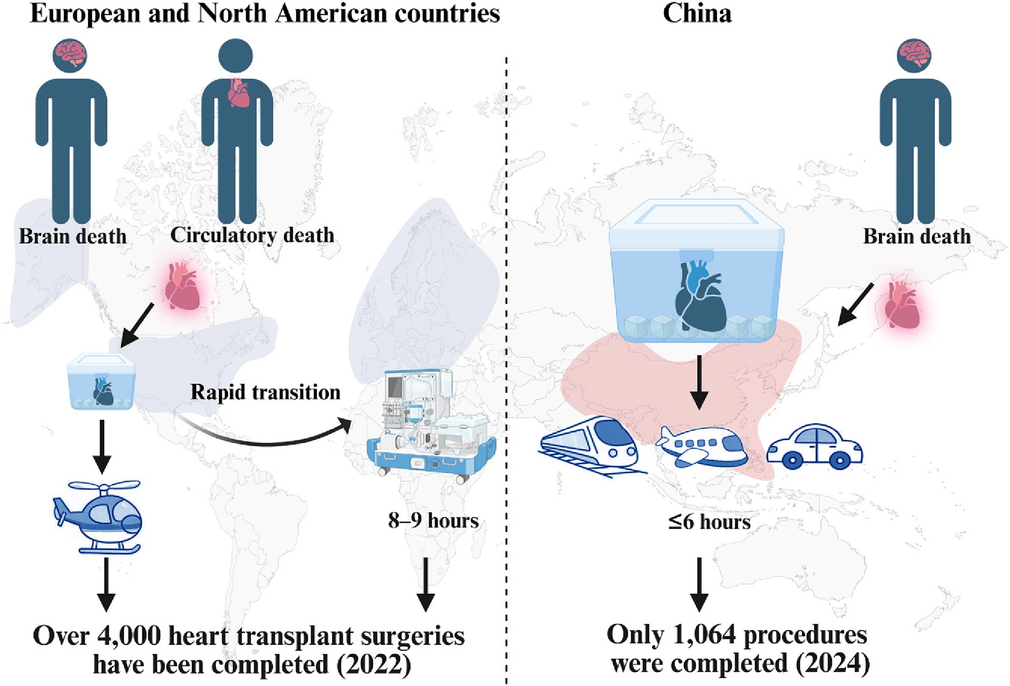
Figure 1.
Comparison of donor cardiac graft preservation in China and abroad.
Despite the emergence of mechanical perfusion technologies, SCS remains the clinical gold standard for donor organ preservation[9]. Its fundamental principle is to provide passive protection by reducing myocardial metabolic activity under hypothermic conditions (0–4 °C). However, current preservation solutions lack well-defined pharmacologically active components that can directly target molecular pathways involved in cold ischemic injury. Consequently, prolonged cold ischemia exceeding 4–6 h inevitably aggravates ischemic damage, thereby increasing the risk of primary graft dysfunction, acute rejection, and even perioperative mortality[10].
During heart procurement and transplantation, donor hearts are subjected to both warm and cold ischemic insults, which differ fundamentally in their underlying pathophysiological mechanisms. Cold ischemia induces a series of unique temperature-dependent injuries, including membrane lipid phase separation[11], mitochondrial dysfunction[12], intracellular calcium overload[13], hyperactivation of autophagy[14], and rebound apoptosis upon reperfusion[15,16]. Despite extensive preclinical evidence, clinical solutions for cold ischemic injury remain challenging. On one front, synthetic and biologic compounds (including cyclosporin A, valproic acid, and mitochondria-targeted peptides) show immense promise in safeguarding organ viability and preventing programmed cell death. However, their clinical translation remains limited. Systemic safety liabilities, poor pharmacokinetics, and off-target distribution represent major barriers to clinical translation and human trials.
This impasse has driven a parallel quest for natural alternatives. Sourced from plants, fungi, and marine animals, these structurally diverse compounds have shown potential to mitigate ischemia-induced damage by regulating a broad spectrum of biological processes, including inflammatory cascades, calcium homeostasis, oxidative stress responses, and apoptotic signaling. However, promising mechanistic findings from in vitro studies have not yet translated into clinical benefits. Currently, translating either synthetic or natural therapeutic strategies into clinically effective treatments remains a major challenge. This review focuses on cold ischemic injury and systematically summarizes its underlying mechanisms using the differences between cold and warm ischemia as a framework. By integrating recent advances from both basic and clinical studies, we comprehensively discuss molecular and pharmacological targets that may be amenable to therapeutic intervention. Particular attention is given to the therapeutic potential of natural products in mitigating cold ischemic injury. Furthermore, we highlight the emerging applications of multi-omics technologies and advanced drug delivery systems in myocardial protection and discuss their potential to improve donor heart preservation strategies.
-
A deeper, comprehensive understanding of the mechanistic differences between cold and warm ischemia is essential for guiding the development and optimization of preservation strategies in heart transplantation. The mechanisms underlying warm ischemic injury have been extensively characterized in various experimental models, such as myocardial infarction models[17] and Langendorff isolated heart perfusion models[18]. In contrast, the mechanisms of cold ischemic injury have not been independently and comprehensively defined. Consequently, studies on cold ischemia often extrapolate from the well-established mechanisms of warm ischemic injury. However, substantial differences exist between these two types of ischemic injury, particularly with respect to energy utilization, glucose metabolism, and mitochondrial function. Metabolomic analyses indicate that their pathological features can be broadly summarized as follows: warm ischemia involves rapid metabolic collapse, whereas cold ischemia is characterized by hypothermia-induced metabolic stagnation[19] (Fig. 2). Distinguishing the mechanistic differences between warm and cold ischemia is essential for identifying optimal cardioprotective targets. Ultimately, the severity of ischemia-reperfusion injury (IRI) following transplantation is determined by the cumulative effects of warm and cold ischemic insults.
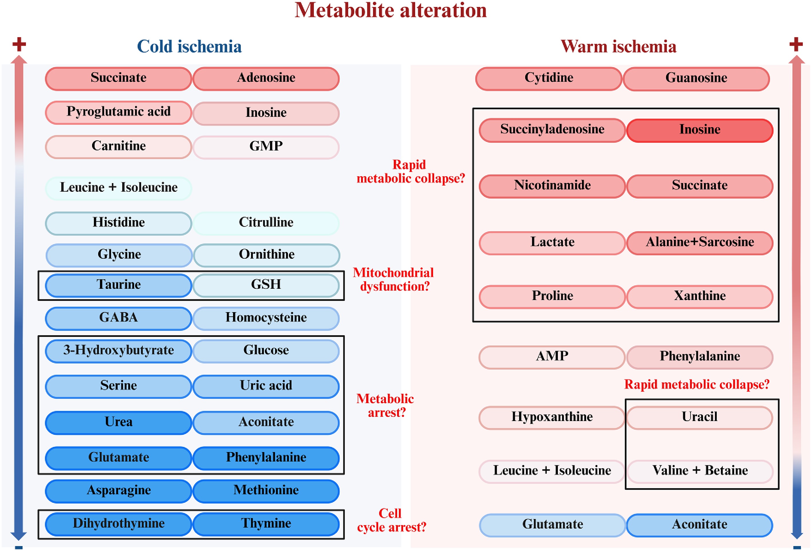
Figure 2.
Comparison of metabolites of warm ischemia and cold ischemia. Metabolites are color-coded based on their abundance changes relative to pre-ischemic baselines, where red and blue indicate upregulation (+) and downregulation (−), respectively (deeper colors denote higher significance).
The core characteristics of warm ischemic injury
-
Warm ischemic injury refers to a pathological condition in which cardiac blood supply is interrupted under normothermic conditions (approximately 37 °C), resulting in myocardial injury. It often occurs in various diseases such as acute myocardial infarction[20] and unstable angina pectoris[21], and therefore research about its pathophysiology, diagnosis, and clinical management has been extensive. Its underlying mechanisms involve disturbances in energy metabolism, calcium overload, oxidative stress, reactive oxygen species (ROS) bursts, activation of cell death pathways, and inflammatory responses. During cardiac transplantation, warm ischemia is inevitable because the myocardial core temperature typically takes more than 15 min to fall to 4 °C following circulatory arrest[22]. This interval constitutes a period of occult warm ischemia, during which cardiomyocytes continue to undergo active metabolism under near-normothermic conditions. The key mechanistic features of ischemia-reperfusion injury are characterized by energy metabolism disruption and oxidative stress[23]. Following abrupt oxygen deprivation, intracellular ATP in cardiomyocytes is rapidly depleted through its conversion to AMP and subsequent degradation. The rapid depletion of ATP compromises the activity of ATP-dependent ion pumps, resulting in ionic imbalance and cellular dysfunction[24]. Although cardiomyocytes initially compensate by increasing glycolytic ATP production, glycolysis alone is insufficient to meet cellular energy demands and progressively becomes impaired, leading to lactate accumulation and further limiting efficient ATP generation[25].
Ischemia decreases the mitochondrial membrane potential and increases the ubiquinone-to-ubiquinol ratio, driving the reverse activity of succinate dehydrogenase (SDH) and promoting succinate accumulation[26]. At the same time, the accumulation of NADH, depletion of ADP, and the reversal of the malate-aspartate shuttle result in the accumulation of fumarate, which disrupts the TCA cycle and prevents succinate oxidation, thus leading to a large accumulation of succinate[26,27]. During reperfusion, rapid oxidation of accumulated succinate by SDH drives excessive electron transfer through the mitochondrial electron transport chain, resulting in a burst of ROS production and oxidative damage. Furthermore, succinate stabilizes hypoxia-inducible factor 1α (HIF-1α) by inhibiting the prolyl hydroxylase domain (PHD) enzyme, thereby amplifying inflammatory signals and exacerbating ischemia-reperfusion injury[26] (Fig. 3).
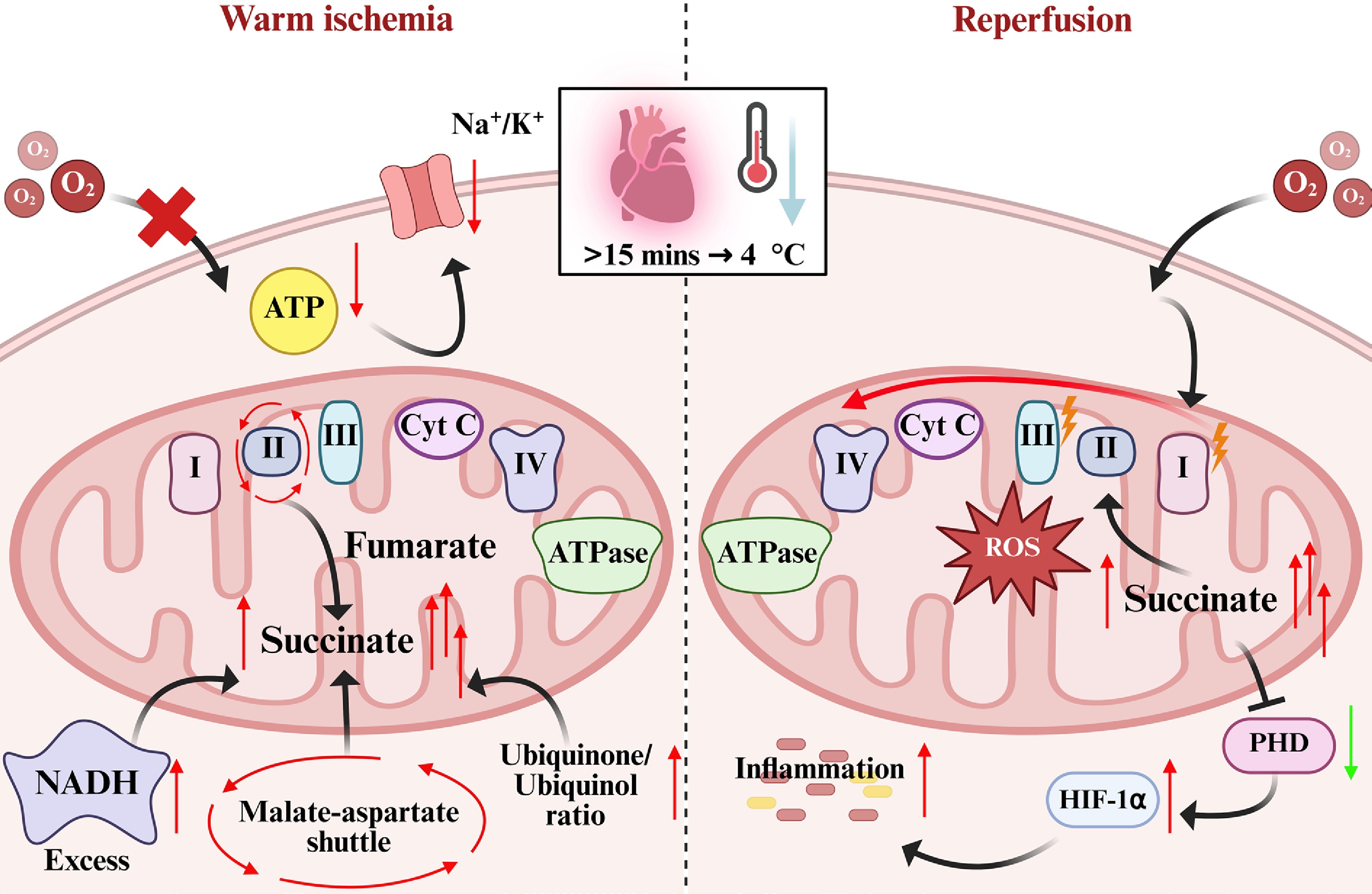
Figure 3.
The core mechanism of warm ischemia and reperfusion injury.
The core characteristics of cold ischemic injury
-
During cold ischemic preservation, low temperatures (0–4 °C) successfully prolong graft survival by suppressing myocardial metabolism. However, metabolic activity does not completely cease; this persistent, low-level metabolism can induce progressive tissue damage upon subsequent reperfusion[28,29].
Energy depletion and metabolic disturbances during static cold storage
-
Firstly, the energy crisis caused by SCS is the core issue at this stage. Under low temperature conditions, most enzyme reactions are inhibited, and the rate of ATP hydrolysis significantly decreases. The ATP/ADP ratio can therefore be relatively maintained, allowing preservation of adenine nucleotide pools. However, in the state of cold ischemia, energy replenishment is also limited, and ion pumps (such as Na+/K+-ATPase) continue to consume ATP, resulting in the continuous depletion of high-energy phosphate compounds[12]. At the same time, lactate accumulates gradually, contrasting with the rapid glycolytic disturbance observed during warm ischemia. This ATP depletion, together with the disruption of energy homeostasis caused by sustained lactate accumulation, directly disrupts cellular ion balance, triggers intracellular Na+ and Ca2+ overload, cellular edema, and mitochondrial dysfunction[13].
Cold-induced cellular and organelle dysfunction during static cold storage
-
Secondly, hypothermia itself can induce specific forms of cellular damage. Low temperatures promote phase separation of membrane lipids in cardiomyocytes, reducing membrane fluidity and compromising the structural integrity and function of membrane-associated proteins[30]. Concurrently, energy depletion and ionic imbalance trigger endoplasmic reticulum (ER) stress, leading to the accumulation of misfolded proteins and activation of the unfolded protein response (UPR)[31]. Moreover, as the central organelles responsible for both energy production and the regulation of apoptosis, mitochondria undergo progressive structural deterioration during cold storage, including matrix swelling and cristae disintegration. These ultrastructural alterations impair oxidative phosphorylation and further exacerbate cellular energy deficits[32]. Evidence from human donor hearts further supports these findings, demonstrating that prolonged cold ischemia induces characteristic contractile band formation, activates autophagy, and causes extensive organelle damage[14].
Oxidative stress and ferroptosis activation during cold ischemia
-
Thirdly, oxidative stress is initiated during cold ischemia. Although the reverse activity of SDH is markedly suppressed at low temperatures, succinate continues to accumulate gradually through residual reverse SDH activity. In addition, hypothermia differentially impairs the activities of mitochondrial electron transport chain complexes, thereby increasing electron leakage and promoting ROS generation[12]. Simultaneously, cold-induced suppression of endogenous antioxidant enzymes, including superoxide dismutase (SOD), catalase (CAT), and glutathione peroxidase (GPX), compromises the ROS-scavenging capacity. Consequently, ROS progressively accumulate throughout the cold ischemic period[15]. Evidence from human donor hearts further demonstrates significantly increased levels of malondialdehyde (MDA), a marker of lipid peroxidation, together with elevated expression of acyl-CoA synthetase long-chain family member 4 (ACSL4), a key regulator of ferroptosis. These findings support the activation of both oxidative stress and ferroptosis[15] (Fig. 4).
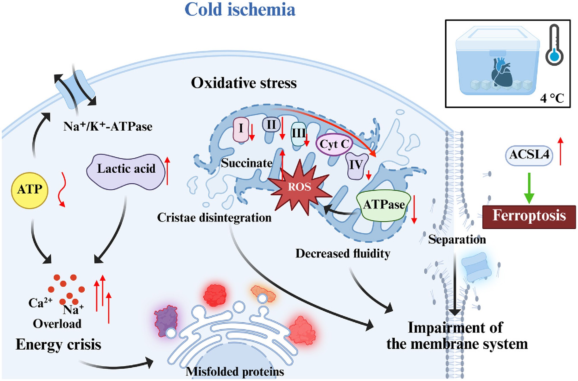
Figure 4.
The core mechanism of cold ischemic injury.
To sum up, cold preservation is not a state of metabolic quiescence but a dynamic pathological process characterized by progressive energy depletion, membrane dysfunction, and oxidative stress. The cumulative effects of these injuries markedly exacerbate tissue damage upon myocardial reperfusion. Unlike warm ischemia, cold ischemia occurs under hypothermic conditions that profoundly suppress mitochondrial metabolism and ATP production. However, this hypometabolic state is far from benign. Prolonged cold storage induces progressive mitochondrial dysfunction, disruption of ionic homeostasis, and accumulation of harmful metabolic intermediates, rendering the graft highly susceptible to oxidative injury during reperfusion. In addition to compromising cardiomyocyte integrity, cold preservation impairs vascular endothelial function and primes inflammatory signaling pathways. Collectively, these multifaceted forms of cold ischemia-induced injury not only determine graft viability but also define key molecular targets for the development of mechanism-based cardioprotective therapies.
-
Succinate metabolism represents the most prominent and mechanistically pivotal target in both cold and warm IRI. Therefore, targeted inhibition of succinate accumulation represents a highly promising therapeutic strategy for mitigating graft injury. Dimethyl malonate (DMM), a well-characterized competitive inhibitor of SDH, can be rationally incorporated into cold storage solutions. It can not only continuously limit succinate accumulation during cold preservation, but also suppress the oxidation of residual succinate prior to reperfusion[33] (Fig. 5). In a mouse model of acute myocardial infarction, pre-ischemic administration of dimethyl malonate (DMM) markedly reduced myocardial damage following ischemia-reperfusion. Mechanistically, DMM exerted cardioprotective effects by limiting the pathological accumulation of ischemic succinate, thereby suppressing the rapid mitochondrial ROS burst that typically occurs during the early phase of reperfusion. This metabolic intervention was accompanied by a significant preservation of cardiac function in treated animals. Supporting these findings, an independent ex vivo study using isolated perfused rat hearts further demonstrated the protective potential of DMM. Specifically, pretreatment with 0.1 mmol·L−1 DMM decreased infarct size from 69% ± 6% in untreated controls to 55% ± 7% after ischemia-reperfusion injury[34]. A major challenge in the clinical translation of DMM-based metabolic modulation is achieving sufficient malonate generation while minimizing toxicity. DMM-derived malonate inhibits SDH; however, excessive SDH inhibition disrupts mitochondrial metabolism and may lead to energy failure and cellular injury[35,36]. Additionally, dimethyl fumarate (DMF) can promote succinate metabolism and reduce succinate accumulation, and also exerts outstanding protective effects in an in vitro rat heart cold preservation model[37] (Fig. 5). DMF has been approved by regulatory agencies, including the US Food and Drug Administration (FDA), for the treatment of relapsing forms of multiple sclerosis and moderate-to-severe plaque psoriasis[38]. Its established clinical application provides a valuable basis for further investigation into its pharmacological behavior and stability under different biological and physicochemical conditions.
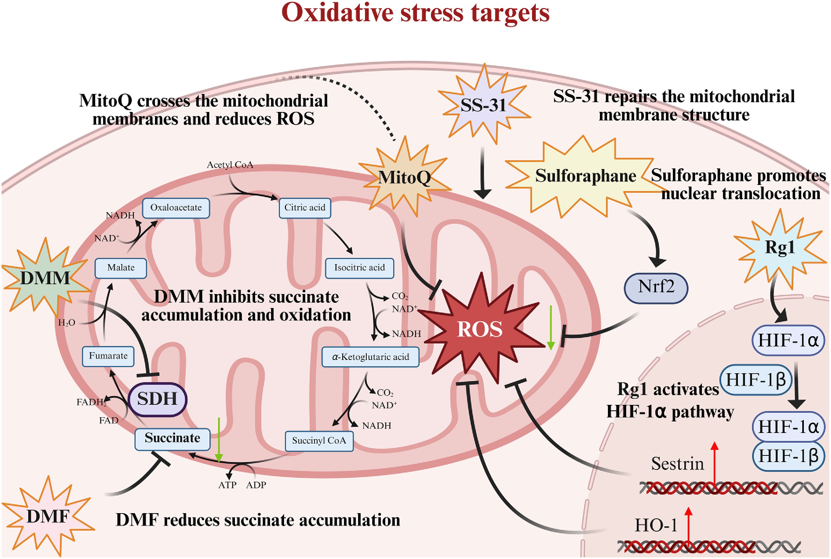
Figure 5.
Several drugs targeting oxidative stress.
Targeting mitochondria-targeted antioxidants
-
Mitochondria are the primary source of ROS and a central target of ischemic injury. Mitochondria-targeted antioxidants can selectively scavenge mitochondrial ROS and thereby preserve mitochondrial integrity and function. As a mitochondria-targeted antioxidant, MitoQ readily crosses mitochondrial membranes, effectively reduces ROS accumulation, and attenuates cardiomyocyte injury[39] (Fig. 5). A study demonstrated that MitoQ pretreatment significantly enhanced the cardioprotective effects of melatonin and mitochondrial transplantation postconditioning. The triple combination of MitoQ, melatonin, and mitochondrial transplantation markedly improved cardiac function, reduced CK-MB levels, and enhanced mitochondrial performance[40]. Despite promising preclinical effects, MitoQ has shown unstable clinical efficacy in oxidative stress-related disorders, including Parkinson's disease, chronic hepatitis C-associated liver injury, and vascular dysfunction[41,42]. In addition, SS-31 peptide, a new mitochondria-targeted antioxidant peptide that mitigates ROS generation by stabilizing and repairing the mitochondrial membrane structure[43,44] (Fig. 5), has advanced to phase II/III clinical trials for mitochondrial disorders, including primary mitochondrial myopathy[45] and heart failure[46]. However, its clinical translation remains limited by peptide-related challenges, such as poor oral bioavailability, short half-life, and the need for parenteral delivery[47].
Targeting endogenous antioxidant defense
-
Endogenous antioxidant defense systems play a pivotal role in enhancing intrinsic myocardial antioxidant capacity upon activation, thereby significantly attenuating ischemic injury[48]. As a potent Nrf2 activator, sulforaphane promotes Nrf2 nuclear translocation and upregulates the expression of downstream antioxidant enzymes, including heme oxygenase-1 (HO-1) and NAD(P)H: quinone oxidoreductase 1 (NQO1). In warm IRI models, Nrf2 activation can effectively inhibit the generation of ROS and prevent cell apoptosis[49] (Fig. 5). The limited chemical stability of sulforaphane (SFN) results in degradation-induced variability in active compound content, which complicates dose control and formulation standardization, thereby limiting its clinical translation[50]. The evidence of ginsenosides further supports this point. Ginsenosides activate the HIF-1α/HO-1[51] and HIF-1α/sestrin pathways[52], which independently and robustly enhance endogenous antioxidant defenses during warm ischemia and subsequent reperfusion[51,52] (Fig. 5). Various ginsenoside monomers have entered different stages of clinical development in China and other Asian countries. Nevertheless, their broader clinical translation continues to be hindered by low bioavailability[53].
Targeting cell death pathways
-
During both cold and warm ischemia, cardiomyocytes undergo multiple forms of regulated cell death, including apoptosis, necrosis, and ferroptosis[54], each contributing to myocardial injury. Modulating these cell death pathways represents a key therapeutic strategy to preserve cardiac graft function. Resveratrol exerts anti-apoptotic effects by upregulating anti-apoptotic B-cell lymphoma-2 (Bcl-2) expression and downregulating pro-apoptotic Bax expression (Fig. 6). In an in vitro rat heart cold ischemia model, resveratrol treatment significantly reduced cardiomyocyte apoptosis and improved graft functional recovery after reperfusion[55]. Although resveratrol has been evaluated in human trials across multiple disease areas, its clinical translation remains constrained by challenges in dose optimization, bioavailability, and reproducibility of therapeutic efficacy[56].
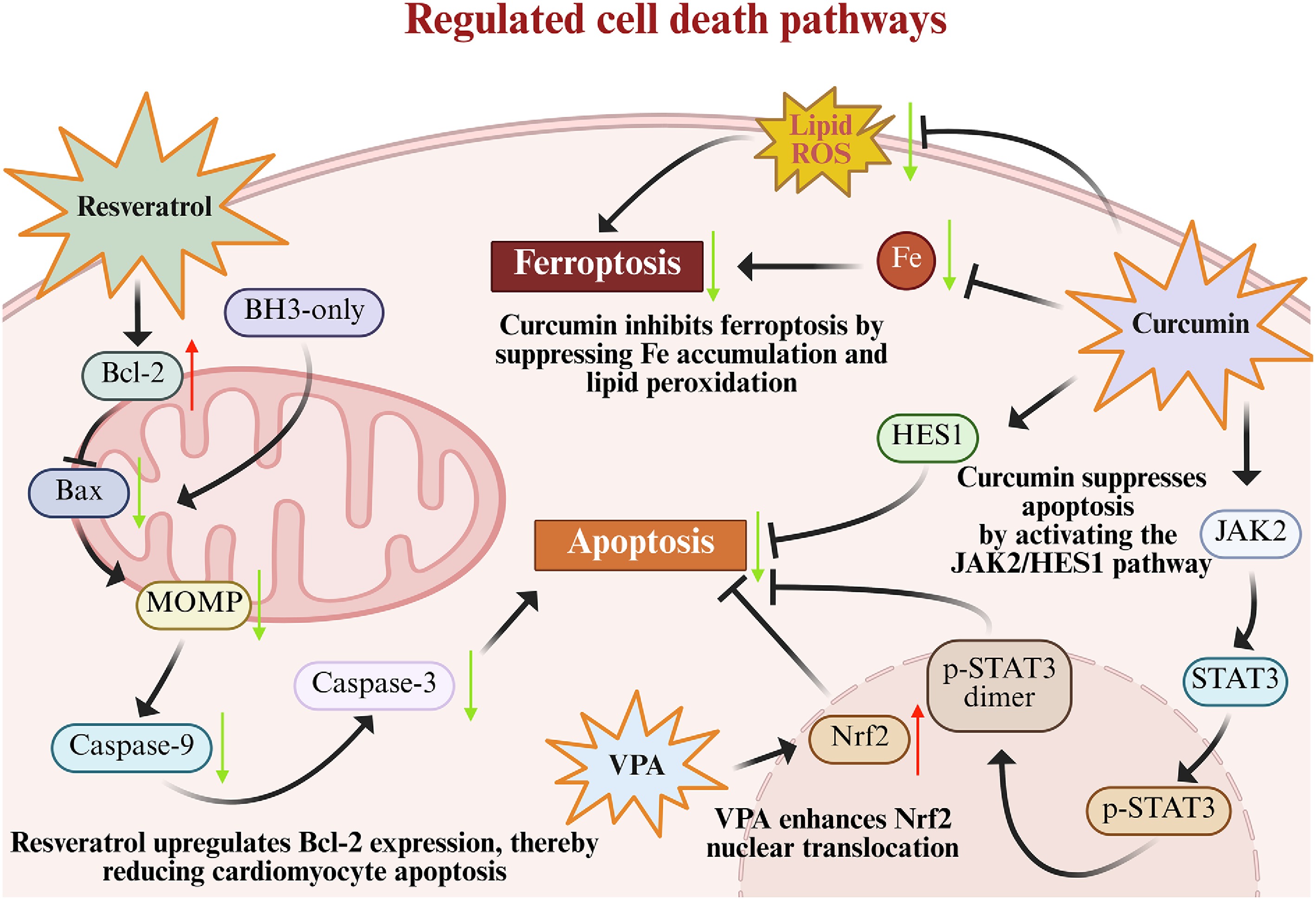
Figure 6.
Several drugs targeting regulated cell death pathways.
Curcumin inhibits ferroptosis, an iron-dependent form of regulated cell death driven by phospholipid peroxidation[57] in cardiomyocytes, by suppressing intracellular iron accumulation and lipid peroxidation, and it suppresses apoptosis by activating the Janus kinase 2/signal transducer and activator of transcription 3 (JAK2/STAT3) pathway (Fig. 6). In warm ischemia-reperfusion models, curcumin markedly reduces infarct size and preserves cardiomyocyte viability[58,59]. Although curcumin is widely recognized as a dietary supplement, its clinical translation is hindered by poor oral bioavailability, inefficient absorption, and intrinsic chemical instability[60]. Furthermore, as a histone deacetylase (HDAC) inhibitor, valproic acid (VPA) inhibits both ferroptosis and apoptosis via enhancing immune-responsive gene 1 (IRG1) upregulation-induced Nrf2 nuclear translocation (Fig. 6). In a porcine heart cold preservation model, VPA extends viable preservation time to 10 h[61]. Although VPA has been widely used as a broad-spectrum antiepileptic agent and mood stabilizer[62], its clinical utility has been constrained by severe adverse effects, including teratogenicity and hepatotoxicity[63,64].
Targeting inflammatory responses
-
During ischemia-reperfusion, activation of the inflammatory signaling further exacerbates myocardial injury. Therefore, inhibiting inflammatory signaling represents an effective strategy to improve cardiac graft preservation outcomes[65]. Luteolin protects cardiomyocytes against cold ischemic injury by suppressing NF-κB-mediated inflammation and restoring calcium homeostasis through modulation of MCU, calmodulin, and Ca2+-Mg2+-ATPase activities[66,67] (Fig. 7). It has been explored in early-phase clinical studies and dietary supplementation trials, including investigations in autism spectrum disorder[68]. Nevertheless, its broader clinical translation is hindered by poor bioavailability.
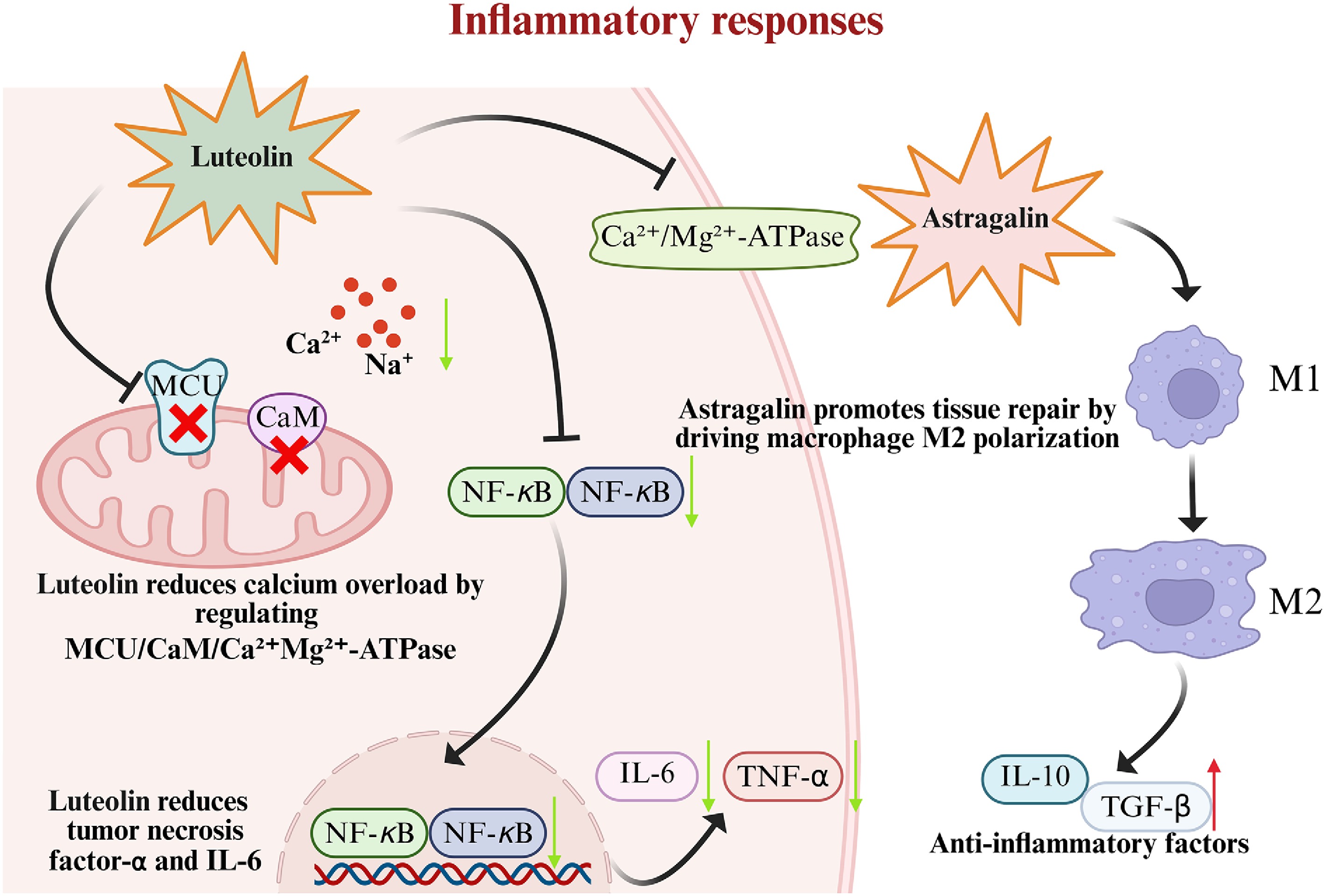
Figure 7.
Several drugs targeting inflammatory responses.
Astragalin has been shown to promote macrophage polarization toward the M2 phenotype, thereby exerting anti-inflammatory, antioxidant, and anti-apoptotic effects. Its protective role has been confirmed in an in vitro rat cardiac cold ischemia model, in which astragalin preserved cardiac graft function after reperfusion[69,70] (Fig. 7). Due to its limited bioavailability, research on astragalin has largely been confined to preclinical animal models.
Targeting cellular organelle function
-
Mitochondria and cell membranes are major targets of ischemic injury, making organelle protection a promising therapeutic strategy for reducing cardiomyocyte damage[71]. Cyclosporin A, a potent mitochondrial permeability transition pore (mPTP) inhibitor, preserves mitochondrial function and attenuates ROS generation and apoptosis[72] (Fig. 8). This agent is a potent immunosuppressive drug with well-established effects on immune regulation. It has been preliminarily applied in clinical heart preservation; however, its broader clinical use remains limited by adverse effects associated with immunosuppression[73].
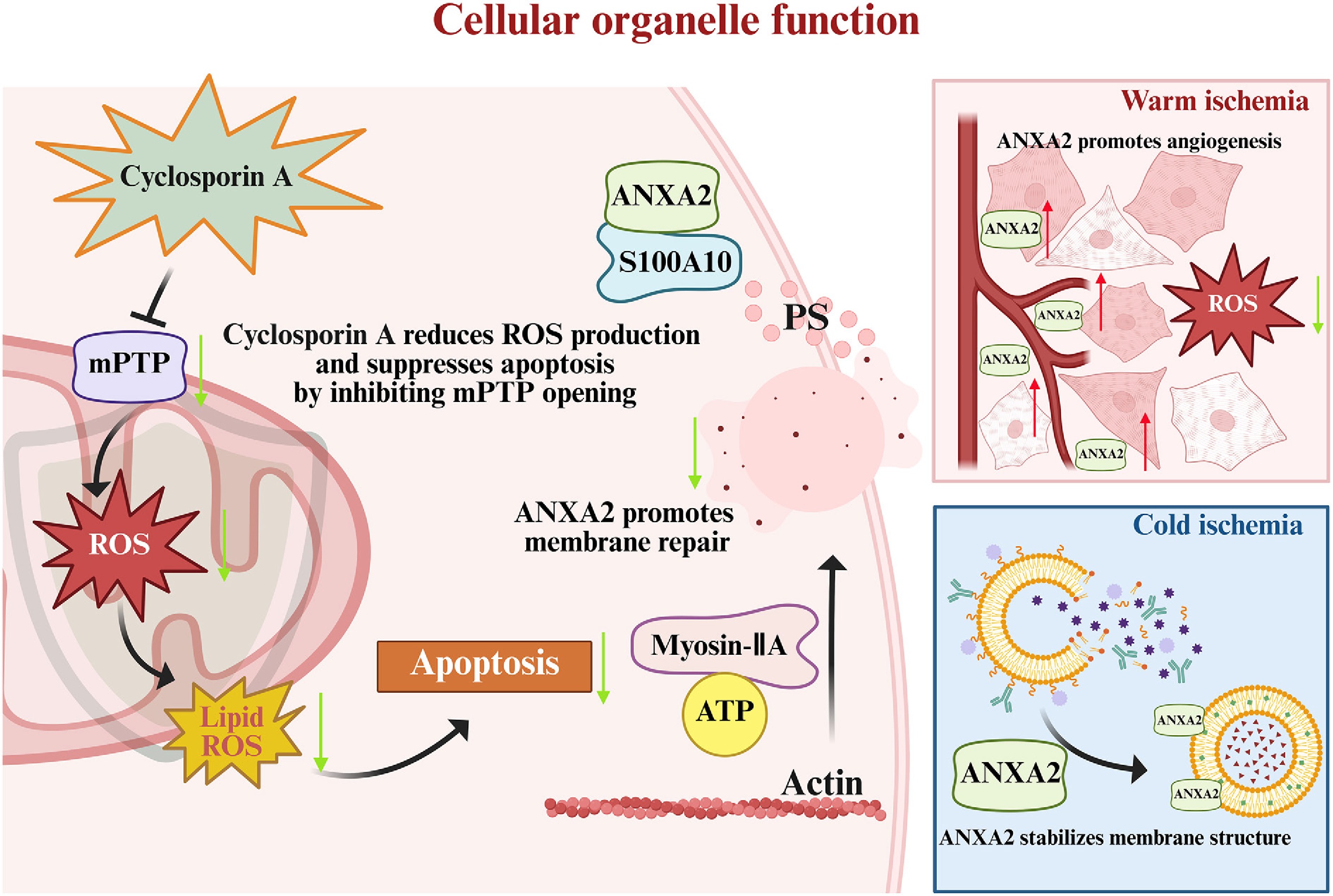
Figure 8.
Several drugs targeting cellular organelle function.
Maintaining cell membrane integrity is critical for cardiomyocyte survival. As a membrane repair-associated protein, Annexin A2 (ANXA2) can specifically bind to phosphatidylserine on damaged cell membranes by forming a heterogeneous tetrameric complex with S100A10. This complex recruits non-muscle myosin IIA and actin, driving actin polymerization and contraction of the injured membrane region. These processes ultimately facilitate membrane resealing[74−76]. ANXA2 exerts its protective effects through distinct mechanisms under cold and warm ischemic conditions. During cold ischemia, low temperatures induce membrane lipid phase separation and reduce membrane fluidity, ultimately leading to membrane rupture. Under these conditions, ANXA2 contributes to membrane stabilization and restoration. During warm ischemia, ANXA2 promotes angiogenesis in the peri-infarct region and attenuates ROS generation and lipid peroxidation[77−79], thereby facilitating membrane repair and cellular recovery (Fig. 8).
Targeting epigenetic regulation
-
Epigenetic regulation (such as histone modification and DNA methylation) plays a crucial role in both cold and warm IRI, and therefore represents a potential target for preservation strategies in heart transplantation[80]. Two histone deacetylase inhibitors, valproic acid (VPA)[61] and trichostatin A (TSA)[81], can affect the expression of genes related to apoptosis and inflammation by regulating the level of histone acetylation, thereby alleviating myocardial IRI[82] (Fig. 9). Additionally, VPA upregulates immune response genes and promotes its production of itaconate, which promotes the nuclear translocation of Nrf2 (Fig. 9).
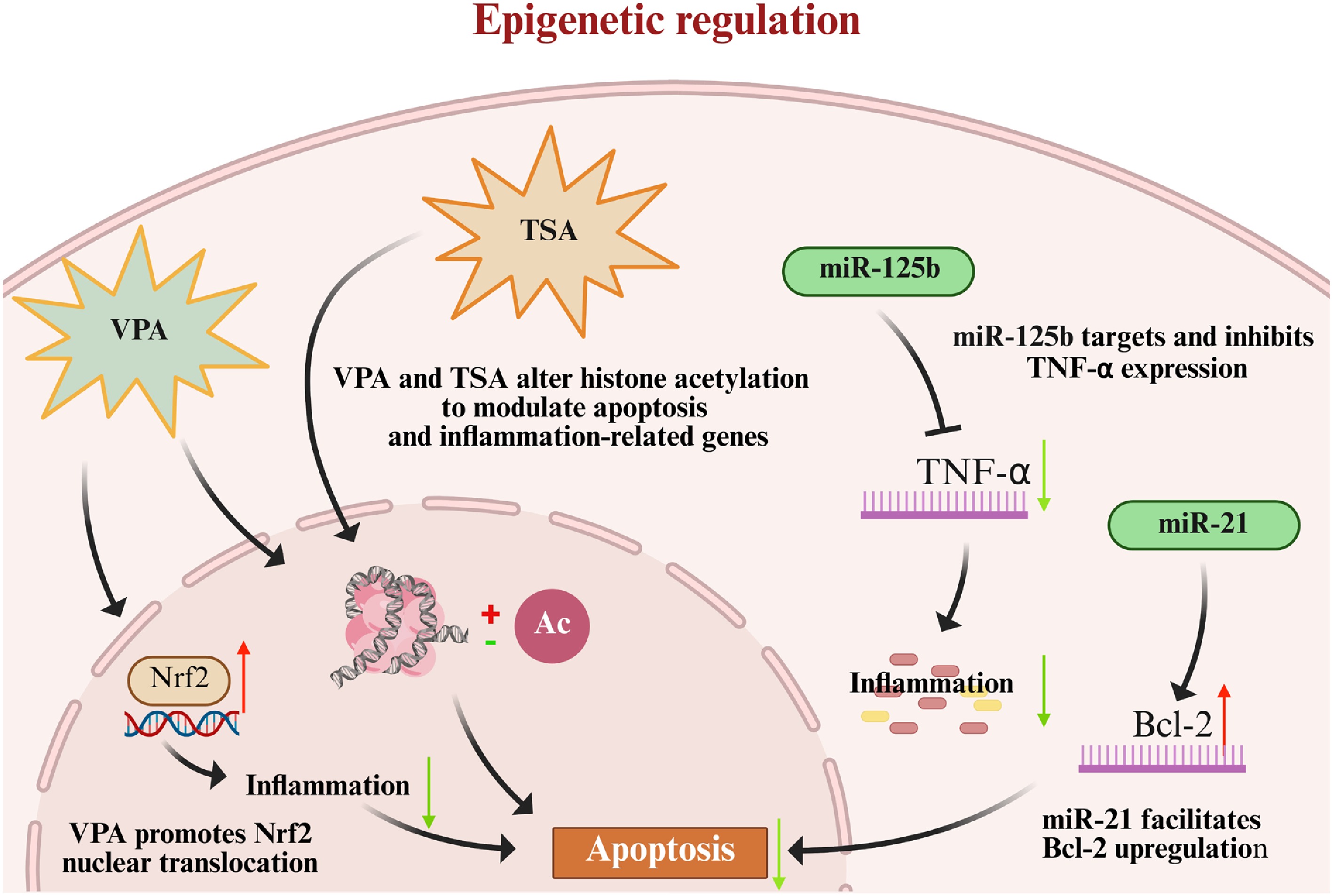
Figure 9.
Several drugs targeting epigenetic regulation.
Moreover, the regulatory effects of microRNAs (miRNAs) have been increasingly recognized. For instance, miR-125b reduces inflammatory responses by inhibiting the expression of TNF-α[83] (Fig. 9); miR-21 inhibits cell apoptosis by upregulating Bcl-2. Experiments have demonstrated that overexpression of miR-21 can significantly alleviate cardiac injury in a rat model of low-temperature cardiac preservation[84] (Fig. 9).
Unfortunately, epigenetic regulators lack organ specificity, limiting their safety in transplantation. Systemic preconditioning may induce donor hypotension and multi-organ toxicity, potentially affecting other transplantable organs[85]. When added to preservation solutions, residual compounds in the isolated heart may be released into the recipient after transplantation and cause off-target effects[86]. miRNAs also exhibit multiple targets and can become dysregulated in complex human physiological networks, raising safety concerns[87]. Therefore, their clinical translation remains constrained by substantial safety challenges.
-
Natural products derived from plants, fungi, and marine animals[88] possess the advantages of diverse skeletal structures[89], low toxicity[90], and multi-target synergistic effects[91]. Therefore, they may be more suitable for injury intervention during heart preservation than single-target synthetic drugs. Single-target synthetic drugs often fail to achieve ideal protection, whereas multi-target synergistic actions of natural products may provide more comprehensive protection. Moreover, some natural products have accumulated sufficient clinical safety data in other disease areas, providing a solid evidence base for their potential application in heart transplantation preservation.
Plant-derived natural products in donor cardiac graft preservation
-
Plant-derived natural products are among the most widely studied types of natural compounds in cardiac graft preservation. Their protective mechanisms mainly focus on anti-oxidation, anti-apoptosis, and anti-inflammation[92]. Astragalin[93] and dihydromyricetin[94] are flavonoid natural products that protect isolated rat hearts from cold ischemic injury through antioxidant, anti-inflammatory, and anti-apoptotic effects. Their protective mechanisms are related to activation of the Nrf2/HO-1 pathway and inhibition of the NF-κB pathway. By inhibiting NF-κB activation, luteolin can regulate the activities of MCU, CaM, and Ca2+-Mg2+-ATPase, reduce calcium overload, and demonstrate a good protective effect in the cardiomyocyte cold preservation model[67].
As a natural product of polyphenols, resveratrol is one of the few natural products capable of protecting against both cold and warm ischemia[95]. It attenuates cardiomyocyte apoptosis and oxidative stress by activating the silent information regulator 1/peroxisome proliferator-activated receptor gamma coactivator 1-alpha (SIRT1/PGC-1α) pathway and upregulating Bcl-2 expression. Curcumin can reduce oxidative stress by activating the Nrf2/HO-1 pathway, and regulate the JAK2/STAT3 and HES1 pathways to inhibit cell apoptosis and ferroptosis, thereby reducing infarct size in the myocardial IRI model[58]; ginsenoside Rg1 and Rb2 have very clear and distinct mechanisms for preventing and treating myocardial injury, where ginsenoside Rg1 enhances endogenous antioxidant ability by activating the HIF-1α/Sestrin2 pathway[52], while ginsenoside Rb2 inhibits inflammatory response and apoptosis by targeting p300 to regulate the level of histone acetylation[96]. Sulforaphane enhances antioxidant capacity and inhibits inflammatory response by activating the Nrf2/HO-1/NQO1 pathway, showing a protective effect in the myocardial warm ischemia model[49].
Fungi-derived natural products in donor cardiac graft preservation
-
Fungi-derived natural products exhibit distinctive chemical scaffolds and pharmacological profiles, and their investigation in cardiac preservation is steadily expanding. Stachatranone F and stachybatranone A are fungal metabolites isolated from Stachybotrys chartarum that exert a cardiomyocyte protective effect by activating the phosphatidylinositol 3-kinase/protein kinase B (PI3K/AKT) signaling pathway and triggering anti-apoptotic cascades, thereby significantly reducing cardiomyocyte apoptosis in the cold ischemia-reperfusion model[97,98]. These promising results highlight their potential as functional additives in cold preservation solutions for enhancing cardiac graft viability and preservation efficacy. Several indoloquinazoline alkaloids named aspergilloids isolated from the fungus Aspergillus clavatonanicus derived from the gut of a centipede showed protective effects in a myocardial cold ischemia model by preventing cold ischemia-induced Ser9 dephosphorylation of GSK3β[99]. diHET is a natural product derived from fungi that exhibits dual biological activities, including promoting regeneration and inhibiting apoptosis. This dual-function activity is relatively uncommon among conventional synthetic drugs[100]. Unfortunately, existing studies are currently only conducted in cellular hypoxia models, and their applicability in the cardiac cold ischemia model remains to be verified. As a natural oxygen carrier extracted from marine invertebrates, M101 showed an exceptional protective effect in static cold ischemia models of steatotic liver disease by reducing biomarkers of reperfusion injury, oxidative stress, and inflammation, thereby improving graft viability and function. Its primary mechanisms involve oxygen delivery capacity and direct anti-inflammatory action. Although M101 is compatible with conventional cold preservation solutions and shows considerable potential for cardiac applications, current evidence remains largely restricted to hepatic models[101]. Therefore, cross-organ validation is warranted to determine its applicability in heart transplantation.
Stage summary of natural products
-
Based on the pathological characteristics of cold ischemic injury, natural products with antioxidant, mitochondrial protective, anti-inflammatory, and cytoprotective properties have been extensively investigated as potential therapeutic candidates for donor heart preservation. Current research on natural products for donor cardiac graft preservation is summarized in Table 1 according to their source, molecular targets, stage of investigation, and applicability to cold and warm ischemia.
Table 1. Research progress on representative natural products for donor cardiac graft preservation.
Source Representation Targets/mechanism Evidence of cold ischemia Evidence of warm ischemia Plants Astragalin**/Dihydromyricetin** Nrf2/HO-1+ and NF-κB− Yes Limited Luteolin* MCU−, CaM−, Ca2+-Mg2+-ATPase−, and NF-κB− Yes Limited Resveratrol** SIRT1/PGC-1α+ and Bcl-2+ Yes Yes Curcumin*** Nrf2/HO-1+, JAK2/STAT3−, and HES1+ Limited Yes Ginsenoside Rg1 and Rb2*** HIF-1α+ and Sestrin+ Limited Yes Sulforaphane*** Nrf2/HO-1/NQO1+ Limited Yes Fungus Stachatranone F* PI3K/AKT+ Yes Limited Stachybatranone A* PI3K/AKT+ Yes Limited Aspergilloids* Ser9/GSK3β− Yes Limited diHET* Apoptosis− Limited Limited Marine animals M101** Oxidative stress− and inflammation− Cross-organ verification Limited Asterisks indicate the experimental stage of the preclinical evaluation: * assessed in cellular models (in vitro); ** evaluated in isolated organ perfusion preservation models (in vitro); *** validated in animal models (in vivo). Plus and minus signs indicate the effects on the pathways or mechanisms: + represents activation; – represents inhibition. As summarized in Table 1, natural products supported by direct experimental evidence in the context of cardiac cold ischemia and preservation are currently limited to astragalin, dihydromyricetin, luteolin, resveratrol, stachatranone F, stachybatranone A, and aspergilloids. Among these compounds, the first four are plant-derived, whereas the latter three are of fungal origin. Although numerous natural products have demonstrated cardioprotective effects in warm ischemia-reperfusion models or in the treatment of cardiovascular diseases, their efficacy and translational potential in the setting of cold ischemia and graft preservation for heart transplantation remain largely unexplored and require further validation.
Bottlenecks of natural products in heart transplantation
-
Although natural products have demonstrated substantial evidence of cardioprotective effects, several fundamental challenges remain in their clinical translation.
Firstly, there is an inherent mismatch between their pharmacokinetic properties and the administration time window. The therapeutic window is only 4–6 h[10], whereas many natural products, such as curcumin, exhibit extremely low oral bioavailability. Although the direct incorporation of curcumin into preservation solutions represents a theoretically feasible approach, its membrane permeability, stability, and biological activity under hypothermic conditions have not been systematically evaluated. Therefore, nanocarrier-based delivery systems, such as lipid encapsulation, may provide a promising strategy to overcome these limitations. For example, encapsulation of curcumin in polyethylene glycol-modified liposomes with particle sizes controlled at 100–200 nm improves solubility and stability, thereby prolonging its therapeutic duration[102,103].
Secondly, natural products exhibit a 'double-edged sword' effect arising from their multi-target characteristics. Unlike traditional single-target drugs, many natural compounds exhibit pleiotropic pharmacological activities through the modulation of multiple molecular pathways[104]. This property may be advantageous for cold ischemia-reperfusion injury, where tissue damage arises from a network of interconnected processes rather than a single pathogenic mechanism. However, the same complexity also presents challenges for target identification and clinical translation. Under the current regulatory framework, clear mechanistic understanding and target characterization are important considerations for drug approval. Consequently, multi-target compounds with poorly understood mechanisms of action face substantial regulatory challenges. Therefore, a more practical and feasible translational strategy is to prioritize the development of natural products with relatively clear targets and well-characterized mechanisms of action. For example, ginsenoside Rb2, a well-recognized p300 inhibitor[96], and stachatranone F, which specifically targets the PI3K/AKT pathway[97], represent natural products with relatively defined molecular mechanisms rather than nonspecific multi-target agents. More importantly, the interaction of stachatranone F with key proteins in the PI3K/AKT pathway has been directly validated through molecular docking and co-immunoprecipitation (Co-IP) analyses, while its mechanism of PI3K activation through the promotion of PI3K phosphorylation has also been elucidated[97]. These findings provide a clearly defined mechanistic basis rather than a merely descriptive association.
Thirdly, pharmacokinetic and toxicological barriers represent major challenges for the clinical translation of natural products. Despite their promising therapeutic potential, the clinical advancement of natural products remains constrained by substantial pharmacokinetic and safety-related challenges. A large proportion of natural compounds suffer from inadequate absorption, limited systemic exposure, rapid biotransformation, and insufficient tissue availability, making it difficult to achieve sustained concentrations required for therapeutic efficacy in vivo[105]. Beyond these pharmacokinetic limitations, uncertainties in dose selection, metabolic stability, off-target activities, and toxicity profiles further hinder their clinical development[104,106]. Moreover, issues such as herb–drug interactions and variability in chemical composition among different preparations complicate quality control and safety assessment, representing additional barriers to their application in transplantation settings where precise and predictable pharmacological effects are essential.
Lastly, the large-scale production of natural products continues to face significant technical bottlenecks. Under extreme environmental conditions, organisms can produce structurally novel and complex secondary metabolites[107]. However, the discovery and development of such compounds are often time-consuming. Many natural products are synthesized through unique biosynthetic enzyme systems, and characterizing these enzymes requires extensive investigation, making large-scale production challenging using conventional chemical synthesis approaches[108]. Therefore, natural products, especially those derived from fungi, remain challenging to produce on a large scale.
-
In vitro mechanical perfusion is not only an organ preservation technique, but also an ideal platform for active therapeutic intervention. It enables the delivery of cytoprotective agents, immunomodulators, and other therapeutic drugs, allowing targeted pre-treatment of grafts before transplantation. In addition, it permits continuous monitoring of graft status during transportation, thereby reducing ischemia-reperfusion injury and optimizing the utilization of marginal donor hearts[109]. However, translating therapeutic agents into this dynamic platform demands strict validation of their solubility and physicochemical stability. Compounds highly effective under static storage may behave unpredictably inside a machine perfusion system where they are exposed to fluid shear stress and continuous oxygenation. For instance, the therapeutic efficacy of curcumin is substantially limited by rapid hydrolytic degradation and susceptibility to oxidative degradation under dynamic agitation[110]. Similarly, some unstable compounds with novel scaffolds may undergo serious chemical cleavage or precipitation risks within perfusates, necessitating advanced formulation strategies to withstand the rigorous biophysical environment of perfusion[111−113]. Studies have successfully delivered adeno-associated virus vectors in the in vitro lung perfusion models to achieve gene therapy, providing a methodological foundation for extending this strategy to heart transplantation[114].
Combined pharmaceutical intervention strategy
-
Adopting a combined pharmacotherapy strategy can achieve synergistic therapeutic effects. For instance, co-administration of drugs targeting succinate metabolism may reduce ROS production at its origin while subsequently suppressing downstream inflammatory responses. Theoretically, this dual-drug approach may be more conducive to prolonging donor heart preservation duration and improving metabolic and functional recovery after ischemic injury. A study using donation after DCD porcine hearts demonstrated that supplementation of the perfusion solution with sevoflurane and remifentanil during in vitro machine perfusion facilitated graft functional recovery and improved post-transplantation survival rates[115]. Based on the pathophysiological mechanisms underlying cold ischemic injury, multi-targeted combination therapy involving the simultaneous modulation of multiple pathways represents a promising strategy for extending the safe preservation window of donor hearts and enhancing the quality of marginal donor organs.
Individual-level analysis and specificity preservation
-
Studies have demonstrated that in donation after DCD hearts, myocardial stress responses and mitochondrial oxidative phosphorylation dysfunction progressively deteriorate with prolonged cold ischemia time[116]. In contrast, brain-dead donor hearts exhibit significantly elevated levels of systemic inflammatory mediators and cardiotoxic substances[117]. Therefore, preservation strategies should be tailored to the characteristics of different donor heart types, allowing the development of targeted intervention approaches. Meanwhile, technologies such as normothermic machine perfusion enable real-time monitoring of injury progression and assessment of donor heart quality by analyzing inflammatory biomarkers in the perfusate. These approaches may ultimately improve the utilization of marginal donor hearts.
-
Our review suggests that cardiac graft preservation represents a critical component of heart transplantation, and mitigating the inevitable ischemic injury is essential for expanding the cardiac donor pool and improving post-transplantation prognosis. Accordingly, it is necessary to clarify the fundamental differences between cold ischemia and warm ischemia injury, and to develop targeted strategies against key molecular pathways involved in cold ischemia-reperfusion injury (CIRI) for advancing cardiac preservation technologies. Unlike warm ischemia, which is primarily characterized by rapid ATP depletion, acute metabolic failure, and immediate cellular injury under normothermic conditions, cold ischemia occurs under hypothermic conditions and induces distinct pathological alterations, including suppressed mitochondrial metabolism, impaired energy production, metabolic imbalance, and enhanced susceptibility to oxidative damage upon reperfusion. Therefore, therapeutic strategies specifically designed for cold ischemic injury should consider these unique biological characteristics rather than simply extrapolating approaches from warm ischemia. Moreover, current research has moved beyond the early paradigm of single antioxidant or anti-inflammatory interventions toward multidimensional regulatory networks involving mitochondrial quality control, programmed cell death, and epigenetic modifications. Unfortunately, pharmacological interventions specifically targeting cold ischemic injury remain scarce, and current approaches largely rely on mechanism-based strategies derived from shared pathological pathways. Developing more targeted therapies that address the unique molecular vulnerabilities of cold ischemia therefore represents an important direction for future research.
Natural products, particularly secondary metabolites derived from fungi and marine animals, represent valuable sources of therapeutic agents owing to their unique structures and diverse biological activities. These compounds can modulate specific pathways and molecular mechanisms that are difficult to achieve with conventional synthetic drugs. Nevertheless, the development of natural product-derived therapeutics continues to face substantial challenges, including chemical complexity, insufficient mechanistic understanding, and difficulties in large-scale production. Future efforts should focus on overcoming these limitations through mechanism-guided activity screening, structural optimization, and synthetic biology approaches, thereby fully exploiting the therapeutic potential of natural products and transforming them into precisely regulated therapeutic tools.
Furthermore, emerging multi-omics technologies are expected to facilitate the development of more accurate preservation strategies, while combined interventions targeting both upstream and downstream pathways may provide synergistic protective effects. In addition, multicenter randomized controlled trials will be indispensable for the clinical translation of molecularly targeted therapeutics. The transition from passive preservation to active protection has opened unprecedented opportunities to improve the utilization of marginal donor hearts. However, significant translational gaps remain. This review also acknowledges several limitations. For example, epigenetic regulatory mechanisms require further systematic evaluation, and translational studies of natural products still need to address interspecies differences during clinical translation. Moreover, the protective effects of many reported compounds have been validated primarily in rodent models, whereas evidence from large-animal studies and human clinical investigations remains limited. Future research should therefore prioritize addressing these challenges and generating the mechanistic and clinical evidence required to advance these therapeutic strategies toward clinical application.
-
Not applicable.
-
The authors confirm their contributions to the work as follows: conceptualization: Sun C, Chi J, Hu Z; figure drawing and discussion: Sun C, Zhao X, Chi J, Hu Z; writing—original draft preparation: Sun C, Chi J, Hu Z; writing—review and editing: Sun C, Zhao X, Chi J, Hu Z, Zhang Y. All authors reviewed the results and approved the final version of the manuscript.
-
Data availability is not applicable to this review as no new data were created or analyzed.
-
The authors declare no conflict of interest.
-
#Authors contributed equally: Chenchen Sun, Xiangwang Zhao
- Copyright: © 2026 by the author(s). Published by Maximum Academic Press on behalf of China Pharmaceutical University. This article is an open access article distributed under Creative Commons Attribution License (CC BY 4.0), visit https://creativecommons.org/licenses/by/4.0/.
-
About this article
Cite this article
Sun C, Zhao X, Chi J, Zhang Y, Hu Z. 2026. Molecular pharmacotargets in donor cardiac graft preservation: mechanisms and translational perspectives. Targetome 2(4): e036 doi: 10.48130/targetome-0026-0039 

Molecular pharmacotargets in donor cardiac graft preservation: mechanisms and translational perspectives
- Received: 16 June 2026
- Revised: 30 July 2026
- Accepted: 04 August 2026
- Published online: 17 August 2026
Abstract: Although heart transplantation remains the definitive therapy for end-stage heart failure, its widespread application is constrained by conventional static cold storage (SCS), where prolonged cold ischemia inflicts irreversible injury. Current preservation solutions lack pharmacologically active components capable of targeting unique hypothermia-induced damage, including lipid phase separation, mitochondrial decay, and restricted succinate clearance, which collectively trigger a devastating oxidative stress burst upon reperfusion. To address this clinical bottleneck, this review systematically deciphers the pathophysiology of cold ischemic injury, framing the discussion around the metabolic distinctions between warm and cold ischemia. We synthesize pivotal molecular interventions targeting oxidative stress cascades, programmed cell death networks (such as apoptosis and ferroptosis), sterile inflammation, organelle impairment, and epigenetic dysregulation, while clarifying their specific applicable scenarios. Crucially, we evaluate the therapeutic superiority of natural products derived from plants, fungi, and marine animals as multi-target synergistic agents, while confronting translational barriers involving their low bioavailability and complex regulatory approval. Ultimately, we outline actionable paradigms to bridge this translational gap, highlighting nanocarrier-based advanced drug delivery and the utilization of in vitro mechanical perfusion as an active therapeutic platform. Integrating these tools with multi-omics profiling allows for personalized, donor-specific preservation strategies. Together, this comprehensive review provides essential theoretical insight and practical guidance to optimize donor graft viability and advance the field of myocardial protection worldwide.