













-
Watermelon (Citrullus lanatus) is valued for its high nutritional content and health benefits, ranking as the third-largest fruit crop worldwide[1,2]. Recent advances in genomic sequencing and functional characterization of watermelon have provided a solid foundation for molecular-assisted breeding, allowing more efficient integration of target traits[3−6]. Owing to strong heterosis, commercial watermelon production relies heavily on F1 hybrids. Male-sterile lines offer an efficient alternative to manual or chemical emasculation, reducing hybrid seed production costs while ensuring seed purity[7], making them a key technology for harnessing heterosis. However, research on the molecular mechanisms underlying male sterility and the regulation of anther development in watermelon remains limited, which hinders the further advancement of the seed production industry. A total of nine male-sterile lines have been reported in watermelon, including the glabrous male-sterile (gms)[8], male sterile 1 (ms-1)[9], male sterile 2 (ms-2)[10], male-sterile dwarf (ms-dw)[11], male sterile 3 (ms-3)[12], DAH3615-MS[13], Se18[14], clapex1[15], and clms1[5]. Among these, only the spontaneous mutant Se18 has been thoroughly studied, in which defective tapetal development during early anther formation leads to male sterility[14].
The tapetum is crucial for pollen development[16]. During early stages, it secretes nutrients for microspores. Later, it undergoes programmed cell death (PCD) to support pollen wall formation. Abnormal tapetum development or PCD can cause pollen abortion[17]. Studies of anther development in Arabidopsis have identified five key transcription factors regulating tapetal development: DYSFUNCTIONAL TAPETUM 1 (AtDYT1), DEFECTIVE IN TAPETAL DEVELOPMENT AND FUNCTION 1 (AtTDF1), ABORTED MICROSPORE (AtAMS), MALE STERILE 188 (AtMS188), and MALE STERILITY1 (AtMS1). These form a conserved AtDYT1-AtTDF1-AtAMS-AtMS188-AtMS1 transcription cascade that precisely regulates tapetum and pollen development[17−20]. Similar genetic pathways exist in rice (OsUDT1-OsTDF1-OsTDR-OsMS188-OsPTC1) and maize (ZmMs32-ZmMs9-ZmbHLH51-ZmMYB84-ZmMs7)[17,21−23].
AtMS188 (MALE STERILE 188, MYB103, MYB80) encodes an R2R3-MYB transcription factor and is expressed at the late stage of tapetum development, with its peak at the tetrad stage[19,24]. Mutations in AtMS188 result in abnormal tapetal PCD, defective callose degradation, and defective pollen exine formation[25−29]. AtMS188 binds to the promoter of AtUNDEAD to regulate tapetal PCD[27]; additionally, AtMS188 regulates AtQRT3 expression for pectin degradation of the tetrad wall[28], and together with AtTEK, co-regulates AtA6 and its homologs (AtA6s) expression for callose degradation of the tetrad wall[29]. Moreover, AtMS188 directly binds to the promoters of genes related to sporopollenin synthesis, such as AtPKSA, AtPKSB, AtMS2, AtCYP703A2, AtCYP704B1, AtACOS5, and AtTKPR1, which promote pollen exine formation[30,31]. Furthermore, AtMS188 directly activates AtMS1 to form a regulatory cascade where AtMS1 controls the synthesis of pollen coat proteins (PCPs)[32].
AtMS1 (MALE STERILITY1) encodes a PHD-finger transcription factor that is highly expressed in the tapetum at stage 9 (haploid microspores are released from tetrads) of anther development[19]. Loss of function of AtMS1 results in abnormal tapetal PCD and defective pollen exine and pollen coat formation[25,32−34]. In Arabidopsis, AtMS1 regulates the expression of both pollen coat protein genes (AtGRP14, AtGRP17, AtGRP18, AtGRP19, AtEXL4, and AtEXL6) and key genes for pollen coat lipid synthesis (AtKCS7, AtKCS15, and AtKCS21)[32,35].
Unlike predominantly self-pollinated model plants (e.g., Arabidopsis, rice) or wind-pollinated crops (e.g., maize), watermelon is a typical insect-pollinated crop that relies heavily on animal vectors for reproduction[36]. This strategy requires the formation of a specialized, adhesive pollen coat containing abundant lipids, phenols, and proteins to facilitate adherence to insect bodies. While the core DYT1-TDF1-AMS-MS188-MS1 pathway is genetically conserved, it remains largely unknown how this regulatory cascade has evolutionarily adapted to coordinate the specific metabolic fluxes required for insect-mediated pollination in Cucurbitaceae. Previous research on the male-sterile line Se18 revealed that a conserved ClDYT1-ClTDF1-ClAMS-ClMS188-ClMS1L1 genetic pathway may be present in watermelon anther development[14,37].
In this study, we employed CRISPR/Cas9 technology to generate ClMS188 and ClMS1L1 knockout lines, both of which exhibited complete male sterility. Using qRT-PCR and RNA in situ hybridization, we demonstrated that ClMS188 and ClMS1L1 are specifically expressed in anther tapetal cells, with their expression peaking at developmental stages 8b and 9, respectively. Sectioning results further revealed that the clms188 and clms1l1 mutants exhibit abnormal tapetal and microspore development, with abnormalities initiated at stages 8b and 9, respectively. Further in vitro and in vivo experiments demonstrated that ClMS188 directly activates ClMS1L1 expression. Transcriptome data revealed altered expression of multiple metabolic pathway genes and anther development regulatory genes in clms188 and clms1l1 mutants. This study investigates the ClMS188–ClMS1L1 regulatory network for watermelon anther and pollen development, advancing knowledge of anther development mechanisms and providing gene resources for hybrid seed production.
-
The WT watermelon inbred line YL was used for experimentation and genetic transformation in this study. Seeds were incubated at 28 °C in darkness for 3 d to induce germination, and the germinated seedlings were then transplanted into soil and grown until the third true leaves were fully expanded (16 h light, 28 °C/8 h dark, 20 °C, 50% relative humidity). After that, the plants were transferred to the Yangling Experimental Base of Northwest A&F University.
Phylogenetic analysis
-
Evolutionary analysis was conducted using the neighbor-joining method in MEGA 7.0, with 1,000 bootstrap replicates implemented[38]. Multiple sequence alignments of ClMS188, ClMS1L1, and other homologous proteins were constructed using GENEDOC software. Sequence accession numbers are listed in Supplementary Table S1.
Construction of gene editing vector and watermelon transformation
-
To generate the gene-edited mutants of ClMS188 and ClMS1L1, we designed two sgRNAs targeting exons of each gene using CRISPR-P v2.0 (
http://crispr.hzau.edu.cn/CRISPR2 ). We amplified the fragment containing both sgRNA sequences from the pCBC vector by PCR, and then inserted it into the CRISPR/Cas9 vector pBSE402 through homologous recombination[39]. The recombinant vector was transformed into Agrobacterium tumefaciens strain EHA105. The watermelon cotyledon explants were infected with the Agrobacterium strain, then subjected to co-cultivation, recovery culture, and selection on antibiotic-containing medium to obtain transgenic plants. After this, GFP fluorescence-positive plants were identified using a handheld fluorescence lamp (LUYOR-3415RG, USA). High-throughput tracking of mutations (Hi-TOM) and next-generation sequencing were employed to validate gene-edited plants[40]. Independently derived homozygous T3 gene-edited mutant lines were used for subsequent studies. All the primers used in this experiment are listed in Supplementary Table S2.RNA in situ hybridization of male anther
-
Sense and antisense RNA probes for ClMS188 and ClMS1L1 were designed from their cDNA sequences and labeled with the DIG RNA Labeling Kit (Roche, Switzerland) following the manufacturer's instructions. WT male floral buds of varying diameters were collected and fixed in 4% paraformaldehyde at 4 °C for 24 h. After dehydration, the samples were infiltrated with paraffin using xylene at 42 °C, allowed to cool naturally, and then embedded and sectioned. The samples were pre-hybridized in pre-hybridization buffer at 42 °C for 3 h, followed by overnight hybridization at 42 °C with the addition of DIG-labeled RNA probes. Finally, the color was developed by incubation with BCIP/NBT solution, and the signals were observed[40]. All the primers used in this experiment are listed in Supplementary Table S2.
Semi-thin resin sectioning
-
WT and mutant male floral buds of varying diameters were collected and fixed in FAA solution at 4 °C for 24 h. Samples were then pre-infiltrated in a 1:1 mixture of 100% ethanol and 7,100 base liquid for 2–3 d. Subsequently, 1 g of hardener I diluted in 100 mL of 7,100 base liquid was added to the samples, followed by incubation at 4°C for 2–3 d. For polymerization, 1 mL of hardener II was diluted in 15 mL of osmotic solution, and the resulting polymerization solution was added to each sample. After an additional polymerization period of 3 d, samples were longitudinally sectioned to 5 μm thickness using a semi-thin microtome (RM2265; Leica). Sections were stained with 1% TBO for 1 min, rinsed with running water, and heated on a hot plate for 2 h. Finally, sections were mounted in neutral resin and imaged under bright-field microscopy[40].
Alexander staining of pollen
-
Male flowers of WT, clms188, and clms1l1 mutants were collected on the same day. Pollen was gently tapped onto glass slides, stained with 100 μL Alexander staining solution, and then covered with coverslips. After staining for 5 min, pollen viability was observed under a microscope (Axio Imager M2; Zeiss) and documented photographically.
Dual-luciferase assay
-
The ClMS1L1 promoter (−1,837 to −1 bp) was cloned into the pGreenII 0800-Luc vector. The ClMS188 CDS was cloned into the pGreenII 62-SK vector[41]. The recombinant vectors were separately transformed into Agrobacterium strain GV3101 (pSoup). Agrobacterium cells containing the constructed effector and reporter (effector : reporter = 9:1) were co-infiltrated into N. benthamiana leaves. The activities of LUC and REN were measured using a Dual-Luciferase reporter kit (Yeasen Biotech, China) with five replicates. All the primers used in this experiment are listed in Supplementary Table S2.
Yeast one-hybrid assay
-
The ClMS188 CDS was cloned into the pGADT7 vector. The ClMS1L1 promoter segment was divided into ClMS1L1-P1 (−1,837 to −1,092 bp) and ClMS1L1-P2 (−1,092 to −1 bp) fragments, which were cloned into the pAbAi vector. Transformed yeast strains were cultured on SD/-Leu medium with or without 80 ng/mL aureobasidin A (AbA) at 30 °C for 3 d, followed by observation and photography. All the primers used in this experiment are listed in Supplementary Table S2.
Electrophoretic mobility shift assay
-
The ClMS188 CDS was fused to an MBP-tag and cloned into the pMAL-C2X vector. This construct was introduced into Escherichia coli Rosetta, and expression was induced with 0.5 mM IPTG. The ClMS188-MBP fusion protein was purified using the LightShift™ Chemiluminescent EMSA Kit (Beyotime, China). Putative target gene promoter fragments were synthesized as biotin-labelled oligonucleotides. All the primers used in this experiment are listed in Supplementary Table S2.
Subcellular localization
-
The ClMS188 CDS and ClMS1L1 CDS were fused to an eGFP tag and cloned into the pGreen vector to construct a p35S::ClMS188-GFP fusion expression vector. NLS-RFP was marked with AtH2B-mCherry. The recombinant vectors were separately transformed into Agrobacterium strain GV3101 (pSoup). Agrobacterium cells containing the constructed vectors were co-infiltrated into N. benthamiana leaves. After 72 h, GFP fluorescence in the injected leaves was observed under an Olympus BX63 microscope (Tokyo, Japan). All the primers used in this experiment are listed in Supplementary Table S2.
Quantitative real-time PCR (qRT-PCR) assays
-
Around 10:00 AM, different tissues and florets of different diameters were collected from WT, clms188, and clms1l1 plants and flash-frozen in liquid nitrogen. Total RNA was extracted with the RNA Simple Total RNA Kit (Tiangen, China), and cDNA was synthesized using the Fast King RT Kit (Tiangen, China). SYBR Green PCR Master Mix (Takara) was used for qRT-PCR. Relative expression levels were normalized against the watermelon β-ACTIN (Gene ID: Cla97C02G026960)[42]. All primers used in this experiment are listed in Supplementary Table S2.
Statistical analysis of RNA-seq data
-
The florets of WT, clms188, and clms1l1 plants from stage 6 to stage 10 (bud diameter 2.0–4.5 mm; Supplementary Tables S3) were collected and flash-frozen in liquid nitrogen[14]. For each sample, three biological replicates were collected from specified tissues from independent WT and mutant plants. RNA extraction, mRNA enrichment, RNA-seq library construction, and sequencing were carried out by Biomarker (Beijing, China). The bioinformatics analysis of RNA-seq data was performed using BMKCloud (
www.biocloud.net ). DEGs were screened according to the screening criteria of fold-change ≥ 2 and FDR < 0.01. The enriched GO classification and KEGG pathways among the overlapping DEGs were identified using BMKCloud. General information regarding the RNA-seq data is listed in Supplementary Tables S4–S10. All primers used in this experiment are listed in Supplementary Table S2. -
According to watermelon genomic annotation, the genomic sequence of ClMS188 contains three exons and two introns; its 966-bp open reading frame (ORF) encodes a 321-amino-acid protein (≈35.5 kDa) (Supplementary Fig. S1a, S1b). Sequence alignment confirms ClMS188 contains a typical R2R3-MYB domain, classifying it as an R2R3-MYB transcription factor (Supplementary Fig. S1c). Phylogenetic analysis shows ClMS188 groups with orthologs from other plants, while its paralogs from Arabidopsis and watermelon form two separate clades (Supplementary Fig. S1d). ClMS188-GFP co-localized with a nuclear marker, confirming its nuclear localization as an R2R3-MYB transcription factor (Fig. 1a).
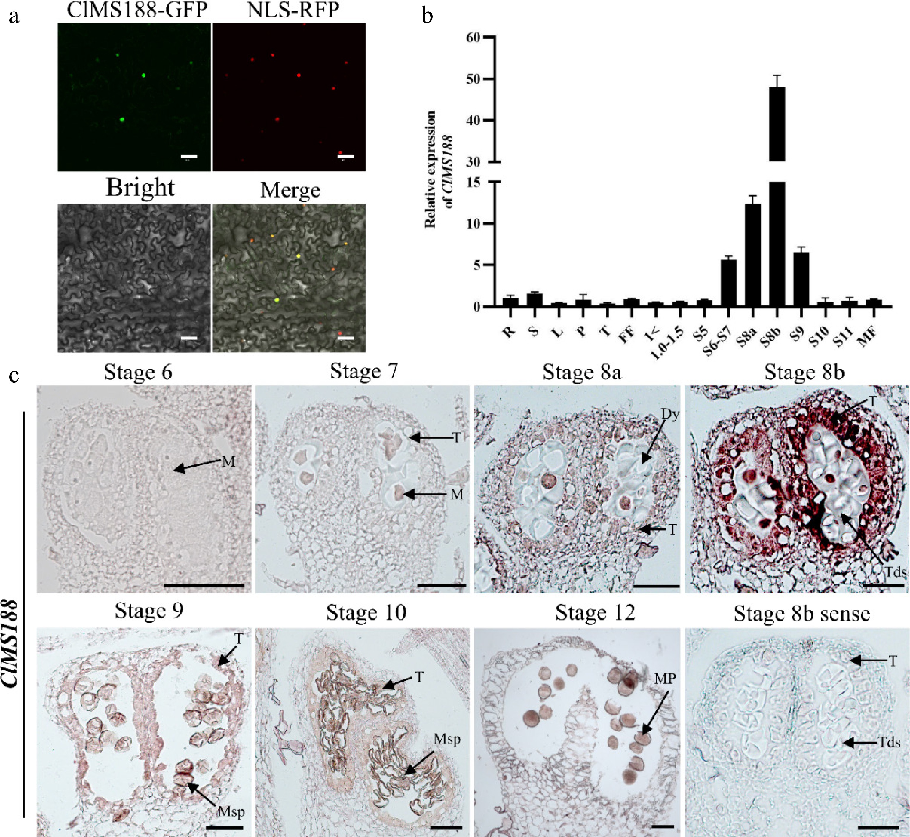
Figure 1.
Subcellular localization and expression analysis of the ClMS188 gene. (a) Subcellular localization of ClMS188. The constructs p35S::ClMS188-GFP and p35S::NLS-RFP were transformed into N. benthamiana leaves and then observed by confocal microscopy. Scale bars, 50 μm. (b) Transcript levels of ClMS188 in the roots (R), stems (S), leaves (L), petioles (P), tendrils (T), female flowers (FF), male floral buds of different diameters, and open male flowers (MF) of WT plants. '> 1' indicates male floral buds with a diameter > 1 mm; 'S5' indicates male floral buds at stage 5 of anther development. Relative expression was normalized to the expression level in WT roots. Data are means ± SDs (n = 3). (c) RNA in situ hybridization of ClMS188 transcript in WT anthers during stages 6–12 is shown. Stage 8b anthers hybridized with the sense probe served as the negative control. Dy, dyad cell; M, microsporocyte; Mp, mature pollen; Msp, microspore; T, tapetum; Tds, tetrads. Scale bars, 20 μm.
Based on the developmental staging criteria for watermelon anther development established in previous research[14] (Supplementary Table S3), we analyzed the expression level of ClMS188 in various tissues of the wild-type (WT) plant by quantitative real-time PCR (qRT-PCR) and RNA in situ hybridization. The qRT-PCR results showed that ClMS188 was generally weakly expressed in vegetative organs (roots, stems, leaves, petioles, tendrils) and female flowers; however, its expression was significantly upregulated during male flower development, especially during stages 6–9, with a peak at stage 8b (Fig. 1b). RNA in situ hybridization showed that ClMS188 transcript levels were low during stages 6–7 (Fig. 1c), followed by a steady increase in the tapetal cells and microsporocytes at stage 8a. At stage 8b (the tetrad stage), ClMS188 expression reached its highest level specifically in the tapetum. During microgametogenesis, ClMS188 transcript abundance decreased sharply in both the tapetum and microspores. These results suggest that ClMS188 is specifically expressed during anther development, with the highest expression level at stage 8b, indicating that the transcription factor ClMS188 may play an important role in this process.
Knockout of ClMS188 results in complete male sterility
-
To further study the function of ClMS188, a dual-target CRISPR/Cas9 vector was constructed using sgRNAs designed to target the first and second exons (Fig. 2a, b). Three ClMS188 mutant lines were obtained: clms188-1 (5-bp deletion) and clms188-2 (1-bp deletion), both of which resulted in frameshifts and premature termination codons, whereas clms188-3 had a 131-bp deletion, leading to a partial loss of the R2R3-MYB protein domain (Fig. 2b; Supplementary Fig. S2). Compared with WT, all clms188 mutants exhibited complete male sterility. For detailed phenotypic analysis, we selected clms188-1 as a representative mutant (Fig. 2c−j). In WT plants, female flowers and male flowers at one-to-two-node lower positions opened synchronously. However, in the clms188 mutants, male flowers at comparable nodes typically remained unopened, although female flowers at adjacent positions flowered normally (Fig. 2c, d). Notably, under low-temperature and low-light conditions, the clms188 mutant occasionally had a single male flower with open petals; however, these flowers displayed significantly shorter petals, reduced anther diameter, and a complete absence of pollen grains (Fig. 2e, f; Supplementary Table S11). Stereomicroscopy showed that WT anthers dehisced normally, releasing abundant, adherent yellow pollen. In contrast, clms188 anthers were indehiscent and lacked visible pollen (Fig. 2g, h; Supplementary Table S11). Alexander staining confirmed abundant viable pollen in WT, whereas no pollen was detected in clms188 (Fig. 2i, j; Supplementary Table S11). To investigate whether the clms188 mutation affects the growth and development of female flowers, fruits, and seeds, we crossed the clms188 mutant (as the female parent) with WT plants (as the male parent). The results showed that the clms188 mutant exhibited no significant differences from WT in female flower opening, number of female flower nodes, fruit size, fruit weight, and seed number (Supplementary Fig. S3; Supplementary Table S11). In summary, our findings indicate that ClMS188 is indispensable for male fertility, as its knockout results in complete male sterility without affecting other traits.
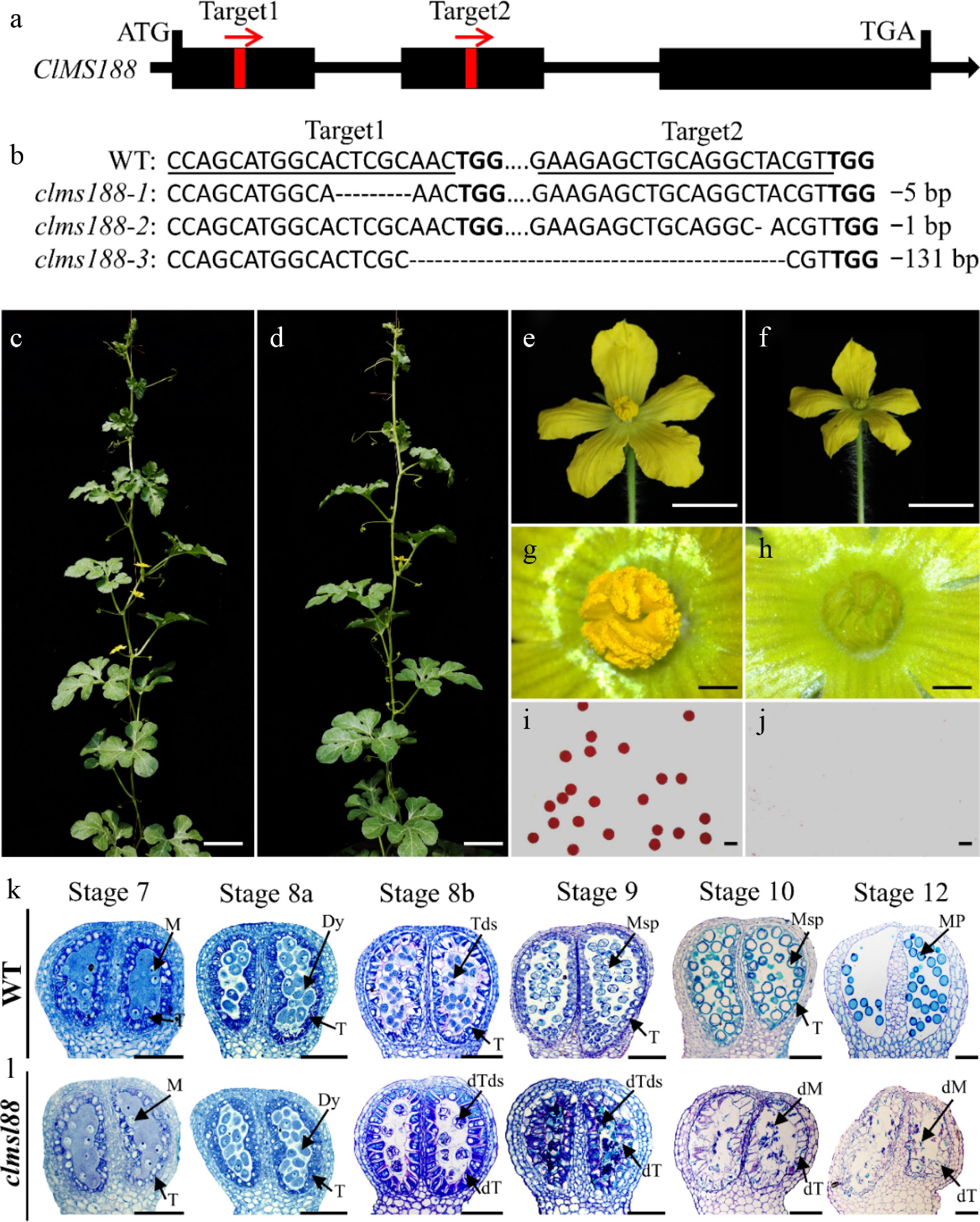
Figure 2.
Knockout of ClMS188 to obtain completely male-sterile mutants. (a) Gene structure and target site positions of ClMS188. Black boxes represent exons; red boxes represent target site locations; red arrows represent target site orientation. (b) Mutation profiles of ClMS188 in three mutant lines identified by sequencing. PAM motifs are in bold; target sites are underlined; '-' indicates deletions. Phenotypes of the (c) WT and (d) clms188. Scale bars, 10 cm. Male flower morphology of (e) WT and (f) clms188. Scale bars, 1 cm. Anther morphology of (g) WT and (h) clms188. Scale bars, 200 μm. Alexander staining of pollen from (i) WT and (j) clms188. Scale bars, 60 μm. Semi-thin sections of (k) WT and (l) clms188 mutant anthers during stages 6–14 are shown. dM, defective microsporocyte; dT, defective tapetum; dTds, defective tetrads. Dy, dyad cell; M, microsporocyte; Mp, mature pollen; Msp, microspore; T, tapetum; Tds, tetrads. Scale bars, 20 μm.
To further characterize the defective clms188 mutant, semi-thin sections of anthers from WT and mutant plants were examined across developmental stages (Fig. 2k, l). No morphological differences were observed from microsporocyte formation (stage 6) through the completion of meiosis I and dyad formation (stage 8a). Starting from stage 8b, anther development in clms188 began to show abnormalities. Specifically, at stage 8b, dyads in WT completed meiosis II and formed tetrads of four haploid microspores, and tapetal cells began to degrade. In contrast, clms188 tetrads were abnormally shaped, with the microspores shrinking and showing irregular morphology, and tapetal cells were enlarged and deeply stained. At stage 9, WT microspores were released into the anther locules, the tapetum degenerated, and the callose was degraded; in clms188, microspores further shrank and became deformed, while the tapetal cells expanded and underwent vacuolation. At stage 10, large vacuoles formed in WT microspores, squeezing the nucleus to the periphery; the microspore wall formed, and the tapetum further degraded; in clms188, microspores continued to shrink, and the tapetum became faintly stained and further enlarged. From stage 11 to stage 12, WT microspores underwent two mitotic divisions to form mature pollen filled with starch and lipids, and the tapetum completely degraded; in clms188, microspores were largely degraded, and the tapetum completely degenerated. Sectioning results showed that the clms188 phenotype is caused by abnormal development of both the tapetum and microspores starting from stage 8b.
ClMS188 directly activates ClMS1L1 expression
-
Studies in Arabidopsis have demonstrated that AtMS188 directly regulates the expression of AtMS1 by binding to its promoter[27,32]. To determine whether ClMS188 directly regulates ClMS1L1 expression during watermelon male flower development, a dual-luciferase (LUC) assay was conducted in N. benthamiana leaves with ClMS188 as the effector and a ClMS1L1 promoter-driven construct as the reporter (Fig. 3a). Compared to the empty vector control, the LUC signals of pClMS1L1::LUC were significantly upregulated by p35S::ClMS188 (Fig. 3b). These data indicate that ClMS188 directly activates ClMS1L1 expression. Previous studies demonstrate that AtMS188 binds to cis-elements sharing a conserved AACC core sequence[27,30−32]. Analysis of the ClMS1L1 promoter (−1,837 bp to −1 bp) revealed eight AACC elements (Fig. 3c). To map the ClMS188 binding region, we divided this promoter segment into two fragments (ClMS1L1-P1 and ClMS1L1-P2) and performed yeast one-hybrid (Y1H) assays (Fig. 3c, d). The Y1H assays preliminarily demonstrated that ClMS188 specifically binds to the ClMS1L1-P1 region, which contains four AACC core sequences (Fig. 3d). To further identify specific binding sites within ClMS1L1-P1, an electrophoretic mobility shift assay (EMSA) was utilized. Four sets of probes containing AACC elements within ClMS1L1-P1 were synthesized, named Probe-1ClMS1L1(AAACCC), Probe-2ClMS1L1(TAAACC), Probe-3ClMS1L1(TAACCA), and Probe-4ClMS1L1(AAACCT) (Fig. 3e). EMSA results showed that ClMS188 bound specifically to Probe-3ClMS1L1, but not to the other probes (Fig. 3e, f). These results demonstrate that ClMS188 directly binds to the TAACCA motif in the ClMS1L1 promoter and activates ClMS1L1 transcription.
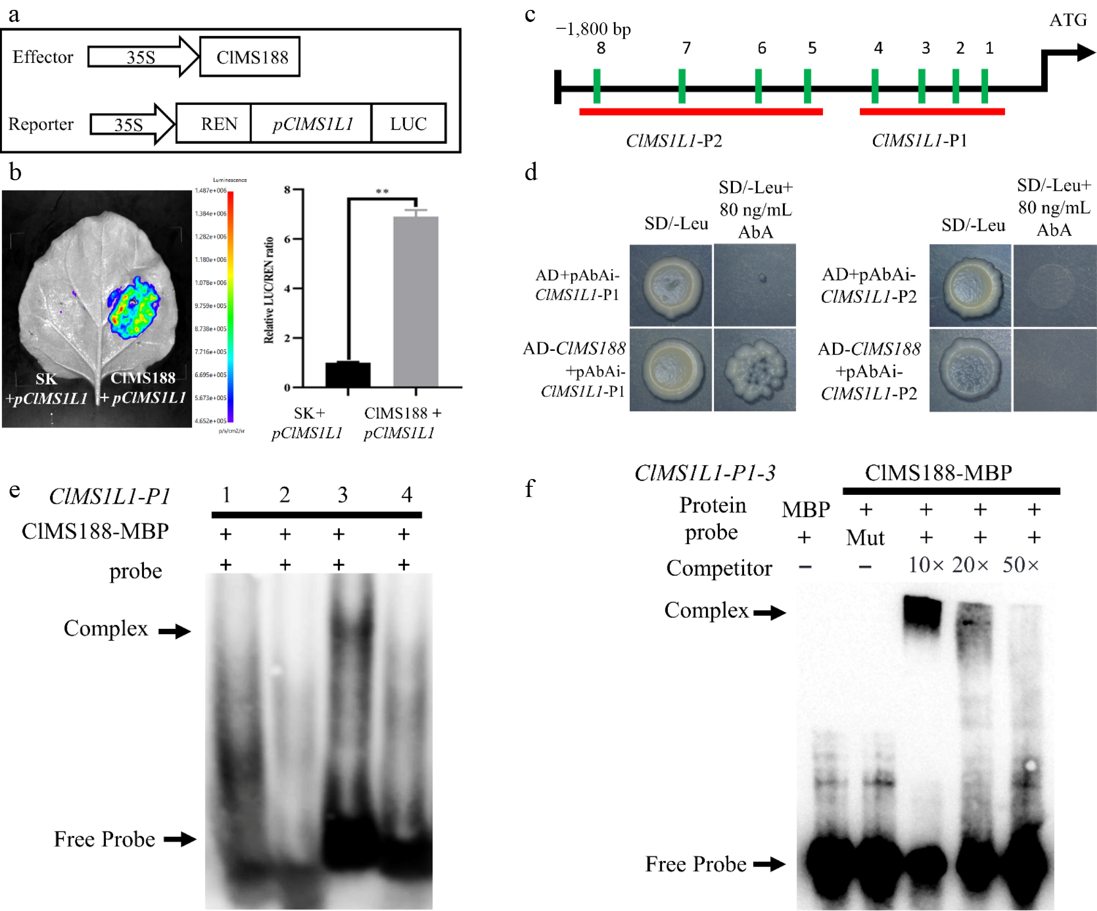
Figure 3.
ClMS188 binds to the promoter of ClMS1L1 and activates its expression. (a) Schematic diagram of effector and reporter constructs for dual-luciferase assay. (b) Dual-luciferase assay to evaluate ClMS188-mediated transactivation of ClMS1L1. The data represent the mean ± SD of five independent experiments. ** Indicates significant differences at p < 0.01. (c) AACC elements in the upstream promoter region of ClMS1L1. (d) Yeast one-hybrid assay of ClMS188 binding to the ClMS1L1 promoter. Yeast cotransformed with the empty vector pGADT7 (AD) and the ClMS1L1 promoter was used as a negative control. Yeast cotransformed with pAbAi-ClMS1L1-P1 and pAbAi-ClMS1L1-P2, along with AD-ClMS188 transformants, grew well on selective medium (SD/-Leu) supplemented with 80 ng/mL AbA. (e), (f) Identification of ClMS188 binding specificity to sites on the ClMS1L1 promoter by electrophoretic mobility shift assays. (e) Binding verification of ClMS188 to four sets of probes, Probe-1ClMS1L1(AAACCC), Probe-2ClMS1L1(TAAACC), Probe-3ClMS1L1(TAACCA), and Probe-4ClMS1L1(AAACCT) on ClMS1L1-P1. (f) ClMS188 binding specificity to sites on the ClMS1L1-P1 promoter of Probe-3ClMS1L1(TAACCA). '−' indicates absence; '+' indicates presence. The arrow indicates the position of the protein-DNA complex after incubation of a biotin-labelled DNA probe with MBP-MS188.
Loss of function of ClMS1L1 leads to complete male sterility
-
To further elucidate the function of ClMS1L1, comprehensive bioinformatics characterization and expression profiling were conducted. The results show that the ClMS1L1 gene consists of four exons and three introns, encoding a 649-amino-acid PHD-finger transcription factor (≈73.7 kDa) containing an N-terminal nuclear localization signal and a C-terminal PHD domain (Supplementary Fig. S4a−S4c). Phylogenetically, ClMS1L1 belongs to the dicot clade of the MS1 family (Supplementary Fig. S4d). ClMS1L1 localizes to the nucleus, as confirmed by fluorescence co-localization with a nuclear marker (Supplementary Fig. S4e). Both qRT-PCR and RNA in situ hybridization showed that ClMS1L1 was weakly expressed at early stages of anther development, increased gradually in the tapetum and microsporocytes, peaked at stage 9, and then declined sharply, suggesting that ClMS1L1 plays a crucial role during these stages (Fig. 4a, b). Furthermore, we examined the expression level of ClMS1L1 in the clms188 mutant background. In comparison with the WT, ClMS1L1 expression was significantly lower in the clms188 mutant at multiple anther developmental stages, further indicating that ClMS1L1 acts downstream of ClMS188 (Supplementary Fig. S4f). Next, we generated three independent ClMS1L1 knockout lines using a dual-target CRISPR/Cas9 strategy targeting its third exon (Fig. 4c). The lines carried distinct mutant alleles: clms1l1-1 (218-bp deletion) and clms1l1-3 (4-bp deletion) result in frameshift and premature truncation, while clms1l1-2 (219-bp deletion) had an in-frame deletion affecting part of the activation domain (Fig. 4d; Supplementary Fig. S5). All mutants exhibited male sterility. We selected clms1l1-1 as the representative mutant for phenotypic analysis (Fig. 4e). In the clms1l1 mutant, male flowers (at nodes comparable to the wild type) remained closed (Fig. 4e). Occasionally, the clms1l1 mutant produced a single male flower with shorter petals, smaller anthers, and no pollen (Fig. 4f; Supplementary Table S11). Stereomicroscopy and Alexander staining confirmed indehiscent anthers with no visible pollen (Fig. 4g, h). Genetic crosses also indicated that clms1l1 mutants are male-sterile without pleiotropic effects on other traits (Supplementary Fig. S6; Supplementary Table S11).
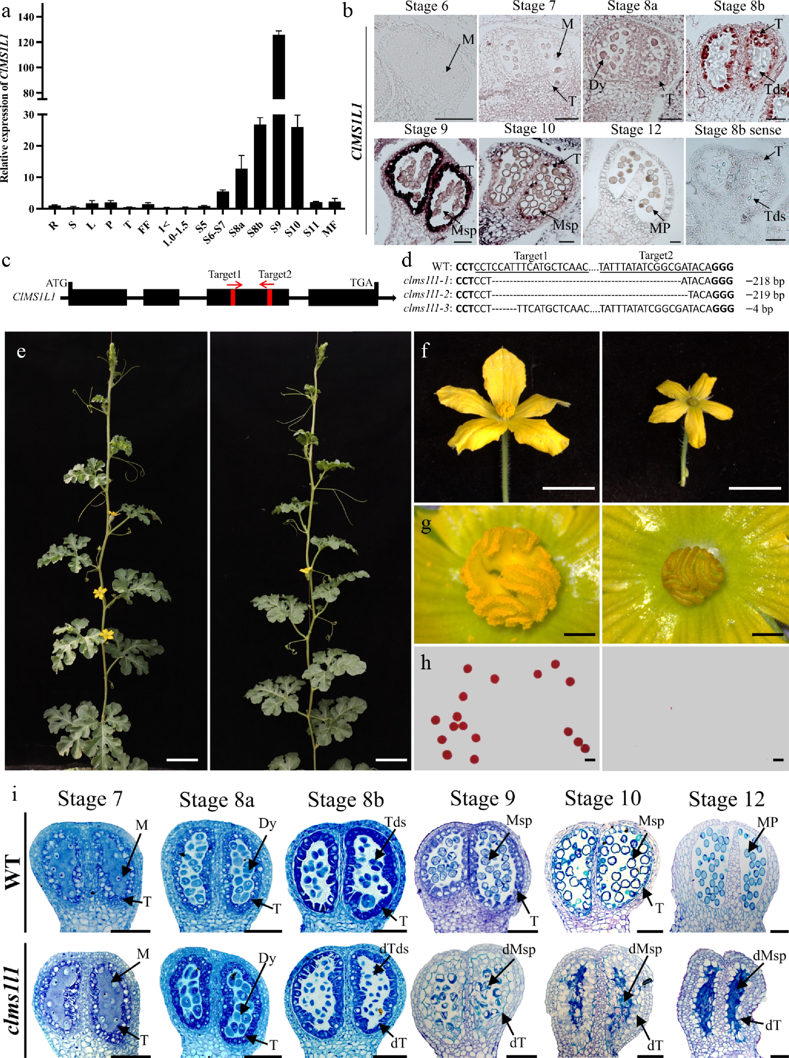
Figure 4.
Expression and function of the ClMS1L1. (a) Transcript levels of ClMS1L1 in the roots (R), stems (S), leaves (L), petioles (P), tendrils (T), female flowers (FF), male floral buds of different diameters, and open male flowers (MF) of WT plants. (b) RNA in situ hybridization of ClMS1L1 transcript in WT anthers during stages 6–12. (c) Gene structure and target site positions of ClMS1L1. (d) Mutation profiles of ClMS1L1 in three mutant lines identified by sequencing. (e) Phenotypes of the WT and ClMS1L1. Scale bars, 10 cm. (f) Male flower morphology of WT and clms1l1. Scale bars, 1 cm. (g) Anther morphology of the WT and clms1l1. Scale bars, 200 μm. (h) Alexander staining of pollen from WT and clms1l1. Scale bars, 60 μm. (i) Semi-thin sections of WT and clms1l1 anthers during stages 6–12. Scale bars, 20 μm.
To examine the phenotypic impact of the clms1l1 mutation, we examined the anther and pollen morphology of WT and clms1l1 at various anther stages (Fig. 4i). Sectioning revealed that clms1l1 showed no visible differences from WT from microsporocyte formation (stage 6) through meiosis II and tetrad formation (stage 8b). At stage 9, the microspores of clms1l1 were stained unevenly, shrank, and became deformed, while the cytoplasm of tapetal cells expanded, and the tapetum exhibited intense staining. At stage 10, the microspores further shrank and began to degrade, and the tapetum became abnormally enlarged, occupying most of the anther locules. From stage 11 to stage 12, the microspore fragments were largely degraded, and the tapetal cells remained expanded. In summary, sectioning results showed that the clms1l1 mutation results in abnormal tapetal and microspore development initiated at stage 9.
Transcriptome analysis of clms188 and clms1l1 mutants
-
To delineate the regulatory networks of ClMS188 and ClMS1L1, we conducted RNA-seq on anthers from WT, clms188, and clms1l1 plants during stages 6−10 (clms1l1-1 and clms188-1 were used as the representative mutants for transcriptome analysis). RNA-seq generated 206.7 million high-quality reads, revealing reproducible intra-genotype and distinct inter-genotype patterns (supported by PCC/PCA) and identifying DEGs (|FC| ≥ 2, FDR < 0.01), 10 of which were validated by qRT-PCR (Supplementary Fig. S7; Supplementary Table S4). In total, 1,875 DEGs were identified in WT vs. clms188 (917 upregulated and 958 downregulated), and 2,266 DEGs were identified in WT vs. clms1l1 (1,282 upregulated and 1,004 downregulated) (Supplementary Fig. S8a, S8b; Supplementary Tables S5, S6). Furthermore, 710 genes overlapped between the DEG lists from the WT vs. clms188 and WT vs. clms1l1 comparisons, of which 439 were co-downregulated, and 271 were co-upregulated (Supplementary Fig. S8c; Supplementary Tables S7, S8). The top 20 most enriched KEGG terms in the analysis of DEGs showed that, for WT vs. clms188, the DEGs were mainly involved in pathways such as plant hormone signal transduction (ko04075), phenylpropanoid biosynthesis (ko00940), pentose and glucuronate interconversions (ko00040), galactose metabolism (ko00052), and glycerolipid metabolism (ko00561) (Supplementary Table S9). For WT vs. clms1l1, the DEGs were mainly involved in plant hormone signal transduction (ko04075), plant–pathogen interaction (ko04626), phenylpropanoid biosynthesis (ko00940), starch and sucrose metabolism (ko00500), and pentose and glucuronate interconversions (ko00040) (Supplementary Table S9). Notably, both comparisons showed significant enrichment for plant hormone signal transduction (ko04075) and phenylpropanoid biosynthesis (ko00940) (Supplementary Fig. S8d, S8e).
We selected genes involved in auxin (IAA), gibberellin (GA), jasmonic acid (JA), and brassinosteroid (BR) signaling pathways of plant hormone signal transduction (ko04075) in both groups for further analysis (Fig. 5; Supplementary Fig. S9a; gene IDs see Supplementary Table S10). The selected genes include: auxin signaling-related genes, including ClLAX, ClIAA family genes, and ClSAUR family genes; GA signaling-related genes, including ClGID1, ClSCL18, and ClPIF1; JA signaling-related genes, including ClJAR1 and ClMYC2 family genes; BR signaling-related genes, including ClBAK1 family genes, and ClBRI1 family genes, ClBKI1, ClTCH4, and ClCYCD3 (Supplementary Fig. S10a). These genes likely play central roles as hormone-related regulators in the ClDYT1-ClTDF1-ClAMS-ClMS188-ClMS1L1 genetic pathway. Furthermore, about half of the differentially expressed genes are specific to either the clms188 or clms1l1 mutant, suggesting that these genes are likely regulated by ClMS188 and ClMS1L1, respectively. These results align with established roles of these hormones in male flower development[43−51].
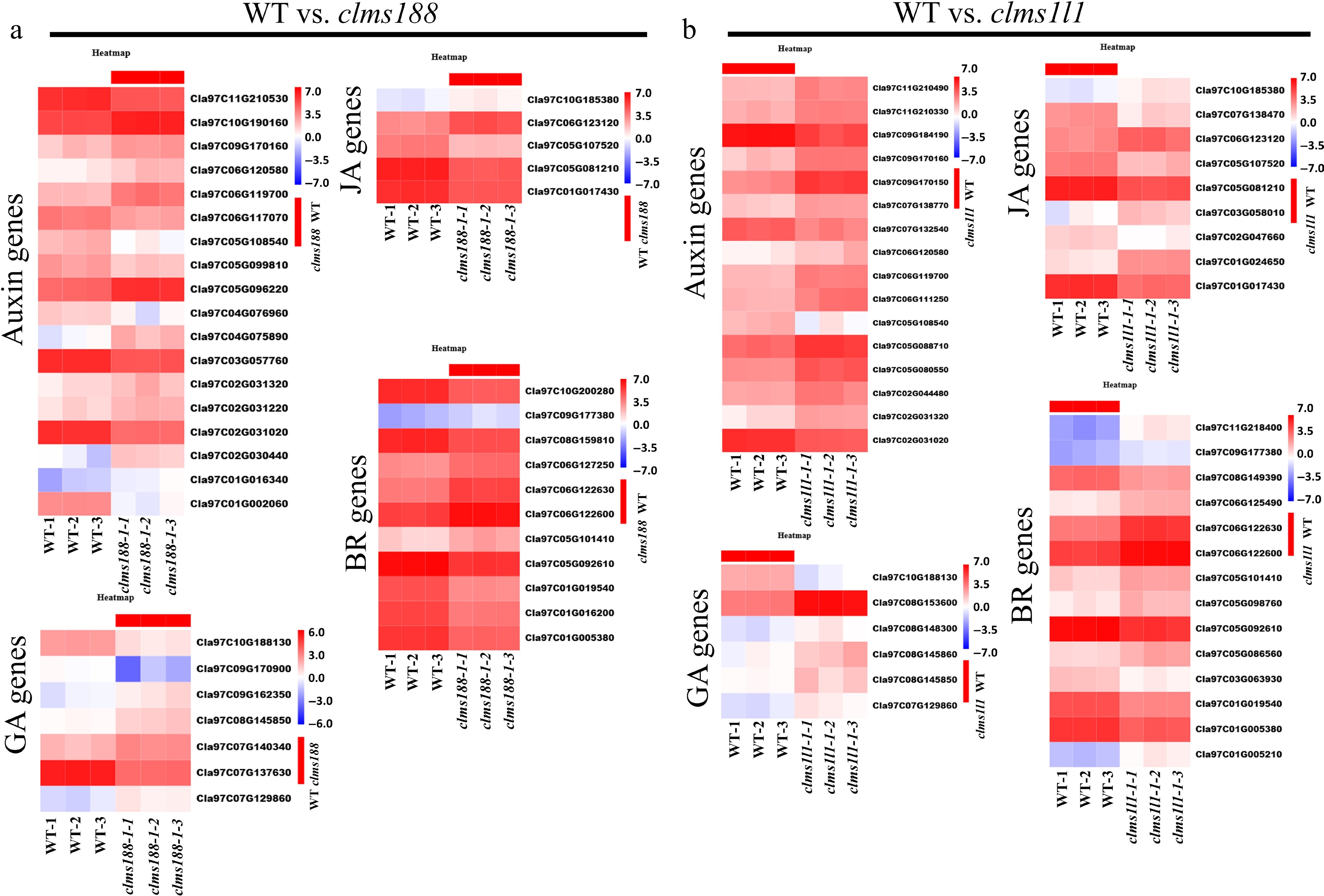
Figure 5.
KEGG enrichment of plant hormone signal transduction pathway in WT vs. clms188 and in WT vs. clms1l1. Heatmaps showing the expression of DEGs associated with auxin, gibberellin, jasmonic acid, and brassinosteroid pathways in (a) WT vs. clms188 and (b) WT vs. clms1l1. The color bar on the right represents expression levels, with red and blue indicating high and low expression, respectively.
Phenylpropanoid biosynthesis pathway (ko00940) produces compounds like lignins, coumarins, and flavonoids, which are essential for pollen and anther development (Supplementary Fig. S9b)[52,53]. Pathway enrichment analysis identified 39 DEGs for WT vs. clms188 and 66 DEGs for WT vs. clms1l1 (Fig. 6a, b; Supplementary Table S10). Of these, 20 DEGs were common to both comparisons, including core phenylpropanoid biosynthesis pathway genes, such as ClPAL family genes, ClC4H, Cl4CL family genes, and ClHCT; lignin biosynthesis pathway genes, such as ClCCR family genes, and peroxidase family genes; and coumarin biosynthesis pathway genes, such as ClGH1 family genes (Supplementary Fig. S10b; Supplementary Table S10). Notably, in comparisons involving the lignin and coumarin biosynthesis pathways, the WT vs. clms188 group exhibited approximately twice as many DEGs as the WT vs. clms1l1 group (Fig. 6a, b; Supplementary Table S10). This implies that ClMS1L1 might adopt a more intricate and specialized regulatory mechanism in these two pathways. Chalcones, serving as precursors in the flavonoid biosynthesis pathway (ko00941), can be further metabolized to flavonols through a series of enzymatic reactions. The key genes of this pathway, such as ClCHS and ClFLS, were enriched (Fig. 6b; Supplementary Table S10). In conclusion, we hypothesize that both ClMS188 and ClMS1L1 play important regulatory roles in the phenylpropanoid biosynthesis and related metabolism pathways.
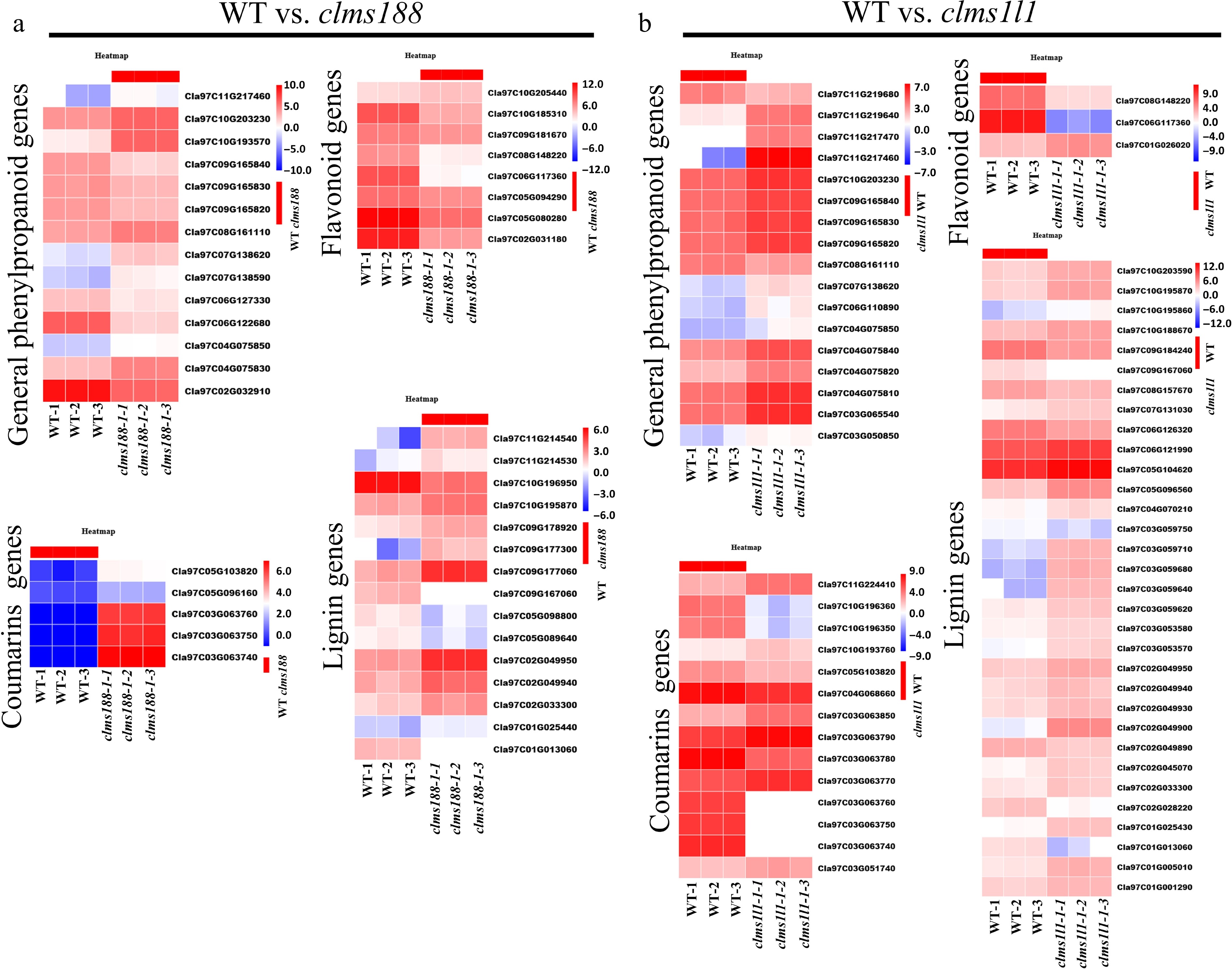
Figure 6.
KEGG enrichment of phenylpropanoid-related pathways in WT vs. clms188 and in WT vs. clms1l1. (a), (b) Heatmaps showing the expression of DEGs related to general phenylpropanoid biosynthesis, lignin, coumarin, and flavonoid metabolism in (a) WT vs. clms188 and (b) WT vs. clms1l1. The color bar on the right represents expression levels, with red and blue indicating high and low expression, respectively.
Identification of potential male sterility-related genes via DEGs analysis in clms188 and clms1l1 mutants
-
Genetic evidence indicates a shared pathway for the synthesis of phenylpropanoids, sporopollenin, and tryphine[54,55]. It has been reported that 13 genes are involved in sporopollenin precursor synthesis and 10 in their transport[55], with studies showing that AtMS188 and OsMS188 regulate multiple genes in this pathway[55,56]. For WT vs. clms188, the expression of 11 homologous genes in the sporopollenin synthesis and transport pathway was altered, including ClACLB1, ClFAS, ClMS2, ClCYP704B1, ClPKSA, ClTKPR1, ClDPW2, and ClLAP3, and the sporopollenin precursor transporter family ClABCG1, ClABCG9, ClABCG26 (Supplementary Fig. S11; Supplementary Table S10). This indicates that ClMS188 plays a critical role in the process of sporopollenin synthesis and transport. Interestingly, for WT vs. clms1l1, the expression of four genes in this pathway—ClCYP704B1, ClTKPR2, ClDPW2, and ClABCG9—also changed, suggesting that these genes may exhibit functional diversity (Supplementary Fig. S11; Supplementary Table S10). Additionally, KEGG analysis demonstrated the enrichment of 12 lipid metabolism pathways in both WT vs. clms188 and WT vs. clms1l1 comparisons, among which the steroid biosynthesis (ko00100) and cutin, suberine, and wax biosynthesis (ko00073) pathways are critical for pollen coat formation (Supplementary Table S9). We found that DEGs in the steroid biosynthesis pathway only change in the comparison of WT vs. clms1l1, such as ClSMT2, ClCAS1, and ClSLM (Supplementary Table S9). Notably, compared to the transcriptome data of the clms1l1 mutant and microarray expression data of the atms1 mutant[34], both ClSLM and AtSLM were found to be differentially expressed, respectively, suggesting that ClSLM and AtSLM may share similar regulatory patterns. However, no differential expression of AtSMT2 and AtCAS1 was detected in the atms1 mutant of Arabidopsis, indicating that these two genes may have undergone functional divergence between Arabidopsis and watermelon (Supplementary Table S9). Furthermore, in the cutin, suberine, and wax biosynthesis pathway, the number of DEGs in the WT vs. clms1l1 was nearly double that in the WT vs. clms188, which means that ClMS1L1 plays an important role in pollen coat formation.
Based on the reported information about male sterility-related genes in Arabidopsis, rice, and maize[17,57,58], we identified 25 homologous male sterility-related genes in WT vs. clms188 (22 downregulated and three upregulated) and 16 homologous male sterility-related genes in WT vs. clms1l1 (nine downregulated and seven upregulated) (gene names and Gene IDs see Table 1). For WT vs. clms188, the ClDYT1-ClTDF1-ClAMS-ClMS188-ClMS1L1 genetic pathway, genes ClDYT1 and ClTDF1 were downregulated, while that of ClAMS was upregulated, suggesting a possible feedback regulatory mechanism between ClMS188 and ClAMS. As expected, ClMS1L1 expression was undetectable, which further validates that ClMS1L1 acts downstream of ClMS188 and is directly regulated by ClMS188 (Fig. 3; Supplementary Table S5). In the sporopollenin-related pathway, the expressions of sporopollenin precursor synthesis- and transport-related genes ClACLB1, ClMS2, ClCYP704B1, ClPKSA, ClTKPR1, ClDPW2, ClLAP3, ClABCG1, ClABCG9, and ClABCG26 were altered (Table 1). In addition, the expressions of the lipid transfer protein genes ClC6 and ClMS44 were upregulated. This indicates that ClMS188 may be involved in the synthesis and transport of sporopollenin and lipid precursors. Recent studies showed that AtMS188 regulates AtQRT3 expression for tetrad wall pectin degradation[28], and together with AtTEK co-regulates AtA6 and its homologs (AtA6s) expression for tetrad wall callose degradation[59]. We found that the expression of ClA6.2, ClA6.5, and ClQRT3 was downregulated in the clms188 mutant. Additionally, the expression of ClCalS5, which regulates the deposition of callose in the tetrad wall, was downregulated. Therefore, ClMS188 may play an important role in anther tetrad wall development. Moreover, the expressions of ClKCS7 and ClKCS15, involved in the synthesis of pollen coat lipids; ClGPAT3, involved in anther cuticle biosynthesis; ClUCL23, involved in the flavonoid metabolism pathway; and ClJAR1, involved in the jasmonic acid biosynthesis pathway, were also downregulated (Table 1). Overall, ClMS188 primarily functions in sporopollenin biosynthesis and tetrad wall development.
Table 1. Identification of the DEGs genes associated with a potential male fertility phenotype.
Gene name Gene ID Log2FC Function of homologs protein function Fertility of homologs gene male sterility (MS) WT vs. clms188 ClDYT1 Cla97C05G107520 −1.69 Early tapetum development Complete MS ClTDF1 Cla97C06G126110 −2.08 Early tapetum development Complete MS ClAMS1 Cla97C02G030840 2.016 Early tapetum development Complete MS ClMS1L1 Cla97C11G207480 −5.98 Tapetum PCD, exine and pollen coat formation Complete MS ClMS2 Cla97C04G069450 −6.31 Sporopollenin synthesis Complete MS ClCYP704B1 Cla97C11G214660 2.29 Sporopollenin synthesis Complete MS ClPKSA Cla97C05G080280 −5.73 Sporopollenin synthesis Partial MS ClTKPR1 Cla97C02G036530 −4.77 Sporopollenin synthesis Partial MS ClDPW2 Cla97C06G119670 −1.69 Sporopollenin synthesis Complete MS ClLAP3 Cla97C05G088990 −12.39 Sporopollenin synthesis Complete MS ClACLB1 Cla97C08G152670 −1.13 Sporopollenin synthesis Thermo-sensitive MS ClABCG1 Cla97C09G181690 −4.85 Sporopollenin transportation Redundant MS ClABCG9 Cla97C10G190610 −4.65 Sporopollenin transportation Complete MS ClABCG26 Cla97C10G188910 −5.69 Sporopollenin transportation Complete MS ClC6 Cla97C05G094260 −2.57 Lipid transportation Partial MS ClMS44 Cla97C05G089860 1.05 Lipid transportation Complete MS ClA6.2 Cla97C03G066330 −1.26 Callose dissolution Redundant MS ClA6.5 Cla97C11G209110 −1.01 Callose dissolution Redundant MS ClQRT3 Cla97C01G025690 −1.14 Pectin dissolution Complete MS ClCalS5 Cla97C02G026770 −2.90 Callose biosynthesis Partial MS ClKCS7 Cla97C10G196270 −2.66 Pollen coat synthesis Partial MS ClKCS15 Cla97C05G096910 −1.19 Pollen coat synthesis Partial MS ClGPAT3 Cla97C09G183960 −3.10 Anther cutin biosynthesis Complete MS ClUCL23 Cla97C11G213980 −1.15 Flavonoid metabolism Partial MS ClJAR1 Cla97C05G081210 −1.49 JA biosynthesis Complete MS WT vs. clms1l1 ClDYT1 Cla97C05G107520 2.25 Early tapetum development Complete MS ClATM1 Cla97C06G117840 −1.09 Tapetum PCD Complete MS ClLBD10 Cla97C02G038990 −2.27 Pollen development Partial MS ClCYP704B1 Cla97C11G214660 1.93 Sporopollenin synthesis Complete MS ClTKPR2 Cla97C05G104620 1.80 Sporopollenin synthesis Partial MS ClDPW2 Cla97C06G119670 2.37 Sporopollenin synthesis Complete MS ClABCG9 Cla97C10G190610 −2.86 Sporopollenin transportation Complete MS ClGPAT3 Cla97C09G183960 2.25 Lipid metabolism Complete MS ClMS44 Cla97C05G089860 −2.32 Lipid transportation Complete MS ClC6 Cla97C05G094260 −1.13 Lipid transportation Partial MS ClLecRK5 Cla97C08G149390 1.43 Callose biosynthesis Complete MS ClG1 Cla97C03G061520 2.00 Callose degradation Complete MS ClKCS5 Cla97C05G089490 −5.12 Pollen coat synthesis Partial MS ClKCS7 Cla97C10G196270 −1.02 Pollen coat synthesis Partial MS ClGELP34 Cla97C07G138280 −1.44 Pollen exine formation Complete MS ClUAM3 Cla97C06G125020 −1.71 Pollen cell wall formation Partial MS Further details and references for these genes are provided in Supplementary Table S12. For WT vs. clms1l1, the ClDYT1-ClTDF1-ClAMS-ClMS188-ClMS1L1 genetic pathway gene ClDYT1 was upregulated. Additionally, the expression of a bHLH transcription factor family member, ClATM1 (which regulates early tapetum development), and an LBD transcription factor family member, ClLBD10 (which regulates male gametophyte development), were downregulated. Pollen coat formation involves pollen coat precursor biosynthesis and transport, which share common biosynthetic pathways with sporopollenin precursor biosynthesis and transport[60,61]. The expressions of sporopollenin precursor synthesis- and transport-related genes ClCYP704B1, ClTKPR2, ClDPW2, and ClABCG9 were upregulated. Moreover, the expression of lipid synthesis gene ClGPAT3 and lipid transport genes ClMS44 and ClC6 were altered. AtKCS family genes AtKCS7, AtKCS15, and AtKCS21 were reported to be downstream of AtMS1 and are involved in the accumulation of pollen coat lipids[35]; the expressions of ClKCS5 and ClKCS7 were downregulated. Furthermore, the expressions of the pollen exine formation-related gene ClGELP34, the pollen wall formation-related gene ClUAM3, the callose synthesis gene ClLecRK5, and the callose degradation gene ClG1 were upregulated (Table 1). These results suggest that ClMS1L1 plays a key role in regulating the lipid metabolism essential for pollen coat formation.
Notably, in the Arabidopsis atms1 mutant background, pollen coat-related genes such as the GRP (glycine-rich protein) family genes (GRP14, GRP16, GRP17, GRP18, GRP20) and the EXL (extracellular lipase protein) family genes (EXL4, EXL5, EXL6) were altered[32], both of which are involved in pollen hydration. However, in the clms1l1 mutant, we did not detect changes in the corresponding homologous genes, suggesting that the genes related to pollen hydration in watermelon may have diverged from those in Arabidopsis (Supplementary Table S9).
-
Although the genetic pathway composed of these five transcription factors that regulates anther development has been identified in Arabidopsis (AtDYT1-AtTDF1-AtAMS-AtMS188-AtMS1), rice (OsUDT1-OsTDF1-OsTDR-OsMS188-OsPTC1), and maize (ZmMs32-ZmMs9-ZmbHLH51-ZmMYB84-ZmMs7)[17,21−23] (see Supplementary Table S13 for details), molecular mechanisms regulating tapetal development and pollen maturation in watermelon anthers are still poorly understood. Through genetic and molecular analyses, we validated the functions of the downstream genes of the ClDYT1-ClTDF1-ClAMS-ClMS188-ClMS1L1 genetic pathway, ClMS188 and ClMS1L1, to study the regulatory relationship. The results demonstrate that ClMS188 directly binds to the TAACCA motif of ClMS1L1 and activates its transcription (Fig. 3). By analyzing the transcriptomic regulatory network of ClMS188 and ClMS1L1, the results further confirm the existence of the complete ClDYT1-ClTDF1-ClAMS-ClMS188-ClMS1L1 genetic pathway in watermelon anther development, which precisely regulates the development of the tapetum and pollen in watermelon (Fig. 7).
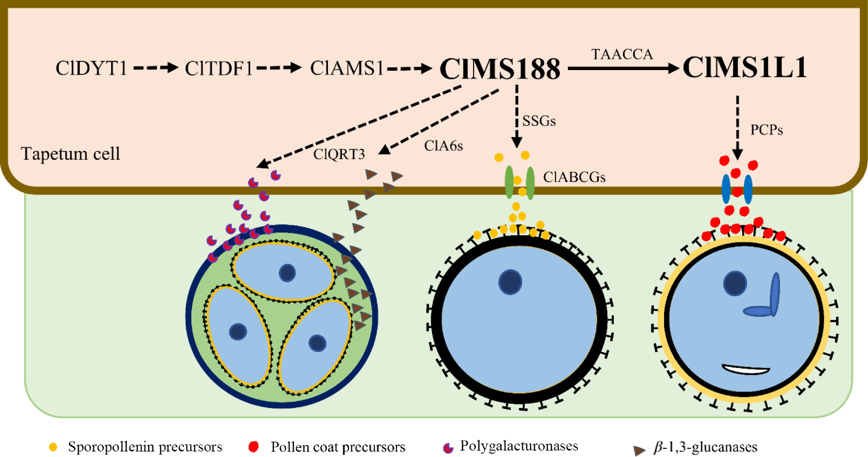
Figure 7.
A proposed model for the regulation of five transcription factors during anther development in watermelon. ClMS188 and ClMS1L1 are downstream components in the ClDYT1-ClTDF1-ClAMS-ClMS188-ClMS1L1 genetic pathway. ClMS188 directly activates ClMS1L1 expression; ClMS188 may control the expression of ClQRT3, ClA6s, and sporopollenin biosynthesis and transport genes. ClMS1L1 may control pollen coat protein formation. The solid arrow represents a validated regulatory mechanism, and the dashed arrows represent predicted regulatory mechanisms. SSGs: sporopollenin biosynthesis genes; PCPs: pollen coat precursors.
ClMS188 plays a crucial role in tetrad wall degradation and sporopollenin precursor formation
-
ClMS188 plays a central role in watermelon anther tapetal development and pollen formation. As a homolog of AtMS188/OsMS188, its expression peaks during the tetrad stage (stage 8b) (Fig. 1b, c). Similar to its function in model plants, transcriptomic analysis revealed that ClMS188 coordinates multiple critical processes in anther development by regulating a set of key genes. This includes controlling genes involved in tapetal development (e.g., ClDYT1, ClTDF1, and ClAMS), tetrad wall degradation (e.g., ClQRT3 and ClA6), and genes regulating sporopollenin biosynthesis and transport (e.g., ClMS2 and the members of the ClABCG gene family) (Table 1). These regulatory functions are essential for both tetrad wall degradation and sporopollenin precursor formation (Fig. 7). Furthermore, our experimental results indicate that ClMS188 directly binds to the TAACCA motif, a specific cis-acting element in the ClMS1L1 promoter, and activates its transcription, thereby promoting pollen coat formation. Analysis of the phenylpropanoid biosynthesis pathway indicated that ClMS188 regulates genes involved in anther wall lignification (e.g., ClCCR and ClCAD) (Fig. 6). This is supported by the more pronounced downregulation of these lignin-related genes in the clms188 mutant compared to the clms1l1 mutant. This mechanism allows ClMS188 to precisely coordinate the mechanical strengthening of the anther wall (via lignin) and the metabolic maturation of pollen grains (via ClMS1L1).
ClMS1L1 is involved in regulating pollen coat formation
-
The orthologs AtMS1/OsPTC1 are highly expressed at the microspore release stage (stage 9), regulating tapetal PCD, pollen exine, and coat formation[25,32,34,62,63]. Similarly, ClMS1L1 expression peaks at stage 9 (Fig. 4a, b). The clms1l1 mutant exhibits aberrant tapetum and microspore development from this stage (Fig. 4i), confirming its crucial role. Transcriptome data show that ClMS1L1 downregulates genes for pollen coat lipids (e.g., ClKCS5/7), pollen exine formation (ClGELP34), and sporopollenin biosynthesis/transport (e.g., ClCYP704B1, ClTKPR2, ClDPW2, and ClABCG9). Genes involved in lipid synthesis/transport (e.g., ClGPAT3, ClMS44, ClC6) were altered. This likely impairs lipid metabolism, reducing pollen adhesiveness and affecting insect pollination. Thus, ClMS1L1 primarily regulates the synthesis and transport of pollen coat precursors and lipids during maturation. The altered Auxin/JA hormone signaling is a metabolic consequence, not a primary cause. Disrupting the phenylpropanoid pathway in mutants blocks the production of sporopollenin and lignin, causing upstream phenolic precursors to accumulate. These intermediates can inhibit/regulate auxin transport and JA biosynthesis; therefore, the hormonal imbalance is a feedback response to the blocked metabolic flux (Figs. 5, 6). This metabolic-hormonal crosstalk synchronizes anther dehiscence with pollen maturation.
ClMS188 and ClMS1L1 can be applied to the SPT system in watermelon
-
The clms188 and clms1l1 mutants are recessive genic male sterility (RGMS) mutants and exhibit no developmental defects in traits including vegetative growth and female reproductive organ development (Supplementary Figs. S3, S6; Supplementary Table S11). Crucially, the clms188 and clms1l1 mutants exhibit a 'clean' male sterility phenotype without pleiotropic defects (Figs. 2, 4). Unlike some male-sterile mutants in other crops that suffer from vegetative growth inhibition or abnormal female fertility, these watermelon mutants maintain normal vegetative vigor and female fruit set. This specificity renders them ideal GMS resources for hybrid seed production. Furthermore, studies on the MS1 homolog in cabbage have demonstrated that a natural allele, Ms-cd1PΔ-597, resulting from a 1-bp deletion in its promoter, confers dominant genic male sterility (DGMS). We propose to use the ABE/GBE technologies developed in our laboratory to precisely mutate the corresponding site in the promoter of ClMS1L1, thereby potentially generating DGMS lines that can serve as a genetic resource for the SPT system[64,65]. They can be utilized to eliminate the labor-intensive manual emasculation process in watermelon breeding, significantly reducing costs while ensuring hybrid purity[7,66].
-
In this study, loss-of-function mutations in ClMS188 and ClMS1L1 resulted in complete male sterility. Expression analyses and anther sectioning demonstrated that ClMS188 and ClMS1L1 are specifically expressed in anthers at stage 8b and stage 9, during which abnormal anther development phenotypes were first observed. LUC, Y1H, and EMSA assays confirmed that ClMS188 directly activates ClMS1L1 transcription. Comparative transcriptomic analyses revealed that ClMS188 coordinates anther development through two separate pathways: one independent of ClMS1L1, which regulates tetrad wall degradation and sporopollenin biosynthesis, and another dependent on ClMS1L1, which controls lipid metabolism for pollen coat formation.
-
The authors confirm their contributions to the paper as follows: conceived and designed the study: Yuan L; executed the experiments: Zhang Q, You X, Peng Y, Zhang Y, Yang F, Zhong Y; provided experimental technical support: Chen X; conducted data analysis: Zhang Q; provided project supervision: Yuan L, Tian S, Luan F; wrote the manuscript: Yuan L, Zhang Q. All authors reviewed the results and approved the final version of the manuscript.
-
The RNA-Seq data were deposited in the NCBI Sequence Read Archive (SRA) database under the accession PRJNA1496536 (https://dataview.ncbi.nlm.nih.gov/object/PRJNA1496536).
-
The authors declare that they have no conflict of interest.
-
accompanies this paper online at: https://doi.org/10.48130/seedbio-0026-0020.
- Supplementary Table S1 Accession numbers of ClMS188, ClMS1L1 and their homologous genes.
- Supplementary Table S2 List of primers and probes used in this study.
- Supplementary Table S3 Comparison table of anther development stage and floral buds diameter.
- Supplementary Table S4 Information regarding the RNA-seq data for the WT , clms188 and clms1l1 mutant.
- Supplementary Table S5 Differentially upregulated and downregulated genes identified in the clms188 mutant anther.
- Supplementary Table S6 Differentially upregulated and downreglated genes identified in the clms1l1 mutant anther.
- Supplementary Table S7 Common up-regulated DEGs between the clms188 and clms1l1 mutants.
- Supplementary Table S8 Common down-regulated DEGs between the clms188 and clms1l1 mutants.
- Supplementary Table S9 KEGG pathway enrichment of WT vs. clms188 and WT vs. clms1l1, steroid biosynthesis pathway gene analysis of WT vs. clms1l1.
- Supplementary Table S10 Hormone, phenylpropanoid and sporopollenin biosynthesis-related DEGs in WT vs. clms188 and WT vs. clms1l1.
- Supplementary Table S11 The statistical analysis of female flower , male flower, fruit and seed for the WT, clms188, and clms1l1.
- Supplementary Table S12 Detailed annotations and supporting references for all genes listed in Table 1.
- Supplementary Table S13 Homolog genes of ClDYT1–ClTDF1–ClAMS–ClMS188–ClMS1L1 genetic pathway in Arabidopsis, rice and maize.
- Supplementary Fig. S1 Schematic representation of ClMS188 genomic structure, protein features, and phylogenetic analysis with R2R3-MYB family proteins.
- Supplementary Fig. S2 Gene editing types in clms188 mutants and the characteristics of the corresponding CIMS188 mutant protein sequences.
- Supplementary Fig. S3 Comparison of female flower opening, fruit size, fruit internal morphology and seed number between WT and clms188 mutant.
- Supplementary Fig. S4 Schematic representation of ClMS1L1 genomic structure, protein features, and phylogenetic analysis with PHD-finger family proteins.
- Supplementary Fig. S5 Gene editing types in clms1l1 mutants and the characteristics of the corresponding ClMS1L1 mutant protein sequences.
- Supplementary Fig. S6 Comparison of female flower opening, fruit size, fruit internal morphology and seed number between WT and clms1l1 mutants.
- Supplementary Fig. S7 Transcriptome quality analysis from anthers of WT, and clms188 and clms1l1 mutants.
- Supplementary Fig. S8 Identification of DEGs in clms188 and clms1l1 vs. the WT.
- Supplementary Fig. S9 A schematic model of plant hormone signal pathway and phenylpropanoid biosynthesis pathway.
- Supplementary Fig. S10 Heatmaps showing the expression of DEGs associated with hormone, phenylpropanoid biosynthesis in both WT vs. clms188 and WT vs. clms1l1.
- Supplementary Fig. S11 Analysis of sporopollenin-related genes in WT vs. clms188 and in WT vs. clms1l1.
- Copyright: © 2026 by the author(s). Published by Maximum Academic Press on behalf of Hainan Yazhou Bay Seed Laboratory. This article is an open access article distributed under Creative Commons Attribution License (CC BY 4.0), visit https://creativecommons.org/licenses/by/4.0/.
-
About this article
Cite this article
Zhang Q, You X, Peng Y, Zhang Y, Yang F, et al. 2026. The R2R3-MYB transcription factor ClMS188 targets ClMS1L1 to orchestrate tapetum development and pollen formation in watermelon. Seed Biology 5: e023 doi: 10.48130/seedbio-0026-0020 

The R2R3-MYB transcription factor ClMS188 targets ClMS1L1 to orchestrate tapetum development and pollen formation in watermelon
- Received: 26 May 2026
- Revised: 23 July 2026
- Accepted: 31 July 2026
- Published online: 09 September 2026
Abstract: Male-sterile lines are crucial genetic materials for investigating the developmental and abortion mechanisms of anthers, and they serve as key parental lines in hybrid seed production. However, research to date has primarily focused on model plants such as Arabidopsis, rice, and maize, whereas the regulatory mechanisms underlying this process remain largely unexplored in Cucurbitaceae. Here, we identified an R2R3-MYB transcription factor, ClMS188, as a key regulator of tapetum and pollen development. The clms188 mutants exhibited complete male sterility due to aberrant tapetum development and tetrad formation. In vitro and in vivo experiments demonstrated that ClMS188 directly binds to the TAACCA motif in the ClMS1L1 promoter and activates its transcription. Similarly, loss-of-function mutants of ClMS1L1 led to defective tapetum and microspore development. Comparative transcriptomic analyses revealed that the expression of key genes in hormone signal transduction and phenylpropanoid biosynthesis pathways was altered in both the clms188 and clms1l1 mutants. Furthermore, the clms188 mutant exhibited disruptions in sporopollenin precursor metabolism and tetrad wall degradation, whereas the clms1l1 mutant showed alterations in genes for pollen coat precursor biosynthesis. Together, these findings establish the ClMS188–ClMS1L1 regulatory module that coordinates tapetum development and pollen formation, and provide a valuable genetic resource for hybrid seed production in watermelon.
-
Key words:
- ClMS188 /
- ClMS1L1 /
- Tapetum and pollen development /
- Male sterile /
- Watermelon