











-
As an ecologically influential and highly diverse branch of the tree of life, fungi are essential components of virtually all terrestrial and aquatic habitats, including soils, plants, rocks, and even the pelagic zones of the ocean[1]. Global fungal species richness is estimated to range from 1.5 to 3.8 million, with 2.5 million widely regarded as the most plausible approximation[2,3], and recent studies indicate that Fungi comprise 19 phyla and 83 classes[4]. In recent years, rapid progress in molecular identification tools has substantially accelerated fungal taxonomic research, leading to the description of numerous taxonomic novelties across a broad range of substrates, including grassland ecosystems, plant tissues, soil, and freshwater habitats[5−10]. Soil, in particular, provides an important habitat for many fungi, enabling their survival and reproduction[6], and a substantial proportion of fungal species complete at least part of their life cycle within this substrate[4]. Among these, saprobic fungi play a pivotal role in decomposing organic matter, a process that supports nutrient cycling, improves soil health, and maintains plant biodiversity[11,12]. Through the secretion of extracellular enzymes, these fungi decompose organic matter and release mineral nutrients into the soil, thereby enhancing soil fertility[13]. These processes also contribute substantially to the breakdown of lignocellulose—the most abundant form of organic carbon on land—playing an essential role in carbon cycling[14]. Rather than existing in isolation, saprobic fungi are integral components of a complex network of soil organisms, interacting synergistically or antagonistically with other fungi, bacteria, and various organisms to maintain overall ecosystem stability[6].
To date, few places on Earth remain untouched by human activities. Consequently, fungi in regions significantly impacted by anthropogenic disturbance, such as cities, warrant further exploration. Soil fungal research remains in its infancy[4], particularly concerning urban soils. Rapid global changes—including climate change, deforestation, drought, heatwaves, population growth, urbanization, infrastructure development, and resource extraction—are exerting a profound influence on fungal communities inhabiting urban soils[6]. While over 250 soil fungal genera are currently known[4], this number is likely to increase substantially through systematic, in-depth investigations across diverse global habitats. In recent years, we have progressively expanded our research scope from keratinophilic fungi in Chinese urban soils to encompass a broader range of fungal taxa within soil environments[6,15−21]. Our preliminary research indicates that environments significantly impacted by human activities harbor rich fungal diversity, even in small areas such as zoos[6]. As a unique and overlooked habitat with distinct disturbance factors (e.g., animal feces, animal hair, and feed), zoos suggest the potential presence of specialized fungal taxa. Therefore, as part of this ongoing research initiative, this study employed culture-dependent methods to investigate fungal taxa at Harbin North Forest Zoo. This study advances current knowledge of microfungal diversity, taxonomy, and phylogenetic relationships in urban soil environments.
-
Soil samples were collected from Harbin North Forest Zoo in northern China on May 6, 2023, following the sampling protocol of Zhang et al.[6]. Samples were placed in Ziploc bags, with collection data recorded accordingly, and maintained at 4 °C during transport[22]. Upon arrival, samples were immediately processed in the laboratory. Fungal isolation was performed using media and protocols previously established by Zhang et al.[6].
Following the method of Zhang et al.[6], macroscopic characteristics of colonies were assessed on PDA, oatmeal agar (OA), and synthetic nutrient-poor agar (SNA) plates. For new Penicillium species, these characteristics were evaluated following Visagie et al.[23]. Morphological features were observed and recorded using either a Zeiss Axio Imager A2 microscope equipped with a Zeiss AxioCam MRc color digital camera (Carl Zeiss AG, Germany), or a Nikon ECLIPSE Ni (Nikon, Japan) compound microscope fitted with a Nikon DS-Ri2 digital camera (Nikon, Japan).
The ex-type strains of the new taxa were deposited at the China General Microbiological Culture Collection Center (CGMCC), China. Dried specimens (dried at 50 °C) and all living cultures were preserved in a non-active state (sterile 30% glycerol at –80 °C) and deposited in the College of Eco-Environmental Engineering, Guizhou Minzu University. MycoBank numbers were registered for each new name.
DNA extraction, PCR amplification, and sequencing
-
Genomic DNA extraction followed the protocol outlined by Zhang et al.[6]. The internal transcribed spacer (ITS) region was amplified and sequenced for each isolate. Sequence identity was subsequently assessed via MEGA BLAST searches against the NCBI GenBank nucleotide database, consistent with the approach of Zhang et al.[6]; isolates exhibiting less than 97% ITS similarity to their closest matches were regarded as putative novel species, prompting amplification of supplementary loci for downstream phylogenetic analysis. Since different taxonomic groups often require distinct loci for phylogenetic inference, this study amplified the specified loci listed in Table 1. Polymerase chain reaction (PCR) was performed using a 25 μL reaction volume, and the amplicons were sequenced by Sangon Biotech (Shanghai) Co., Ltd, China. Newly generated sequences have been deposited in GenBank under the accession numbers listed in Supplementary Table S1.
Table 1. Loci, primers, and PCR amplification procedures used in this study.
Gene/
locusPrimer Primer sequence (5’-3’) Annealing temperature (°C) Ref. ITS ITS1 TCCGTAGGTGAACCTGCGG 55 [24] ITS5 GGAAGTAAAAGTCGTAACAAGG ITS4 TCCTCCGCTTATTGATATGC LSU LR0R ACCCGCTGAACTTAAGC 55 [25] LR5 ATCCTGAGGGAAACTTC SSU NS1 GTAGTCATATGCTTGTCTC 55 [24] NS4 CTTCCGTCAATTCCTTTAAG TEF1 EF1-983F GCYCCYGGHCAYCGTGAYTTYAT 55 [26,27] EF1-2218R ATGACACCRACRGCRACRGTYTG EF1-728F CATCGAGAAGTTCGAGAAGG EF-2 GGAAGTACCAGTGATCATGTT EF–986R TACTTGAAGGAACCCTTACC ACT ACT-512F ATGTGCAAGGCCGGTTTCGC 51 [27] ACT-783R TACGAGTCCTTCTGGCCCAT RPB2 fRPB2-5f GAYGAYMGWGATCAYTTYGG 52 [28,29] RPB150 CTGGGGWGATCARAAGAAGG fRPB2-7cR CCCATRGCTTGYTTRCCCAT TUB Bt2a GGTAACCAAATCGGTGCTGCTTTC 51 [30,31] Bt2b ACCCTCAGTGTAGTGACCCTTGGC BT2916 CTCAGCCTCAGTGAACTCCAT 58 BT1819R TTCCGTCCCGACAACTTCGT CaM CF1 GCCGACTCTTTGACYGARGAR 55 [32] CF4 TTTYTGCATCATRAGYTGGAC ITS: the nuclear internal transcribed spacers, LSU: the nuclear large subunit ribosomal RNA, SSU: the nuclear ribosomal small subunit rRNA, TEF1: the translation elongation factor 1-alpha, ACT: the actin, RPB2: the second largest subunit of RNA polymerase II, TUB: the beta-tubulin. Phylogenetic analyses
-
Phylogenetic analyses were performed to determine the taxonomic placement of the putative novel taxa central to this investigation. These analyses were based on the ITS and LSU regions, as well as the TEF, RPB2, and TUB genes, markers selected based on recommendations in recent literature. Information on all reference strains is summarized in Supplementary Table S2. Consensus sequences were assembled with Lasergene v6.0 (DNASTAR). The DNA loci were aligned with MAFFT v7.037[33] and trimmed with MEGA v6.06[34]. The 'Concatenate Sequence' function in PhyloSuite v.1.2.3 was used to concatenate the loci[35]. Phylogenetic analyses were performed using both Bayesian and Maximum-Likelihood algorithms. The dataset was partitioned by gene locus. The best evolutionary models for phylogenetic analyses were selected independently for each locus using ModelFinder[36] under the corrected Akaike Information Criterion (AICc). Maximum Likelihood (ML) analysis was performed in IQ-TREE v1.6.11 using the ultrafast bootstrap algorithm with 10,000 replicates[37]. Bayesian Inference (BI) was conducted using MrBayes v3.2[38] via PhyloSuite[35]. Two independent Markov Chain Monte Carlo (MCMC) runs, each employing four chains, were executed simultaneously for 5 × 10⁷ generations, sampling a tree every 1,000 generations. A burn-in of 25% was discarded. Chain convergence was monitored and verified using Tracer v1.5[39], ensuring that all parameters reached stationarity well before the 25% cutoff and that effective sample size (ESS) values exceeded 200. The multi-locus phylogenetic tree was visualized in FigTree v1.4.2, and the final layout was annotated with ML bootstrap values and BI posterior probabilities using Microsoft PowerPoint (Microsoft, 2019).
-
In this study, 278 taxa were isolated and assigned to four phyla, 14 classes, 47 orders (three undetermined), 99 families (ten undetermined), 168 genera (seven undetermined), and 278 species (44 undescribed taxa) (Supplementary Table S3). By integrating multi-locus phylogenetic inference with detailed morphological examination, we described and illustrated two new families, four new genera, and 20 new species. The treatment of each new taxon was based on the latest and most comprehensive literature for that specific group.
Taxonomy
-
Phylum Ascomycota Caval.-Sm.
Class Dothideomycetes O.E. Erikss & Winka
Microthyriales G. Arnaud
Microthyriaceae Sacc.
Pseudosoloacrosporiella Crous.
Crous et al. erected the genus Pseudosoloacrosporiella with P. cryptomeriae as the type species[40]. This species was originally recovered from leaves of Cryptomeria japonica (Cupressaceae) in the Netherlands[40]. It remains uncertain which family Pseudosoloacrosporiella belongs to within the order Microthyriales (Dothideomycetes). The genus is characterized by conidiophores that are mostly reduced to conidiogenous cells. Conidiogenous cells possess one to three sympodial loci at their apices, undergoing rhexolytic conidiogenesis, which results in the formation of tiny collarettes at denticulate loci[40]. Ramoconidia are fusoid to ellipsoid, tapering towards both ends, septate, light brown, guttulate, and smooth-walled[40].
Phylogenetic relationships within Microthyriales were inferred using a combined ITS and LSU dataset, with Kirschsteiniothelia lignicola (MFLUCC 10-0036) designated as the outgroup. The final alignment included 60 taxa and spanned 1,348 nucleotide positions (ITS, 536 bp; LSU, 812 bp), including gaps. The best-fit models were TVMe + I + G4 (ITS) and GTR + F + I + G4 (LSU) for the ML analysis, whereas those for the BI analysis were SYM + I + G4 (ITS) and GTR + F + I + G4 (LSU). Phylogenetically, CGMCC 3.29784 and ZY 23.009 formed a distinct subclade sister to Pseudosoloacrosporiella cryptomeriae (CBS 148441), with strong support (100% bootstrap support [BS]/1 posterior probability [PP], Fig. 1). Additionally, CGMCC 3.29783, ZY 23.005, ZY 23.006, and ZY 23.007 formed a single, well-supported clade (100/1) that was clearly separated from other taxa in the order Microthyriales (Fig. 1).
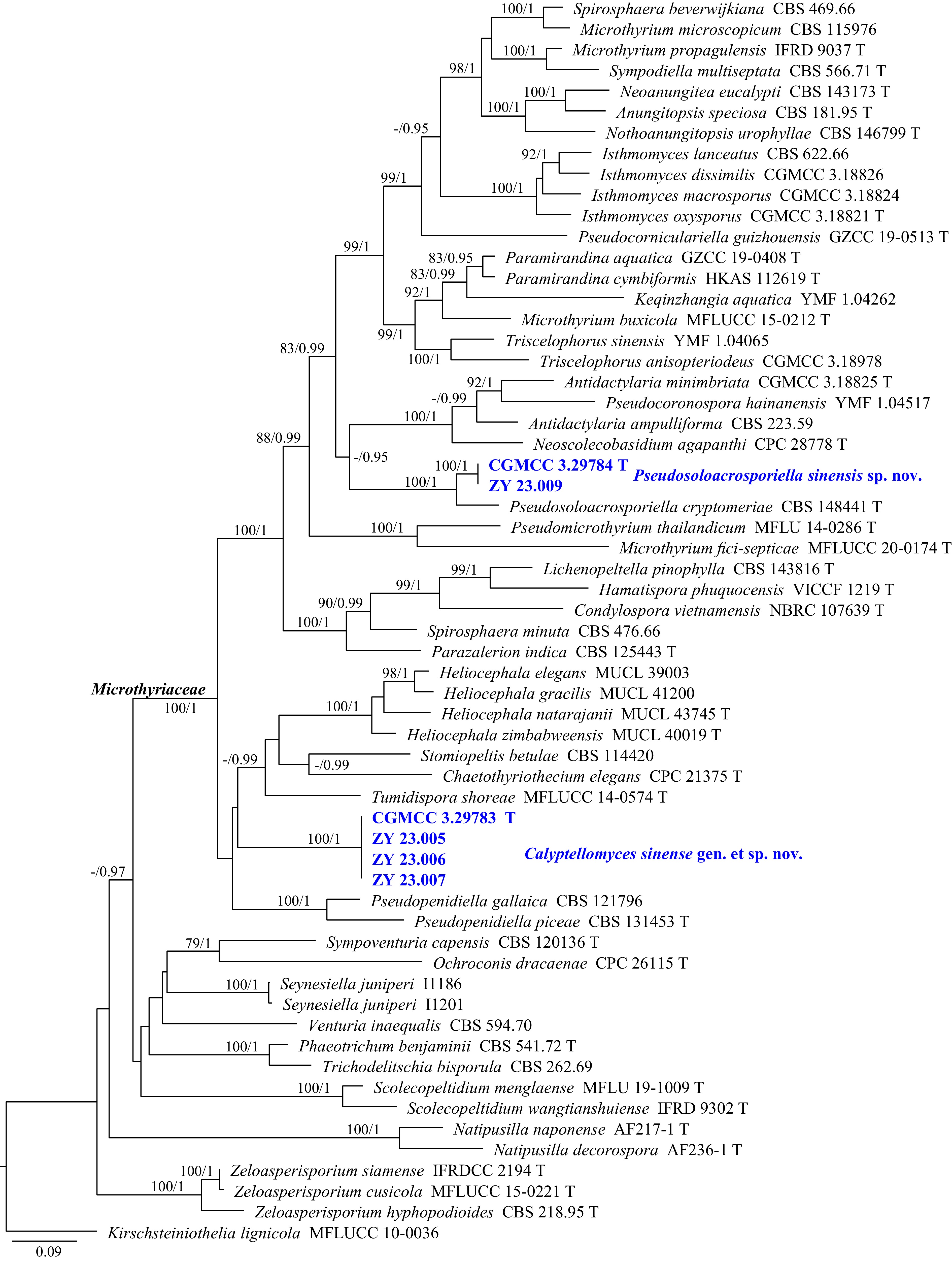
Figure 1.
Phylogram generated from ML analysis based on the combined dataset of ITS and LSU sequence data. Numbers at branches indicate support values (IQ-TREE-BS/BI-PP) above 70%/0.90. The tree is rooted to Kirschsteiniothelia lignicola (MFLUCC 10-0036). Newly generated sequences are in bold and blue, and 'T' indicates type-derived sequences. The scale bar represents the expected number of changes per site.
Pseudosoloacrosporiella sinensis Y.J. Wu, Zhi Y. Zhang & Y.F. Han sp. nov. Fig. 2
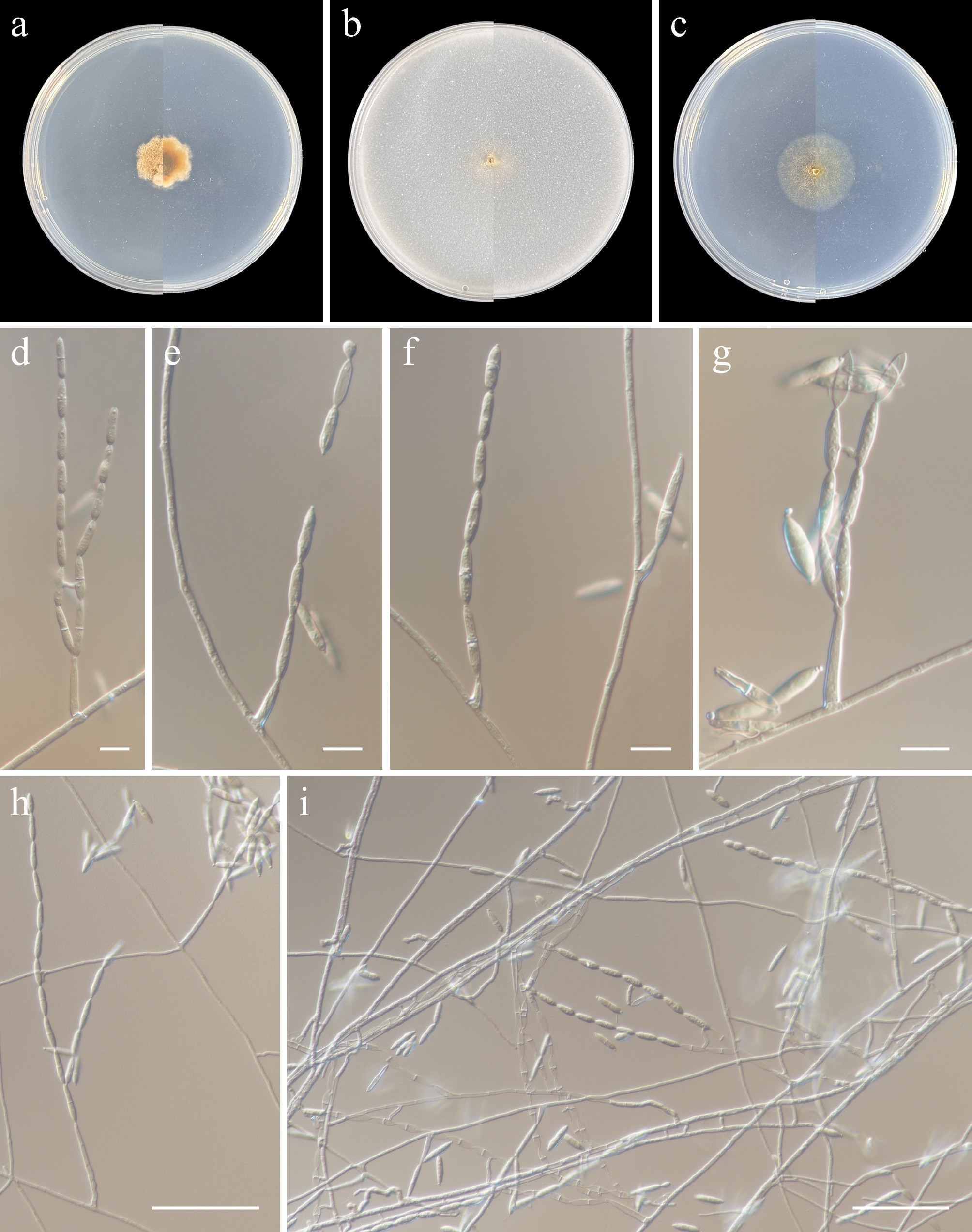
Figure 2.
Pseudosoloacrosporiella sinensis (from holotype ZY H-23.008). (a)–(c) Upper and reverse views of cultures on PDA, OA, and SNA after 14 d at 25 °C. (d)–(i) Conidiogenous cells and conidia. Scale bars: (d), (g) 10 μm, (h), (i), 50 μm.
MycoBank number: MB862606
Etymology: Named after the type locality, China.
Description: Hyphae branched, septate, hyaline, smooth-walled, 1–3 μm wide. Asexual morph Conidiophores solitary, erect, hyaline, smooth, subcylindrical with apical taper, zero-to-one-septate, mostly reduced to conidiogenous cells. Conidiogenous cells 15.5–26 × (1–)2–4 µm, with one to two sympodial apical loci with rhexolytic conidiogenesis, leaving minute collarettes on denticulate loci, 0.5 µm diam. Ramoconidia cylindrical-fusoid, tapering towards both ends, hyaline, smooth, zero-to-one-septate, 12.5–21 × 2.5–3.5 µm, with one to two apical loci. Conidia in long unbranched chains, zero-to-one-septate, cylindrical-fusoid, hyaline, smooth, (6–)11.5–20.5 × 2.5–4 µm (av. 15.1 × 2.9); loci rhexolytic, 0.5 µm diam., with minute marginal frill. Sexual morph not observed.
Culture characteristics (14 d at 25 °C): Colony on PDA reaching 18–21 mm diam., felted, compact, plicated, convex, yellow, margin dentate; reverse brown to yellow. Colony on OA reaching 10–11 mm diam., flat, pale yellow, aerial mycelia extremely sparse, reverse pale yellow. Colony on SNA reaching 23–25 mm diam., cottony, margin entire, pale brown, aerial mycelia sparse, reverse pale brown.
Material examined: China, Heilongjiang Province, Harbin City, soil collected from the green belts of Harbin Northern Forest Zoo, 127.1051° E, 45.3977° N, 8 May 2023, Zhi-Yuan Zhang (holotype ZY H-23.008, dried culture; culture ex-type CGMCC 3.29784, ibid., ZY 23.008). China, Heilongjiang Province, Harbin City, soil collected from the green belts of Harbin Northern Forest Zoo, 127.1131° E, 45.3912° N, 8 May 2023, Zhi-Yuan Zhang, isotype ZY H-23.009, ex-isotype ZY 23.009.
Notes: To date, Pseudosoloacrosporiella contains only one species, P. cryptomeriae. Phylogenetically, P. sinensis formed a distinct subclade closely allied to P. cryptomeriae (Fig. 1). Morphologically, P. sinensis is distinguished from P. cryptomeriae by the shape and size of its conidia (cylindrical-fusoid, 11.5–20.5 × 2.5–4 µm in P. sinensis vs fusoid-ellipsoid, 12–13 × 3 µm in P. cryptomeriae)[40]. Additionally, pairwise comparison of ITS and LSU loci reveals that P. sinensis (ex-type CGMCC 3.29784) differs from P. cryptomeriae (ex-type CBS 148441) by 8.5% (49/577 bp, 25 gaps) in ITS and 4.9% (40/810 bp, no gaps) in LSU.
Calyptellomyces Y.J. Wu, Zhi Y. Zhang & Y.F. Han, gen. nov.
MycoBank number: MB862607
Etymology: The name refers to the cup-shaped lower part of the conidial septum.
Description: Hyphae branched, septate, hyaline, smooth-walled. Asexual morph Conidiophores solitary, erect, hyaline to brown, smooth, subcylindrical to cylindrical, one-to-six-septate. Conidiogenous cells with one apical conidiogenous locus, rarely one at each end, hyaline to brown, smooth, subcylindrical to cylindrical. Conidia solitary, hyaline to pale brown, elliptical to subglobose, smooth, zero-to-one-septate, cup-shaped at the lower part, hemispherical to campanulate at the upper part. Sexual morph not observed.
Type species: Calyptellomyces sinensis Y.J. Wu, Zhi Y. Zhang & Y.F. Han
Notes: Saccardo erected the family Microthyriaceae with the sexual genus Microthyrium designated as the type genus[41]. To date, this family comprises 18 genera[4,42], namely Antidactylaria, Arnaudiella, Chaetothyriothecium, Hamatispora, Isthmomyces, Keqinzhangia, Microthyrium, Neoanungitea, Nothoanungitopsis, Nothomicrothyrium, Paramicrothyrium, Paramirandina, Pseudocorniculariella, Pseudocoronospora, Pseudomicrothyrium, Pseudopenidiella, Triscelophorus, and Tumidispora. Of these, Antidactylaria, Hamatispora, Isthmomyces, Keqinzhangia, Neoanungitea, Paramirandina, Pseudocorniculariella, Pseudocoronospora, Pseudopenidiella, and Triscelophorus are asexual genera[42]. Asexual morphs in Microthyriaceae are characterized by micronematous to macronematous, mononematous, unbranched or branched, hyaline or brown conidiophores, some reduced to conidiogenous cells, integrated, terminal, determinate or sympodial, mono- to polyblastic conidiogenous cells, and subcylindrical to ellipsoid, obclavate, pale brown, verruculose, aseptate to multi-septate conidia, solitary or in branched chains, sometimes radial on compact heads; ramoconidia, when present, are subcylindrical to fusoid-ellipsoid, pale brown, verruculose, aseptate[43]. Phylogenetic analysis of combined ITS and LSU sequence data showed that Calyptellomyces formed a separate branch in Microthyriaceae (Fig. 1). Morphologically, Calyptellomyces is distinguished from other genera within Microthyriaceae by the following characteristics: conidiophores solitary, erect, subcylindrical to cylindrical; conidiogenous cells with one apical conidiogenous locus, rarely one at each end; conidia solitary, elliptical to subglobose, zero-to-one-septate, cup-shaped at the lower part and hemispherical to campanulate at the upper part; ramoconidia absent; and sexual morph not observed[42].
Calyptellomyces sinensis Y.J. Wu, Zhi Y. Zhang & Y.F. Han sp. nov. Fig. 3
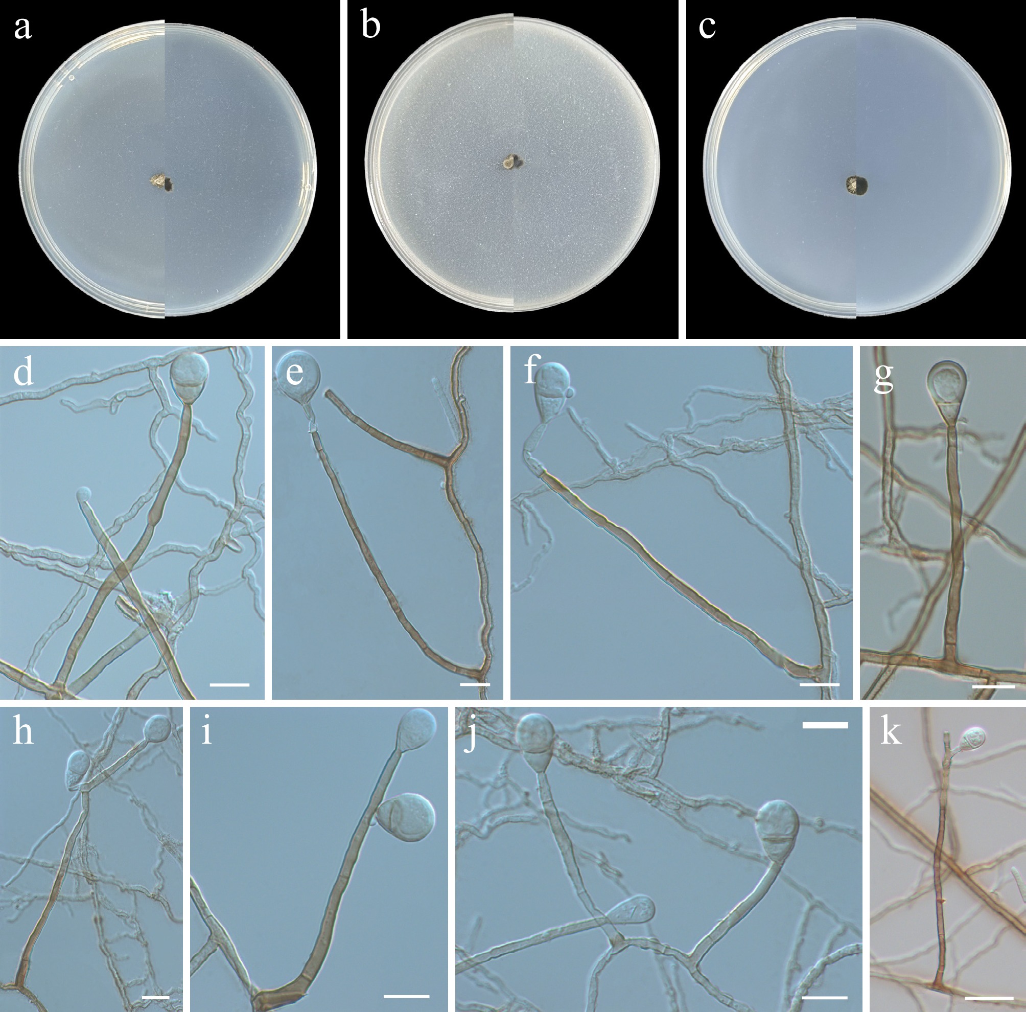
Figure 3.
Calyptellomyces sinensis (from holotype ZY H-23.004). (a)–(c) Upper and reverse views of cultures on PDA, OA, and SNA after 14 d at 25 °C. (d)–(k) Conidiophores, conidiogenous cells, and conidia. Scale bars: (d)–(j) 10 μm, (k) 20 μm.
MycoBank number: MB862608
Etymology: Named after the type locality, China.
Description: Hyphae branched, septate, hyaline to pale brown, smooth-walled, 1–3 μm wide. Asexual morph Conidiophores solitary, erect, hyaline to brown, smooth, subcylindrical to cylindrical, one-to-six-septate, 8.5–96 µm. Conidiogenous cells with one apical conidiogenous locus, rarely one at each end, hyaline to brown, smooth, subcylindrical to cylindrical, 3.5–10 × 1.5–3.5 µm (av. 7.3 × 2.3). Conidia solitary, hyaline to pale brown, elliptical to subglobose, smooth, zero-to-one-septate, cup-shaped at the lower part, hemispherical to campanulate at the upper part, 10–19.5 × 6–14 µm (av. 14.4 × 8.9). Ramoconidia: not observed. Sexual morph: not observed.
Culture characteristics (14 d at 25 °C): Colony on PDA reaching 4–6 mm diam., felty, margin slightly undulate, olive to black, aerial mycelia sparse, reverse black. Colony on OA reaching 3–5 mm diam., raised at center, felty to pulverulent, margin slightly undulate, olive to black, aerial mycelia sparse, reverse black. Colony on SNA reaching 5–6 mm diam., flat, felty, margin entire, black, aerial mycelia sparse, reverse black.
Material examined: China, Heilongjiang Province, Harbin City, soil collected from the green belts of Harbin Northern Forest Zoo, 127.105° E, 45.3952° N, 8 May 2023, Zhi-Yuan Zhang (holotype ZY H-23.004, dried culture; culture ex-type CGMCC 3.29783, ibid., ZY 23.004). China, Heilongjiang Province, Harbin City, soil collected from the green belts of Harbin Northern Forest Zoo, 127.1131° E, 45.3912° N, 8 May 2023, Zhi-Yuan Zhang, isotype ZY H-23.005, ex-isotype ZY 23.005, ibid., ZY 23.006, ibid., 23.007.
Pleosporales Luttrell ex M.E. Barr
Pleosporales, erected by Luttrell[44], encompasses saprotrophic, endophytic, pathogenic, parasitic, hyperparasitic fungi, as well as epiphytes, and exhibits cosmopolitan distribution[45−47]. This order is characterized by perithecioid ascomata with papillate apices; bitunicate asci; pseudoparaphyses; and ascospores that are mostly septate, variable in shape, pigmented, and either surrounded by a gelatinous sheath or lacking one[45,46,48−51]. Recent studies indicate that Pleosporales comprises 94 families, accounting for one quarter of all reported taxa within the Dothideomycetes, thereby representing the most species-rich order in this class[4].
Phylogenetic relationships within Pleosporales were inferred using a multi-locus (SSU, ITS, LSU, TEF1, RPB2, and TUB) dataset, with Hysterium angustatum (CBS 236.34) and H. rhizophorae (MFLUCC 15-0950) designated as the outgroup. The final alignment included 106 taxa and spanned 4,471 nucleotide positions (SSU, 1,018 bp; ITS, 498 bp; LSU, 830 bp; TEF1, 886 bp; RPB2, 896 bp; and TUB, 343 bp), including gaps. The best-fit models were TIM2+F+I+G4 (SSU), TIM3 + F + I + G4 (TEF1), and GTR + F + I + G4 (ITS, LSU, RPB2, and TUB) for the ML analysis, whereas GTR + F + I + G4 was applied across all six loci for BI analysis. Phylogenetically, CGMCC 3.29786, ZY 23.014, ZY 23.015, and ZY 23.016 formed a single, well-supported clade (100/1, Fig. 4) that was clearly separated from other families. Furthermore, another group of our new isolates (CGMCC 3.29785, ZY 23.011–23.012) formed a distinct subclade nested within the Pyrenochaetopsidaceae and was closely related to P. dalbergiae.
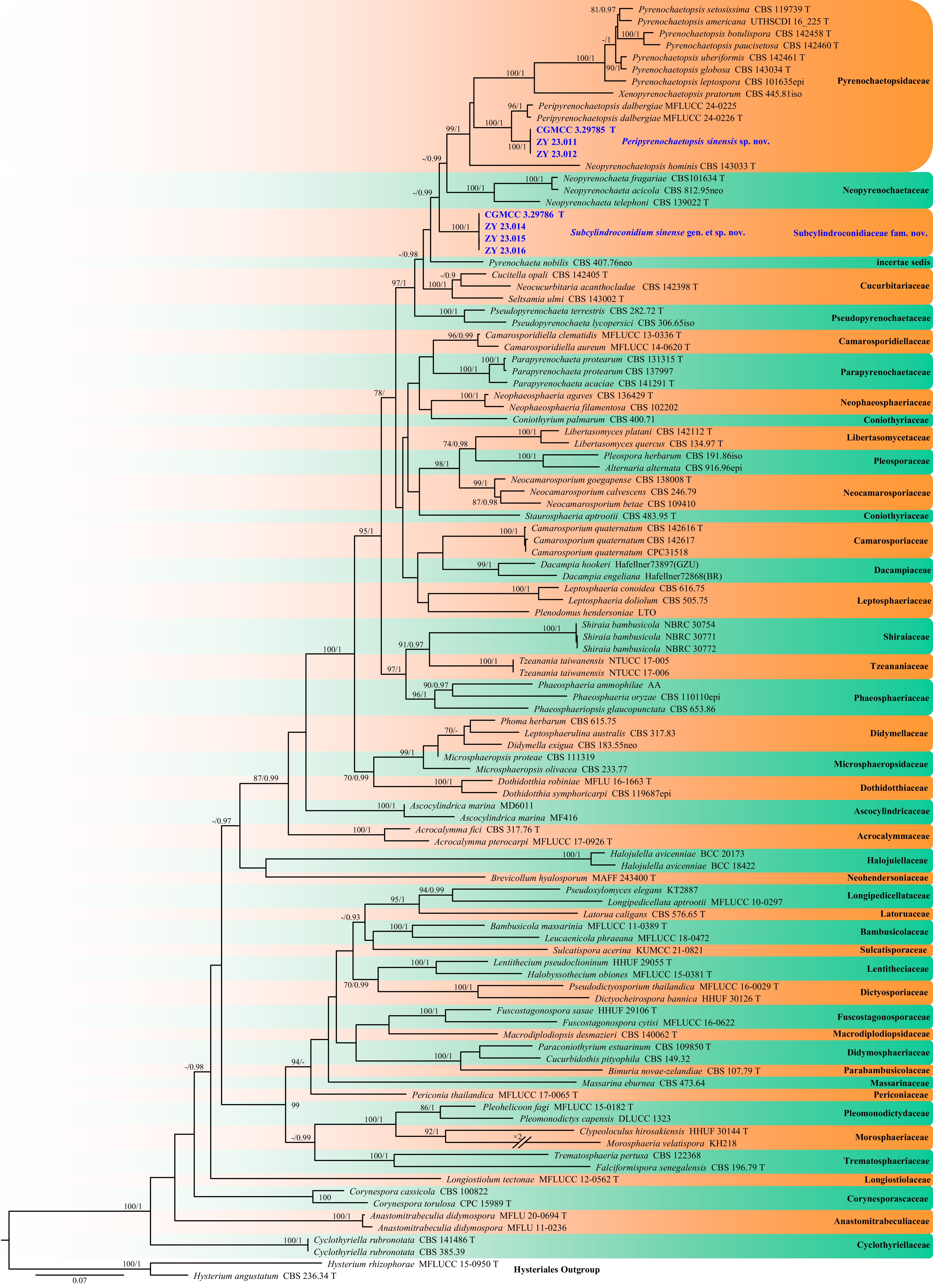
Figure 4.
Phylogram generated from ML analysis based on the combined dataset of SSU, ITS, LSU, TEF1, RPB2, and TUB sequence data. Numbers at branches indicate support values (IQ-TREE-BS/BI-PP) above 70%/0.90. The tree is rooted to Hysterium angustatum (CBS 236.34) and H. rhizophorae (MFLUCC 15-0950). Newly generated sequences are in bold and blue, and 'T' indicates type-derived sequences. The scale bar represents the expected number of changes per site.
Pyrenochaetopsidaceae Valenz.-Lopez, Crous, Cano, Guarro & Stchigel
Pyrenochaetopsidaceae was proposed as a new family by Valenzuela-Lopez et al., designating Pyrenochaetopsis as its type genus[52]. This family was established to accommodate two novel genera harbouring Pyrenochaeta species that resolved outside Cucurbitariaceae in phylogenetic analyses. The family comprises four genera: Neopyrenochaetopsis, Xenopyrenochaetopsis, Pyrenochaetopsis, and Peripyrenochaetopsis[4,52,53].
Peripyrenochaetopsis Samarad., Lumyong & K.D. Hyde
Samaradiwakara et al. established the genus Peripyrenochaetopsis, designating P. dalbergiae as its type species[53]; this remains the only valid species currently recognized in this genus. This species is saprobic on dead leaves of Dalbergia cultrata.
Peripyrenochaetopsis sinensis Y.J. Wu, Zhi Y. Zhang & Y.F. Han sp. nov. Fig. 5
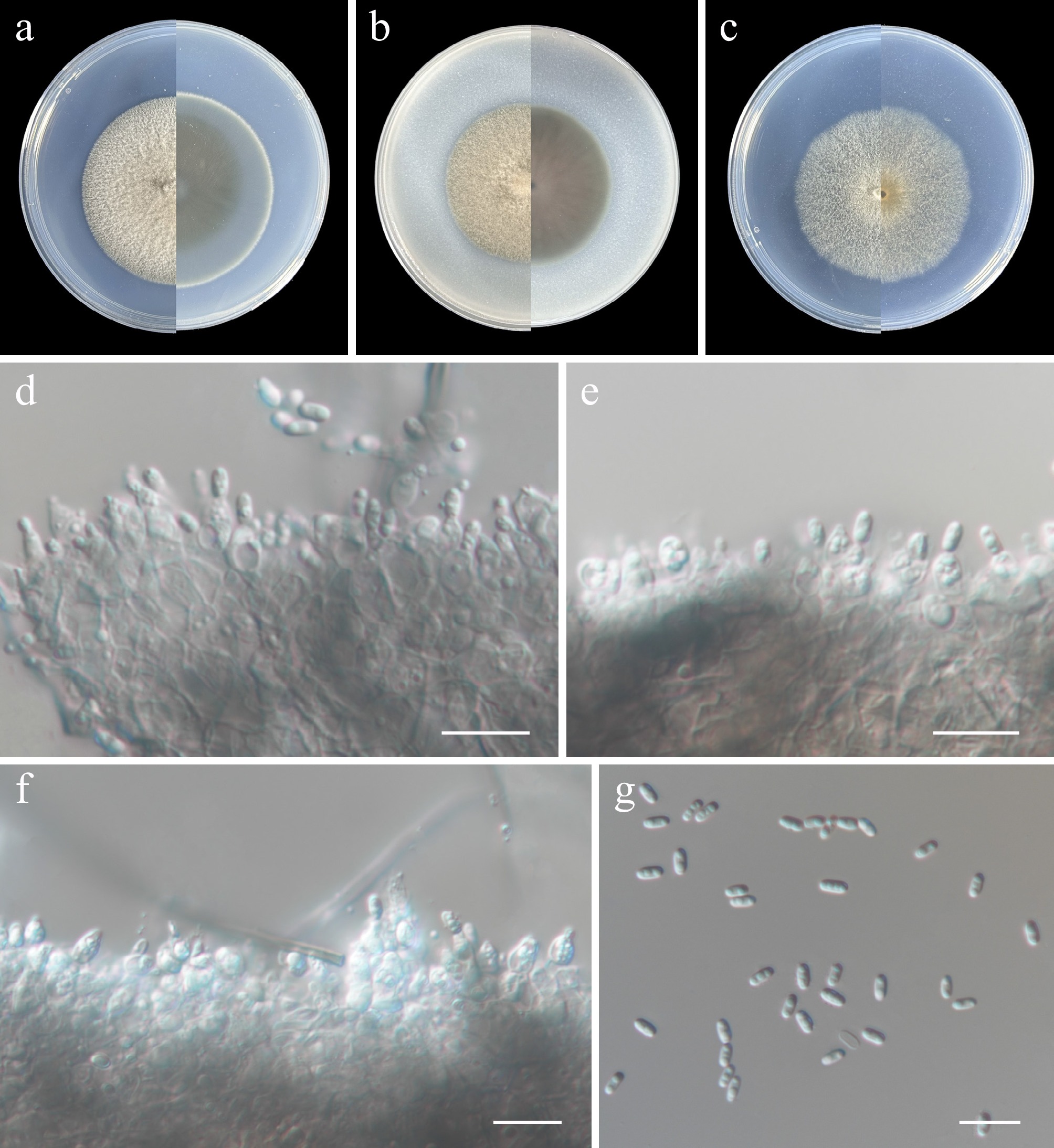
Figure 5.
Peripyrenochaetopsis sinensis (from holotype ZY H-23.010). (a)–(c) Upper and reverse views of cultures on PDA, OA, and SNA after 14 d at 25 °C. (d)–(f) Conidiogenous cells, conidia, and conidiomatal wall. (g) Conidia. Scale bars: (d)–(g) 10 μm.
MycoBank number: MB862609
Etymology: Named after the type locality, China.
Description: Hyphae branched, septate, hyaline to light brown, thick- and smooth-walled, 1–3 μm wide. Asexual morph sporulated on PDA, OA, and SNA. Conidiomata, pycnidial, solitary, semi-immersed to immersed, subglobose to ovoid, light brown to dark brick, glabrous, covered with mycelium, elongated with a short, semi-papillate, cylindrical neck. Conidiomatal wall 3.5–13.5 μm wide, pseudoparenchymatous, composed of four to five layers of light brown cells of textura angularis. Conidiogenous cells phialidic, discrete or integrated, 3.5–6.5 × 2.5–3.5 μm (av. 5.2 × 3.2 μm, n = 30), ampulliform, subcylindrical, hyaline. Conidia aseptate, hyaline, cylindrical to allantoid, guttulate, thin- and smooth-walled, 2.5–4.5 × 1–2 μm (av. 3.5 × 1.7 μm, n = 30). Sexual morph not observed.
Culture characteristics (14 d at 25 °C): Colony on PDA reaching 55–56 mm diam., raised at the centre, cottony, circular, margin regular, papyrus white; reverse concrete grey in the center, papyrus white at the edges. Colony on OA reaching 46–47 mm diam., raised at the centre, cottony, circular, margin regular, mouse grey; reverse pastel violet. Colony on SNA reaching 51–53 mm diam., cottony, circular, margin regular, grey white; reverse grey white.
Material examined: China, Heilongjiang Province, Harbin City, soil collected from the green belts of Harbin Northern Forest Zoo, 127.1036° E, 45.3966° N, 8 May 2023, Zhi-Yuan Zhang (holotype ZY H-23.010, dried culture; culture ex-type CGMCC 3.29785, ibid., ZY 23.010). China, Heilongjiang Province, Harbin City, soil collected from the green belts of Harbin Northern Forest Zoo, 127.1027° E, 45.3975° N, 8 May 2023, Zhi-Yuan Zhang, isotype ZY H-23.011, ex-isotype ZY 23.011, ibid., ZY 23.012.
Notes: A novel saprobic taxon, Peripyrenochaetopsis sinensis, was isolated from soil in China. In P. sinensis, the conidiogenous cells are ampulliform to subcylindrical, 3.5–6.5 × 2.5–3.5 μm, and the conidia are guttulate. In contrast, P. dalbergiae has subglobose to ampulliform conidiogenous cells, 2–4.5 × 2–4 μm, and its conidia are mostly eguttulate[53]. Additionally, pairwise comparison of ITS and TEF1 loci reveals that P. sinensis (ex-type CGMCC 3.88888) differs from P. dalbergiae (ex-type MFLUCC 24-0226) by 4.2% (22/522 bp, three gaps) in ITS and 3% (28/928 bp, no gaps) in TEF1.
Subcylindroconidiaceae Y.J. Wu, Zhi Y. Zhang & Y.F. Han fam. nov.
MycoBank number: MB862610
Etymology: Derived from the subcylindro- (subcylindrical) and conidium (conidia), referring to the subcylindrical conidiogenous cells.
Description: Hyphae branched, septate. Asexual morph Conidiomata pycnidial, pale brown to brown, solitary, setose, globose to subglobose, papillate, ostiolate. Conidiophores cylindrical to subcylindrical, aseptate. Conidiogenous cells phialidic, solitary, rarely two borne on the same conidiophore, subcylindrical to cylindrical, tapering towards the apex. Conidia solitary, aseptate, cylindrical. Sexual morph not observed.
Type: Subcylindroconidium Y.J. Wu, Zhi Y. Zhang & Y.F. Han
Notes: A new family, Subcylindroconidiaceae, is erected here to accommodate the monotypic genus Subcylindroconidium. The genus is diagnosed by pycnidial, solitary, setose, globose to subglobose, papillate, and ostiolate conidiomata; cylindrical to subcylindrical and aseptate conidiophores; phialidic conidiogenous cells that are solitary (rarely two borne on the same conidiophore), subcylindrical to cylindrical, and tapering towards the apex; and solitary, aseptate, cylindrical conidia. These characteristics resemble those of Cucurbitariaceae, Neopyrenochaetaceae, Pseudopyrenochaetaceae, and Pyrenochaetopsidaceae within the Pleosporales. However, Subcylindroconidiaceae is phylogenetically distinct and was initially identified based on its asexual state. Multi-locus (SSU, ITS, LSU, TEF1, RPB2, and TUB) phylogenetic analyses placed Subcylindroconidiaceae between Cucurbitariaceae and Neopyrenochaetaceae (Fig. 4). In this study, abundant sporulation of the asexual state was noted on PDA, OA, and SNA media, yet the sexual morph could not be obtained in culture. Therefore, further collections and studies are needed to establish the sexual-asexual relationship for Subcylindroconidiaceae.
Subcylindroconidium Y.J. Wu, Zhi Y. Zhang & Y.F. Han gen. nov.
MycoBank number: MB862611
Etymology: Refers to the subcylindrical conidiogenous cells.
Description: Hyphae branched, septate, pale brown to hyaline. Asexual morph Conidiomata pycnidial, pale brown to brown, solitary, setose, globose to subglobose, papillate, ostiolate. Conidiophores aseptate, hyaline, cylindrical to subcylindrical. Conidiogenous cells phialidic, solitary, hyaline, rarely two borne on the same conidiophore, subcylindrical to cylindrical, tapering towards the apex. Conidia solitary, aseptate, hyaline, cylindrical. Sexual morph not observed.
Type: Subcylindroconidium sinense Y.J. Wu, Zhi Y. Zhang & Y.F. Han
Subcylindroconidium sinense Y.J. Wu, Zhi Y. Zhang & Y.F. Han sp. nov. Fig. 6
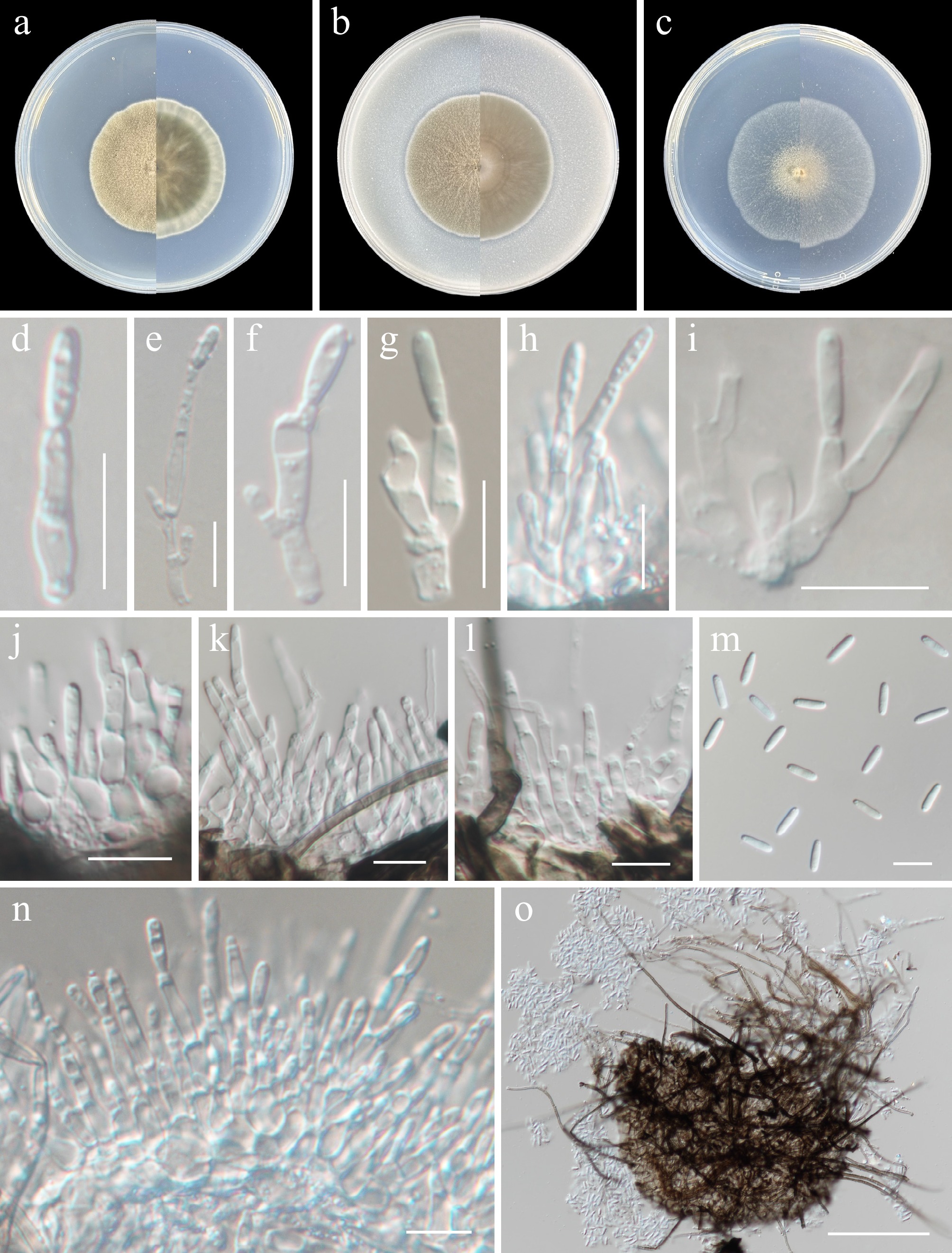
Figure 6.
Subcylindroconidium sinense (from holotype ZY H-23.013). (a)–(c) Upper and reverse views of cultures on PDA, OA, and SNA after 14 d at 25 °C. (d)–(l), (n) Conidiogenous cells. (m) Conidia. (o) Pycnidia. Scale bars: (d)–(g) 10 μm.
MycoBank number: MB862612
Etymology: Named after the type locality, China.
Description: Hyphae branched, septate, pale brown to hyaline, 1–3 μm wide. Asexual morph Conidiomata pycnidial, pale brown to brown, solitary, setose, globose to subglobose, papillate, ostiolate. Conidiophores cylindrical to subcylindrical, aseptate, hyaline. Conidiogenous cells phialidic, hyaline, solitary, rarely two borne on the same conidiophore, subcylindrical to cylindrical, tapering towards the apex, 9.5–16 × 1.5–3 μm. Conidia solitary, aseptate, hyaline, cylindrical, 7–9 × 1.5–2.5 μm (av. 8.2 × 2.2, n = 30 μm). Sexual morph not observed.
Culture characteristics (14 d at 25 °C): Colony on PDA reaching 45–46 mm diam., cottony, circular, margin regular, green beige to light ivory; reverse grey white to papyrus white. Colony on OA reaching 45–47 mm diam., cottony, circular, margin regular, dark ivory; reverse mouse grey. Colony on SNA reaching 48–51 mm diam., cottony, circular, margin regular, grey white; reverse grey white.
Material examined: China, Heilongjiang Province, Harbin City, soil collected from the green belts of Harbin Northern Forest Zoo, 127.1193° E, 45.3903° N, 8 May 2023, Zhi-Yuan Zhang (holotype ZY H-23.013, dried culture; culture ex-type CGMCC 3.29786, ibid., ZY 23.013). China, Heilongjiang Province, Harbin City, soil collected from the green belts of Harbin Northern Forest Zoo, 127.1136° E, 45.3893° N, 8 May 2023, Zhi-Yuan Zhang, isotype ZY H-23.014, ex-isotype ZY 23.014, ibid., ZY 23.015, ibid., ZY 23.016.
Notes: A megablast search against the NCBI GenBank nucleotide database revealed that the closest hits for the ITS sequence corresponded to an uncultured fungus [clone CMH429, GenBank KF800520.1; Identities = 514/525 (98%), one gap (0%)] and uncultured Pleosporales [clone SW_2d_H02, GenBank JF449840.1; Identities = 495/502 (99%), one gap (0%)]. For the TEF1 sequence, the closest hit was Phaeosphaeriaceae sp. [isolate PN33, GenBank ON952520.1; Identities = 870/898 (97%), no gap (0%)]. In contrast, only distant matches were retrieved for the RPB2 sequence via a blastn, with the highest similarity observed to Neopyrenochaeta cercidis [GenBank MK434908.1; 900/1021 (88%), no gaps (0%)]. For the TUB sequence, the closest hit corresponded to Neopyrenochaeta acicola [isolate PN31, GenBank ON866492.1; Identities = 587/651 (90%), seven gaps (1%)].
Sporormiaceae Munk
Westerdykella Stolk
Stolk established the genus Westerdykella to accommodate W. ornata, which was isolated from soil in Mozambique[54]. To date, Westerdykella comprises 15 species, all of which have molecular sequences available[55]. The majority of these species were originally described only from their sexual morph, whereas W. dispersa is unusual in producing both sexual and asexual morphs in the same culture medium[56]. The asexual morph of Westerdykella is characterized by superficial or submerged, globose to subglobose or irregular-elongate, and olive to olive-black, or brown to dark brown conidiomata with an ostiole. The conidia are hyaline, globose to oval or pyriform, and borne on simple, short conidiophores[57−59].
Based on the molecular data used by Cheng et al.[55], the phylogenetic dataset for this study was constructed by incorporating data from the proposed new species. Phylogenetic relationships of the genus Westerdykella were inferred using a multi-locus (ITS, LSU, and TUB) dataset, with Preussia funiculata (Huhndorf 2577) and P. typharum (CBS 107.69) designated as the outgroup. The final alignment included 59 taxa and spanned 2,238 nucleotide positions (ITS, 433 bp; LSU, 854 bp; and TUB, 951 bp), including gaps. The best-fit models were GTR + I + G4 (ITS) and TIM2 + F + I + G4 (LSU, TUB) for the ML analysis, whereas those for the BI analysis were SYM + I + G4 (ITS) and GTR + F + I + G4 (LSU, TUB). Phylogenetically, CGMCC 3.29787, ZY 23.018, and ZY 23.019 formed a single, well-supported clade (100/1, Fig. 7).
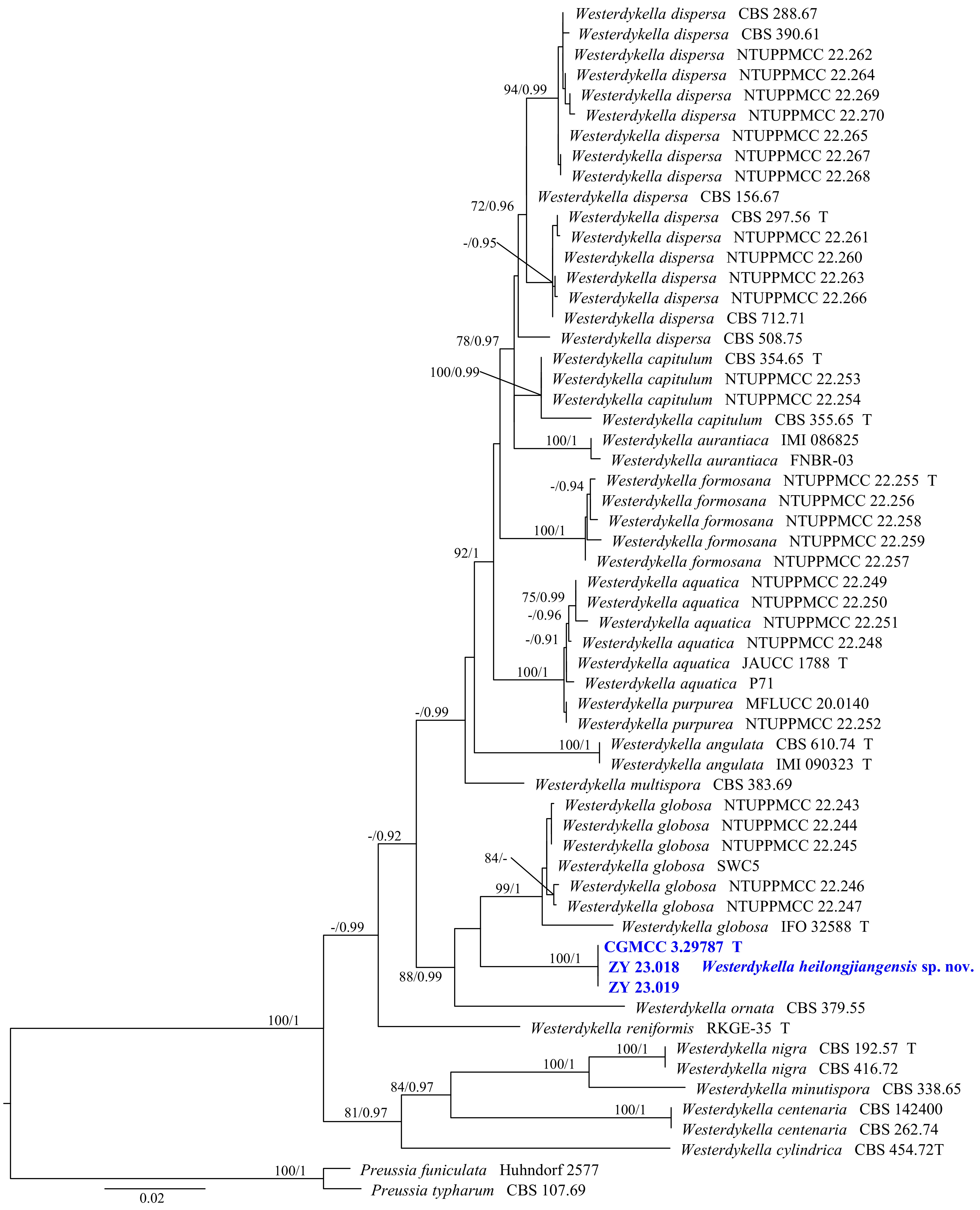
Figure 7.
Phylogram generated from ML analysis based on the combined dataset of ITS, LSU, and TUB sequence data. Numbers at branches indicate support values (IQ-TREE-BS/BI-PP) above 70%/0.90. The tree is rooted to Preussia funiculata (Huhndorf 2577) and P. typharum (CBS 107.69). Newly generated sequences are in bold and blue, and 'T' indicates type-derived sequences. The scale bar represents the expected number of changes per site.
Westerdykella heilongjiangensis Y.J. Wu, Zhi Y. Zhang & Y.F. Han sp. nov. Fig. 8
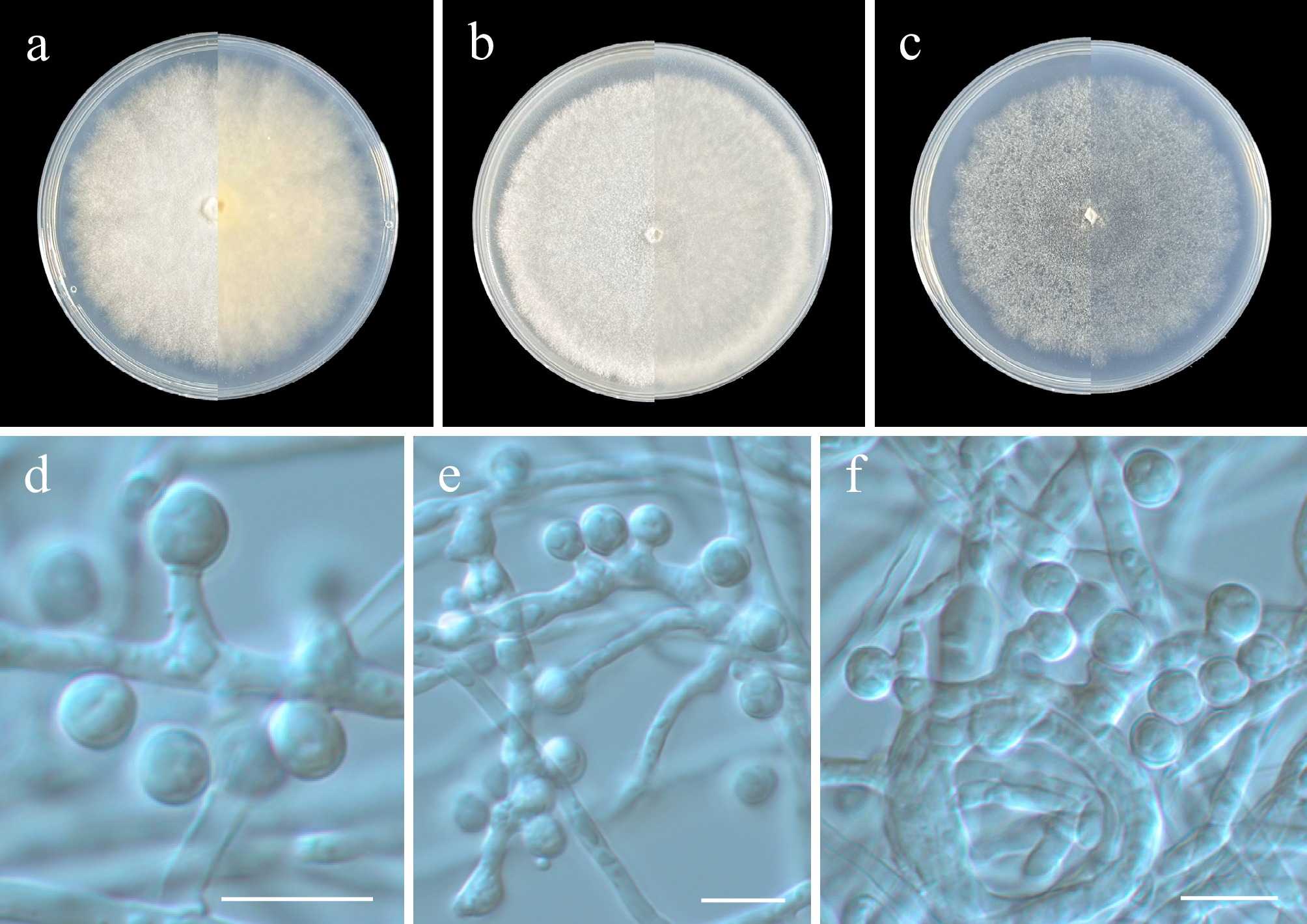
Figure 8.
Westerdykella heilongjiangensis (from holotype ZY H-23.017). (a)–(c) Upper and reverse views of cultures on PDA, OA, and SNA after 14 d at 25 °C. (d)–(f) Conidiogenous cells and conidia. Scale bars: (d)-(f) 10 μm.
MycoBank number: MB862613
Etymology: Named after the type locality, Heilongjiang Province, China.
Description: Hyphae branched, septate, hyaline, smooth and thin-walled, 1–2.5 µm wide. Asexual morph Conidiophores simple, hyaline, smooth, often reduced to conidiogenous cells. Conidiogenous cells simple, hyaline, smooth, subcylindrical to cylindrical, arising from hyphae or hyphal coils. Conidia hyaline, aseptate, smooth, borne on short protrusions or conidiogenous cells, subglobose to ellipsoidal, 4.5–7.5 × 4–6 µm (av. 5.8 × 5.1 µm, n = 30). Sexual morph not observed.
Culture characteristics (14 d at 25 °C): Colony on PDA reaching 79–81 mm diam., cottony, white, nearly circular, margin indistinct; reverse white. Colony on OA reaching 76–78 mm diam., cottony, white, nearly circular, margin indistinct; reverse white. Colony on SNA reaching 68–73 mm diam., felty, white, margin indistinct; reverse white.
Material examined: China, Heilongjiang Province, Harbin City, soil collected from the green belts of Harbin Northern Forest Zoo, 127.1050° E, 45.3952° N, 8 May 2023, Zhi-Yuan Zhang (holotype ZY H-23.017, dried culture; culture ex-type CGMCC 3.29787, ibid., ZY 23.017). China, Heilongjiang Province, Harbin City, soil collected from the green belts of Harbin Northern Forest Zoo, 127.1193° E, 45.3903° N, 8 May 2023, Zhi-Yuan Zhang, isotype ZY H-23.018, ex-isotype ZY 23.018, ibid., ZY 23.019.
Notes: Phylogenetically, Westerdykella heilongjiangensis formed a distinct subclade and was closely related to W. globosa and W. ornata (Fig. 7). Morphologically, since the sexual morph of W. heilongjiangensis is unknown, and the asexual morphs of W. globosa and W. ornata are unknown, we were unable to compare their morphological differences[54,55,60]. However, pairwise comparison of the ITS, LSU, and TUB loci reveals that W. heilongjiangensis (ex-type CGMCC 3.29787) differs from W. globosa (ex-type IFO 32588) by 5.4% (29/536 bp, eight gaps) in ITS, and differs from W. ornata (CBS 379.55) by 7.3% (34/461 bp, five gaps) in ITS, 2.5% (24/938 bp, 11 gaps) in LSU, and 6.2% (63/1006 bp, two gaps) in TUB.
Venturiales Y. Zhang ter, C.L. Schoch & K.D. Hyde
Sympoventuriaceae Y. Zhang ter, C.L. Schoch & K.D. Hyde
Scolecobasidium E.V. Abbott
Abbott established the genus Scolecobasidium to accommodate S. terreum, designated as the type species, and S. constrictum[61]. This genus is diagnosed by having creeping, septate hyphae; producing non-erect, aseptate conidiophores as short lateral branches from aerial hyphae; and forming elongate, two-celled, smooth, light olivaceous to nearly hyaline conidia that are borne singly on short, terminal, thread-like sterigmata, with one to three sterigmata per conidiophore[61]. Scolecobasidium is a large genus whose members exhibit diverse lifestyles as endophytes, saprophytes, or plant pathogens, and can also act as opportunistic pathogens in animals and humans[62].
Phylogenetic relationships within the genus Scolecobasidium were inferred using a multi-locus (SSU, ITS, LSU, TUB, and TEF1) dataset, with Guizhoumyces aciculaea (GUCC 18195) and Sympoventuria capensis (CBS 120136) designated as the outgroup. The final alignment included 67 taxa and spanned 3,433 nucleotide positions (SSU, 1,215 bp; ITS, 556 bp; LSU, 809 bp; TUB, 481 bp; and TEF1, 372 bp), including gaps. The best-fit models were TIM2 + F + I + G4 (SSU, LSU), TIM + F + I + G4 (ITS), GTR + F + I + G4 (TUB), and TPM2u + F + I + G4 (TEF1) for the ML analysis, whereas GTR + F + I + G4 was applied across all five loci for BI analysis. Phylogenetically, CGMCC 3.29788, ZY 23.021, and ZY 23.022 formed a single, well-supported clade (100/1, Fig. 9).
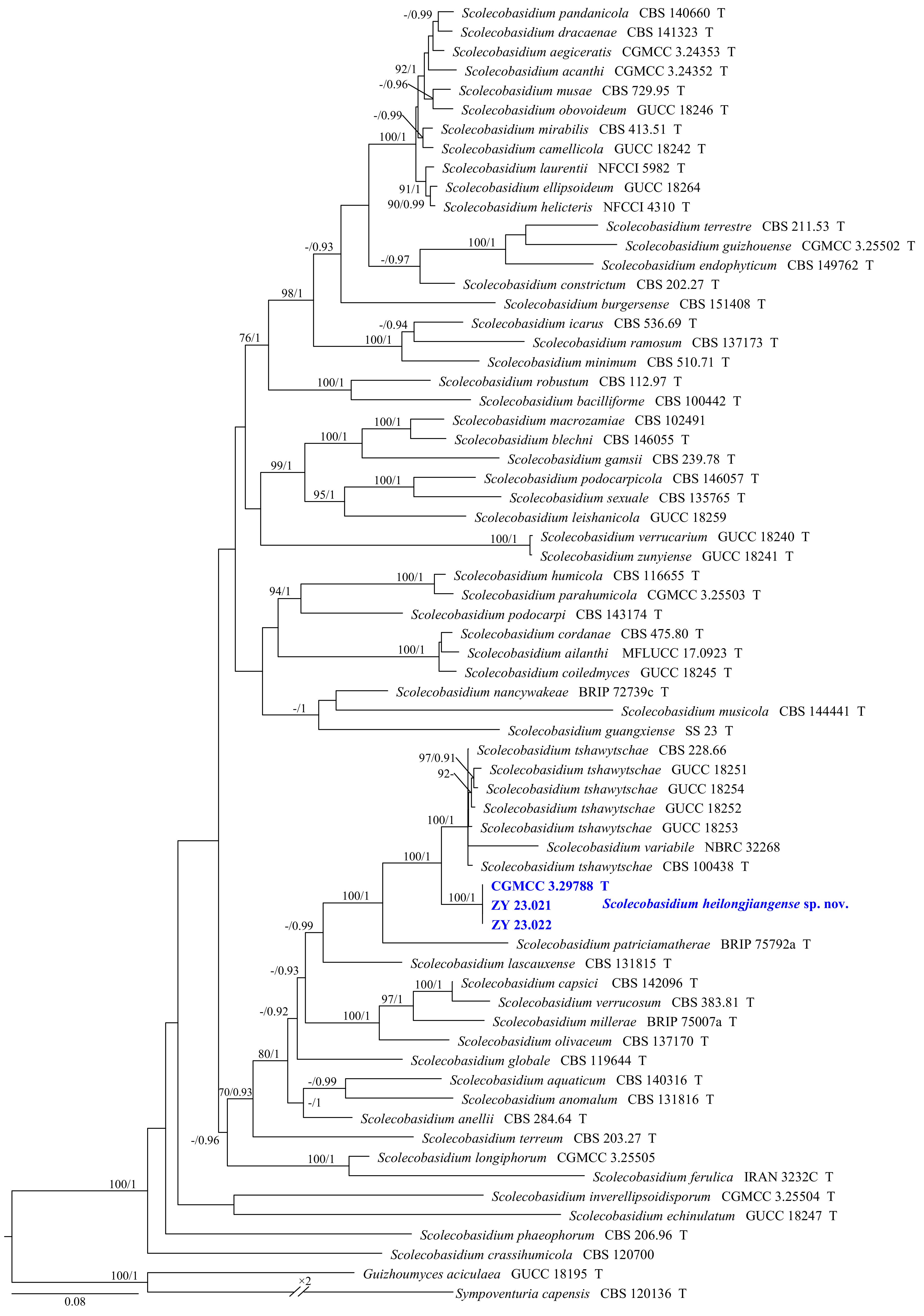
Figure 9.
Phylogram generated from ML analysis based on the combined dataset of SSU, ITS, LSU, TUB, and TEF1 sequence data. Numbers at branches indicate support values (IQ-TREE-BS/BI-PP) above 70%/0.90. The tree is rooted to Guizhoumyces aciculaea (GUCC 18195) and Sympoventuria capensis (CBS 120136). Newly generated sequences are in bold and blue, and 'T' indicates type-derived sequences. The scale bar represents the expected number of changes per site.
Scolecobasidium heilongjiangense Y.J. Wu, Zhi Y. Zhang & Y.F. Han sp. nov. Fig. 10
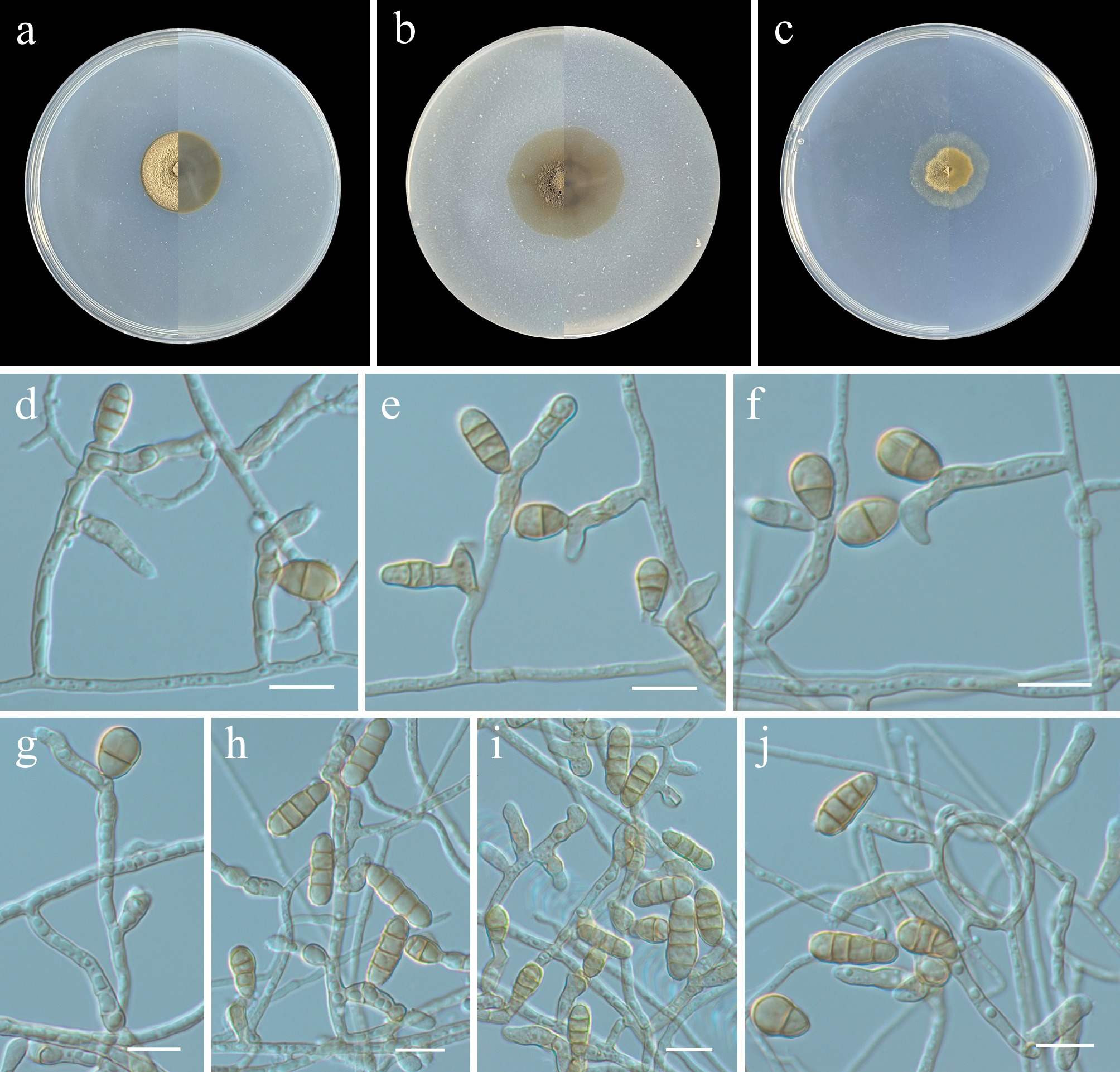
Figure 10.
Scolecobasidium heilongjiangense (from holotype ZY H-23.020). (a)–(c) Upper and reverse views of cultures on PDA, OA, and SNA after 14 d at 25 °C. (d)–(j) Conidiogenous cells and conidia. Scale bars: (d)–(j) 10 μm.
MycoBank number: MB862614
Etymology: Named after the type locality, Heilongjiang Province, China.
Description: Hyphae branched, septate, hyaline to pale brown, smooth and thin-walled, 1–3 µm wide. Asexual morph Conidiophores mononematous, terminal or intercalary and arising laterally from the hyphae or hyphal coils, simple or sparingly branched, straight or flexuous, 0–1-septate, smooth, thin-walled, cylindrical to narrowly clavate, hyaline, subhyaline or pale brown, (4–)6–19(–30) × 1.5–3.5 µm, often reduced to conidiogenous cells. Conidiogenous cells mono- or polyblastic, integrated, terminal or intercalary in conidiophores and hyphae, subcylindrical, clavate, or ampulliform, sympodial, straight or geniculate, with small shoulders resulting from sympodial extensions, aseptate, subhyaline to pale brown, 4.5–7.5 × 2.5–4 µm, frequently emerging directly from the hyphae. Conidial secession rhexolytic. Conidia acropleurogenous, solitary, subcylindrical with sometimes slightly swollen in the middle, obovoid, ellipsoidal, pale brown, minutely echinulate to verruculose, 1–3(–4)-septate, sometimes constricted at the septa, 6–16(–19) × 4–7 µm (av. 11.3 × 4.6 µm, n = 30). Chlamydospores and sexual morph not observed.
Culture characteristics (14 d at 25 °C): Colony on PDA reaching 23–24 mm diam., pale brown, slightly raised to umbonate, dense, margin regularly; reverse basalt grey. Colony on OA reaching 28–30 mm diam., flat, grey beige, aerial mycelia sparse, dense at centre, reverse grey beige. Colony on SNA reaching 20–22 mm diam., flocculent, grey beige, aerial mycelia sparse, dense at centre, reverse grey beige.
Material examined: China, Heilongjiang Province, Harbin City, soil collected from the green belts of Harbin Northern Forest Zoo, 127.1028° E, 45.3988° N, 8 May 2023, Zhi-Yuan Zhang (holotype ZY H-23.020, dried culture; culture ex-type CGMCC 3.29788, ibid., ZY 23.020). China, Heilongjiang Province, Harbin City, soil collected from the green belts of Harbin Northern Forest Zoo, 127.1027° E, 45.3975° N, 8 May 2023, Zhi-Yuan Zhang, isotype ZY H-23.021, ex-isotype ZY 23.021, ibid., ZY 23.022.
Notes: Phylogenetically, Scolecobasidium heilongjiangense formed a distinct subclade and was closely related to S. tshawytschae and S. patriciamatherae (Fig. 9). Morphologically, the conidiogenous cells of S. heilongjiangense are subcylindrical, clavate, or ampulliform, while those of S. tshawytschae are lageniform or ampulliform[62,63]. The conidia of S. heilongjiangense are subcylindrical, obovoid, or ellipsoidal, averaging 11.3 × 4.6 µm, whereas the conidia of S. tshawytschae are cylindrical or slightly clavate, averaging 16.4 × 5.2 µm[62,63]. Additionally, due to the lack of morphological characterization of S. patriciamatherae[64], we compared only its molecular sequence differences with those of S. heilongjiangense. Pairwise comparison of the ITS and LSU loci reveals that S. heilongjiangense (ex-type CGMCC 3.29458) differs from S. patriciamatherae (ex-type BRIP 75792a) by 28.8% (212/735 bp, 93 gaps) in ITS and 3.8% (35/904 bp, ten gaps) in LSU.
Class Eurotiomycetes O.E. Erikss. & Winka
Chaetothyriales M.E. Barr
Herpotrichiellaceae Munk
Exophiala J.W. Carmich.
Carmichael established the genus Exophiala, designating E. salmonis as its type species[65]. As a black fungal genus within Herpotrichiellaceae, Exophiala occupies a broad ecological range, occurring in substrates such as soil and water, and occasionally colonizing the human body as an opportunistic pathogen[66]. To date, the taxonomic structure of this family remains unresolved, which is primarily due to the polyphyletic nature of most of its genera[67,68], including Exophiala[69]. This study proposes one new species based on the taxonomic framework established by Torres-Garcia et al.[69].
Phylogenetic relationships of the genus Exophiala were inferred using a multi-locus (ITS, LSU, TUB, and TEF1) dataset, with Cladophialophora behniae (CBS 146975) and C. scillae (CBS 116461) designated as the outgroup. The final alignment included 61 taxa and spanned 1,917 nucleotide positions (ITS, 536 bp; LSU, 828 bp; TUB, 333 bp; and TEF1, 220 bp), including gaps. The best-fit models were TIM2 + F + I + G4 (ITS, LSU, and TUB) and TIM3e + I + G4 (TEF1) for the ML analysis, whereas those for the BI analysis were GTR + F + I + G4 (ITS, LSU, and TUB) and GTR + I + G4 (TEF1). Phylogenetically, CGMCC 3.29789 and ZY 23.024 formed a single, well-supported clade (100/1, Fig. 11).
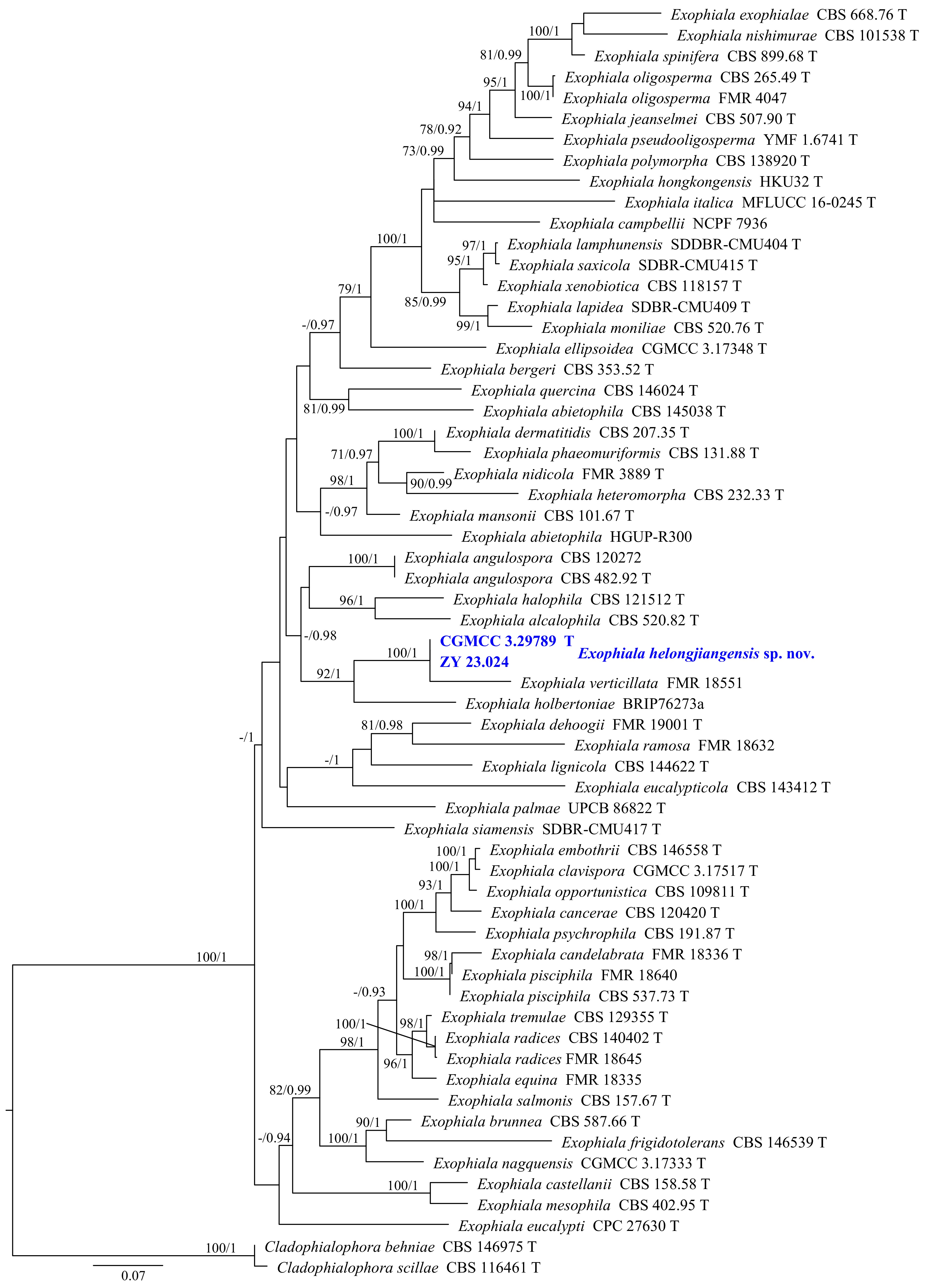
Figure 11.
Phylogram generated from ML analysis based on the combined dataset of ITS, LSU, TUB, and TEF1 sequence data. Numbers at branches indicate support values (IQ-TREE-BS/BI-PP) above 70%/0.90. The tree is rooted to Cladophialophora behniae (CBS 146975) and C. scillae (CBS 116461). Newly generated sequences are in bold and blue, and 'T' indicates type-derived sequences. The scale bar represents the expected number of changes per site.
Exophiala heilongjiangensis Y.J. Wu, Zhi Y. Zhang & Y.F. Han sp. nov. Fig. 12
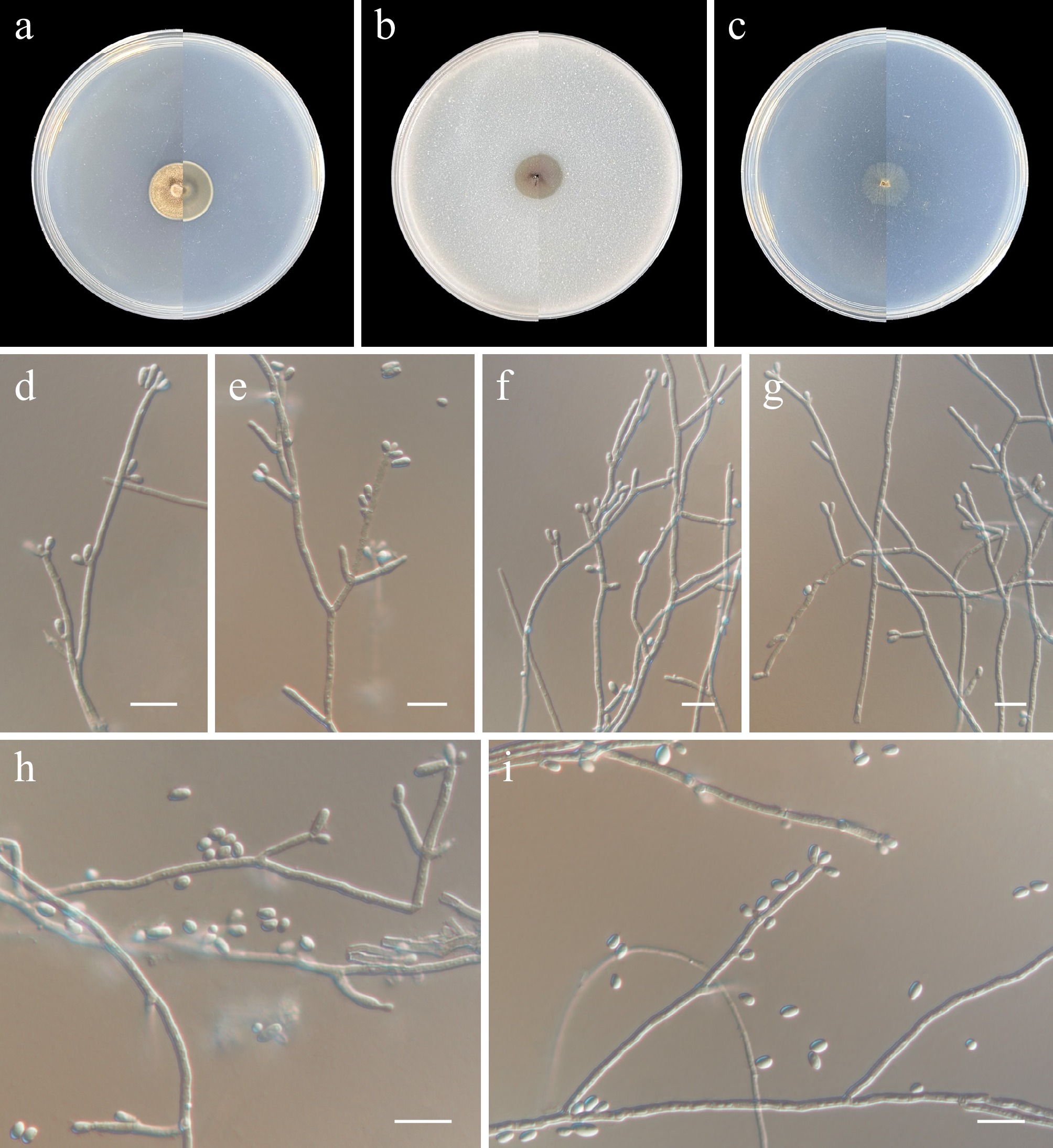
Figure 12.
Exophiala heilongjiangensis (from holotype ZY H-23.023). (a)–(c) Upper and reverse views of cultures on PDA, OA, and SNA after 14 d at 25 °C. (d)–(i) Conidiogenous cells and conidia. Scale bars: (d)–(i) 10 μm.
MycoBank number: MB862615
Etymology: Named after the type locality, Heilongjiang Province, China.
Description: Hyphae branched, septate, hyaline, smooth-walled, 1.5–2.5 µm wide. Asexual morph Conidiophores often reduced to conidiogenous cells growing directly or on short supporting cells from vegetative hyphae. Conidiogenous cells discrete, lateral or terminal, or intercalary, hyaline smooth-walled, cylindrical, slightly tapered towards the tip, with scars bearing conidia, inconspicuously sympodial, conidiogenous loci also inconspicuously annellidic, 5–16 × 1.5–2.5 µm. Conidia aseptate, hyaline, smooth-walled, obovoid to cylindrical, straight or slightly curved, 2.5–7 × 1.5–2.5 µm (av. 4.4 × 1.9 µm, n = 30), often with a narrow, truncate basal scar, forming slimy heads on the conidiogenous loci. Chlamydospores and sexual morph not observed.
Culture characteristics (14 d at 25 °C): Colony on PDA reaching 17–19 mm diam., flat, pale grey, margin entire; reverse cream-whitish. Colony on OA reaching 12–14 mm diam., flat, velvety, grass green, margin entire; reverse grass green. Colony on SNA reaching 13–15 mm diam., flat, pale, aerial hyphae sparse, margin entire; reverse pale.
Material examined: China, Heilongjiang Province, Harbin City, soil collected from the green belts of Harbin Northern Forest Zoo, 127.1051° E, 45.3977° N, 8 May 2023, Zhi-Yuan Zhang (holotype ZY H-23.023, dried culture; culture ex-type CGMCC 3.29789, ibid., ZY 23.023). China, Heilongjiang Province, Harbin City, soil collected from the green belts of Harbin Northern Forest Zoo, 127.1136° E, 45.3893° N, 8 May 2023, Zhi-Yuan Zhang, isotype ZY H-23.024, ex-isotype ZY 23.024.
Notes: Phylogenetically, Exophiala heilongjiangensis formed a distinct subclade and was closely related to E. verticillata (Fig. 11). Morphologically, E. heilongjiangensis is distinguished from E. verticillata by having conidiophores reduced to conidiogenous cells, and by the shape and size of its conidia (obovoid to cylindrical, 2.5–7 × 1.5–2.5 µm in E. heilongjiangensis vs guttulate, ellipsoidal to broadly ellipsoidal or subcylindrical, 1.5–4 × 0.5–2.5 µm in E. verticillata)[69].
Eurotiales G.W. Martin ex Benny & Kimbr.
Aspergillaceae Link
Penicillium Link
Recent studies have shown that Penicillium consists of the subgenera Aspergilloides and Penicillium, with subgenus Penicillium including 13 sections and 39 series, and the subgenus Aspergilloides including 19 sections and 64 series[70].
Phylogenetic relationships of the genus Penicillium were inferred using a multi-locus (ITS, TUB, CaM, and RPB2) dataset, with Talaromyces longistipes (CGMCC 3.25509) and T. parapalmae (CGMCC 3.25510) designated as the outgroup. The final alignment included 78 taxa and spanned 2,132 nucleotide positions (ITS, 471 bp; TUB, 362 bp; CaM, 454 bp; and RPB2, 845 bp), including gaps. The best-fit models were TN + I + G4 (TUB), GTR + F + I + G4 (CaM and ITS), and TIM2 + F + I + G4 (RPB2) for the ML analysis, whereas those for the BI analysis were GTR + I + G4 (TUB) and GTR + F + I + G4 (CaM, ITS, and RPB2). Phylogenetically, our isolates (CGMCC 3.29790, ZY 23.026–23.027) and (CGMCC 3.29791, ZY 23.029) formed distinct subclades with high support values (100/1), respectively, and were separated from other species within Penicillium (Fig. 13).
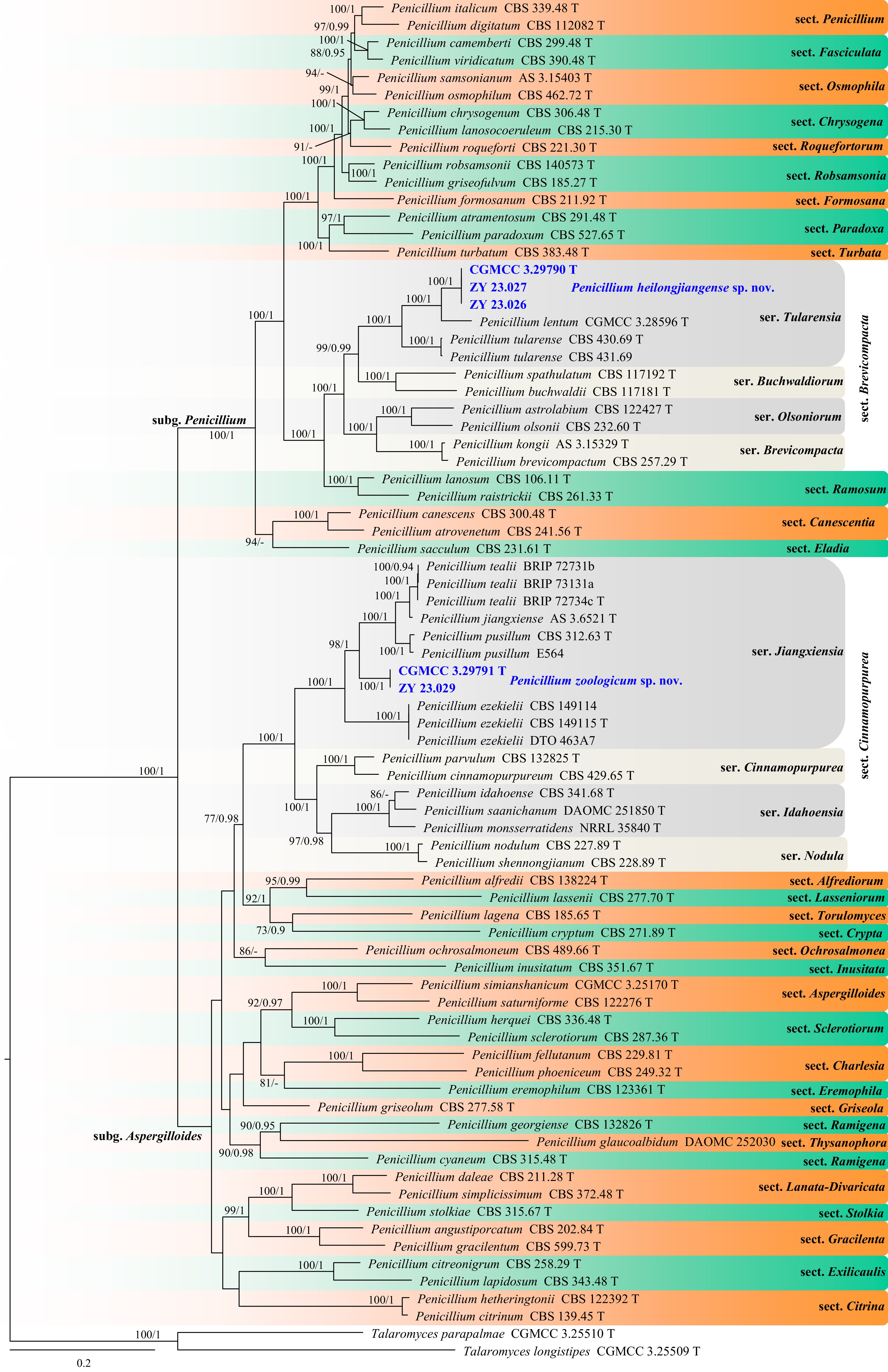
Figure 13.
Phylogram generated from ML analysis based on the combined dataset of TUB, CaM, ITS, and RPB2 sequence data. Numbers at branches indicate support values (IQ-TREE-BS/BI-PP) above 70%/0.90. The tree is rooted to Talaromyces longistipes (CGMCC 3.25509) and T. parapalmae (CGMCC 3.25510). Newly generated sequences are in bold and blue, and 'T' indicates type-derived sequences. The scale bar represents the expected number of changes per site.
Penicillium heilongjiangense Y.J. Wu, Zhi Y. Zhang & Y.F. Han sp. nov. Fig. 14
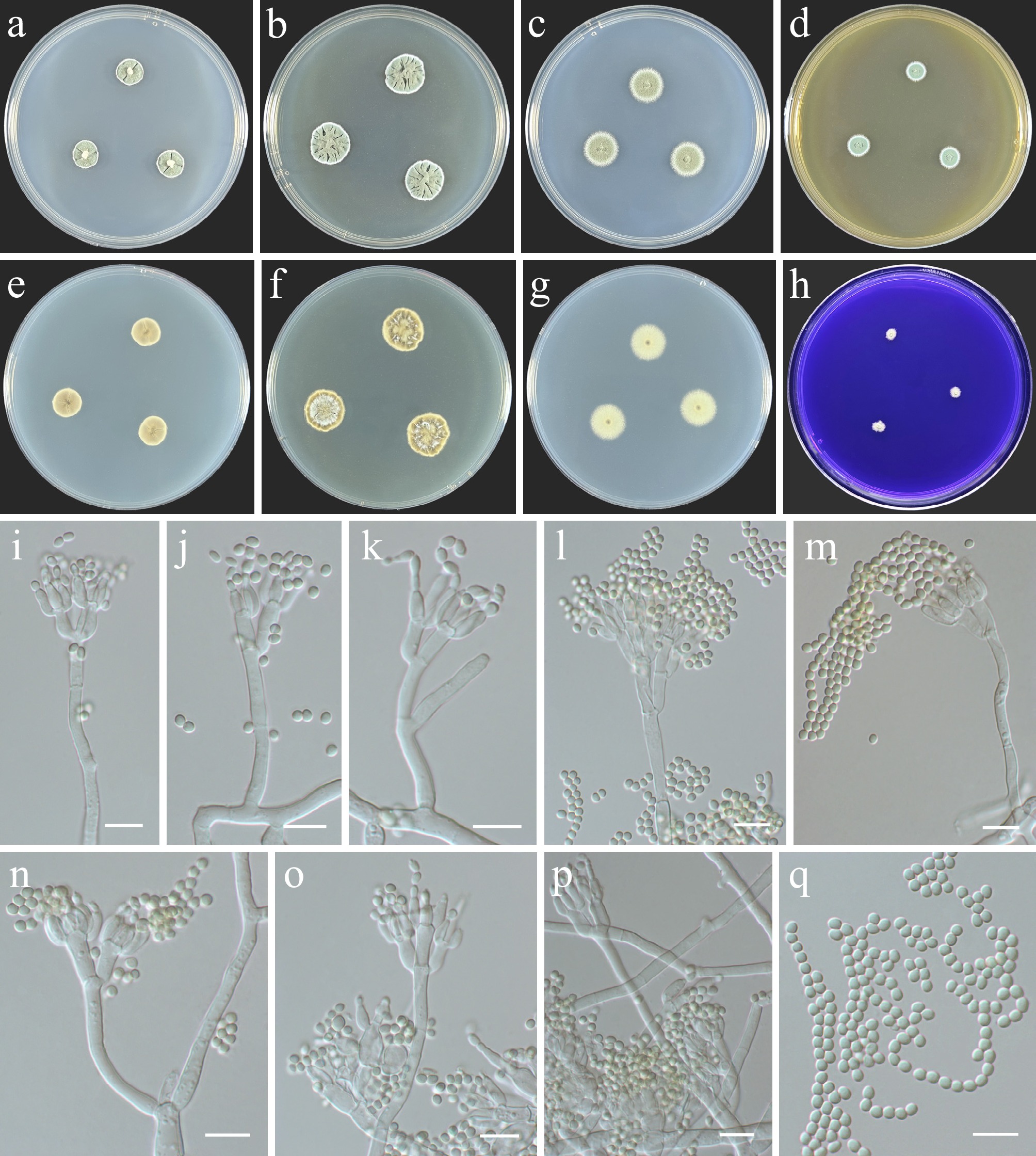
Figure 14.
Penicillium heilongjiangense (from holotype ZY H-23.025). (a)–(h) Colonies on medium at 25 °C for 7 d (left to right, top row: CYA, YES, DG18, and MEA obverse; second row: CYA reverse, YES reverse, DG18 reverse, and CREA obverse). (i)–(p) Conidiophores and conidia. (q) Conidia. Scale bars: (i)–(q) 10 μm.
MycoBank number: MB862616
Infrageneric classification. Subgenus Penicillium, section Brevicompacta, series Tularensia.
Etymology: Named after the type locality, Heilongjiang Province, China.
Colony diameter (7 d, in mm): CYA 10–14; CYA 30 °C, 37 °C no growth; MEA 8–9; YES 14–16; DG18 12–14; CREA 4–5.
Colony characteristics (7 d). CYA at 25 °C: Colonies flat with elevated grooves, radially sulcate; oval to circular; mycelium white; powdery, floccose areas present; sporulation good, conidia antique green; exudate absent; reverse green beige; soluble pigment absent. MEA at 25 °C: Colonies low, plane, wide, entire; mycelium white; powdery, margins floccose; sporulation good, conidia bluish gray-green; exudate absent; reverse zinc yellow; soluble pigment absent. YES at 25 °C: Colonies deep, radially sulcate, raised at center, margins low, irregular; mycelium white; powdery; sporulation good, conidia glaucous-green; exudate absent; reverse papyrus white to green beige; soluble pigment absent. DG18 at 25 °C: Colonies low, plane, margins low, wide, entire; mycelium white; texture velvety to slightly floccose; sporulation good, conidia bluish gray-green; exudate absent; reverse oyster white; soluble pigment absent. CREA at 25 °C: Weak growth, no acid production.
Description: Hyphae branched, septate, hyaline, smooth, 1.5–6 μm diam. Asexual morph Sclerotia absent. Conidiophores biverticillate, up to 99 μm long, stipes vesiculate up to 4 μm diam. Metulae divergent, two to six per stipe, 6–13 × 3.5–5.5 μm. Phialides ampulliform, two to six per metula, 5–13 × 2.5–4 μm. Conidia elongated ellipsoidal to subglobose, smooth-walled, 2–3 × 1.5–2.5 μm (av. 2.7 × 2.3 µm, n = 30). Sexual morph not observed.
Material examined: China, Heilongjiang Province, Harbin City, soil collected from the green belts of Harbin Northern Forest Zoo, 127.1003° E, 45.3992° N, 8 May 2023, Zhi-Yuan Zhang (holotype ZY H-23.025, dried culture; culture ex-type CGMCC 3.29790, ibid., ZY 23.025). China, Heilongjiang Province, Harbin City, soil collected from the green belts of Harbin Northern Forest Zoo, 127.1097° E, 45.3926° N, 8 May 2023, Zhi-Yuan Zhang, isotype ZY H-23.026, ex-isotype ZY 23.026, ibid., ZY 23.027.
Notes: The section Brevicompacta comprises four series, among which the series Tularensia consists of P. lentum and P. tularense[70]. Penicillium tularense produces light brown to pale tan cleistothecia, which are not found in P. heilongjiangense or P. lentum[71]. Penicillium heilongjiangense produces ellipsoidal to subglobose conidia, while P. lentum produces broadly ellipsoidal conidia[72]. Additionally, pairwise comparison of TUB, CaM, and ITS loci reveals that P. heilongjiangense (ex-type CGMCC 3.29790) differs from P. lentum (ex-type CGMCC 3.28596) by 9.4% (45/477 bp, five gaps) in TUB, 10.6% (52/487 bp, 14 gaps) in CaM, and 2% (12/585 bp, 1 gap) in ITS, and differs from P. tularense (ex-type CBS 430.69) by 16.8% (80/475 bp, 13 gaps) in TUB, 16.2% (73/449 bp, 16 gaps) in CaM, and 3% (17/565 bp, 1 gap) in ITS.
Penicillium zoologicum Y.J. Wu, Zhi Y. Zhang & Y.F. Han sp. nov. Fig. 15
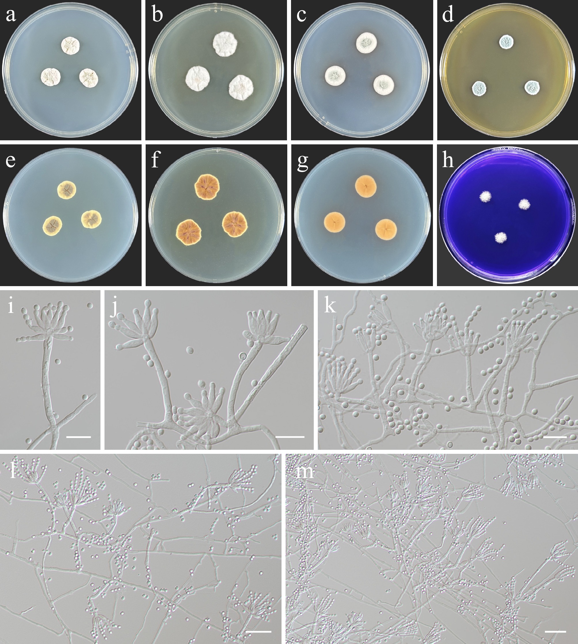
Figure 15.
Penicillium zoologicum (from holotype ZY H-23.028). (a)–(h) Colonies on medium at 25 °C for 7 d (left to right, top row: CYA, YES, DG18, and MEA obverse; second row: CYA reverse, YES reverse, DG18 reverse, and CREA obverse). (i)–(m) Conidiogenous cells and conidia. Scale bars: (i)–(k) 10 μm, (l), (m) 20 μm.
MycoBank number: MB862617
Infrageneric classification. Subgenus Aspergilloides, section Cinnamopurpurea, series Jiangxiensia.
Etymology: The specific epithet 'zoologicum' (Lat.) refers to the type locality, Harbin Northern Forest Zoo.
Colony diameter (7 d, in mm): CYA 12–14; CYA 30 °C 7–9; 37 °C no growth; MEA 9–10; YES 15–17; DG18 13–15; CREA 7–8.
Colony characteristics (7 d). CYA at 25 °C: Colonies deep, raised at center, margins low, entire; mycelium white; texture velutinous; sporulation sparse, conidia papyrus white; exudate clear; reverse white aluminium to oyster white; soluble pigment absent. MEA at 25 °C: Colonies moderately deep, raised at center, margins low, entire; mycelium white; texture velutinous; sporulation good, conidia papyrus white; exudate absent; reverse beige brown to light ivory; soluble pigment absent. YES at 25 °C: Colonies moderately deep, radially sulcate, irregular; mycelium white; texture floccose; sporulation sparse, conidia papyrus white; exudate absent; reverse salmon orange; soluble pigment absent. DG18 at 25 °C: Colonies moderately deep, raised at center, margins low, entire; mycelium white; texture floccose; sporulation sparse, conidia papyrus white; exudate absent; reverse dahlia yellow; soluble pigment beige red. CREA at 25 °C: Weak growth, no acid production.
Description: Hyphae branched, septate, hyaline, smooth, 1–2.5 μm wide. Asexual morph Conidiophores monoverticillate. Stipes smooth-walled, 15 – 46.5 × 2 – 3.5 µm. Vesicles absent or present, up to 7.5 µm. Phialides ampulliform, two to six per vesicle, 5.5–14.5 × 2.5–3.5 μm. Conidia globose to subglobose, smooth-walled, 2–3.5 μm (av. 2.6 µm, n = 30). Sclerotia absent. Sexual morph not observed.
Material examined: China, Heilongjiang Province, Harbin City, soil collected from the green belts of Harbin Northern Forest Zoo, 127.1071° E, 45.3948° N, 8 May 2023, Zhi-Yuan Zhang (holotype ZY H-23.028, dried culture; culture ex-type CGMCC 3.29791, ibid., ZY 23.028). China, Heilongjiang Province, Harbin City, soil collected from the green belts of Harbin Northern Forest Zoo, 127.1028° E, 45.3988° N, 8 May 2023, Zhi-Yuan Zhang, isotype ZY H-23.029, ex-isotype ZY 23.029.
Notes: Penicillium zoologicum phylogenetically belongs to section Cinnamopurpurea, series Jiangxiensia, and, similar to other members of this series, predominantly produces monoverticillate conidiophores. The section Cinnamopurpurea comprises four series, among which the series Jiangxiensia consists of P. ezekielii, P. jiangxiense, P. pusillum, and P. tealii[70]. Penicillium ezekielii can be differentiated from P. zoologicum by its production of cylindrical to ellipsoidal, rough-walled conidia, while P. zoologicum produces globose to subglobose, smooth-walled conidia[73]. Penicillium jiangxiense can be differentiated from P. zoologicum by its small conidia (2–2.5 × 1.5–2 µm)[74]. Penicillium pusillum produces sclerotia, which are not observed in P. zoologicum[75]. Furthermore, P. tealii differs from P. zoologicum by lacking conidiophores and conidia, instead producing abundant sclerotia[73]. Moreover, pairwise comparison of TUB, CaM, and ITS loci reveals that P. zoologicum (ex-type CGMCC 3.88888) differs from P. ezekielii (ex-type CBS 149115) by 13.4% (65/485 bp, 20 gaps) in TUB, 17.2% (82/475 bp, 19 gaps) in CaM, and 5.4% (32/588 bp, 13 gaps) in ITS; from P. jiangxiense (ex-type AS 3.6521) by 10.4% (48/458 bp, ten gaps) in TUB, 16.8% (82/487 bp, 17 gaps) in CaM, and 4.7% (27/564 bp, nine gaps) in ITS; from P. pusillum (ex-type CBS 312.63) by 10.1% (47/465 bp, 11 gaps) in TUB, 13.6% (94/689 bp, 21 gaps) in CaM, and 5.3% (30/564 bp, eight gaps) in ITS; from P. tealii (ex-type BRIP72734c) by 10.9% (53/485 bp, 20 gaps) in TUB and 4.3% (23/532 bp, four gaps) in ITS.
Onygenales Cif. ex Benny & Kimbr.
Onygenaceae Berk.
Nannizziopsis Currah
Patouillard established the genus Rollandina to accommodate R. capitata[76]. Later, Apinis described another new species of the genus, R. vriesii[77]. However, re-examination of the type specimen of R. capitata yielded descriptions that differed among researchers[77,78]. Given that pure cultures of R. capitata, the type species of Rollandina, are unknown, Von Arx considered this genus to be doubtful[79]. Currah endorsed von Arx's view, treating Rollandina as a nomen confusum and establishing the new genus Nannizziopsis within the family Onygenaceae to accommodate R. vriesii[80]. Based on an integrated analysis of physiology, morphology, and multi-locus phylogeny from Nannizziopsis and several chrysosporium-like isolates, Stchigel et al. proposed a new family, Nannizziopsidaceae[81]. However, in the larger phylogeny of Onygenales, this family does not form a distinct clade separate from Onygenaceae[82,83].
In the large-scale phylogeny of Onygenales, Nannizziopsis mirabilis is nested within Neogymnomycetaceae, separating it from other Nannizziopsis species (which belong to Onygenaceae)[82]. Therefore, this study excluded it from the phylogenetic analysis of the genus. Phylogenetic relationships of the genus Nannizziopsis were inferred using a combined ITS and LSU dataset, with Paranannizziopsis longispora (CBS 133990) and P. tardicrescens (CBS 142038) designated as the outgroup. The final alignment included 28 taxa and spanned 1,170 nucleotide positions (ITS, 559 bp; LSU, 611 bp), including gaps. The best-fit models were TN + F + G4 (ITS) and TIM3 + F + I +G4 (LSU) for the ML analysis, whereas those for the BI analysis were GTR + F + G4 (ITS) and GTR + F + I + G4 (LSU). Phylogenetically, CGMCC 3.29792 and ZY 23.031 formed a single, well-supported clade (100/1, Fig. 16).
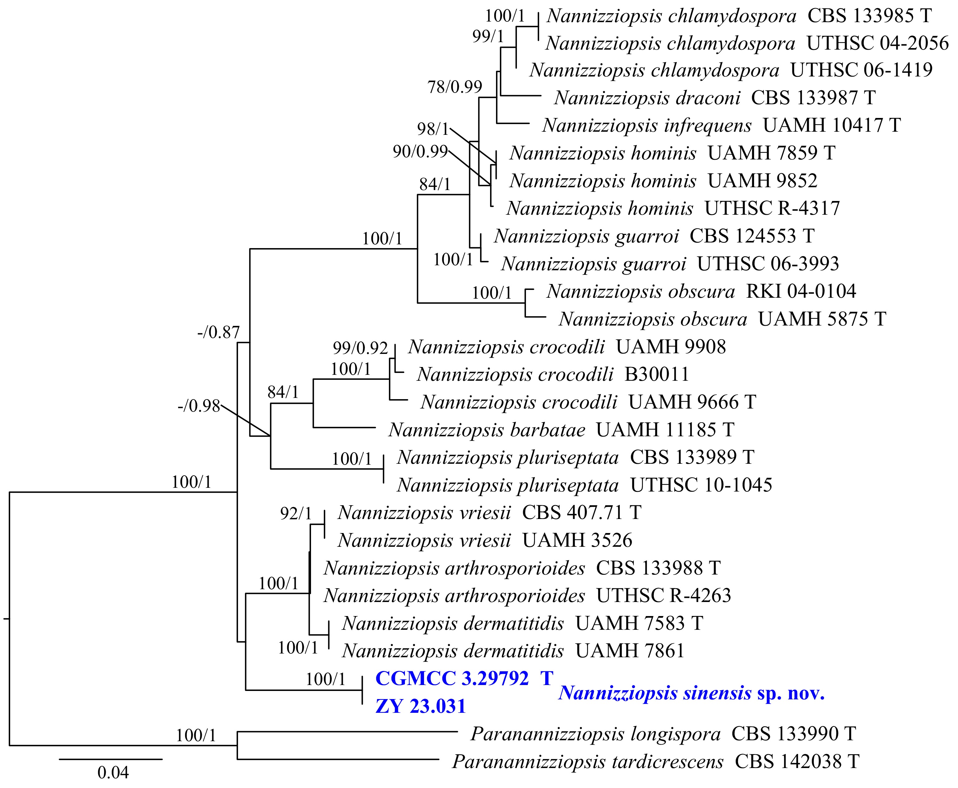
Figure 16.
Phylogram generated from ML analysis based on the combined dataset of ITS and LSU sequence data. Numbers at branches indicate support values (IQ-TREE-BS/BI-PP) above 70%/0.90. The tree is rooted to Paranannizziopsis longispora (CBS 133990) and P. tardicrescens (CBS 142038). Newly generated sequences are in bold and blue, and 'T' indicates type-derived sequences. The scale bar represents the expected number of changes per site.
Nannizziopsis sinensis Y.J. Wu, Zhi Y. Zhang & Y.F. Han sp. nov. Fig. 17
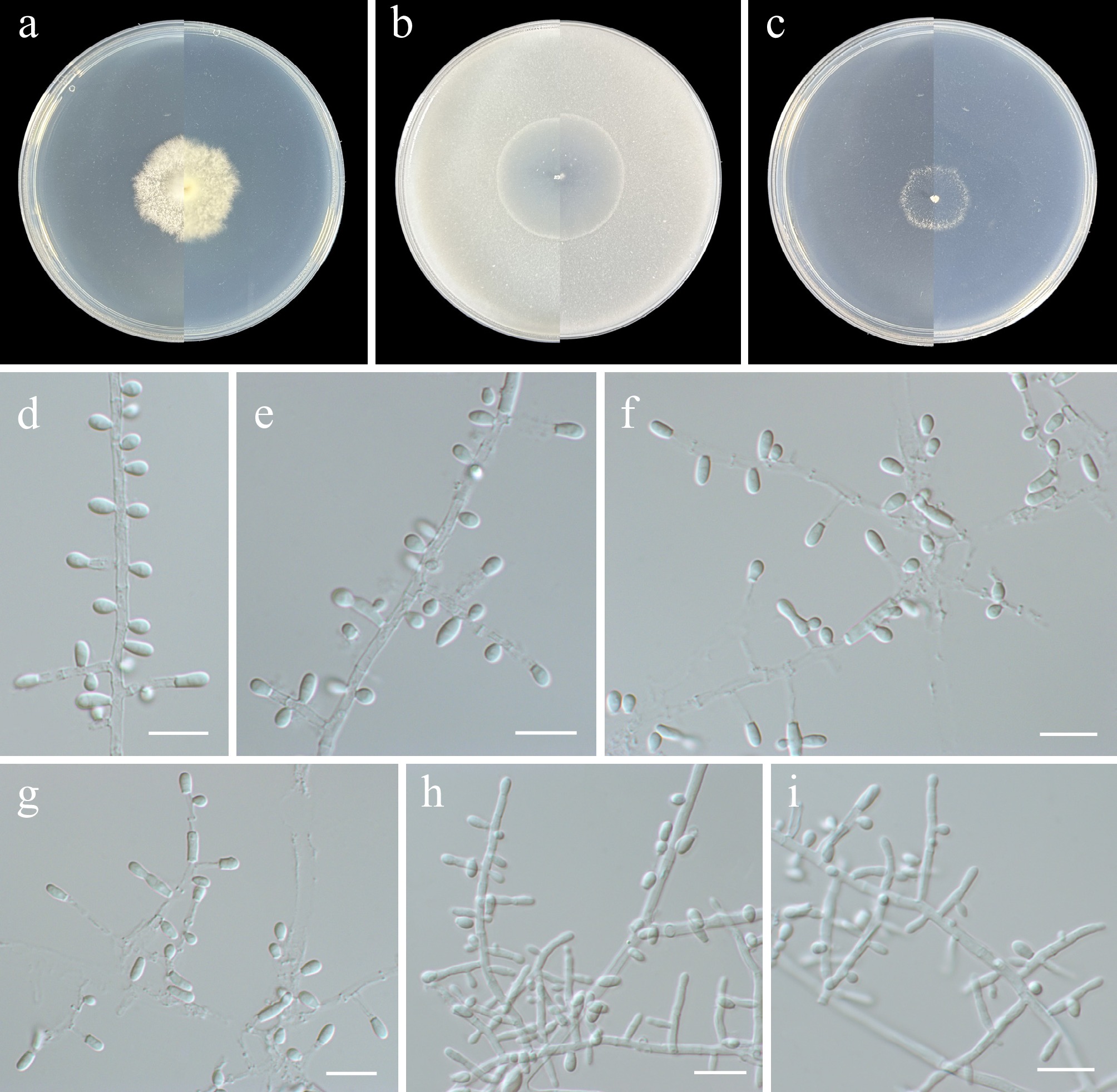
Figure 17.
Nannizziopsis sinensis (from holotype ZY H-23.030). (a)–(c) Upper and reverse views of cultures on PDA, OA, and SNA after 14 d at 25 °C. (d)–(f), (h), (i) Fertile hyphae bearing mostly sessile conidia. (g) Intercalary conidia. Scale bars: (d)–(i) 10 μm.
MycoBank number: MB862618
Etymology: Named after the type locality, China.
Description: Hyphae branched, septate, hyaline, smooth-walled, 1–4 μm wide. Asexual morph Conidia 1(–2)-celled, mostly sessile, also produced on short protrusions or terminal, hyaline, thin- and smooth-walled, pyriform, obovate, or claviform to cylindrical, 2.5–10.5 × 1.5–2.5 μm (av. 4.8 × 2.1, n = 30 μm). Intercalary conidia rarely, doliiform to cylindrical, 4–6 × 2–3 μm. Arthroconidia absent. Chlamydospores and sexual morph not observed.
Culture characteristics (14 d at 25 °C): Colony on PDA reaching 28–33 mm diam., felted, slightly cottony at centre, white, reverse white. Colony on OA reaching 32–34 mm diam., flat, aerial mycelium sparse, white, circular, margin regular, reverse white. Colony on SNA reaching 17–19 mm in diam., flat, felted, sparse aerial mycelium, white, reverse white.
Material examined: China, Heilongjiang Province, Harbin City, soil collected from the green belts of Harbin Northern Forest Zoo, 127.1093° E, 45.3903° N, 8 May 2023, Zhi-Yuan Zhang (holotype ZY H-23.030, dried culture; culture ex-type CGMCC 3.29792, ibid., ZY 23.030). China, Heilongjiang Province, Harbin City, soil collected from the green belts of Harbin Northern Forest Zoo, 127.1143° E, 45.3898° N, 8 May 2023, Zhi-Yuan Zhang, isotype ZY H-23.031, ex-isotype ZY 23.031.
Notes: Phylogenetically, Nannizziopsis sinensis formed a distinct subclade and separated from other species in Nannizziopsis (Fig. 16). Morphologically, within the genus Nannizziopsis, the sexual morphs of N. sinensis, N. arthrosporioides, N. chlamydospore, N. dermatitidis, N. draconi, N. guarroi, and N. pluriseptata remain unknown[81,84,85]. Among these species, only N. sinensis lacks arthroconidia, and it produces intercalary conidia only rarely.
Class Leotiomycetes O.E. Erikss. & Winka
Helotiales Nannf.
Hyphodiscaceae Ekanayaka & K.D. Hyde
Cistella Quél.
The type species of the genus Cistella is C. dentata. To date, molecular sequences for the vast majority of species in this genus are unavailable. Several studies have examined the taxonomic placement of this genus solely on the basis of the molecular sequences of Cistella albidolutea (KUS-F52678). Johnston et al. classified it within Vandijckellaceae but did not clarify which order it belongs to[86]. In contrast, Ekanayaka et al. placed it within the family Calloriaceae, order Rhytismatales[87]. Cistella species occur on diverse substrates, including soil, plant litter, needles, and cones, and are widespread, mainly in temperate zones.
Phylogenetic relationships of the genus Cistella and related taxa were inferred using a multi-locus (ITS, LSU, and RPB2) dataset, with Chlorencoelia torta (KUS-F 52256) designated as the outgroup. The final alignment included 37 taxa and spanned 2,162 nucleotide positions (ITS, 575 bp; LSU, 840 bp; and RPB2, 747 bp), including gaps. The best-fit models were SYM + I + G4 (ITS), TN + F + I + G4 (LSU), and GTR + F + I + G4 (RPB2) for the ML analysis, whereas those for the BI analysis were SYM + I + G4 (ITS) and GTR + F + I + G4 (LSU, RPB2). Phylogenetically, CGMCC 3.29793 and ZY 23.033 formed a single, well-supported clade (100/1, Fig. 18).
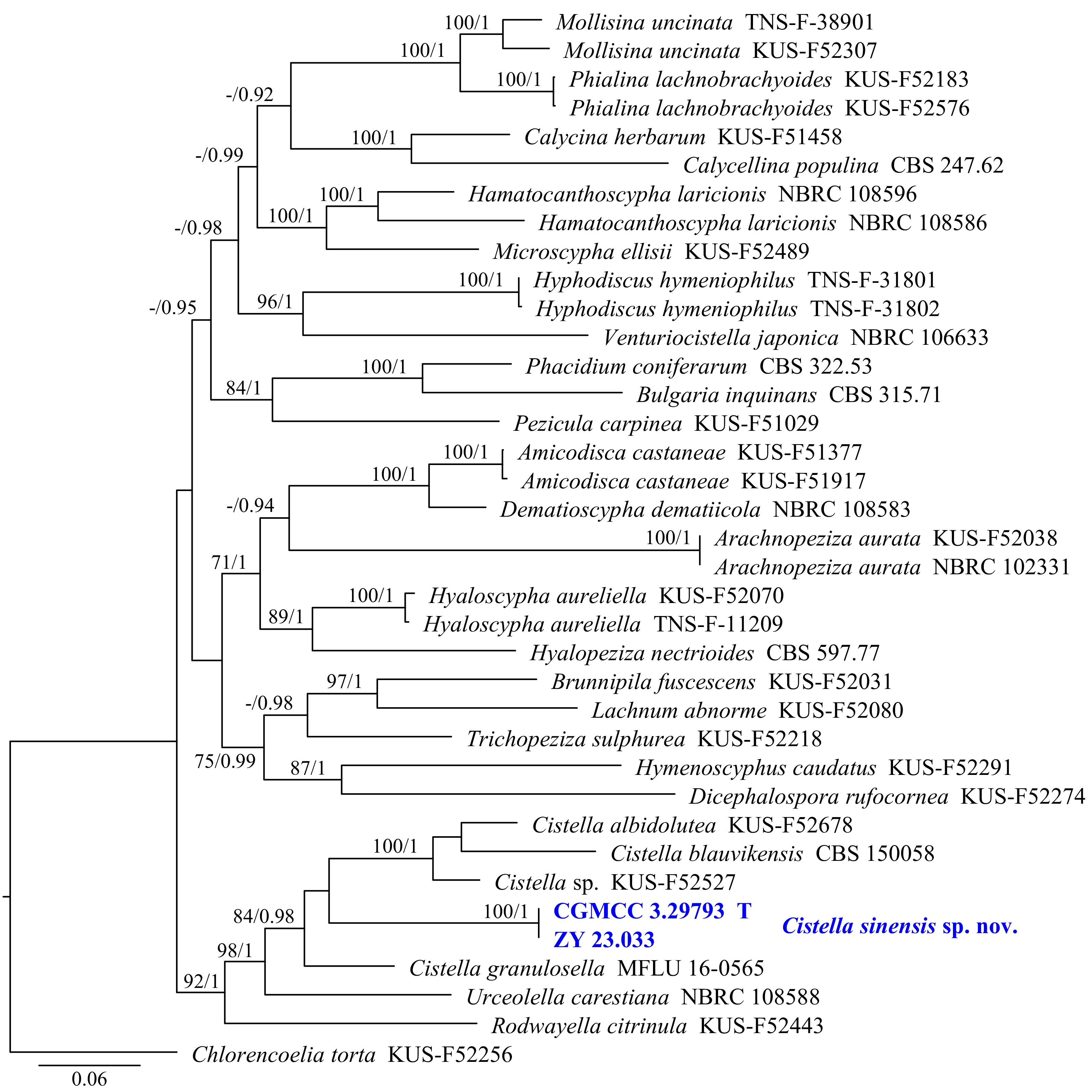
Figure 18.
Phylogram generated from ML analysis based on the combined dataset of ITS, LSU, and RPB2 sequence data. Numbers at branches indicate support values (IQ-TREE-BS/BI-PP) above 70%/0.90. Newly generated sequences are in bold and blue, and 'T' indicates type-derived sequences. The tree is rooted to Chlorencoelia torta (KUS-F 52256). The scale bar represents the expected number of changes per site.
Cistella sinensis Y.J. Wu, Zhi Y. Zhang & Y.F. Han sp. nov. Fig. 19
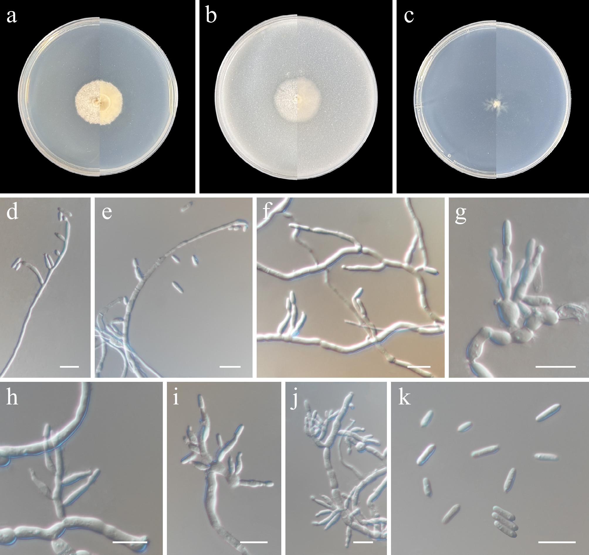
Figure 19.
Cistella sinensis (from holotype ZY H-23.032). (a)–(c) Upper and reverse views of cultures on PDA, OA, and SNA after 14 d at 25 °C. (d)–(f) Phialides and conidia. (g)–(j) Conidiophores and phialides. (k) Conidia. Scale bars: (d)–(k) 10 μm.
MycoBank number: MB862619
Etymology: Named after the type locality, China.
Description: Hyphae branched, septate, hyaline, smooth-walled, up to 3 μm wide. Asexual morph Conidiophores solitary, arising directly from aerial, substratal mycelium, unbranched, subcylindrical, straight or slightly curved, up to 10 μm long, 1.5–3 μm wide at base, aseptate, hyaline, smooth-walled. Phialides lateral, terminal, ampulliform, subcylindrical, hyaline, septate, smooth-walled, 4.5–15.5 × 1.5–3 μm. Conidia solitary, aseptate, cylindrical with enlarged at the base, straight, with both ends rounded, hyaline, smooth-walled, 4.5–22 × 1.5–1.9 μm (av. 6.8 × 1.9, n = 30 μm). Sexual morph not observed.
Culture characteristics (14 d at 25 °C): Colony on PDA reaching 24–27 mm diam., flat, felty, white, margin entire, reverse white. Colony on OA reaching 22–24 mm diam., flat, moderate aerial mycelium, felty, dusty, white, margin entire, reverse creamy white. Colony on SNA reaching 6–15 mm diam., aerial hyphae sparse, white, margin irregular, reverse white.
Material examined: China, Heilongjiang Province, Harbin City, soil collected from the green belts of Harbin Northern Forest Zoo, 127.1003° E, 45.3992° N, 8 May 2023, Zhi-Yuan Zhang (holotype ZY H-23.032, dried culture; culture ex-type CGMCC 3.29793, ibid., ZY 23.032). China, Heilongjiang Province, Harbin City, soil collected from the green belts of Harbin Northern Forest Zoo, 127.1133° E, 45.3921° N, 8 May 2023, Zhi-Yuan Zhang, isotype ZY H-23.033, ex-isotype ZY 23.033.
Notes: Phylogenetically, Cistella sinensis (CGMCC 3.29793 and ZY 23.111), C. albidolutea (KUS-F52678), C. blauvikensis (CBS 150058), C. granulosella (MFLU 16-0565), and Cistella sp. (KUS-F52527) clustered into one group with a moderate support value (84/0.98, Fig. 18). To date, C. blauvikensis is the only species within the genus Cistella established based on its anamorph[88]. Beyond this, the presence of anamorphs in other species remains unconfirmed. Morphologically, C. sinensis differs from C. blauvikensis in its solitary, aseptate, cylindrical conidia[88].
Hamatocanthoscyphaceae Ekanayaka & K.D. Hyde
Hyalodendriella Crous
Crous et al. established the genus Hyalodendriella to accommodate H. betulae[89]. To date, Hyalodendriella consists of H. betulae and H. bialowiezensis[89,90]. The genus is diagnosed by dimorphic conidiophores, conidia that turn brown with age, and thickened scars. The microconidiophores arise as lateral branches from the hyphae, are septate, and bear terminal conidiogenous cells. The macroconidiophores are septate, subcylindrical, straight to curved, with an apical rachis, and have numerous aggregated loci. The conidia are limoniform to ellipsoid, aseptate, arranged in short chains, tapering toward the ends, and are prominently apiculate with distinctly thickened and darkened tips that are non-refractive.
Phylogenetic relationships of the genus Hyalodendriella were inferred using a multi-locus (ITS, LSU, and RPB2) dataset, with Leotia lubrica (AFTOL-ID 1) designated as the outgroup. The final alignment included 32 taxa and spanned 2,104 nucleotide positions (ITS, 515 bp; LSU, 842 bp; and RPB2, 747 bp), including gaps. The best-fit models were SYM + I + G4 (ITS), TN + F + I + G4 (LSU), and GTR + F + I + G4 (RPB2) for the ML analysis, whereas those for the BI analysis were SYM + I + G4 (ITS) and GTR + F + I + G4 (LSU, RPB2). Phylogenetically, CGMCC 3.29794 and ZY 23.035 formed a single, well-supported subclade (100/1, Fig. 20).
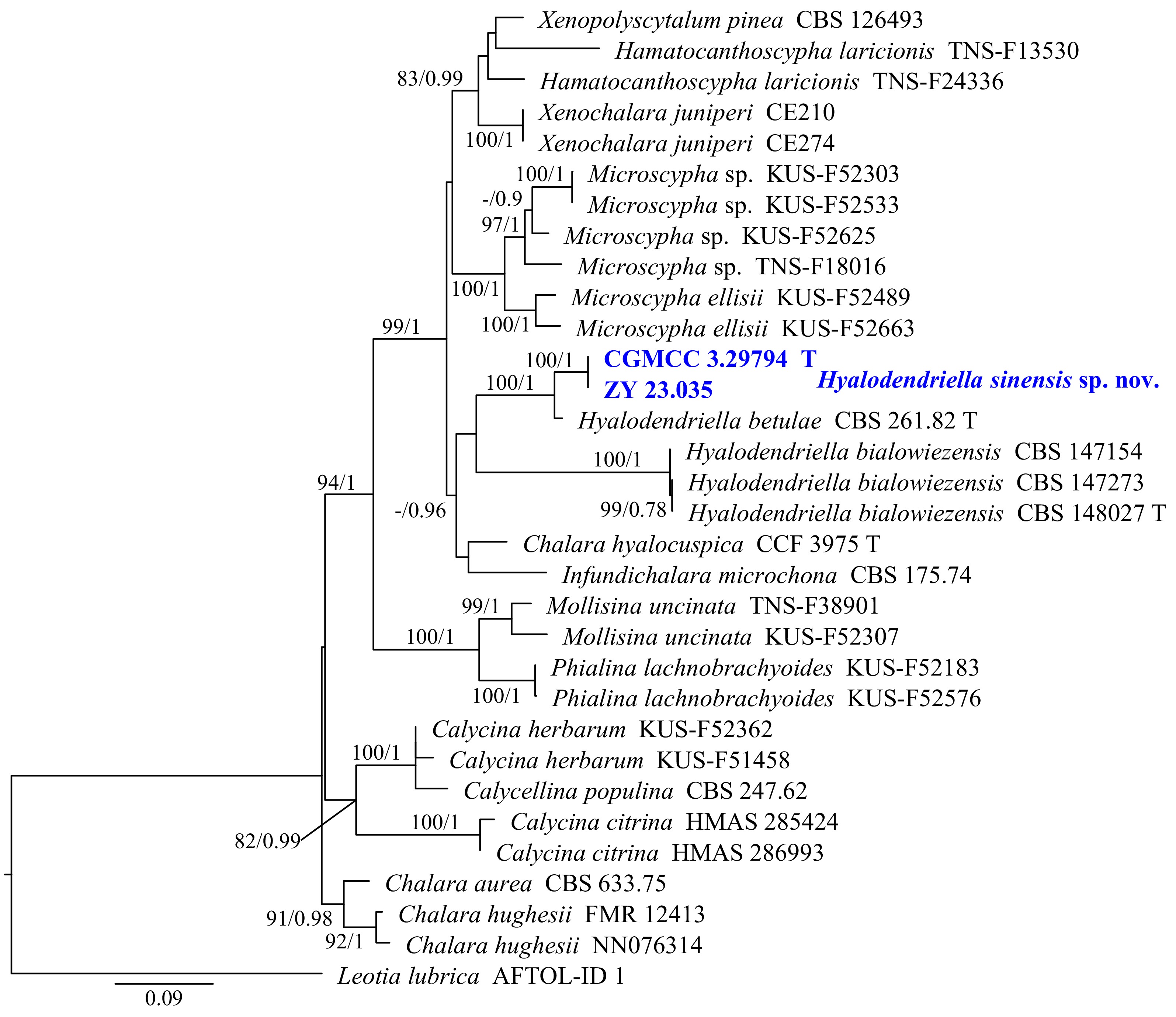
Figure 20.
Phylogram generated from ML analysis based on the combined dataset of ITS, LSU, and RPB2 sequence data. Numbers at branches indicate support values (IQ-TREE-BS/BI-PP) above 70%/0.90. Newly generated sequences are in bold and blue, and 'T' indicates type-derived sequences. The tree is rooted to Leotia lubrica (AFTOL-ID 1). The scale bar represents the expected number of changes per site.
Hyalodendriella sinensis Y.J. Wu, Zhi Y. Zhang & Y.F. Han sp. nov. Fig. 21
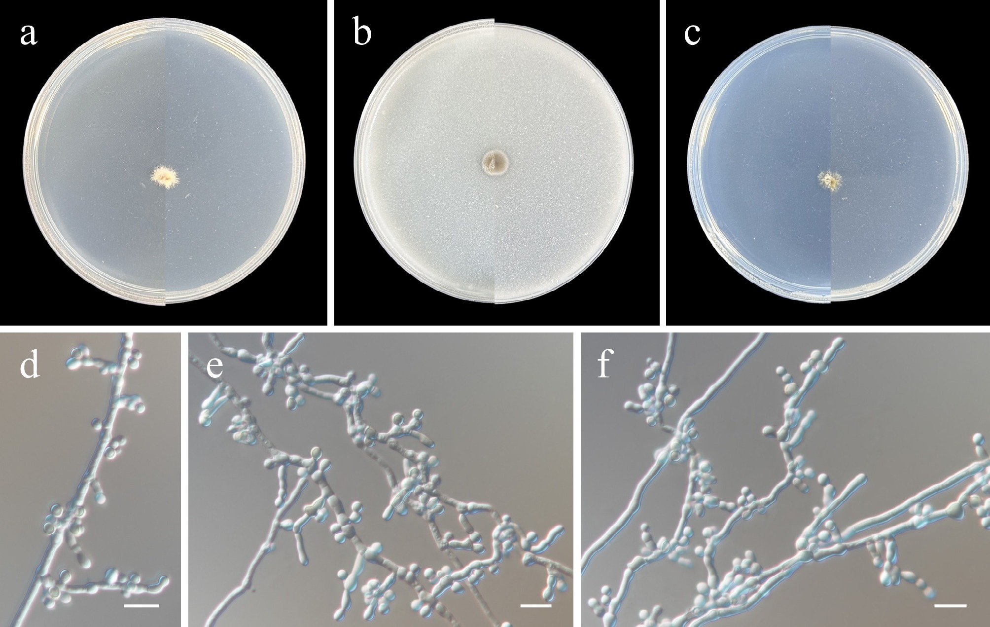
Figure 21.
Hyalodendriella sinensis (from holotype ZY H-23.034). (a)–(c) Upper and reverse views of cultures on PDA, OA, and SNA after 14 d at 25 °C. (d)–(f) Conidiogenous cells and conidia. Scale bars: (d)–(f) 10 μm.
MycoBank number: MB862620
Etymology: Named after the type locality, China.
Description: Hyphae branched, septate, hyaline, smooth-walled, 1–3.5 μm wide. Asexual morph Conidiophores simple, often reduced to conidiogenous cells. Conidiogenous cells terminal, lateral, terminal, hyaline, smooth, pyriform, clavate to cylindrical, or irregular, 3.5–7 × 2–4 µm. Conidia solitary, smooth, aseptate, pyriform, ellipsoidal to subglobose, hyaline to pale brown, 2.5–6 × 2.5–4 µm (av. 3.8 × 3, n = 30 μm). Sexual morph not observed.
Culture characteristics (14 d at 25 °C): Colony on PDA reaching 7–9 mm diam., flat, white, margin irregular, reverse white. Colony on OA reaching 7–9 mm diam., flat, flocculent, grayish-white, circular, margin regular, reverse pale. Colony on SNA reaching 7–9 mm in diam., powdery, white, margin irregular, reverse white.
Material examined: China, Heilongjiang Province, Harbin City, soil collected from the green belts of Harbin Northern Forest Zoo, 127.1071° E, 45.3948° N, 8 May 2023, Zhi-Yuan Zhang (holotype ZY H-23.034, dried culture; culture ex-type CGMCC 3.29794, ibid., ZY 23.034). China, Heilongjiang Province, Harbin City, soil collected from the green belts of Harbin Northern Forest Zoo, 127.0971° E, 45.3994° N, 8 May 2023, Zhi-Yuan Zhang, isotype ZY H-23.035, ex-isotype ZY 23.035.
Notes: Phylogenetically, Hyalodendriella sinensis formed a distinct subclade and was closely related to H. betulae (Fig. 20). Additionally, the phylogenetic tree suggests that Hyalodendriella may be polyphyletic. Morphologically, H. sinensis differs from H. betulae and H. bialowiezensis in having conidiophores often reduced to conidiogenous cells and solitary, pyriform, ellipsoidal, or subglobose conidia[89,90].
Leptodontidiaceae Hern.-Restr., Crous & Gené
Leptodontidium de Hoog.
De Hoog established the genus Leptodontidium to accommodate L. trabinellum[91]. To date, the sexual morph of Leptodontidium remains unknown. The genus comprises 14 taxa, including saprobes and terrestrial species, though some have since been reallocated to other genera (Index Fungorum 2026). Recent phylogenetic studies have included four species in this genus[92].
Phylogenetic relationships of the genus Leptodontidium were inferred using a combined ITS and LSU dataset, with Lauriomyces bellulus (FMR 12188 and CBS 517.93) designated as the outgroup. The final alignment included 19 taxa and spanned 1,322 nucleotide positions (ITS, 485 bp; LSU, 837 bp), including gaps. The best-fit models were TIM2 + G4 (ITS) and TN + F + I + G4 (LSU) for the ML analysis, whereas those for the BI analysis were SYM + G4 (ITS) and GTR + F + I + G4 (LSU). Phylogenetically, CGMCC 3.29795 and ZY 23.037 formed a single, well-supported subclade (100/1, Fig. 22).
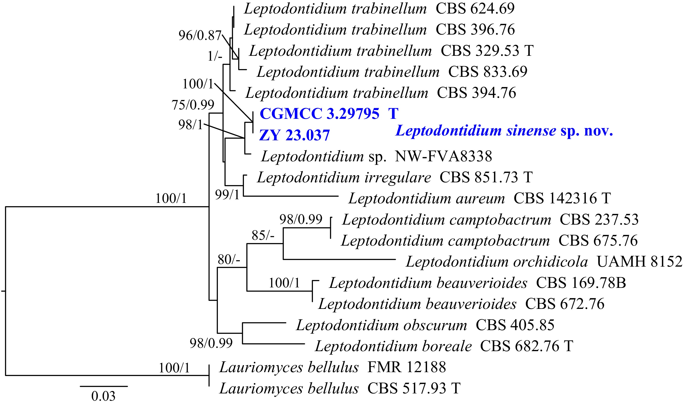
Figure 22.
Phylogram generated from ML analysis based on the combined dataset of ITS and LSU sequence data. Numbers at branches indicate support values (IQ-TREE-BS/BI-PP) above 70%/0.90. The tree is rooted to Lauriomyces bellulus (FMR 12188 and CBS 517.93). Newly generated sequences are in bold and blue, and 'T' indicates type-derived sequences. The scale bar represents the expected number of changes per site.
Leptodontidium sinense Y.J. Wu, Zhi Y. Zhang & Y.F. Han sp. nov. Fig. 23
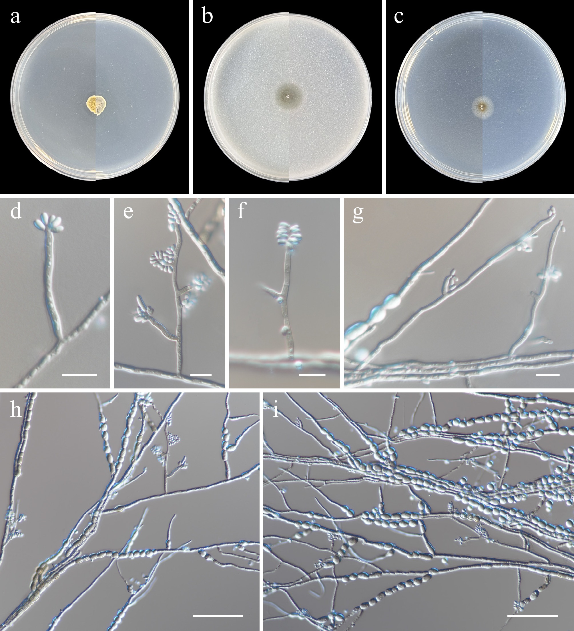
Figure 23.
Leptodontidium sinense (from holotype ZY H-23.036). (a)–(c) Upper and reverse views of cultures on PDA, OA, and SNA after 14 d at 25 °C. (d)–(g) Conidiophores, conidiogenous cells, and conidia. (h), (i) Conidia and chlamydospores. Scale bars: (d)–(g) 10 μm; (h), (i) 50 μm.
MycoBank number: MB862621
Etymology: Named after the type locality, China.
Description: Hyphae branched, septate, hyaline, smooth-walled, 1–4 μm wide. Asexual morph Conidiophores solitary, erect, straight, simple or irregularly branched, cylindrical to subcylindrical, medium brown, thick-walled, finely roughened, one-to-six-septate, 14–61 × 1.5–3.5 µm. Conidiogenous cells terminal, lateral, subcylindrical, hyaline or medium brown, somewhat paler than the rest of the conidiophore, smooth, 3.5–19 × 1–2.5 µm. Conidia hyaline, smooth, aseptate, elliptical to subcylindrical, apex obtuse, straight to slightly curved, 2–4.5(–8.5) × 1.5–2.5 µm (av. 4.1 × 2, n = 30 μm), aggregating in mucoid mass. Chlamydospores terminal or intercalary, solitary or in simple or branched chains, zero-to-one-septate, ellipsoidal to subglobose, subcylindrical to cylindrical, 3.5–14 × 3.5–7 μm (av. 9.4 × 5.5, n = 30 μm), hyaline becoming brown, smooth. Sexual morph not observed.
Culture characteristics (14 d at 25 °C): Colony on PDA reaching 8–10 mm diam., aerial mycelium dense, powdery, pale yellow to white, elliptical, margin regular, reverse gray. Colony on OA reaching 11–13 mm diam., flat, aerial mycelium sparse, pale bluish-green, circular, margin regular, reverse pale bluish-green. Colony on SNA reaching 10–12 mm in diam., flat, powdery, sparse aerial mycelium, pale brown, circular, margin regular, reverse pale brown.
Material examined: China, Heilongjiang Province, Harbin City, soil collected from the green belts of Harbin Northern Forest Zoo, 127.1043° E, 45.3898° N, 8 May 2023, Zhi-Yuan Zhang (holotype ZY H-23.036, dried culture; culture ex-type CGMCC 3.29795, ibid., ZY 23.036). China, Heilongjiang Province, Harbin City, soil collected from the green belts of Harbin Northern Forest Zoo, 127.1133° E, 45.3921° N, 8 May 2023, Zhi-Yuan Zhang, isotype ZY H-23.037, ex-isotype ZY 23.037.
Notes: Phylogenetically, Leptodontidium sinense formed a distinct subclade closely related to L. aureum, L. irregulare, L. trabinellum, and an undescribed strain NW-FVA8338 (Fig. 22). Morphologically, L. sinense differs from L. aureum in its conidial shape and size (elliptical to subcylindrical, 2–4.5 × 1.5–2.5 µm in L. sinense vs obovoid to oblong, 4.5–8 × 2–3 μm)[93]. Leptodontidium sinense differs from L. irregulare in lacking beauveria-like conidia[94]. Leptodontidium sinense differs from L. trabinellum in having elliptical to subcylindrical conidia[93].
Class Sordariomycetes O.E. Erikss. & Winka
Chaetosphaeriales Huhndorf, A.N. Mill. & F.A. Fernández
Chaetosphaeriaceae Réblová, M.E. Barr & Samuels
Codinaea Maire
Maire established the genus Codinaea with C. aristata as the type species[95]. Recently, Réblová et al. proposed a narrow concept for the genus Codinaea sensu stricto, based on combined morphological and phylogenetic evidence[96]. Subsequently, Wu & Diao validated the accuracy of this narrow concept, described several species, and developed a key for the genus[97]. Here, we follow the taxonomic framework of Wu & Diao[97] and describe a new species.
Phylogenetic relationships of the genus Codinaea were inferred using a multi-locus (ITS, LSU, and TEF1) dataset, with Nimesporella sp. (KUNCC 23-13365) and N. capillaceae (IMI 358908) designated as the outgroup. The final alignment included 40 taxa and spanned 2,234 nucleotide positions (ITS, 480 bp; LSU, 859 bp; and TEF1, 895 bp), including gaps. The best-fit models were TIM2 + F + I + G4 (ITS), TIM3 + F + I + G4 (LSU), and TIM + F + I + G4 (TEF1) for the ML analysis, whereas GTR + F + I + G4 was applied across all three loci for the BI analysis. Phylogenetically, CGMCC 3.29796, ZY 23.039, ZY 23.040, and ZY 23.041 formed a single, well-supported subclade (100/1, Fig. 24).
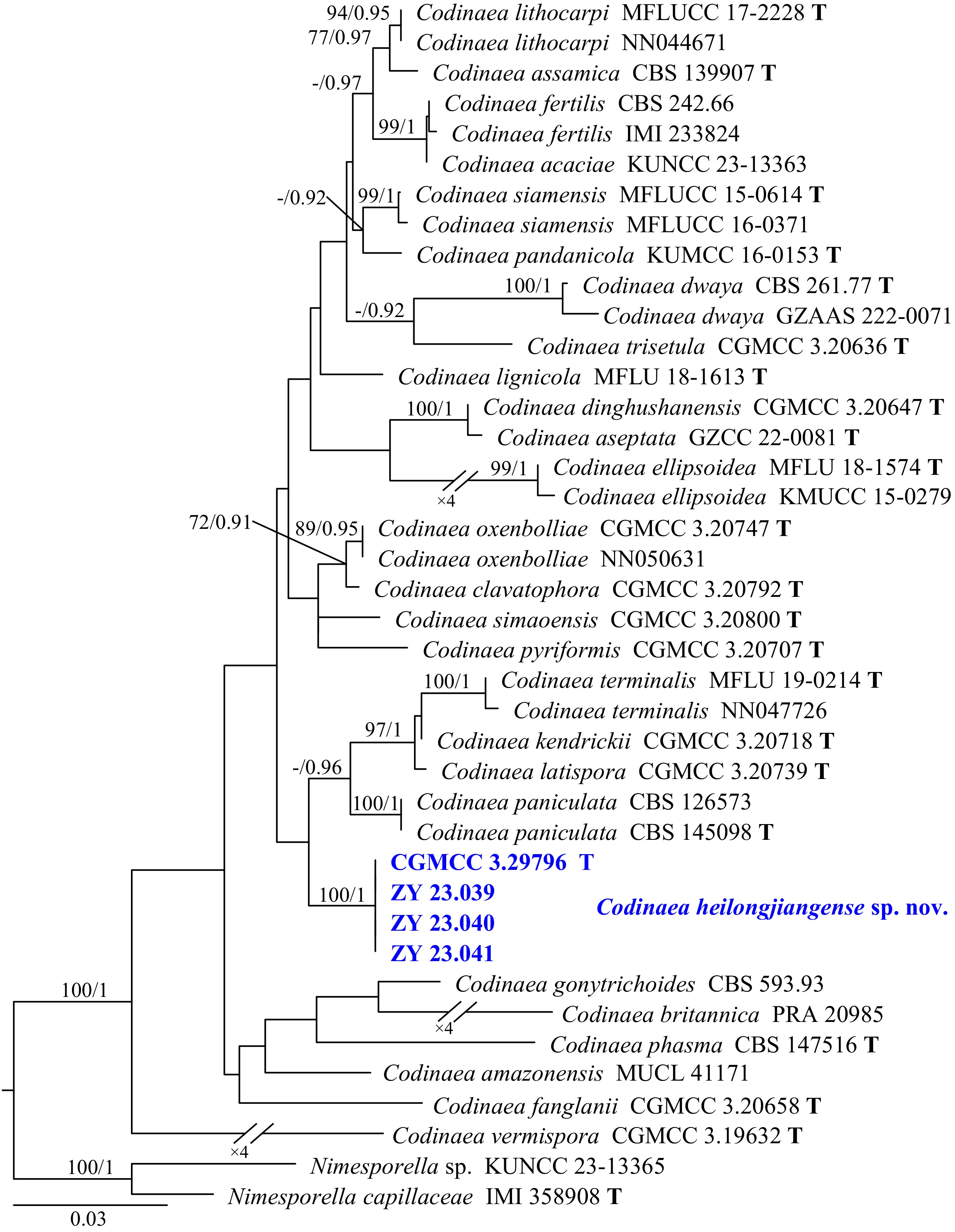
Figure 24.
Phylogram generated from ML analysis based on the combined dataset of ITS, LSU, and TEF1 sequence data. Numbers at branches indicate support values (IQ-TREE-BS/BI-PP) above 70%/0.90. The tree is rooted to Nimesporella sp. (KUNCC 23-13365) and N. capillaceae (IMI 358908). Newly generated sequences are in bold and blue, and 'T' indicates type-derived sequences. The scale bar represents the expected number of changes per site.
Codinaea heilongjiangensis Y.J. Wu, Zhi Y. Zhang & Y.F. Han sp. nov. Fig. 25
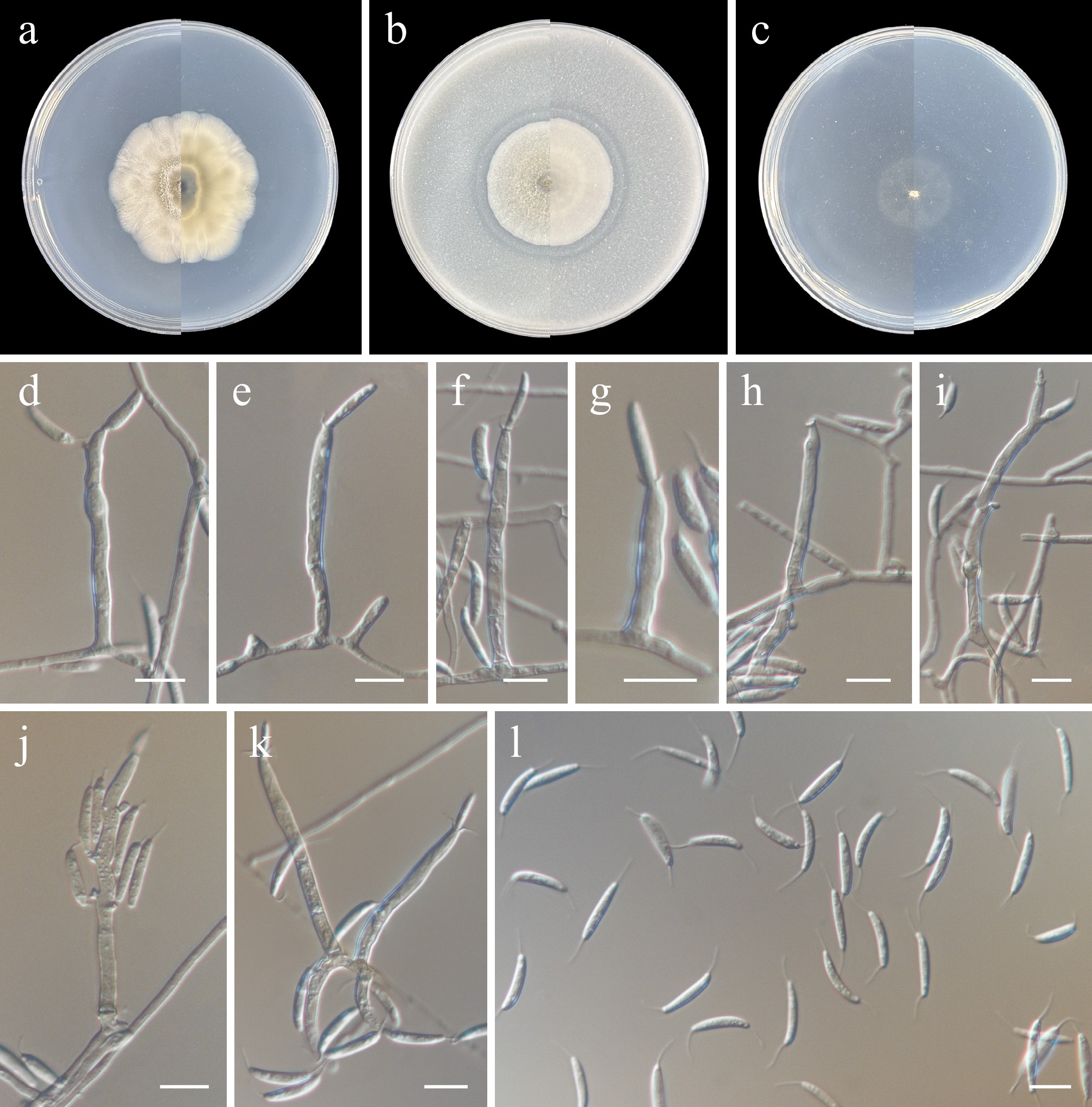
Figure 25.
Codinaea heilongjiangensis (from holotype ZY H-23.038). (a)–(c) Upper and reverse views of cultures on PDA, OA, and SNA after 14 d at 25 °C. (d)–(h), (k) Conidiophores and conidiogenous cells with funnel-shaped collarettes. (i), (j) Percurrently and sympodially proliferated conidiophores and conidiogenous cells bearing funnel-shaped collarettes. (l) Conidia. Scale bars: (d)–(l) 10 μm.
MycoBank number: MB862800
Etymology: Named after the type locality, Heilongjiang Province, China.
Description: Hyphae branched, septate, hyaline, smooth-walled, 1–2.5 μm wide. Asexual morph Conidiophores macronematous, mononematous, arising from superficial mycelium, erect, straight, or slightly curved, cylindrical then becoming geniculate due to sympodial proliferation, smooth, zero–two-septate, hyaline, with sympodial proliferation, 10.5–30.5 × 2.5–4 µm. Conidiogenous cells integrated, cylindrical, obclavate, hyaline, smooth-walled, with a funnel-shaped collarette of 1–2 µm deep and 3–5 µm wide, with a narrow sporulating locus of 2–2.5 µm wide. Conidia acrogenous, solitary, lunate, straight, or slightly curved, 10.5–16 × 2–3 µm, aseptate, hyaline, smooth, apex acute to rounded, base slightly truncate to rounded, with one setula at each end, 4–8 µm long. Setae not observed. Sexual morph not observed.
Culture characteristics (14 d at 25 °C): Colony on PDA reaching 41–45 mm diam., felty, compact, plicated, convex, margin undulate, white, grey to grey-brown; reverse grey-brown. Colony on OA reaching 33–35 mm diam., ulotrichy, low convex, margin entire, white to gray; reverse white to pale gray. Colony on SNA reaching 19–22 mm diam., flat, colorless, aerial mycelia extremely sparse; reverse colorless.
Material examined: China, Heilongjiang Province, Harbin City, soil collected from the green belts of Harbin Northern Forest Zoo, 127.1043° E, 45.3898° N, 8 May 2023, Zhi-Yuan Zhang (holotype ZY H-23.038, dried culture; culture ex-type CGMCC 3.29796, ibid., ZY 23.038). China, Heilongjiang Province, Harbin City, soil collected from the green belts of Harbin Northern Forest Zoo, 127.1028° E, 45.3988° N, 8 May 2023, Zhi-Yuan Zhang, isotype ZY H-23.039, ex-isotype ZY 23.039, ibid., ZY 23.040, ibid., ZY 23.041.
Notes: To date, the genus Codinaea comprises 36 species[97,98], among which molecular sequences are available for 27 species. Phylogenetically, C. heilongjiangensis formed an independent lineage distinct from other Codinaea species (Fig. 24). Morphologically, it resembles C. vermiformis, C. dwaya, C. trisetula, and C. pyriformis in lacking sterile or fertile setae and in having solitary or irregularly grouped conidiophores[97]. However, it can be distinguished from these species by its conidiogenous cells, which are sometimes polyphialidic with sympodial proliferation, and by its lunate conidia[97].
Coniochaetales Huhndorf, A.N. Mill. & F.A. Fernández
Coniochaetaceae Malloch & Cain
Coniochaeta (Sacc.) Cooke
Cooke established the genus Coniochaeta with C. ligniaria as the type species[99]. As a genus of pleomorphic yeasts within the order Coniochaetales, Coniochaeta has a global distribution[100−102]. Members of the genus exhibit both sexual and asexual morphs during their life cycle. In the sexual morph, ascomata are setose, dark brown to black, and either pyriform with an ostiole or globose without one; the asci contained within are thin-walled and produce single-celled, smooth ascospores with an elongated germ slit[103,104]. The asexual morph, in contrast, produces colonies varying from pink-salmon to dark brown, together with phialidic conidiogenous cells[105−107].
Phylogenetic relationships of the genus Coniochaeta were inferred using a combined ITS and LSU dataset, with Phialemonium obovatum (CBS 279.76) and P. limoniforme (CBS 139049) designated as the outgroup. The final alignment included 91 taxa and spanned 1,060 nucleotide positions (ITS, 502 bp; LSU, 558 bp), including gaps. The best-fit models were GTR + F + I + G4 (ITS) and TIM + F + I + G4 (LSU) for the ML analysis, whereas those for the BI analysis were GTR + F + I + G4 (ITS) and GTR + I + G4 (LSU). Phylogenetically, CGMCC 3.29797 and ZY 23.043 formed a single, well-supported subclade (100/1, Fig. 26).
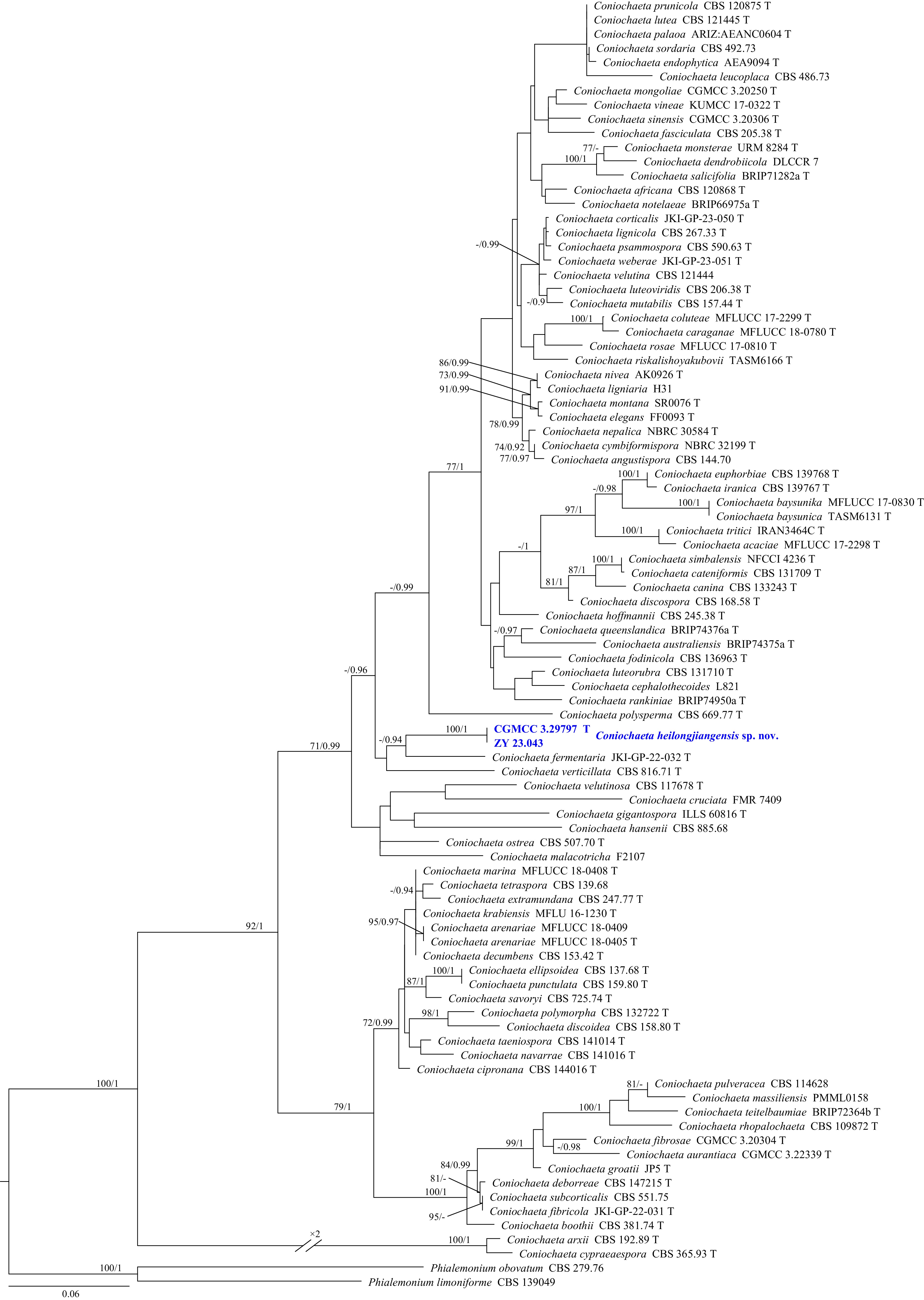
Figure 26.
Phylogram generated from ML analysis based on the combined dataset of ITS and LSU sequence data. Numbers at branches indicate support values (IQ-TREE-BS/BI-PP) above 70%/0.90. The tree is rooted to Phialemonium obovatum (CBS 279.76) and P. limoniforme (CBS 139049). Newly generated sequences are in bold and blue, and 'T' indicates type-derived sequences. The scale bar represents the expected number of changes per site.
Coniochaeta heilongjiangensis Y.J. Wu, Zhi Y. Zhang & Y.F. Han sp. nov. Fig. 27
Figure 27.
Coniochaeta heilongjiangensis (from holotype ZY H-23.042). (a)–(c) Upper and reverse views of cultures on PDA, OA, and SNA after 14 d at 25 °C. (d)–(f) Conidiophores, conidiogenous cells, and conidia. (g) Conidia. (d)–(g) Scale bars: 10 μm.
MycoBank number: MB862622
Etymology: Named after the type locality, Heilongjiang Province, China.
Description: Hyphae branched, septate, hyaline, smooth-walled, 1–3 μm wide. Asexual morph Conidiophores arising from hyphae, cylindrical, clavate, subglobose, or irregular, straight or slightly curved, smooth or slightly roughened, hyaline. Conidiogenous cells phialidic, rarely subcylindrical, with a slender neck at the apex, hyaline, smooth, single or in clusters on conidiophores or hyphae, 6–20 μm in length and 2–3.5 μm in width at the swollen portion. Conidia solitary, aseptate, smooth, hyaline, terminal, ovoid, subglobose, 3–5 × 2.5–3 μm (av. = 3.7 × 2.5 μm, n = 30). Sexual morph not observed.
Culture characteristics (14 d at 25 °C): Colony on PDA reaching 14–19 mm diam., compact, convex with cracked surface, margin crenate, white; reverse white. Colony on OA reaching 26–28 mm diam., flat, white, aerial mycelia sparse, margin entire, reverse white. Colony on SNA reaching 10–14 mm diam., flat, dendritic branching, white, margin irregular; reverse white.
Material examined: China, Heilongjiang Province, Harbin City, soil collected from the green belts of Harbin Northern Forest Zoo, 127.0971° E, 45.3994° N, 8 May 2023, Zhi-Yuan Zhang (holotype ZY H-23.042, dried culture; culture ex-type CGMCC 3.29797, ibid., ZY 23.042). China, Heilongjiang Province, Harbin City, soil collected from the green belts of Harbin Northern Forest Zoo, 127.1006° E, 45.3982° N, 8 May 2023, Zhi-Yuan Zhang, isotype ZY H-23.043, ex-isotype ZY 23.043.
Notes: Phylogenetically, Coniochaeta heilongjiangensis formed a distinct subclade closely related to C. fermentaria (Fig. 26). However, C. heilongjiangensis differs from C. fermentaria by the presence of conidiophores and the absence of chlamydospores[108]. Additionally, C. heilongjiangensis differs from other Coniochaeta species by its unknown sexual morph, distinct conidiophores, and obovoid conidia.
Hypocreales Lindau
Hypocreales (Sordariomycetes) comprises fungi exhibiting a remarkable diversity of lifestyles[109]. These fungi occupy an extensive range of ecological niches, including endophytic, herbicolous, corticolous, caulicolous, foliicolous, fungicolous, entomogenous, bryophilous, lichenicolous, algicolous, and coprophilous habits. Additionally, some species are associated with humans and animals, some occur on myxomycetes, and others have been isolated from leaf litter, soil, water and air, in terrestrial, freshwater and marine habitats[110]. The latest research indicates that this order comprises 29 families and one clade (Trichonectria-Cylindromonium)[111].
Phylogenetic relationships of Hypocreales were inferred using a multi-locus (ITS, LSU, RPB2, and TEF1) dataset, with Phialemonium atrogriseum (CBS 604.67), P. obovatum (CBS 279.76), and P. thermophilum (CBS 734.71) designated as the outgroup. The final alignment included 125 taxa and spanned 2,863 nucleotide positions (ITS, 518 bp; LSU, 779 bp; RPB2, 758 bp; and TEF1, 808 bp), including gaps. The best-fit models were GTR + F + I + G4 (ITS, RPB2, and TEF1) and TIM3 + F + I + G4 (LSU) for the ML analysis, whereas GTR + F + I + G4 was applied across all four loci for the BI analysis. Phylogenetically, CGMCC 3.29798, ZY 23.045, ZY 23.046, ZY 23.047, and ZY 23.048 formed a single, well-supported clade (100/1) near Myrotheciomycetaceae (Fig. 28). Furthermore, CGMCC 3.29799, ZY 23.050, and ZY 23.051 formed a separate, well-supported subclade (100/1) within Sarocladiaceae.
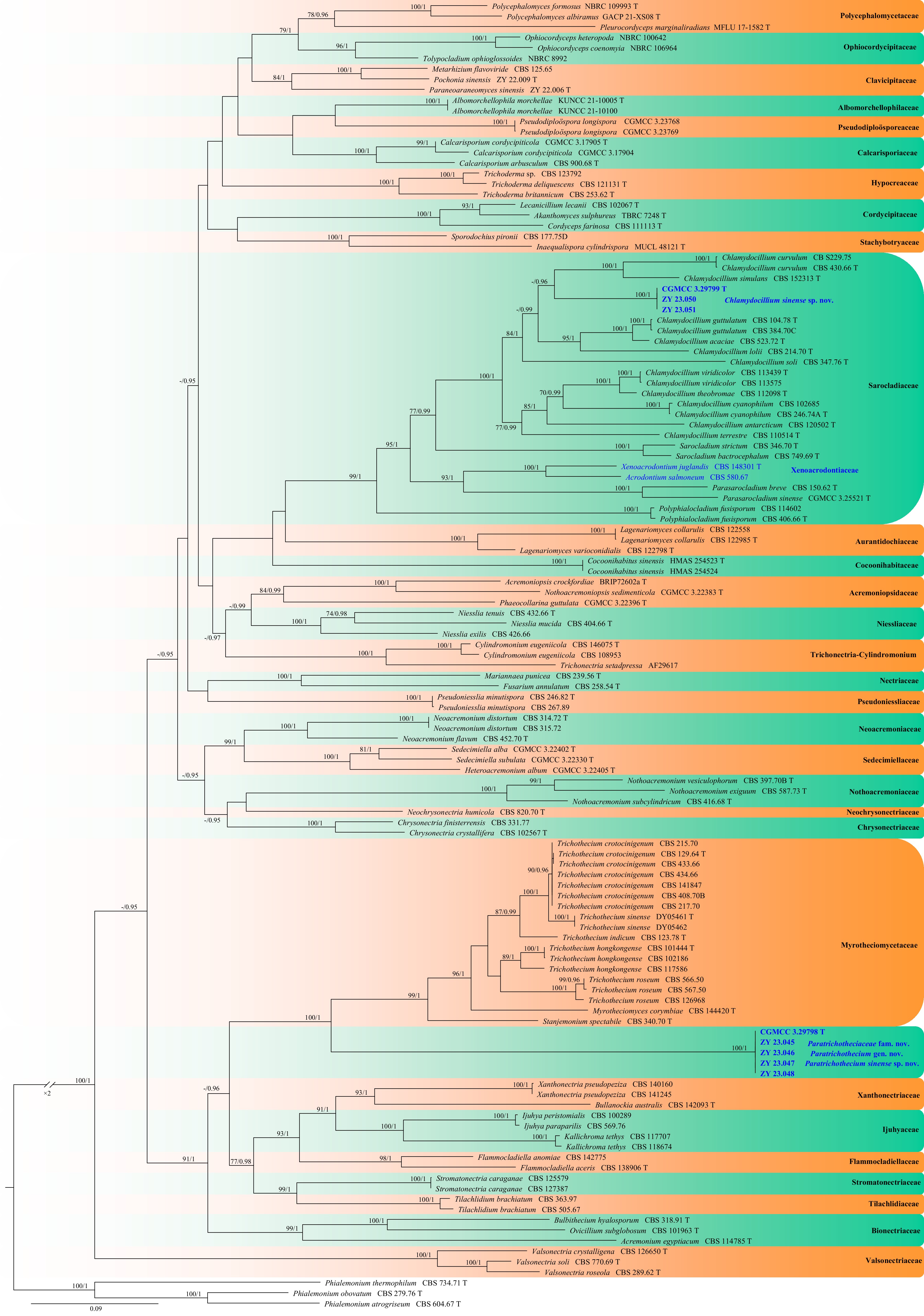
Figure 28.
Phylogram generated from ML analysis based on the combined dataset of ITS, LSU, RPB2, and TEF1 sequence data. Numbers at branches indicate support values (IQ-TREE-BS/BI-PP) above 70%/0.90. The tree is rooted to Phialemonium atrogriseum (CBS 604.67), P. obovatum (CBS 279.76), and P. thermophilum (CBS 734.71). Newly generated sequences are in bold and blue, and 'T' indicates type-derived sequences. The scale bar represents the expected number of changes per site.
Paratrichotheciaceae Y.J. Wu, Zhi Y. Zhang & Y.F. Han fam. nov.
MycoBank number: MB862623
Etymology: Named after the type genus Paratrichothecium.
Description: Hyphae branched, septate. Asexual morph Conidiophores simple, rarely branched, laterally arising from vegetative hyphae, straight. Conidiogenous cells phialidic, solitary, cylindrical to clavate, monophialidic, occasionally two borne on the same conidiophore, tapering towards the apex. Conidia unicellular, subcylindrical to cylindrical, aggregated in heads. Sexual morph not observed.
Type: Paratrichothecium Y.J. Wu, Zhi Y. Zhang & Y.F. Han
Notes: In this study, multi-locus (ITS, LSU, RPB2, and TEF1) phylogenetic analyses resolved the new family as a distinct lineage closely allied to Myrotheciomycetaceae, which is diagnosed by superficial conidiomata, unbranched to branched conidiophores (solitary or sporodochial, with or without a basal stroma), phialidic or retrogressively conidiogenous cells, and zero-to-one-septate, fusoid-ellipsoid conidia with a subobtuse apex and a truncate, unthickened wall — features that together set the new family apart from Paratrichotheciaceae[112].
Paratrichothecium Y.J. Wu, Zhi Y. Zhang & Y.F. Han gen. nov.
MycoBank number: MB862624
Etymology: Named after the genus Trichothecium, indicating its phylogenetic proximity to Trichothecium.
Description: Hyphae branched, septate, hyaline, smooth-walled. Asexual morph Conidiophores simple, rarely branched, laterally arising from vegetative hyphae, straight. Conidiogenous cells phialidic, solitary, cylindrical to clavate, smooth- and thin-walled, hyaline, monophialidic, occasionally two borne on the same conidiophore, tapering towards the apex. Conidia unicellular, smooth- and thin-walled, hyaline, subcylindrical to cylindrical, aggregated in heads. Chlamydospores and sexual morph not observed.
Type: Paratrichothecium sinense Y.J. Wu, Zhi Y. Zhang & Y.F. Han
Paratrichothecium sinense Y.J. Wu, Zhi Y. Zhang & Y.F. Han sp. nov. Fig. 29
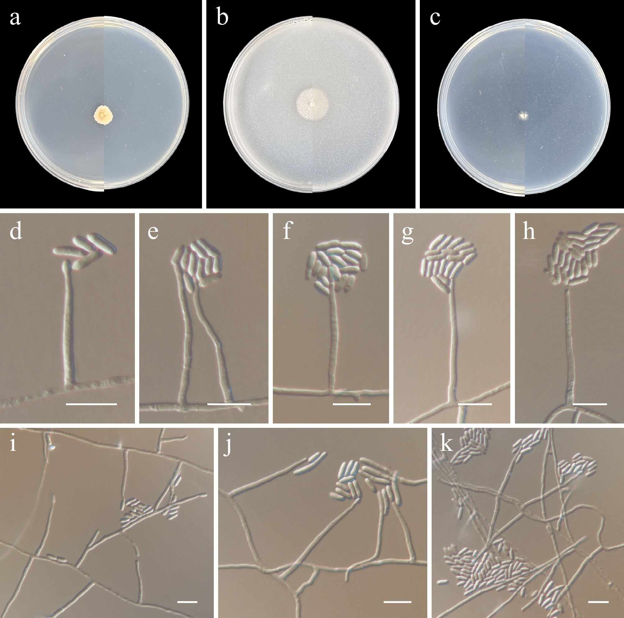
Figure 29.
Paratrichothecium sinense (from holotype ZY H-23.044). (a)–(c) Upper and reverse views of cultures on PDA, OA, and SNA after 14 d at 25 °C. (d)–(k) Phialidic and conidia. (d)–(k) Scale bars: 10 μm.
MycoBank number: MB862625
Etymology: Named after the type locality, China.
Description: Hyphae branched, septate, hyaline, smooth-walled, 1–2 μm wide. Asexual morph Conidiophores simple, rarely branched, laterally arising from vegetative hyphae, straight. Conidiogenous cells phialidic, solitary, cylindrical to clavate, smooth- and thin-walled, hyaline, monophialidic, occasionally two borne on the same conidiophore, tapering towards the apex, 20.5–89 × 2.5–4 μm. Conidia unicellular, smooth- and thin-walled, hyaline, subcylindrical to cylindrical, aggregated in heads, 7.5–19.5 × 3.5–5 μm (av. 11.9 × 3.8, n = 30 μm). Chlamydospores and sexual morph not observed.
Culture characteristics (14 d at 25 °C): Colony on PDA reaching 7–11 mm diam., flocculent, wrinkled, pure white, margin entire; reverse pure white. Colony on OA reaching 15–17 mm diam., flat, flocculent, white, margin entire; reverse white. Colony on SNA growing slowly, reaching 4–5 mm diam., flat, sparse aerial mycelium, white; reverse white.
Material examined: China, Heilongjiang Province, Harbin City, soil collected from the green belts of Harbin Northern Forest Zoo, 127.1050° E, 45.3952° N, 8 May 2023, Zhi-Yuan Zhang (holotype ZY H-23.044, dried culture; culture ex-type CGMCC 3.29798, ibid., ZY 23.044). China, Heilongjiang Province, Harbin City, soil collected from the green belts of Harbin Northern Forest Zoo, 127.1131° E, 45.3912° N, 8 May 2023, Zhi-Yuan Zhang, isotype ZY H-23.045, ex-isotype ZY 23.045, ibid., ZY 23.046, ibid., ZY 23.047, ibid., ZY 23.048.
Notes: A megablast search of the ITS sequence against the NCBI nucleotide database revealed the highest similarity to two uncultured fungal sequences: uncultured fungus [isolate OTU 229, GenBank KT328709.1; Identities = 535/550 (97%), two gaps (0%)] and uncultured fungus [clone 3993_661, GenBank MW215116.1; Identities = 496/532 (93%), 15 gaps (2%)]. For the LSU sequence, the closest match was Claviceps panicoidearum [TNS:F-96498, GenBank LC681738.1; Identities = 829/884 (94%), six gaps (0%)]. In contrast, a blastn search of the TEF1 sequence returned only distant matches, the closest being Myrothecium roridum [strain 785, GenBank JF724135.1; Identities = 848/954 (89%), 20 gaps (2%)]. Similarly, the RPB2 sequence showed highest similarity to Volutella minutissima [voucher GMB5311, GenBank PX373366.1; Identities = 795/1071 (74%), 45 gaps (4%)].
Sarocladiaceae L. Lombard
Synonym: Xenoacrodontiaceae Crous
The family Xenoacrodontiaceae was established by Crous et al. to accommodate Xenoacrodontium, a monospecific hyphomycetous genus[40]. To date, the genus is represented by a single species, X. juglandis, described from Juglans regia (Juglandaceae) in the Netherlands[40]. Crous et al., when establishing Xenoacrodontiaceae, conducted a phylogenetic analysis with very limited taxon sampling, which did not include data from most families of Hypocreales, and used only LSU sequences[40]. Interestingly, two recent comprehensive taxonomic studies on acremonium-like fungi in Hypocreales have overlooked Xenoacrodontiaceae[111,113]. In this study, the type strain of Xenoacrodontium juglandis and another strain CBS 580.67 (‘Acrodontium salmoneum’) were nested within the Sarocladiaceae clade (Fig. 28). Therefore, we suggest treating Xenoacrodontiaceae as a synonym of Sarocladiaceae, because Sarocladiaceae is the earlier name. Accordingly, Xenoacrodontium is herein transferred to Sarocladiaceae. Consequently, this family currently comprises five genera: Chlamydocillium, Parasarocladium, Polyphialocladium, Sarocladium, and Xenoacrodontium.
Xenoacrodontium Crous
Type: Xenoacrodontium juglandis Crous
Notes: Crous et al. erected the genus Xenoacrodontium, which is monotypic, comprising only X. juglandis[40], a species distinguished from other Sarocladiaceae genera by its subulate, sympodially proliferating conidiogenous cells bearing a rachis with multiple subdenticulate loci[40]. According to Videira et al., Acrodontium is classified within Teratosphaeriaceae, yet the genus is polyphyletic, since a number of acrodontium-like taxa actually belong to other orders[114]. One such isolate, CBS 580.67 (a Houston, Texas, USA culture contaminant identified as A. salmoneum), clustered with X. juglandis CBS 148301 in a subclade within Sarocladiaceae and may represent an undescribed second species[40]; as the strain was unavailable to us, however, we withhold formal description at this time.
Chlamydocillium Zare & W. Gams
Chlamydocillium was proposed by Zare and Gams for C. cyanophilum, a soil-derived species with a verticillium-like asexual morph and numerous intercalary chlamydospores, but was initially not assigned to any family[115]. Subsequently, Hou et al. placed the genus in Sarocladiaceae based on phylogenetic evidence[113]. To date, the genus Chlamydocillium comprises 11 species, with sequences available for all species[111,113,116].
Chlamydocillium sinense Y.J. Wu, Zhi Y. Zhang & Y.F. Han sp. nov. Fig. 30
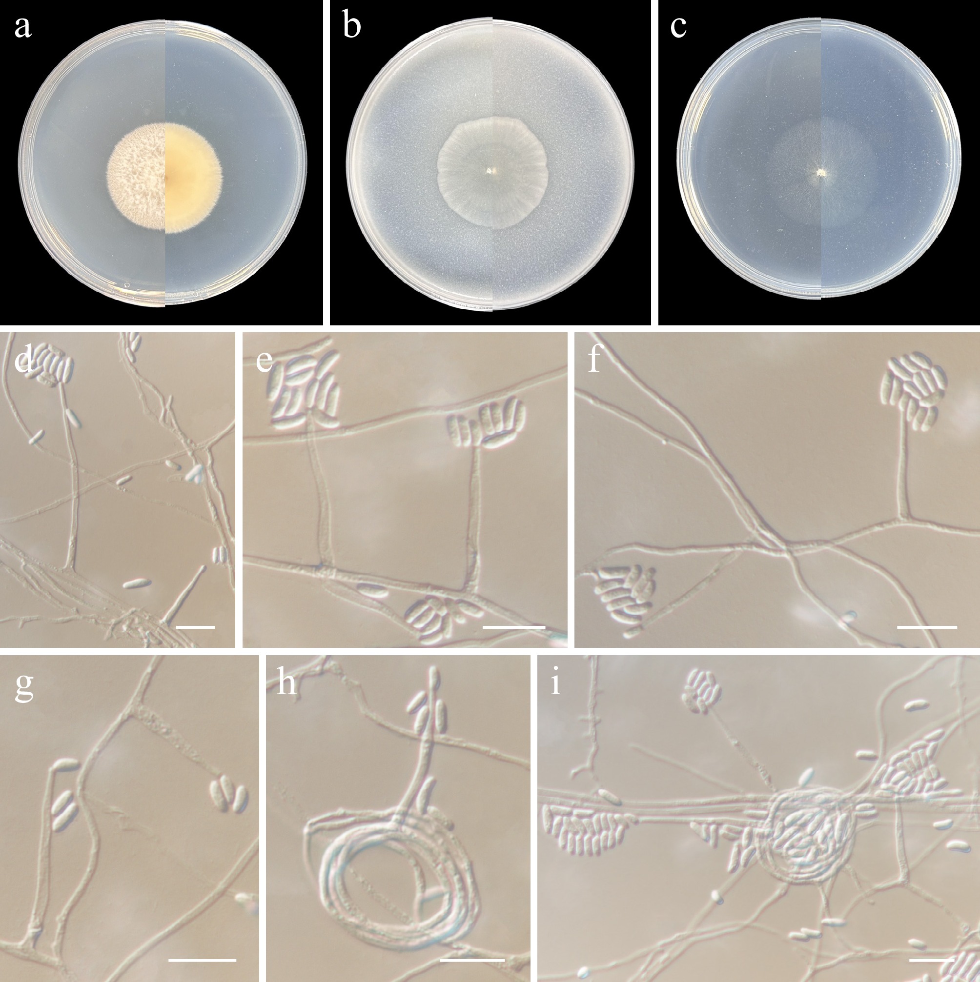
Figure 30.
Chlamydocillium sinense (from holotype ZY H-23.049). (a)–(c) Upper and reverse views of cultures on PDA, OA, and SNA after 14 d at 25 °C. (d)–(g) Monophialides. (h), (i) Conidiophores radiating out from coils formed by the mycelia. (d)–(i) Scale bars: 10 μm.
MycoBank number: MB862626
Etymology: Named after the type locality, China.
Description: Hyphae branched, septate, hyaline, smooth-walled, up to 2 μm wide. Asexual morph Sporulation abundant, phalacrogenous, rarely nematogenous. Conidiophores solitary, erect, arising directly from aerial, substratal mycelium, or mycelial coils, unbranched, up to 15 μm long, 0.5–2 μm wide at base, aseptate or with a single septum in basal part, hyaline, smooth-walled. Phialides lateral, subulate, or subcylindrical, hyaline, smooth-walled, 10.5–49 μm long, 1–2.5 μm wide at base, commonly with inconspicuous periclinal thickening and collarette at conidiogenous loci; polyphialides not observed. Conidia aseptate, cylindrical, ellipsoid, or short reniform, straight or curved, with both ends rounded, slightly apiculate at the base, hyaline, thin-, smooth-walled, 3–8.5 × 1.5–2.5 μm (av. 5 × 1.9, n = 30 μm), eguttulate, arranged in slimy heads. Chlamydospores and sexual morph not observed.
Culture characteristics (14 d at 25 °C): Colony on PDA reaching 34–35 mm diam., flat, densely and radially covered thick, mycelial ropes, felty, moist, creamy white, margin filiform, reverse apricot, with apricot radial lines. Colony on OA reaching 34–35 mm diam., flat, membranous with sparse aerial mycelium, white, margin entire, reverse concolourous. Colony on SNA reaching 33–34 mm diam., flat, sparse aerial mycelium, white, margin entire, reverse concolourous.
Material examined: China, Heilongjiang Province, Harbin City, soil collected from the green belts of Harbin Northern Forest Zoo, 127.1019° E, 45.3977° N, 8 May 2023, Zhi-Yuan Zhang (holotype ZY H-23.049, dried culture; culture ex-type CGMCC 3.29799, ibid., ZY 23.049). China, Heilongjiang Province, Harbin City, soil collected from the green belts of Harbin Northern Forest Zoo, 127.1097° E, 45.3926° N, 8 May 2023, Zhi-Yuan Zhang, isotype ZY H-23.050, ex-isotype ZY 23.050, ibid., ZY 23.051.
Notes: Phylogenetically, Chlamydocillium sinense formed an independent lineage distinct from other Chlamydocillium species (Fig. 28). Morphologically, C. sinense can be distinguished from congeneric species by the following features: chlamydospores absent; phialides arising directly from aerial or substratal mycelium, or from mycelial coils; and polyphialides and adelophialides absent[111,113,115,116].
Sordariales Chadef. ex D. Hawksw. & O.E. Erikss.
Lasiosphaeriaceae Nannf.
Nannfeldt erected the family Lasiosphaeriaceae[117], which is the largest and most diverse family of the Sordariales. However, the family remains a paraphyletic group. It usually comprises coprophilous plant debris inhabitants and soil-borne taxa that develop paraphysate ascomata with different ascomatal wall structures, cylindrical or clavate unitunicate asci, which usually possess a non-amyloid apical apparatus, and mostly two-celled ascospores with a dark apical cell and a hyaline basal cell, smooth-walled or with different types of ornamentation, with germ pores and usually bearing mucilaginous appendages[118].
Phylogenetic relationships of the genus Heilongjiangomyces and related taxa were inferred using a multi-locus (ITS, LSU, RPB2, and TUB) dataset, with Microthecium fimicola (CBS 967.97), M. quandrangulatum (CBS 112763), and M. tenuissimum (CBS 112764) designated as outgroup taxa. The final alignment included 53 taxa and spanned 2,342 nucleotide positions (ITS, 537 bp; LSU, 524 bp; RPB2, 845 bp; and TUB, 436 bp), including gaps. The best-fit models were TIM2 + F + I + G4 (ITS, TUB) and TIM3 + F + I + G4 (LSU, RPB2) for the ML analysis, whereas those for the BI analysis were GTR + F + I + G4 (ITS, LSU, and RPB2) and GTR + I + G4 (TUB). Phylogenetically, CGMCC 3.29800, ZY 23.053, ZY 23.054, and ZY 23.055 formed a single, well-supported clade (100/1, Fig. 31).
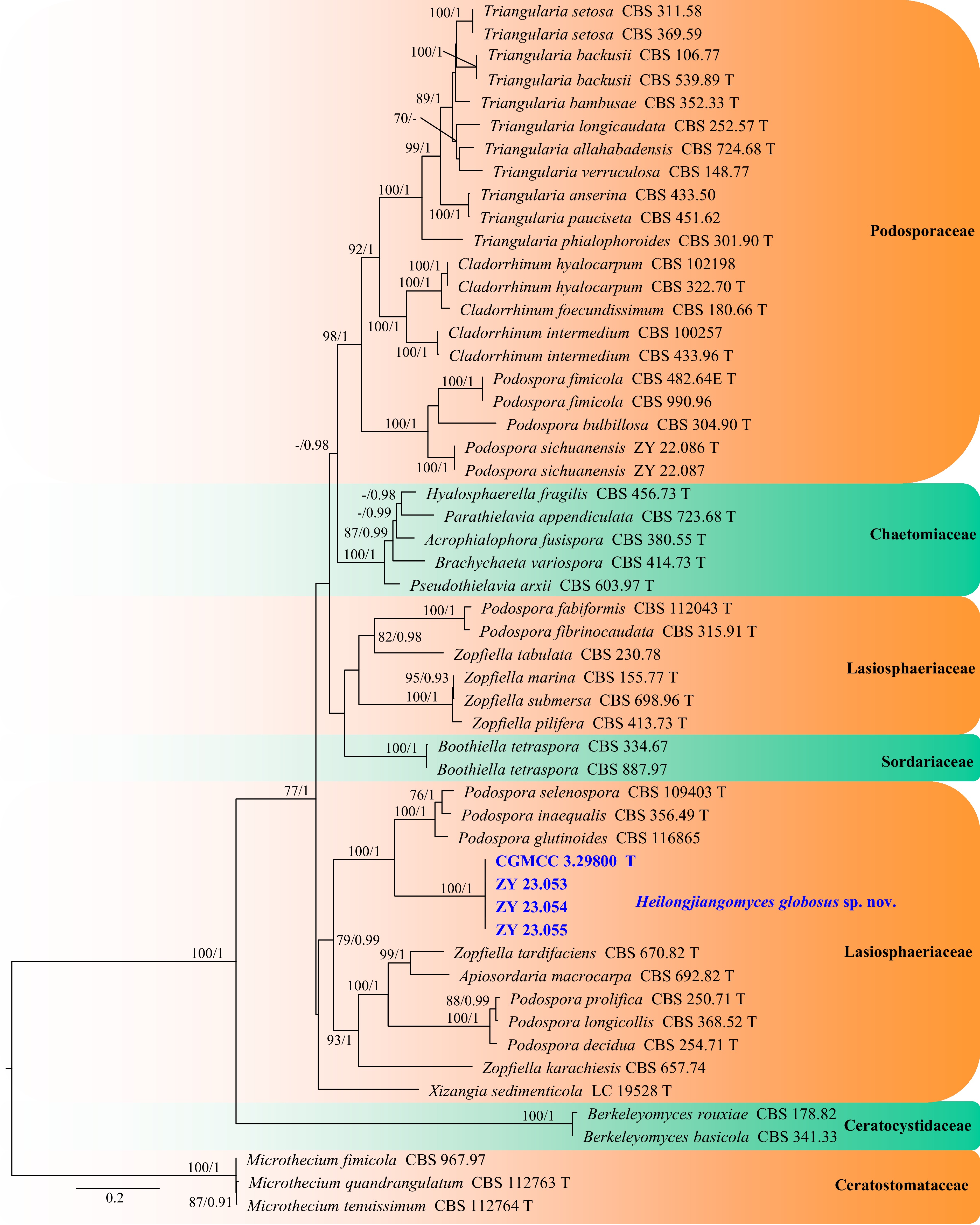
Figure 31.
Phylogram generated from ML analysis based on the combined dataset of ITS, LSU, RPB2, and TUB sequence data. Numbers at branches indicate support values (IQ-TREE-BS/BI-PP) above 70%/0.90. The tree is rooted to Microthecium fimicola (CBS 967.97), M. quandrangulatum (CBS 112763), and M. tenuissimum (CBS 112764). Newly generated sequences are in bold and blue, and 'T' indicates type-derived sequences. The scale bar represents the expected number of changes per site.
Heilongjiangomyces Y.J. Wu, Zhi Y. Zhang & Y.F. Han, gen. nov.
MycoBank number: MB862628
Etymology: Named after the type locality, Heilongjiang Province, China.
Description: Hyphae, branched, septate, smooth-walled. Asexual morph Conidiophores solitary, erect, hyaline, smooth, subcylindrical to cylindrical, aseptate, usually reduced to conidiogenous cells. Conidiogenous cells solitary, hyaline, smooth, flask-shaped to lageniform, cylindrical to subcylindrical. Conidia hyaline, smooth, aseptate, subglobose to globose, arranged in slimy heads. Sexual morph not observed.
Type species: Heilongjiangomyces globosus Y.J. Wu, Zhi Y. Zhang & Y.F. Han
Notes: According to Hyde et al., Sordariales comprises 11 families[4]. Some of these families have been taxonomically clarified, such as Chaetomiaceae and Podosporaceae[119,120]. However, many groups still require the collection of new specimens and molecular data for confirmation. For example, Zopfiella is polyphyletic, with its members distributed across multiple families, including Bombardiaceae, Lasiosphaeriaceae, Naviculisporaceae, and Schizotheciaceae[119−121]. Most taxa in Sordariales are established based on teleomorphs, and the anamorphs of many groups remain unclear. Recently, Phurbu et al. established Xizangia, a genus belonging to Lasiosphaeriaceae, based on anamorphic characters[122], which belongs to Lasiosphaeriaceae. We herein introduce Heilongjiangomyces gen. nov. As in Xizangia, the teleomorph of this genus remains unknown, and the two genera share morphological similarity. However, the conidiogenous cells of Heilongjiangomyces are flask-shaped to lageniform, cylindrical to subcylindrical.
Heilongjiangomyces globosus Y.J. Wu, Zhi Y. Zhang & Y.F. Han sp. nov. Fig. 32
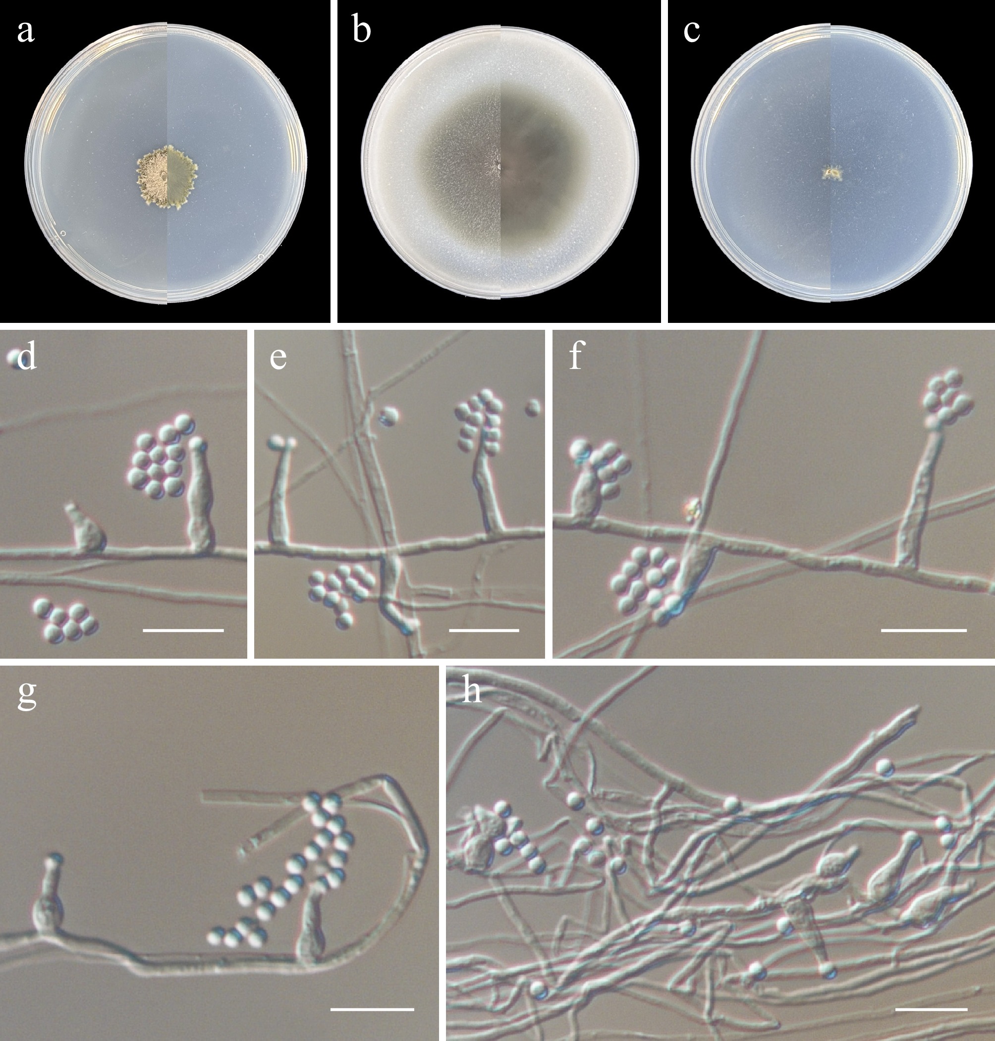
Figure 32.
Pseudosoloacrosporiella sinensis (from holotype ZY H-23.052). (a)–(c) Upper and reverse views of cultures on PDA, OA, and SNA after 14 d at 25 °C. (d)–(h) Conidiogenous cells and conidia. Scale bars: (d)–(h) 10 μm.
MycoBank number: MB862629
Etymology: The epithet 'globosus' refers to the globose conidia of this species.
Description: Hyphae, branched, septate, hyaline, smooth-walled, 0.5–2.5 µm diam. Asexual morph Conidiophores solitary, erect, hyaline, smooth, subcylindrical to cylindrical, aseptate, usually reduced to conidiogenous cells. Conidiogenous cells solitary, hyaline, smooth, flask-shaped to lageniform, 4.5–11 µm long, 3–4 µm wide at the base, 1–2 µm wide at the apex, or cylindrical to subcylindrical, 11–24.5 × 1.5–2.5 µm. Conidia hyaline, smooth, aseptate, subglobose to globose, 1.5–3 µm, arranged in slimy heads. Sexual morph not observed.
Culture characteristics (14 d at 25 °C): Colony on PDA reaching 17–21 mm diam., flocculent, dense, dark ivory, margin irregular; reverse beige grey. Colony on OA reaching 52–55 mm diam., flat, pebble grey, aerial mycelia sparse, reverse pebble grey. Colony on SNA reaching 5–7 mm diam., flocculent, white, aerial mycelia sparse, reverse white.
Material examined: China, Heilongjiang Province, Harbin City, soil collected from the green belts of Harbin Northern Forest Zoo, 127.1133° E, 45.3921° N, 8 May 2023, Zhi-Yuan Zhang (holotype ZY H-23.052, dried culture; culture ex-type CGMCC 3.29800, ibid., ZY 23.052). China, Heilongjiang Province, Harbin City, soil collected from the green belts of Harbin Northern Forest Zoo, 127.1027° E, 45.3975° N, 8 May 2023, Zhi-Yuan Zhang, isotype ZY H-23.053, ex-isotype ZY 23.053, ibid., ZY 23.054, ibid., ZY 23.055.
Notes: A megablast search of the ITS sequences against the NCBI nucleotide database revealed the highest similarity to a series of uncultured fungal clones: uncultured fungus [clone CMH204, GenBank KF800295.1; Identities = 523/526 (99%), three gaps], uncultured fungus [clone OTU925, GenBank MF971821.1; Identities = 520/527 (99%), three gaps], uncultured Sordaria [clone 10J70C48, GenBank HG937028.1; Identities = 519/527 (98%), three gaps], uncultured Sordaria [clone 10S50C02, GenBank HG937029.1; Identities = 519/527 (98%), three gaps], uncultured Lasiosphaeriaceae [clone NG_N_G12, GenBank GU055601.1; Identities = 519/527 (98%), four gaps], uncultured soil fungus [clone C149, GenBank JX489839.1; Identities = 518/527 (98%), three gaps], and uncultured fungus [clone YF6, GenBank KJ734954.1; Identities = 518/527 (98%), three gaps]. For the LSU sequence, the closest match was likewise an uncultured Lasiosphaeriaceae clone [NG_N_G12, GenBank GU055601.1; Identities = 520/523 (99%), one gap (0%)]. In contrast, a blastn search of the RPB2 sequence returned only distant matches, the closest being Podospora fimbriata [strain CBS 144.54, GenBank AY780189.1; Identities = 691/844 (82%), 11 gaps (1%)]. Similarly, the TUB sequence showed highest similarity to Schizothecium aloides [strain CBS 879.72, GenBank AY999159.1; Identities = 352/432 (81%), 30 gaps (6%)].
Xylariales Nannf.
Xylariaceae Tul. & C. Tul.
Ascotricha Berk.
Berkeley established Ascotricha, designating A. chartarum as its type species[123]. The genus is diagnosed by ascomata bearing long hairs with shorter, hyaline branches, brown, ellipsoidal ascospores possessing an equatorial germ slit, and asexual morphs of the geniculosporium type[123,124].
Phylogenetic relationships of the genus Ascotricha were inferred using a combined ITS and TUB dataset, with Lopadostoma dryophilum (CBS 133213) and L. turgidum (CBS 133207) designated as the outgroup. The final alignment included 19 taxa and spanned 1,102 nucleotide positions (ITS, 536 bp; TUB, 566 bp), including gaps. The best-fit models were TIM2e + G4 (ITS) and TPM2u + F + G4 (TUB) for the ML analysis, whereas those for the BI analysis were GTR + F + G4 (ITS) and HKY + F + G4 (TUB). Phylogenetically, CGMCC 3.29801 and ZY 23.057 formed a single, well-supported clade (100/1, Fig. 33).
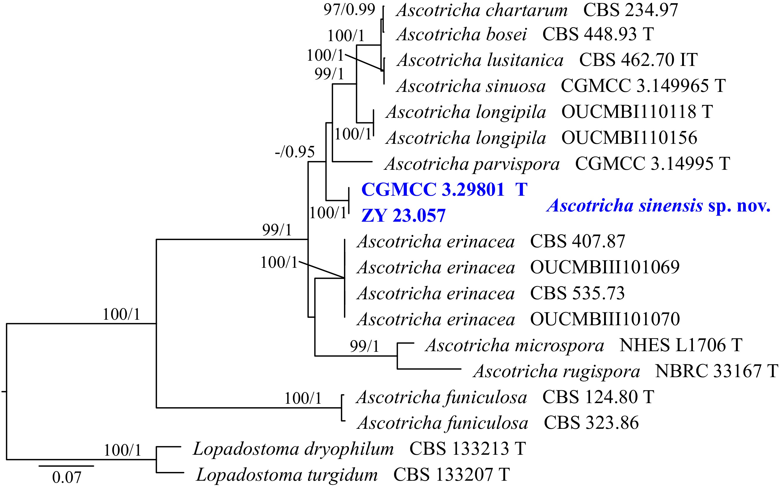
Figure 33.
Phylogram generated from ML analysis based on the combined dataset of ITS and TUB sequence data. Numbers at branches indicate support values (IQ-TREE-BS/BI-PP) above 70%/0.90. The tree is rooted to Lopadostoma dryophilum (CBS 133213) and L. turgidum (CBS 133207). Newly generated sequences are in bold and blue, and 'T' indicates type-derived sequences. The scale bar represents the expected number of changes per site.
Ascotricha sinensis Y.J. Wu, Zhi Y. Zhang & Y.F. Han sp. nov. Fig. 34
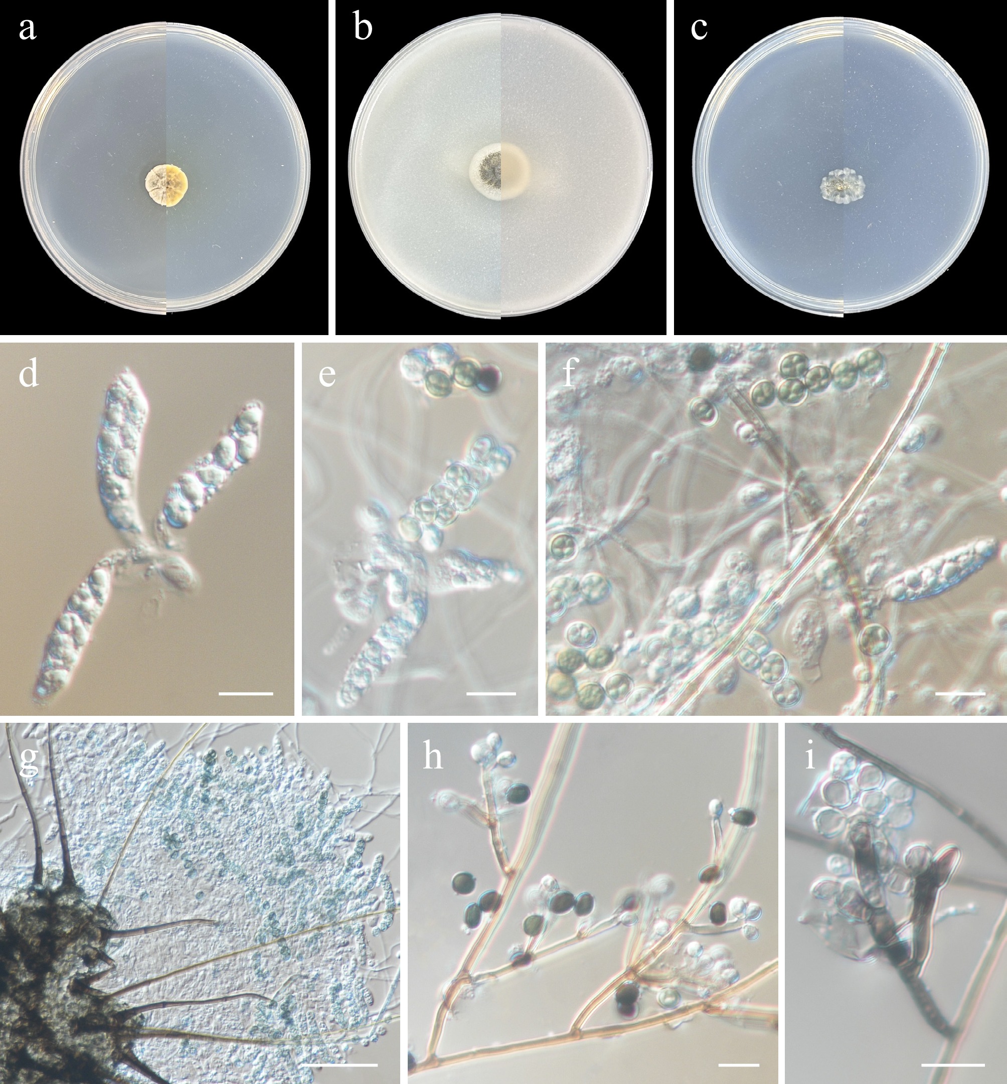
Figure 34.
Ascotricha sinensis (from holotype ZY H-23.056). (a)–(c) Upper and reverse views of cultures on PDA, OA, and SNA after 14 d at 25 °C. (d)–(f) Asci and ascospores. (g) Ascomata and asci. (h), (i) Conidiophores, conidiogenous cells, and conidia. Scale bars: (d)–(i) 10 μm.
MycoBank number: MB862630
Etymology: Named after the type locality, China.
Description: Hyphae branched, septate, hyaline, smooth-walled, up to 3 μm wide. Sexual morph Ascomata superficial, dark brown to black, ostiolate, subglobose or spherical below, 100–200 μm diam, with a short cylindrical neck. Ascomatal hairs dark brown to black, erect, rigid, whip-like, simple, remotely septate, 3–5.5 μm diam at the base, tapering above, up to 185 μm long; shorter hairs hyaline to black, claviform, 10–12 × 2–3.6 μm. Asci cylindrical, thin-walled, eight-spored, with an inamyloid apical ring, deliquescing after the spores have matured. Ascospores uniseriate, pale brown to dark brown when mature, discoid, 5–7 × 4.5–6 μm (av. = 6.5 × 5.5 μm, n = 30), with several protrusions and multiple furrows. Asexual morph Conidiophores erect, dark brown below, becoming paler or hyaline apically, smooth, length variable, 1.5–2 μm diam. Conidiogenous cells integrated, terminal, slightly geniculate, 9.5–20 × 1.5–2.5 μm (av. = 13.7 × 2.2 μm, n = 30). Conidia ellipsoidal to pyriform, hyaline, 4–6 × 3–5 μm (av. = 5 × 4 μm, n = 30), with inconspicuous warts and a slightly protruding basal hilum.
Culture characteristics (14 d at 25 °C): Colony on PDA reaching 12–13 mm diam., fluffy, elliptical, petal-like, with several furrows, beige to cream, margin slightly undulate; reverse light ivory to cream. Colony on OA reaching 16–18 mm diam., powdery, brown green to white from centre to margin, margin entire, reverse white. Colony on SNA reaching 10–13 mm diam., powdery, brown green to white from centre to margin, margin radially striate with lobate edge, reverse white.
Material examined: China, Heilongjiang Province, Harbin City, soil collected from the green belts of Harbin Northern Forest Zoo, 127.1193° E, 45.3903° N, 8 May 2023, Zhi-Yuan Zhang (holotype ZY H-23.056, dried culture; culture ex-type CGMCC 3.29801, ibid., ZY 23.056). China, Heilongjiang Province, Harbin City, soil collected from the green belts of Harbin Northern Forest Zoo, 127.1193° E, 45.3903° N, 8 May 2023, Zhi-Yuan Zhang, isotype ZY H-23.057, ex-isotype ZY 23.057.
Notes: To date, the genus Ascotricha comprises 19 species[124,125]. Unfortunately, few sequences for Ascotricha are available. Phylogenetically, A. sinensis formed an independent lineage, distinct from other Ascotricha species (Fig. 33). Morphologically, A. sinensis differs from other Ascotricha species in producing both sexual and asexual morphs simultaneously, and in possessing ascospores bearing several protrusions and multiple furrows[124,125].
Nemania Gray
Gray erected the genus Nemania, designating N. serpens as its type species[126]. Species of Nemania are distributed worldwide in temperate and tropical regions[127]. Nemania is characterised by carbonaceous, multiperitheciate, effused-pulvinate stromata with papillate ostioles and variable presence of soft, whitish, brownish, grey or yellow internal tissue, not releasing pigments in 10% KOH, unitunicate cylindrical asci, and ascospores with a conspicuous or inconspicuous germ slit of spore length to much less than spore length[128]. Most Nemania species have been described from sexual morphs, with only a few known from asexual morphs.
Phylogenetic relationships of the genus Nemania were inferred using a multi-locus (ITS, LSU, RPB2, and TUB) dataset, with Coniolarelia limoniispora (MUCL 29409), Rosellinia aquila (MUCL 51703), and R. britannica (MFLU 17-302) designated as the outgroup. The final alignment included 61 taxa and spanned 2,928 nucleotide positions (ITS, 510 bp; LSU, 829 bp; RPB2, 1,039 bp; and TUB, 550 bp), including gaps. The best-fit models were TIM2 + F + I + G4 (ITS), TIM + F + I + G4 (LSU), TIM3 + F + I + G4 (RPB2), and TN + F + I + G4 (TUB) for the ML analysis, whereas GTR + F + I + G4 was applied across all four loci for the BI analysis. Phylogenetically, CGMCC 3.29802 and ZY 23.059 formed a single, well-supported clade (100/1, Fig. 35).
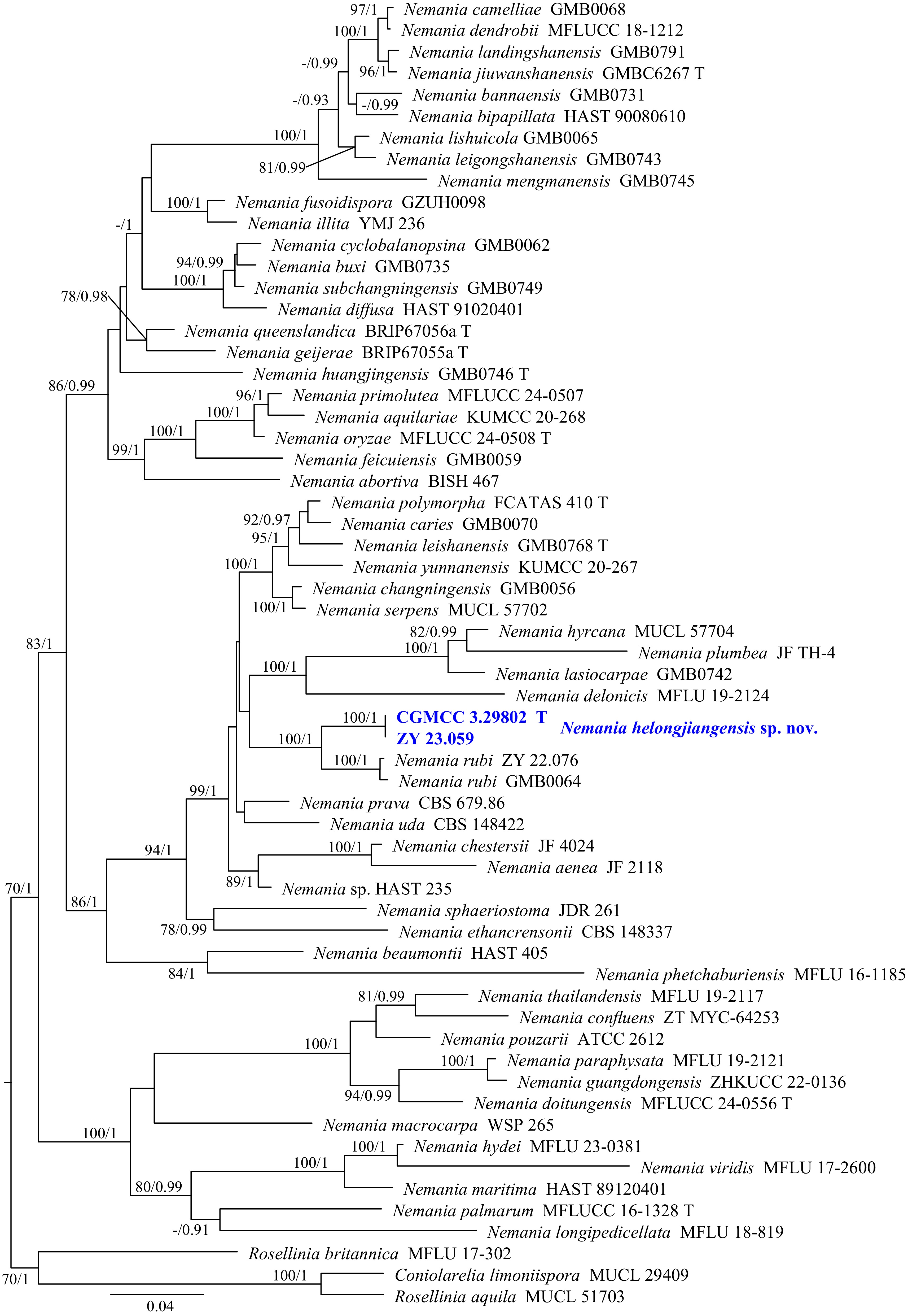
Figure 35.
Phylogram generated from ML analysis based on the combined dataset of ITS, LSU, RPB2, and TUB sequence data. Numbers at branches indicate support values (IQ-TREE-BS/BI-PP) above 70%/0.90. The tree is rooted to Coniolarelia limoniispora (MUCL 29409), Rosellinia aquila (MUCL 51703), and R. britannica (MFLU 17-302). Newly generated sequences are in bold and blue, and 'T' indicates type-derived sequences. The scale bar represents the expected number of changes per site.
Nemania heilongjiangensis Y.J. Wu, Zhi Y. Zhang & Y.F. Han sp. nov. Fig. 36
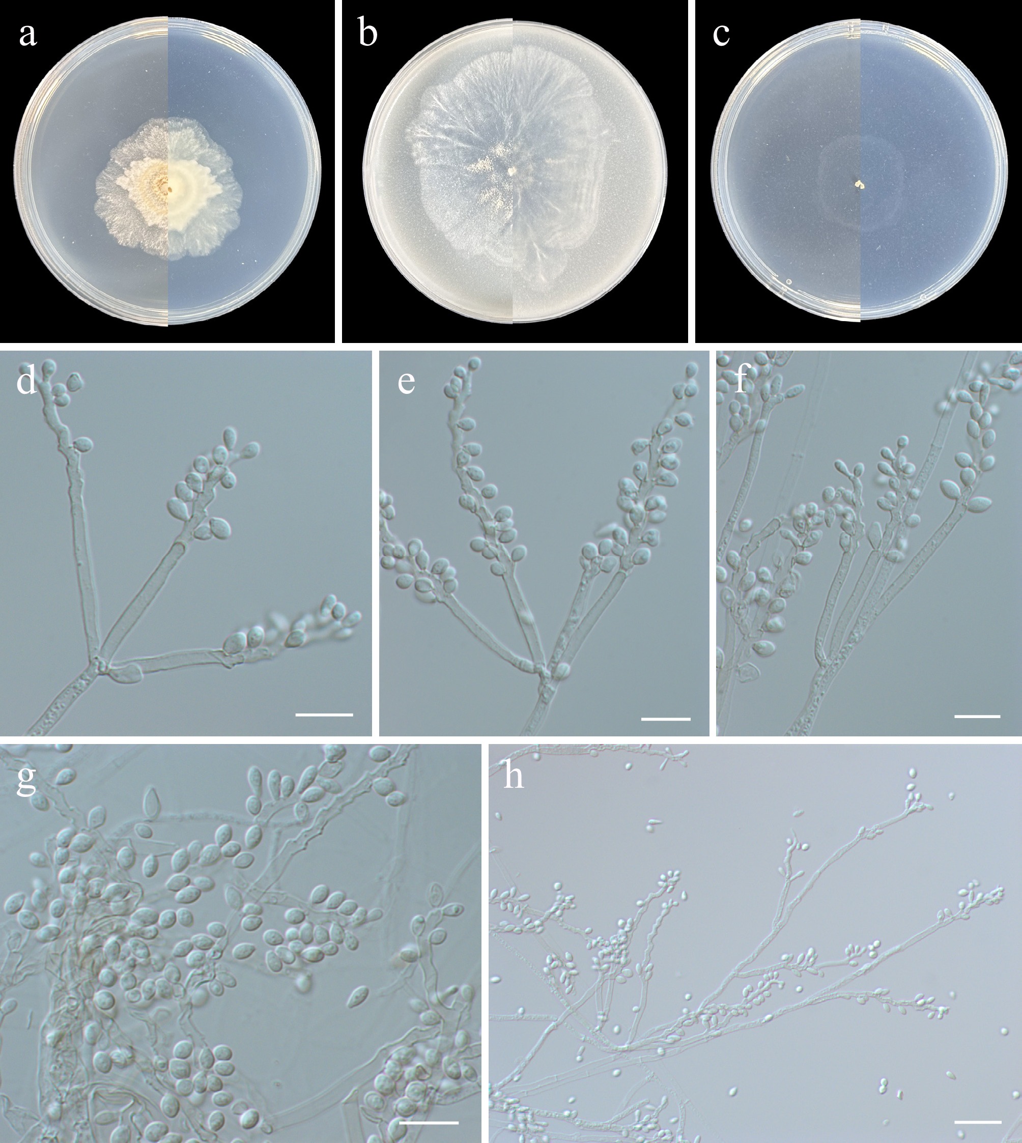
Figure 36.
Nemania heilongjiangensis (from holotype ZY H-23.058). (a)–(c) Upper and reverse views of cultures on PDA, OA, and SNA after 14 d at 25 °C. (d)–(f), (h) Conidiogenous cells and conidia. (g) Conidia. Scale bars: (d)–(g) 10 μm, (h) 20 μm.
MycoBank number: MB862631
Etymology: Named after the type locality, Heilongjiang Province, China.
Description: Hyphae branched, septate, hyaline, smooth-walled, 1–4 μm wide. Asexual morph geniculosporium-like. Conidiophores variable in length, hyaline to light brown. Conidiogenous cells hyaline, smooth, 18.5–79 × 1.5–3 µm, subcylindrical, with a regular lower portion and a curved upper portion bearing numerous conidiogenous loci. Conidia solitary, hyaline, smooth, ellipsoid to subglobose, base truncate, 3–6 × 2–4 µm (av. 4.3 × 2.8 µm, n = 30). Sexual morph not observed.
Culture characteristics (14 d at 25 °C): Colony on PDA reaching 36–42 mm diam., felted, slightly cottony at centre, white to pale yellow, margin irregular, reverse white. Colony on OA reaching 52–66 mm diam., flat, powdery at centre, white, margin irregular, reverse white. Colony on SNA reaching 30–27 mm diam., flat, aerial mycelium sparse, white, reverse white.
Material examined: China, Heilongjiang Province, Harbin City, soil collected from the green belts of Harbin Northern Forest Zoo, 127.1065° E, 45.3962° N, 8 May 2023, Zhi-Yuan Zhang (holotype ZY H-23.058, dried culture; culture ex-type CGMCC 3.29802, ibid., ZY 23.058). China, Heilongjiang Province, Harbin City, soil collected from the green belts of Harbin Northern Forest Zoo, 127.1028° E, 45.3988° N, 8 May 2023, Zhi-Yuan Zhang, isotype ZY H-23.059, ex-isotype ZY 23.059.
Notes: Phylogenetically, Nemania heilongjiangensis is closely related to N. rubi, with high support (100/1, Fig. 35). Pi et al. established N. rubi based on the sexual morph[129], and later Zhang et al. described the asexual morph of this species[6]. Morphologically, N. heilongjiangensis has longer conidiogenous cells compared to N. rubi (18.5–79 × 1.5–3 µm in N. heilongjiangensis vs 50 × 2–3.5 µm in N. rubi)[6]. Additionally, pairwise comparison of the ITS, LSU, RPB2, and TUB loci reveals that N. heilongjiangensis (ex-type CGMCC 3.29802) differs from N. rubi (holotype GMB0064) by 5.4% (26/481 bp, five gaps) in ITS, 1.1% (9/801 bp, no gaps) in LSU, 7.1% (77/1081 bp, no gaps) in RPB2, and 7.1% (25/349 bp, no gaps) in TUB.
-
As one of the most complex ecosystems in nature, soil harbors a vast array of microbial communities. Fungi, the second-largest biological group after insects[130], remain understudied in soil[4], particularly in urban environments. This study systematically investigates soil fungal diversity at the Harbin North Forest Zoo to enhance understanding of this diversity in urban soils.
Currently, fungal taxonomic research is in a phase of rapid development, with new taxa being published at an unprecedented rate. However, this rapid progress is accompanied by growing concerns. An integrative approach, in which several independent lines of evidence are drawn upon for the recognition and circumscription of fungal species, has accordingly gained wide currency among mycologists[6]. Morphological data are thus increasingly assessed in concert with multi-locus phylogenies, an approach that markedly reduces the risk of assigning morphologically convergent but phylogenetically unrelated lineages to a single species[131]. However, phylogenetic analyses must also meet rigorous requirements, most notably adequate breadth and depth of taxon sampling; otherwise, they may yield erroneous results, thereby introducing new confusion into fungal taxonomic studies. The family Xenoacrodontiaceae, reassessed here, illustrates this problem. The family was originally proposed by Crous et al. on the basis of a tree inferred from a narrowly sampled dataset, comprising LSU sequences of only 60 strains drawn from four hypocrealean families[40]. Subsequently, more comprehensive studies on Hypocreales have largely overlooked Xenoacrodontiaceae in their phylogenetic analyses[111,113]. In the present study, a four-locus dataset (ITS, LSU, RPB2, and TEF1) comprising 125 strains was assembled, covering 29 hypocrealean families together with the Trichonectria-Cylindromonium clade. The results indicated that Xenoacrodontiaceae is nested within Sarocladiaceae and is closely related to Parasarocladium (Fig. 28). Xenoacrodontiaceae is therefore reduced to synonymy with Sarocladiaceae, and Xenoacrodontium is recombined accordingly.
Many recently reported higher-level new taxa, such as Catenuliconidiaceae, Longirostraceae, Neochrysonectriaceae, Novolignincolaceae, and Pseudoniessliaceae, are based on a single species[8,111,113,132,133]. Similar to these families, the new families proposed in this study, Paratrichotheciaceae and Subcylindroconidiaceae, each include only a single species. However, phylogenetically, both form distinct subclades and are respectively separated from other families within Hypocreales (Paratrichotheciaceae) and Pleosporales (Subcylindroconidiaceae) (Figs 4 and 28). Furthermore, megablast searches of multiple gene loci against the NCBI GenBank nucleotide database yielded low similarity to target strains or matched only 'uncultured fungus'. To avoid confusion, we tentatively designate them as new families. Nevertheless, to confirm these taxa, additional isolates will be needed in the future.
There is no consensus on the classification of 'dark matter fungi'—those detected only from environmental DNA without physical specimens[134−136]. However, Tedersoo et al. have recently proposed 30 new taxonomic names spanning the phylum-to-order ranks using phylogenetic inference based on long-read sequences that cover most of the ribosomal RNA operon[137]. There is a growing trend to establish new fungal taxa based solely on DNA sequences, which can, to some extent, provide legitimate status for taxa that are unculturable under laboratory conditions[138]. The fungal taxonomic system is still being refined. Many described species still lack molecular sequences, and it is concerning whether the current classification system, built on existing data, can withstand the impact of DNA-only naming. Until a consensus is reached, we propose that the following points be considered when establishing new fungal taxa: (1) Morphological descriptions must be provided for taxa that sporulate in natural environments and/or under cultivation conditions; (2) For taxa that are culturable in the laboratory but do not sporulate, multi-locus phylogenetic analyses must be included, incorporating at least the ITS, LSU, and one protein-coding gene; (3) For taxa unculturable under laboratory conditions, a more conservative approach should be adopted, requiring additional gene loci or even genomic data.
-
This study employed a culture-based approach using three media to systematically investigate fungal diversity in soils from the Harbin Northern Forest Zoo. The findings enhance our understanding of soil fungi in environments significantly impacted by human activities. Furthermore, by integrating morphological characteristics with phylogenetic analysis, the study established two new fungal families, four new genera, and twenty new species, thereby advancing our knowledge of fungal taxonomy.
-
Not applicable.
-
The authors confirm their contributions to the paper as follows: conceptualization: Zhang ZY, Han YF; methods, software, experiment and data organization: Wu YJ, Zhang MY, Gu RY, Feng Y; writing original draft: Wu YJ, Zhang ZY; review and editing: Sun BD, Wang Y, Tao G, Zhang ZY, Han YF, Feng Y; funding acquisition: Zhang ZY. All authors reviewed the results and approved the final version of the manuscript.
-
The data that support the findings of this study are available in the National Center for Biotechnology Information (NCBI) databases and the MycoBank repository.
-
The authors declare no competing interests.
-
accompanies this paper online at: https://doi.org/10.48130/mycosphere-0026-0021.
- Supplementary Table S1 Isolates and sequence accession numbers of new species.
- Supplementary Table S2 The reference isolates and their GenBank accession numbers used for phylogenetic analysis in this study.
- Supplementary Table S3 All species obtained in this study.
- Copyright: © 2026 by the author(s). Published by Maximum Academic Press, Fayetteville, GA. This article is an open access article distributed under Creative Commons Attribution License (CC BY 4.0), visit https://creativecommons.org/licenses/by/4.0/.
-
About this article
Cite this article
Wu YJ, Zhang MY, Gu RY, Sun BD, Wang Y, et al. 2026. Culturable mycobiota from Harbin North Forest Zoo in China. Mycosphere 17: e020 doi: 10.48130/mycosphere-0026-0021 

Culturable mycobiota from Harbin North Forest Zoo in China
- Received: 13 March 2026
- Revised: 28 August 2026
- Accepted: 09 October 2026
- Published online: 29 October 2026
Abstract: The Harbin North Forest Zoo is an important center for science education, featuring a favorable ecological environment and rich vegetation with a forest cover exceeding 95.8%. Such conditions are conducive to substantial fungal diversity, as soil-inhabiting fungi are known to contribute critically to nutrient turnover and the broader stability of ecosystem processes. Systematic investigations of soil fungal taxa in wildlife parks remain limited at present. This study investigated soil fungal taxa in Harbin North Forest Zoo using culture-dependent methods to expand knowledge of fungal diversity in these habitats. In total, 278 fungal taxa were recovered, spanning four phyla, 14 classes, 47 orders (three undetermined), 99 families (ten undetermined), 168 genera (seven undetermined), and 278 species (44 undescribed taxa). Based on this dataset, comprehensive taxonomic descriptions together with multi-locus phylogenetic inferences are presented for two novel families, four novel genera, and 20 novel species. The newly introduced taxa include the following: Ascotricha sinensis, Calyptellomyces sinensis, Chlamydocillium sinense, Cistella sinensis, Codinaea heilongjiangensis, Coniochaeta heilongjiangensis, Exophiala heilongjiangensis, Heilongjiangomyces globosus, Hyalodendriella sinensis, Leptodontidium sinense, Nannizziopsis sinensis, Nemania heilongjiangensis, Paratrichothecium sinense, Penicillium heilongjiangense, Penicillium zoologicum, Peripyrenochaetopsis sinensis, Pseudosoloacrosporiella sinensis, Scolecobasidium heilongjiangense, Subcylindroconidium sinense, and Westerdykella heilongjiangensis. Of these, Paratrichotheciaceae (Paratrichothecium sinense) and Subcylindroconidiaceae (Subcylindroconidium sinense) are introduced as two new families, while Calyptellomyces (C. sinensis), Subcylindroconidium (S. sinense), Paratrichothecium (P. sinense), and Heilongjiangomyces (H. globosus) are introduced as new genera. This study further demonstrates that wildlife parks harbor high fungal diversity, including numerous previously undescribed taxa, thereby enhancing our understanding of soil fungal taxa in anthropogenic environments.
-
Key words:
- 20 New taxa;
- Fungal diversity;
- Multi-locus phylogeny;
- Taxonomy;
- Urban soils fungi