













-
Wild plants in general, and pulses in particular, have a vast and almost unexplored potential to provide novel protein sources and more sustainable food chains[1]. The use of lupin (Lupinus sp.) seeds as food and stock feed has attracted much interest in recent years due to their potential as an alternative protein source, as they contain very high levels of amino acids and nitrogenous compounds[2−4]. Edible seeds of lupins are a valuable pulse crop with high levels of protein (30%–40%), carbohydrates and fiber (up to 40%), minerals, vitamins, and other beneficial ingredients, including alkaloids and polyphenols[5−9]. Nutritional values rank this genus as a promising agricultural crop today and in the future[10]. Lupins also have the potential to help diversify agriculture with 'new' native plant crops. This approach seeks to achieve higher resilience for agricultural systems through diverse cropping. Enriching agriculture and food diversity by de novo domestication or commercialization of local plants is essential in a changing global economy[2,11−14].
Today, only four species of lupins are used commercially: L. angustifolius, L. luteus, L. mutabilis, and L. albus[15]. Wild lupins, on the other hand, are not considered food crops, primarily because of their bitterness and potential health risks[16]. Bitterness in lupin species is conventionally attributed to their quinolizidine alkaloid (QA) content, which are secondary metabolites synthesized by species of the Fabaceae family and, more specifically, the Genisteae tribe[17]. Different lupin species accumulate various amounts of these toxic and bitterness-contributing compounds in their seeds. The QA content of the four edible species is significantly lower than that in the non-edible species[18]. In recent years, many attempts have been made to decipher QA biosynthetic pathways in lupin seeds, resulting in potential ways to manipulate their accumulation[6,7,18−22].
In many plant species, including lupins, toxicity effects can act as a two-edged sword, i.e., they can be harmful and beneficial depending on the intended usage, ecological context, time and dosage exposure, etc. Alkaloids, in particular, can act as defense compounds in plants against pathogens and predators due to their relative toxicity[23]. Understanding alkaloid biosynthesis and mechanisms of action is essential to monitor the production of specific alkaloids, discover new bioactive molecules, and sustainably exploit them against targets of interest, such as herbivores, pathogens, or human physiological conditions[24].
Historically, mammals were fed lupin seeds with low QA content, considered 'sweet' when their QA levels did not exceed 500 mg·kg−1 on a dry weight (DW) basis[25]. More recently, the cut-off was revisited and set at 200 mg·kg−1 DW[18,21]. Lupin seeds with high QA contents are considered hazardous when consumed by mammals. For example, introducing alkaloid-rich lupin seeds as a protein supplement in grain feed harmed pigs[23]. The QA dosage detected in the lupin-grain mixture ranged from 51 to 1,245 mg·kg−1 DW. These pigs exhibited symptoms ranging from feed rejection (50%) to vomiting (20%) and even death, linearly correlating with QA content[23]. Other recent research showed that QAs transferred to milk when dairy cows were fed a diet containing a high lupin content (1 kg of lupin seeds per day), raising a potential indirect human health concern[26]. However, lupin species and varieties with low QA contents have long been used to feed dairy cows as a protein supplement. For this reason, monitoring QA amounts produced in lupin seeds is an important step towards their exploitation as a crop. Indeed, different lupin species exhibit different QA compositions[18], indicating that the origins and mechanisms of QA biosynthesis remain a key knowledge gap. Addressing this gap would support the domestication of wild lupins as novel crops. An overview of the major QAs produced by the various commercial species reveals substantial chemodiversity. Cultivated lupins showed good correlation between low concentrations of QAs and the following environmental conditions: high light exposure and standard diurnal cycles, adequate hydration, low temperature, sufficient mineral nutrition, and high soil pH[9].
We captured the unique QAs produced by two rough-seed wild lupins native to Israel, L. pilosus and L. palaestinus, the latter of which is endemic to sandy soils on the Israeli coastline. We characterized their ionome profiles in whole seeds and at the tissue level to explore the connection between their ecology and phytochemical profiles. The overarching objective was to assess the food and feed potential of these native crop wild relatives, and understand how pedological origin dictates phytochemical profile. We compared their full QA content and bulk and tissue-specific ionome to that of L. albus, an edible, non-local commercial, smooth-seed cultivated lupin crop. Revealing how the phytochemical profiles of wild lupins are linked to their origins may open new avenues for supporting the use of wild plants.
-
Twenty different populations of L. pilosus and ten of L. palaestinus were identified in various locations in Israel from 2017 to 2020 (Fig. 1 and Table 1). To minimize confounding environmental variation, populations were selected within the same broad Mediterranean climatic setting as much as possible, such that the key contrast among sites was geological substrate (basaltic vs calcareous/limestone bedrock; Fig. 1, Table 1). Identification of the populations was conducted during the flowering period, when the two species can be easily distinguished by flower color. L. pilosus has dark blue-purple flowers and is widely distributed in Mediterranean climates, whereas L. palaestinus has pale lilac flowers and is restricted to sandy soils along the Israeli coastline[27,28]. Pods containing 2 to 4 seeds were randomly collected from more than 100 representative individuals in each population, yielding a minimum of 350 seeds for each population. To ensure correct identification, plants were tagged during blooming in spring, and pods devoid of visual fungal infections on the outer surface were collected in early summer. Pods were first air-dried in the sun for 12 h and then air-dried for 14 d in the shade, after which time seeds were removed from the pods and inspected. Only healthy brown seeds were transferred to labeled paper bags, stored in sealed plastic containers, and kept at −4 °C for 7 d, after which time the seeds were transferred to new paper bags and kept in a dark, dry, and well-ventilated cupboard for another three months.
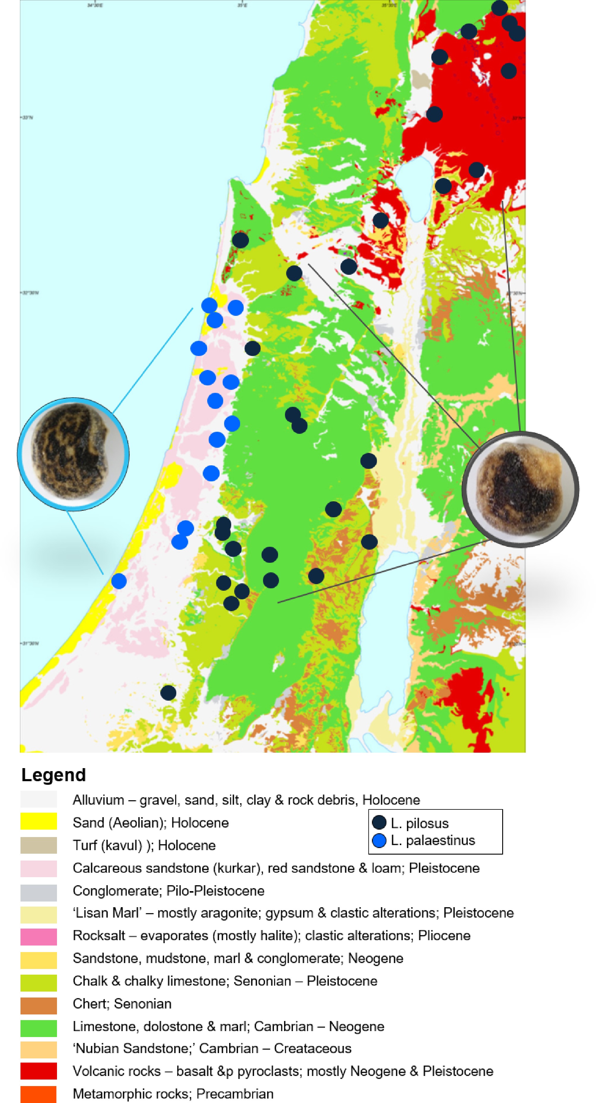
Figure 1.
Sampling locations of the populations of local wild lupins Lupinus pilosus (dark grey dots; photo of seed on the right side of the map), and L. palaestinus (light blue dots; photo of seed on the left side of the map), overlain on the geological map of Israel. The background map is taken from the Geological Survey of Israel, the governmental website:
www.gov.il/en/pages/israel-map-1-200k .Table 1. Ecological description of sampling sites from which wild Lupinus pilosus (PIL) and L. palaestinus (PA) populations were collected in Israel between 2017 and 2020.
Region Population code Collection site Soil bedrock X coordinate Y coordinate Mean temperature 2006–2020 (°C) Altitude (m) Golan Heights PIL-1 Mapalim Junction B 35.75115 32.98624 18 510 Hula Valley PIL-2 Naḥal Ḥamdal B 35.67416 33.09425 19 470 Judean Hills PIL-3 Tel Socoh South L 34.97057 31.68486 20 320 Golan Heights PIL-4 Ofir Viewpoint B 35.66266 32.8149 22 210 Golan Heights PIL-5 South-west Ḥispin B 35.79299 32.84563 18 420 Golan Heights PIL-10 Ḥazeka Road B 34.93600 31.69999 20 330 Judean Hills PIL-11 Matta L 35.06057 31.71652 19 600 Golan Heights PIL-13 Fares Road (Vineyard) B 32.950322 35.86401 17 730 Golan Heights PIL-14 Tel Fazra B 35.69216 33.22469 19 410 Judean Hills PIL-18 Khirbet Kanim L 34.96411 31.66636 19 390 Judean Hills PIL-19 Tel esh-Sharia L 34.68087 31.39089 20 180 Golan Heights PIL-20 Avital B 35.78519 33.10365 15 1,000 Carmel Mountains PIL-21 Makura B 35.42362 32.16415 21 230 Samaria Mountains PIL-23 Awartha L 35.28638 32.16152 18 520 Samaria Mountains PIL-25 Shecḥem Mountains L 35.01352 32.3207 17 800 Samaria Mountains PIL-26 Kedumim L 35.15738 32.21239 20 400 Carmel Mountains PIL-30 Kerem Maharal South B 34.99145 32.64432 20 65 Judean Hills PIL-33 Sarisa L 35.06529 31.79748 19 650 Lower Galilee PIL-34 Ahuzat Barak B 35.33889 32.6431 21 160 Lower Galilee PIL-36 Nau'ra B 35.38378 32.60564 21 110 Sharon Coastal Plain PA-2 Pardes Ḥana L 34.974200 32.4728 20 30 Sharon Coastal Plain PA-3 Ilanot West L 34.907942 32.29253 20 50 Sharon Coastal Plain PA-5 Bnei-Tzion North L 34.86837 32.21983 20 40 Sharon Coastal Plain PA-7 Ilanot East L 34.89259 32.29232 20 40 Sharon Coastal Plain PA-10 Netanya L 34.85319 32.32145 21 30 Sharon Coastal Plain PA-11 Ḥirbet Samara L 34.87782 32.39219 20 20 Sharon Coastal Plain PA-12 Hod Ha-Sharon L 34.88387 32.14996 20 30 Sharon Coastal Plain PA-15 Tel Mond-Kurkar L 34.91846 32.2566 20 50 Coastal Plain South PA-16 Ashqelon L 34.57425 31.66878 21 30 Coastal Plain East PA-17 Sitriya L 34.84209 31.88985 21 80 Commercial product ALB-sh
(L. albus-sweet)NA NA NA NA NA NA Soil bedrock: B, basalt; L, limestone; NA, not applicable. Finally, the seeds were stored at −20 °C until analysis. All these seeds form a wild lupin collection of L. pilosus and L. palaestinus from Israel. The edible, non-local L. albus-sweet (ALB-sh in Table 1) was purchased from a commercial store (a 1 kg plastic bag, Sobhi Nakhly and Sons Ltd, best before October 8th, 2021) and included as a reference species. On one occasion (spring 2023), immature, still green pods of both L. pilosus and L. palaestinus were collected at selected sites (L. pilosus in an open field at the Agricultural Research Organization, Volcani Institute; L. palaestinus in an open field on Tachach Boulevard, Rishon LeZion). Seeds of representative populations were photographed under a Nikon SMZ25 binocular equipped with a Nikon DS-Ri2 camera and captured using NIS Elements software.
For bulk analyses, when powdered samples were used, frozen seeds of L. pilosus and L. palaestinus were dried for 48 h using a lyophilizer at –80 °C, 0.1 bar (Freeze dryer Gamma 2-20, Martin Christ, Osterode, Germany), and ground to a fine powder using a FOSS CM-290 Cemotec, Labtec line grinder. Freeze-drying led to a weight loss of 10%, and the material was stored at –80 °C until it was analyzed. These analyses were performed on five biological replicates, selected randomly and comprising a minimum of 20 seeds from each of the 30 populations, and on five replicates of L. albus, yielding 160 samples.
For tissue-specific analyses, sample preparation followed the protocol by Vogel-Mikuš et al.[29]: representative seeds of each species (randomly picked from a pool of seeds stored in a paper bag) were first scarified by removing a small (up to 0.5 × 0.5 cm2) piece of the testa using a scalpel, and then left to imbibe in MilliQ for 4 h at 5 °C. Imbibed seeds were halved transversally to expose cotyledons using a fresh platinum-coated razor blade (SPI Supplies®, West Chester, PA, USA), and the testa (i.e., the seed coat) was removed. Cotyledon halves were placed in hand-made tin foil containers filled with tissue-freezing medium (TissueTek, Sigma) and finally frozen in liquid nitrogen. Longitudinal 60 µm thick sections were cut in a cryotome (Leica, Germany) at −20 °C, positioned between pre-cooled filter papers, placed in custom-made aluminum containers, and covered with fitting stainless-steel lids. Sections were freeze-dried in these containers at −94 °C and 0.012 mbar for 3 d in a lyophilizer (Labogene, Denmark). Dried sections were inspected, and those with intact morphology were sandwiched between two Pioloform foils stretched over aluminum holders. Each photo of the seeds shows the scale bar for size estimation and correlation (Fig. 2a–c).
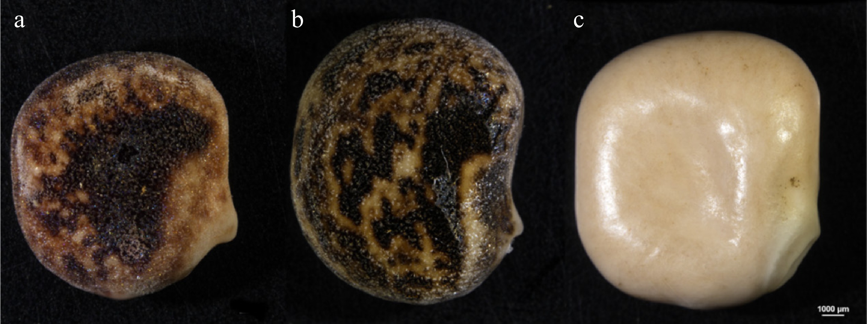
Figure 2.
Representative seeds of Lupinus species in this study: two local species: (a) L. pilosus, (b) L. palaestinus, and (c) L. albus; edible reference species.
Scanning Electron Microscopy (SEM) coupled with Electron Dispersive Spectrometry (EDX)
-
A Tescan Essence Mira S6124 FEG electron column v1.2.0.0 was used for imaging the surface of the seed (testa) and the surface of tissue inside the seed (cotyledons) of the three Lupinus species. Seeds were cut with a scalpel and were not coated before analysis. Using the Oxford Instrument EDX detector (BSE, 10 keV) and EDX-explorer15 software, relative amounts of carbon (C; standard used: C vitamin), nitrogen (N; standard used: BN), oxygen (O; standard used: SiO2), sulfur (S; standard used: FeS2), potassium (K; standard used: KBr), and calcium (Ca; standard used: Wollastonite) were measured in four replicates for each sample. Fragments were scanned and analyzed for their elemental composition. Scans of seed testa and cotyledons of L. pilosus (PL-10, see Table 1), L. palaestinus (PA-5), and L. albus (ALB-sh) were made in triplicate. Magnifications and other scanning parameters are detailed at the bottom of each photo.
Microparticle-induced X-ray emission (micro-PIXE)
-
Quantitative distribution of phosphorus (P), sulfur (S), chlorine (Cl), K, Ca, manganese (Mn), iron (Fe), and zinc (Zn) in longitudinal sections of seeds of the three species were determined using micro-PIXE at the ion accelerator of the Jožef Stefan Institute (Ljubljana, Slovenia) as described previously[29−31]. In short, accelerated protons were focused to approximately 1 × 1 µm2 beam size, scanning the sample sandwiched between the Pioloform foils stretched over an aluminum holder in a vacuum chamber. Interaction between protons and atoms in the sample induces X-ray emission characteristics for each element. X-rays were detected by a four-segmented SDD detector (PNDetector, Münich, Germany), and spectra were recorded using Oxford Microbeam Data Acquisition (OMDA[29] Q-3, Oxford Microbeams Ltd, Bicester, UK) software, which saves pixel-by-pixel spectra in a list mode file. These spectra were processed in GeoPIXE II software[32]. Distribution maps were generated, and tissue-specific concentrations were extracted from the maps using Fiji[33] by importing numerical matrices generated in GeoPIXE II, followed by cell type (i.e., tissue) selection in the image using Brush tool, saving the selection to the Region of Interest Manager to calculate average concentrations in the selected tissues, all in Fiji software. In addition to determining the average concentration of elements in the entire section, the allocation of elements in the image was used to discern the cotyledons (rich in Ca), apparent cell types rich in P and K, and the remaining cell types in L. pilosus and L. palaestinus. The element profiles in L. albus were different as no element profile was evident for the cotyledons; the P-rich cells were not rich in K, while, in contrast, Mn-rich cell types were found.
Liquid Chromatography/Tandem Mass Spectrometry (LC-MS/MS)
-
The LC-MS/MS method used has been described in detail in Namdar et al.[18]. In brief, ground samples (2 g) were weighed and placed in 50 mL screw cap polypropylene tubes. A solution of 40 mL methanol/water (1:1 v/v) containing 1% formic acid was added to each sample. The mixture was shaken vigorously for 30 min on a rotary tumbler and then centrifuged for 15 min at 3,500 rpm. One ml of the supernatant was transferred into a 50 mL screw cap polypropylene tube and diluted with 49 mL distilled water. Five hundred µL of the diluted sample extract was filtered prior to injection.
Samples were analyzed on a Waters Acquity BEH C18 1.7 µm, 100 m × 2.1 m column, with analysis performed using a Waters Acquity UPLC system coupled with a Waters Xevo TQ-XS MS/MS spectrometer. Blank extracts with external matrix calibration were run for quantification. The column temperature was set at 50 °C. For mobile phase solvent A, 10 mM ammonium carbonate in water pH 9.0, and methanol as mobile phase solvent B were used. The flow rate was set at 0.4 mL·min−1, with a linear gradient of mobile phase B% running at 0% for 1 min, then going up for 7 min until reaching 40%, followed by another rise for 4 min to 80%, and then returning to 0% B in 0.2 min. The column was equilibrated at 0% B for 2 min. Injection volume was 3 µL.
MS conditions: The capillary voltage used was 3.0 kV with 30 V cone voltage. Source temperature was set at 150 °C, desolvation gas (N2) flow at 1,000 L·hr−1 at 600 °C, cone gas (N2) flow at 150 L·hr−1. Argon was used as the CID gas at a pressure of 10−3 mbar. Solvent discard was done at 0–1.5 min and at 13–14.2 min.
QAs were identified and quantified by external 9-point calibration lines (0–200 ng·mL−1) in diluted blank extract. A set of 15 QAs was available for this purpose. For the identification of QAs that had no certified standard reference available, GC-MS analysis assisted in the identification of QAs, as detailed below. This QA quantification was used to reveal the connection between environmental conditions and QA development, using multiple statistical tools (see herein).
Gas Chromatography coupled with Mass Spectroscopy (GC-MS)
Sample preparation for GC-MS
-
Lupin samples (15 g from each lupin variety included in the project) were dried for 48 h using a lyophilizer (Freeze dryer LMC-2, Martin Christ, Osterode, Germany) and then ground to a fine powder using a FOSS CM-290 Cemotec, Labtec line grinder. The QA extraction method used here follows[7]: 2 g of ground lupin seeds were weighed, and 9 mL of a solvent mixture of ethyl acetate and ammonia solution (NH4OH 25%), both HPLC quality at an 8:1 volumetric ratio, were added to the ground lupin samples and left for 18 h in the refrigerator (4 °C). The alkaloid extracts were filtered using a 45 µm mesh, followed by a 22 µm mesh and directly injected into the GC-MS.
Compounds identification by GC-MS
-
GC-MS analyses were carried out using an Agilent 7890B gas chromatograph coupled to a 5977A mass spectrometer (electron multiplier potential 2 kV, filament current 0.35 mA, electron energy 70 eV, and the spectra were recorded over the range of m/z 40 to 500). An Agilent 7683 autosampler was used for the sample introduction. A 1 μL aliquot of each sample was injected into the GC-MS using a 1:10 split-ratio injection mode. Helium was the carrier gas at a constant flow of 1.1 mL·s−1. An isothermal hold at 50 °C was maintained for 2 min, followed by a heating gradient of 6 °C·min−1 to 300 °C, and the final temperature was held for 4 min. A 3-min solvent delay was applied. A 30 m, 0.25 mm ID, 5% cross-linked phenylmethyl siloxane capillary column (HP-5MS) with 0.25 μm film thickness was used for separation, and the injection port temperature was 220 °C. The MS interface temperature was 280 °C.
Peak assignments were performed using a spectral library (NIST 17.0) and compared with the MS data obtained from the injection of standards.
Statistical analysis
-
Variability in bulk and tissue-specific element concentrations was first assessed using ANOVA on Ranks. When significant, Dunn's test at p < 0.05 was used to determine the significant differences between species in SigmaPlot v12 (Systat Software Inc., London, UK). Boxplot comparison analysis and hierarchical clustering were carried out using R v4.0.2[34]. For comparing the total QA variable, the assumptions of normality and homogeneity of residuals variance were first assessed by visually plotting sample quantiles against theoretical quantiles and residuals against fitted values, followed by Shapiro-Wilk, and Levene's tests. The QA variable did not meet the above assumptions, even after applying square root and log transformations, and was therefore treated as non-parametric. Kruskal-Wallis and Dunn post hoc tests were used to assess differences between species using the kruskal.test base function and dunn.test function of the FSA package[35]. Due to the absence of QAs in the L. albus samples, it was removed from the analysis to avoid statistical bias. It is, therefore, marked with an asterisk (*) instead of letters as the other tested species.
For multivariate phytochemical analysis, individual QA data were pre-processed using median normalization, log10 transformation, and Pareto scaling to minimize technical variation and achieve a normal distribution. Principal Component Analysis (PCA) was performed to visualize global metabolic differences between populations, and to identify loading contributions. To evaluate the statistical significance of soil-type effects on the overall QA profile, Permutational Multivariate Analysis of Variance (PERMANOVA) based on Euclidean distances was executed using the 'adonis2' function in the 'vegan' package[25]. To identify soil-specific biomarkers, an Orthogonal Partial Least Squares Discriminant Analysis (OPLS-DA) was conducted using the 'ropls' package[25]. Candidate biomarkers were selected based on Variable Importance in Projection (VIP) scores > 1.0. Hierarchical clustering and heat maps of the different lupin species and their QAs studied here were processed using the as.dendrogram function of the pheatmap package[36], following scaling by scale base function.
-
Lupinus pilosus and L. palaestinus seeds differed in visual traits such as size, shape, color, and surface texture compared to L. albus (Fig. 2). Visual inspection suggests that L. pilosus produced the smallest seeds, followed by L. palaestinus and L. albus. However, by weight, the lightest seeds were those of L. palaestinus (0.35 ± 0.02 g per seed DW), followed by L. pilosus (0.50 ± 0.02 g per seed DW) and L. albus (0.60 ± 0.02 g per seed DW). Most striking was the visible difference in the seed coat texture. The seed testa of L. albus was smooth and shiny (Fig. 2c), while the seed testa of L. pilosus and L. palaestinus were rough, somewhat hairy, and marked with black patches in various patterns (Fig. 2a, b).
Additionally, L. albus had a considerably larger diameter. After Lupinus pilosus collection, the green and soft seeds were manually peeled and left to dry. Within 5 h, the seeds hardened, decreased in size, and changed color (Fig. 3a, b, Supplementary File 1).
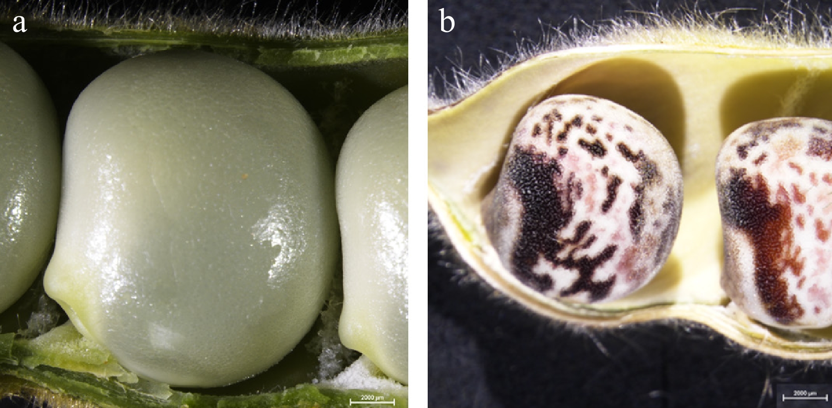
Figure 3.
Lupinus pilosus seeds in a pod immediately after harvest and exposure to the (a) air, and (b) after 5 h of air drying.
The differences in the seed testa texture among the three Lupinus species were particularly evident under scanning electron microscope visualization (Fig. 4a, c, e). Despite differences in the testa texture, the micrographs of the cotyledons of these three species were not as contrasting (Fig. 4b, d, f).
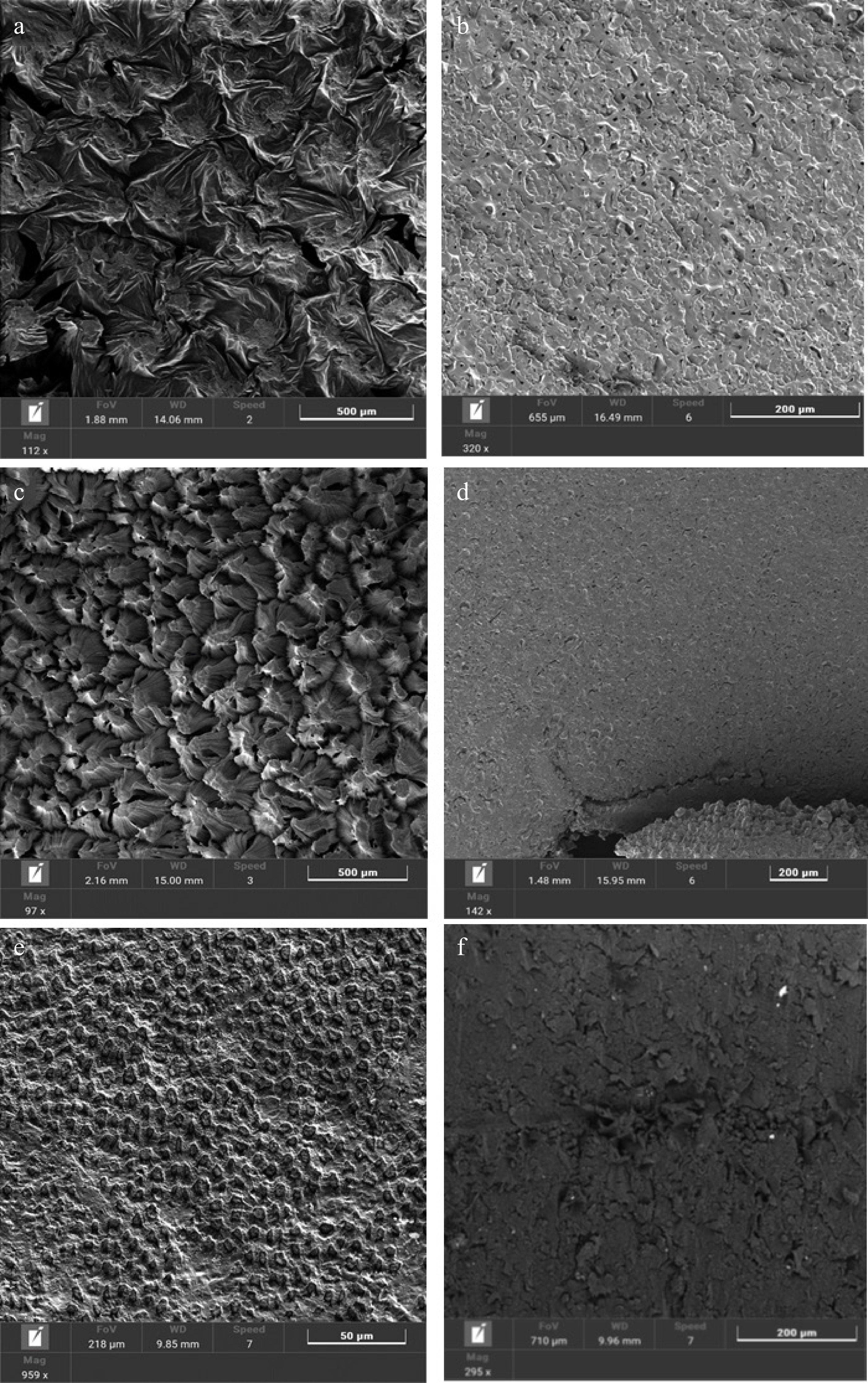
Figure 4.
Seed surface (panels on the left) and cotyledons (panels on the right) of the three Lupinus species studied. Representative scanning electron micrographs of rough-seeded lupins (a), (b) L. pilosus (PIL-5); (c), (d) L. palaestinus (PA-10), smooth-seeded sweet lupin; and (e),(f) L. albus (ALB-sh).
Elemental profiles
-
Edible seeds are key to ensuring sufficient essential elemental intake. There was a significant difference in bulk element concentrations in all measured elements except in Fe, Cu, and Zn among the three species (Supplementary Table S1). Point-by-point analysis to capture element composition of the seed testa and cotyledons was performed using SEM-EDX at 10 kV. Under these analytical conditions, C and O were the most prominent elements in seed testa and cotyledons, followed by N, whose concentration in the seed testa was too low to be detected (Table 2). Sulfur was detected only in the cotyledons of L. pilosus and L. palaestinus. K was detected in both tissues of all three species, while Ca was detected only in seed testa. Surprisingly, Mn, a micronutrient typically occurring at concentrations below 0.05% in seeds, was detected in seed testa of L. albus and L. pilosus, where Al was also found.
Table 2. Elemental analysis (SEM-EDX) of L. pilosus, L. palaestinus, and L. albus cotyledons and seed testa.
Element L. pilosus L. palaestinus L. albus Cotyledon
Wt% (st dev)Seed testa
Wt% (st dev)Cotyledon
Wt% (st dev)Seed testa
Wt% (st dev)Cotyledon
Wt% (st dev)Seed testa
Wt% (st dev)C 63.13 (2.71)ab 49.92 (3.05) 61.05 (1.54)b 47.97 (3.58) 68.08 (13.53)a 63.65 (5.01) O 28.29 (2.48) 45.02 (1.69)a 25.31 (1.58) 50.75 (4.42)a 29.73 (12.49) 18.31 (4.63)b K 0.34 (0.09) 1.05 (0.31) 2.55 (3.23) 0.75 (0.35) 0.80 (0.56) 0.34 (0.61) Ca 2.24 (1.18) 1.34 (1.09) 0.44 (0.02) Al 0.50 (0.12) 0.72 (0.34) 0.69 (0.55) N 7.33 (1.07) 9.64 (2.14) 1.72 (1.27) S 0.17 (0.11) 0.96 (0.32) Mn 15.64 (0.26)a Significant value < 0.05; significance (marked by lowercase letters a, b) was detected for C in the cotyledon, and for Mn and O in the testa. The averages (n = 3 biological samples and n = 4 scan sites on each sample) are shown with standard deviations. Empty cells = element not detected. A quantitative localization analysis using micro-PIXE was used to precisely capture the differences in tissue-specific element profiles between these three Lupinus species. Distribution maps over 1.2 mm × 1.2 mm scan areas of the seed longitudinal section for P, S, Cl, K, Ca, and Mn revealed several distinct features of the Lupinus seeds (Fig. 5). Most prominently, a peculiar dispersal of cell-types with high P, K, and Mn concentrations was identified in L. pilosus and L. palaestinus seeds. In contrast, S, Cl, and Ca concentrations tended to be much lower in these same cell types, and vice versa (Fig. 5, Table 3, Supplementary Figs S1, S2). This dispersion of element-specific cell-types was not apparent in L. albus, where the presence of 4–7 cell layers of characteristically high P concentrations was observed in the cotyledons. This area also did not align with the much more numerous Mn-rich cell layer, which contained extreme Mn concentrations exceeding 0.9%Wt (Fig. 5, Table 3, Supplementary Fig. S3).
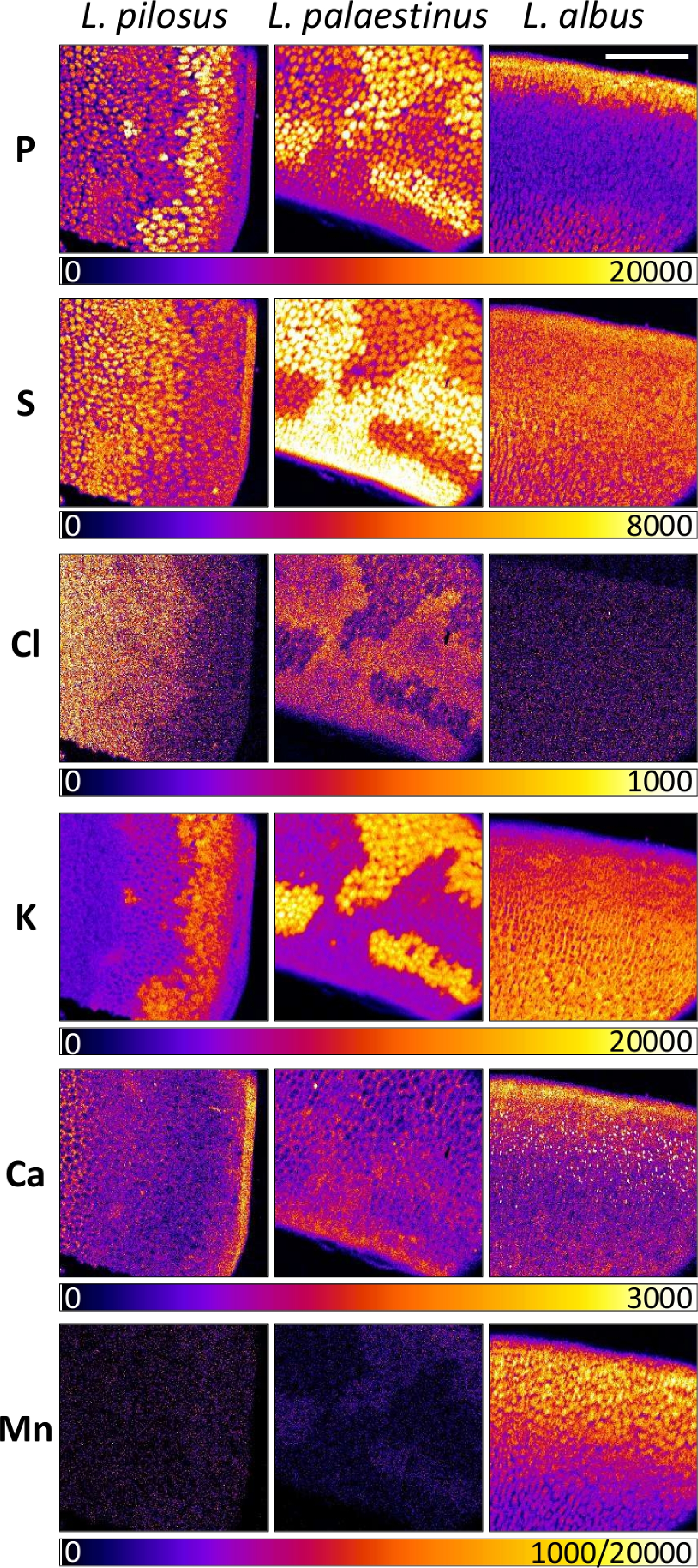
Figure 5.
Representative quantitative distribution maps of phosphorus (P), sulfur (S), chlorine (Cl), potassium (K), calcium (Ca), and manganese (Mn) in longitudinally cut seeds of three different Lupinus species. Scale bar represents 500 µm and the scale is the same for all maps. Color legends represent minimum and maximum displayed concentrations in mg·kg−1 dry weight. There are two maxima values for Mn: the lower for L. pilosus and L. palaestinus, and the higher for L. albus.
Table 3. Relative concentration (out of the total amount measured, averaged, in mg·kg−1 dry weight) of phosphorus (P), sulphur (S), chlorine (Cl), potassium (K), calcium (Ca), manganese (Mn), iron (Fe), and zinc (Zn) in the entire section of cotyledons of Lupinus pilosus, L. palaestinus, and L. albus analyzed with micro-PIXE.
Wt%stat L. pilosus (n = 6) L. palaestinus (n = 6) L. albus (n = 7) P 42.37 b 44.39a 23.70b S 13.4 b 12.55 a 12.78b Cl 2.18a 1.04 a 0.12b K 35.59 35.24 35.81 Ca 5.44b 5.53 a 4.36b Mn 0.77b 1.08 b 22.89a Fe 0.22 b 0.17 b 0.34a Zn 0.25 0.17 0.27 Different letters next indicate significant differences between the species for each element separately (ANOVA on Ranks, followed by Dunn's post hoc test at p < 0.05). QA profiles
-
The QA profiles for 30 populations of the two wild local lupin species were investigated. Five specimens were sampled from each population. The total QA amounts (mg·kg−1 DW seed material) in the two wild lupins were compared to the total QA profiles detected in the edible reference lupin species, as described in Fig. 6. The results show that, in general, L. pilosus was richer in QAs than L. palaestinus, with L. pilosus containing an average 10,300 ± 960 QA mg·kg−1 DW, which was approximately 1.03% of DW, and L. palaestinus containing 6,090 ± 1,110 mg·kg−1 DW, which was approximately 0.61% of DW (Fig. 6). For comparison, the total QA profiles of three wild L. albus populations were also presented in Fig. 6.
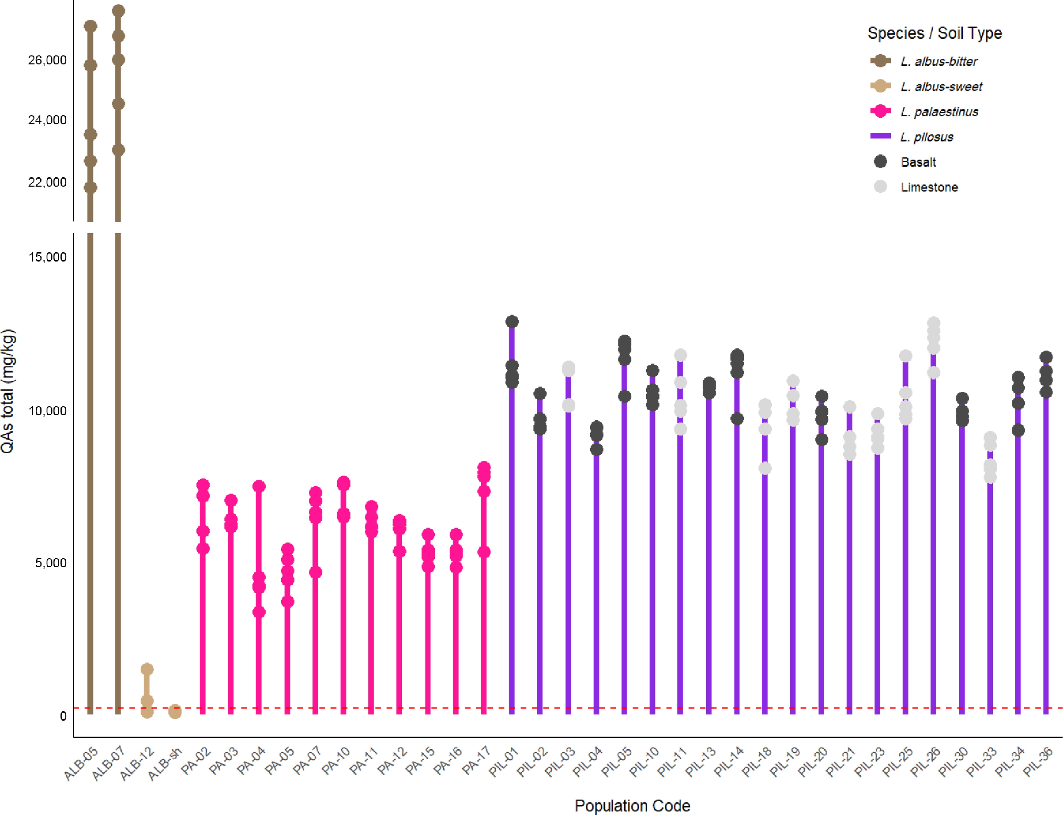
Figure 6.
Total quinolizidine alkaloid (QA) levels were detected in L. palaestinus and L. pilosus, and in one cultivar of the non-local edible L. albus. The dashed red line marks the level of QAs in food considered safe for human consumption[21]. Total QAs of three wild L. albus populations were added for comparison (for details regarding these species, see Namdar et al.[18]). For the Population Code, please see Table 1.
Differences in the QA compositions of the L. pilosus populations suggested that substrate origin significantly shapes the seed phytochemical profile (Fig. 7). Populations grown on basaltic soils exhibited higher levels of lupanine and multiflorine, whereas those from calcareous sediments produced greater amounts of sparteine and albine, along with conjugated esters of 13α-hydroxylupanine and 13α-hydroxymultiflorine. Multivariate statistical analysis further resolved this phytochemical divergence. PCA (Fig. 8a) demonstrated that 77.71% of the total metabolic variance was captured by the first two components (PC1: 55.54%, PC2: 22.17%), with distinct clustering by soil type (PERMANOVA: F1,95 = 27.99, p < 0.001). The OPLS-DA model (Q2 = 0.733, R2Y = 0.755) identified Sparteine (VIP = 1.68) as the primary biomarker for calcareous populations, with concentrations 3.5-fold higher than in basaltic ones (290 vs 81 µg·g−1). Conversely, multiflorine derivatives, specifically 13α-Tigloyloxymultiflorine (VIP = 1.59) and 13α-Hydroxymultiflorine (VIP = 1.51), were more prominent in seeds from basaltic origins (Fig. 8b). In contrast, L. palaestinus, which grows in relatively uniform sandy calcareous environments, displayed lower inter-population diversity in QA profiles, making soil-driven impacts less evident for this species.
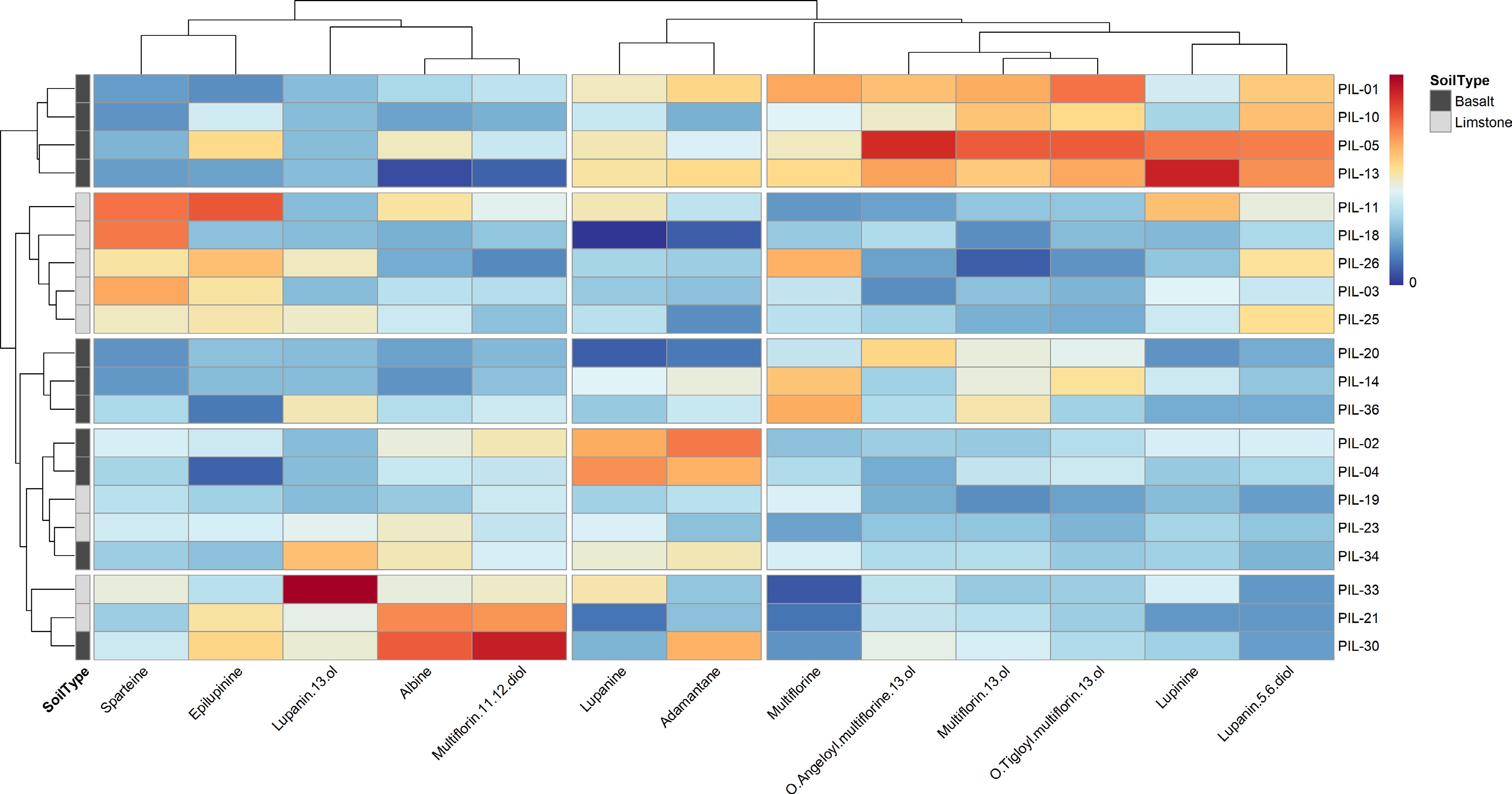
Figure 7.
QA distribution (on a 0–10 scale) in L. pilosus based on their geological distribution. For the original raw data, see Namdar et al.[18].
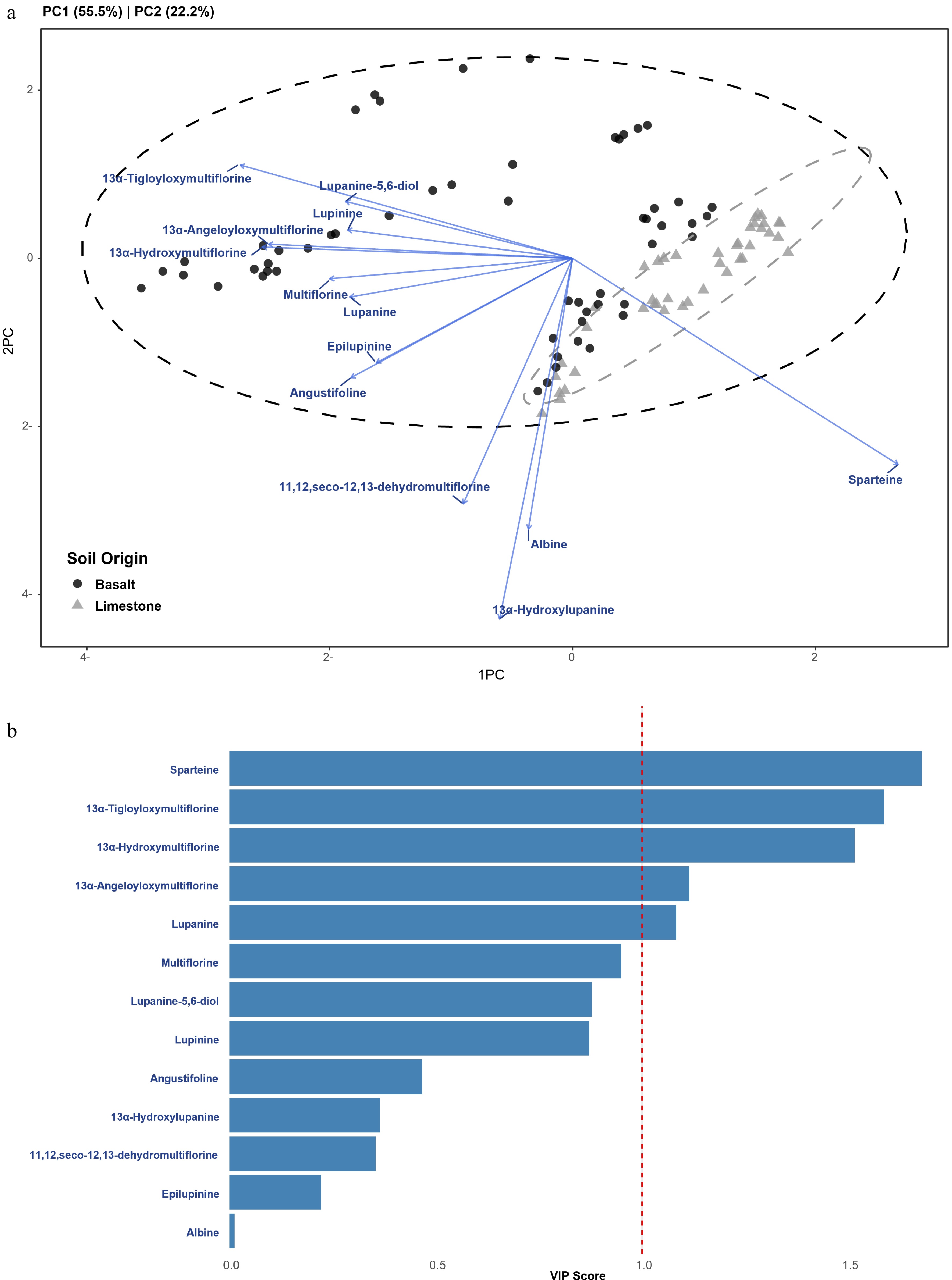
Figure 8.
Multivariate statistical analysis of quinolizidine alkaloid (QA) profiles in Lupinus pilosus seeds originating from basaltic and calcareous soils. (a) PCA Biplot: Principal Component Analysis (PCA) displaying the metabolic grouping of populations by soil origin. The first two components account for 77.71% of the total variance (PC1: 55.54%, PC2: 22.17%). Points represent individual samples (Basalt = black circles, Limestone = grey triangles), with 95% confidence ellipses. Blue vectors indicate the loading contributions of specific alkaloids to the principal components. (b) VIP Scores: top 10 predictive alkaloids ranked by variable importance in projection scores from the OPLS-DA model (Q2 = 0.733, R2Y = 0.755). Variables with VIP > 1.0 (indicated by the red dashed line) are considered significant discriminants for soil-type classification.
-
It has long been accepted that QA levels in lupin seeds need to remain low if they are to be developed into a potential food or feed crop[21]. The two wild lupin species studied here demonstrate lower QA levels than those detected in the bitter varieties of the four commercialized lupin species[18]. This suggests that these wild lupin species could be used as the basis for a new crop. However, their use as food for humans and animals depends on the ability to de-bitter them without compromising their beneficial biological activity and desirable sensory qualities, which remains a major challenge in transforming wild lupins into crops. The distinguished difference in chemical profiles noted between wild and commercial lupin seeds may be attributed to the different stages in which the two types of seeds were processed. Fresh and immature lupin seeds are paled-skin and relatively large as they still contain high amounts of water. As they mature, the seeds dry, significantly shrink, and turn darker colored (see Supplemental File 1). While the commercial seeds may have been preserved as close as possible to their fresh and immature stage (they are whitish and relatively large), wild lupin seeds were sampled upon maturation, in their driest form. This fits with ethnographic descriptions reporting that pre-mature wild seeds were best for consumption and thus collected in this stage[37,38].
The spatial distribution of elements in the smooth and rough seeds, as revealed using the SEM-EDX, showed uniformity across all three analyzed species. However, analysis also highlighted some differences between plant parts; specifically seed testa and cotyledons. Even though the seeds of L. pilosus and L. palaestinus were collected from different locations, the elemental concentrations for each species remained fairly constant, indicating a stronger phylogenetic than ecological effect, as shown previously for leaves of various plants[39]. The most distinct element profile in seeds was found in L. albus, with extremely high Mn concentrations, which aligns with other reports[40,41]. This species has been described as Mn accumulating[42,43], therefore only limited amounts of these seeds should be consumed, not to exceed 11 mg/person per day[44].
Lupin seeds generally contain higher N levels and are conjugated in different forms[41,45]. However, SEM-EDX analyses showed that seed testa contains no measurable N, and all N available in the seed is concentrated in the cotyledon (Table 2). This information is essential for accurately calculating N content in lupin seeds and selecting an adequate de-bittering approach. Importantly, this indicates that removing the seed testa cannot be considered de-bittering. Detection of S only in wild lupin cotyledons may be a proxy for proteins consisting of S-containing amino acids, or as part of secondary metabolites with a specific odor, typical for cooked Lupinus seeds (Table 2).
Very high concentrations of Mn were detected in a commercial sample of L. albus. This is intriguing data, confirming previous observations[37,39]. As mentioned above, we noticed that upon harvest, lupin seeds tend to dry, shrink, and harden significantly. We monitored this process by placing a L. pilosus seed under the binocular and taking a snapshot every 15 min to compose a time-lapse video of the change occurring. When we tasted the seeds in their green and fresh stage, they were sweet, bearing almost no bitterness. Plant-gatherers also report that the seeds bear almost no bitterness at their pre-mature, green stage[37,38]. Therefore, apparently, maturation and the drying process make the seeds bitter[46]. To verify this suggestion, fresh green wild lupin seeds of the three species should be further analyzed for their Mn and QA production and accumulation to show their accumulation during the maturation process, i.e., from fresh and green, to dry and brownish-patterned.
QA profiles across species and their origin
-
Low levels of alkaloids are essential in transforming wild lupin varieties into crops. The QA content in L. pilosus is around 1% of its dry weight, and QA levels detected in L. palaestinus are somewhat lower (Fig. 4). Beyond general lower QA levels, lupin species designed for food and feed should be free of highly toxic QAs such as thermopsine, cysteine, and anagyrine. These QAs are absent in the natural populations of L. pilosus and L. palaestinus studied here.
Other QAs may be desirable for their varied biological activities[20,47−58]. Inter alia, it has already been suggested[18] that the chemotaxonomy based on alkaloid content dictates QA composition for each species. In other words, QA profiles are relatively consistent within each species, but there are significant differences in QA profiles between species. We note that a given species produces either high levels of multiflorine/lupanine derivatives, or high levels of lupinine/sparteine derivatives, but not both, in a 'switch on/off' biochemical mechanism (Fig. 7, for absolute concentrations see Namdar et al.[18]). Our multivariate analysis of L. pilosus further elucidates this mechanism, showing that while both pathways are present, their relative partitioning is significantly modulated by the environment. PCA and OPLS-DA models revealed that 77.71% of the metabolic variance is driven by soil-origin-specific partitioning (Fig. 8). Thus, while L. pilosus and L. palaestinus produce high amounts of multiflorine and lupinine, they do not produce toxic QAs. In general, L. pilosus was richer in QAs than L. palaestinus, and L. pilosus contained, on average, around 1% of DW, while L. palaestinus contained approximately 0.6% of DW. This is 30- to 50-fold higher than the 0.02% threshold concentration established in 2001 by the Australia New Zealand Food Authority (ANZFA)[21]. However, analysis of a larger set of lupin species by us identified only two varieties of L. albus and one variety of L. luteus that meet this threshold. In contrast, other edible lupins demonstrate higher QA levels up to 20-fold the recommended dosage[18]. Reports on elevated levels in commercially grown lupin varieties from Europe show that the ANZFA standard is difficult to achieve in the region, often attributed to less favorable growing conditions[59−61].
Further analysis of QA levels in local wild lupins points to the possible contribution of the ecological system of the lupine origin. For example, L. pilosus grows in diverse ecological systems. We note a strong correlation between the QA compositions and the geological/soil bedrock on which the plants grow (Figs 7, 8a). This soil-driven divergence was statistically validated by PERMANOVA (F1,95 = 27.99, p < 0.001), confirming that the substrate acts as a primary determinant of the seed's chemical signature. L. pilosus grown in basaltic soils tends to contain relatively higher amounts of lupanine and multiflorine and their derivatives, with 13α-hydroxymultiflorine and its tigloyl esters identified as key biomarkers (VIP > 1.5). In contrast, L. pilosus populations grown on limestone contain higher amounts of sparteine and epilupinine. Notably, OPLS-DA identified Sparteine (VIP = 1.68) as the most significant discriminant for calcareous populations, exhibiting a 3.5-fold increase compared to basaltic origins (290 vs 81 µg·g−1). This type of inter-population diversity is ecologically intriguing and should be investigated further, since environmental conditions are related to seed survival and seed bank density[62]. The dramatic shift toward sparteine production in calcareous soils suggests a specific biosynthetic adaptation to alkaline environments, possibly linked to nutrient availability or localized herbivory pressures. Importantly, our sampling aimed to reduce climatic/environmental confounding by focusing on sites that fall within a broadly comparable climatic context; therefore, the dominant contrast among the compared populations is the underlying substrate (basalt vs calcareous/limestone). Nevertheless, because this is an observational field comparison, we cannot fully exclude additional co-varying factors (e.g., microclimate, vegetation context, or local land-use history) that may differ among sites. Future work using a common-garden design and/or reciprocal soil transplants would provide a stronger causal test of substrate-driven effects on QA profiles and ionomes. In comparison, L. palaestinus grows in relatively uniform environments with sandy calcareous soil beds. Their QA profiles show relatively low diversity between populations, and thus, the ecological impact on their QA production is more elusive.
Previous work on East-Mediterranean lupins has focused on species-level relationships and on ecological/physiological responses to soil conditions[27], but, to the best of our knowledge, no study has yet examined whether L. pilosus populations from basalt and limestone differ genetically. Our current data therefore, cannot distinguish between local adaptation and environmentally induced plasticity, and we agree that a future population-genetic study using molecular markers would be the appropriate way to address this question.
-
This study provides valuable insights into the potential of L. pilosus and L. palaestinus, wild lupin species native to Israel, as promising candidates for future crop development. By analyzing the geographic and pedological influences on their phytochemical composition, particularly their QA profiles, we have uncovered significant variations tied to their environmental origins, which were statistically validated as distinct metabolic fingerprints. Our research highlights that the ecological and geological contexts in which these lupins grow play a decisive role in shaping their alkaloid content, specifically driving a predictable biosynthetic trade-off between sparteine and multiflorine pathways. The application of robust multivariate statistical modeling (PCA and OPLS-DA) confirmed that soil bedrock acts as a primary determinant of these chemical signatures, with sparteine and specific multiflorine esters emerging as high-impact predictive biomarkers (VIP > 1.5) for limestone and basaltic populations, respectively. The research highlights the novel discovery that the ecological and geological contexts in which these lupins grow play a decisive role in shaping their alkaloid content, presenting opportunities to leverage these natural variations for specific agricultural and nutritional applications. The detailed analysis provided by advanced techniques such as LC-MS/MS, micro-PIXE, and SEM-EDX, integrated with multivariate informatics, has allowed us to pinpoint these unique relationships, contributing to a deeper understanding of the chemotaxonomy of wild lupins. These findings suggest that the immediate influence of soil bedrock on alkaloid biosynthesis presents both a strategic opportunity and a technical challenge for lupin development. Whether these variations stem from long-term genetic adaptations of specific accessions or from immediate physiological plasticity, they highlight that site selection is as critical as genotype selection. This offers a tool for developing customized alkaloid profiles for nutritional applications, while simultaneously posing a challenge in managing and preventing the expression of undesirable alkaloids when introducing varieties to different pedological environments.
-
Not applicable.
-
The authors confirm their contributions to the paper as follows: study conception and design: Shelef O, Namdar D; data collection: Ben-Simchon E, Cohen O, Namdar D, Zemach H; analysis and interpretation of results: Shelef O, Namdar D, Zemach H, Ben-Simchon E, Sternberg M, Mulder P, Pongrac P, Kelemen M; draft manuscript preparation: Shelef O, Namdar D, Zemach H, Ben-Simchon E, Mulder P. All authors reviewed the results and approved the final version of the manuscript.
-
The datasets generated during and/or analyzed during the current study are available from the corresponding author OS on reasonable request.
-
This work is dedicated to the memory of our colleague and collaborator, Dr. Patrick Mulder, who sadly passed away shortly after the manuscript was completed. We gratefully acknowledge his valuable contributions to this study. The authors thank Smadar Wininger and Zvika Amitay for helping with fieldwork and Uri Goldfisher and Jonatan Fireman for their help with field and lab work. They are also grateful to Kinneret Rosenblatt for her help in preparing Fig. 1 and to Maša Andlovic for her help in ImageJ analysis. This work was supported by the Slovenian Research and Innovation Agency through research core funding [No. P1-0112 and P1-0212] and project funding [J4-3091 and N4-0346]. The RADIATE project enabled access to the nuclear microprobe under the Grant Agreement 824096 from the EU Research and Innovation programme HORIZON 2020.
-
The authors declare that they have no conflict of interest.
-
accompanies this paper online at: https://doi.org/10.48130/cas-0026-0015.
- Supplementary Table S1 Bulk sodium (Na), magnesium (Mg), phosphorus (P), potassium (K), calcium (Ca), manganese (Mn), iron (Fe), copper (Cu) and zinc (Zn) concentrations (mg kg-1 dry weight).
- Supplementary Fig. S1 Cell-type specific concentrations of phosphorus (P), sulphur (S), chlorine (Cl), potassium (K), calcium (Ca), manganese (Mn), iron (Fe) and zinc (Zn) in longitudinal sections of Lupinus pilosus seed.
- Supplementary Fig. S2 Cell-type specific concentrations of phosphorus (P), sulphur (S), chlorine (Cl), potassium (K), calcium (Ca), manganese (Mn), iron (Fe) and zinc (Zn) in longitudinal sections of Lupinus palaestinus seed.
- Supplementary Fig. S3 Cell-type specific concentrations of phosphorus (P), sulphur (S), chlorine (Cl), potassium (K), calcium (Ca), manganese (Mn), iron (Fe) and zinc (Zn) in longitudinal sections of Lupinus albus seed.
- Supplementary File 1 Time-lapse observation of lupin seed maturation.
- Copyright: © 2026 by the author(s). Published by Maximum Academic Press, Fayetteville, GA. This article is an open access article distributed under Creative Commons Attribution License (CC BY 4.0), visit https://creativecommons.org/licenses/by/4.0/.
-
About this article
Cite this article
Shelef O, Namdar D, Zemach H, Ben-Simchon E, Mulder P, et al. 2026. Soil-origin effects on wild lupin seed traits and their potential for crop development. Circular Agricultural Systems 6: e017 doi: 10.48130/cas-0026-0015 

Soil-origin effects on wild lupin seed traits and their potential for crop development
- Received: 19 January 2026
- Revised: 04 April 2026
- Accepted: 08 April 2026
- Published online: 02 July 2026
Abstract: This paper explores the effect of soil type at the site of origin on lupin seed phytochemistry. We examined wild populations of lupins originating from basaltic and calcareous soils and analyzed their seed morphology and phytochemical profiles, with a particular focus on secondary metabolites—namely quinolizidine alkaloids and ionomes. The two lupin species studied, Lupinus pilosus and L. palaestinus, are native to Israel and were collected from wild populations. The overarching goal of this study is to leverage this knowledge to facilitate the domestication of these crop wild relatives for food or feed applications. We sampled 30 populations of the two species across Israel, characterized their environments at the landscape scale, and integrated this data with quinolizidine alkaloid profiles obtained from the seeds. This approach enabled us to elucidate the biochemical and ecological contexts underlying alkaloid formation. This study reveals that L. pilosus and L. palaestinus can be distinguished based on their seed phytochemical profiles. We suggest that these distinguished profiles are related to their soil origin. Our results highlight the influence of pedological origin—basaltic vs calcareous soils on both quinolizidine alkaloid composition and ionomic profiles in wild lupins. These insights may ultimately support the development of novel crops, contribute to agricultural diversification, and enhance crop resilience.
-
Key words:
- Agricultural diversification /
- Crop resilience /
- Crop wild relatives /
- Lupin /
- Phytochemistry /
- Quinolizidine alkaloid