








Search
2026 Volume 2026
Article Contents
REVIEW ARTICLE   Open Access    

Nanotherapeutics in the treatment of cancer: transforming cancer care

More Information
  • Nanotechnology is revolutionizing oncological interventions through nanotherapeutics, which employ nanoparticles (NPs) to significantly increase treatment precision and efficacy. These NP vectors deliver anticancer agents directly to tumors, reducing damage to healthy cells by utilizing the established enhanced permeability and retention (EPR) effect. In response to targeted biochemical signals, NPs ensure precise drug release within tumors. Additionally, they function as contrast agents to improve cancer imaging and diagnostics, facilitating early diagnosis and the development of personalized therapies. NP-based combination therapies effectively combat drug resistance by delivering multiple therapeutic agents targeting diverse molecular pathways. Nanomedicines provide accurate drug delivery in blood cancers, reducing side effects and potentially enhancing hematopoietic stem cell transplantation. Furthermore, NPs modulate intracellular signaling in hematological cancers, offering opportunities for pioneering treatments. This article highlights the multifaceted roles of NPs in advancing cancer treatment, pointing toward significant improvements and future research directions.
  • 加载中
  • [1] DeSantis CE, Lin CC, Mariotto AB, Siegel RL, Stein KD, et al. 2014. Cancer treatment and survivorship statistics, 2014. CA: A Cancer Journal for Clinicians 64(4):252−271 doi: 10.3322/caac.21235

    CrossRef   Google Scholar

    [2] Ward E, DeSantis C, Robbins A, Kohler B, Jemal A. 2014. Childhood and adolescent cancer statistics, 2014. CA: A Cancer Journal for Clinicians 64(2):83−103 doi: 10.3322/caac.21219

    CrossRef   Google Scholar

    [3] Hu J, Hu K, Cheng Y. 2016. Tailoring the dendrimer core for efficient gene delivery. Acta Biomaterialia 35:1−11 doi: 10.1016/j.actbio.2016.02.031

    CrossRef   Google Scholar

    [4] Sanchez-Garcia L, Martín L, Mangues R, Ferrer-Miralles N, Vázquez E, et al. 2016. Recombinant pharmaceuticals from microbial cells: a 2015 update. Microbial Cell Factories 15(1):33 doi: 10.1186/s12934-016-0437-3

    CrossRef   Google Scholar

    [5] Chudy M, Tokarska K, Jastrzębska E, Bułka M, Drozdek S, et al. 2018. Lab-on-a-chip systems for photodynamic therapy investigations. Biosensors and Bioelectronics 101:37−51 doi: 10.1016/j.bios.2017.10.013

    CrossRef   Google Scholar

    [6] Mitragotri S, Burke PA, Langer R. 2014. Overcoming the challenges in administering biopharmaceuticals: formulation and delivery strategies. Nature Reviews Drug Discovery 13(9):655−672 doi: 10.1038/nrd4363

    CrossRef   Google Scholar

    [7] Liu X, Wu F, Ji Y, Yin L. 2019. Recent advances in anti-cancer protein/peptide delivery. Bioconjugate Chemistry 30(2):305−324 doi: 10.1021/acs.bioconjchem.8b00750

    CrossRef   Google Scholar

    [8] Sandbhor P, Palkar P, Bhat S, John G, Goda JS. 2024. Nanomedicine as a multimodal therapeutic paradigm against cancer: on the way forward in advancing precision therapy. Nanoscale 16(13):6330−6364 doi: 10.1039/d3nr06131k

    CrossRef   Google Scholar

    [9] Zheng C, Li M, Ding J. 2021. Challenges and opportunities of nanomedicines in clinical translation. BIO Integration 2(2):57−60 doi: 10.15212/bioi-2021-0016

    CrossRef   Google Scholar

    [10] Dadwal A, Baldi A, Kumar Narang R. 2018. Nanoparticles as carriers for drug delivery in cancer. Artificial Cells, Nanomedicine, and Biotechnology 46(sup2):295−305 doi: 10.1080/21691401.2018.1457039

    CrossRef   Google Scholar

    [11] Nasrollahzadeh M, Sajadi SM, Sajjadi M, Issaabadi Z. 2019. An introduction to green nanotechnology. In Interface Science and Technology. Volume 28. Amsterdam: Elsevier. pp. 1−27 doi: 10.1016/b978-0-12-813586-0.00001-8
    [12] Emerich DF, Thanos CG. 2003. Nanotechnology and medicine. Expert Opinion on Biological Therapy 3(4):655−663 doi: 10.1517/14712598.3.4.655

    CrossRef   Google Scholar

    [13] Alexis F, Rhee JW, Richie JP, Radovic-Moreno AF, Langer R, et al. 2008. New frontiers in nanotechnology for cancer treatment. Urologic Oncology: Seminars and Original Investigations 26(1):74−85 doi: 10.1016/j.urolonc.2007.03.017

    CrossRef   Google Scholar

    [14] Moghimi SM, Hunter AC, Murray JC. 2005. Nanomedicine: current status and future prospects. The FASEB Journal 19(3):311−330 doi: 10.1096/fj.04-2747rev

    CrossRef   Google Scholar

    [15] Langer R. 1998. Drug delivery and targeting. Nature 392:5−10

    Google Scholar

    [16] Farokhzad OC, Langer R. 2009. Impact of nanotechnology on drug delivery. ACS Nano 3(1):16−20 doi: 10.1021/nn900002m

    CrossRef   Google Scholar

    [17] Park K. 2016. Drug delivery research: the invention cycle. Molecular Pharmaceutics 13(7):2143−2147 doi: 10.1021/acs.molpharmaceut.6b00015

    CrossRef   Google Scholar

    [18] Park K. 2013. Facing the truth about nanotechnology in drug delivery. ACS Nano 7(9):7442−7447 doi: 10.1021/nn404501g

    CrossRef   Google Scholar

    [19] Gowda R. 2013. Use of nanotechnology to develop multi-drug inhibitors for cancer therapy. Journal of Nanomedicine & Nanotechnology 4(6):184 doi: 10.4172/2157-7439.1000184

    CrossRef   Google Scholar

    [20] Yu M, Wu J, Shi J, Farokhzad OC. 2016. Nanotechnology for protein delivery: overview and perspectives. Journal of Controlled Release 240:24−37 doi: 10.1016/j.jconrel.2015.10.012

    CrossRef   Google Scholar

    [21] Misra R, Acharya S, Sahoo SK. 2010. Cancer nanotechnology: application of nanotechnology in cancer therapy. Drug Discovery Today 15(19−20):842−850 doi: 10.1016/j.drudis.2010.08.006

    CrossRef   Google Scholar

    [22] Sun D, Sun X, Zhang X, Wu J, Shi X, et al. 2024. Emerging chemodynamic nanotherapeutics for cancer treatment. Advanced Healthcare Materials 13(22):e2400809 doi: 10.1002/adhm.202400809

    CrossRef   Google Scholar

    [23] Singh R, Dheek M, Chauhan SB, Singh I. 2025. Nanotherapeutics in cancer-associated infections. In Applications of Nanotherapeutics and Nanotheranostics in Managing Infectious Diseases. Amsterdam: Elsevie. pp. 553−581 doi: 10.1016/b978-0-443-28836-4.00023-8
    [24] Bae Y, Jang WD, Nishiyama N, Fukushima S, Kataoka K. 2005. Multifunctional polymeric micelles with folate-mediated cancer cell targeting and pH-triggered drug releasing properties for active intracellular drug delivery. Molecular BioSystems 1(3):242−250 doi: 10.1039/b500266d

    CrossRef   Google Scholar

    [25] Nasongkla N, Bey E, Ren J, Ai H, Khemtong C, et al. 2006. Multifunctional polymeric micelles as cancer-targeted, MRI-ultrasensitive drug delivery systems. Nano Letters 6(11):2427−2430 doi: 10.1021/nl061412u

    CrossRef   Google Scholar

    [26] Shen H, Shi S, Zhang Z, Gong T, Sun X. 2015. Coating solid lipid nanoparticles with hyaluronic acid enhances antitumor activity against melanoma stem-like cells. Theranostics 5(7):755−771 doi: 10.7150/thno.10804

    CrossRef   Google Scholar

    [27] Bassas-Galia M, Follonier S, Pusnik M, Zinn M. 2017. Natural polymers: a source of inspiration. In Bioresorbable Polymers for Biomedical Applications. Amsterdam: Elsevier. pp. 31−64 doi: 10.1016/b978-0-08-100262-9.00002-1
    [28] Kirpotin DB, Drummond DC, Shao Y, Shalaby MR, Hong K, et al. 2006. Antibody targeting of long-circulating lipidic nanoparticles does not increase tumor localization but does increase internalization in animal models. Cancer Research 66(13):6732−6740 doi: 10.1158/0008-5472.can-05-4199

    CrossRef   Google Scholar

    [29] Ali Khawar I, Kim JH, Kuh HJ. 2015. Improving drug delivery to solid tumors: priming the tumor microenvironment. Journal of Controlled Release 201:78−89 doi: 10.1016/j.jconrel.2014.12.018

    CrossRef   Google Scholar

    [30] Garg P, Pareek S, Kulkarni P, Salgia R, Singhal SS. 2024. Nanoengineering solutions for cancer therapy: bridging the gap between clinical practice and translational research. Journal of Clinical Medicine 13(12):3466 doi: 10.3390/jcm13123466

    CrossRef   Google Scholar

    [31] Raj M, Chopra D, Kumar Upadhyay S, Singh M, Sharma AK. 2024. Recent advances in cancer therapy: drug delivery, nanotherapeutics, and future perspectives. Anticancer Therapeutics: Bionanotechnology, Nanomedicine and Phytochemicals. Germany: De Gruyter. pp. 273−316 doi: 10.1515/9783111349541-009
    [32] Wang X, Yang L, Chen Z, Shin DM. 2008. Application of nanotechnology in cancer therapy and imaging. CA: A Cancer Journal for Clinicians 58(2):97−110 doi: 10.3322/ca.2007.0003

    CrossRef   Google Scholar

    [33] Forte E, Fiorenza D, Torino E, Costagliola di Polidoro A, Cavaliere C, et al. 2020. Radiolabeled PET/MRI nanoparticles for tumor imaging. Journal of Clinical Medicine 9(1):89 doi: 10.3390/jcm9010089

    CrossRef   Google Scholar

    [34] Lin PC, Lin S, Wang PC, Sridhar R. 2014. Techniques for physicochemical characterization of nanomaterials. Biotechnology Advances 32(4):711−726 doi: 10.1016/j.biotechadv.2013.11.006

    CrossRef   Google Scholar

    [35] Rodrigues T, Reker D, Schneider P, Schneider G. 2016. Counting on natural products for drug design. Nature Chemistry 8(6):531−541 doi: 10.1038/nchem.2479

    CrossRef   Google Scholar

    [36] Bukhari B, Naveed M, Makhdoom SI, Jabeen K, Asif MF, et al. 2021. A comparison between organic and inorganic nanoparticles: prime nanoparticles for tumor curation. Nano 16(13):2130011 doi: 10.1142/s1793292021300115

    CrossRef   Google Scholar

    [37] Bodunde OP, Ikumapayi OM, Akinlabi ET, Oladapo BI, Adeoye AOM, et al. 2021. A futuristic insight into a "nano-doctor": a clinical review on medical diagnosis and devices using nanotechnology. Materials Today: Proceedings 44:1144−1153 doi: 10.1016/j.matpr.2020.11.232

    CrossRef   Google Scholar

    [38] Greene MK, Johnston MC, Scott CJ. 2021. Nanomedicine in pancreatic cancer: current status and future opportunities for overcoming therapy resistance. Cancers 13(24):6175 doi: 10.3390/cancers13246175

    CrossRef   Google Scholar

    [39] Park SH, Oh SG, Mun JY, Han SS. 2006. Loading of gold nanoparticles inside the DPPC bilayers of liposome and their effects on membrane fluidities. Colloids and Surfaces B: Biointerfaces 48(2):112−118 doi: 10.1016/j.colsurfb.2006.01.006

    CrossRef   Google Scholar

    [40] Hua S, Wu SY. 2013. The use of lipid-based nanocarriers for targeted pain therapies. Frontiers in Pharmacology 4:143 doi: 10.3389/fphar.2013.00143

    CrossRef   Google Scholar

    [41] Kim CH, Lee SG, Kang MJ, Lee S, Choi YW. 2017. Surface modification of lipid-based nanocarriers for cancer cell-specific drug targeting. Journal of Pharmaceutical Investigation 47(3):203−227 doi: 10.1007/s40005-017-0329-5

    CrossRef   Google Scholar

    [42] Iyer AK, Su Y, Feng J, Lan X, Zhu X, et al. 2011. The effect of internalizing human single chain antibody fragment on liposome targeting to epithelioid and sarcomatoid mesothelioma. Biomaterials 32:2605−2613 doi: 10.1016/j.biomaterials.2010.11.073

    CrossRef   Google Scholar

    [43] Liu Y, Quan X, Li J, Huo J, Li X, et al. 2023. Liposomes embedded with PEGylated iron oxide nanoparticles enable ferroptosis and combination therapy in cancer. National Science Review 10:nwac167 doi: 10.1093/nsr/nwac167

    CrossRef   Google Scholar

    [44] Marasini N, Ghaffar KA, Skwarczynski M, Toth I. 2017. Liposomes as a vaccine delivery system. In Micro and Nanotechnology in Vaccine Development. Amsterdam: Elsevier. pp. 221−239 doi: 10.1016/b978-0-323-39981-4.00012-9
    [45] Zylberberg C, Matosevic S. 2016. Pharmaceutical liposomal drug delivery: a review of new delivery systems and a look at the regulatory landscape. Drug Delivery 23(9):3319−3329 doi: 10.1080/10717544.2016.1177136

    CrossRef   Google Scholar

    [46] Wang X, Liu X, Li Y, Wang P, Feng X, et al. 2017. Sensitivity to antitubulin chemotherapeutics is potentiated by a photoactivable nanoliposome. Biomaterials 141:50−62 doi: 10.1016/j.biomaterials.2017.06.034

    CrossRef   Google Scholar

    [47] Han B, Yang Y, Chen J, Tang H, Sun Y, et al. 2020. Preparation, characterization, and pharmacokinetic study of a novel long-acting targeted paclitaxel liposome with antitumor activity. International Journal of Nanomedicine 15:553−571 doi: 10.2147/ijn.s228715

    CrossRef   Google Scholar

    [48] O'Brien MER, Wigler N, Inbar M, Rosso R, Grischke E, et al. 2004. Reduced cardiotoxicity and comparable efficacy in a phase IIItrial of pegylated liposomal doxorubicin HCl(CAELYX™/Doxil®) versus conventional doxorubicin forfirst-line treatment of metastatic breast cancer. Annals of Oncology 15(3):440−449 doi: 10.1093/annonc/mdh097

    CrossRef   Google Scholar

    [49] Geisberg CA, Sawyer DB. 2010. Mechanisms of anthracycline cardiotoxicity and strategies to decrease cardiac damage. Current Hypertension Reports 12(6):404−410 doi: 10.1007/s11906-010-0146-y

    CrossRef   Google Scholar

    [50] Zocchi MR, Tosetti F, Benelli R, Poggi A. 2020. Cancer nanomedicine special issue review anticancer drug delivery with nanoparticles: extracellular vesicles or synthetic nanobeads as therapeutic tools for conventional treatment or immunotherapy. Cancers 12(7):1886 doi: 10.3390/cancers12071886

    CrossRef   Google Scholar

    [51] Vader P, Mol EA, Pasterkamp G, Schiffelers RM. 2016. Extracellular vesicles for drug delivery. Advanced Drug Delivery Reviews 106:148−156 doi: 10.1016/j.addr.2016.02.006

    CrossRef   Google Scholar

    [52] Antes TJ, Middleton RC, Luther KM, Ijichi T, Peck KA, et al. 2018. Targeting extracellular vesicles to injured tissue using membrane cloaking and surface display. Journal of Nanobiotechnology 16(1):61 doi: 10.1186/s12951-018-0388-4

    CrossRef   Google Scholar

    [53] Witwer KW, Wolfram J. 2021. Extracellular vesicles versus synthetic nanoparticles for drug delivery. Nature Reviews Materials 6(2):103−106 doi: 10.1038/s41578-020-00277-6

    CrossRef   Google Scholar

    [54] Jeppesen DK, Zhang Q, Franklin JL, Coffey RJ. 2023. Extracellular vesicles and nanoparticles: emerging complexities. Trends in Cell Biology 33(8):667−681 doi: 10.1016/j.tcb.2023.01.002

    CrossRef   Google Scholar

    [55] Chiang CL, Ma Y, Hou YC, Pan J, Chen SY, et al. 2023. Dual targeted extracellular vesicles regulate oncogenic genes in advanced pancreatic cancer. Nature Communications 14:6692 doi: 10.1038/s41467-023-42402-3

    CrossRef   Google Scholar

    [56] Masood F, Chen P, Yasin T, Fatima N, Hasan F, et al. 2013. Encapsulation of Ellipticine in poly-(3-hydroxybutyrate-co-3-hydroxyvalerate) based nanoparticles and its in vitro application. Materials Science and Engineering: C 33(3):1054−1060 doi: 10.1016/j.msec.2012.11.025

    CrossRef   Google Scholar

    [57] Ganta S, Talekar M, Singh A, Coleman TP, Amiji MM. 2014. Nanoemulsions in translational research—opportunities and challenges in targeted cancer therapy. AAPS PharmSciTech 15(3):694−708 doi: 10.1208/s12249-014-0088-9

    CrossRef   Google Scholar

    [58] Kim B, Pena CD, Auguste DT. 2019. Targeted lipid nanoemulsions encapsulating epigenetic drugs exhibit selective cytotoxicity on CDH1/FOXM1+ triple negative breast cancer cells. Molecular Pharmaceutics 16(5):1813−1826 doi: 10.1021/acs.molpharmaceut.8b01065

    CrossRef   Google Scholar

    [59] Mahato R. 2017. Nanoemulsion as targeted drug delivery system for cancer therapeutics. Journal of Pharmaceutical Sciences and Pharmacology 3(2):83−97 doi: 10.1166/jpsp.2017.1082

    CrossRef   Google Scholar

    [60] Ganta S, Devalapally H, Shahiwala A, Amiji M. 2008. A review of stimuli-responsive nanocarriers for drug and gene delivery. Journal of Controlled Release 126(3):187−204 doi: 10.1016/j.jconrel.2007.12.017

    CrossRef   Google Scholar

    [61] Zahr AS, Davis CA, Pishko MV. 2006. Macrophage uptake of core–shell nanoparticles surface modified with poly(ethylene glycol). Langmuir 22(19):8178−8185 doi: 10.1021/la060951b

    CrossRef   Google Scholar

    [62] Stolnik S, Daudali B, Arien A, Whetstone J, Heald CR, et al. 2001. The effect of surface coverage and conformation of poly(ethylene oxide) (PEO) chains of poloxamer 407 on the biological fate of model colloidal drug carriers. Biochimica et Biophysica Acta (BBA) - Biomembranes 1514(2):261−279 doi: 10.1016/S0005-2736(01)00376-5

    CrossRef   Google Scholar

    [63] Alvarez-Lorenzo C. 2010. Poloxamine-based nanomaterials for drug delivery. Frontiers in Bioscience E 2(2):424−440 doi: 10.2741/e102

    CrossRef   Google Scholar

    [64] Ganta S, Deshpande D, Korde A, Amiji M. 2010. A review of multifunctional nanoemulsion systems to overcome oral and CNS drug delivery barriers. Molecular Membrane Biology 27(7):260−273 doi: 10.3109/09687688.2010.497971

    CrossRef   Google Scholar

    [65] Maranhao JA, Valduga CJ, Tavares ER, Kretzer IF, Maria AD, et al. 2011. Novel formulation of a methotrexate derivative with a lipid nanoemulsion. International Journal of Nanomedicine 6:2285−2295 doi: 10.2147/ijn.s18039

    CrossRef   Google Scholar

    [66] Winter E, Pizzol CD, Locatelli C, Silva AH, Conte A, et al. 2014. In vitro and in vivo effects of free and chalcones-loaded nanoemulsions: insights and challenges in targeted cancer chemotherapies. International Journal of Environmental Research and Public Health 11(10):10016−10035 doi: 10.3390/ijerph111010016

    CrossRef   Google Scholar

    [67] Mein JR, Lian F, Wang XD. 2008. Biological activity of lycopene metabolites: implications for cancer prevention. Nutrition Reviews 66(12):667−683 doi: 10.1111/j.1753-4887.2008.00120.x

    CrossRef   Google Scholar

    [68] Chen YJ, Inbaraj BS, Pu YS, Chen BH. 2014. Development of lycopene micelle and lycopene chylomicron and a comparison of bioavailability. Nanotechnology 25(15):155102 doi: 10.1088/0957-4484/25/15/155102

    CrossRef   Google Scholar

    [69] Lu W, Zhang G, Zhang R, Flores LG II, Huang Q, et al. 2010. Tumor site–specific silencing of NF-κB p65 by targeted hollow gold nanosphere–mediated photothermal transfection. Cancer Research 70(8):3177−3188 doi: 10.1158/0008-5472.can-09-3379

    CrossRef   Google Scholar

    [70] Periasamy VS, Athinarayanan J, Alshatwi AA. 2016. Anticancer activity of an ultrasonic nanoemulsion formulation of Nigella sativa L. essential oil on human breast cancer cells. Ultrasonics Sonochemistry 31:449−455 doi: 10.1016/j.ultsonch.2016.01.035

    CrossRef   Google Scholar

    [71] Migotto A, Carvalho VFM, Salata GC, da Silva FWM, Yan CYI, et al. 2018. Multifunctional nanoemulsions for intraductal delivery as a new platform for local treatment of breast cancer. Drug Delivery 25(1):654−667 doi: 10.1080/10717544.2018.1440665

    CrossRef   Google Scholar

    [72] Natesan S, Sugumaran A, Ponnusamy C, Thiagarajan V, Palanichamy R, et al. 2017. Chitosan stabilized camptothecin nanoemulsions: development, evaluation and biodistribution in preclinical breast cancer animal mode. International Journal of Biological Macromolecules 104:1846−1852 doi: 10.1016/j.ijbiomac.2017.05.127

    CrossRef   Google Scholar

    [73] Liang S, Sun C, Yang P, Ma PA, Huang S, et al. 2020. Core-shell structured upconversion nanocrystal-dendrimer composite as a carrier for mitochondria targeting and catalase enhanced anti-cancer photodynamic therapy. Biomaterials 240:119850 doi: 10.1016/j.biomaterials.2020.119850

    CrossRef   Google Scholar

    [74] Duan Y, Dhar A, Patel C, Khimani M, Neogi S, et al. 2020. A brief review on solid lipid nanoparticles: part and parcel of contemporary drug delivery systems. RSC Advances 10(45):26777−26791 doi: 10.1039/d0ra03491f

    CrossRef   Google Scholar

    [75] Hou D, Xie C, Huang K, Zhu C. 2003. The production and characteristics of solid lipid nanoparticles (SLNs). Biomaterials 24(10):1781−1785 doi: 10.1016/S0142-9612(02)00578-1

    CrossRef   Google Scholar

    [76] Uner M, Yener G. 2007. Importance of solid lipid nanoparticles (SLN) in various administration routes and future perspectives. International Journal of Nanomedicine 2(2):289−300 doi: 10.2147/IJN.S1235

    CrossRef   Google Scholar

    [77] Akanda M, Getti G, Nandi U, Mithu MS, Douroumis D. 2021. Bioconjugated solid lipid nanoparticles (SLNs) for targeted prostate cancer therapy. International Journal of Pharmaceutics 599:120416 doi: 10.1016/j.ijpharm.2021.120416

    CrossRef   Google Scholar

    [78] Jiang Y, Huo S, Hardie J, Liang XJ, Rotello VM. 2016. Progress and perspective of inorganic nanoparticle-based siRNA delivery systems. Expert Opinion on Drug Delivery 13(4):547−559 doi: 10.1517/17425247.2016.1134486

    CrossRef   Google Scholar

    [79] Han G, Ghosh P, Rotello VM. 2007. Functionalized gold nanoparticles for drug delivery. Nanomedicine 2(1):113−123 doi: 10.2217/17435889.2.1.113

    CrossRef   Google Scholar

    [80] Samadian H, Hosseini-Nami S, Kamrava SK, Ghaznavi H, Shakeri-Zadeh A. 2016. Folate-conjugated gold nanoparticle as a new nanoplatform for targeted cancer therapy. Journal of Cancer Research and Clinical Oncology 142(11):2217−2229 doi: 10.1007/s00432-016-2179-3

    CrossRef   Google Scholar

    [81] Tang Y, Liang J, Wu A, Chen Y, Zhao P, et al. 2017. Co-delivery of trichosanthin and albendazole by nano-self-assembly for overcoming tumor multidrug-resistance and metastasis. ACS Applied Materials & Interfaces 9(32):26648−26664 doi: 10.1021/acsami.7b05292

    CrossRef   Google Scholar

    [82] Xu H, Wang Y, Pei Z, Ji W, Pei Y. 2019. NIRF turn-on nanoparticles based on the tumor microenvironment for monitoring intracellular protein delivery. Chemical Communications 55(99):14930−14933 doi: 10.1039/c9cc07768e

    CrossRef   Google Scholar

    [83] Farzin A, Etesami SA, Quint J, Memic A, Tamayol A. 2020. Magnetic nanoparticles in cancer therapy and diagnosis. Advanced Healthcare Materials 9(9):1901058 doi: 10.1002/adhm.201901058

    CrossRef   Google Scholar

    [84] Knežević NŽ, Gadjanski I, Durand JO. 2019. Magnetic nanoarchitectures for cancer sensing, imaging and therapy. Journal of Materials Chemistry B 7(1):9−23 doi: 10.1039/c8tb02741b

    CrossRef   Google Scholar

    [85] Santhosh PB, Ulrih NP. 2013. Multifunctional superparamagnetic iron oxide nanoparticles: promising tools in cancer theranostics. Cancer Letters 336(1):8−17 doi: 10.1016/j.canlet.2013.04.032

    CrossRef   Google Scholar

    [86] Lim SY, Shen W, Gao Z. 2015. Carbon quantum dots and their applications. Chemical Society Reviews 44(1):362−381 doi: 10.1039/c4cs00269e

    CrossRef   Google Scholar

    [87] Desmond LJ, Phan AN, Gentile P. 2021. Critical overview on the green synthesis of carbon quantum dots and their application for cancer therapy. Environmental Science: Nano 8(4):848−862 doi: 10.1039/d1en00017a

    CrossRef   Google Scholar

    [88] Fam SY, Chee CF, Yong CY, Ho KL, Mariatulqabtiah AR, et al. 2020. Stealth coating of nanoparticles in drug-delivery systems. Nanomaterials 10(4):787 doi: 10.3390/nano10040787

    CrossRef   Google Scholar

    [89] Liu Z, Tabakman S, Welsher K, Dai H. 2009. Carbon nanotubes in biology and medicine: in vitro and in vivo detection, imaging and drug delivery. Nano Research 2(2):85−120 doi: 10.1007/s12274-009-9009-8

    CrossRef   Google Scholar

    [90] Orive G, Gascón AR, Hernández RM, Domínguez-Gil A, Pedraz JL. 2004. Techniques: new approaches to the delivery of biopharmaceuticals. Trends in Pharmacological Sciences 25(7):382−387 doi: 10.1016/j.tips.2004.05.006

    CrossRef   Google Scholar

    [91] Zafar Razzacki S, Thwar PK, Yang M, Ugaz VM, Burns MA. 2004. Integrated microsystems for controlled drug delivery. Advanced Drug Delivery Reviews 56(2):185−198 doi: 10.1016/j.addr.2003.08.012

    CrossRef   Google Scholar

    [92] Arayne MS, Sultana N, Qureshi F. 2007. Review: nanoparticles in delivery of cardiovascular drugs. Pakistan Journal of Pharmaceutical Sciences 20:315−325

    Google Scholar

    [93] Patra JK, Baek KH. 2014. Green nanobiotechnology: factors affecting synthesis and characterization techniques. Journal of Nanomaterials 2014:417305 doi: 10.1155/2014/417305

    CrossRef   Google Scholar

    [94] Joseph RR, Venkatraman SS. 2017. Drug delivery to the eye: what benefits do nanocarriers offer? Nanomedicine 12(6):683−702 doi: 10.2217/nnm-2016-0379

    CrossRef   Google Scholar

    [95] He H, Chen Y, Li Y, Song Z, Zhong Y, et al. 2018. Effective and selective anti-cancer protein delivery via all-functions-in-one nanocarriers coupled with visible light-responsive, reversible protein engineering. Advanced Functional Materials 28(14):1706710 doi: 10.1002/adfm.201706710

    CrossRef   Google Scholar

    [96] Blanco E, Shen H, Ferrari M. 2015. Principles of nanoparticle design for overcoming biological barriers to drug delivery. Nature Biotechnology 33(9):941−951 doi: 10.1038/nbt.3330

    CrossRef   Google Scholar

    [97] Esmaeili J, Jalise SZ, Tebyani A. 2025. The role of functional groups in tissue adhesives: a mini-review. Regenerative Engineering and Translational Medicine doi: 10.1007/s40883-025-00455-4

    CrossRef   Google Scholar

    [98] Habibi S, Bahramian S, Zare Jalise S, Mehri S, Ababzadeh S, et al. 2025. Novel strategies in breast cancer management: from treatment to long-term remission. Critical Reviews in Oncology/Hematology 211:104715 doi: 10.1016/j.critrevonc.2025.104715

    CrossRef   Google Scholar

    [99] Zare Jalise S, Kheirkhah AH, Poondla N, Sardarabadi H, Sheykhhasan M. 2026. Nanotherapeutics for colorectal cancers using nanobiomaterials. In Current Applications of Nanobiomaterials, eds Arunachalam K, Anand K, Kumar SSD, Mohandoss S, Prasad R, et al. Volume 2. Cham: Springer. pp. 1−40 doi: 10.1007/978-3-032-08576-4_1
    [100] Huskić I, Pekov IV, Krivovichev SV, Friščić T. 2016. Minerals with metal-organic framework structures. Science Advances 2(8):e1600621 doi: 10.1126/sciadv.1600621

    CrossRef   Google Scholar

    [101] González-Toro DC, Ryu JH, Chacko RT, Zhuang J, Thayumanavan S. 2012. Concurrent binding and delivery of proteins and lipophilic small molecules using polymeric nanogels. Journal of the American Chemical Society 134(16):6964−6967 doi: 10.1021/ja3019143

    CrossRef   Google Scholar

    [102] Zhang XQ, Xu X, Bertrand N, Pridgen E, Swami A, et al. 2012. Interactions of nanomaterials and biological systems: implications to personalized nanomedicine. Advanced Drug Delivery Reviews 64:1363−1384 doi: 10.1016/j.addr.2012.08.005

    CrossRef   Google Scholar

    [103] Liu S. 2012. Epigenetics advancing personalized nanomedicine in cancer therapy. Advanced Drug Delivery Reviews 64(13):1532−1543 doi: 10.1016/j.addr.2012.08.004

    CrossRef   Google Scholar

    [104] Deng S, Gigliobianco MR, Censi R, Di Martino P. 2020. Polymeric nanocapsules as nanotechnological alternative for drug delivery system: current status, challenges and opportunities. Nanomaterials 10(5):847 doi: 10.3390/nano10050847

    CrossRef   Google Scholar

    [105] Khalid AD, Ur-Rehman N, Tariq GH, Ullah S, Buzdar SA, et al. 2023. Functional bioinspired nanocomposites for anticancer activity with generation of reactive oxygen species. Chemosphere 310:136885 doi: 10.1016/j.chemosphere.2022.136885

    CrossRef   Google Scholar

    [106] Pei Y, Li M, Hou Y, Hu Y, Chu G, et al. 2018. An autonomous tumor-targeted nanoprodrug for reactive oxygen species-activatable dual-cytochrome c/doxorubicin antitumor therapy. Nanoscale 10:11418−11429 doi: 10.1039/c8nr02358a

    CrossRef   Google Scholar

    [107] Kwon S, Ko H, You DG, Kataoka K, Park JH. 2019. Nanomedicines for reactive oxygen species mediated approach: an emerging paradigm for cancer treatment. Accounts of Chemical Research 52(7):1771−1782 doi: 10.1021/acs.accounts.9b00136

    CrossRef   Google Scholar

    [108] Ramesh R, Amreddy N, Muralidharan R, Babu A, Mehta M, et al. 2015. Tumor-targeted and pH-controlled delivery of doxorubicin using gold nanorods for lung cancer therapy. International Journal of Nanomedicine 10:6773−6788 doi: 10.2147/ijn.s93237

    CrossRef   Google Scholar

    [109] Roovers RC, Laeremans T, Huang L, De Taeye S, Verkleij AJ, et al. 2007. Efficient inhibition of EGFR signalling and of tumour growth by antagonistic anti-EGFR Nanobodies. Cancer Immunology, Immunotherapy 56(3):303−317 doi: 10.1007/s00262-006-0180-4

    CrossRef   Google Scholar

    [110] Reck M, Kranich AL, Steinbach AK, Gatzemeier U. 2005. Neue molekulare therapien beim nichtkleinzelligen lungenkarzinom am beispiel der EGFR/HER1-Inhibition. EGFR/HER1 inhibition: an example of new targeted therapies in non-small cell lung cancer. Medizinische Klinik 100(12):785−793 doi: 10.1007/s00063-005-1130-1

    CrossRef   Google Scholar

    [111] Dienst A, Grunow A, Unruh M, Rabausch B, Nör JE, et al. 2005. Specific occlusion of murine and human tumor vasculature by VCAM-1–targeted recombinant fusion proteins. JNCI: Journal of the National Cancer Institute 97(10):733−747 doi: 10.1093/jnci/dji130

    CrossRef   Google Scholar

    [112] Litman T, Brangi M, Hudson E, Fetsch P, Abati A, et al. 2000. The multidrug-resistant phenotype associated with overexpression of the new ABC half-transporter, MXR (ABCG2). Journal of Cell Science 113(11):2011−2021 doi: 10.1242/jcs.113.11.2011

    CrossRef   Google Scholar

    [113] Gabizon A, Horowitz AT, Goren D, Tzemach D, Mandelbaum-Shavit F, et al. 1999. Targeting folate receptor with folate linked to extremities of poly(ethylene glycol)-grafted liposomes: in vitro studies. Bioconjugate Chemistry 10(2):289−298 doi: 10.1021/bc9801124

    CrossRef   Google Scholar

    [114] Maeda H, Wu J, Sawa T, Matsumura Y, Hori K. 2000. Tumor vascular permeability and the EPR effect in macromolecular therapeutics: a review. Journal of Controlled Release 65(1−2):271−284 doi: 10.1016/S0168-3659(99)00248-5

    CrossRef   Google Scholar

    [115] Jhaveri AM, Torchilin VP. 2014. Multifunctional polymeric micelles for delivery of drugs and siRNA. Frontiers in Pharmacology 5:77 doi: 10.3389/fphar.2014.00077

    CrossRef   Google Scholar

    [116] Carmeliet P, Jain RK. 2000. Angiogenesis in cancer and other diseases. Nature 407(6801):249−257 doi: 10.1038/35025220

    CrossRef   Google Scholar

    [117] Maeda H. 2001. The enhanced permeability and retention (EPR) effect in tumor vasculature: the key role of tumor-selective macromolecular drug targeting. Advances in Enzyme Regulation 41(1):189−207 doi: 10.1016/S0065-2571(00)00013-3

    CrossRef   Google Scholar

    [118] Green MR, Manikhas GM, Orlov S, Afanasyev B, Makhson AM, et al. 2006. Abraxane®, a novel Cremophor®-free, albumin-bound particle form of paclitaxel for the treatment of advanced non-small-cell lung cancer. Annals of Oncology 17(8):1263−1268 doi: 10.1093/annonc/mdl104

    CrossRef   Google Scholar

    [119] Zhu S, Qian L, Hong M, Zhang L, Pei Y, et al. 2011. RGD-modified PEG-PAMAM-DOX conjugate: in vitro and in vivo targeting to both tumor neovascular endothelial cells and tumor cells. Advanced Materials 23(12):H84−H89 doi: 10.1002/adma.201003944

    CrossRef   Google Scholar

    [120] Chen Z. 2010. Small-molecule delivery by nanoparticles for anticancer therapy. Trends in Molecular Medicine 16(12):594−602 doi: 10.1016/j.molmed.2010.08.001

    CrossRef   Google Scholar

    [121] Cirillo G, Vittorio O, Kunhardt D, Valli E, Voli F, et al. 2019. Combining carbon nanotubes and chitosan for the vectorization of methotrexate to lung cancer cells. Materials 12(18):2889 doi: 10.3390/ma12182889

    CrossRef   Google Scholar

    [122] Arvizo RR, Rana S, Miranda OR, Bhattacharya R, Rotello VM, et al. 2011. Mechanism of anti-angiogenic property of gold nanoparticles: role of nanoparticle size and surface charge. Nanomedicine: Nanotechnology, Biology and Medicine 7(5):580−587 doi: 10.1016/j.nano.2011.01.011

    CrossRef   Google Scholar

    [123] Arvizo RR, Miranda OR, Thompson MA, Pabelick CM, Bhattacharya R, et al. 2010. Effect of nanoparticle surface charge at the plasma membrane and beyond. Nano Letters 10(7):2543−2548 doi: 10.1021/nl101140t

    CrossRef   Google Scholar

    [124] Gaster RS, Hall DA, Nielsen CH, Osterfeld SJ, Yu H, et al. 2009. Matrix-insensitive protein assays push the limits of biosensors in medicine. Nature Medicine 15(11):1327−1332 doi: 10.1038/nm.2032

    CrossRef   Google Scholar

    [125] Galanzha EI, Shashkov EV, Kelly T, Kim JW, Yang L, et al. 2009. In vivo magnetic enrichment and multiplex photoacoustic detection of circulating tumour cells. Nature Nanotechnology 4(12):855−860 doi: 10.1038/nnano.2009.333

    CrossRef   Google Scholar

    [126] Rukh L, Ullah S, Naqvi MAQ, Ahmad I, Nawaz MY, et al. 2024. Cytotoxicity and genotoxicity induced by metal-based nanoparticles in humans and animals: nanoparticle toxicity. Letters in Animal Biology 4(2):01−10 doi: 10.62310/liab.v4i2.143

    CrossRef   Google Scholar

    [127] Vanivska K, Dianová L, Halo M Jr, Štefunková N, Lenický M, et al. 2024. Toxicity of nanoparticles on animal and human organism: cell response. Journal of Microbiology, Biotechnology and Food Sciences 14:e10844 doi: 10.55251/jmbfs.10844

    CrossRef   Google Scholar

    [128] Bhardwaj LK, Rath P, Choudhury M. 2023. A comprehensive review on the classification, uses, sources of nanoparticles (NPs) and their toxicity on health. Aerosol Science and Engineering 7(1):69−86 doi: 10.1007/s41810-022-00163-4

    CrossRef   Google Scholar

    [129] Sengul AB, Asmatulu E. 2020. Toxicity of metal and metal oxide nanoparticles: a review. Environmental Chemistry Letters 18(5):1659−1683 doi: 10.1007/s10311-020-01033-6

    CrossRef   Google Scholar

    [130] Sharma N, Das Kurmi B, Singh D, Mehan S, Khanna K, et al. 2024. Nanoparticles toxicity: an overview of its mechanism and plausible mitigation strategies. Journal of Drug Targeting 32(5):457−469 doi: 10.1080/1061186X.2024.2316785

    CrossRef   Google Scholar

    [131] Sharifi S, Behzadi S, Laurent S, Forrest ML, Stroeve P, et al. 2012. Toxicity of nanomaterials. Chemical Society Reviews 41(6):2323−2343 doi: 10.1039/c1cs15188f

    CrossRef   Google Scholar

    [132] He W, Li X, Morsch M, Ismail M, Liu Y, et al. 2022. Brain-targeted codelivery of Bcl-2/Bcl-xl and Mcl-1 inhibitors by biomimetic nanoparticles for orthotopic glioblastoma therapy. ACS Nano 16(4):6293−6308 doi: 10.1021/acsnano.2c00320

    CrossRef   Google Scholar

    [133] Fan L, Li F, Zhang H, Wang Y, Cheng C, et al. 2010. Co-delivery of PDTC and doxorubicin by multifunctional micellar nanoparticles to achieve active targeted drug delivery and overcome multidrug resistance. Biomaterials 31(21):5634−5642 doi: 10.1016/j.biomaterials.2010.03.066

    CrossRef   Google Scholar

    [134] Park SS, Lee DM, Lim JH, Lee D, Park SJ, et al. 2018. Pyrrolidine dithiocarbamate reverses Bcl-xL-mediated apoptotic resistance to doxorubicin by inducing paraptosis. Carcinogenesis 39(3):458−470 doi: 10.1093/carcin/bgy003

    CrossRef   Google Scholar

    [135] van Vlerken LE, Duan Z, Seiden MV, Amiji MM. 2007. Modulation of intracellular ceramide using polymeric nanoparticles to overcome multidrug resistance in cancer. Cancer Research 67(10):4843−4850 doi: 10.1158/0008-5472.can-06-1648

    CrossRef   Google Scholar

    [136] Jabr-Milane LS, van Vlerken LE, Yadav S, Amiji MM. 2008. Multi-functional nanocarriers to overcome tumor drug resistance. Cancer Treatment Reviews 34(7):592−602 doi: 10.1016/j.ctrv.2008.04.003

    CrossRef   Google Scholar

    [137] Zhang C, Zhang X, Zhao W, Zeng C, Li W, et al. 2019. Chemotherapy drugs derived nanoparticles encapsulating mRNA encoding tumor suppressor proteins to treat triple-negative breast cancer. Nano Research 12(4):855−861 doi: 10.1007/s12274-019-2308-9

    CrossRef   Google Scholar

    [138] Babu A, Templeton AK, Munshi A, Ramesh R. 2013. Nanoparticle-based drug delivery for therapy of lung cancer: progress and challenges. Journal of Nanomaterials 2013(1):863951 doi: 10.1155/2013/863951

    CrossRef   Google Scholar

    [139] Talekar M, Trivedi M, Shah P, Ouyang Q, Oka A, et al. 2016. Combination wt-p53 and microRNA-125b transfection in a genetically engineered lung cancer model using dual CD44/EGFR-targeting nanoparticles. Molecular Therapy 24(4):759−769 doi: 10.1038/mt.2015.225

    CrossRef   Google Scholar

    [140] Dong Y, Liao H, Yu J, Fu H, Zhao D, et al. 2019. Incorporation of drug efflux inhibitor and chemotherapeutic agent into an inorganic/organic platform for the effective treatment of multidrug resistant breast cancer. Journal of Nanobiotechnology 17(1):125 doi: 10.1186/s12951-019-0559-y

    CrossRef   Google Scholar

    [141] Beh CW, Seow WY, Wang Y, Zhang Y, Ong ZY, et al. 2009. Efficient delivery of Bcl-2-targeted siRNA using cationic polymer nanoparticles: downregulating mRNA expression level and sensitizing cancer cells to anticancer drug. Biomacromolecules 10(1):41−48 doi: 10.1021/bm801109g

    CrossRef   Google Scholar

    [142] Zhao Y, Huan ML, Liu M, Cheng Y, Sun Y, et al. 2016. Doxorubicin and resveratrol co-delivery nanoparticle to overcome doxorubicin resistance. Scientific Reports 6:35267 doi: 10.1038/srep35267

    CrossRef   Google Scholar

    [143] Jurczyk M, Kasperczyk J, Wrześniok D, Beberok A, Jelonek K. 2022. Nanoparticles loaded with docetaxel and resveratrol as an advanced tool for cancer therapy. Biomedicines 10(5):1187 doi: 10.3390/biomedicines10051187

    CrossRef   Google Scholar

    [144] Pinzon-Daza M, Campia I, Kopecka J, Garzon R, Ghigo D, et al. 2013. Nanoparticle- and liposome-carried drugs: new strategies for active targeting and drug delivery across blood-brain barrier. Current Drug Metabolism 14(6):625−640 doi: 10.2174/1389200211314060001

    CrossRef   Google Scholar

    [145] Jurczyk M, Jelonek K, Musiał-Kulik M, Beberok A, Wrześniok D, et al. 2021. Single- versus dual-targeted nanoparticles with folic acid and biotin for anticancer drug delivery. Pharmaceutics 13(3):326 doi: 10.3390/pharmaceutics13030326

    CrossRef   Google Scholar

    [146] Li M, Wu Y, He C, Li X, Tao J, et al. 2023. Mitochondria-targeting small-molecule NIR-II fluorescent probes for imaging and treatment of tumor (adv. therap. 11/2023). Advanced Therapeutics 6(11):2370031 doi: 10.1002/adtp.202370031

    CrossRef   Google Scholar

    [147] Al-Thani AN, Jan AG, Abbas M, Geetha M, Sadasivuni KK. 2024. Nanoparticles in cancer theragnostic and drug delivery: a comprehensive review. Life Sciences 352:122899 doi: 10.1016/j.lfs.2024.122899

    CrossRef   Google Scholar

    [148] Prieložná J, Mikušová V, Mikuš P. 2024. Advances in the delivery of anticancer drugs by nanoparticles and chitosan-based nanoparticles. International Journal of Pharmaceutics: X 8:100281 doi: 10.1016/j.ijpx.2024.100281

    CrossRef   Google Scholar

    [149] Lin Y, Chen X, Wang K, Liang L, Zhang H. 2024. An overview of nanoparticle-based delivery platforms for mRNA vaccines for treating cancer. Vaccines 12(7):727 doi: 10.3390/vaccines12070727

    CrossRef   Google Scholar

    [150] Desai N, Chavda V, Singh TRR, Thorat ND, Vora LK. 2024. Cancer nanovaccines: nanomaterials and clinical perspectives. Small 20(35):2401631 doi: 10.1002/smll.202401631

    CrossRef   Google Scholar

    [151] Baljon JJ, Kwiatkowski AJ, Pagendarm HM, Stone PT, Kumar A, et al. 2024. A cancer nanovaccine for co-delivery of peptide neoantigens and optimized combinations of STING and TLR4 agonists. ACS Nano 18(9):6845−6862 doi: 10.1021/acsnano.3c04471

    CrossRef   Google Scholar

    [152] Kheirkhah AH, Habibi S, Yousefi MH, Mehri S, Ma B, et al. 2024. Finding potential targets in cell-based immunotherapy for handling the challenges of acute myeloid leukemia. Frontiers in Immunology 15:1460437 doi: 10.3389/fimmu.2024.1460437

    CrossRef   Google Scholar

    [153] Guo Y, Hu P, Shi J. 2024. Nanomedicine remodels tumor microenvironment for solid tumor immunotherapy. Journal of the American Chemical Society 146(15):10217−10233 doi: 10.1021/jacs.3c14005

    CrossRef   Google Scholar

    [154] Liang R, Xie J, Li J, Wang K, Liu L, et al. 2017. Liposomes-coated gold nanocages with antigens and adjuvants targeted delivery to dendritic cells for enhancing antitumor immune response. Biomaterials 149:41−50 doi: 10.1016/j.biomaterials.2017.09.029

    CrossRef   Google Scholar

    [155] Wang Y, Li J, Nakahata S, Iha H. 2024. Complex role of regulatory T cells (tregs) in the tumor microenvironment: their molecular mechanisms and bidirectional effects on cancer progression. International Journal of Molecular Sciences 25(13):7346 doi: 10.3390/ijms25137346

    CrossRef   Google Scholar

    [156] Zhong SJ, Liu X, Kaneko T, Feng Y, Hovey O, et al. 2024. Peptide blockers of PD-1-PD-L1 interaction reinvigorate PD-1-suppressed T cells and curb tumor growth in mice. Cells 13(14):1193 doi: 10.3390/cells13141193

    CrossRef   Google Scholar

    [157] Lang C, Lantos A, Megyesfalvi Z, Egger F, Hoda MA, et al. 2022. Clinical and prognostic implications of CD47 and PD-L1 expression in surgically resected small-cell lung cancer. ESMO Open 7(6):100631 doi: 10.1016/j.esmoop.2022.100631

    CrossRef   Google Scholar

    [158] Ma GL, Lin WF. 2023. Immune checkpoint inhibition mediated with liposomal nanomedicine for cancer therapy. Military Medical Research 10(1):20 doi: 10.1186/s40779-023-00455-x

    CrossRef   Google Scholar

    [159] Guo Y, Li Y, Zhang M, Ma R, Wang Y, et al. 2024. Polymeric nanocarrier via metabolism regulation mediates immunogenic cell death with spatiotemporal orchestration for cancer immunotherapy. Nature Communications 15:8586 doi: 10.1038/s41467-024-53010-0

    CrossRef   Google Scholar

    [160] Hiep Tran T, Thu Phuong Tran T. 2024. Current status of nanoparticle-mediated immunogenic cell death in cancer immunotherapy. International Immunopharmacology 142:113085 doi: 10.1016/j.intimp.2024.113085

    CrossRef   Google Scholar

    [161] Lei J, Qi S, Yu X, Gao X, Yang K, et al. 2024. Development of mannosylated lipid nanoparticles for mRNA cancer vaccine with high antigen presentation efficiency and immunomodulatory capability. Angewandte Chemie International Edition 63(13):e202318515 doi: 10.1002/anie.202318515

    CrossRef   Google Scholar

    [162] Banerjee K, Gautam SK, Kshirsagar P, Ross KA, Spagnol G, et al. 2019. Amphiphilic polyanhydride-based recombinant MUC4β-nanovaccine activates dendritic cells. Genes & Cancer 10(3−4):52−62 doi: 10.18632/genesandcancer.189

    CrossRef   Google Scholar

    [163] Korzun T, Moses AS, Jozic A, Grigoriev V, Newton S, et al. 2024. Lipid nanoparticles elicit reactogenicity and sickness behavior in mice via toll-like receptor 4 and myeloid differentiation protein 88 axis. ACS Nano 18(36):24842−24859 doi: 10.1021/acsnano.4c05088

    CrossRef   Google Scholar

    [164] García-Fernández C, Virgilio T, Latino I, Guerra-Rebollo M, Gonzalez SF, et al. 2024. Stealth mRNA nanovaccines to control lymph node trafficking. Journal of Controlled Release 374:325−336 doi: 10.1016/j.jconrel.2024.08.018

    CrossRef   Google Scholar

    [165] Cao Y, Long J, Sun H, Miao Y, Sang Y, et al. 2023. Dendritic cell-mimicking nanoparticles promote mRNA delivery to lymphoid organs. Advanced Science 10(33):2302423 doi: 10.1002/advs.202302423

    CrossRef   Google Scholar

    [166] Ni K, Luo T, Culbert A, Kaufmann M, Jiang X, et al. 2020. Nanoscale metal–organic framework co-delivers TLR-7 agonists and anti-CD47 antibodies to modulate macrophages and orchestrate cancer immunotherapy. Journal of the American Chemical Society 142:12579−12584 doi: 10.1021/jacs.0c05039

    CrossRef   Google Scholar

    [167] Lian S, Xie X, Lu Y, Jia L. 2019. Checkpoint CD47 function on tumor metastasis and immune therapy. Onco Targets and Therapy 12:9105−9114 doi: 10.2147/OTT.S220196

    CrossRef   Google Scholar

    [168] Pham LM, Poudel K, Phung CD, Nguyen TT, Pandit M, et al. 2021. Preparation and evaluation of dabrafenib-loaded, CD47-conjugated human serum albumin-based nanoconstructs for chemoimmunomodulation. Colloids and Surfaces B: Biointerfaces 208:112093 doi: 10.1016/j.colsurfb.2021.112093

    CrossRef   Google Scholar

    [169] Du C, Jiang J, Wan C, Pan G, Kong F, et al. 2022. AntiPD-L1 antibody conjugated Au-SPIOs nanoplatform for enhancing radiosensitivity and triggering anti-tumor immune response. Scientific Reports 12:19542 doi: 10.1038/s41598-022-23434-z

    CrossRef   Google Scholar

    [170] Alhallak K, Sun J, Wasden K, Guenthner N, O'Neal J, et al. 2021. Nanoparticle T-cell engagers as a modular platform for cancer immunotherapy. Leukemia 35(8):2346−2357 doi: 10.1038/s41375-021-01127-2

    CrossRef   Google Scholar

    [171] Zhang Y, Zhang X, Gao Y, Liu S. 2025. Principles of lipid nanoparticle design for mRNA delivery. BMEMat 3(1):e12116 doi: 10.1002/bmm2.12116

    CrossRef   Google Scholar

    [172] Zhu M, Wang R, Nie G. 2014. Applications of nanomaterials as vaccine adjuvants. Human Vaccines & Immunotherapeutics 10(9):2761−2774 doi: 10.4161/hv.29589

    CrossRef   Google Scholar

    [173] Jacob EM, Huang J, Chen M. 2024. Lipid nanoparticle-based mRNA vaccines: a new frontier in precision oncology. Precision Clinical Medicine 7(3):pbae017 doi: 10.1093/pcmedi/pbae017

    CrossRef   Google Scholar

    [174] Zhang Z, Song P, Zhao Y, Li Y, Shao K, et al. 2024. Pharmacological effects and related mechanisms of Ganoderma Lucidum polysaccharides at the cellular level. Highlights in Science, Engineering and Technology 91:422−429 doi: 10.54097/vd1ry878

    CrossRef   Google Scholar

    [175] Lin Q, Zhou H, Zeng J, Zeng M, Kraithong S, et al. 2024. Bioactive polysaccharides mediate ferroptosis to modulate tumor immunotherapy. International Journal of Biological Macromolecules 279:135147 doi: 10.1016/j.ijbiomac.2024.135147

    CrossRef   Google Scholar

    [176] Xie J, Lin D, Li J, Zhou T, Lin S, et al. 2023. Effects of Ganoderma lucidum polysaccharide peptide ameliorating cyclophosphamide-induced immune dysfunctions based on metabolomics analysis. Frontiers in Nutrition 10:1179749 doi: 10.3389/fnut.2023.1179749

    CrossRef   Google Scholar

    [177] Murphy EJ, Fehrenbach GW, Abidin IZ, Buckley C, Montgomery T, et al. 2023. Polysaccharides—naturally occurring immune modulators. Polymers 15(10):2373 doi: 10.3390/polym15102373

    CrossRef   Google Scholar

    [178] Xing J, Liu Z, Huang Y, Qin T, Bo R, et al. 2016. Lentinan-modified carbon nanotubes as an antigen delivery system modulate immune response in vitro and in vivo. ACS Applied Materials & Interfaces 8(30):19276−19283 doi: 10.1021/acsami.6b04591

    CrossRef   Google Scholar

    [179] Rai SN, Mishra D, Singh P, Singh MP, Vamanu E, et al. 2023. Biosynthesis and bioapplications of nanomaterials from mushroom products. Current Pharmaceutical Design 29(13):1002−1008 doi: 10.2174/1381612829666230417083133

    CrossRef   Google Scholar

    [180] Wu Y, Liang X, Mao C, Jiang Y. 2023. The distinct properties of polysaccharide nanoparticles tune immune responses against mRNA antigen via stimulator of interferon genes-mediated autophagy and inflammasome. ACS Nano 17(21):21782−21798 doi: 10.1021/acsnano.3c07632

    CrossRef   Google Scholar

    [181] Cordeiro AS, Farsakoglu Y, Crecente-Campo J, de la Fuente M, González SF, et al. 2021. Carboxymethyl-β-glucan/chitosan nanoparticles: new thermostable and efficient carriers for antigen delivery. Drug Delivery and Translational Research 11(4):1689−1702 doi: 10.1007/s13346-021-00968-9

    CrossRef   Google Scholar

    [182] Sanz-Ortega L, Rojas JM, Barber DF. 2020. Improving tumor retention of effector cells in adoptive cell transfer therapies by magnetic targeting. Pharmaceutics 12(9):812 doi: 10.3390/pharmaceutics12090812

    CrossRef   Google Scholar

    [183] Hawkins RE, Gilham DE, Debets R, Eshhar Z, Taylor N, et al. 2010. Development of adoptive cell therapy for cancer: a clinical perspective. Human Gene Therapy 21(6):665−672 doi: 10.1089/hum.2010.086

    CrossRef   Google Scholar

    [184] Darcy PK, Neeson PJ. 2015. Adoptive immunotherapy: a new era for the treatment of cancer. Immunotherapy 7(5):469−471 doi: 10.2217/imt.15.37

    CrossRef   Google Scholar

    [185] Shrestha B, Zhang Y, Yu B, Li G, Boucher JC, et al. 2020. Generation of antitumor T cells for adoptive cell therapy with artificial antigen presenting cells. Journal of Immunotherapy 43(3):79−88 doi: 10.1097/cji.0000000000000306

    CrossRef   Google Scholar

    [186] Zhang Q, Wei W, Wang P, Zuo L, Li F, et al. 2017. Biomimetic magnetosomes as versatile artificial antigen-presenting cells to potentiate T-cell-based anticancer therapy. ACS Nano 11(11):10724−10732 doi: 10.1021/acsnano.7b04955

    CrossRef   Google Scholar

    [187] Li SY, Liu Y, Xu CF, Shen S, Sun R, et al. 2016. Restoring anti-tumor functions of T cells via nanoparticle-mediated immune checkpoint modulation. Journal of Controlled Release 231:17−28 doi: 10.1016/j.jconrel.2016.01.044

    CrossRef   Google Scholar

    [188] Demento SL, Cui W, Criscione JM, Stern E, Tulipan J, et al. 2012. Role of sustained antigen release from nanoparticle vaccines in shaping the T cell memory phenotype. Biomaterials 33(19):4957−4964 doi: 10.1016/j.biomaterials.2012.03.041

    CrossRef   Google Scholar

    [189] Emami Nejad A, Najafgholian S, Rostami A, Sistani A, Shojaeifar S, et al. 2021. The role of hypoxia in the tumor microenvironment and development of cancer stem cell: a novel approach to developing treatment. Cancer Cell International 21(1):62 doi: 10.1186/s12935-020-01719-5

    CrossRef   Google Scholar

    [190] Estrella V, Chen T, Lloyd M, Wojtkowiak J, Cornnell HH, et al. 2013. Acidity generated by the tumor microenvironment drives local invasion. Cancer Research 73(5):1524−1535 doi: 10.1158/0008-5472.can-12-2796

    CrossRef   Google Scholar

    [191] Wan D, Song Y, Lu X, Huang Y, Zhang J, et al. 2024. Nanoscale "precision strike": tumor microenvironment-responsive smart micelles for efficient targeted drug delivery. Nano Research 17(9):8360−8367 doi: 10.1007/s12274-024-6848-2

    CrossRef   Google Scholar

    [192] Srinivasarao DA, Shah S, Famta P, Vambhurkar G, Jain N, et al. 2025. Unravelling the role of tumor microenvironment responsive nanobiomaterials in spatiotemporal controlled drug delivery for lung cancer therapy. Drug Delivery and Translational Research 15(2):407−435 doi: 10.1007/s13346-024-01673-z

    CrossRef   Google Scholar

    [193] Chen Y, Ma H, Wang W, Zhang M. 2021. A size-tunable nanoplatform: enhanced MMP2-activated chemo-photodynamic immunotherapy based on biodegradable mesoporous silica nanoparticles. Biomaterials Science 9(3):917−929 doi: 10.1039/d0bm01452d

    CrossRef   Google Scholar

    [194] Wang D, Wang T, Yu H, Feng B, Zhou L, et al. 2019. Engineering nanoparticles to locally activate T cells in the tumor microenvironment. Science Immunology 4(37):eaau6584 doi: 10.1126/sciimmunol.aau6584

    CrossRef   Google Scholar

    [195] Xian S, Chen X, Ren S, Chen X, Wang H. 2024. Ionizable STING-activating nanoadjuvants enhance tumor immunogenicity and potentiate immunotherapy efficacy in solid tumors. Cancer Research 84(18):3044−3057 doi: 10.1158/0008-5472.can-23-3511

    CrossRef   Google Scholar

    [196] Ritu, Chandra P, Das A. 2023. Immune checkpoint targeting antibodies hold promise for combinatorial cancer therapeutics. Clinical and Experimental Medicine 23(8):4297−4322 doi: 10.1007/s10238-023-01201-2

    CrossRef   Google Scholar

    [197] Feng L, Dong Z, Tao D, Zhang Y, Liu Z. 2018. The acidic tumor microenvironment: a target for smart cancer nano-theranostics. National Science Review 5(2):269−286 doi: 10.1093/nsr/nwx062

    CrossRef   Google Scholar

    [198] Qin J, Zhu Y, Zheng D, Zhao Q. 2021. pH-sensitive polymeric nanocarriers for antitumor biotherapeutic molecules targeting delivery. Bio-Design and Manufacturing 4(3):612−626 doi: 10.1007/s42242-020-00105-4

    CrossRef   Google Scholar

    [199] Bulbake U, Doppalapudi S, Kommineni N, Khan W. 2017. Liposomal formulations in clinical use: an updated review. Pharmaceutics 9(2):12 doi: 10.3390/pharmaceutics9020012

    CrossRef   Google Scholar

    [200] Tornesello AL, Tagliamonte M, Tornesello ML, Buonaguro FM, Buonaguro L. 2020. Nanoparticles to improve the efficacy of peptide-based cancer vaccines. Cancers 12(4):1049 doi: 10.3390/cancers12041049

    CrossRef   Google Scholar

    [201] Kulkarni JA, Cullis PR, van der Meel R. 2018. Lipid nanoparticles enabling gene therapies: from concepts to clinical utility. Nucleic Acid Therapeutics 28(3):146−157 doi: 10.1089/nat.2018.0721

    CrossRef   Google Scholar

    [202] Woodman C, Vundu G, George A, Wilson CM. 2021. Applications and strategies in nanodiagnosis and nanotherapy in lung cancer. Seminars in Cancer Biology 69:349−364 doi: 10.1016/j.semcancer.2020.02.009

    CrossRef   Google Scholar

    [203] Martin JD, Cabral H, Stylianopoulos T, Jain RK. 2020. Improving cancer immunotherapy using nanomedicines: progress, opportunities and challenges. Nature Reviews Clinical Oncology 17(4):251−266 doi: 10.1038/s41571-019-0308-z

    CrossRef   Google Scholar

    [204] Goldberg MS. 2019. Improving cancer immunotherapy through nanotechnology. Nature Reviews Cancer 19(10):587−602 doi: 10.1038/s41568-019-0186-9

    CrossRef   Google Scholar

    [205] Lee KW, Jeong D, Na K. 2013. Doxorubicin loading fucoidan acetate nanoparticles for immune and chemotherapy in cancer treatment. Carbohydrate Polymers 94(2):850−856 doi: 10.1016/j.carbpol.2013.02.018

    CrossRef   Google Scholar

    [206] Gurung S, Khan F, Gunassekaran GR, Do Yoo J, Poongkavithai Vadevoo SM, et al. 2020. Phage display-identified PD-L1-binding peptides reinvigorate T-cell activity and inhibit tumor progression. Biomaterials 247:119984 doi: 10.1016/j.biomaterials.2020.119984

    CrossRef   Google Scholar

    [207] Boland JL, Zhou Q, Martin M, Callahan MK, Konner J, et al. 2019. Early disease progression and treatment discontinuation in patients with advanced ovarian cancer receiving immune checkpoint blockade. Gynecologic Oncology 152(2):251−258 doi: 10.1016/j.ygyno.2018.11.025

    CrossRef   Google Scholar

    [208] Sharpe AH, Pauken KE. 2018. The diverse functions of the PD1 inhibitory pathway. Nature Reviews Immunology 18(3):153−167 doi: 10.1038/nri.2017.108

    CrossRef   Google Scholar

    [209] Zang X, Zhao X, Hu H, Qiao M, Deng Y, et al. 2017. Nanoparticles for tumor immunotherapy. European Journal of Pharmaceutics and Biopharmaceutics 115:243−256 doi: 10.1016/j.ejpb.2017.03.013

    CrossRef   Google Scholar

    [210] Paulis LE, Mandal S, Kreutz M, Figdor CG. 2013. Dendritic cell-based nanovaccines for cancer immunotherapy. Current Opinion in Immunology 25(3):389−395 doi: 10.1016/j.coi.2013.03.001

    CrossRef   Google Scholar

    [211] Shao K, Singha S, Clemente-Casares X, Tsai S, Yang Y, et al. 2015. Nanoparticle-based immunotherapy for cancer. ACS Nano 9(1):16−30 doi: 10.1021/nn5062029

    CrossRef   Google Scholar

    [212] Guo Y, Wang D, Song Q, Wu T, Zhuang X, et al. 2015. Erythrocyte membrane-enveloped polymeric nanoparticles as nanovaccine for induction of antitumor immunity against melanoma. ACS Nano 9:6918−6933 doi: 10.1021/acsnano.5b01042

    CrossRef   Google Scholar

    [213] Perica K, De León Medero A, Durai M, Chiu YL, Bieler JG, et al. 2014. Nanoscale artificial antigen presenting cells for T cell immunotherapy. Nanomedicine: Nanotechnology, Biology and Medicine 10(1):119−129 doi: 10.1016/j.nano.2013.06.015

    CrossRef   Google Scholar

    [214] Nawaz W, Xu S, Li Y, Huang B, Wu X, et al. 2020. Nanotechnology and immunoengineering: how nanotechnology can boost CAR-T therapy. Acta Biomaterialia 109:21−36 doi: 10.1016/j.actbio.2020.04.015

    CrossRef   Google Scholar

    [215] Khawar MB, Afzal A, Abbasi MH, Sheikh N, Sun H. 2023. Nano-immunoengineering of CAR-T cell therapy against tumor microenvironment: the way forward in combating cancer. OpenNano 10:100124 doi: 10.1016/j.onano.2023.100124

    CrossRef   Google Scholar

    [216] Nguyen NT, Huang K, Zeng H, Jing J, Wang R, et al. 2021. Nano-optogenetic engineering of CAR T cells for precision immunotherapy with enhanced safety. Nature Nanotechnology 16(12):1424−1434 doi: 10.1038/s41565-021-00982-5

    CrossRef   Google Scholar

    [217] Miliotou AN, Papadopoulou LC. 2018. CAR T-cell therapy: a new era in cancer immunotherapy. Current Pharmaceutical Biotechnology 19(1):5−18 doi: 10.2174/1389201019666180418095526

    CrossRef   Google Scholar

    [218] Zeng Q, Liu Z, Niu T, He C, Qu Y, et al. 2023. Application of nanotechnology in CAR-T-cell immunotherapy. Chinese Chemical Letters 34(3):107747 doi: 10.1016/j.cclet.2022.107747

    CrossRef   Google Scholar

    [219] Fu R, Li H, Li R, McGrath K, Dotti G, et al. 2021. Delivery techniques for enhancing CAR T cell therapy against solid tumors. Advanced Functional Materials 31(44):2009489 doi: 10.1002/adfm.202009489

    CrossRef   Google Scholar

    [220] Tao B, Du R, Zhang X, Jia B, Gao Y, et al. 2023. Engineering CAR-NK cell derived exosome disguised nano-bombs for enhanced HER2 positive breast cancer brain metastasis therapy. Journal of Controlled Release 363:692−706 doi: 10.1016/j.jconrel.2023.10.007

    CrossRef   Google Scholar

    [221] Baghery Saghchy Khorasani A, Yousefi AM, Bashash D. 2022. CAR NK cell therapy in hematologic malignancies and solid tumors; obstacles and strategies to overcome the challenges. International Immunopharmacology 110:109041 doi: 10.1016/j.intimp.2022.109041

    CrossRef   Google Scholar

    [222] Kim KS, Han JH, Park JH, Kim HK, Choi SH, et al. 2019. Multifunctional nanoparticles for genetic engineering and bioimaging of natural killer (NK) cell therapeutics. Biomaterials 221:119418 doi: 10.1016/j.biomaterials.2019.119418

    CrossRef   Google Scholar

    [223] Varghese RJ, Sakho EHM, Parani S, Thomas S, Oluwafemi OS, et al. 2019. Introduction to nanomaterials: synthesis and applications. In Nanomaterials for Solar Cell Applications. Amsterdam: Elsevier. pp. 75−95 doi: 10.1016/b978-0-12-813337-8.00003-5
    [224] Harish V, Ansari MM, Tewari D, Gaur M, Yadav AB, et al. 2022. Nanoparticle and nanostructure synthesis and controlled growth methods. Nanomaterials 12(18):3226 doi: 10.3390/nano12183226

    CrossRef   Google Scholar

    [225] Saleh TA. 2020. Nanomaterials: classification, properties, and environmental toxicities. Environmental Technology & Innovation 20:101067 doi: 10.1016/j.eti.2020.101067

    CrossRef   Google Scholar

    [226] Mirzayans R, Murray D. 2023. Intratumor heterogeneity and treatment resistance of solid tumors with a focus on polyploid/senescent giant cancer cells (PGCCs). International Journal of Molecular Sciences 24(14):11534 doi: 10.3390/ijms241411534

    CrossRef   Google Scholar

    [227] Xu Q, Lan X, Lin H, Xi Q, Wang M, et al. 2023. Tumor microenvironment-regulating nanomedicine design to fight multi-drug resistant tumors. WIREs Nanomedicine and Nanobiotechnology 15:e1842 doi: 10.1002/wnan.1842

    CrossRef   Google Scholar

    [228] Housman G, Byler S, Heerboth S, Lapinska K, Longacre M, et al. 2014. Drug resistance in cancer: an overview. Cancers 6(3):1769−1792 doi: 10.3390/cancers6031769

    CrossRef   Google Scholar

    [229] Zhang Q, Feng Y, Kennedy D. 2017. Multidrug-resistant cancer cells and cancer stem cells hijack cellular systems to circumvent systemic therapies, can natural products reverse this? Cellular and Molecular Life Sciences 74(5):777−801 doi: 10.1007/s00018-016-2362-3

    CrossRef   Google Scholar

    [230] Davodabadi F, Sajjadi SF, Sarhadi M, Mirghasemi S, Nadali Hezaveh M, et al. 2023. Cancer chemotherapy resistance: mechanisms and recent breakthrough in targeted drug delivery. European Journal of Pharmacology 958:176013 doi: 10.1016/j.ejphar.2023.176013

    CrossRef   Google Scholar

    [231] Cruz E, Kayser V. 2019. Monoclonal antibody therapy of solid tumors: clinical limitations and novel strategies to enhance treatment efficacy. Biologics: Targets and Therapy 13:33−51 doi: 10.2147/BTT.S166310

    CrossRef   Google Scholar

    [232] Yoo H, Kim Y, Kim J, Cho H, Kim K. 2024. Overcoming cancer drug resistance with nanoparticle strategies for key protein inhibition. Molecules 29(17):3994 doi: 10.3390/molecules29173994

    CrossRef   Google Scholar

    [233] Yang C, Ding Y, Mao Z, Wang W. 2024. Nanoplatform-mediated autophagy regulation and combined anti-tumor therapy for resistant tumors. International Journal of Nanomedicine 19:917−944 doi: 10.2147/IJN.S445578

    CrossRef   Google Scholar

    [234] He S, Gou X, Zhang S, Zhang X, Huang H, et al. 2024. Nanodelivery systems as a novel strategy to overcome treatment failure of cancer. Small Methods 8(1):2301127 doi: 10.1002/smtd.202301127

    CrossRef   Google Scholar

    [235] Ashique S, Garg A, Hussain A, Farid A, Kumar P, et al. 2023. Nanodelivery systems: an efficient and target-specific approach for drug-resistant cancers. Cancer Medicine 12(18):18797−18825 doi: 10.1002/cam4.6502

    CrossRef   Google Scholar

    [236] Maimaitijiang A, He D, Li D, Li W, Su Z, et al. 2024. Progress in research of nanotherapeutics for overcoming multidrug resistance in cancer. International Journal of Molecular Sciences 25(18):9973 doi: 10.3390/ijms25189973

    CrossRef   Google Scholar

    [237] Wilhelm S, Tavares AJ, Dai Q, Ohta S, Audet J, et al. 2016. Analysis of nanoparticle delivery to tumours. Nature Reviews Materials 1:16014 doi: 10.1038/natrevmats.2016.14

    CrossRef   Google Scholar

    [238] Reya T, Morrison SJ, Clarke MF, Weissman IL. 2001. Stem cells, cancer, and cancer stem cells. Nature 414(6859):105−111 doi: 10.1038/35102167

    CrossRef   Google Scholar

    [239] Miyata K, Christie RJ, Kataoka K. 2011. Polymeric micelles for nano-scale drug delivery. Reactive and Functional Polymers 71(3):227−234 doi: 10.1016/j.reactfunctpolym.2010.10.009

    CrossRef   Google Scholar

    [240] Xu W, Ling P, Zhang T. 2013. Polymeric micelles, a promising drug delivery system to enhance bioavailability of poorly water-soluble drugs. Journal of Drug Delivery 2013(1):340315 doi: 10.1155/2013/340315

    CrossRef   Google Scholar

    [241] Sztandera K, Gorzkiewicz M, Klajnert-Maculewicz B. 2019. Gold nanoparticles in cancer treatment. Molecular Pharmaceutics 16(1):1−23 doi: 10.1021/acs.molpharmaceut.8b00810

    CrossRef   Google Scholar

    [242] Cai W, Gao T, Hong H, Sun J. 2008. Applications of gold nanoparticles in cancer nanotechnology. Nanotechnology, Science and Applications 1:17−32 doi: 10.2147/NSA.S3788

    CrossRef   Google Scholar

    [243] England CG, Gobin AM, Frieboes HB. 2015. Evaluation of uptake and distribution of gold nanoparticles in solid tumors. The European Physical Journal Plus 130(11):231 doi: 10.1140/epjp/i2015-15231-1

    CrossRef   Google Scholar

    [244] Paciotti GF, Kingston DGI, Tamarkin L. 2006. Colloidal gold nanoparticles: a novel nanoparticle platform for developing multifunctional tumor-targeted drug delivery vectors. Drug Development Research 67(1):47−54 doi: 10.1002/ddr.20066

    CrossRef   Google Scholar

    [245] Izci M, Maksoudian C, Gonçalves F, Aversa L, Salembier R, et al. 2022. Gold nanoparticle delivery to solid tumors: a multiparametric study on particle size and the tumor microenvironment. Journal of Nanobiotechnology 20(1):518 doi: 10.1186/s12951-022-01727-9

    CrossRef   Google Scholar

    [246] Dendrou CA, Fugger L, Friese MA. 2015. Immunopathology of multiple sclerosis. Nature Reviews Immunology 15(9):545−558 doi: 10.1038/nri3871

    CrossRef   Google Scholar

    [247] Kranz LM, Diken M, Haas H, Kreiter S, Loquai C, et al. 2016. Systemic RNA delivery to dendritic cells exploits antiviral defence for cancer immunotherapy. Nature 534(7607):396−401 doi: 10.1038/nature18300

    CrossRef   Google Scholar

    [248] Reddy ST, van der Vlies AJ, Simeoni E, Angeli V, Randolph GJ, et al. 2007. Exploiting lymphatic transport and complement activation in nanoparticle vaccines. Nature Biotechnology 25(10):1159−1164 doi: 10.1038/nbt1332

    CrossRef   Google Scholar

    [249] Purcell BP, Lobb D, Charati MB, Dorsey SM, Wade RJ, et al. 2014. Injectable and bioresponsive hydrogels for on-demand matrix metalloproteinase inhibition. Nature Materials 13(6):653−661 doi: 10.1038/nmat3922

    CrossRef   Google Scholar

    [250] Moon JJ, Suh H, Bershteyn A, Stephan MT, Liu H, et al. 2011. Interbilayer-crosslinked multilamellar vesicles as synthetic vaccines for potent humoral and cellular immune responses. Nature Materials 10(3):243−251 doi: 10.1038/nmat2960

    CrossRef   Google Scholar

    [251] Lynn GM, Laga R, Darrah PA, Ishizuka AS, Balaci AJ, et al. 2015. In vivo characterization of the physicochemical properties of polymer-linked TLR agonists that enhance vaccine immunogenicity. Nature Biotechnology 33(11):1201−1210 doi: 10.1038/nbt.3371

    CrossRef   Google Scholar

    [252] Hani U, Jaswanth Gowda BH, Haider N, Ramesh K, Paul K, et al. 2023. Nanoparticle-based approaches for treatment of hematological malignancies: a comprehensive review. AAPS PharmSciTech 24(8):233 doi: 10.1208/s12249-023-02670-0

    CrossRef   Google Scholar

    [253] Samal P, Begum S. 2022. Drug loaded nanomaterials for hematological malignancies diagnosis and enhanced targeted therapy. In Advanced Nanomaterials for Point of Care Diagnosis and Therapy. Amsterdam: Elsevier. pp. 383−398 doi: 10.1016/b978-0-323-85725-3.00016-7
    [254] Deshantri AK, Varela Moreira A, Ecker V, Mandhane SN, Schiffelers RM, et al. 2018. Nanomedicines for the treatment of hematological malignancies. Journal of Controlled Release 287:194−215 doi: 10.1016/j.jconrel.2018.08.034

    CrossRef   Google Scholar

    [255] Weber AL, Rahemtullah A, Ferry JA. 2003. Hodgkin and non-Hodgkin lymphoma of the head and neck Clinical, pathologic, and imaging evaluation. Neuroimaging Clinics of North America 13(3):371−392 doi: 10.1016/S1052-5149(03)00039-X

    CrossRef   Google Scholar

    [256] Shanbhag S, Ambinder RF. 2018. Hodgkin lymphoma: a review and update on recent progress. CA: A Cancer Journal for Clinicians 68(2):116−132 doi: 10.3322/caac.21438

    CrossRef   Google Scholar

    [257] Visani G, Isidori A. 2011. Doxorubicin variants for hematological malignancies. Nanomedicine 6(2):303−306 doi: 10.2217/nnm.10.152

    CrossRef   Google Scholar

    [258] Tuscano JM, Martin SM, Ma Y, Zamboni W, O'Donnell RT. 2010. Efficacy, biodistribution, and pharmacokinetics of CD22-targeted pegylated liposomal doxorubicin in a B-cell non-Hodgkin's lymphoma xenograft mouse model. Clinical Cancer Research 16(10):2760−2768 doi: 10.1158/1078-0432.CCR-09-3199

    CrossRef   Google Scholar

    [259] Ashley JD, Quinlan CJ, Schroeder VA, Suckow MA, Pizzuti VJ, et al. 2016. Dual carfilzomib and doxorubicin–loaded liposomal nanoparticles for synergistic efficacy in multiple myeloma. Molecular Cancer Therapeutics 15(7):1452−1459 doi: 10.1158/1535-7163.mct-15-0867

    CrossRef   Google Scholar

    [260] Russo D, Piccaluga P, Michieli M, Michelutti T, Visani G, et al. 2002. Liposomal daunorubicin (DaunoXome) for treatment of poor-risk acute leukemia. Annals of Hematology 81(8):462−466 doi: 10.1007/s00277-002-0509-9

    CrossRef   Google Scholar

    [261] Davis T, Farag SS. 2013. Treating relapsed or refractory Philadelphia chromosome-negative acute lymphoblastic leukemia: liposome-encapsulated vincristine. International Journal of Nanomedicine 8:3479−3488 doi: 10.2147/IJN.S47037

    CrossRef   Google Scholar

    [262] Raj TAS, Smith AM, Moore AS. 2013. Vincristine sulfate liposomal injection for acute lymphoblastic leukemia. International Journal of Nanomedicine 8:4361−4369 doi: 10.2147/IJN.S54657

    CrossRef   Google Scholar

    [263] Rivera D, Kadia TM, Montalban-Bravo G, Faderl S, Sasaki K, et al. 2021. Liposomal cytarabine and daunorubicin (CPX-351) in combination with gemtuzumab ozogamicin (GO) in relapsed refractory (R/R) acute myeloid leukemia (AML) and post-hypomethylating agent (post-HMA) failure high-risk myelodysplastic syndrome (HR-MDS). Blood 138:2323 doi: 10.1182/blood-2021-149840

    CrossRef   Google Scholar

    [264] Dong Y, Wu X, Chen X, Zhou P, Xu F, et al. 2021. Nanotechnology shaping stem cell therapy: recent advances, application, challenges, and future outlook. Biomedicine & Pharmacotherapy 137:111236 doi: 10.1016/j.biopha.2021.111236

    CrossRef   Google Scholar

    [265] Yi DK, Nanda SS, Kim K, Tamil Selvan S. 2017. Recent progress in nanotechnology for stem cell differentiation, labeling, tracking and therapy. Journal of Materials Chemistry B 5(48):9429−9451 doi: 10.1039/c7tb02532g

    CrossRef   Google Scholar

    [266] Habibi S, Khamisipour GH, Obeidi N, Zare Jaliseh S. 2020. Direct differentiation of human primary fibroblast into hematopoietic-like stem cells. Cell Journal 22:141−147 doi: 10.22074/cellj.2020.6846

    CrossRef   Google Scholar

    [267] Marano F, Hussain S, Rodrigues-Lima F, Baeza-Squiban A, Boland S. 2011. Nanoparticles: molecular targets and cell signalling. Archives of Toxicology 85(7):733−741 doi: 10.1007/s00204-010-0546-4

    CrossRef   Google Scholar

    [268] Huang L, Huang J, Huang J, Xue H, Liang Z, et al. 2020. Nanomedicine–a promising therapy for hematological malignancies. Biomaterials Science 8(9):2376−2393 doi: 10.1039/d0bm00129e

    CrossRef   Google Scholar

    [269] Dorrance AM, Neviani P, Ferenchak GJ, Huang X, Nicolet D, et al. 2015. Targeting leukemia stem cells in vivo with antagomiR-126 nanoparticles in acute myeloid leukemia. Leukemia 29(11):2143−2153 doi: 10.1038/leu.2015.139

    CrossRef   Google Scholar

    [270] Johnson DT, Zhou J, Kroll AV, Fang RH, Yan M, et al. 2022. Acute myeloid leukemia cell membrane-coated nanoparticles for cancer vaccination immunotherapy. Leukemia 36(4):994−1005 doi: 10.1038/s41375-021-01432-w

    CrossRef   Google Scholar

    [271] Chen F, Shi Y, Zhang J, Liu Q. 2020. Nanoparticle-based drug delivery systems for targeted epigenetics cancer therapy. Current Drug Targets 21(11):1084−1098 doi: 10.2174/1389450121666200514222900

    CrossRef   Google Scholar

    [272] Su FY, Chuang EY, Lin PY, Chou YC, Chen CT, et al. 2014. Treatment of chemotherapy-induced neutropenia in a rat model by using multiple daily doses of oral administration of G-CSF-containing nanoparticles. Biomaterials 35(11):3641−3649 doi: 10.1016/j.biomaterials.2014.01.020

    CrossRef   Google Scholar

    [273] Hazan-Halevy I, Landesman-Milo D, Rosenblum D, Mizrahy S, Ng BD, et al. 2016. Immunomodulation of hematological malignancies using oligonucleotides based-nanomedicines. Journal of Controlled Release 244:149−156 doi: 10.1016/j.jconrel.2016.07.052

    CrossRef   Google Scholar

    [274] Ghosh S, Lalani R, Patel V, Bardoliwala D, Maiti K, et al. 2019. Combinatorial nanocarriers against drug resistance in hematological cancers: opportunities and emerging strategies. Journal of Controlled Release 296:114−139 doi: 10.1016/j.jconrel.2019.01.011

    CrossRef   Google Scholar

    [275] Zhang R, Billingsley MM, Mitchell MJ. 2018. Biomaterials for vaccine-based cancer immunotherapy. Journal of Controlled Release 292:256−276 doi: 10.1016/j.jconrel.2018.10.008

    CrossRef   Google Scholar

    [276] Thomas SN, Vokali E, Lund AW, Hubbell JA, Swartz MA. 2014. Targeting the tumor-draining lymph node with adjuvanted nanoparticles reshapes the anti-tumor immune response. Biomaterials 35(2):814−824 doi: 10.1016/j.biomaterials.2013.10.003

    CrossRef   Google Scholar

    [277] Caster JM, Wang AZ. 2017. Applying nanotherapeutics to improve chemoradiotherapy treatment for cancer. Therapeutic Delivery 8(9):791−803 doi: 10.4155/tde-2017-0043

    CrossRef   Google Scholar

    [278] Gavas S, Quazi S, Karpiński TM. 2021. Nanoparticles for cancer therapy: current progress and challenges. Nanoscale Research Letters 16(1):173 doi: 10.1186/s11671-021-03628-6

    CrossRef   Google Scholar

    [279] Prajapati A, Rangra S, Patil R, Desai N, Jyothi VGSS, et al. 2024. Receptor-targeted nanomedicine for cancer therapy. Receptors 3(3):323−361 doi: 10.3390/receptors3030016

    CrossRef   Google Scholar

    [280] Sun Y. 2016. Tumor microenvironment and cancer therapy resistance. Cancer Letters 380(1):205−215 doi: 10.1016/j.canlet.2015.07.044

    CrossRef   Google Scholar

    [281] Yang S, Gao H. 2017. Nanoparticles for modulating tumor microenvironment to improve drug delivery and tumor therapy. Pharmacological Research 126:97−108 doi: 10.1016/j.phrs.2017.05.004

    CrossRef   Google Scholar

    [282] Taylor S, Spugnini EP, Assaraf YG, Azzarito T, Rauch C, et al. 2015. Microenvironment acidity as a major determinant of tumor chemoresistance: proton pump inhibitors (PPIs) as a novel therapeutic approach. Drug Resistance Updates 23:69−78 doi: 10.1016/j.drup.2015.08.004

    CrossRef   Google Scholar

    [283] Aslan B, Ozpolat B, Sood AK, Lopez-Berestein G. 2013. Nanotechnology in cancer therapy. Journal of Drug Targeting 21(10):904−913 doi: 10.3109/1061186X.2013.837469

    CrossRef   Google Scholar

    [284] Arafat M, Sakkal M, Beiram R, AbuRuz S. 2024. Nanomedicines: emerging platforms in smart chemotherapy treatment—a recent review. Pharmaceuticals 17(3):315 doi: 10.3390/ph17030315

    CrossRef   Google Scholar

    [285] Abu Dayyih W, Hailat M, Albtoush S, Albtoush E, Abu Dayah A, et al. 2024. Nanomedicine advancements in cancer therapy: a scientific review. Jordan Journal of Pharmaceutical Sciences 17(3):506−529 doi: 10.35516/jjps.v17i3.2384

    CrossRef   Google Scholar

    [286] Al Refaai KA, AlSawaftah NA, Abuwatfa W, Husseini GA. 2024. Drug release via ultrasound-activated nanocarriers for cancer treatment: a review. Pharmaceutics 16(11):1383 doi: 10.3390/pharmaceutics16111383

    CrossRef   Google Scholar

    [287] Chaudhari NS, Pandey AP, Patil PO, Tekade AR, Bari SB, et al. 2014. Graphene oxide based magnetic nanocomposites for efficient treatment of breast cancer. Materials Science and Engineering: C 37:278−285 doi: 10.1016/j.msec.2014.01.007

    CrossRef   Google Scholar

    [288] Guo R, Zhang L, Qian H, Li R, Jiang X, et al. 2010. Multifunctional nanocarriers for cell imaging, drug delivery, and near-IR photothermal therapy. Langmuir 26(8):5428−5434 doi: 10.1021/la903893n

    CrossRef   Google Scholar

    [289] Wu YL, Yin H, Zhao F, Li J. 2013. Multifunctional hybrid nanocarriers consisting of supramolecular polymers and quantum dots for simultaneous dual therapeutics delivery and cellular imaging. Advanced Healthcare Materials 2(2):297−301 doi: 10.1002/adhm.201200183

    CrossRef   Google Scholar

    [290] Xu J. 2019. Multifunctional nanocarriers for combination cancer therapy. Thesis. University of Pittsburgh, USA
    [291] Nel AE, Mei KC, Liao YP, Liu X. 2022. Multifunctional lipid bilayer nanocarriers for cancer immunotherapy in heterogeneous tumor microenvironments, combining immunogenic cell death stimuli with immune modulatory drugs. ACS Nano 16(4):5184−5232 doi: 10.1021/acsnano.2c01252

    CrossRef   Google Scholar

    [292] Ju Y, Kim CJ, Caruso F. 2023. Functional ligand-enabled particle assembly for bio–nano interactions. Accounts of Chemical Research 56(13):1826−1837 doi: 10.1021/acs.accounts.3c00172

    CrossRef   Google Scholar

    [293] Loganathan P, Magzoub M. 2023. Peptide functionalized nanocarriers for targeted drug delivery in cancer therapy. Biophysical Journal 122(3):291a doi: 10.1016/j.bpj.2022.11.1649

    CrossRef   Google Scholar

    [294] Kumar R, Dkhar DS, Kumari R, Divya, Mahapatra S, et al. 2022. Ligand conjugated lipid-based nanocarriers for cancer theranostics. Biotechnology and Bioengineering 119(11):3022−3043 doi: 10.1002/bit.28205

    CrossRef   Google Scholar

    [295] Wang H, Ouyang W, Liu H. 2024. Tumor microenvironment responsive nanozymes for multimodal imaging of tumors. Nano TransMed 3:100032 doi: 10.1016/j.ntm.2024.100032

    CrossRef   Google Scholar

    [296] Zhu M, Ren G, Guo J, Chen X, Long R, et al. 2024. Hypoxia-responsive nanomicelle based on 2-nitroimidazole for tumor treatment through chemotherapy and modulation of the tumor redox microenvironment. ACS Applied Nano Materials 7(11):12452−12465 doi: 10.1021/acsanm.4c00826

    CrossRef   Google Scholar

    [297] Garello F, Cavallari E, Capozza M, Ribodino M, Parolisi R, et al. 2025. MRI detection of free-contrast agent nanoparticles. Magnetic Resonance in Medicine 93(2):761−774 doi: 10.1002/mrm.30292

    CrossRef   Google Scholar

    [298] Wang Y, Sun Z, Chen Z, Wu Y, Gu Y, et al. 2019. In vivo photoacoustic/single-photon emission computed tomography imaging for dynamic monitoring of aggregation-enhanced photothermal nanoagents. Analytical Chemistry 91(3):2128−2134 doi: 10.1021/acs.analchem.8b04585

    CrossRef   Google Scholar

    [299] Rouchota M, Adamiano A, Iafisco M, Fragogeorgi E, Pilatis I, et al. 2021. Optimization of in vivo studies by combining planar dynamic and tomographic imaging: workflow evaluation on a superparamagnetic nanoparticles system. Molecular Imaging 2021:6677847 doi: 10.1155/2021/6677847

    CrossRef   Google Scholar

    [300] Fan L, Yang J, Leung KC, Song C, Li Q. 2018. Noninvasive real-time monitoring of local drug release using nano-Au-absorbed self-decomposable SiO2 carriers. Nanoscale 10(32):15332−15338 doi: 10.1039/c8nr03782e

    CrossRef   Google Scholar

    [301] Barenholz Y. 2012. Doxil®—The first FDA-approved nano-drug: lessons learned. Journal of Controlled Release 160(2):117−134 doi: 10.1016/j.jconrel.2012.03.020

    CrossRef   Google Scholar

    [302] Von Hoff DD, Ervin T, Arena FP, Chiorean EG, Infante J, et al. 2013. Increased survival in pancreatic cancer with nab-paclitaxel plus gemcitabine. New England Journal of Medicine 369(18):1691−1703 doi: 10.1056/nejmoa1304369

    CrossRef   Google Scholar

    [303] Desai N, Trieu V, Yao Z, Louie L, Ci S, et al. 2006. Increased antitumor activity, intratumor paclitaxel concentrations, and endothelial cell transport of cremophor-free, albumin-bound paclitaxel, ABI-007, compared with cremophor-based paclitaxel. Clinical Cancer Research 12(4):1317−1324 doi: 10.1158/1078-0432.ccr-05-1634

    CrossRef   Google Scholar

    [304] Lancet JE, Uy GL, Newell LF, Lin TL, Ritchie EK, et al. 2021. CPX-351 versus 7+3 cytarabine and daunorubicin chemotherapy in older adults with newly diagnosed high-risk or secondary acute myeloid leukaemia: 5-year results of a randomised, open-label, multicentre, phase 3 trial. The Lancet Haematology 8(7):e481−e491 doi: 10.1016/S2352-3026(21)00134-4

    CrossRef   Google Scholar

    [305] Adams D, Gonzalez-Duarte A, O'Riordan WD, Yang CC, Ueda M, et al. 2018. Patisiran, an RNAi therapeutic, for hereditary transthyretin amyloidosis. New England Journal of Medicine 379(1):11−21 doi: 10.1056/nejmoa1716153

    CrossRef   Google Scholar

    [306] Autio KA, Dreicer R, Anderson J, Garcia JA, Alva A, et al. 2018. Safety and efficacy of BIND-014, a docetaxel nanoparticle targeting prostate-specific membrane antigen for patients with metastatic castration-resistant prostate cancer: a phase 2 clinical trial. JAMA Oncology 4(10):1344 doi: 10.1001/jamaoncol.2018.2168

    CrossRef   Google Scholar

    [307] Miller K, Cortes J, Hurvitz SA, Krop IE, Tripathy D, et al. 2016. HERMIONE: a randomized Phase 2 trial of MM-302 plus trastuzumab versus chemotherapy of physician's choice plus trastuzumab in patients with previously treated, anthracycline-naïve, HER2-positive, locally advanced/metastatic breast cancer. BMC Cancer 16(1):352 doi: 10.1186/s12885-016-2385-z

    CrossRef   Google Scholar

    [308] Santos MA. 2020. Towards overcoming limitations on MRI-guided focused ultrasound hyperthermia-mediated drug delivery using thermosensitive liposomes. Thesis. University of Toronto, Canada
    [309] Zuckerman JE, Davis ME. 2015. Clinical experiences with systemically administered siRNA-based therapeutics in cancer. Nature Reviews Drug Discovery 14(12):843−856 doi: 10.1038/nrd4685

    CrossRef   Google Scholar

    [310] Amani A, Begdelo JM, Yaghoubi H, Motallebinia S. 2019. Multifunctional magnetic nanoparticles for controlled release of anticancer drug, breast cancer cell targeting, MRI/fluorescence imaging, and anticancer drug delivery. Journal of Drug Delivery Science and Technology 49:534−546 doi: 10.1016/j.jddst.2018.12.034

    CrossRef   Google Scholar

    [311] Chen JS, Chen J, Bhattacharjee S, Cao Z, Wang H, et al. 2020. Functionalized nanoparticles with targeted antibody to enhance imaging of breast cancer in vivo. Journal of Nanobiotechnology 18(1):135 doi: 10.1186/s12951-020-00695-2

    CrossRef   Google Scholar

    [312] Xu J, Li R, Yan D, Zhu L. 2024. Biomimetic modification of siRNA/chemo drug nanoassemblies for targeted combination therapy in breast cancer. ACS Applied Materials & Interfaces 16(44):59765−59776 doi: 10.1021/acsami.4c11064

    CrossRef   Google Scholar

    [313] Pourali P, Neuhöferová E, Dzmitruk V, Svoboda M, Stodůlková E, et al. 2024. Bioproduced nanoparticles deliver multiple cargoes via targeted tumor therapy in vivo. ACS Omega 9(31):33789−33804 doi: 10.1021/acsomega.4c03277

    CrossRef   Google Scholar

    [314] Kim T, Han HS, Yang K, Kim YM, Nam K, et al. 2024. Nanoengineered polymeric RNA nanoparticles for controlled biodistribution and efficient targeted cancer therapy. ACS Nano 18(11):7972−7988 doi: 10.1021/acsnano.3c10732

    CrossRef   Google Scholar

    [315] Abbasi Dezfouli S, Rajendran AP, Claerhout J, Uludag H. 2023. Designing nanomedicines for breast cancer therapy. Biomolecules 13(10):1559 doi: 10.3390/biom13101559

    CrossRef   Google Scholar

    [316] Wang J, Zhu X, Jiang H, Ji M, Wu Y, et al. 2024. Cancer cell-derived exosome based dual-targeted drug delivery system for non-small cell lung cancer therapy. Colloids and Surfaces B: Biointerfaces 244:114141 doi: 10.1016/j.colsurfb.2024.114141

    CrossRef   Google Scholar

    [317] He ZH, Qi LJ, He XY, Han D, Liao XR, et al. 2024. Enhancing targeted cancer therapy through multiple drug delivery by silk peptide nanoparticles. Nano Select 5(5):2300176 doi: 10.1002/nano.202300176

    CrossRef   Google Scholar

    [318] Zhou J, Wan S, Wu Y, Hu H, Liu Y, et al. 2024. Cancer cell membrane-camouflaged paclitaxel/PLGA nanoparticles for targeted therapy against lung cancer. Biomedicine & Pharmacotherapy 177:117102 doi: 10.1016/j.biopha.2024.117102

    CrossRef   Google Scholar

    [319] Abdel-Hafez SM, Gallei M, Wagner S, Schneider M. 2024. Inhalable nano-structured microparticles for extracellular matrix modulation as a potential delivery system for lung cancer. European Journal of Pharmaceutics and Biopharmaceutics 204:114512 doi: 10.1016/j.ejpb.2024.114512

    CrossRef   Google Scholar

    [320] Ali Taheri R, Fathi H, Sharafi A, Mirzaei M, Jafari S, et al. 2024. Niosomes loaded with gold nanoparticles for enhanced radiation therapy in lung cancer. Nanomedicine 19(27):2257−2270 doi: 10.1080/17435889.2024.2393071

    CrossRef   Google Scholar

    [321] Wang Z, Xing H, Liu A, Guan L, Li X, et al. 2023. Multifunctional nano-system for multi-mode targeted imaging and enhanced photothermal therapy of metastatic prostate cancer. Acta Biomaterialia 166:581−592 doi: 10.1016/j.actbio.2023.05.014

    CrossRef   Google Scholar

    [322] Wang Z, Liu A, Li X, Guan L, Xing H, et al. 2023. Multifunctional nanoprobe for multi-mode imaging and diagnosis of metastatic prostate cancer. Talanta 256:124255 doi: 10.1016/j.talanta.2023.124255

    CrossRef   Google Scholar

    [323] Hasan M, Pathan MKM, Kabir MF. 2024. Functionalized mesoporous silica nanoparticles as potential drug delivery vehicle against colorectal cancer. Journal of Medical and Health Studies 5(3):56−62 doi: 10.32996/jmhs.2024.5.3.7

    CrossRef   Google Scholar

    [324] Kadyr S, Zhuraliyeva A, Yermekova A, Makhambetova A, Kaldybekov DB, et al. 2024. PLGA-PEG nanoparticles loaded with Cdc42 inhibitor for colorectal cancer targeted therapy. Pharmaceutics 16(10):1301 doi: 10.3390/pharmaceutics16101301

    CrossRef   Google Scholar

    [325] Liu R, Miao Y, Wen K, Yang Y, Xu D, et al. 2024. Salmonella Biomimetic nanoparticles for photothermal-chemotherapy of colorectal cancer. Nano Letters 24(43):13851−13860 doi: 10.1021/acs.nanolett.4c04609

    CrossRef   Google Scholar

    [326] Li Y, Chen J, Xia Q, Shang J, He Y, et al. 2024. Photothermal Fe3O4 nanoparticles induced immunogenic ferroptosis for synergistic colorectal cancer therapy. Journal of Nanobiotechnology 22(1):630 doi: 10.1186/s12951-024-02909-3

    CrossRef   Google Scholar

    [327] Mann RA, Hossen ME, Withrow ADM, Burton JT, Blythe SM, et al. 2024. Mesoporous silica nanoparticles-based smart nanocarriers for targeted drug delivery in colorectal cancer therapy. arXiv Preprint:2409.18809 doi: 10.48550/arXiv.2409.18809

    CrossRef   Google Scholar

    [328] Sun R, Chen Y, Pei Y, Wang W, Zhu Z, et al. 2024. The drug release of PLGA-based nanoparticles and their application in treatment of gastrointestinal cancers. Heliyon 10(18):e38165 doi: 10.1016/j.heliyon.2024.e38165

    CrossRef   Google Scholar

    [329] Huang HL, Chen KW, Liao HW, Wang LY, Peng SL, et al. 2024. Nanoparticles for augmenting therapeutic potential and alleviating the effect of di(2-ethylhexyl) phthalate on gastric cancer. ACS Applied Materials & Interfaces 16(15):18285−18299 doi: 10.1021/acsami.3c15976

    CrossRef   Google Scholar

    [330] Lin X, Huang M, Chen J, Zhou X, Zhong Z, et al. 2023. Manganese-based nanoparticles for chemodynamic therapy of gastrointestinal cancer. Nan Fang Yi Ke Da Xue Xue Bao 43(8):1432−1439 (in Chinese) doi: 10.12122/j.issn.1673-4254.2023.08.22

    CrossRef   Google Scholar

    [331] Huang HL, Lai CH, Tsai WH, Chen KW, Peng SL, et al. 2024. Nanoparticle-enhanced postbiotics: revolutionizing cancer therapy through effective delivery. Life Sciences 337:122379 doi: 10.1016/j.lfs.2023.122379

    CrossRef   Google Scholar

    [332] Martins A, Ferreira BC, Gaspar M, Vieira S, Lopes J, et al. 2024. 1114: Potential of concomitant radiation therapy with gold nanoparticles for pancreatic cancer. Radiotherapy and Oncology 194:S5369−S5371 doi: 10.1016/s0167-8140(24)01578-0

    CrossRef   Google Scholar

    [333] Nakayama M, Noda A, Akasaka H, Tominaga T, McCorkell G, et al. 2024. Ultrasound-stimulated microbubbles for treatment of pancreatic cancer cells with radiation and nanoparticles: in vitro study. Journal of Medical Physics 49(3):326−334 doi: 10.4103/jmp.jmp_30_24

    CrossRef   Google Scholar

    [334] Martins A, Ferreira BC, Gaspar MM, Vieira S, Lopes J, et al. 2024. Enhanced cytotoxicity against a pancreatic cancer cell line combining radiation and gold nanoparticles. Pharmaceutics 16(7):900 doi: 10.3390/pharmaceutics16070900

    CrossRef   Google Scholar

    [335] Nedelcu A, Mocan T, Sabau LI, Matea CT, Tabaran F, et al. 2024. In vitro photothermal therapy of pancreatic cancer mediated by immunoglobulin G-functionalized silver nanoparticles. Scientific Reports 14:14417 doi: 10.1038/s41598-024-63142-4

    CrossRef   Google Scholar

    [336] Sugo Y, Kimura A, Koizumi T, Oyama K, Taguchi M. 2024. Development of gelatin nanoparticles for positron emission tomography diagnosis in pancreatic cancer. Radiochimica Acta 112(12):971−980 doi: 10.1515/ract-2024-0304

    CrossRef   Google Scholar

    [337] Spadea A, Tirella A, Rios de la Rosa JM, Lallana E, Mehibel M, et al. 2024. Targeting hypoxia-inducible factor-1α in pancreatic cancer: siRNA delivery using hyaluronic acid-displaying nanoparticles. Pharmaceutics 16(10):1286 doi: 10.3390/pharmaceutics16101286

    CrossRef   Google Scholar

    [338] Rehman U, Parveen N, Sheikh A, Abourehab MAS, Sahebkar A, et al. 2022. Polymeric nanoparticles-siRNA as an emerging nano-polyplexes against ovarian cancer. Colloids and Surfaces B: Biointerfaces 218:112766 doi: 10.1016/j.colsurfb.2022.112766

    CrossRef   Google Scholar

    [339] Guo C, Cheng X, Yang Y, Wang L, Wang W, et al. 2025. Aptamer-modified GSH-degradable honokiol polyprodrug nanoparticles for ovarian cancer-specific targeting therapy. Bioorganic & Medicinal Chemistry Letters 123:130215 doi: 10.1016/j.bmcl.2025.130215

    CrossRef   Google Scholar

    [340] Ye M, Ye R, Wang Y, Guo M, Zhu M, et al. 2024. Targeted pH-responsive biomimetic nanoparticle-mediated starvation-enhanced chemodynamic therapy combined with chemotherapy for ovarian cancer treatment. International Journal of Pharmaceutics 661:124426 doi: 10.1016/j.ijpharm.2024.124426

    CrossRef   Google Scholar

    [341] Li G, Shi S, Tan J, He L, Liu Q, et al. 2024. Highly efficient synergistic chemotherapy and magnetic resonance imaging for targeted ovarian cancer therapy using hyaluronic acid-coated coordination polymer nanoparticles. Advanced Science 11(41):2309464 doi: 10.1002/advs.202309464

    CrossRef   Google Scholar

    [342] Tunki L, Kulhari H, Vadithe LN, Kuncha M, Bhargava S, et al. 2019. Modulating the site-specific oral delivery of sorafenib using sugar-grafted nanoparticles for hepatocellular carcinoma treatment. European Journal of Pharmaceutical Sciences 137:104978 doi: 10.1016/j.ejps.2019.104978

    CrossRef   Google Scholar

    [343] Yu N, Su M, Wang J, Liu Y, Yang J, et al. 2024. Long-term exposure of fresh and aged nano zinc oxide promotes hepatocellular carcinoma malignancy by up-regulating claudin-2. International Journal of Nanomedicine 19:9989−10008 doi: 10.2147/ijn.s478279

    CrossRef   Google Scholar

    [344] Gharibkandi NA, Wawrowicz K, Walczak R, Majkowska-Pilip A, Wierzbicki M, et al. 2024. 109Pd/109mAg in-vivo generator in the form of nanoparticles for combined β-Auger electron therapy of hepatocellular carcinoma. EJNMMI Radiopharmacy and Chemistry 9(1):59 doi: 10.1186/s41181-024-00293-9

    CrossRef   Google Scholar

    [345] Chen H, Liu J, Cao Z, Li J, Zhang H, et al. 2025. Enhancing hepatocellular carcinoma therapy with DOX-loaded SiO2 nanoparticles via mTOR-TFEB pathway autophagic flux inhibition. Journal of Nanobiotechnology 23(1):27 doi: 10.1186/s12951-025-03107-5

    CrossRef   Google Scholar

    [346] Yang X, Zhang Q, Li D, Hu L, Wang Y, et al. 2024. A multifunctional nanodrug increases the therapeutic sensitivity of lenvatinib to hepatocellular carcinoma by inhibiting the stemness of hepatic cancer stem cells. Advanced Healthcare Materials 13(29):2401398 doi: 10.1002/adhm.202401398

    CrossRef   Google Scholar

    [347] Fawaz W, Hanano A, Murad H, Yousfan A, Alghoraibi I, et al. 2024. Polymeric nanoparticles loaded with vincristine and carbon dots for hepatocellular carcinoma therapy and imaging. Scientific Reports 14:24520 doi: 10.1038/s41598-024-75332-1

    CrossRef   Google Scholar

    [348] Du B, Chong Y, Jiang X, Yu M, Lo UG, et al. 2021. Hyperfluorescence imaging of kidney cancer enabled by renal secretion pathway dependent efflux transport. Angewandte Chemie International Edition 60(1):351−359 doi: 10.1002/anie.202010187

    CrossRef   Google Scholar

    [349] Dai T, Wang Q, Zhu L, Luo Q, Yang J, et al. 2024. Combined UTMD-nanoplatform for the effective delivery of drugs to treat renal cell carcinoma. International Journal of Nanomedicine 19:8519−8540 doi: 10.2147/IJN.S459960

    CrossRef   Google Scholar

    [350] Ferreira LAB, Garcia-Fossa F, Radaic A, Durán N, Fávaro WJ, et al. 2020. Biogenic silver nanoparticles: in vitro and in vivo antitumor activity in bladder cancer. European Journal of Pharmaceutics and Biopharmaceutics 151:162−170 doi: 10.1016/j.ejpb.2020.04.012

    CrossRef   Google Scholar

    [351] Wang B, Zhang K, Wang J, Zhao R, Zhang Q, et al. 2020. Poly(amidoamine)-modified mesoporous silica nanoparticles as a mucoadhesive drug delivery system for potential bladder cancer therapy. Colloids and Surfaces B: Biointerfaces 189:110832 doi: 10.1016/j.colsurfb.2020.110832

    CrossRef   Google Scholar

    [352] Wu Z, Li B, Qie Y, Wu S, Qi F, et al. 2024. Targeted inhibition of lymphovascular invasion formation with CREKA peptide-modified silicasomes to boost chemotherapy in bladder cancer. Nano Letters 24(33):10186−10195 doi: 10.1021/acs.nanolett.4c02485

    CrossRef   Google Scholar

    [353] Liu F, Guo C, Li X, Li Y, Xu S, et al. 2024. A versatile nano-transformer for efficient localization-specific imaging and synergistic therapy of bladder cancer. Nano Today 54:102116 doi: 10.1016/j.nantod.2023.102116

    CrossRef   Google Scholar

    [354] Li Y, Shan S, Zhang R, Sun C, Hu X, et al. 2024. Imaging and downstaging bladder cancer with the 177Lu-labeled bioorthogonal nanoprobe. ACS Nano 18(26):17209−17217 doi: 10.1021/acsnano.4c04303

    CrossRef   Google Scholar

    [355] Wang P, Wei X, Shen L, Xu K, Wen Z, et al. 2024. Amplification-free analysis of bladder cancer microRNAs on wrinkled silica nanoparticles with DNA-functionalized quantum dots. Analytical Chemistry 96(12):4860−4867 doi: 10.1021/acs.analchem.3c05204

    CrossRef   Google Scholar

    [356] Gdowski A, Kim L, Lai X, Ma X, Raupp J, et al. 2024. Pre-clinical safety and efficacy evaluation of sodium chloride nanoparticles for treating non-muscle invasive bladder cancer. Journal of Clinical Oncology 42(16):e14591 doi: 10.1200/jco.2024.42.16_suppl.e14591

    CrossRef   Google Scholar

    [357] Yang J, Liu B, Wang Q, Yan H, Li G, et al. 2024. Carboxylated mesoporous silica nanoparticle-nucleic acid Chimera conjugate-assisted delivery of siRNA and doxorubicin effectively treat drug-resistant bladder cancer. Biomedicine & Pharmacotherapy 178:117185 doi: 10.1016/j.biopha.2024.117185

    CrossRef   Google Scholar

    [358] Pereira Santos Carvalho I, Bueno Silva L, Luis Ferraz do Amaral R, Nader Chrysostomo-Massaro T, Dias de Lima Fragelli B, et al. 2024. Evaluation of in vivo and in vitro efficacy of solasonine/solamargine-loaded lipid-polymer hybrid nanoparticles against bladder cancer. International Journal of Pharmaceutics 661:124411 doi: 10.1016/j.ijpharm.2024.124411

    CrossRef   Google Scholar

  • Cite this article

    Rad MK, Razani E, Bashash D. 2026. Nanotherapeutics in the treatment of cancer: transforming cancer care. European Journal of Cancer Care 2026: e010 doi: 10.48130/ejcc-0026-0007
    Rad MK, Razani E, Bashash D. 2026. Nanotherapeutics in the treatment of cancer: transforming cancer care. European Journal of Cancer Care 2026: e010 doi: 10.48130/ejcc-0026-0007

Figures(2)  /  Tables(3)

Article Metrics

Article views(434) PDF downloads(45)

Other Articles By Authors

Review Article   Open Access    

Nanotherapeutics in the treatment of cancer: transforming cancer care

European Journal of Cancer Care  2026 Article number: e010  (2026)  |  Cite this article

Abstract: Nanotechnology is revolutionizing oncological interventions through nanotherapeutics, which employ nanoparticles (NPs) to significantly increase treatment precision and efficacy. These NP vectors deliver anticancer agents directly to tumors, reducing damage to healthy cells by utilizing the established enhanced permeability and retention (EPR) effect. In response to targeted biochemical signals, NPs ensure precise drug release within tumors. Additionally, they function as contrast agents to improve cancer imaging and diagnostics, facilitating early diagnosis and the development of personalized therapies. NP-based combination therapies effectively combat drug resistance by delivering multiple therapeutic agents targeting diverse molecular pathways. Nanomedicines provide accurate drug delivery in blood cancers, reducing side effects and potentially enhancing hematopoietic stem cell transplantation. Furthermore, NPs modulate intracellular signaling in hematological cancers, offering opportunities for pioneering treatments. This article highlights the multifaceted roles of NPs in advancing cancer treatment, pointing toward significant improvements and future research directions.

    • Cancer remains one of the most formidable global health problems of the 21st century. In 2022, the International Agency for Research on Cancer recorded roughly 20 million new diagnoses and 9.7 million deaths, and epidemiological models predict both figures will continue to climb as populations age, urbanise, and adopt carcinogenic lifestyles[17]. Despite undeniable progress, the standard therapeutic arsenal—surgery, radiotherapy, cytotoxic chemotherapy, targeted antibodies, and immune-checkpoint inhibitors—still leaves many patients with refractory or relapsing disease. Dose-limiting toxicities restrict drug intensity, off-target tissue damage compromises quality of life, and intrinsic or acquired multidrug resistance has become a major cause of treatment failure. Consequently, the clinical community is actively searching for technologies that can deliver agents more precisely, overcome biological barriers, and engage tumours on multiple fronts without exacerbating systemic harm[8].

      Nanotechnology offers a fundamentally new toolkit for addressing these challenges. When materials are engineered at dimensions between 1 and 100 nm, they display quantum and surface phenomena that bulk matter cannot: sizes comparable to biomacromolecules permit passage through fenestrated vasculature, a high surface-area-to-volume ratio supports dense functionalisation with targeting ligands, and tunable core–shell architectures enable controlled drug loading and release[9,10]. Nanoparticles (NPs) can therefore be designed not merely as passive drug carriers but as dynamic therapeutic systems that sense their environment, navigate complex physiological pathways, and discharge payloads on command[11]. The underlying principle, engineering matter from the bottom up, has already reshaped fields as diverse as micro-electronics and energy storage; its translation into oncology now appears similarly transformative[1214].

      The conceptual foundations of modern nanomedicine delivery systems were laid by pioneering work from groups led by Langer & Farokhzad[15,16], Park[17], and their collaborators, who established the principles of controlled drug release, polymer–drug interactions, and translational design constraints that continue to guide the field today. Langer's early demonstrations that polymeric matrices could safely and predictably modulate drug release kinetics transformed controlled delivery from a theoretical concept into a clinically viable strategy, enabling sustained, localized, and stimuli-responsive therapies[16]. In parallel, Park and colleagues critically articulated the biological and translational barriers facing nanoparticle-based drug delivery—particularly issues of burst release, reproducibility, scale-up, and the mismatch between in vitro performance and in vivo outcomes thereby shaping a more rigorous, mechanism-driven approach to nanocarrier design[18].

      Over the past three years, momentum in translational nanomedicine has accelerated dramatically. The success of lipid-nanoparticle (LNP) platforms in delivering mRNA vaccines for COVID-19 validated large-scale, Good-Manufacturing-Practice production of clinically safe NPs and propelled a new wave of oncology vaccines and CRISPR-based gene editors into early-phase trials[1922]. Stimuli-responsive hydrogels that transition from liquid to solid once injected now trap chemotherapeutics or immunomodulators inside surgical cavities and release them as pH, enzymatic activity, or reactive-oxygen-species (ROS) concentrations rise in the tumour micro-environment[23]. Exosome-mimetic vesicles, created by serial extrusion of donor cells, reproduce the protein corona of native extracellular vesicles and thereby evade phagocytic clearance while homing to metastatic niches. Metallic nanostructures, most notably 20- to 150-nm gold nanoshells, convert harmless near-infra-red light into localised thermal energy, achieving ablation of otherwise inoperable lesions, while superparamagnetic iron-oxide particles double as magnetic-resonance contrast agents and hyperthermia triggers. Even DNA-based nanorobots have reached proof-of-concept in animals: by carrying thrombin behind a strand-displacement gate that opens only on a tumour-specific membrane marker, these constructs occlude the feeding vasculature from within, starving malignant tissue without affecting healthy vessels[13].

      Yet the field still confronts critical knowledge gaps. Head-to-head comparisons that connect an NP's composition, shape, or surface chemistry to its pharmacokinetics, tumour penetration, immune profile, and long-term safety remain sparse[24,25]. While hydrogels, liposomes, and polymeric micelles dominate the existing literature, newer vehicles, ROS-generating carbon dots, metal-organic frameworks, dendrimers, and biological vesicles have introduced functions that go beyond payload delivery, actively modulating oxidative stress, metabolism, and the host immune response[26,27]. Understanding where each platform excels or fails is essential, because a carrier optimised for, say, siRNA transport across the blood–brain barrier will be sub-optimal for photothermal ablation of a deep-seated sarcoma, and vice versa[28,29]. Furthermore, regulatory approval pathways are uneven: although several NP-containing new molecular entities won United States Food and Drug Administration clearance in 2024, others face delays due to manufacturability or toxicology concerns[30,31]. Systematic, comparative data can therefore accelerate translational decisions and de-risk clinical investment[19,32,33].

      This work is presented as a narrative review aimed at providing a comprehensive and up-to-date synthesis of nanoparticle (NP) platforms applied to cancer therapy. The literature was identified through structured searches of major scientific databases, including PubMed, Scopus, Google Scholar, and ScienceDirect. Publications from January 2010 to December 2025 were considered, with particular emphasis placed on studies published within the last five years to capture recent technological advances and translational progress. Peer-reviewed original articles, review papers, and relevant clinical trial reports were included based on their relevance to nanocarrier design, physicochemical properties, biological interactions, preclinical performance, clinical evaluation, and regulatory status. In addition, clinical trial registries, regulatory databases, and publicly available industrial pipeline resources were consulted to provide an accurate overview of the current clinical and translational landscape of cancer nanomedicine.

      The present review addresses this unmet need by providing a comprehensive, up-to-date synthesis of contemporary NP platforms used, or poised to be used, in cancer therapy. We first retrace the conceptual progression from classical liposomes to today's smart nanoconstructs, emphasising how incremental design choices have solved (or revealed) key biological bottlenecks. We then collate and evaluate evidence on eight major families, such as liposomes, polymeric micelles, nanoreinforced injectable hydrogels, dendrimers, metallic and carbon nanostructures, LNPs for nucleic acids, and biologically derived exosomes, highlighting for each the relationship between physicochemical parameters and in vivo behaviour. Special attention is given to ROS-generating nanocarriers and nano-robotic systems, which seek to turn oxidative stress and microvascular dynamics from liabilities into therapeutic assets. Clinical trial registries, regulatory databases, and industrial pipelines are mined to provide an objective snapshot of what has reached the bedside, what is in translation, and what remains conceptual.

      By correlating design principles with pre-clinical and clinical outcomes, our goal is to deliver a practical framework that helps researchers and clinicians select or engineer the most suitable nanocarrier for a given oncologic challenge. In doing so, we hope to speed the transition from elegant nanoscale concepts to tangible patient benefit and to illuminate the pathways by which nanotechnology can convert cancer's biological complexity from an insurmountable hindrance into a map for precision intervention.

    • NPs exhibit distinct characteristics that make them highly effective carriers for anticancer therapies. Their small size allows for precise, targeted drug delivery, whereas their large surface area provides substantial drug-loading capacity. Additionally, NPs can be engineered to possess specific physicochemical properties, such as surface charge, size, or hydrophobicity, that optimize drug encapsulation and controlled release[34,35]. NPs are generally classified into two main categories: organic and inorganic. Organic NPs are derived primarily from carbon-based materials, whereas inorganic NPs are composed of noncarbon substances[36]. However, although they are carbon-based, certain materials, such as graphene, fullerenes, and carbon nanotubes, are sometimes classified as inorganic[37,38].

    • Organic NPs, particularly liposomes, have been widely investigated for their potential in drug delivery. Liposomes are structured entities composed of cholesterol and either natural or synthetic phospholipids. They feature a lipid bilayer that encases a core, which can encapsulate both hydrophobic and hydrophilic pharmaceutical compounds. This versatility makes liposomes ideal for targeted drug delivery. They can be administered orally or intravenously, with the latter being the more common route for cancer therapies. The drug delivery mechanism of liposomes involves accumulation at tumor sites, internalization by cancer cells, and the controlled release of the therapeutic agents. Two fundamental strategies are employed to target liposomes specifically to cancer cells: passive targeting, which exploits the EPR effect within the tumor microenvironment (TME), and active targeting, which involves surface modifications of liposomes. These modifications may include functionalization with antibodies, peptides, small molecules, or carbohydrates to improve targeting specificity. Liposomes have effectively delivered various anticancer drugs, including doxorubicin and paclitaxel, chemotherapeutic drugs, and nucleic acids. For example, paclitaxel-loaded liposomes have demonstrated superior efficacy and enhanced bioavailability compared with free paclitaxel, whereas liposomal doxorubicin has shown a reduction in cardiotoxicity without compromising efficacy in breast cancer treatment[35,3949].

    • Extracellular vesicles (EVs), including exosomes (Exos), are membrane-bound particles secreted by prokaryotic and eukaryotic cells. EVs range in size from the nanoscale to the microscale and comprise lipids, proteins, and nucleic acids, including microRNAs. These vesicles hold considerable promise for targeted cancer therapies, as they can be engineered to enhance drug delivery and elicit specific anticancer effects. However, a key obstacle remains in scaling up the production of EVs for clinical application. Recent research indicates that EVs can act as carriers for conventional anticancer drugs and molecules designed to stimulate immune responses against tumors. For example, Exos loaded with A Disintegrin and Metalloprotease-10 inhibitors can prevent the release of decoy ligands that hinder tumor cell recognition, whereas NPs carrying aminobisphosphonates can stimulate an immune response via γδ T cells, bridging innate and adaptive immunity[5054].

      A novel approach involving dual-targeted EVs (dtEVs) has been developed to deliver therapeutic RNA for treating pancreatic ductal adenocarcinoma (PDAC) in mice. These dtEVs, generated by transfecting cells with plasmid DNAs encoding RNA and protein, achieve high RNA loading. When combined with the chemotherapy agent gemcitabine, dtEVs, which include a tissue-targeting peptide and a humanized monoclonal antibody, effectively suppressed PDAC tumors and metastasis, leading to increased survival rates in animal models. This approach offers considerable promise for scalable, efficient delivery of gene therapies targeting solid tumors, such as PDAC[55].

    • Nanoemulsions are fine oil‒water mixtures that efficiently deliver therapeutic agents to tumor sites. These systems use biocompatible oils as carriers, exploiting the EPR effect for passive tumor targeting. Additionally, surface modifications using peptides, small molecules, or antibodies specific to tumor-associated antigens (TAAs) are actively being investigated to increase targeting precision. Nanoemulsions show significant promise in overcoming multidrug resistance (MDR) and accommodating diverse drug combinations[5659].

      Moreover, nanoemulsions can be conjugated with antibodies or their fragments to facilitate highly specific and selective drug delivery. The antigen‒antibody binding affinity enhances cancer cell-targeted delivery and uptake of drug-loaded nanoemulsions, thereby improving therapeutic efficacy. Furthermore, the nanocarrier‒antibody complex can be designed to respond to specific stimuli, further increasing its precision in targeting cancerous tissues[60].

      Although nanoemulsions can be combined with oligonucleotides, their use has been hindered by instability, rapid clearance from biological fluids, and limited intracellular penetration. However, substituting phosphodiester linkages with phosphorothionate bonds can improve resistance to enzymatic degradation. Conjugation with polyethylene glycol (PEG), cationic liposomes, micelle polyelectrolyte complexes (PEC), lipidic DNA, and pH-responsive nanocarriers has been investigated to mitigate these limitations and enhance the efficacy of cancer therapies[6164].

      In an innovative study, Moura et al. created lipid nanoemulsions that bind Low-density lipoprotein (LDL) receptors, facilitating targeted drug delivery to tissues exhibiting receptor overexpression, including tumor cells. They encapsulated methotrexate for leukemia therapy and demonstrated through in vitro assays that the nanoemulsion exhibited significantly higher cellular uptake and toxicity against cancer cells compared to the free drug[65]. Similarly, Winter et al. formulated chalcone-encapsulating nanoemulsions for leukemia treatment. Research conducted in vitro and in vivo revealed that chalcone-loaded nanoemulsions effectively triggered apoptosis in cancer cells, resulting in antileukemic effects comparable to free chalcones. However, they reported that free chalcones were more toxic to VERO cells compared to the nanoencapsulated chalcones, with in vivo tests showing that the free drug caused significant weight loss, liver damage due to oxidative stress, and inflammation[66].

      Lycopene (LP), a compound with potential anticancer properties found in tomatoes, is often limited by poor stability and bioavailability[67]. A recent investigation aimed to overcome these obstacles by developing a nanoemulsion formulation containing LP and gold NPs. This formulation was tested on the human colon cancer cell line HT-29, and the results indicated that smaller gold NP sizes increased the cytotoxicity of the formulation. Additionally, increasing the LP content increased the percentage of early apoptotic cells. The simultaneous treatment and nanoemulsion formulation resulted in increased levels of apoptotic and necrotic cells. The emulsifier used did not significantly affect cell viability but played a role in enhancing the stability of the formulation. Ultimately, treatment with the nanoemulsion led to the downregulation of tumor markers and induced significant apoptotic effects in the cells[68,69].

      The use of nanoemulsions as a potential approach for treating breast cancer has also been investigated, showing promising results. Periasamy et al. developed a nanoemulsion from Nigella sativa L. essential oil and showed that it could trigger apoptosis in MCF-7 breast cancer cells. This formulation could be an efficient carrier for targeted drug delivery in breast cancer treatment[70]. Another promising approach involves the local administration of C6 ceramide-loaded nanoemulsions. Researchers have developed bioadhesive nanoemulsions containing ceramide, which are surface-modified with chitosan. This modification enables localized targeting of tumors and pretumor lesions, reducing systemic toxicity. Compared with the free drug formulation, nanoencapsulation of C6 ceramide significantly lowered the concentration required to decrease the viability of MCF-7 cells. The inclusion of tributyrin in the oil phase further enhanced this reduction. In vivo studies revealed that administering the nanoemulsion intraductally led to extended drug retention in mammary tissues. Similarly, Natesan et al. used a chitosan-modified nanoemulsion to encapsulate camptothecin. The in vitro and in vivo results indicated that the nanoemulsion formulation was superior to the free drug in targeting and treating cancer[71,72].

    • Dendrimers are synthetic nanocarriers with a branched, tree-like structure, offering extensive surface customization and precise targeting capabilities. These NPs can be tailored for specific applications, allowing targeted drug delivery. Dendrimers can exploit the EPR effect for passive tumor targeting and can be combined with other nanocarriers. Owing to their high drug-loading capacity and versatile release mechanisms, these materials are adaptable to various therapeutic strategies. Surface modifications with antibodies, vitamins, or peptides have addressed common challenges in dendrimer-based drug delivery, including their rapid clearance from the body[73].

      Solid lipid NPs (SLNs) are a colloidal drug delivery system that utilizes solid lipids instead of liquid ones. Common lipids employed in SLNs include fatty acids, steroids, and triglycerides. SLN models typically consist of solid solution and core–shell types. These nanoparticles can be administered through various routes, such as parenteral, oral, rectal, ophthalmic, and topical. Due to their large surface area, SLNs exhibit high drug release rates, supporting uniform distribution and controlled release. Passive targeting in SLNs relies primarily on the EPR effect, whereas active targeting involves the recognition of tumor-specific receptors or transporters that are overexpressed on cancer cell surfaces. Additionally, codelivery systems, which enable the concurrent delivery of two therapeutic agents, have been explored to increase treatment efficacy[7477].

      Various cancer cell lines treated with vorinostat-loaded SLNs have demonstrated improved pharmacokinetics and increased efficacy against MDR cancer cells. Table 1 summarizes the different NP types and their key characteristics.

      Table 1.  Summary of types of NPs and their key characteristics.

      Nano-drug delivery system Key characteristics Liposomes Extracellular vehicles Nanoemulsions Dendrimers Inorganic NPs Carbon quantum dots Solid lipid NPs
      General Nanosized carriers are
      designed to improve
      drug solubility, stability, and delivery to target sites.
      Biocompatible, versatile in drug loading Natural origin, involved in cell communication Enhances solubility, stable formulation Highly branched,
      precise drug delivery
      Targets specific cells, encapsulates poorly soluble drugs Bright fluorescence for imaging, biocompatible Solid at room temperature, controlled release
      Targeting ability Ability to deliver drugs specifically to tumor sites, increasing efficacy and minimizing side effects. Active or passive targeting Targeting based on cellular recognition and uptake Passive targeting Active targeting
      through specific
      surface ligands
      Highly precise targeting mechanisms Surface modification
      for targeting
      Can be engineered for targeted release
      Formulation
      stability
      Physical stability of NPs
      under physiological
      conditions, preventing
      early release or degradation
      of the drug.
      Stable in biological fluids Stability influenced
      by lipid bilayer
      Thermodynamically stable Stable due to dendritic architecture Good stability, but may require matrix
      stabilizers
      Stable and resistant to photobleaching Stability influenced by lipid matrix composition
      Encapsulation
      ability
      Ability to encapsulate a
      variety of therapeutic agents, including hydrophilic and hydrophobic drugs.
      High encapsulation efficiency Encapsulates
      proteins, RNA,
      DNA
      Excellent for poorly soluble drugs High versatility in
      drug loading
      Capable of carrying various compounds, including genes Effective for small molecules and drugs Good encapsulation of hydrophobic and hydrophilic drugs
      Therapeutic applications Use in targeted therapy,
      imaging, and combination treatments for enhanced effectiveness against cancer.
      Chemotherapy, vaccine delivery Delivering proteins, biomolecules, and drugs Delivery of lipophilic pharmaceuticals Gene therapy, immunotherapeutics Imaging, photothermal therapy, combination therapy Bioimaging, drug delivery Chemotherapy, anti-inflammatory agents, local anesthetics
      Synthesis methods Various techniques for NP preparation impact their size, shape, and stability. Thin film hydration, reversed-phase evaporation Isolation from biological sources, synthetic methods High-energy emulsification, microfluidics Divergent/convergent synthesis methods Chemical vapor deposition, sol-gel, coprecipitation Hydrothermal
      synthesis, chemical oxidation
      High shear mixing, solvent evaporation
      Size range The size of NPs influences
      their biological behavior and interaction with cells.
      50–500 nm 50–1,000 nm 100 –1,000 nm 1–10 nm 5–100 nm 2–10 nm 50–1,000 nm
      Surface modifications Modifying NP surfaces to improve biocompatibility and target specificity. PEGylation,
      targeting ligands
      Natural surface proteins, engineered ligands Surfactants for stability Surface
      functionalization
      with peptides
      Coatings to enhance stability and targeting Surface polymerization, functional groups PEGylation, lipid coating
      Biocompatibility The compatibility of NPs with biological systems influences
      their safety and efficacy.
      Generally, high biocompatibility Biocompatible, derived from cells Varies depending on the emulsifier High, but depends
      on surface groups
      Can vary; gold and
      silica are often biocompatible
      Generally, high,
      minimal toxicity
      High biocompatibility, ideal for drug delivery
      Regulatory considerations Regulations surrounding the approval and use of NPs in clinical settings. FDA-compliant
      for certain formulations
      Growing interest in regulation Evolving regulations
      for emulsion products
      FDA guidance for dendrimer
      applications
      Must meet safety and efficacy standards Emerging guidelines
      for nanomaterials
      Regulatory scrutiny similar to liposomes
      Future directions Potential advancements and innovations in NP technology
      for improved cancer
      treatment.
      Personalized
      medicine
      applications
      Use in targeted therapies and diagnostics Increasing applications in cosmetics Expanding to include immune modulation Development of multifunctional NPs Advances in imaging applications Enhanced targeting and formulation improvements
    • Inorganic NPs constitute a significant class of drug delivery systems in cancer therapy. These NPs include diverse materials, including gold and silver NPs, carbon quantum dots (CQDs), carbon nanotubes, metal oxide particles, and mesoporous silica NPs. They are known for their precise targeting ability, favorable pharmacokinetic profiles, capacity to encapsulate poorly soluble drugs, and utility in diagnostic applications. The development of drug delivery systems using these NPs requires careful consideration of their size and physicochemical properties[78].

      Gold and silver NPs can effectively transport small and large drug molecules to tumor sites. Targeting strategies often take advantage of the EPR effect and the altered pH and redox potential of the TME. However, these metallic NPs are prone to aggregation and may exhibit increased toxicity to healthy cells, limiting their application unless they are conjugated with specific polymers such as PEG[7981].

      Magnetic NPs have proven to be especially beneficial in the treatment of cancer, especially in hyperthermic therapy. These particles can be composed of pure metals or composites with polymers and are typically delivered to the target site via a catheter or hypodermic needle injection. A heat source, such as microwave radiation, is then applied to induce hyperthermia at the tumor site, raising the local temperature to approximately 42 °C. This temperature increase induces cancer cell death by exploiting the leaky vascularity of tumors while sparing surrounding normal tissues. Moreover, magnetic NPs are employed in theranostics, photodynamic therapy, photothermal ablation, biosensing, drug delivery, and MRI owing to the superparamagnetic properties of iron oxide NPs[8285].

      CQDs are fluorescent carbon-based NPs crucial in cancer imaging and drug delivery. Their superior targeting capabilities are facilitated by their ability to undergo surface modifications, enabling attachment to multiple targeting molecules. The fluorescence properties of CQDs make them particularly valuable for bioimaging, increasing their potential in theranostic applications. CQDs are characterized by low toxicity, high biocompatibility, efficient targeting, and additional diagnostic functionality, positioning them as promising emerging platforms for nanodrug delivery[8688].

    • NPs derived from organic and inorganic materials are instrumental in advancing cancer treatment. Their unique properties, such as size, shape, and surface characteristics, are critical in determining the efficacy of nanobased drug delivery systems. These properties directly influence therapeutic outcomes by enhancing drug solubility, facilitating selective tumor targeting, and improving bioavailability[8991]. NPs in the optimal size range of 10–100 nm are particularly effective for cancer therapy. This range allows for efficient drug transport while exploiting the EPR effect, facilitating accumulation at tumor sites. NPs smaller than 10 nm can leak from blood vessels and potentially harm healthy cells, whereas kidneys rapidly excrete particles less than 1–2 nm in size. Conversely, the immune system quickly clears NPs larger than 100 nm. The surface properties of these NPs, such as charge, hydrophobicity, and functionalization, are pivotal in enhancing their stability, increasing their targeting precision, and minimizing off-target effects[9295].

      In addition to determining targeting efficiency, the physicochemical characteristics of nanoparticles critically govern their biological fate following systemic administration. Nanoparticle size strongly influences circulation time, biodistribution, and clearance, with particles in the 10–100 nm range demonstrating prolonged blood residence and enhanced tumor accumulation[96]. Particle shape further modulates vascular margination and cellular internalization, as rod-shaped or disk-like nanoparticles often exhibit improved adhesion to endothelial surfaces compared with spherical counterparts. Surface properties, including charge, hydrophobicity, and chemical functionalization, dictate interactions with plasma proteins, leading to protein corona formation that can alter nanoparticle identity and biological behavior[97]. These surface-mediated interactions influence opsonization, uptake by the mononuclear phagocyte system, and subsequent clearance. At the cellular level, nanoparticles enter tumor and immune cells through multiple endocytic pathways, including clathrin-mediated endocytosis, caveolae-dependent uptake, and macropinocytosis, with the dominant pathway determined by nanoparticle size and surface chemistry[98]. Following internalization, intracellular trafficking through endosomal and lysosomal compartments regulates drug release, degradation, and therapeutic efficacy. Consequently, rational nanoparticle design must integrate size, shape, and surface engineering to optimize systemic stability, cellular uptake, and intracellular drug availability in cancer therapy[99].

      Significant efforts are directed toward understanding how to avoid toxic effects in vital organs while ensuring effective drug distribution. Therapeutic NPs (TNPs) offer adaptable characteristics, such as customizable sizes and surface features, which allow for conjugation with tumor-specific molecules, enabling more targeted treatments[100,101]. As the understanding of cancer progresses, TNPs are becoming increasingly integral to personalized cancer therapies. However, challenges remain, including limited drug-loading capacity, the need for regulated drug release, and difficulties in ensuring accurate intracellular and tissue distributions. Additionally, the concurrent targeting of both tumor cells and the TME presents further hurdles. Overcoming these challenges will require structural refinement, the design of multiligand targeting systems, extensive pharmacokinetic and pharmacodynamic analyses, and thorough quantification of tissue distribution[65,101104]. Various nanocarrier formulations utilize distinct strategies to address these issues. For example, some nanocarriers generate reactive oxygen species (ROS) upon entering the bloodstream, leading to DNA damage and tumor cell apoptosis[105107].

      Active targeting, a promising approach for cancer therapy, leverages the overexpression of specific receptors on tumor cells. Nanocarriers can be engineered with ligands or antibodies that bind to these receptors, increasing the specificity of drug delivery. For example, the epidermal growth factor receptor (EGFR) is often overexpressed in various cancers, leading to the development of EGFR-targeted nanocarriers to improve delivery efficiency. While active targeting provides increased specificity and reduces systemic toxicity, challenges remain, such as receptor heterogeneity across different cancer types and the potential for receptor downregulation or mutation during treatment[108113].

      In contrast, passive targeting exploits the inherent properties of tumor sites, including the EPR effect and unique TME characteristics, to concentrate nanocarriers at the tumor site. In tumors, abnormal blood vessel formation and poor lymphatic drainage lead to enhanced particle retention and EPR. However, the high interstitial fluid pressure within the TME can hinder uniform NP distribution, resulting in particle accumulation primarily around blood vessels and the tumor periphery. To address this, many nanocarriers are designed to exploit TME features such as acidic pH, increased redox potential, and enzyme variations, enabling more consistent drug delivery throughout the tumor[114117].

      Targeting the TME remains a critical area of research. Nanocarriers can be functionalized with specific ligands to guide their accumulation in cancerous tissues, leveraging the EPR effect. However, the effectiveness of this approach may be limited in smaller tumors with less developed vasculature. For example, the chemotherapeutic agent Abraxane accumulates in tumors via albumin binding to the gp60 receptor and secreted protein acidic and rich in cysteine (SPARC). Additionally, various targeting ligands, such as lymphatic peptide 1 (LyP-1), Cys-Arg-Glu-Lys-Ala (CREKA), Arg-Gly-Asp (RGD), and urokinase-type plasminogen activator (uPA), have been utilized to enhance drug delivery by targeting endothelial cells in the tumor vasculature[118120].

      Reducing systemic toxicity is a significant benefit of NP-based drug delivery. Targeted strategies facilitate accurate delivery of drugs to cancerous locations, thus limiting contact with healthy tissues and decreasing off-target effects. This targeted approach allows higher drug doses at the tumor site, improving therapeutic efficacy while protecting normal cells from toxic effects. Additionally, NPs can protect encapsulated drugs from premature degradation and enzymatic breakdown, further enhancing the therapeutic potential of cancer treatments[24,121].

      NPs also hold promise for advancing cancer biomarker discovery and diagnostics. NPs can be engineered to detect circulating tumor cells, identify prognostic markers, and assess treatment efficacy. For example, fluorescent silver or dendrimer-based nanocomposites can label cells for imaging, whereas quantum dots are used to detect early-stage cancers. Magnetic NPs can capture circulating tumor cells, enabling tumor detection that is often not possible with traditional imaging techniques[122125].

      The molecular structure of NPs significantly influences their toxicity and stability. Key factors, including size, shape, surface charge, and chemical composition, determine their interactions with biological systems, which can result in toxicological effects such as oxidative stress, inflammation, and genetic damage[126128]. Metallic NPs, including silver, gold, and iron oxides, induce cellular damage by crossing membranes and affecting organelles such as mitochondria and nuclei. This can lead to the disruption of metabolic processes and mutagenic changes[126,127,129]. Additionally, the bioaccumulation of NPs in critical organs such as the liver, kidneys, and brain raises concerns about long-term health effects[129]. As a result, comprehensive studies are needed to assess the toxicokinetic behavior of these particles. Strategies such as surface modification or encapsulation can help mitigate these risks while preserving the therapeutic efficacy of NPs in cancer treatment[130,131].

    • Cancer cells often develop resistance to therapeutic agents through the dysregulation of proteins such as B-cell lymphoma 2 (Bcl-2) and nuclear factor kappa-light-chain-enhancer of activated B cells (NF-kB). The overexpression of Bcl-2, in particular, contributes to resistance, making it a target for NP-based therapies. One promising strategy is the use of NPs to deliver small interfering RNAs (siRNAs) that target Bcl-2, often in combination with chemotherapeutic agents[132]. Similarly, inhibitors of NF-kB, such as pyrrolidine dithiocarbamate (PDTC) and curcumin, have been incorporated into NP-based combination therapies to overcome drug resistance[133,134]. Another strategy is to activate proapoptotic factors directly. For example, combining ceramide with paclitaxel has enhanced therapeutic efficacy in drug-resistant tumor models[135]. Furthermore, NP-mediated ceramide delivery has been shown to restore p53 expression, a critical tumor suppressor that can potentially reverse drug resistance[136].

      NP-based gene therapy targeting p53 has shown promise in treating lung and breast cancers by promoting apoptosis and inhibiting tumor proliferation[137139]. Several NP-based drug delivery systems work by blocking drug efflux pumps while promoting apoptosis, thus overcoming both pump-mediated and non-pump-mediated resistance mechanisms[140]. For example, an amphiphilic cationic NP complex encapsulating paclitaxel along with the Bcl-2 convertor gene has demonstrated the ability to effectively inhibit the growth of drug-resistant cancer cells[141]. Additionally, the codelivery of doxorubicin and resveratrol using NPs has demonstrated substantial cytotoxicity in doxorubicin-resistant breast cancer cells by downregulation of Bcl-2 and NF-kB expression and inhibiting efflux transporter expression[142,143]. Folic acid-conjugated NPs containing resveratrol and docetaxel have also proven effective in treating MDR prostate cancer[144,145].

      Mitochondrion-targeted NPs, which influence efflux transporters and apoptotic pathways, reduce ATP production and disrupt the mitochondrial outer membrane, ultimately leading to cell death[146]. NP-based strategies collectively present promising solutions for overcoming drug resistance in cancer by simultaneously targeting multiple molecular pathways (Fig. 1).
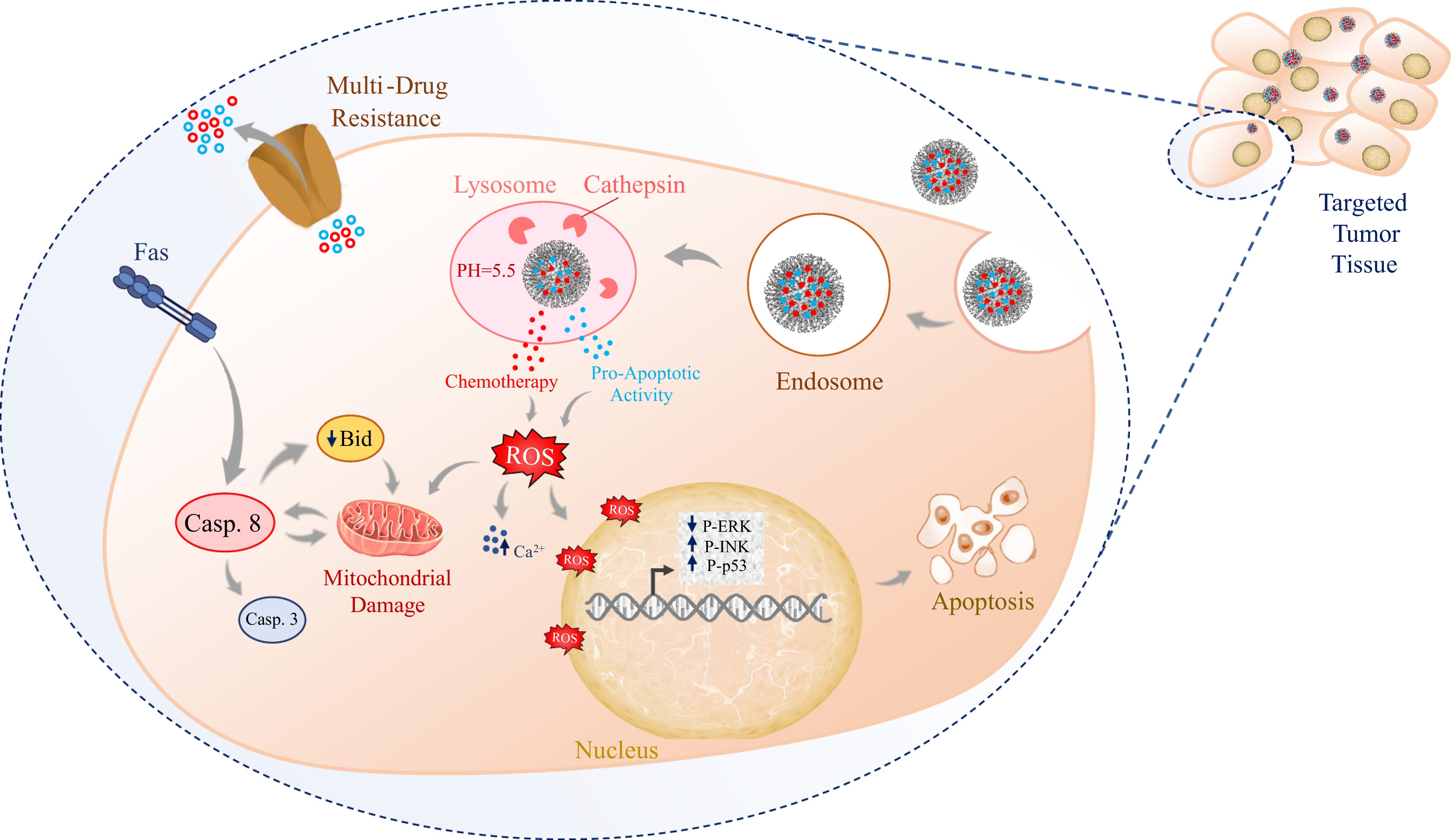
      Figure 1. 

      The diagram depicts the sequential process by which targeted nanoparticles are delivered to tumor cells to induce apoptosis with optimal efficacy. Initially, nanoparticles penetrate the cell via endocytosis, circumventing MDR channels. Once inside the cell, the nanoparticles engage key caspase cascade elements, initiating apoptotic signaling pathways. This interaction promotes the production of ROS within the apoptosis pathway, further amplifying cellular death. The nanoparticles ultimately trigger apoptosis in tumor cells through precise targeting and efficient delivery. This highlights the potential of nanoparticle-based drug delivery systems to counteract drug resistance mechanisms, thereby improving the effectiveness of cancer therapies. Reactive oxygen species (ROS), caspase-8 (Casp. 8), caspase-3 (Casp. 3), extracellular signal-regulated kinase (ERK), c-Jun N-terminal kinase (JNK), tumor protein p53 (P53), and multi-drug resistance (MDR).

    • Antitumor immunotherapy aims to modulate or activate the immune system to target cancer cells while minimizing damage to healthy tissue. NPs are vital in drug delivery, providing a targeted, controlled drug release platform, improving pharmacokinetics and biodistribution, and mitigating the risks of systemic toxicity. This is achieved through their ability to protect drugs from degradation, facilitate controlled release, and target specific tissues[147,148]. Chitosan-based NPs, for example, are nontoxic, biodegradable, and mucoadhesive, enabling precise cancer cell targeting[148]. In cancer vaccines, NPs act as delivery vehicles and adjuvants, increasing immunogenicity by efficiently delivering antigens and adjuvants to immune cells. Lipid NPs, for example, improve mRNA vaccine stability and the immune response, which is crucial for antitumor immunity[149,150]. NPs also enable the codelivery of peptide neoantigens and adjuvants such as STING and TLR4 agonists, enhancing CD8+ T-cell activation and improving immune checkpoint blockade therapy efficacy[151].

      Despite its potential, significant clinical challenges remain in the context of tumor immunotherapy. A major hurdle is the immune system's difficulty in distinguishing between malignant and normal cells owing to their molecular similarities. For example, in chimeric antigen receptor T-cell (CAR-T) therapy, managing 'off-target effects', unintended attacks on healthy cells, remains a substantial challenge[152]. Moreover, immune cells or cytokines delivered intravenously often fail to reach tumor sites because of the immunosuppressive microenvironment of solid tumors. As a result, immunotherapy tends to be more effective against hematological cancers, with limited efficacy against solid tumors[153].

    • Dendritic cells (DCs) are key antigen-presenting cells (APCs) that initiate immune responses and ensure self-tolerance. Nanotechnology enhances cancer therapy by improving antigen delivery to DCs, triggering a stronger immune response against tumor cells. However, regulatory T cells (Tregs) within the TME suppress cytotoxic T lymphocytes (CTLs), dampening overall T-cell activity. Targeted nanodelivery systems designed to reduce Treg numbers at the tumor site can help mitigate this suppression[154,155].

      Tumor cells often evade immune detection by overexpressing immune-evasive proteins such as programmed death-ligand 1 (PD-L1) and CD47, which block T-cell functions[156,157]. Nanocarriers engineered to deliver inhibitors targeting these proteins can enhance immunotherapy efficacy. In addition to direct tumor cell death, NP drugs can trigger immunogenic cell death (ICD) through ROS production, further amplifying the immune response. Thus, integrating nanotechnology into cancer immunotherapy holds significant promise by stimulating immune responses and overcoming tumor-mediated immune evasion[158,159].

    • Nanocarriers enhance tumor immunotherapy by delivering a range of agents, including TAAs, immune checkpoint inhibitors, mRNA-based vaccines, immunomodulatory cytokines, and natural polysaccharides. These strategies aim to improve the ability of the immune system to recognize and eliminate cancer cells, contributing to both cancer prevention and treatment[160,161].

      The use of TAAs is crucial for reactivating immune responses. NP-based delivery systems offer advantages over direct antigen injections by protecting antigens from degradation and ensuring targeted delivery to DCs for efficient antigen presentation and CTL activation. Several nanogel and nanovaccine formulations, such as those incorporating long peptide antigens or recombinant MUC4β subunits, have demonstrated potent antigen-specific T-cell responses[162]. Additionally, targeted delivery of antigens to APCs combined with immunostimulatory agents such as Toll-Like Receptor 4 (TLR4) agonists can further enhance antitumor immunity. The delivery of antigens to local lymph nodes via nanocarriers promotes both CTL and humoral immune responses, underscoring the therapeutic potential of these systems in cancer immunotherapy[163165].

      Monoclonal antibodies (mAbs) have revolutionized cancer immunotherapy by targeting tumor cells with high specificity. However, their poor pharmacokinetics often limit their efficacy, which can be improved by encapsulating them in nanocarriers. NPs can deliver immune-modulating agents such as anti-programmed cell death protein 1 (PD-1) or anti-CD47 antibodies, increasing immune responses and attacks on cancer cells. These NPs can also play multiple roles in the cancer immune cycle, including antigen presentation, T-cell activation, and tumor eradication. Furthermore, combining CAR-T-cell therapy with bispecific antibodies in NP formats has shown significant antitumor efficacy in preclinical models[166170].

      Despite promising results from nucleic acid-based immunotherapy in preclinical studies, clinical trials have underperformed due to nucleic acid delivery challenges. Biocompatible, cell-targeting nanomaterials, such as lipid NPs, have improved the delivery of mRNA-based vaccines, leading to robust CD8+ T-cell responses and tumor shrinkage in animal models. Moreover, combining mRNA therapies with cytokines or immunoadjuvants in NPs has further enhanced immune activation and improved tumor responses[171173].

      Polysaccharides, such as lentinan and Ganoderma lucidum glycopeptides, have long been recognized for their ability to modulate immune responses. While their clinical use has been limited by stability and bioavailability issues, recent advances in NP carriers have increased their stability and delivery[174177]. Studies have shown that polysaccharide-loaded nanocarriers can stimulate immune cell activation, expand T-cell populations, and synergize with chemotherapy, offering significant potential in cancer immunotherapy[178181].

    • Adoptive T-cell transfer (ACT) has shown substantial efficacy in cancer treatment, but several challenges remain, including poor tumor infiltration, limited functional persistence, and suboptimal targeting. Recent advances in nanotechnology offer strategies to overcome these barriers. Traditional ex vivo T-cell expansion using natural APCs can be time-consuming and inconsistent, leading to efforts to develop artificial APCs (aAPCs) to improve antitumor T-cell expansion[182185].

      For example, Zhang et al. engineered biomimetic magnetosomes and magnetic nanoclusters coated with leukocyte membrane fragments and functionalized them with T-cell stimulatory components. These aAPCs enhanced antigen-specific CTL expansion and guided CTL migration to tumor sites via MRI, significantly inhibiting tumor growth in mouse models[186]. Similarly, Li et al. designed polymer NPs that delivered cytotoxic T-lymphocyte antigen 4 (CTLA-4) siRNA to T-cells, leading to enhanced T-cell activation and proliferation within the TME[187].

      Additionally, Demento et al. developed poly(lactic-co-glycolic acid) (PLGA)-based NPs modified with lipopolysaccharides to promote DC maturation, further enhancing T-cell responses and boosting both humoral and cellular immunity[188].

    • Tumor tissues exhibit unique microenvironments characterized by acidity, hypoxia, and enzyme overexpression, which distinguish them from normal tissues[189,190]. These factors contribute to the low immunogenicity of cancer cells and the immunosuppressive nature of the TME. Enhancing the effectiveness of anticancer therapies necessitates remodeling the TME and enhancing immune cell infiltration. Multifunctional nanodrug delivery systems are being developed to target tumor tissues, improve drug uptake and release, and modulate the immunosuppressive TME. For example, microenvironment-responsive systems that respond to specific signals within the tumor can improve drug retention and penetration. Examples include systems that codeliver PD-L1 antibodies and photosensitizers, resulting in significant tumor growth inhibition[191194]. Additionally, systems that exploit the acidic TME to activate prodrugs or inhibit immunosuppressive enzymes have shown promise in enhancing local and systemic antitumor effects[195198].

      Immune checkpoint inhibitors, including anti-PD-1 and anti-CTLA-4 antibodies, have revolutionized melanoma and non-small cell lung cancer (NSCLC) treatments by restoring the body's antitumor immune response. This approach functions by counteracting the inhibitory effects of PD-1 and CTLA-4, which normally suppress the activity of CD8+ αβ T lymphocytes, thereby restoring their antitumor capabilities[199205]. However, these therapies have been less effective for other tumors, including colorectal and ovarian cancers. In murine models, liposomal doxorubicin targeted PD-L1-expressing tumor cells via specific peptides, disrupting the PD-L1/PD-1 interaction and enhancing CD8+ T-cell-mediated antitumor activity[206]. NK cells and γδ T cells, key components of the innate immune system, exhibit reduced expression of immune checkpoint molecules, which enhances their cytotoxic activity against tumors. Consequently, nanoformulated activators targeting these immune cells may constitute an ideal strategy for treating tumors that do not respond to anti-PD-1 and/or anti-CTLA-4 therapies. Further investigation is necessary to understand how NP-based drugs can effectively trigger γδ T cells[207,208].

      The effective delivery of immunomodulatory agents to targeted cells is essential for the development of nanovaccines, NP-based formulations designed to induce antigen-specific immune responses[209]. NPs confer numerous advantages, including protection of payloads, extended half-life, reduced toxicity, and enhanced delivery to APCs or TAAs[210]. Some NPs possess inherent immunostimulatory properties, provoking robust T-cell and antibody responses against tumors. The size of the NPs significantly influences their efficacy, and they can be used to deliver DC stimulatory molecules that activate APCs in tumor-draining lymph nodes. Combining antigens with adjuvants in NPs has demonstrated potential, with particle size playing a crucial role in immunogenicity. Moreover, targeting specific subsets of DCs with tailored NPs can enhance cytotoxic T-cell responses. The combination of distinct NP types with complementary functionalities remains an intriguing avenue for future exploration[211213].

      Nanotechnology-assisted adoptive cell therapies (ACTs), such as CAR-T-cell therapy, also hold promise for enhancing cancer treatment outcomes. Strategies involving the incorporation of NPs or cytokines into CAR-T cells have shown potential for increasing their cytotoxic activity, improving treatment outcomes[214216]. However, challenges such as side effects, high costs, and limited efficacy remain[217219]. Research into combination therapies and alternative immune cells, such as CAR-NK cells or CAR-macrophages, could further improve the clinical efficacy of ACT (Fig. 2)[220222].

      Figure 2. 

      The diagram illustrates the enhanced delivery of DC stimulatory molecules via nanoparticles, facilitating the activation of APCs or TAAs within the tumor-draining lymph nodes. This mechanism can amplify the cytotoxic potential of nanotechnology-assisted ACT, exemplified by CAR-T cells. The figure shows that DCs interact with nanoparticles through their receptors, initiating CAR-T-cell activation via class I and additional receptors. CAR-T cells, in turn, recognize and bind to tumor cells via a specific receptor, leading to their destruction through the release of perforin and granzyme. Chimeric antigen receptor (CAR), tumor-associated antigens (TAA), major histocompatibility complex class I (Class I MHC), T cell receptor (TCR), and TNF-related apoptosis-inducing ligand (TRAIL).

    • NMs, defined by at least one dimension in the nanoscale range (1–100 nm), exhibit unique mechanical, optical, electrical, and catalytic properties that distinguish them from bulk materials. NMs are categorized on the basis of their dimensionality into zero-dimensional (0D), one-dimensional (1D), two-dimensional (2D), and three-dimensional (3D), including structures such as nanospheres, nanotubes, and nanosheets. Their properties can be fine-tuned through modifications in size, shape, synthesis, and functionalization, and they can be composed of both inorganic materials (e.g., metals and oxides) and organic materials (e.g., polymeric NPs and nanofilms)[223225].

      The resistance of solid tumors to traditional systemic treatments is a multifaceted challenge influenced by multiple biological and environmental factors. A significant challenge is heterogeneity within tumors, which includes subpopulations of cells, such as polyploid giant cancer cells (PGCCs), that can survive treatment and repopulate the tumor, contributing to therapy resistance[226]. Additionally, the TME plays a crucial role in supporting tumor survival through factors such as hypoxia and acidity, which protect the tumor and facilitate its adaptation to treatment, presenting further barriers to effective therapy[227]. Traditional chemotherapy often fails due to mechanisms such as drug inactivation, efflux, and DNA repair, which are further complicated by the presence of cancer stem cells (CSCs) that exhibit inherent resistance to many treatments[228,229]. Furthermore, systemic chemotherapy struggles to deliver high concentrations of drugs at the tumor site, leading to limited effectiveness and significant side effects[230]. Although monoclonal antibodies are more targeted, their poor tissue penetration and heterogeneous distribution contribute to acquired resistance[231]. Nanocarriers provide an effective approach to overcoming the resistance of solid tumors to traditional treatments by targeting key resistance mechanisms, such as the overexpression of apoptosis inhibitors (e.g., Bcl-2, IAPs, and Akt) and drug efflux proteins such as P-gp, which are commonly observed in resistant tumors. These NP-based systems increase the solubility, stability, and targeted delivery of anticancer drugs via the EPR effect, concentrating them in tumor tissues to improve efficacy and reduce off-target toxicity[232]. Moreover, nanocarriers can codeliver autophagy regulators and anticancer drugs, offering a synergistic effect that increases treatment efficacy while reducing systemic toxicity[233]. Designed for precise targeting, deep penetration, and controlled drug release, nanocarriers help overcome MDR, minimize side effects, and improve biocompatibility[234,235]. The codelivery of multiple drugs and MDR reversal agents also shows promise in inhibiting tumor metastasis and recurrence (Table 2)[236].

      The delivery of NMs to solid tumors has been a central research focus for over a decade. Early studies emphasized improving delivery via passive or active targeting strategies, primarily leveraging the EPR effect. The EPR effect takes advantage of the abnormal vascular structures found in tumors, allowing NMs to extravasate from blood vessels and penetrate tumor tissues[29,237]. However, recent studies indicate that the EPR effect is often less effective than previously thought, with some researchers proposing that NMs may enter tumors through active mechanisms rather than solely relying on leaky blood vessels[114,238240].

      A meta-analysis by Wilhelm et al. revealed that only 0.7% of intravenously administered organic and inorganic NPs accumulate in solid tumors in preclinical models, irrespective of the targeted strategy used. While this low accumulation may suffice for small tumors, it is insufficient for larger tumors because of size and cellular composition variations. These findings emphasize that therapeutic success depends on the precision of targeting mechanisms and the proficiency of drug release processes. Despite the low accumulation rate, this figure is still higher than the efficacy seen with conventional therapies, although it does not account for tumor size, dosage, or variability in the EPR effect across different models[237].

      The size of gold-based NMs is particularly important for optimizing delivery efficiency. The thickness and permeability of the stromal barrier, directly related to tumor size, can affect the penetration of NMs into the tumor. Tailoring the size of NMs to match the specific dimensions of individual tumors may improve therapeutic outcomes by enhancing diagnostic and therapeutic efficiency. However, the biological heterogeneity of tumors complicates the prediction of NM effectiveness[241243]. The variability in tumor perfusion, vessel permeability, and angiogenesis further challenges the reliable prediction of outcomes from NP-based treatments[244,245].

      Table 2.  Utilizing NPs to promote antitumor immune reactions.

      Approach Tumor model(s) Key outcome(s) Ref.
      Delivery of TAAs to APCs OVA-expressing melanoma, thymoma, T-cell lymphoma, colon adenocarcinoma - Efficient internalization of TAAs by dendritic cells (DCs).
      - Potent cellular and humoral immune responses.
      - Enhanced immune response of tumor-specific T cells against antigens.
      - Tumor growth slowed down.
      [246,247]
      Delivery of adjuvants to
      TDLNs (Tumor-Draining Lymph Nodes)
      Melanoma, B-cell lymphoma - Efficient internalization of adjuvants by dendritic cells.
      - Strong cellular and humoral immune responses.
      - Reduction in tumor size.
      - Prolonged survival time.
      - Immune stimulation of DCs through TLR-ligand modulation.
      [248,249]
      Codelivery of antigen and adjuvant Melanoma, OVA-expressing - Immune responses from both CD4 and CD8 T cells.
      - Strong CD8 T-cell response independent of additional stimuli.
      - Production of INF-gamma.
      - Balanced antibody response (IgG1 and IgG2).
      - Reduced regulatory T-cell count, enhancing vaccine effectiveness.
      - Vigorous production of antigen-specific cytotoxic T lymphocytes (CTLs).
      [250,251]
    • Current chemotherapeutic and molecularly targeted agents have improved outcomes in treating hematological malignancies (HMs), but long-term remission or complete remission remains achievable in only a subset of patients. This limited success can be attributed to the nonspecific nature of these therapies, which inadvertently affect healthy tissues and cause off-target side effects[252]. To sustain therapeutic concentrations at the tumor site, high drug doses and frequent administrations are frequently required, exacerbating these adverse effects[253]. Consequently, there is a growing need for drug-delivery systems that minimize healthy tissue exposure while ensuring precise and effective delivery to malignant cells[254].

      HMs, particularly lymphomas, often involve the spleen, which plays a key role in immune regulation and can accumulate intravenously administered NPs. The targeting of inflammatory monocytes in the spleen via NPs loaded with siRNA has shown promise in reducing tumor growth in lymphoma models[255,256].

      Several formulations have been developed and tested in nanomedicine for drug delivery in blood cancers. These include PEGylated liposomal doxorubicin (PLD) and non-PEGylated liposomal doxorubicin (NPLD), which are used in treating various HMs. For example, PLD in combination with the proteasome inhibitor bortezomib has shown encouraging results in multiple myeloma treatment[257259]. Additionally, liposomal daunorubicin has been explored as a treatment option for HM[260]. Liposomal vincristine has also been approved for treating Philadelphia chromosome-negative acute lymphoblastic leukemia (ALL)[261,262]. Recently, approved combinations, such as liposomal cytarabine + daunorubicin (CPX-351/VYXEOSTM), have demonstrated efficacy in high-risk secondary acute myeloid leukemia (AML)[263]. Moreover, polymer-based nanomedicines, including polymeric micelles, have emerged as tailored solutions to enhance drug stability, circulation, and targeting, offering considerable potential in hematological cancer treatment.

      In addition to optimizing drug delivery, nanotechnology has also been investigated to improve hematopoietic stem cell transplantation (HSCT), a critical treatment for blood cancers such as leukemia. However, challenges such as recurrence and graft-vs-host disease (GVHD) remain. Nanomaterials are being investigated to expand hematopoietic stem cells (HSCs) ex vivo, addressing the issue of limited donor cell availability. Hydrogels, microcavity arrays, and NPs have shown promise in supporting HSC expansion in laboratory settings, suggesting that they may improve HSCT outcomes[264266].

      Additionally, NPs have been engineered to target signaling pathways involved in blood cancers[254,267,268], with promising results in animal models. NPs have also been studied for their ability to modulate intracellular pathways related to epigenetics, cell death, and metabolism, potentially opening new treatment options. These advancements in nanomedicine could enhance therapeutic outcomes while minimizing side effects in blood cancer treatment[268271]. Furthermore, NP-based delivery systems can stimulate immune responses by promoting the release of antitumor cytokines. For example, recombinant granulocyte‒monocyte colony stimulatory factor (rGM-CSF) delivered through nanoformulations has been designed to enhance tumor cell clearance by macrophages[272]. NPs loaded with siRNAs have been utilized to target tumor-associated macrophages, indicating their promising role in cancer immunotherapy, although potential side effects remain a concern[273,274]. Peptide- and protein-based nanoformulations have been explored for their ability to enhance the efficacy of cancer vaccines[275], and NP-based drug formulations have demonstrated promise in stimulating antitumor immune effector cells[187,276].

    • Nanocarriers demonstrate significant potential in cancer therapy; however, several challenges impede their successful translation into clinical application. A considerable challenge in oncology is tumor biology's complex and heterogeneous nature, which poses substantial obstacles to effectively targeting and delivering chemotherapeutic agents. MDR, driven by mechanisms such as the overproduction of efflux pumps or alterations in drug targets, further reduces the effectiveness of nanocarriers[277,278]. This issue, coupled with the varied molecular profiles of tumor cells, makes selective targeting difficult while sparing healthy tissue.

      Nanocarriers must be able to target and infiltrate diverse tumor cell types, but inconsistent receptor expression across different tumor types limits their specificity. This variability reduces the success of receptor-targeted therapies[279]. Additionally, the TME, with factors such as hypoxia, elevated interstitial fluid pressure, and acidic pH, presents significant obstacles to drug release and distribution. Hypoxia can reduce drug sensitivity, whereas interstitial fluid pressure can limit the penetration of nanocarriers into deeper tumor regions[280282].

      Off-target toxicity is another major concern. Despite efforts to improve nanocarrier selectivity, unintended accumulation in healthy tissues may lead to harmful side effects. Nanomaterials may provoke immune responses or accumulate in organs such as the liver, spleen, or kidneys, resulting in chronic toxicity[283]. Moreover, the long-term fate of nanocarriers is not well understood, and interactions with the immune system could cause immunogenicity or impair immune function. Therefore, controlling nanocarrier distribution and clearance is crucial for reducing off-target effects. Comprehensive preclinical evaluations of pharmacokinetics, biocompatibility, and long-term safety are essential.

      The scalability of nanocarrier production also presents challenges. Manufacturing nanocarriers requires precise control over size, shape, and surface properties to ensure consistent therapeutic outcomes[284]. Even small deviations in these parameters can impact biodistribution, uptake, and efficacy. Ensuring consistent quality at a commercial scale is vital for successful clinical use. Furthermore, regulatory approval of nanomedicines remains slow, with few products approved due to the challenges in proving their safety and efficacy. Regulatory agencies require extensive data on physicochemical properties, pharmacokinetics, toxicity, and efficacy, which is often time-consuming and expensive[285].

      Researchers are developing stimuli-responsive nanocarriers to increase the precision of drug delivery triggered by ultrasonic, light, or temperature factors. These systems can potentially regulate drug release specifically at the tumor site, minimizing systemic exposure and reducing associated side effects. However, designing such systems is complex, and practical limitations, such as the depth of stimulus penetration, must be addressed. For example, light-responsive nanocarriers face limitations in terms of tissue penetration, whereas ultrasound and magnetic fields, which allow deeper penetration, require optimized protocols[284,286]. Furthermore, the possibility of inadvertently damaging healthy tissues remains a concern, and achieving uniform activation within heterogeneous tumors remains challenging.

      Despite these obstacles, innovative strategies are emerging. Functionalizing nanomaterials, such as graphene oxide, is a promising approach to improving nanocarrier targeting and cytotoxicity. With its large surface area and capacity to absorb near-infrared light, graphene oxide emerges as an ideal candidate for drug delivery and photothermal therapy. Furthermore, developing multifunctional, 'smart' nanocarriers that can respond to multiple stimuli or deliver combination therapies may increase the precision and effectiveness of cancer treatments[60,287].

      One promising approach to mitigate the challenges of nanocarrier-based cancer treatment is the development of multifunctional nanocarriers. These specialized carriers are designed with distinct characteristics that allow them to navigate the complex TME and efficiently deliver therapeutic agents to the tumor site[288291]. Several strategies can be utilized to enhance the performance of nanocarriers: (1) Targeting specificity: Nanocarriers can be altered with targeting ligands (such as antibodies or peptides) that recognize specific receptors on cancer cells, increasing their selectivity for tumor cells. This modification enhances the accumulation of therapeutic agents at the tumor site, improving their effectiveness while reducing systemic toxicity[292294]. (2) Stimuli-responsive behavior: Nanocarriers can be engineered to respond to specific conditions in the TME, such as acidic pH, elevated enzyme levels, or hypoxia. For example, pH-sensitive nanocarriers exploit the acidic microenvironment of tumors to trigger the release of drugs directly at the tumor site, reducing the impact on healthy tissues[198,295,296]. (3) Enhanced imaging capabilities and theranostics: Nanocarriers can be integrated with imaging agents, allowing noninvasive tracking of their distribution in the body. These real-time data can help optimize treatment regimens and enable personalized treatment approaches. Furthermore, the development of theranostic nanocarriers, which combine therapeutic agents with diagnostic imaging tools, holds promise for real-time monitoring of treatment responses and adaptive management (Table 3)[297300].

      Table 3.  Some recent advancements in nanoparticle-based imaging and targeted drug delivery for solid tumor types.

      Tumor type NPs type NPs feature Key advancements Ref.
      Breast cancer (MCF-7) Oleic acid-Fe3O4 NPs (OA-Fe3O4) incorporated into PLA-PEG-PLA copolymer (BMNPs/EPPT/drug) - Spherical morphology (179–203 nm)
      - Magnetic
      - Drug-loaded
      - pH-sensitive release
      - Targeted delivery via EPPT peptides
      - Increased drug release at lower pH
      - Enhanced in vitro cytotoxicity (lower IC₅₀)
      - Selective interaction with breast cancer receptors
      - Significant tumor volume reduction in vivo.
      [310]
      HER-2-positive breast cancer Generation 5 poly(amidoamine) dendrimers conjugated with gold NPs, chelated gadolinium, and anti-HER-2 antibody - Bimodal imaging (CT and MRI)
      - HER-2 specific targeting
      - Encapsulated gold NPs
      - Gadolinium for MRI
      - Enhanced MRI signal intensity (~20%)
      - Improved CT resolution and contrast (2-fold)
      - Specific targeting and internalization in HER-2 positive cells
      - Potential for early detection and therapeutic monitoring.
      [311]
      HER2-positive breast cancer cRGD-modified RBC membrane-coated multidrug nanocomplexes - Coated with red blood cell membrane (RBCm)
      - Cyclic Arg-Gly-Asp (cRGD) modification for targeting
      - Combinational delivery of Polo-like kinase 1 siRNA (siPlk1) and neratinib
      - Enhanced drug stability and tumor accumulation
      - Overcomes challenges of in vivo codelivery of oligonucleotide drugs and chemotherapy agents
      - Combination therapy significantly improved antitumor efficacy in HER2-positive breast cancer, both in vitro and in vivo.
      [312]
      Breast cancer Biologically produced Gold NPs (AuNPs) - Round AuNPs (13 ± 1.3 nm)
      - Zeta potential of −35.8 ± 1.3 mV
      - Conjugated with multiple cargoes (Paclitaxel, Transferrin, and
      anti-miR-135b)
      - Multicargo delivery system with effective tumor targeting
      - Higher in vitro cytotoxicity
      - Enhanced tumor-specific toxicity
      - Suppression of miR-135b expression
      - Superior to current drug carriers.
      [313]
      Breast cancer Polymeric siRNA NPs (PRNs) - NPs (50–200 nm) made using rolling circle transcription (RCT)
      - Coated with biopolymers
      (PLL, PLG, HA)
      - Size- and surface-tuned PRNs improve systemic delivery and anticancer effects
      - HA-layered NPs (~200 nm) exhibit the best targeting and therapeutic efficacy due to CD44 receptor targeting.
      [314]
      Breast cancer Lipid-substituted polyethyleneimine (PEI) polymers (lipopolymers) - siRNA/lipopolymer nanocomplexes
      - Optimized with anionic additives (phosphate buffer and N-Lauroylsarcosine Sodium Salt) for gene silencing
      - Achieved > 80% gene silencing and > 70% cell killing with minimal cytotoxicity in MDA-MB-231 breast cancer cells
      - > 95% cell uptake
      - Persistent effects for at least 6 d.
      [315]
      Lung cancer (NSCLC) Superparamagnetic iron oxide NPs (SPIONs) combined with Exos derived from NSCLC cells - Dual-targeting system using SPIONs for magnetic targeting
      - Exos for homing and targeted delivery
      - Doxorubicin (DOX) loaded for tumor suppression
      - Developed a 'dual-targeting' drug delivery platform
      - Exosome-SPIONs loaded with DOX provide optimal tumor tissue delivery
      - Effective tumor suppression in NSCLC models
      - Reduced toxicity to normal tissues.
      [316]
      Lung cancer (EGFR-mutant, PC-9 cells) Silk peptide NPs (CUR/ERL@SP) - Dual-drug coloaded (curcumin and erlotinib)
      - 150 nm in size
      - Enhanced cell internalization
      - Codelivery of curcumin and erlotinib enhances drug efficiency
      - Improved cellular uptake
      - Downregulation of EGFR and proteins involved in tumor invasion
      - More effective therapeutic strategy for EGFR-mutant lung cancer.
      [317]
      Lung cancer (A549 cells) Paclitaxel-loaded PLGA NPs (AM@PTX-NPs) - A549 cell membrane biomimetic NPs
      - 10.90% drug loading efficiency
      - Spherical shape
      - A549 cell membrane camouflaged NPs significantly enhanced targeting
      - Tumor growth inhibition: 73% vs 37.39% for unmodified PTX-NPs
      - Improved strategy for targeted cancer treatment.
      [318]
      Lung cancer (A549 cells) Collagenase-loaded AcMD nanostructured microparticles (MPs) - Hollow, corrugated morphology
      - Median volume diameters:
      2.58 ± 1.35 to 3.01 ± 0.68 µm
      - MMAD: 1.93 ± 0.06 to 2.80 ± 0.10 µm
      - FPF: 68.02 ± 6.86% to 69.62 ± 2.01%
      - Enzymatic stability: 89.5 ± 6.7%
      - Collagenase-loaded microparticles improved nanoparticle penetration into tumor spheroids (A549/MRC-5 coculture)
      - Enhanced delivery of therapeutic NPs
      - ECM-modulation for better intratumoral penetration.
      [319]
      Lung cancer (A549 cells) Niosomes containing gold NPs (Nio-AuNPs) - Hydrodynamic diameter of AuNPs: 38.85 ± 0.85 nm
      - Nio-AuNPs diameter: 127.8 ± 2.92 nm
      - Spherical shape (confirmed by TEM and FE-SEM)
      - Combination of Nio-AuNPs with X-ray radiation (XRT) showed a synergistic effect
      - Increased cytotoxicity and apoptosis induction in A549 cells
      - Superior to XRT or AuNPs alone.
      [320]
      Metastatic prostate cancer (PCa) Au/Mn nanodots-luteinizing hormone-releasing hormone (AMNDs-LHRH) nanosystem - Multimode imaging (FL/CT/MR)
      - Targeted drug delivery
      - Photothermal therapy
      - Fluorescence-guided surgery
      - Targeting GnRH-R positive PCa and metastases for accurate preoperative CT/MR diagnosis
      - Fluorescence visualization for surgery
      - Improved photothermal therapy for metastatic PCa
      - Enhanced diagnostic accuracy and therapeutic effect.
      [321]
      Bone metastatic prostate cancer (PCa) Au/Gd nanodots - Multimode imaging (optical and X-ray)
      - Good biocompatibility
      - High biosafety
      - High-precision detection of metastatic prostate cancer
      - Complementary multimode imaging for accurate diagnosis and treatment guidance
      - Enhanced biosafety and biocompatibility in vitro and in vivo.
      [322]
      Colorectal cancer Functionalized mesoporous silica NPs (MSN-NH2 and MSN-COOH) - Spherical shape
      - Mesoporous structure
      - pH-responsive drug release
      - Surface functionalization with amino (positive charge) and carboxyl groups (negative charge)
      - Controlled release of doxorubicin at physiological pH (7.4) to reduce systemic toxicity
      - Enhanced drug release at acidic pH (5.5), targeting TME
      - Improved therapeutic efficacy by exploiting pH-sensitive drug release.
      [323]
      Colon cancer Poly(lactide-co-glycolide)-block-poly(ethylene glycol)-carboxylic acid endcap NPs (PLGA-PEG-COOH NPs) - CASIN encapsulated in PLGA-PEG-COOH NPs
      - Targeted delivery system
      - Improved bioavailability
      - Reduced drug elimination
      - Development of a nanoparticle-based delivery system for CASIN
      - Overcomes rapid drug elimination and low bioavailability
      - Enables targeted inhibition of Cdc42 in colon cancer.
      [324]
      Colorectal cancer Salmonella membrane-coated bacilliform gold nanorods (SM-AuNRs) - Gold nanorods coated with Salmonella membrane proteins
      - Mimics Salmonella morphology and surface
      - Targeted tumor cell internalization
      - Mucus barrier penetration
      - Development of biomimetic SM-AuNRs for enhanced tumor targeting and penetration
      - Superior photothermal and chemotherapeutic effects for colorectal cancer treatment.
      [325]
      Colorectal cancer GOx@FeNPs (glucose oxidase-loaded iron NPs) - Dual-targeted NPs with cRGD peptide and anisamide (AA)
      - Combines photothermal therapy (PTT), ferroptosis, immune checkpoint blockade (αPD-L1), and MRI
      - Combination of PTT, ferroptosis, and αPD-L1 blockade for enhanced CRC treatment
      - Dual-targeting (cRGD and AA) improves tumor targeting
      - Induces immunogenic cell death (ICD) and enhances immune response
      - Achieves over 90% tumor inhibition
      - MRI capability for integrated diagnosis and therapy.
      [326]
      Colorectal cancer Hyaluronidase-responsive MSN-HA/DOX, DOX/SLN-PEG-Biotin, Galactosylated chitosan-functionalized MSNs - Functionalized mesoporous silica NPs (MSNs)
      - Surface modifications with hyaluronic acid (HA), polyethylene glycol (PEG), biotin, and galactosylated chitosan
      - Dual-stimulus release (hyaluronidase-responsive)
      - Enhanced tumor inhibition via dual-stimulus release and biotin-targeting for CRC cells
      - Controlled release through asialoglycoprotein receptors
      - Significant in vitro and in vivo efficacy improvements over conventional chemotherapy.
      [327]
      Gastrointestinal cancer Poly(lactic-co-glycolic acid) (PLGA)-based NPs Biodegradable, biocompatible, and tunable properties such as the ratio of PLA to PGA, molecular weight, crystallinity, and preparation process - Diagnosis
      - Chemotherapy
      - Radiotherapy
      - Novel treatments like immunotherapy, gene therapy, and photothermal therapy
      [328]
      Gastric cancer pH-responsive NPs with TPGS-conjugated fucoidan - pH-responsive
      - Targeted delivery to P-selectin protein
      - Demonstrated enhanced antigastric tumor efficacy
      - Reduced expression of malignant proteins
      - Promising clinical therapeutic potential
      [329]
      Gastric and colon cancer Glucose oxidase-loaded manganese-based mesoporous silica NPs (MSN@Mn-GOx) - Solid spheres (~100 nm)
      - Fenton-like properties
      - pH-responsive (stronger at pH 6.0)
      - Zeta potential: -35 mV
      - Enhanced ROS production
      - Inhibition of gastric and colon cancer cell proliferation
      - MRI-based tumor imaging enhancement
      - Potential therapeutic option for gastric cancer
      [330]
      Gastric cancer Arginine-chitosan and fucoidan NPs - Protects postbiotic (SGMNL-133) from gastric acid degradation
      - Facilitates mucus penetration
      - Enhances interaction with cancer cells
      - Successful isolation and optimization of GMNL-133 (SGMNL-133) to inhibit gastric cancer cell proliferation
      - Development of NP formulation to improve in vivo delivery of SGMNL-133
      - Enhanced anti-gastric tumor efficacy and reduced tissue inflammation.
      [331]
      Pancreatic adenocarcinoma AuNPs (Gold NPs) - Coated with hyaluronic acid and oleic acid (HAOA-AuNPs)
      - Coated with bombesin peptides (BBN-AuNPs)
      - Spherical shape
      - Size: 83 ± 20 nm (HAOA-AuNPs)
      - Size: 49 ± 12 nm (BBN-AuNPs)
      - Significant reduction in cell viability when combined with Radiation Therapy (RT)
      - Improved therapeutic effect when RT was combined with AuNPs
      - AuNPs at different concentrations (200-400 μM for HAOA-AuNPs, 50-200 μM for BBN-AuNPs) showed varying reductions in cell viability (up to 37%)
      - RT + AuNPs combination showed up to a 26% reduction in viability at 72 h postirradiation
      [332]
      Pancreatic cancer (MIAPaCa-2 and PANC-1 cell lines) Titanium dioxide NPs
      (H2O2-modified)
      - Modified with hydrogen peroxide
      - Used in combination with ultrasound-stimulated microbubbles (USMB) and X-rays
      - USMB + X-rays showed a significant radiation enhancement effect and increased ROS in MIAPaCa-2 cells
      - No enhancement effect observed in PANC-1 cells
      - USMB did not improve nanoparticle-induced radiosensitization in either cell line when combined
      [333]
      Pancreatic adenocarcinoma AuNPs (Gold NPs) - Coated with hyaluronic acid and oleic acid (HAOA-AuNPs)
      - Coated with bombesin peptides (BBN-AuNPs)
      - Spherical shape
      - Size: 83 ± 20 nm (HAOA-AuNPs)
      - Size: 49 ± 12 nm (BBN-AuNPs)
      - Significant reduction in cell viability when combined with Radiation Therapy (RT)
      - RT + AuNPs showed up to 37% reduction in cell viability
      - Combination of RT and AuNPs led to improved therapeutic efficacy, with up to 26% reduction in cell viability at 72 h postirradiation
      - Improved effect observed with both HAOA-AuNPs and BBN-AuNPs compared to RT alone
      [334]
      Pancreatic cancer (PANC-1 cell line) Silver NPs (AgNPs) - Functionalized with IgG molecules
      - NIR light absorption (808 nm, 2 W)
      - Used for photothermal therapy
      - Photo-excitation of IgG-functionalized silver NPs induced dysfunction in the Golgi apparatus
      - Activated caspase-3 apoptotic pathway, leading to cellular apoptosis
      - Demonstrated as a potential novel therapy for pancreatic cancer via photothermal treatment using a laser
      [335]
      Pancreatic cancer Gelatin-based NPs - Fabricated via radiation-induced crosslinking
      - Size: 5–20 nm
      - Labeled with 64Cu
      - Negative surface potential
      - Developed as a novel imaging agent for PET
      - In vivo evaluation showed accumulation of NPs in pancreatic tumors
      - 64Cu-labeled gelatin NPs show promise for next-generation PET imaging in pancreatic cancer
      [336]
      Pancreatic cancer Hyaluronic acid (HA)-displaying NPs - Composed of positively charged chitosan (CS)
      - Complexed with small interfering RNA (siRNA)
      - Two formulations: low molecular weight (LMW) CS and high molecular weight (HMW) CS
      - Targeted siRNA therapy to downregulate HIF-1α and enhance treatment outcomes
      - Both LMW and HMW CS formulations showed an ability to knock down HIF-1α in vitro and in vivo
      - LMW CS showed faster uptake kinetics, while HMW CS was more effective in gene knockdown
      - Potential to enhance treatment in the hypoxic TME, a characteristic of pancreatic cancer
      [337]
      Ovarian cancer Polymeric-based NPs siRNA delivery, serum stability enhancement, targeted delivery - Developing polymeric NPs for siRNA delivery to target specific genes involved in OC prognosis and overcoming chemo-resistance. [338]
      Ovarian cancer Honokiol (HK)-based polyprodrug NPs - Glutathione (GSH)-sensitive HK polyprodrug
      - Conjugated with EpCAM-specific aptamer and polyethylene glycol (PEG)
      - High drug loading and GSH-responsive drug release
      - Developed a GSH-sensitive HK polyprodrug for targeted ovarian cancer therapy
      - A/P-PHK NP40 (aptamer-modified and PEG-modified prodrug) showed the highest targeting ability for EpCAM-overexpressing ovarian cancer cells
      - Exhibited enhanced cell growth inhibition compared to free HK and control HK NPs
      - Novel strategy for improving the delivery and targeting of HK in ovarian cancer treatment
      [339]
      Ovarian cancer Arsenic-manganese complex NPs (ATO-Mn) - Arsenic trioxide (ATO) and manganese (Mn2+) complexed in NPs
      - pH-sensitive release of arsenic (As3+) and manganese (Mn2+) ions
      - SKOV3 cell membrane-encapsulated NPs for homologous targeting
      - Glucose oxidase-based starvation therapy
      - MR imaging for real-time ATO dose distribution monitoring
      - Achieved synergistic effects combining starvation therapy, chemodynamic therapy, and chemotherapy
      - ATO and Mn2+ release triggered by tumor acidity,
      generating reactive oxygen species (ROS) through
      Fenton-like reaction
      - Improved biocompatibility and therapeutic efficacy compared to free ATO administration
      - Self-enhanced chemodynamic therapy with glucose oxidase supporting H2O2 restoration and reducing cellular acidity
      - Innovative approach for multimodal diagnosis and treatment of ovarian cancer
      [340]
      Ovarian Cancer HA@PFG NPs - Cisplatin prodrug (Pt-COOH)
      - Fe3+ and natural polyphenols (Gossypol)
      - Stable structure
      - Controllable drug release behavior
      - High drug loading capacity
      - pH-sensitive release at tumor sites
      - Ferroptosis promotion
      - Combined MRI imaging with cisplatin-based chemotherapy for improved diagnosis and therapy
      - Synergistic effect of Pt-COOH (chemotherapy) and Gossypol (pro-apoptotic) for tumor cell killing
      - Fe3+ release at tumor sites facilitates ferroptosis and MRI imaging of ovarian cancer
      - Improved therapeutic efficacy in patient-derived tumor xenograft (PDX) model
      - Ameliorated OC symptoms through IL-6 signaling
      pathways
      [341]
      Hepatocellular carcinoma - Solid lipid NPs (SLN)
      - PEGylated galactose-conjugated SLN (GAL-SSLN)
      - Targeted delivery
      - Enhanced cytotoxicity
      - Apoptosis induction
      - Improved pharmacokinetics
      - GAL-SSLN demonstrated superior targeting and therapeutic efficacy compared to traditional Sorafenib-loaded SLN in HCC.
      - Enhanced liver-specific drug delivery and efficacy.
      [342]
      Hepatocellular carcinoma Nano Zinc Oxide (nZnO) - Aged nZnO with physicochemical transformation
      - Investigated for promotional effects on liver cancer progression
      - Studied the promotional effects of aged nano zinc oxide (nZnO) on HCC cell progression (HepG2)
      - Focused on the impact of long-term exposure to subtoxic doses of NPs
      - Explored how NMs, specifically nZnO, may influence liver cancer progression, highlighting the challenges in assessing chronic exposure to NPs
      [343]
      Hepatocellular carcinoma Inorganic NPs - Accumulate in the liver due to EPR effect
      - Enhanced accumulation in HCC tumors with leaky vasculature
      - Explored the use of inorganic NPs in liver cancer therapy, leveraging their ability to accumulate in HCC tumors through the EPR effect
      - Identified the potential of inorganic NPs to overcome the challenge of liver accumulation and enhance therapeutic effects in HCC treatment
      - Highlighted the use of low- and high-LET radiation from the same radionuclide for enhanced treatment strategies in HCC
      [344]
      Liver cancer Silica NPs (DOX-loaded) - Loaded with doxorubicin (DOX) and MRI contrast agent
      - Coated with pH-responsive and tumor cell-targeting polymers
      - Targeted delivery with controlled drug release in acidic environments
      - Achieved integrated diagnostic and therapeutic strategy for liver cancer
      - pH-responsive polymer coating enables controlled release of DOX in the tumor's acidic environment
      - Inhibited autophagic flux by targeting the autophagy‒lysosome pathway and regulating TFEB nuclear translocation
      - Promoted tumor cell death and enhanced therapeutic efficacy in liver cancer treatment
      [345]
      Hepatocellular carcinoma siCD24-Lenvatinib-MnO@PLAP nanomedicine - Incorporates manganese oxide (MnO), lenvatinib (Len), and siRNA against CD24 (siCD24)
      - Micelles composed of methoxypolyethylene glycol (mPEG), poly-L-lysine (PLLys), and polyasparagyl (PAsp(PIP)) triblock copolymer
      - Responsive to TME (TME)
      - MRI contrast agent (Mn2+)
      - Synergistic effect of siCD24 and lenvatinib reduces CSC stemness and enhances therapeutic efficacy
      - Inhibited CD24 expression and HIF-1α, reducing drug resistance in CSCs
      - Improved lenvatinib therapy for HCC with enhanced tumor-targeting and reduced stem cell resistance
      - In situ production of Mn2+ for MRI monitoring of therapeutic progress
      [346]
      Hepatoblastoma Vincristine-loaded polycaprolactone NPs with carbon dots (Vin@PCL-CDs) - Carbon dots encapsulated with vincristine sulfate
      - Polycaprolactone (PCL) biodegradable NPs
      - Spherical, uniformly sized
      (~200 nm)
      - Excellent colloidal stability
      - Improved vincristine pharmacokinetics and targeted delivery to liver cancer cells
      - Prolonged release of both vincristine and carbon dots, enhancing the therapeutic effect
      - Enhanced cancer cell inhibition and targeted delivery compared to free vincristine
      - Carbon dots enabled bioimaging with clear fluorescence, not cytotoxic, offering potential in imaging and cancer research
      [347]
      Kidney cancer ICG-PEG45 (Indocyanine Green conjugated with PEG) - Renal-tubule-secreted near-infrared-emitting fluorophore
      - Effluxed via P-glycoprotein transporter in normal tissues
      - Retained in cancerous tissues with low P-glycoprotein expression
      - Enhanced kidney cancer targeting through renal tubular secretion
      - Hyperfluorescence imaging for better detection and visualization of tumors.
      [348]
      Renal cell carcinoma ZnPP@G-PP NPs (ZnPP@PLGA-PFP NPs) - Sound-sensitive, multifunctional nanoplatform
      - Sonosensitizer (ZnPP) loaded in NPs
      - G250-targeting ligand for tumor specificity
      - Encapsulation efficiency and good stability
      - Multimodal imaging (PA/US) capabilities
      - Designed for targeted sonodynamic therapy (SDT) and multimodal imaging
      - Effective tumor targeting via G250 ligand
      - LIFU (low-intensity focused ultrasound) irradiation enhances 1O2 production, improving the SDT effect
      - In vitro and in vivo studies showed significant therapeutic effects with effective tumor penetration and suppression
      - Real-time diagnostic and therapeutic monitoring with PA/US imaging
      - Excellent biocompatibility and therapeutic potential for RCC treatment
      [349]
      Bladder cancer Silver NPs (AgNPs) - Biogenic synthesis from Fusarium sp.
      - Dose- and time-dependent cytotoxicity
      - Induces apoptosis
      - Inhibits cell migration and proliferation
      - AgNPs demonstrated antitumoral activity in NMIBC models
      - Tumor regression (57.13%) and benign lesions observed in animal studies
      - Potential as a cost-effective alternative for bladder cancer treatment
      [350]
      Bladder cancer PAMAM-modified Mesoporous Silica NPs (MSNPs) - Modified with poly(amidoamine) (PAMAM) dendrimers
      - Mucoadhesive properties
      - Controlled drug release triggered by acidic pH
      - PAMAM-modified MSNPs exhibited enhanced mucoadhesion, particularly with two-generation PAMAM
      - Sustained doxorubicin release
      - Demonstrates potential as a mucoadhesive drug delivery system to improve bladder cancer chemotherapy.
      [351]
      Bladder cancer Lipid-coated Mesoporous Silica NPs (Silicasomes) - Combination of cisplatin (chemotherapy) and tirofiban (antiplatelet agent)
      - TME-targeted
      - Prevents LVI formation and enhances drug delivery
      - Developed a nanodrug that targets the TME, combining chemotherapy and antiplatelet therapy
      - Silicasomes enable targeted delivery and synergistic treatment
      - Inhibition of platelet function to prevent LVI formation, reduce metastasis, and enhance cisplatin efficacy
      - Demonstrated improved antitumor activity without significant adverse effects, offering a promising strategy for bladder cancer treatment
      [352]
      Bladder cancer CTMF NPs (Cascading Transformative Multifunctional Nanotransformer) - Composed of CuS NPs (photothermal agent)
      - Encapsulated perfluoro-15-crown-5-ether (PFCE, 19F MRI agent)
      - Coated with Mn2+-polyphenol shell (signal quencher and therapeutic agent)
      - TME-activatable
      - Dual imaging (19F MRI and photoacoustic) for tumor tracking
      - Stepwise morphologic transformation to enhance drug penetration and overcome transport barriers
      - TME-activatable, zero-background 19F MRI for tumor imaging and ratiometric photoacoustic imaging for guidance
      - Polyphenol-enhanced cell adhesion and in situ generation of quinones for efficient tumor targeting and internalization
      - Laser-triggered decomposition, oxidative stress amplification, and mitochondrial damage leading to cuproptosis and immunogenic cell death
      - Synergistic tumor therapy, providing a versatile, precise, and efficient bladder cancer treatment strategy with imaging-guided capabilities
      [353]
      Bladder Cancer Iron oxide nanoparticle coated with hyaluronic acid (HA) - HA coating for mucosa penetration
      - DBCO for bioorthogonal reaction with azide receptor on bladder cancer cells
      - Labeled with 177Lu for internal irradiation
      - Magnetic resonance imaging (MRI) for targeted imaging
      - Multifunctional nanodrug that integrates mucosa penetration, targeting, and internal irradiation for bladder cancer therapy
      - Facilitates bladder cancer downstaging and bladder-preserving therapy
      - Improved cellular internalization through bioorthogonal reaction
      - Targeted MRI for visualizing both nonmuscle-invasive and muscle-invasive bladder cancer
      - Demonstrated inhibition of metastasis and potential for preserving the bladder during treatment
      [354]
      Bladder Cancer Wrinkled silica NPs (WSNs) - WSNs used as microreactors for multiplex miRNA analysis
      - Conjugated with S9.6 antibody for DNA/miRNA duplex binding
      - ssDNA labeled with quantum dots (QDs) for miRNA identification
      - Fluorescence-based detection
      - Enables multiplex miRNA detection without enzymes or nucleic acid amplification
      - Achieves high sensitivity (down to 5 fM) and wide dynamic range (six orders of magnitude)
      - High specificity to distinguish single-base mutation sequences
      - Effective for clinical serum specimen analysis, improving the accuracy and reliability of BC diagnosis
      [355]
      Non-Muscle Invasive Bladder Cancer Sodium Chloride NPs
      (NaCl-NPs)
      - Intravesical instillation of NaCl-NPs for local treatment
      - No systemic or local toxicity at high doses
      - Treatment administered posttransurethral resection of bladder tumors (TURBT)
      - Safe and effective alternative to traditional therapies (e.g., BCG, intravesical chemotherapy)
      - 2.55-fold reduction in tumor development compared to saline, similar to gemcitabine
      - Improved overall survival (70% at 60 d vs 40% for saline group)
      - Potential for safer, more tolerable treatment regimens to prevent recurrence of NMIBC
      [356]
      Bladder Cancer (Drug-resistant) Doxorubicin (DOX)/COOH-mesoporous silica nanoparticle (MSN)/Polyethylenimine (PEI)/Nucleic acid chimeras - COOH-mesoporous silica NPs (MSN) with PEI coating
      - Controlled drug release for over 48 h
      - Targeted delivery of DOX and siRNA
      - DOX/MSN/Chimera significantly inhibits PI3K expression and promotes apoptosis in drug-resistant bladder cancer cells
      - Reduces tumor volume in vivo
      - Improved reduction in chemotherapy-induced toxicity to normal tissues
      - Potential for personalized and targeted therapy for drug-resistant bladder cancer
      [357]
      Bladder Cancer Lipid-Polymer hybrid NPs (LPHNP) loaded with Solasonine (SS) and Solamargine (SM) - Average size: 130 nm
      - Polydispersity index: 0.22
      - Positive zeta potential (indicating chitosan coating)
      - High encapsulation efficiency (91.08% for SS, 88.35% for SM)
      - Mucoadhesive properties
      - Nanoencapsulated SS/SM showed enhanced anticancer effects compared to free SS/SM, with twofold lower IC50 in 3D bladder cancer cell culture
      - In vivo antitumoral effect with significant reduction in bladder volume
      - High encapsulation efficiency and stability
      - Identified systemic toxicity and liver damage at high doses, suggesting the need for further safety evaluations
      [358]

      Despite extensive preclinical success, only a limited number of nanocarrier-based formulations have successfully translated into clinical practice, underscoring the gap between experimental promise and clinical reality. Examining both successful and unsuccessful clinical cases provides critical insight into the factors governing nanomedicine efficacy, safety, and translational feasibility.

      Among the most successful examples is Doxil®, a PEGylated liposomal formulation of doxorubicin approved for the treatment of ovarian cancer, multiple myeloma, and Kaposi's sarcoma. Doxil® demonstrated reduced cardiotoxicity and improved pharmacokinetics compared with free doxorubicin, largely due to prolonged circulation time and controlled drug release. This formulation highlights the importance of surface modification and size optimization in minimizing off-target toxicity while maintaining therapeutic efficacy[48,301]. Similarly, Abraxane®, an albumin-bound paclitaxel nanoparticle, achieved clinical success by eliminating the need for toxic solvents and exploiting endogenous albumin transport pathways to enhance tumor accumulation. Abraxane® showed improved tolerability and efficacy in breast, pancreatic, and lung cancers, illustrating how simplified nanocarrier design and biologically inspired targeting can facilitate clinical translation[302,303].

      Another notable success is CPX-351 (Vyxeos®), a liposomal formulation co-encapsulating cytarabine and daunorubicin at a fixed synergistic ratio for the treatment of high-risk acute myeloid leukemia. By maintaining optimal drug ratios in vivo, CPX-351 significantly improved overall survival compared with conventional chemotherapy, demonstrating the advantage of nanocarriers in combination drug delivery[304]. In addition, although not developed for cancer, Onpattro® (patisiran)—a lipid nanoparticle–delivered siRNA therapeutic—provides strong clinical validation for nucleic acid delivery platforms that are now being adapted for oncological applications[305].

      In contrast, several nanocarrier systems have shown limited or unsuccessful outcomes in clinical trials, highlighting persistent translational challenges. BIND-014, a prostate-specific membrane antigen (PSMA)-targeted polymeric nanoparticle delivering docetaxel, failed to demonstrate superior efficacy in Phase II trials despite promising preclinical data. This failure has been attributed to insufficient tumor accumulation and heterogeneous receptor expression, emphasizing the limitations of active targeting strategies in complex tumor environments[306]. Similarly, MM-302, a HER2-targeted liposomal doxorubicin, did not improve survival in metastatic breast cancer, likely due to poor tumor penetration and variability in HER2 expression[307].

      Stimulus-responsive nanocarriers have also encountered clinical challenges. ThermoDox®, a heat-sensitive liposomal formulation of doxorubicin designed for localized drug release during hyperthermia, failed to meet primary endpoints in Phase III trials. Inconsistent heat application, limited control over stimulus activation, and tumor heterogeneity were identified as major barriers[308]. Furthermore, CALAA-01, a cyclodextrin-based siRNA nanoparticle, was discontinued in early-phase trials due to immune activation and safety concerns, illustrating the difficulties associated with systemic nucleic acid delivery[309].

      These clinical cases underscore that successful nanomedicine translation depends not only on innovative design but also on robust control of pharmacokinetics, tumor penetration, safety, and manufacturability. They reinforce the need for realistic preclinical models, improved patient stratification, and simplified, scalable nanocarrier systems to overcome the biological and regulatory challenges limiting the clinical impact of nanotechnology in cancer therapy.

    • Nanotherapeutics, especially via engineered NPs, are revolutionizing cancer treatment, offering significant advantages over traditional therapeutic modalities. Nanotechnology enables more effective drug delivery and reduces systemic toxicity, a longstanding limitation of conventional therapies. Owing to their small size and large surface area, NPs facilitate targeted drug delivery, improving the transport of therapeutic agents to tumor sites through the EPR effect. Furthermore, these particles can be engineered for controlled drug release, promoting sustained and targeted drug delivery that optimizes therapeutic efficacy. Beyond their function in drug delivery, NPs are pivotal in cancer imaging and diagnostics, enabling earlier detection and supporting the development of personalized treatment strategies. The versatility of NPs extends to combination therapies, where they can help overcome drug resistance by concurrently delivering multiple agents that target distinct molecular pathways. In the context of HMs, nanomedicines have demonstrated promise in improving treatment specificity while minimizing adverse side effects. Nanocarriers, such as liposomes and polymeric NPs, are particularly notable for their potential in this domain. Advancements in NP-based stem cell expansion and the modulation of intracellular signaling pathways further expand the therapeutic potential of NPs, offering new strategies for treatment. The potential of nanotechnology in oncology remains substantial, with ongoing efforts aimed at enhancing the drug-loading capacity of NPs, optimizing release mechanisms, and refining tumor targeting. Integrating nanotechnology with emerging immunotherapies, such as CAR-T-cell therapies, also holds considerable promise for improving treatment efficacy and patient outcomes.

      • Not applicable.

      • Not applicable.

      • The authors confirm their contributions to the paper as follows: figure, table and draft manuscript preparation: Rad MK, Razani E; manuscript review and editing: Bashash D. All authors reviewed the results and approved the final version of the manuscript.

      • Data sharing not applicable to this article as no datasets were generated or analyzed during the current study.

      • The authors declare that they have no conflict of interest.

      • Copyright © 2026 by the author(s). European Journal of Cancer Care by Maximum Academic Press on behalf of John Wiley & Sons Ltd. This is an open access article under the terms of the Creative Commons Attribution License, which permits use, distribution and reproduction in any medium, provided the original work is properly cited.
    Figure (2)  Table (3) References (358)
  • About this article
    Cite this article
    Rad MK, Razani E, Bashash D. 2026. Nanotherapeutics in the treatment of cancer: transforming cancer care. European Journal of Cancer Care 2026: e010 doi: 10.48130/ejcc-0026-0007
    Rad MK, Razani E, Bashash D. 2026. Nanotherapeutics in the treatment of cancer: transforming cancer care. European Journal of Cancer Care 2026: e010 doi: 10.48130/ejcc-0026-0007

Catalog

    /

    DownLoad:  Full-Size Img  PowerPoint
    Return
    Return