












-
The host immune system represents an integrated, evolutionarily conserved surveillance network that maintains tissue homeostasis and defends against microbial encounters through the involvement of both innate and adaptive immunity[1,2]. Innate immunity deploys rapid, pattern-triggered responses, whereas adaptive immunity confers antigenic specificity and durable memory. This functional synergy between these two modules is critical for defending the organismal body, and its subversion underpins pathologies ranging from infectious diseases, autoimmunity, and cancer immune evasion[3,4].
Recently, histone lactylation has emerged as a pivotal research frontier, fundamentally reshaping our understanding of metabolic-epigenetic crosstalk in immunity[5]. In 2019, histone lactylation modification was identified and characterized using high-sensitivity mass spectrometry, changing the traditional concept that lactate is merely a byproduct of glycolysis and metabolic pathways. Lactate was, for the first time, revealed to regulate the expression of target genes by covalently modifying histones, opening a new avenue of research in epigenetic regulation[6]. Since then, developing new products, histone lactylation has provided a new perspective for understanding the regulation of immune function. For instance, histone lactylation plays an essential role in immune cell activation and differentiation[7,8]. It can also dynamically regulate the expression of immune-related genes, thus influencing the intensity and duration of the host immune response[6,7]. Here, we summarize the 'writers', 'erasers', and 'readers' responsible for histone lactylation in mice and humans and provide an in-depth overview of the functions and mechanisms of histone lactylation in immune regulation. Moreover, we focus on the regulatory role of histone lactylation in immune-related diseases, providing a theoretical basis for the development of novel immunotherapy strategies in clinical settings.
-
For a relatively long time, lactic acid has been regarded as the end product of glycolysis and a simple metabolic intermediate in organisms[9]. Under aerobic conditions, glucose in cells is converted into pyruvate through glycolysis, thereby entering the mitochondrial matrix to participate in the tricarboxylic acid cycle (TCA) for complete oxidative energy production[10]. In contrast, under anaerobic or hypoxic conditions, pyruvate is converted into lactic acid by lactate dehydrogenase. This process maintains continuous glycolysis and generates a small amount of ATP[11]. However, a groundbreaking study conducted by the team of Dr. Yingming Zhao in 2019 completely overturned this traditional understanding of lactate. They reported for the first time that lactic acid can act as a signaling molecule directly involved in the regulation of gene expression by covalently modifying the lysine (K) residues in the histone tail[6]. This seminal discovery established a new paradigm for understanding how lactate functionally modifies histones and how these lactylated histones contribute to gene expression regulation.
-
Histones are a group of alkaline proteins that tightly bind to DNA in eukaryotic cells[12]. The N-terminal tail region of histones contains numerous post-translational modifications (PTMs) that can regulate specific gene expression by altering nucleosome structures or recruiting non-histone proteins[13]. Among these PTMs, histone lactylation exhibits unique modification sites. The identified histone lactylation modifications cover most histones, for instance, histone 2A (H2A), histone 2B (H2B), histone 3 (H3), and histone 4 (H4) (Fig. 1)[6]. Using mass spectrometry and techniques like CUT & Tag, numerous histone lactylation sites have been characterized in both mice and humans. These include H2AK11, H2AK13, H2AK115, H2BK5, H2BK6, H2BK11, H2BK15, H2BK16, H2BK20, H2BK23, H2BK43, H2BK58, H2BK85, H2BK108, H2BK116, H2BK120, H3K9, H3K14, H3K18, H3K23, H3K27, H3K56, H3K79, H4K5, H4K8, H4K12, H4K16, H4K31, H4K77, H4K79, and H4K91[6,14,15]. However, the molecular confirmation of many of them remains limited. For example, H2A is supposed to possess a limited number of lactylation sites. The investigation into lactylation modification of H2A at K11, K13, and K115 lacks definitive evidence and requires further advanced analytical validation[6]. For H2B, the lactylation at the K58 site has been experimentally validated, while the other potential lactylation sites on H2B remain elusive[15]. H3 exhibits the most densely characterized lactylation sites, with multiple responsible sites (K9, K14, K18, K23, K27, and K56) confirmed via molecular or biochemical assays[6,16,17]. H4 lactylation modifications are localized at both the N-terminal and C-terminal tails' K residues. Among them, K5, K8, K12, K16, K79, and K91 have so far been experimentally validated for lactylation modification[6,14,18,19]. We have summarized all these lactylation sites of histones (mice and humans) in Table 1.
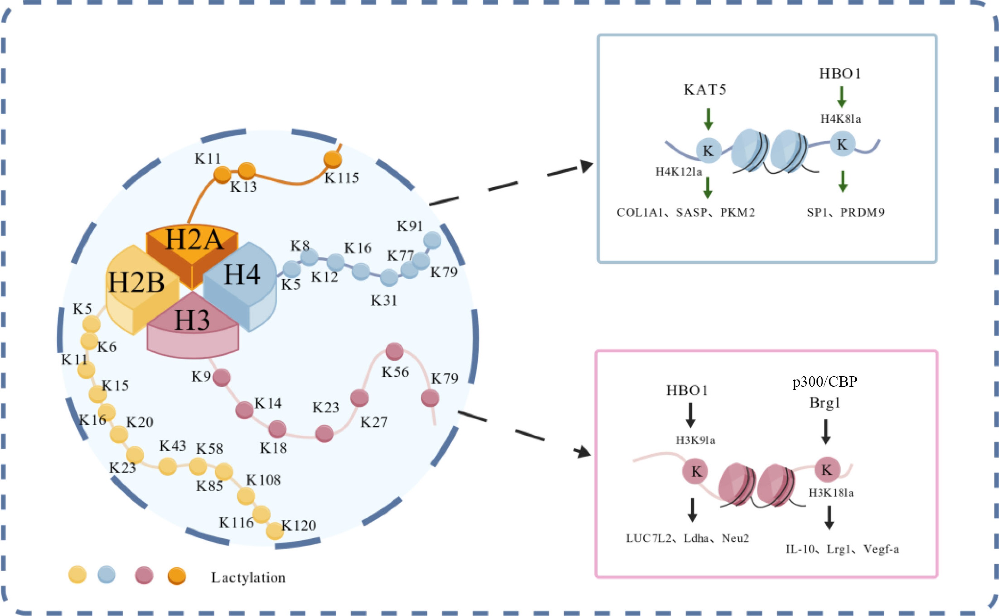
Figure 1.
Summary of histone lactylation modification sites, and a summary of reported histone lactylation sites including H2A, H2B, H3, and H4. A brief introduction was given on the lactylation of the K9/K18 sites of H3 and the K12/K18 sites of H4. The figure was created with BioGDP.com[20].
Table 1. Summary of histone lactylation sites in humans and mice.
Histone Lactylation sites Species H2A K11, K13, K115 Human K11, K115 Mouse H2B K5, K11, K15, K16, K20, K23, K43, K58, K85,
K108, K116, K120Human K5, K11, K15, K16, K20, K58, K85, K108 Mouse H3 K9, K18, K23, K27, K79 Human K14, K18, K23, K27, K56 Mouse H4 K5, K8, K12, K16, K31, K77, K79, K91 Human K8, K12, K31, K77, K79, K91 Mouse -
During histone lactylation modification, 'writers', 'erasers', and 'readers' act as core elements, responsible for catalyzing and/or eliminating histone lactylation, as well as recognizing and binding to specific histone lactylation moieties (Fig. 2; Supplementary Table S1).
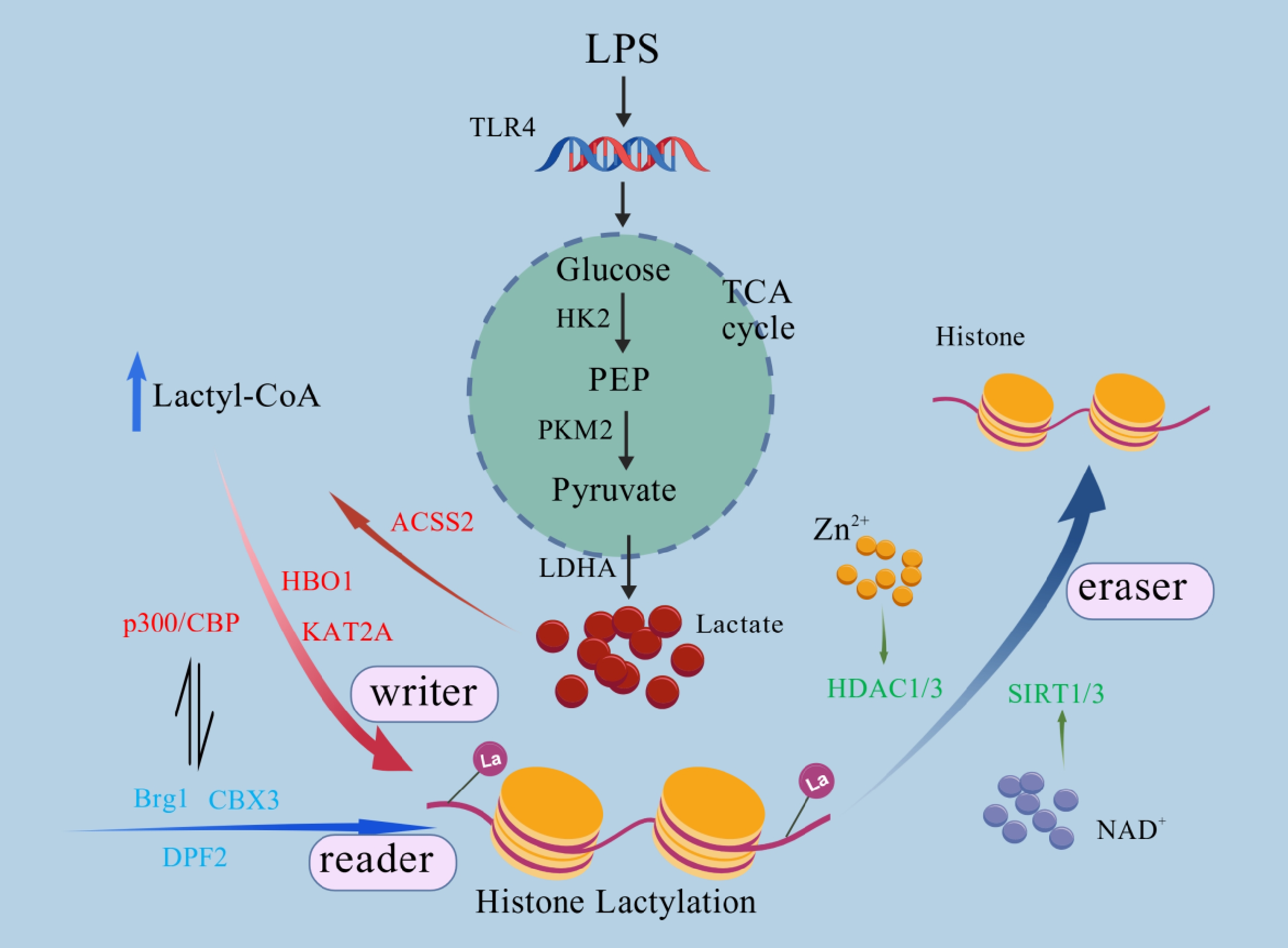
Figure 2.
The potential mechanism of histone lactylation in innate immunity. The metabolic regulation mechanism of lipopolysaccharide (LPS)-mediated histone lactylation activation of the toll-like receptor 4 (TLR4) pathway by LPS can induce the Warburg effect, promote glycolysis, and produce lactic acid in large quantities through the hexokinase 2 (HK2), pyruvate kinase M2 (PKM2), and lactate dehydrogenase A (LDHA) pathways. Nucleated lactic acid is converted into lactic acid coenzyme A by Acyl-CoA Synthetase Short Chain Family Member 2 (ACSS2), which mediates histone lactylation under the mediation of modified writing and reader proteins. However, Zn2+ and NAD+ can activate HDAC1/3 and SIRT1/3 to remove the milk acylation modification and jointly regulate the dynamic reversible equilibrium of the modification. The figure was created with BioGDP.com[20].
Histone lactylation relies on 'writers' that catalyze the covalent binding of lactate to histone lysine residues. For instance, histone acetyltransferase p300/CBP, which belongs to the lysine acetyltransferase (KAT) family, acts as a potential enzyme to drive histone lactylation[6]. Overexpression of p300/CBP in human embryonic kidney 293T cells (HEK293T) increases histone lactylation levels, whereas the loss of p300/CBP reduces histone lactylation levels in human colon cancer cells (HCT116) and HEK293T cells[6]. The use of the specific p300/CBP small-molecule inhibitor A-485 in surgical mice can significantly reduce histone lactylation levels in ischemic brains without affecting lactate production[21]. Besides p300/CBP, another KAT family protein, histone acetyltransferase binding to ORC1 (HBO1), regulates the transcription of downstream genes by mediating lactylation at the H3K9 site (H3K9la) during tumor progression (Fig. 2)[22]. These studies highlight the KAT family enzymes as essential 'writers' of histone lactylation.
On the other hand, histone lactylation 'erasers' mainly belong to the histone deacetylase (HDAC) family and exhibit broad-spectrum deacetylation activity. Among them, HDAC1, HDAC2, HDAC3 (class I HDAC), and HDAC6 (class II HDAC) have been shown to effectively remove lactate groups from lactylated histones[23,24]. These enzymes hydrolyze the amide bond between lysine residues and lactate groups in lactylated histones through a zinc ion-dependent catalytic mechanism[25,26]. In addition, the Sirtuins (SIRT) family (class III HDAC) proteins are also involved in the erasure of lactate from lactylated histones (Fig. 2)[27−29]. Researchers have confirmed that SIRT1 and SIRT3 act as 'erasers' of lactylated histones[28].
'Readers' specifically recognize histone lactylation modifications, recruit downstream regulatory complexes (such as transcription factors or chromatin remodeling factors), and convert epigenetic signals into specific biological functions[6]. Currently, research on histone lactylation 'readers' is relatively limited, but preliminary evidence suggests that the bromodomain family proteins are one part of them[30−32]. The classic function of bromodomain proteins is to recognize histone acetylation modifications, but some of them have been reported to recognize and bind to certain lactylated histones. For instance, bromodomain-containing protein 4 (BRD4) and Brahma-related gene 1 (Brg1) can recognize H4K8la and H3K18la, respectively[32,33]. In addition, Double PHD fingers 2 (DPF2) has been identified to directly bind to H3K14la (Fig. 2)[31]. Whether there are other histone lactylation 'readers' (such as proteins containing BRD or PHD finger domains) and how 'readers' distinguish similar histone PTMs (lactylation and acetylation) are awaiting further in-depth investigations.
-
Histone lactylation, which serves as a pivotal epigenetic modification linking metabolism and immunity, profoundly influences immune responses by regulating immune cell function[6,7]. It not only shapes innate immune memory through modifications at sites such as H3K18la, enhancing the body's long-term defense against pathogens, but also induces immune cells to adopt a suppressive phenotype within the tumor microenvironment, thereby promoting immune evasion. The balance of histone lactylation is directly related to immune homeostasis and disease progression[7]. The underlying molecular mechanisms by which histone lactylation regulates immunity are primarily rooted in its direct influence on epigenetic modifications[6,8]. Through the covalent alteration of histone lysine residues, histone lactylation modifies chromatin structure and alters gene transcriptional activity, thereby impacting the progression of immune diseases and inflammation[6,8].
H2A and H2B lactylation
-
Research on H2A and H2B lactylation regulatory mechanisms is sparse in both mice and humans, with H2BK58la being an exception[6,15]. During hepatocellular carcinoma (HCC) tumorigenesis, H2BK58la promotes lactic acid accumulation in cancer cells due to local hypoxia, activates N-myc downstream-regulated gene 1 (NDRG1) transcription, and modulates the GSK-3β-p53 pathway to counteract cellular senescence[15]. The impact of H2BK58la on transcription factor function represents an emerging research area, with future investigations focusing on how this regulation affects disease-related gene expression.
H3 lactylation
-
H3 lactylation influences chromatin openness and gene expression, with research particularly concentrated in areas such as immune metabolism and tumors. In the dimension of immune regulation, multiple H3 lactylation modification sites have been identified to play a central role, among which H3K18la is the most critical and widely studied[6,34]. In addition, H3K9la and H3K14la have been confirmed by numerous studies to be involved in immune-related gene expression regulation[35,36]. Additionally, other H3 lactate modification sites, such as H3K23la, H3K27la, and H3K56la, together form an important epigenetic basis for lactate-mediated immune function regulation (Fig. 3)[37−39].
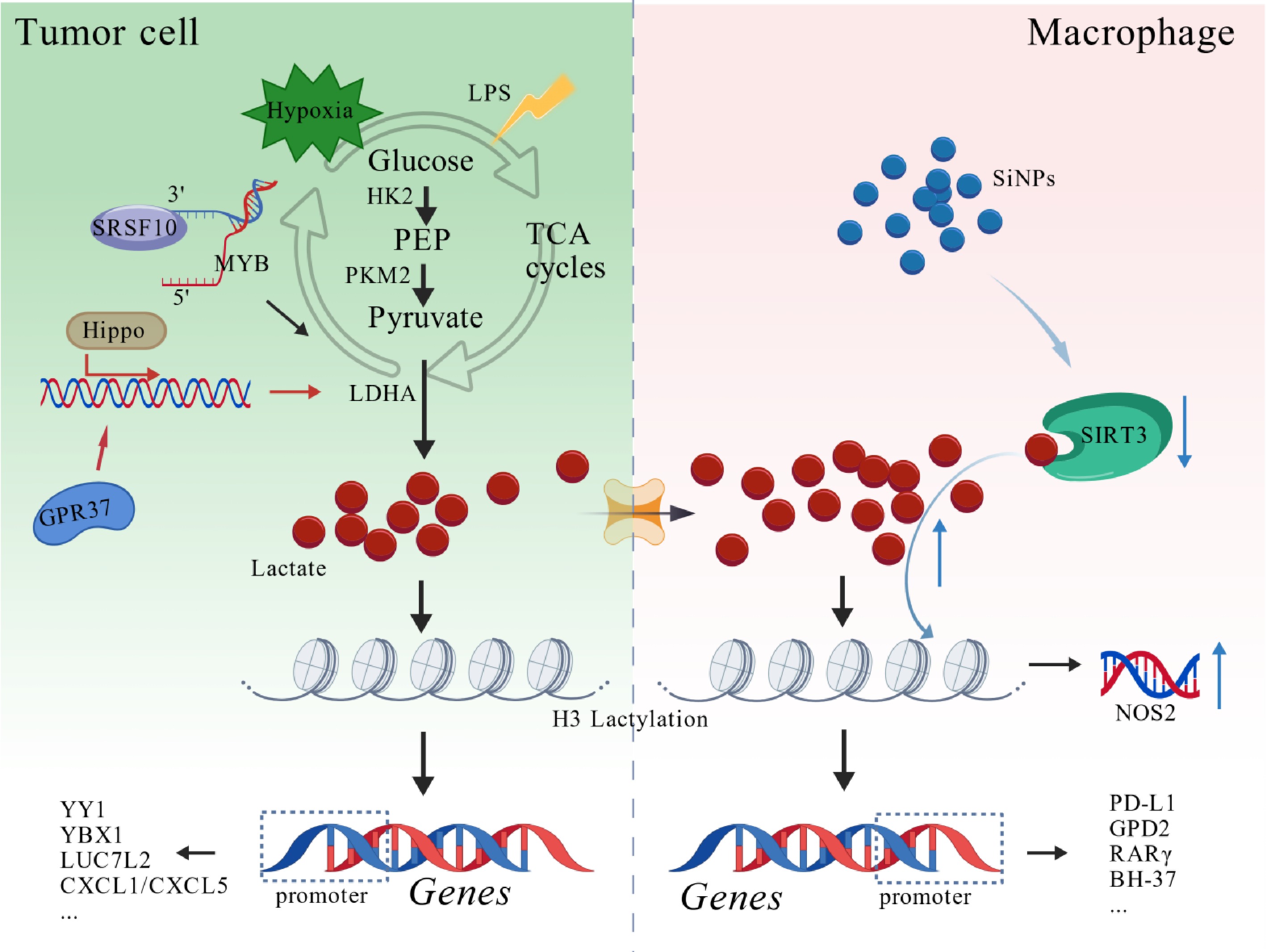
Figure 3.
H3 lactylation: a bridge between metabolism and epigenetics. In a tumor microenvironment stimulated by hypoxia or LPS, serine-and arginine-rich splicing factor 10 (SRSF10)-MYB and G protein-coupled receptor 37 (GPR37)/Hippo pathway activation can upregulate LDHA, promote lactic acid production and histone lactylation, and activate target gene transcription. Tumor-derived lactic acid can enter macrophages, induce lactylation, and drive gene transcription. Silica nanoparticles can inhibit SIRT3 activity in macrophages, lead to the accumulation of H3 lactylation, activate Nitric Oxide Synthase 2 (NOS2), amplify pro-inflammatory reactions, and mediate the polarization of M1 macrophages and the process of pulmonary fibrosis. The figure was created with BioGDP.com[20].
H3K9la
-
Histone lactylation of H3K9la is an important epigenetic regulatory mechanism, that can play different regulatory roles depending on physiological and pathological contexts (Supplementary Table S2). This modification is widely involved in many kinds of disease processes: in many kinds of malignant tumors, it can mediate tumor proliferation, invasion, distant metastasis, chemotherapy resistance and tumor immune escape by regulating downstream target genes and constructing a positive feedback loop of signals, and at the same time, many naturally active substances and proteins related to modification can play a role in inhibiting cancer by downregulating H3K9la[16,40−45]. In the cardiovascular field, H3K9la participates in pathophysiological processes such as vascular calcification, atherosclerosis, and angiogenesis, and various active molecules can improve vascular-related diseases by regulating this modification[46−48]. In autoimmune diseases, H3K9la can mediate the activation of inflammatory pathways and promote the occurrence and development of rheumatoid arthritis. Targeted regulation of this modification can effectively inhibit inflammation and relieve symptoms[49,50].
H3K14la
-
As a multifunctional epigenetic lactylation modification, H3K14la has both immune regulation and pathological driving activity and plays an important role in the occurrence and development of malignant tumors, metabolic dysfunction, nervous disorders, and respiratory diseases (Supplementary Table S2). In infectious diseases, H3K14la is the core regulator of organ injury in sepsis. Lactic acid accumulation induced by sepsis can enrich the H3K14la modification of iron metabolism-related gene promoter regions, activate endothelial cells, and further aggravate septic lung injury[51]. In malignant tumors, lactic acid produced by tumor metabolic reprogramming can upregulate H3K14la modification, participate in the regulation of malignant progression, chemotherapy resistance, and tumor microenvironment remodeling, and mediate pathological processes such as chemotherapy resistance of liver cancer, activation of oncogenes of cervical cancer, and tumor microenvironment disorder of lung adenocarcinoma[52,53]. In metabolic diseases, H3K14la closely regulates the progression of diabetic nephropathy, and lactic acid-induced high expression of H3K14la can trigger renal tubular epithelial-interstitial transformation, activate the renal fibrosis pathway, and promote renal fibrosis and disease deterioration[54,55]. In neurological and respiratory diseases, H3K14la mediates a variety of injury mechanisms. An abnormal increase in H3K14la after cerebral hemorrhage can disturb calcium homeostasis, induce iron death of neurons, and aggravate nerve injury[56]. At the same time, it can activate inflammatory and aging pathways, promote the aging of lung epithelial cells, and participate in the pathological process of chronic obstructive pulmonary disease[57].
H3K18la
-
H3K18la is the first identified and most extensively studied H3 lactylation. It is involved in regulating tumors, infectious diseases, metabolic and fibrotic diseases, and neurological diseases. Considering the existing studies on H3K18la, we complied a table summarizing its major immunological functions (Supplementary Table S3).
H3K23la, H3K27la, and H3K56la
-
Currently, research on H3K27la mainly focuses on its role in immune regulation and tumorigenesis, while research on H3K23la's immune function in intestinal inflammation. Gegen Qinlian decoction (GQD) treatments effectively inhibit the production of lactate, reduce H3K23la, and thus suppress inflammation and polarization of M1 macrophages[58]. For H3K27la, it was reported to modulate immune-related gene expression and sustain long-term immune memory in trained monocytes[59]. In metabolic reprogramming of gastric adenocarcinoma (GAC), 26S proteasome non-ATPase regulatory subunit 14 (PSMD14) stabilizes 6-phosphofructo-2-kinase/fructose-2,6-bisphosphatase 2 (PFKFB2), which enhances glycolysis, leading to lactate accumulation and H3K27la induction[39]. Additionally, H3K56la was reported to promote tumorigenesis of liver cancer stem cells (LCSCs)[38,40].
H4 lactylation
-
Similar to H3 lactylation, H4 lactylation facilitates chromatin opening by neutralizing the positive charge of lysine residues, diminishing histone-DNA binding affinity[60,61]. Previous studies have demonstrated that lactylation modifications at H4K5, H4K8, H4K12, H4K16, H4K79, and H4K91 sites play essential roles in inflammation response regulation, immune cell activation, and tumor immune escape (Fig. 4)[14,62−65].
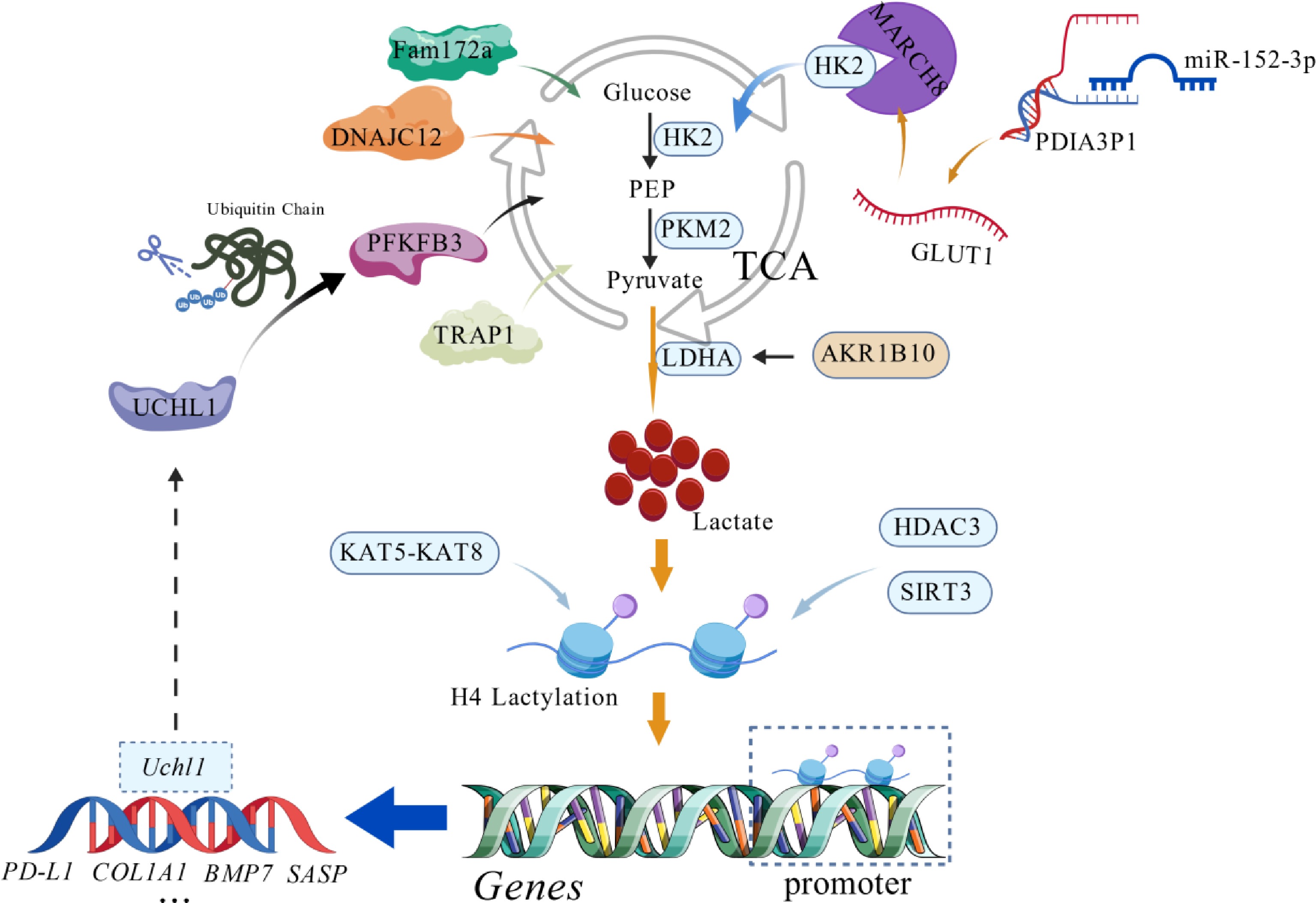
Figure 4.
H4 lactylation: a bridge between metabolism and epigenetics. Synergistic regulation of H4 lactylation by multiple molecular pathways. Family with sequence similarity 172, member A (Fam172a), DNAJC12, and tumor necrosis factor receptor-associated protein 1 (TRAP1) indirectly regulate lactic acid production by regulating glycolysis, while Aldo-keto reductase family 1 B10 (AKR1B10) directly promotes lactic acid accumulation by activating LDHA, thus regulating gene lactylation. Protein disulfide isomerase A family 3 pseudogene 1 (PDIA3P1) competitively adsorbs microRNA-152-3p (miR-152-3p), stabilizes glucose transporter 1 GLUT1 and HK2, strengthens glycolysis, and improves the level of lactylation. Ubiquitin carboxy-terminal hydrolase L1 (UCHL1) can stabilize 6-phosphofructo-2-kinase/fructose-2,6-diphosphatase 3 (PFKFB3) to promote lactic acid production and H4K8la modification, and is positively activated by lactylation, forming an amplification loop. Lysine acetyltransferase 5 (KAT5) and lysine acetyltransferase 8 (KAT8) positively promoted lactylation, while HDAC3/SIRT3 negatively inhibited modification, maintaining its dynamic balance together. The figure was created with BioGDP.com[20].
H4K5la
-
As a key lactate-dependent epigenetic modification, H4K5la is primarily involved in tumorigenesis and immune escape[66−68]. For example, in neuroblastoma (NB), the low expression of DNAJC12 (DnaJ heat shock protein family member C12) activates glycolysis, promoting lactic acid production and the increase of H4K5la. The latter upregulates COL1A1 (encoding type-I collagen) transcription, accelerating the development of NB. Inhibiting glycolysis, reducing H4K5la, or targeting COL1A1 can alleviate the invasion of NB cells[66]. In bladder cancer (BCa), lactate dehydrogenase A (LDHA) drives lactic acid production to induce H4K5la, activating programmed death ligand-1 (PD-L1) transcription to assist immune escape[62]. In acute myeloid leukemia (AML), signal transducer and activator of transcription 5 (STAT5) enhances glycolysis and lactic acid accumulation, promotes nuclear translocation of E3-binding protein (E3BP), enriches H4K5la at the PD-L1 promoter region, and upregulates PD-L1 expression[68,69].
H4K8la
-
Many pathophysiological disorders are regulated by the lactic acid modification H4K8la, and their core functions focus on nerve injury repair, malignant tumor progression, and immune response regulation (Supplementary Table S3). In the nervous system, H4K8la plays a significant role in pathological regulation, which can continuously activate the modification level through a glycolysis-related positive feedback loop and participate in the process of spinal cord injury. At the same time, it can aggravate the brain edema caused by subarachnoid hemorrhage by regulating the downstream target gene and Wnt/β-catenin signal pathway, induce nerve cell apoptosis, and mediate the deterioration of nervous system injury[33,70,71]. In tumors, the abnormal enrichment of H4K8la mediated by lactic acid accumulation can promote the malignant progression of esophageal squamous cell carcinoma by activating downstream oncogene transcription and matrix-related pathways, and can also upregulate the expression of drug-resistance-related genes, maintain the drug-resistance phenotype of multiple myeloma, and activate gastric cancer microenvironment-related pathways to reshape the tumor matrix[63,72,73]. At the level of immune regulation, the expression level of H4K8la directly affects the differentiation of macrophages, and its downregulation can inhibit the differentiation of M1 macrophages and the expression of markers, thus regulating the anti-tumor and inflammatory immune response of the body[58].
H4K12la
-
Lactic acid-mediated epigenetic modification of H4K12la is involved in the regulation of multi-system diseases, which can affect the disease process, including tumor progression, neurometabolic damage, inflammatory response, and cell homeostasis (Supplementary Table S3). In the field of tumors, lactic acid accumulation caused by abnormal glycolysis activation can significantly upregulate the modification level of H4K12la and mediate chemotherapy resistance and malignant progression of many tumors through multiple mechanisms such as super-enhancer activating target gene transcription, inhibiting iron ferroptosis of tumor cells, and weakening anti-tumor immunity. Targeted inhibition of lactic acid modification can cooperate with anti-tumor drugs to block tumor development[64,74−77]. In nervous system and metabolic-related diseases, H4K12la can participate in neuropathological injuries such as Alzheimer's disease and diabetic cognitive impairment by continuously activating microglia, mediating cell apoptosis, and triggering abnormal signal pathway activation[78−81]. At the same time, it is widely involved in the process of chronic lung disease, skin lesions, chronic kidney disease, and intestinal inflammation and aggravates the pathological damage of multiple organs by regulating cell aging, macrophage polarization, and tissue fibrosis[82−85]. In the regulation of inflammation and cell homeostasis, H4K12la can respond to abnormal glycolytic metabolism, be enriched in an aging-related secretory phenotype, participate in pathological remodeling of vascular smooth muscle cells, and play an important regulatory role in inflammatory response, injury repair, and cell homeostasis maintenance[58,86].
H4K16la, H4K79la, and H4K91la
-
Compared with the above H4 lactylation, research on the immune function of H4K16la, H4K79la, and H4K91la is still in its preliminary stage[14,65]. For instance, it was reported that knocking out succinate-coenzyme A (CoA) ligase GDP-forming subunit beta (SUCLG2) can inhibit the proliferation of GBM and promote cell apoptosis through the mediation of H4K16la, while liver kinase B1 (LKB1) inhibits telomerase activity through H4K16la, leading to the aging of lung adenocarcinoma (LUAD) cells[65,87]. However, their specific functions in immune cell activation, inflammatory response, or tumor immunity have not been clearly reported. In addition, H4K79la and H4K91la have been proven to regulate the expression of downstream glycolytic genes in breast cancer (BC) cells and participate in tumor metabolic reprogramming, but whether such modifications affect the tumor immune microenvironment through metabolic immune interaction still needs to be further explored[14].
-
Currently, the research on lactylation sites and mechanisms of H2A and H2B in humans and mice is still scarce. On the one hand, the overall modification abundance of H2A/H2B is much lower than that of H3 and H4, and some sites are folded tightly, which makes them difficult to hydrolyze[88]. In addition, the N-terminal tail of H2A/H2B is dense with lysine (for example, H2BK5/11/15/16/20), and multiple sites coexist with short peptide segments and high homology, which makes it difficult to distinguish isomers and accurately identify sites[6]. On the other hand, the steric hindrance of H2A/H2B is large, and the modifying enzyme preferentially recognizes H3/H4, so the lactic acid modification efficiency is low[89]. At the same time, the commercial antibody of H2A/H2B-specific lactic acid is scarce, which limits the verification of related functions. In addition, the existing research focuses on the lactate sites of H3 and H4 with a clear mechanism, which leads to the biological function and regulation mechanism of H2A and H2B not being systematically explained.
Histone lactylation is a specific epigenetic marker of the Warburg effect, which forms a metabolic antagonistic relationship with acetylation: lactic acid and lactyl-CoA drive lactate, while acetyl-CoA mediates acetylation. The writing enzymes of the two pathways partially overlap, and members of the KAT family, such as p300/CBP and lysine acetyltransferase 2A (KAT2A), can catalyze two modifications at the same time, and the modification type is mainly determined by coenzyme supply. For example, p300/CBP, as a core co-catalyst, preferentially binds acetyl-CoA and mediates acetylation under physiological conditions; however, under the condition of metabolic stress, lactyl-CoA accumulates in large quantities, competitively occupies the p300/CBP active center, and then induces lactic acid[90]. However, the molecular dynamics mechanism of the competition between lactyl-CoA and acetyl-CoA for binding p300/CBP is not clear, and the relevant conclusions mostly depend on in vitro experiments; verification under physiological and pathological conditions in vivo is still insufficient. In addition, only a few loci, such as H3K18la and H3K14la, are associated with glycolysis activation. It is not clear whether the lactylation of these loci can independently drive glycolysis conversion, and the synergistic regulation of other unidentified histone lactylation loci cannot be ruled out. At the reading level, the specific molecular chain of how to recruit downstream transcriptional machinery and initiate gene expression after histone lactylation remains unresolved; at the same time, the lactate-specific erasing enzyme has not been discovered, and its dynamic reversible regulation and downstream signaling pathway need to be systematically clarified.
-
Significant research progress has been made in histone lactylation, contributing to the regulation of immunity and linking immune–metabolic pathways. Nevertheless, numerous challenges remain to be explored in future studies. First, the mechanism underlying the conversion of lactate into long-term epigenetic memory has not been fully elucidated[8]. For instance, when immune cells are stimulated, the mTOR-HIF-1α signaling axis initiates metabolic reprogramming, enhances glucose uptake, and produces large amounts of lactate via glycolysis to serve as a substrate for histone lactylation[91]. It remains a critical unresolved issue how lactate, as a metabolic product, is precisely converted into stable, heritable epigenetic marks within cells to sustain long-term immune memory. Additionally, during the maintenance of immune memory after BCG vaccination, the molecular mechanisms and regulatory networks underlying the persistent existence and functional role of histone lactylation modifications in monocytes require further exploration.
Histone PTMs are a complex, dynamic regulatory system, and there may be interactions, synergies, or antagonistic relationships between different modifications[13,92]. For example, histone lactylation may compete with other lysine modifications, such as methylation and ubiquitination, at the same modification site. Under microenvironments such as stress, hypoxia, and inflammation, these modifications may dynamically change and together shape the expression profile of target genes[6,93]. However, limitations in research studies on the specific synergistic modes and molecular mechanisms of these compounds. For instance, it is currently unclear how H3K18la modification and H3K27ac (acetylation modification) interact to co-regulate the transcriptional activation of genes in the promoter regions of inflammation-related genes.
Additionally, research on the impact of genetic polymorphisms (e.g., LDHA and EP300) on individual differences in trained immunity is still in its infancy[8,94]. Although genome-wide association studies (GWAS) have identified that polymorphisms in LDHA and EP300 genes significantly affect trained immune responses, the specific mechanisms of these effects and how to use such genetic information to predict and regulate individual immune responses require extensive research[8]. Currently, there is a lack of systematic and in-depth studies on how variations in the LDHA and EP300 genes among different individuals influence lactate metabolism and histone lactylation, thereby leading to individual differences in immune responses. Future studies should focus on lactylation omics and genomic data to further elucidate the interaction between histone lactylation and DNA. Furthermore, single-cell lactylomics research should be conducted through in vivo models to explore the heterogeneity of immune cell responses and the mechanism of lactylation in immune diseases.
Despite these challenges, research on histone lactylation has broad applications in fields such as tumor immunotherapy and vaccine design optimization. In tumor immunotherapy, an in-depth understanding of the role of histone lactylation in regulating immune cell function is expected to provide key targets for developing new immunotherapeutic strategies. Studies have found that in the tumor microenvironment (TME), the metabolic state and histone modification patterns of immune cells undergo changes that affect their ability to perform immune surveillance and kill tumor cells. By regulating the level of histone lactylation, it is possible to reshape immune cell function and enhance their antitumor activity. For example, increased histone lactylation levels may facilitate the polarization of macrophages towards the anti-tumor M1 phenotype, thereby enhancing their phagocytic and cytotoxic effects on tumor cells[95]. Additionally, developing specific inhibitors or activators targeting enzymes associated with histone lactylation modification (such as p300/CBP) may emerge as a new strategy for tumor immunotherapy. In certain diseases, lactate-related genes can act as new biomarkers for early diagnosis, while alterations in histone lactylation levels across various diseases may also serve as biomarkers for disease diagnosis and prognostic assessment[96,97].
In vaccine design optimization, research on histone lactylation offers novel insights for enhancing the immune efficacy of vaccines[8]. Vaccination is a pivotal strategy for preventing infectious diseases; however, the immune efficacy of some current vaccines still needs to be enhanced. An in-depth exploration of mechanisms underlying histone lactylation in trained immunity will facilitate optimization of vaccine design and vaccination protocols. By adjusting adjuvant components or administration routes to potentiate metabolic reprogramming and histone lactylation modification in immune cells, it may be feasible to elevate the level of vaccine-induced immune memory and the magnitude of immune responses. The development of novel adjuvants capable of promoting immune cells to produce more lactate or enhancing histone lactylation modification is anticipated to boost vaccine immunogenicity, thereby enabling vaccines to exert a more prominent role in preventing infectious diseases. In the future, as research on histone lactylation continues to advance, it is anticipated that more breakthroughs will be achieved in these application areas, thereby conferring substantial benefits to human health.
-
(1) A paradigm shift in emerging epigenetic understanding, histone lactylation exhibits a unique and critical role in immunity.
(2) Histone lactylation serves as a specific epigenetic marker of immune memory while also orchestrating a core regulatory function in modulating inflammatory responses, thus underscoring its multifaceted immunological significance.
(3) Elucidating the intricate interplay between histone lactylation and immunity promises to enrich the 'immune–metabolic' regulatory framework, unveiling novel therapeutic targets and strategies for managing immune-mediated diseases (e.g., sepsis, rheumatoid arthritis, and inflammation in the tumor microenvironment).
This work is supported by the National Natural Science Foundation of China (32100702). We feel deeply grateful to the researchers for their contributions to science, and we extend our sincere thanks to those whose work could not be cited in the present article due to space limitations. All the figures were created with BioGDP.com.
-
This study is based exclusively on published literature and publicly available data datasets, and ethical approval is not applicable.
-
The authors confirm their contributions to the paper as follows: study conception and design, manuscript drafting, and data interpretation: Kong F, Ali U; manuscript revision, supervision, conceptualization, and funding: Ji S. All authors reviewed the results and approved the final version of the manuscript.
-
The datasets used/or analyzed during the current study are available from the corresponding author on reasonable request.
-
The authors declare they have no conflict of interest.
-
accompanies this paper online at: https://doi.org/10.48130/epi-0026-0008.
-
# Authors contributed equally: Fanrui Kong, Umar Ali
- Supplementary Table S1 Summary of writers, erasers, and readers in histone lactylation.
- Supplementary Table S2 Disease-associated histone lactylation sites and their molecular effectors.
- Supplementary Table S3 Summary of physiological and pathological studies on H3K18la.
- Copyright: © 2026 by the author(s). Published by Maximum Academic Press, Fayetteville, GA. This article is an open access article distributed under Creative Commons Attribution License (CC BY 4.0), visit https://creativecommons.org/licenses/by/4.0/.
-
About this article
Cite this article
Kong F, Ali U, Ji S. 2026. The emergent role of histone lactylation in immunoregulation and diseases. Epigenetics Insights 19: e011 doi: 10.48130/epi-0026-0008 

The emergent role of histone lactylation in immunoregulation and diseases
- Received: 03 April 2026
- Revised: 02 June 2026
- Accepted: 09 July 2026
- Published online: 11 September 2026
Abstract: The intricate interplay between metabolic pathways and immune mechanisms is crucial for advancing immunometabolism research. The pathophysiology of metabolic dysfunction is associated with abnormal immune responses that contribute to inflammation, autoimmunity, and tumorigenesis. Elucidating the immune-metabolic interface is essential for developing innovative targeted therapeutic strategies and biomarkers for early disease diagnosis. In recent studies, histone lactylation has emerged as a novel epigenetic modification. As an initial regulatory mode of metabolic reprogramming, histone lactylation plays a crucial role in controlling the expression of genes related to the host immune system. Here, we summarize the potential 'writers', 'erasers', and 'readers' of histone lactylation and critically evaluate its multifaceted roles in directing immune modulations. Moreover, we delineate the translational potential of targeting the histone lactylation axis for novel immunotherapies and vaccine strategies.
-
Key words:
- Histone lactylation /
- Epigenetic modification /
- Immunity /
- Signaling molecules /
- Immune dysfunction