












-
In mammals, nuclear methylation of DNA is an epigenetic mark composed of a methyl group in the fifth position of the pyrimidine ring of cytosine (5-methylcytosine, or 5mC). 5mC is a highly conserved and widespread epigenetic mark in both plants and animals[1−3], but variable in fungi and arthropods[1]. 5mC was first reported in mammalian DNA in 1951[4]. DNA methylation also occurs in mitochondria, but at a low frequency. An example is the promoter of the TFAM (mitochondrial transcription factor A) gene, involved in DNA compaction[5]. The role of this methylation is not yet fully understood. Methylation can also be found in adenine (m6A) in both DNA and RNA, and it has been widely implicated in nuclear splicing, stability, translation rate, and the subcellular targeting of mRNA[6]. The role of m6A in mammalian cells remains elusive, although some studies suggest that it is involved in RNA polymerase pausing.
5mC plays a crucial role in development by regulating gene expression and ensuring the formation of the 200 different cell types required for a functional organism. The impact of 5mC during development is paralleled by other epigenetic modifications like histone post-translational modifications, histone variants, and miRNA variations[7]. There is a tight relationship and interdependence of all these actors. In contrast, when a cell divides in adults, the only goal of the mother cell is to generate two identical daughter cells with the same cell phenotype. In this respect, 5mC patterns must be faithfully transmitted[8,9]. However, this does not mean that our epigenome is constant. In fact, it changes throughout our entire lifespan, probably from birth onwards. For example, maternal vitamin supplementation during pregnancy and lactation can alter DNA methylation patterns in breastfed infants[10]. Breastfed infants of intervention mothers showed an increase in DNA methylation in 217 CpGs and a decrease in 213 CpGs. The affected genes are involved in collagen metabolic processes and the regulation of apoptosis[10]. Drastic changes have also been observed during ageing[11].
It is noteworthy that such a small chemical modification can have such a profound impact on cellular life, governing cell fate and phenotype through gene expression and repression, as well as maintaining genome integrity[12]. This is because proteins, due to their highly complex structural domains, can read, erase, or establish this small chemical modification[12]. These proteins are involved in maintaining, repairing, and inheriting these DNA methylation patterns. These patterns are constantly exposed to alteration through the spontaneous deamination of 5mC, leading to the formation of a G:T mismatch; however, such lesions are repaired in the majority of cases.
In this review, we will discuss the potential interaction between methyl-CpG binding domain 4 (MBD4) and Ubiquitin-like containing PHD and Ring Finger domains 1 (UHRF1) that ensures DNA methylation repair following spontaneous deamination. This is based on the assumption that, as these two proteins belong to the same macromolecular complex, they must act in coordination with each other[13,14]. We will specifically refer to the repair of spontaneous (hydrolytic) deamination of 5-methylcytosine, which converts 5-methylcytosine to thymine and generates a mutagenic G mismatch. These lesions are recognized by MBD4, a DNA glycosylase that initiates base excision repair to restore the correct G base pair while preserving CpG methylation fidelity. Our proposed functional interplay between MBD4 and UHRF1 is therefore considered within the context of repair of deamination-induced G mismatches and the subsequent restoration of maintenance DNA methylation. This hypothesis does not address TET-mediated oxidative DNA demethylation or passive loss of methylation during DNA replication, although these processes also contribute to the broader regulation of DNA methylation dynamics. However, before discussing our proposed model in this context, we will review the localization and roles of 5mC.
-
There are different and complementary methods of analysing DNA methylation, which are described elsewhere[11,15]. Unlike in plants, 5mC in mammals covers most of the genome, except in CpG islands. These patterns are established during early embryogenesis thanks to the activity of DNMT3A and DNMT3B, which are de novo DNA methyltransferases[16,17]. In highly differentiated tissues, 5mC is mainly found in a CpG context, a condition that allows a symmetric methylation pattern to appear, i.e., fully methylated DNA. Approximately 45% of CpGs are found within repetitive elements and are presumed to be constitutively methylated[18]. Considering that there are 29 million CpGs in the haploid genome, DNA methylation patterns in normal human cells remain largely unexplored, but some general methylation patterns are established. Firstly, 7% of all CpGs are located within CpG islands (CGIs), most of which are unmethylated[19]. Most 5mCs are found in repetitive sequences, gene bodies, and intergenic regions[1,20]. In the human brain, 6%–8% of CGIs are methylated[21]. 5mC can also be found in stem cells, at a proportion of 25% (vs CG), in CHG and CHH trinucleotides (where H represents A, C, or T)[22,23].
Array techniques allow analysis of up to 930,000 CpGs at once[24], and whole-genome bisulfite sequencing[25] can be used to analyze all CpG sites, of which there are over 28 million in the human genome[26]. There are around 30,000 CpG islands in the human genome. In human DNA, 5mC is found in around 1.5% of the genome[27]. CpG sites are also found at significantly higher densities in gene-rich areas of human chromosomes compared with gene-poor areas[28]. In somatic cells, 5mC occurs almost exclusively in the context of the symmetrical methylation of paired CpG sites, where a cytosine nucleotide is located next to a guanine nucleotide.
DNA methylation in promoters
-
In vertebrates, 60%–80% of all CpG dinucleotides undergo methylation. Exceptions to this are CGIs, which are short regions of DNA with a high concentration of CpG dinucleotides. Indeed, CpG sites tend to cluster at promoter regions at higher densities than elsewhere in the genome, leading to the definition of the term CGIs. These CGIs are located in gene promoters and are usually unmethylated in normal cells. However, a small fraction (6%) of these islands become methylated during early development or tissue differentiation[29]. Another pathological situation in which promoters become hypermethylated is in the promoters of tumor suppressor genes in cancer[30,31]. Examples of these genes include RB1, CDKN2A/p16IN4A, p73, CDH13, SHP1, SOCS3, 3OST2, BRCA1, CDX2, CDKN2A, RUNX3, FOXO4, PPARG, PML, MEG3, 14-3-3σ, and PTEN[30,31]. CpG methylation is usually associated with gene repression when present in promoters, but its presence in gene bodies is correlated with gene expression[20]. However, recently it has been suggested that RNA methylation has a stronger effect on gene expression than does DNA methylation within gene bodies and promoters[32].
Hemi-methylated DNA, in which a methyl group is present on cytosines on only one strand, is rarely found in the genome outside of DNA replication. This is in contrast to 5fC and 5caC, as 5fC and 5caC exhibit no or low symmetry with respect to the opposite strand and thus exhibit behavior similar to 5hmC[33,34]. Hemi-methylation and 5hmC are states of DNA methylation that are read by the UHRF family of proteins[35,36].
DNA methylation in enhancers and super-enhancers
-
Enhancers are DNA sequences that play a key role in regulating gene expression in a spatiotemporal manner, governing the appearance of the gene expression program and forming the basis of the cell phenotype[37]. More specifically regarding enhancers, a 2023 study showed that the Tandem-Tudor domain of human UHRF1 prefers histones bearing H3K4me1 in combination with H3K9me2/3. H3K4me1 is a classic enhancer mark. The authors reported that UHRF1-TTD binds to enhancers and promoters of cell-type-specific genes, with a potential repressive effect on some of these genes[38]. A 2024 study supports this finding: it identifies enrichment of the hUHRF1 TTD domain on young repetitive/transposable elements that overlap with known enhancers, particularly L1PA/SVA/HERVK regions bearing H3K4me1-K9me2/3 marks. The authors conclude that UHRF1-TTD can bind to TE enhancers or enhancers derived from transposable elements[39]. Concerning MBD4 was reported to bind a methylated CpG island in an enhancer region located in the Early growth response protein 2 gene intron 1[40]. However, it is not yet known whether MBD4 co-localises with UHRF1 at enhancers.
Super-enhancers (SEs) are regulatory DNA elements that govern cell-specific changes in gene expression in various biological processes, including development, differentiation, and disease progression, in a disease- and tissue-specific manner[41]. SEs are regions of up to 300 kb and can regulate gene expression by acting on the transcription start sites (TSS) of genes, even when these are separated by up to 1.5 Mbp[42]. SEs can only exert their regulatory role in gene expression when gene promoters are unmethylated; methylation of SEs leads to gene silencing, whereas an unmethylated status allows gene expression. However, maximal gene expression is only reached with both an unmethylated promoter and an unmethylated SE[41]. Recent studies suggest that DNA methylation can alter SE organization and influence the expression of downstream target genes[43] (references therein). In an interesting study, it was shown that UHRF1 deficiency reduced DNA 5-mC modification and altered super-enhancer distribution, resulting in transglutaminase-2 overexpression[43]. As with enhancers, it is not yet known whether MBD4 co-localizes with UHRF1 at SEs. We cannot exclude this possibility, especially in the case of methylated DNA repair when G:T mismatches occur.
DNA methylation in gene bodies
-
The role of 5mC methylation in gene bodies is crucial for maintaining genome stability and ensuring the correct transcription of genes, thereby preventing the initiation of spurious transcription events. While the functional role of gene body methylation remains unclear, it is known to be associated with transcriptional elongation, splicing, regulation of alternative promoters, and modulation of expression levels through interaction with methyl-binding proteins such as MeCP2. Within the central nervous system, gene body methylation tends to decrease as gene expression increases[44].
In the liver, hypermethylation of the gene body primarily occurs in genes involved in lipid metabolism[45]. It has been demonstrated that differential methylation in gene bodies and enhancer elements correlates inversely with ageing-related gene expression[45]. A stronger correlation was observed between age-related differential gene expression and early-life promoter and gene body methylation patterns. Indeed, significant changes in DNA methylation were observed during the first 5 years of life, with 110,726 changes (14.13%) observed in the 783,659 CpG sites investigated. Of these, 59,749 and 50,977 CpGs were hyper- or hypo-methylated, respectively[46]. The subsequent 5 years are characterized by fewer changes in methylated CpGs: only 460 CpG dyads were found, of which 130 (28%) and 330 (72%) were hyper- or hypo-methylated, respectively[46]. Loss of methylation was observed in CpG-poor regions, such as open sea locations including intronic and intergenic regions, regardless of age. Conversely, methylation gain occurred in CpG-dense regions, such as CpG islands and gene promoters, in both age intervals examined. Nevertheless, given that changes in the composition of peripheral blood mononuclear cells occur during healthy ageing[47], modifications in DNA methylation patterns may also contribute to changes in blood cell subpopulations. Therefore, ageing may have a smaller impact on DNA methylation patterns within a given cell subpopulation. Investigating specific blood cell populations would provide a clearer understanding of the impact of ageing on these patterns.
In mouse ESC ChIP-seq, about 10.2% of Uhrf1-enriched regions were annotated around promoters or gene bodies, while most were intergenic; the authors also noted that Uhrf1 occupancy correlated more strongly with histone marks than with 5mC/5hmC[48]. Functionally, UHRF1 depletion can reduce DNA methylation at gene body loci, as a 2024 study reported progressive loss of methylation at promoters, the stanniocalcin 2 gene body, and pericentric heterochromatin after UHRF1 knockdown[49]. We have observed that loss of MBD4 in mouse embryonic fibroblasts caused hypomethylation affecting promoters, gene bodies, and repetitive elements, which supports a role for MBD4 at methylated genic regions, although loss-of-function methylation changes are not identical to direct binding evidence[50]. A cooperation of UHRF1 and MBD4, but rather in a context of G:T mismatch, in gene bodies, therefore cannot be excluded.
DNA methylation in repetitive elements
-
Repetitive sequences, which are present in several hundred to several thousand copies per genome, account for more than half of the genome. In the yeast Cryptococcus neoformans, 5mC methylation occurs in transposon-rich repeats and requires the DNA methyltransferase Dnmt5[51]. The vast majority of 5mC is found in repetitive sequences and transposable elements, which are concentrated in heterochromatin, such as pericentromeres, centromeres, and telomeres[2,28,52]. UHRF1 associates with newly replicated repetitive sequences, specifically pericentromeric α-satellite and Alu sequences; loss of LSH reduced UHRF1 association with these repeats[53]. Furthermore, we have observed that MBD4 localizes at repetitive elements, making cooperation between UHRF1 and MBD4 likely at these elements but rather in the context of a G:T mismatch[50].
It is now clear that the epigenetic code at repetitive elements contributes to cell identity, i.e., the cell's phenotype. Therefore, the fundamental question arising from this dogma is how DNA methylation patterns are established and inherited from mother cells to daughter cells. Do these patterns need to be inherited from a template, or are they set automatically during DNA replication? In the well-accepted model of DNA methylation pattern duplication at CpG, a hemi-methylated DNA profile is generated due to the appearance of a newly synthesized DNA strand. The hemi-methylated DNA is recognized by the SRA domain of UHRF1 (Fig. 1), which subsequently recruits DNMT1 to methylate the complementary DNA strand, thereby recovering DNA methylation on both strands and achieving an asymmetric state. However, in non-CpG contexts, the mechanism by which the methylation patterns are duplicated is not yet elucidated but may involve UHRF1, considering that the latter also putatively contributes to establishing methylation patterns in CHG and CHH contexts (H = A, C, or T)[54].
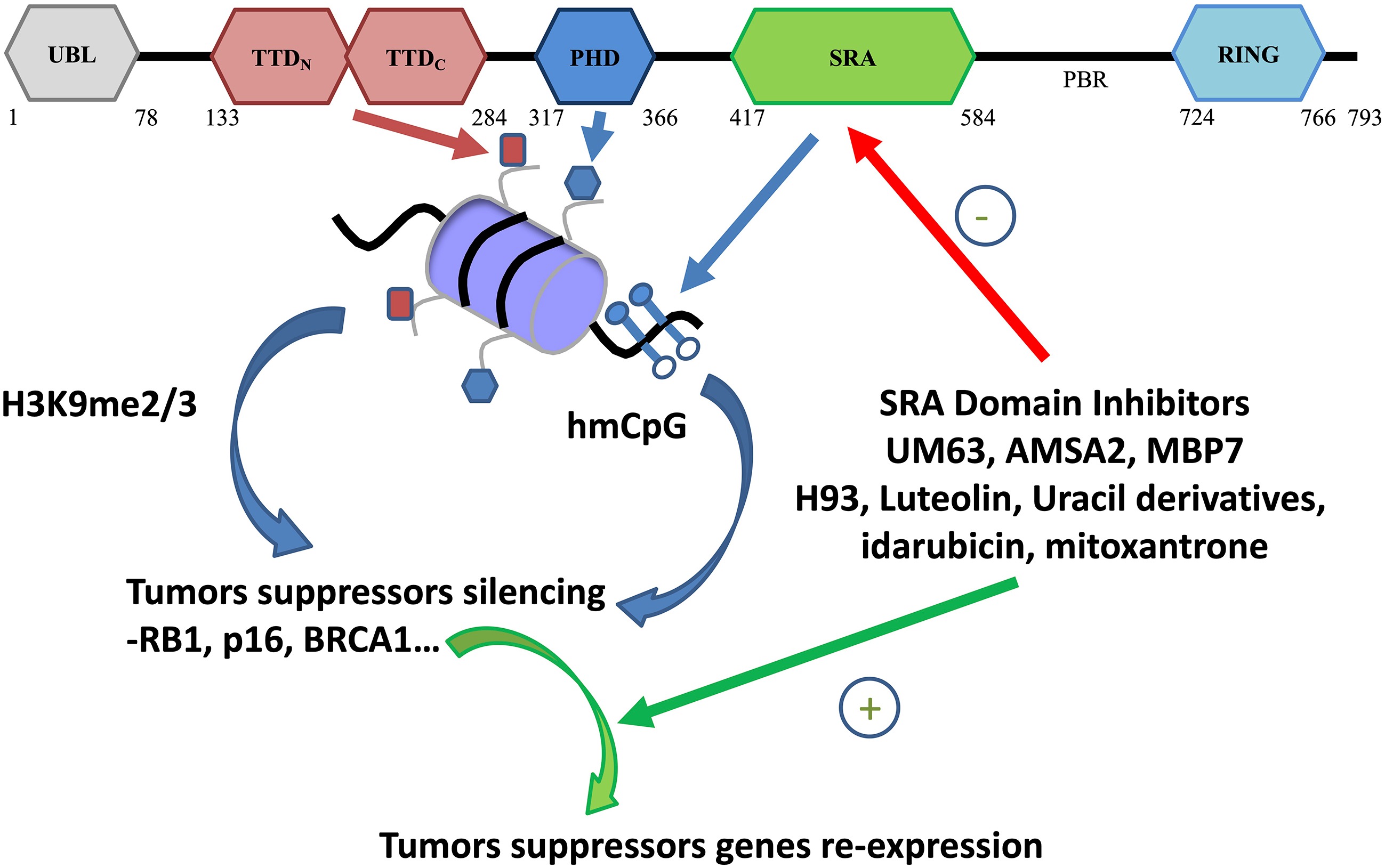
Figure 1.
Structural domains of UHRF1 and their role in tumor suppressor gene expression. The RING (Really Interesting New Gene) domain exhibits the E3 ligase activity of UHRF1. The UBL (ubiquitin-like) domain interacts with E2-ubiquitin conjugate[121]. The TTD binds di/trimethylated lysine 9 on histone H3 (H3K9me2/3; ochre rectangle on nucleosome), and the PHD binds unmodified arginine 2 on histone H3 (blue hexagon on nucleosome). The Tandem Tudor Domain (TTD) that senses the presence of di- or tri-methylated Lysine 9 of histone H3 (me2/me3K9H3) and the SRA (Set and Ring Associated) that binds to hemi-methylated DNA (hmCpGs) domain are involved in the silencing of TSGs. Various inhibitors of the SRA domain, such as UM63[122], AMSA2[122], MBP7[123], H93[124], Uracil derivatives (NSC232005)[125], idarubicin, and mitoxantrone[126], have been identified as being able to induce the re-expression of TSGs.
Role of CpG methylation
-
Although 5mC was suspected to be involved in gene expression regulation a long time ago[55], it has only more recently been demonstrated to play a role in gene regulation and cell differentiation[56].
DNA methylation is a fundamental and evolutionarily conserved epigenetic modification that shapes mammalian genome function by regulating gene expression, maintaining cellular identity, and ensuring genome stability. During development, dynamic waves of methylation and demethylation establish lineage-specific regulatory landscapes, silencing pluripotency genes while activating differentiation programs. Methylation patterns are tightly integrated with chromatin context, influencing transcription factor accessibility and reinforcing repressive or permissive chromatin states. At promoters and enhancers, DNA methylation modulates gene-specific transcriptional outputs, whereas gene-body methylation contributes to transcriptional fidelity and exon definition[7]. The maintenance machinery, particularly DNMT1 and UHRF1, preserves methylation patterns through cell division, enabling stable epigenetic inheritance. Conversely, active demethylation mediated by TET enzymes supports regulatory plasticity in development and environmental adaptation. Aberrant methylation landscapes emerge in ageing, senescence, and cancer, where global hypomethylation coexists with focal hypermethylation at regulatory elements, contributing to genomic instability and altered gene expression. Advances in single-cell and long-read sequencing have revealed previously unappreciated heterogeneity and context-specific methylation states, underscoring its multifaceted roles across tissues and disease states. External experiences trigger signaling pathways that drive site-specific methylation and demethylation of neuronal DNA, reshaping gene expression in ways that support long-term memory formation[57]. Overall, DNA methylation acts as a versatile regulatory layer that integrates developmental cues, chromatin architecture, and environmental signals to control genome function throughout life[7].
DNA methylation is thus now widely recognized as a key epigenetic factor influencing gene transcription and silencing. The mechanism by which 5mC exerts regulatory roles over gene expression is probably multifaceted and includes the stability of DNA interactions with sequence-specific DNA-binding proteins, such as transcription factors (TFs), as well as interactions with methyl-DNA-binding domain (MBD) proteins. These proteins then recruit protein complexes that favour chromatin condensation[58].
This is the result of the specific ability of proteins to read this chemical modification, and of proteins that can maintain and transmit this epigenetic mark faithfully to subsequent generations[59,60]. The absence or loss of fidelity in maintaining DNA methylation patterns can lead to cell death in the best cases, and to cells with altered functions in the worst cases, resulting in various pathologies. The most well-known and well-studied example of altered DNA methylation patterns is cancer[59,60]. DNA methylation is already considered a therapeutic target, given that two drugs, decitabine and azacytidine, are commercially available[61].
Spontaneous 5mC demethylation and the BER pathway
-
The transition of cytosine to thymine in methylated CpG is the most common type of DNA mutation and is usually caused by the spontaneous deamination of 5mC. The propensity of 5-methylcytosine (5mC) to undergo spontaneous deamination has exerted a significant influence on the evolutionary trajectory of the human genome. This type of methylation damage is the main cause of somatic mutations that accumulate with age. This deamination process gives rise to G:T mismatches that need to be repaired as G:C in order to avoid a C to T mutation. This type of DNA mutation is usually repaired by the base excision repair (BER) pathway[62]. It is estimated that endogenous DNA damage covers a range of between 20,000 and 100,000 lesions per cell per day[63]. This damage often takes the form of deamination, alkylation, or oxidation of bases in the DNA and may be caused by hydrolysis, exposure to reactive oxygen species, or metabolites. The hydrolytic deamination of 5-methylcytosine (5mC) and cytosine (C) to form thymine (T) and uracil (U), respectively, poses a constant threat to genome integrity. These spontaneous events occur at a rate of 2–300 lesions per cell per day and, if left uncorrected, lead to C:G to T:A transitions in the subsequent round of DNA replication.
BER is an evolutionarily conserved and essential DNA repair mechanism that relies on a highly coordinated series of protein–protein interactions to recognize, excise, and repair damaged bases[64,65]. As such, BER serves as the major surveillance system for base lesions caused by endogenous and exogenous attacks. Key enzymes in this process include DNA glycosylases, apurinic/apyrimidinic endonuclease 1 (APE1), polynucleotide kinase-phosphatase (PNKP), DNA polymerase β (Pol β), ligase IIIα (LigIIIα), poly (ADP-ribose) polymerases (PARP1 and PARP2), and X-ray repair cross-complementing protein 1 (XRCC1). These enzymes work together in a tightly regulated molecular network to catalyse BER.
The BER pathway is initiated by one of several DNA glycosylases, which recognize and excise specific DNA lesions in a coordinated manner. The two main G:T glycosylases, Methyl-CpG Domain Protein 4 (MBD4) and Thymine DNA Glycosylase (TDG), remove thymine that is produced when 5-methylcytosine is deaminated. These glycosylases also remove a variety of other base lesions, including G:U, and act preferentially at CpG sites throughout the genome[66].
-
MBD4 was cloned in the past by a two-hybrid approach using MLH1, a mismatch repair (MMR) protein, as a 'bait'[67]. MBD4 (Methyl Binding Domain 4) is a CpG methyl-binding protein and the only such protein with glycosylase activity embedded in its C-terminal domain (Fig. 2)[68]. The domain separating the C- and N-terminal domains may serve as a fusion point between them and is also called the intervening region[66]. MBD4 activity is primarily ensured by its binding to methylated DNA via its methyl-binding domain. This binding enables the glycosylase domain to recognize the G/T mismatch and remove the thymine. The failure to excise these lesions can lead to mutagenesis, which highlights the importance of removing thymine and uracil from mismatched base pairs. Available data suggest that MBD4 is involved in the base excision repair (BER) of G/T mismatches resulting from the deamination of 5-methylcytosine (5mC) at CpG dinucleotides. Indeed, a germline deficiency in the MBD4 gene can cause and predispose to multiple tumours[66]. MBD4 is involved in active demethylation due to its ability to excise 5-hydroxymethyluracil from a double-stranded CpG dinucleotide in vitro[69]. Consequently, MBD4 can be considered a safeguard of DNA methylation patterns. Some studies have linked MBD4 to apoptosis due to its interaction with MutL homolog 1 (MLH1), a protein functionally associated with apoptosis[70,71].
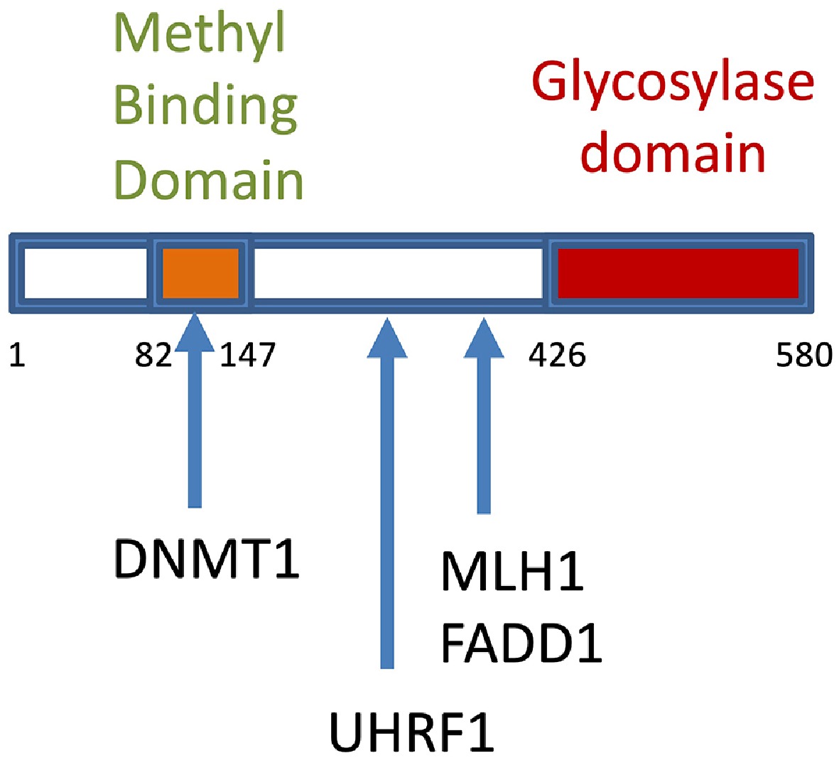
Figure 2.
Structural domains of MBD4 and interacting proteins. The region between the methyl-binding domain and the glycosylase domain is called 'the intervening region', in which the binding of UHRF1 occurs[111]. Numerals correspond to the number of amino acids delineating the structural domains of MBD4. MLH1 interacts with the glycosylase domain, whereas DNMT1 interacts with the methyl-binding domain[114,127]. MBD4 was shown to directly interact with proapoptotic Fas-associated death domain protein (FADD) in a complex with MLH1[128].
Enzymatic activity of MBD4
-
MBD4's glycosylase activity corresponds to the hydrolysis of the N-glycosidic bond between the base and the deoxyribose, creating an abasic site. As this is the only activity that has been detected so far, the enzyme is classified as monofunctional. To our knowledge, no mammalian glycosylase with bifunctional activity in a methylated context exists. However, ROS1 in plants has such a bifunctional role. In bifunctional enzymes, glycosylase activity is coupled with lyase activity, which leads to a break in the DNA backbone at the 3′ side of the abasic site, resulting in the complete removal of the nucleotide from the DNA.
We recently conducted research into the role of MBD4 in maintaining DNA methylation and its mechanism[50]. We hypothesized that MBD4 may exhibit bifunctional activity in conjunction with other proteins. We demonstrated that the MBD4 complex exhibits glycosylase and lyase activities when the DNA is methylated. Among the most abundant proteins in the purified MBD4 complex, we noted the presence of two proteins involved in the mismatch repair pathway: MLH1 and PMS2. Furthermore, we observed that MBD4 is a methyl-directed glycosylase enzyme with lyase activity that is specific to G/T mismatches in a CpG context, due to its association with the MMR protein MLH1. We suggested that this association induces structural modifications favouring the glycosylase catalytic site to optimally engage with the substrate, thereby enabling it to exert its full activity.
In order to repair DNA methylation patterns, that is, to fully recover methylated DNA (on both strands), hemi-methylated DNA must be recognized by a protein or a tandem of proteins in order to methylate the newly synthesized DNA strand. So far, only a few such proteins have been reported to have such capacity, one of which is UHRF1[59]. We will now review some information on UHRF1 to show the impact that an interaction between MBD4 and UHRF1 can have.
-
We discovered the human UHRF1 protein in 2000 and named it ICBP90 (Inverted CCAAT Box-Binding Protein)[72]. First identified as a transcription factor of the TOP2A gene[72], the real breakthrough came in 2007 and 2008 when several studies (including ours) demonstrated the cooperation and association of UHRF1 with DNA methyltransferase 1 (DNMT1), and the role of the SRA domain was deciphered[35,73−78]. The SRA domain is present only in the UHRF family in mammalian cells[35]. It was first reported in the human UHRF1 version and was called the G9a domain[72]. It was later named the SRA domain (Set and RING-associated).
So, how does the UHRF1/DNMT1 tandem work? When a mother cell divides, the DNA must be duplicated to ensure that each daughter cell has an identical genome and epigenome, including the DNA methylation patterns. Both strands of DNA are methylated in a CpG context, and during replication they separate to allow the DNA polymerase to generate a new complementary strand. The result is an intermediate state called hemi-methylated DNA, which is recognized by UHRF1 thanks to its SRA domain. Once bound, UHRF1 helps DNMT1 identify the cytosine that will be methylated, thereby ensuring that the patterns are faithfully duplicated. This is how methylation patterns are inherited and how UHRF1 participates in DNA methylation maintenance. However, other mechanisms, such as antagonizing active demethylation by TET2, might also contribute[79]. Given that UHRF1 can read the H3K9me2/3 modification[80], UHRF1 is also likely to be involved in the inheritance of the histone code. We have proposed the existence of a machinery for replicating the epigenetic code (ECREM), which includes UHRF1, DNMT1, HDAC1, and PCNA[59,81], and that within this complex UHRF1 plays the role of a conductor.
Several publications have already reviewed the knowledge on UHRF1 structure and roles in detail[9,30,31,35,59,82−89]. Nevertheless, a brief overview of UHRF1-mediated regulation of TSG expression follows.
UHRF1 and TSGs
-
Tumor suppressor genes are crucial for regulating a cell's diverse functions. Their primary function is to prevent tumor formation. However, when tumors are formed, it is too late to rely on them because their expression is inactivated, either through mutation or through epigenetic silencing[90]. An interesting cancer therapeutic strategy would be to reactivate silenced TSGs through demethylation using CRISPR-mediated DNA methylation editing[91]. Several natural compounds have been identified that reactivate TSG expression by downregulating UHRF1 expression[92], some of which target its SRA domain (Fig. 1). At present, the inhibitors of the SRA domain have only been tested using purified proteins and/or cell-line models, and to our knowledge no clinical data are available.
An impressive number of UHRF1-regulated TSGs have been identified in the past. Among these genes, we can find RB1, p16INK4A, p73, CDH13 and SHP1, SOCS3 and 3OST2, BRCA1, TUSC3, CDX2, CDKN2A, RUNX3, FOXO4, PPARG and PML, MEG3[93], 14-3-3σ, TXNIP and PTEN[94].
Knockdown of UHRF1 was annotated and categorized into 488 hypermethylated and 1,696 hypomethylated promoters, as well as 1,149 hypermethylated and 5,501 hypomethylated gene bodies (39,112,494). Further analysis revealed 21 downregulated oncogenes and 15 upregulated tumour suppressor genes (TSGs)[54]. These results suggest that UHRF1 has a significant impact on the balance of oncogene and TSG expression. In addition, 15 tumor suppressor genes (TSGs) that are specific to lung cancer have been found to be regulated by UHRF1 through DNA hypermethylation[95]. These genes are EMP2, CSRNP1, ZDHHC2, PAFAH1B1, CD44, GABARAP, EPHA3, NR4A1, SRGAP3, CHST10, PIN1, HIPK2, ZNF185, TMEM127, and CBL[95]. The authors conducted functional genomic screens in primary tumor cells derived from a genetically engineered mouse model of lung cancer driven by the activation of the Kras G12D allele and the loss of p53. They concluded that the broad effect observed across a large number of TSGs supports the role of UHRF1 in lung cancer. However, for both studies, the reason why UHRF1 regulates so many TSGs within one cell type and what the triggering factor is remains unclear. Nevertheless, let's try to imagine a scenario. If we consider that UHRF1 colocalizes with DNMT1 and PCNA[96] at the replication fork, it is easily understandable that UHRF1 meets at a moment the promoters of all silenced TSGs. Therefore, when UHRF1 is decreased or silenced, all tumor suppressor genes that are silenced through hypermethylation may putatively be re-expressed, as their promoter does not anymore encounter UHRF1 and therefore cannot be fully methylated. This remains, of course, a hypothesis, but it is very likely to be verifiable.
Interestingly, several products of TSGs regulated by UHRF1 are also able to interact with the UHRF1 protein. These genes include RB1[97], BRCA1[13], and Tip60[98,99]. This interaction sometimes requires post-translational modifications. For instance, BRCA1 recruits UHRF1 to DNA double-strand breaks (DSBs) during the S phase of the cell cycle. This process requires the BRCT domain of BRCA1 and the phosphorylation of Ser674 in UHRF1[13]. However, the significance of this feature is unclear as it appears contradictory: if UHRF1 is silencing a gene, why does it need to interact with it? Alternatively, it may act as inhibitory feedback control, whereby the product of a gene counteracts the inhibitory capacity of the regulator, creating a finely tuned regulatory mechanism. Conversely, it is noteworthy that some tumour suppressor genes (TSGs) are able to regulate UHRF1 expression. For example, p53 and p73 exert an inhibitory effect on UHRF1 expression in response to DNA damage[100−102].
Although the mechanism by which UHRF1 silences tumor suppressor genes (TSGs) is the maintenance of promoter hypermethylation, it is not yet clear whether UHRF1 is involved in establishing the hypermethylation of TSG promoters or only in its inheritance following DNA duplication. Nevertheless, there are some areas that warrant further investigation. For example, a reduction in UHRF1/DNMT1 interaction has been suggested as an explanation for the global hypomethylation that occurs in cancer cells[103]. Whether this hypomethylation leads to hypermethylation of TSGs is not yet evidenced, but is highly possible. Accordingly, it has been shown that the STUB1-UHRF1/DNMT1 axis increases DNA methylation of the PLA2G2A promoter in cholangiocarcinoma cells, leading to its silencing[104]. Through this capacity to repress a high number of TSGs, UHRF1 plays an important role in various types of cancer[88,89,105,106].
Interestingly, MBD4 was shown to be associated with hypermethylation of CDKN2A/p16IN4A and hMLH1 gene promoters[107]. Transcriptional repression by MBD4 was shown to be histone deacetylase 1 (HDAC1) dependent, and MBD4 directly binds to Sin3A and HDAC1. HDAC1 belongs to the UHRF1 complex[108], further supporting that MBD4 may cooperate with UHRF1 to maintain hypermethylation of TSG promoters, at least for CDKN2A/p16INK4A and hMLH1 genes. Another tumor suppressor gene, namely p15ink4b, was also shown to be protected from demethylation by MBD4[109] and is known to be regulated by UHRF1[110]. Together, these studies support a putative cooperation between MBD4 and UHRF1 in regulating a few TSGs, but a physical cooperation between these two proteins in that kind of regulation requires further investigation. Indeed, it would be valuable to assess whether UHRF1 and MBD4 co-localize at tumor-suppressor gene promoters as part of the same macromolecular complex. Such an association would imply that they contribute to maintaining TSG promoter methylation patterns following G:T mismatches and thus maintaining the « cancer phenotype ».
-
Considering that MBD4 generates a gap, resulting in hemi-methylation of DNA after refilling, in cooperation with other BER machinery members, we hypothesized that UHRF1 is an essential partner of the MBD4/MLH1 tandem in maintaining DNA methylation patterns. The presence of MBD4 and UHRF1 in the same macromolecular complex has already been reported[111] and thus is not new. Furthermore, this study identified the MBD4 structural domain involved in interacting with UHRF1 as its intervening region (Fig. 2). Such cooperation makes sense when considering the following arguments. For example, it has been demonstrated that the BER glycosylase MBD4 binds preferentially to active chromatin and to DNA that is replicated early. This correlates with a lower mutational burden in these domains[112]. Furthermore, MBD4 has been identified as the primary enzyme responsible for 5mC deamination repair in humans. However, this cooperation may be confined to heterochromatin, since previous studies have shown that MBD4 and UHRF1 are found together at chromocenters during heterochromatin replication and formation[111]. Furthermore, MBD4 may protect UHRF1 by recruiting USP7, a protein known to protect UHRF1 from ubiquitination[113].
Figure 3 illustrates the sequence of events for MBD4/UHRF1 dialogue. We are qualifying the interaction between MBD4 and UHRF1 as a dialogue and not as a cooperation since the literature supports a direct interaction between the two proteins[111]. If the proteins belong to the same macromolecular complex with no direct interaction, we would rather talk of cooperation. Once the incorrect nucleotide has been removed by MBD4 with the assistance of MLH1, the BER machinery repairs the gap, resulting in a cytosine facing a guanosine once more. This results in the formation of hemimethylated DNA, meaning that only one strand is methylated. However, MBD4 and the BER machinery alone are insufficient to restore fully methylated DNA. Therefore, there are two possibilities. The first is that the methylation is not repaired. However, this is unlikely, as our functional study of mouse embryonic fibroblasts shows that MBD4 is required to preserve DNA methylation within promoters and retroelements[50]. The second possibility is intervention by UHRF1, which recognizes the hemi-methylated state and recruits DNMT1 to methylate cytosine on the newly synthesized DNA strand. Considering that MBD4, UHRF1, and DNMT1 are partners[35,73,111,114], this is the most likely mechanism.
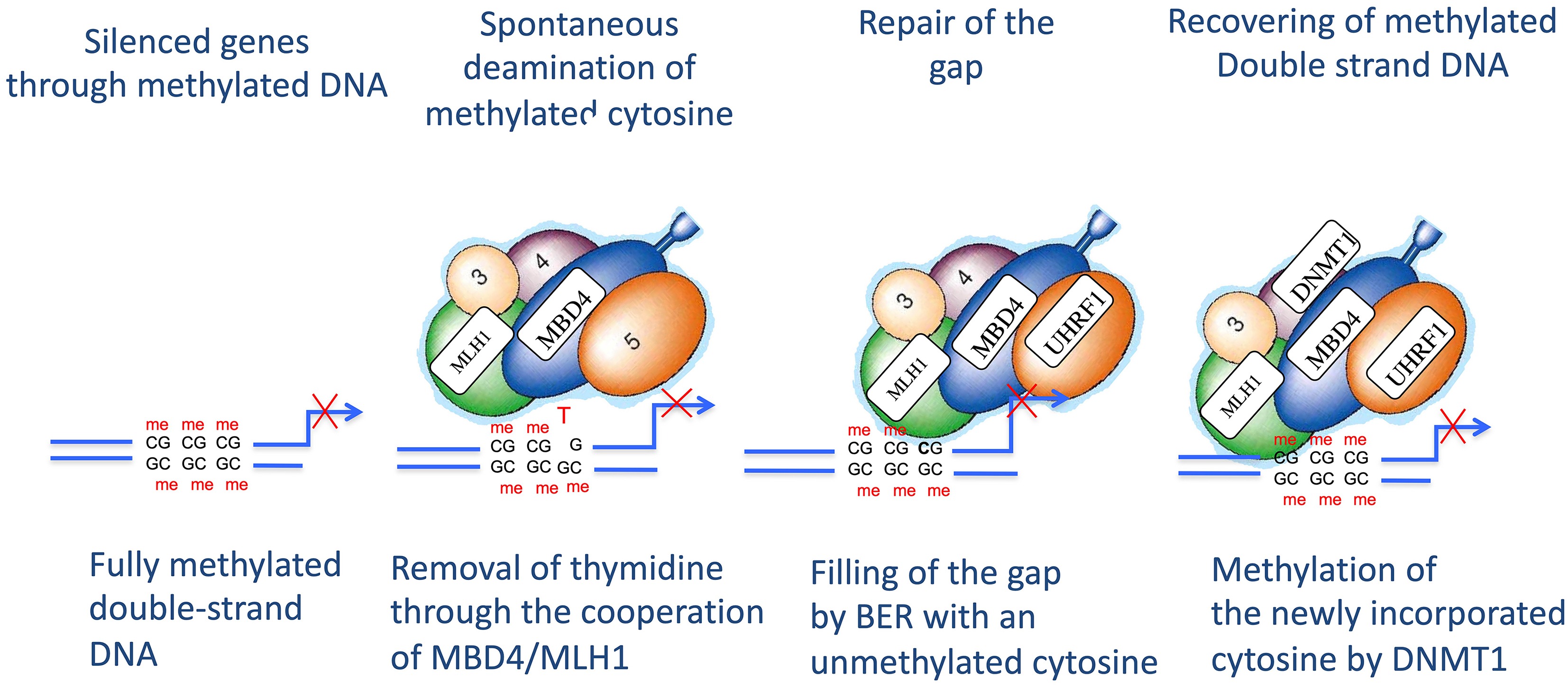
Figure 3.
Proposed model of the dialogue between UHRF1 and MBD4, in the presence of their respective partners, in relation to repairing DNA methylation patterns. After removal of the T:G mismatch by MBD4/MLH1, the gap is filled thanks to the intervention of the BER machinery. The filling of the gap generates a hemi-methylated state of the DNA that is sensed by UHRF1. The cooperation of the latter with DNMT1 allows recovery of fully methylated DNA, i.e., both DNA strands are methylated. As outlined in the text, this model assumes that the MBD4–UHRF1 complex is pre-assembled before encountering a G:T mismatch.
The other fundamental question is how MBD4 recruits UHRF1. We have long hypothesized that, during DNA replication, the fully assembled UHRF1 complex closely follows the replication machinery[81]. This would allow it, on one hand, to duplicate DNA methylation patterns and, when necessary, on the other hand, to promote repair of G:T mismatches. We therefore believe that MBD4 is already part of the UHRF1 complex before encountering a mismatch. This model is consistent with the need for an efficient process: otherwise, it would be time-consuming and potentially unreliable for MBD4 to depend on the subsequent recruitment of UHRF1. This is the concept illustrated in Fig. 3, which proposes that the UHRF1–MBD4 association is already established when MBD4 encounters a mismatch.
-
It is clear from the literature that UHRF1 and MBD4 have the potential to cooperate in the maintenance and repair of DNA methylation patterns through a yet unresolved molecular dialogue. One of the most intriguing challenges is understanding why UHRF1 and MBD4 require direct interaction, given that UHRF1, according to our model (illustrated in Fig. 3), does not intervene immediately after MBD4. Indeed, before UHRF1 becomes involved, the gap must first be filled by a ligase, such as ligase IIIa. Only after this event can UHRF1 sense the presence of hemi-methylated DNA, subsequently engaging in dialogue with DNMT1 (Fig. 1). The existence of this direct interaction is perplexing, unless we consider that MBD4 acts as a conductor, first instructing the ligase to act, and then introducing a change in the structure of UHRF1 to facilitate hemi-methylated DNA recognition.
An important unresolved question is therefore how MBD4-mediated repair could influence the subsequent recruitment or activity of UHRF1 despite the apparent temporal separation between completion of base excision repair and maintenance methylation. Although no direct molecular mechanism has yet been demonstrated, several non-mutually exclusive models can be envisioned. First, MBD4-mediated repair may induce transient local changes in chromatin architecture or DNA accessibility that persist after gap filling and create a favorable context for UHRF1 recognition of hemi-methylated CpG sites. Second, progression through the repair process may be accompanied by post-translational modifications of MBD4, UHRF1, or associated repair factors that function as molecular switches coordinating the transition from DNA repair to restoration of the epigenetic landscape. Third, MBD4 may participate in multiprotein repair complexes that remain associated with chromatin after DNA synthesis, allowing scaffold proteins or chromatin remodelers to relay information to UHRF1 without requiring a stable direct interaction between the two proteins. Finally, transient interactions with BER proteins, PCNA, chromatin assembly factors, or histone-modifying enzymes could provide a molecular bridge linking repair completion to UHRF1 recruitment during chromatin restoration. Interestingly, UHRF1 is known to interact and/or belong to the same macromolecular complex as PCNA, chromatin remodelers, and histone-modifying enzymes[81,96]. These models are not mutually exclusive and generate experimentally testable predictions. For example, time-resolved chromatin immunoprecipitation, proximity-labelling proteomics, live-cell imaging, and mutational analyses of candidate post-translational modification sites could distinguish whether UHRF1 recruitment depends on persistent chromatin alterations, transient signalling events, or assembly of higher-order repair complexes. However, as illustrated in Fig. 3, we are favouring a model consistent with the existence of an association between MBD4 and UHRF1 prior to MBD4 encountering a G:T mismatch. In this context, the macromolecular complex containing the MBD4–UHRF1 tandem is supposed to intervene just after the DNA replication machinery. It would indeed be time-consuming and potentially unreliable for MBD4 to depend on the subsequent random recruitment of UHRF1. Figure 3 illustrates this concept, proposing that the UHRF1–MBD4 association is already established when MBD4 encounters a mismatch.
Future studies should directly test the proposed coupling between MBD4-mediated base excision repair, UHRF1 recruitment, and DNMT1-dependent maintenance methylation. First, the MBD4–UHRF1 interaction should be examined in relevant cell types using endogenous co-immunoprecipitation, reciprocal pull-down assays, and proximity ligation assays, particularly under conditions that increase cytosine deamination or activate BER. Second, an in vitro reconstitution system using purified MBD4, BER components, UHRF1, DNMT1, and defined methylated/deaminated DNA substrates would allow direct assessment of whether MBD4-initiated repair promotes UHRF1 binding and DNMT1-mediated remethylation. Third, single-cell and single-molecule approaches, including live-cell imaging, targeted DNA damage systems, proximity labeling, or single-molecule tracking, could resolve the temporal order of MBD4, UHRF1, and DNMT1 recruitment at deamination/repair sites. Together, these approaches would provide direct mechanistic evidence for the proposed BER–UHRF1–DNMT1 cascade.
If the proposed functional interplay between MBD4 and UHRF1 is confirmed, its disruption could have important pathological consequences beyond defective repair of deamination-induced G mismatches. Loss of MBD4 function may impair not only the removal of mutagenic lesions but also the faithful restoration of DNA methylation patterns following base excision repair, potentially resulting in mutation accumulation, epigenetic instability, and aberrant transcriptional regulation. This possibility may be particularly relevant in cancers, where MBD4 mutations have been associated with increased mutational burden and genomic instability. Within the framework of our hypothesis, defective MBD4 could therefore compromise both genome integrity and maintenance methylation by altering the chromatin environment or molecular signals required for efficient UHRF1 function. More broadly, because disturbances in DNA repair and epigenetic regulation are common features of neurodegenerative and inflammatory disorders, impaired coordination between MBD4-mediated repair and UHRF1-dependent maintenance methylation could contribute to disease-associated epigenetic remodeling. Although these concepts remain speculative, they provide experimentally testable predictions and offer a potential mechanistic link between DNA repair, epigenetic maintenance, and human disease.
One of the main remaining challenges is to investigate whether the UHRF1–MBD4 axis plays a role in maintaining the cancer phenotype. Indeed, both UHRF1[85,88,98,115,116] and MBD4 are overexpressed in some types of cancer[117]. This dual overexpression reinforces the suspicion that these two proteins act together in cancer cells to maintain their genome integrity and thus maintain the cancer signature. However, whether the UHRF1/MBD4 tandem is involved in whole epigenome integrity maintenance is far from certain. Indeed, co-localization of MBD4 and UHRF1 is found mainly at chromocenters constituted of heterochromatin[111]. Therefore, it cannot be ruled out that the repair of DNA methylation patterns following 5mC deamination outside chromocenters or heterochromatin requires different factors than the UHRF1/MBD4 tandem. Indeed, UHRF1 is involved in DNA repair upon double-strand breaks and undergoes auto-ubiquitination, which allows the recruitment of the DNA repair machinery[14,87,118−120]. In this case, it is likely that UHRF1 requires partners other than MBD4. It is not known whether spontaneous demethylation induces the auto-ubiquitination of UHRF1, but this would be difficult to identify as it may be a rare event within a cell.
In conclusion, we propose that the MBD4/UHRF1 tandem acts as an important suppressor of epimutation to maintain the integrity of the epigenome of normal cells and cancer cells as well. Given that alterations in this dialogue may contribute to various diseases, particularly cancer, elucidating the dialogue between the UHRF1/MBD4 tandem and its other complex partners could contribute to the development of precision medicine strategies.
-
Not applicable.
-
The authors confirm their contributions to the paper as follows: draft manuscript preparation: Bronner C; literature review: Bronner C, Hamiche A; manuscript editing: Bronner C, Mousli M, Hamiche A. All authors reviewed the results and approved the final version of the manuscript.
-
Data sharing is not applicable to this article as no datasets were generated or analyzed during the current study.
-
The authors declare that they have no conflict of interest.
- Copyright: © 2026 by the author(s). Published by Maximum Academic Press, Fayetteville, GA. This article is an open access article distributed under Creative Commons Attribution License (CC BY 4.0), visit https://creativecommons.org/licenses/by/4.0/.
-
About this article
Cite this article
Bronner C, Mousli M, Hamiche A. 2026. A working hypothesis: a dialogue between UHRF1 and MBD4, with the help of BER, to ensure the integrity of genomic DNA methylation patterns at heterochromatin. Epigenetics Insights 19: e012 doi: 10.48130/epi-0026-0009 

A working hypothesis: a dialogue between UHRF1 and MBD4, with the help of BER, to ensure the integrity of genomic DNA methylation patterns at heterochromatin
- Received: 30 January 2026
- Revised: 25 June 2026
- Accepted: 28 July 2026
- Published online: 08 October 2026
Abstract: The DNA methyltransferase 1 (DNMT1)/Ubiquitin-like containing PHD and Ring Finger domains 1 (UHRF1) tandem ensures the faithful transmission of DNA methylation profiles from a mother cell (even a cancer cell) to its daughter cells. Five-methylcytosine (5mC) undergoes spontaneous deamination, leading to the generation of a thymine and thus a G:T mismatch. This mismatch is repaired through the action of two glycosylases, one of which is methyl-CpG binding domain 4 (MBD4). After repair, i.e., removal of thymine and filling the gap, DNA is found in a hemi-methylated state (only one DNA strand is methylated). However, how cells re-establish fully methylated patterns, in which both DNA strands carry methylation marks, remains unresolved. Here, we present a new paradigm for the repair and maintenance of DNA methylation patterns through the dialogue between MBD4 and UHRF1 that can operate in the context of the spontaneous demethylation of cytosine and recover fully methylated DNA. If validated experimentally, this proposed MBD4–UHRF1 functional interplay could provide a mechanistic framework linking repair of deamination-induced G mismatches with maintenance of DNA methylation fidelity, offering new insights into the epigenetic instability observed in cancer and other diseases.