













-
Blue mold rot is most commonly found in pears during postharvest storage and is caused by Penicillium expansum[1]. Postharvest illnesses have a negative impact on the edible safety, nutritional value, and commercial worth of fresh pear fruit, resulting in significant losses each year. The rise of fungicide-resistant organisms and the irreparable harm chemical fungicides do to human health and the environment limit their usage[2]. Over the past decade, the use of antagonistic yeast for biological control has become a mature and promising means[3]. However, the viability of antagonistic yeasts is easily affected by adverse abiotic factors such as pH, temperature, and oxidative stress under postharvest conditions, resulting in variable and often reduced biocontrol efficacy. Consequently, increasing attention has been directed toward the development of stable resistance-inducing elicitors that effectively activate host defense responses. In this context, yeast cell wall components have emerged as promising alternatives to living cells, owing to their structural stability, safety, and strong potential to activate fruit defense mechanisms.
The yeast cell wall constitutes 15%–30% of the cell's dry weight and serves as the outermost dynamic organelle of yeast[4]. It consists of mannoprotein, β-glucan, chitin, and proteins connected by glycan and glycolipid anchors[5]. Although yeast cell walls vary by species, they typically consist of β-1,3-glucan polysaccharides coupled with chitin to create a basic scaffolding structure[6]. The cell wall interacts with the environment through a multitude of receptors in addition to its vital roles in maintaining cell permeability and shape. Furthermore, yeast cell walls are commonly utilized as supplements in farm animal feed because they may prevent intestinal infections and modulate immunological function[7]. Previous studies in our laboratory showed that cell wall polysaccharides from Saccharomyces cerevisiae and Rhodosporidium paludigenum could induce disease resistance in fruit by enhancing endogenous nitric oxide production, activating defense responses, and increasing antioxidant enzyme activities[8]. Kluyveromyces marxianus is a non-pathogenic yeast strain widely recognized for its essential role in the fermentation of traditional dairy products such as kefir and cheese. With further study, K. marxianus is regarded as a promising probiotic[9]. It has been shown that it can regulate intestinal inflammation in people and trigger apoptosis in human cancer cells[10]. Within the context of postharvest biology, Zheng et al. demonstrated that K. marxianus effectively suppresses blue mold rot and reduces patulin accumulation in apples, highlighting its antifungal potential[11]. As a well-established food-grade microorganism, K. marxianus holds considerable promise for applications in food safety, agriculture, and environmental management. Despite its broad applicability, however, studies investigating the use of its cell wall components as inducers of fruit disease resistance remain notably limited.
Our preliminary observations revealed that inactivated K. marxianus elicited stronger resistance against P. expansum in pear fruit compared to inactivated S. cerevisiae, although the underlying mechanisms remained unexplored. To achieve a comprehensive understanding, we investigated the physiological and molecular responses activated by the cell wall treatment. Furthermore, we isolated β-glucan and mannan from the K. marxianus cell wall and employed transcriptomic analysis to delineate their respective contributions to defense-related gene expression. This integrated approach aims to establish a mechanistic foundation for the application of the K. marxianus cell wall as a novel and effective food-grade elicitor in postharvest fruit protection.
-
K. marxianus, Cryptococcus laurentii, Rhodotorula mucilaginosa, and S. cerevisiae were purchased from the China General Microbiological Culture Collection Center (CGMCC) and cultured in yeast extract peptone dextrose medium at 28 °C and 200 rpm for 24 h. K. marxianus (CGMCC 2.1442) was preserved in our laboratory. The yeast was then collected by centrifugation and rinsed with sterile water. Penicillium expansum (CGMCC 3.7898) was cultivated for 7 d at 25 °C on potato dextrose agar (PDA). Pears (Pyrus pyrifolia Nakai. cv. 'Shuijing'), free of visible disease, were purchased from Handan, Hebei, China. The fruit were surface-disinfected with 0.1% (v/v) NaClO for 3 min, rinsed with tap water, and air-dried before use. Glucose, N-acetylglucosamine, and D-mannose were bought from Aladdin Chemical Co. (Shanghai, China).
Inhibition of pear blue mold rot by inactivated yeast
-
Five evenly distributed wounds of uniform size and depth were formed on the surface of each pear fruit using a sterilized punch. To obtain inactivated yeast bodies, a 1 × 10⁸ cells mL−1 yeast suspension was made using a hemocytometer and inactivated for 20 min in a water bath at 100 °C. Each hole was given 20 μL of liquid as follows: (1) sterile water (control); (2) 1 × 10⁸ cells mL−1 inactivated K. marxianus suspension; (3) 1 × 10⁸ cells mL−1 inactivated S. cerevisiae suspension; (4) 1 × 10⁸ cells mL−1 inactivated R. mucilaginosa suspension; (5) 1 × 10⁸ cells mL−1 inactivated C. laurentii suspension. After drying at room temperature, nine pears were chosen at random and stored in a room with a constant temperature of 25 °C and 90% relative humidity for 24 h. Then, each wound was individually treated with 20 μL of a 1 × 10⁴ spores mL−1 P. expansum spore suspension. The pears were rewrapped and put in a storage room. The lesion diameter and disease incidence were measured and documented on a daily basis. Each treatment was replicated three times, with nine pears each, and the whole experiment was performed twice. The inactivated yeast was freeze-dried into white powder and observed by scanning electron microscopy (GeminiSEM 300, Carl Zeiss, Germany).
Preparation of yeast cell wall
-
Yeast cultivated in YPD medium was harvested, centrifuged at 4,000 rpm, and washed with sterile water three times. A 100 mg sample of inactivated yeast was suspended in 0.5 mL of sterile water, and then acid-washed glass beads were added. The samples were shaken and crushed in an automatic tissue grinder at 70 Hz for 15 min. The totally fragmented yeast was centrifuged at 4,000 rpm for 10 min, and the precipitate was washed three times in sterile water. The cleansed yeast cell walls were freeze-dried and kept at −20 °C.
Inhibitory effect of K. marxianus and S. cerevisiae cell wall on pear blue mold rot
-
Cell wall suspensions were made by adding 0.5% (w/v) of K. marxianus and S. cerevisiae yeast cell wall to sterile water. The pears were washed and punched with three holes of similar size. Each hole received 20 μL of liquid as follows: (1) sterile water (control); (2) 0.5% (w/v) K. marxianus cell wall suspension; (3) 0.5% (w/v) S. cerevisiae cell wall suspension. Pears were wrapped and stored at 25 °C for 24 h before accessing each wound with a 20 μL 1 × 10⁴ spores mL−1 P. expansum spore suspension. The disease incidence and lesion diameter were recorded following the same procedure.
Optimal treatment conditions for K. marxianus cell wall inhibiting pear blue mold rot
-
To create cell wall suspensions of varying concentrations, 0.1% (w/v), 0.5% (w/v), and 1% (w/v) of K. marxianus cell walls were mixed with sterile water. Four equally spaced holes were punched in the pear fruit. 20 μL sterile water (control) and 20 μL of the above three kinds of K. marxianus cell wall suspension with different concentrations were added to the wounds, respectively. Then, the pears were sealed and cultured at 25 °C for 24 h. P. expansum spores were then added to each wound, and the pear fruit continued to be incubated at 25 °C.
Pear fruit was treated with 0.5% (w/v) K. marxianus cell wall at 25 °C for 2, 12, 24, and 36 h, respectively. Then, a 20 μL 1 × 10⁴ spores mL−1 P. expansum spore suspension was added to each wound. Subsequently, the lesion diameter and disease incidence of pears were recorded daily. The treatment time was defined as the interval between the addition of K. marxianus cell wall and the addition of spores.
Analysis of cell wall monosaccharide composition by high-performance liquid chromatography (HPLC)
-
The monosaccharide content of several yeast cell walls was measured by HPLC using the technique of Sun et al. with minor changes[12]. About 4 mg of dried yeast cell wall powder was combined with 2 M trifluoroacetic acid in an ampoule, sealed, and hydrolyzed in an oven at 120 °C for 8 h. Upon reaching room temperature, 200 μL of anhydrous ethanol was added to the hydrolysate, followed by nitrogen blow-drying, which was performed three times. Subsequently, it was neutralized with NaOH. The hydrolysate was derivatized with 1-phenyl-3-methyl-5-pyrazolone (PMP) and incubated in a water bath at 70 °C for 30 min. After adding chloroform, the mixture's lower organic phase was discarded three times. The same procedure was used to derivatize standard monosaccharide samples.
After passing the sample through a 0.22 μm filter membrane, the derivatives were analyzed using a Promosil-C18 column (4.6 mm × 150 mm × 5 μm, Bonna-Agela Technologies, China) linked to a HPLC Ultimate 3000 system (Thermo Scientific, USA). The flow rate was 1 mL min−1, and the injection volume was 20 μL. The polysaccharide content of the samples was calculated from their monosaccharide content.
Comparison of induced resistance between commercial polysaccharide mixtures and the K. marxianus cell wall
-
Commercial mannan, β-glucan, and chitin were utilized to create mixed polysaccharide suspensions according to the proportion of polysaccharides in the K. marxianus cell wall. A 20 μL sample of sterile water, 20 μL of 0.5% (w/v) K. marxianus cell wall suspension, and 20 μL of the mixture were added to each wound of the pear fruit, and the pears were then sealed and incubated at 25 °C for 24 h. After adding 20 μL of a 1 × 10⁴ spores mL−1 spore suspension, the pear fruit was cultured at 25 °C and recorded every day.
Characterization of the yeast cell wall
-
Fourier transform infrared (FTIR) spectra of K. marxianus and S. cerevisiae cell walls were acquired using an FTIR spectrometer (Nicolet 5700, Thermo Fisher Scientific, MA, USA) in the wavenumber range 4,000–400 cm−1. Scanning electron microscopy (SU8010, Hitachi, Japan) was used to investigate the surface structure of the freeze-dried yeast cell wall.
Reactive oxygen species (ROS) measurement in pear fruit
-
P. expansum spore suspensions were applied to pear fruit wounds after 24 h of adding sterile water and 0.5% (w/v) K. marxianus cell wall. The pears were then kept at 25 °C for 24 h. The flesh of the pear was cut at the wound site and immediately sectioned in a freezing microtome (Leica, Germany). Tissue slices were stained with 100 μM 2',7'-dichlorodihydrofluorescein diacetate (DCFH-DA) for 30 min in the dark and washed twice with phosphate buffer saline (PBS, 50 mM, pH 6.5). The photos were captured using a Nikon Eclipse Ti fluorescent microscope (Nikon, Japan) at an exposure period of 600 ms.
Measurement of enzymatic activity and content
-
Pear fruit wounds were treated with 20 μL of sterile water and 20 μL of 0.5% (w/v) K. marxianus cell wall for 24 h. As the 0 h sample, a sterile scalpel was used to retrieve the pulp around the wound of a pear fruit. After adding 20 μL of a 1 × 10⁴ spores mL−1 P. expansum spore suspension to the wounds, pear tissues were sampled after 12, 24, and 36 h. Fresh samples were placed in liquid nitrogen immediately after removal and then frozen at −80 °C. To extract different crude enzyme solutions, approximately 0.9 g of frozen pear fruit samples were ground and broken in a tissue grinder with various buffers.
The protein concentration of crude enzyme liquid was determined by the Bicinchoninic Acid method of the kit instructions (Yeasen, Shanghai, China). Superoxide dismutase (SOD), catalase (CAT), glutathione reductase (GR), peroxidase (POD), phenylalanine ammonia lyase (PAL), chitinase, polyphenol oxidase (PPO), and glutamate decarboxylase (GAD) enzyme activity was determined according to the kit instructions (Jiancheng, Nanjing, China). β-1,3-glucanase activity was determined by the 3,5-dinitrosalicylic acid method[13]. The concentrations of GAD, L-glutamate (Glu), γ-aminobutyric acid (GABA), and GABA transaminase (GABA-T) were measured using the kit (D&B, Shanghai, China) after 36 h of selecting pear samples. All enzyme activities and contents were calculated using the protein concentration of the sample as the denominator.
Extraction of β-glucans and mannans
-
The yeast cell walls obtained above were extracted with 2% (w/v) NaOH at a ratio of 1:20 (g dry cell wall/mL solution) in a water bath at 80 °C for 2 h. This extraction was performed twice. After extraction, the mixture was cooled to room temperature and centrifuged at 10,000 rpm for 10 min at 4 °C. The pellets were washed three times with sterile water and then freeze-dried to obtain β-glucan.
The supernatant containing water-soluble polysaccharides was collected and neutralized to pH 7 using 4 mol/L HCl. Trichloroacetic acid (TCA) was then added to a final concentration of 4% (w/v) to remove proteins, followed by incubation at 4 °C for 4 h. The mixture was centrifuged at 10,000 rpm for 10 min, and the precipitate was discarded. The supernatant was concentrated by rotary evaporation, mixed with three volumes of absolute ethanol, and incubated at 4 °C overnight to precipitate polysaccharides. After centrifugation at 10,000 rpm at 4 °C, the precipitate was collected, redissolved in distilled water, and dialyzed using an MD77 dialysis membrane under running water for 2 d to remove small molecules[14]. The dialysate was finally freeze-dried to obtain mannan.
Analysis of gene expression and transcriptomics
-
The treatment of pear fruit followed the same procedure. Total RNA was extracted from about 2.7 g of frozen pear samples according to the kit instructions (Plant Total RNA Isolation Kit Plus, FOREGENE). A micro spectrophotometer (NanoDrop 1000, Thermo-Fisher, USA) was used to determine RNA concentration and quality. The cDNA was produced using TransScript All-in-One First-Strand cDNA Synthesis SuperMix (Transgen Biotech, Beijing, China) according to the manufacturer's instructions. Supplementary Table S1 lists the gene-specific primers built using P. pyrifolia sequences from the National Center for Biotechnology Information GenBank. For RT-qPCR, the Hieff qPCR SYBR Green Master Mix (Yeasen, Shanghai, China) was used. Actin was used as a reference gene in pear fruit.
The obtained glucan and mannan were separately prepared as 0.5% (w/v) polysaccharide solutions or suspensions. Three wounds with uniform depth were made on each pear. Each wound was treated with 20 μL of one of the following solutions: (1) sterile water (control); (2) 0.5% mannan solution; (3) 0.5% glucan suspension. After incubation at 25 °C for 24 h, 0.3 g of tissue surrounding each wound was excised using a sterile scalpel, immediately frozen in liquid nitrogen, and stored at −80 °C. Each treatment included three biological replicates, and the samples were sent to Biomarker Technologies Co., Ltd. for transcriptome analysis.
Statistical analysis
-
Statistical significance for multiple comparisons in each experiment was established using one-way ANOVA and the Prism 9.0 program (GraphPad Software, Inc.). The data were reported as mean ± SEM (n ≥ 3). p < 0.05 was defined as statistically significant.
-
Previous work in our laboratory demonstrated that live K. marxianus effectively controls P. expansum infection in pear fruit through direct biocontrol mechanisms[15]. However, whether non-viable forms of this yeast, particularly its cell wall, could confer protection by activating host resistance remained unexplored. To address this question, pear fruit were treated with various inactivated yeasts and subsequently challenged with P. expansum. All inactivated yeast treatments significantly enhanced fruit resistance to blue mold rot when applied 24 h prior to pathogen inoculation, though their effectiveness varied considerably. Notably, inactivated K. marxianus, C. laurentii, and R. mucilaginosa exhibited superior protective effects, resulting in lower disease incidence and smaller lesion diameters compared to S. cerevisiae (Fig. 1a and b). To determine whether this protection arose from induced resistance rather than direct antifungal activity, we compared treatments where inactivated yeasts were applied simultaneously with pathogen inoculation or 24 h in advance. The markedly stronger inhibition observed in the pre-treatment group confirmed that inactivated yeasts primarily function by priming the fruit's innate defense responses (Supplementary Fig. S1). Microscopic examination revealed morphological differences among the inactivated yeast strains, with S. cerevisiae, C. laurentii, and R. mucilaginosa displaying ellipsoidal shapes while K. marxianus exhibited a more elongated morphology (Fig. 1c). These structural variations may contribute to the observed differences in resistance-inducing efficacy, potentially through differential interactions with fruit surface receptors. The yeast cell wall is the outermost structure necessary to maintain the growth and morphology of yeast[16]. The inactivated yeast lost its physiological activity and mainly depended on the surface cell wall coming into contact with the pear. Sun et al. found that the R. paludigenum cell wall induced resistance against P. expansum in pear fruit[17], suggesting that yeast cell wall may be closely related to the enhancement of fruit resistance. Since C. laurentii and R. mucilaginosa showed resistance-inducing effects comparable to those of K. marxianus, and considering the food-grade safety and industrial relevance of K. marxianus, S. cerevisiae and K. marxianus were selected as representative yeasts for subsequent cell wall extraction and mechanistic studies.
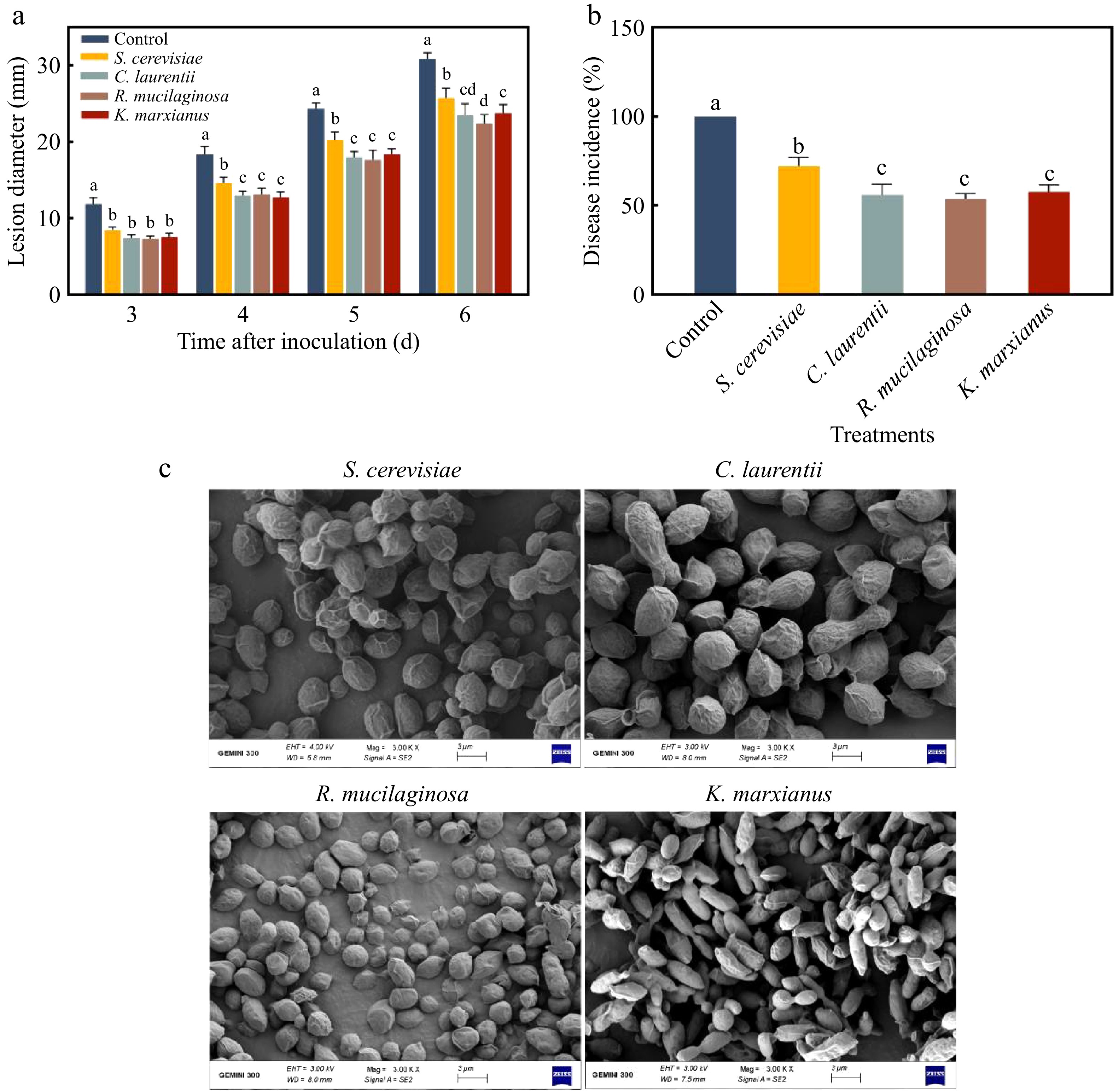
Figure 1.
(a) Lesion diameter and (b) disease incidence of different inactivated yeasts induced resistance to pear blue mold. (c) SEM image of inactivated yeast. Letters indicate significant differences (p < 0.05). Error bars represent mean ± SEM of three replicates.
Effect of yeast cell wall on inducing pear resistance to inhibit P. expansum
-
After choosing 0.5% (w/v) K. marxianus and S. cerevisiae cell wall suspension to treat pear fruit for 24 h, respectively, it was discovered that the diameter and occurrence of lesions in pear fruit treated with K. marxianus cell wall were smaller than those in other groups (Fig. 2a and b). On the fourth and sixth days, the lesion diameters in the S. cerevisiae cell wall group were 13 and 23.8 mm, respectively, whereas in the K. marxianus cell wall group they were 9.2 and 20.8 mm. Similar experiments, such as 1% yeast cell wall as a bioinducer, greatly decreased disease incidence of cherry tomato fruit, which verified the broad-spectrum nature of the yeast cell wall[18]. Figure 2c and d show that the 0.5% (w/v) K. marxianus cell wall has the best inhibitory effect. Figure 2e and f show that lesion width and incidence decreased with treatment time from 2 to 24 h and rose with treatment duration of 36 h. Resistance peaked at 24 h and declined at 36 h, likely due to the transient nature of induced plant defenses. The incorporation of the yeast cell wall into pear fruit may stimulate plant systemic defense signals, such as systemic acquired resistance (SAR). The yeast cell wall could induce the pear fruit to produce a systemic defensive 'memory', protecting the fruit from later infection by pathogenic fungi. To sum up, the treatment concentration of 0.5% (w/v) and the optimal treatment time of 24 h were selected for follow-up experiments.
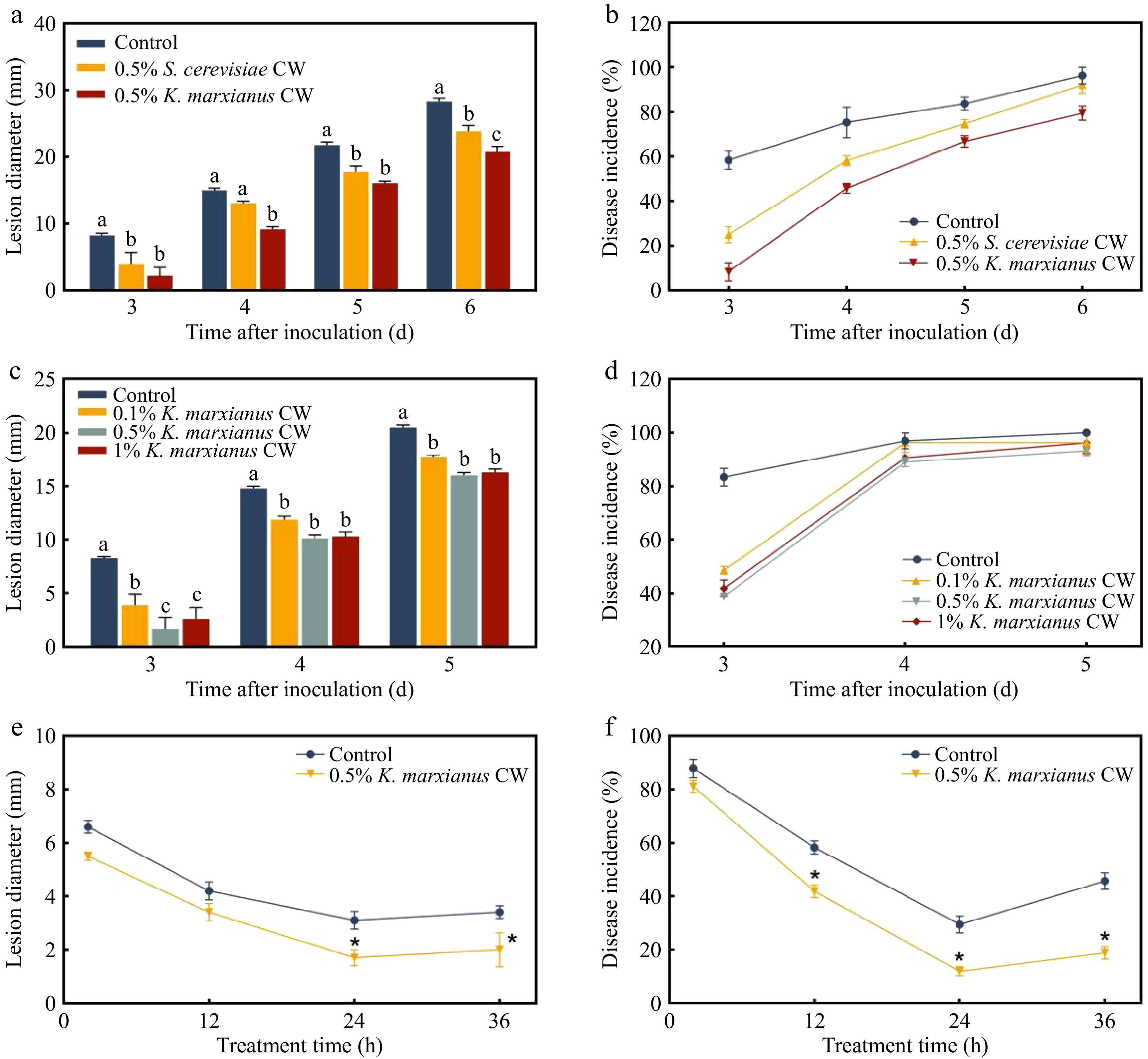
Figure 2.
(a) Lesion diameter and (b) disease incidence of different yeast cell walls induced pear resistance to P. expansum. (c) Lesion diameter and (d) disease incidence of varying concentration gradients of K. marxianus yeast cell wall generated resistance in pear fruit. On day 3, the (e) lesion diameter and (f) disease incidence of pear fruit were subjected to 0.5% (w/v) K. marxianus cell wall at various treatment durations. Control: sterile water; CW: cell wall. Different letters indicate significant differences (p < 0.05). Bars represent mean ± SEM.
Characterization of K. marxianus and S. cerevisiae cell walls
-
The results of HPLC analysis of polysaccharide components and contents in the cell wall are shown in Fig. 3a and b. S. cerevisiae's cell wall polysaccharides included 53.45% mannan, 2.56% chitin, and 43.99% glucan, whereas K. marxianus' cell wall contained 60.37% mannan, 1.29% chitin, and 38.34% glucan (Fig 3c). Both the K. marxianus cell wall and the mixture generated using the K. marxianus cell wall polysaccharide ratio had strong inhibitory effects on blue mold rot. However, the lesion diameters and incidence of pear fruit treated with K. marxianus cell wall were much smaller than those treated with the mixture (Fig. 3d and e). It should be noted that the mass-based comparison between the whole cell wall and isolated polysaccharides does not represent a strictly dose-equivalent system, due to differences in composition and physical state. Therefore, the observed differences mainly reflect functional characteristics rather than quantitative potency. Mannan is non-toxic and contains many health benefits, including balancing gut microorganisms, regulating immunity, and other functions[19]. Chitin has antifungal properties and has been reported as a pattern recognition molecule that recognizes plant receptors and activates the defense system[20]. β-Glucan has been shown to induce plant disease resistance and to enhance the efficacy of biocontrol yeasts against postharvest diseases[21]. Although the chitin and glucan content of the K. marxianus cell wall was slightly lower than that of S. cerevisiae, the cell wall's structure and morphology also played an important role. This was confirmed by the fact that the induction effect of the polysaccharide mixture was significantly lower than that of the K. marxianus cell wall. The structure formed by polysaccharide crosslinking in the cell wall may influence the effect of induced resistance.
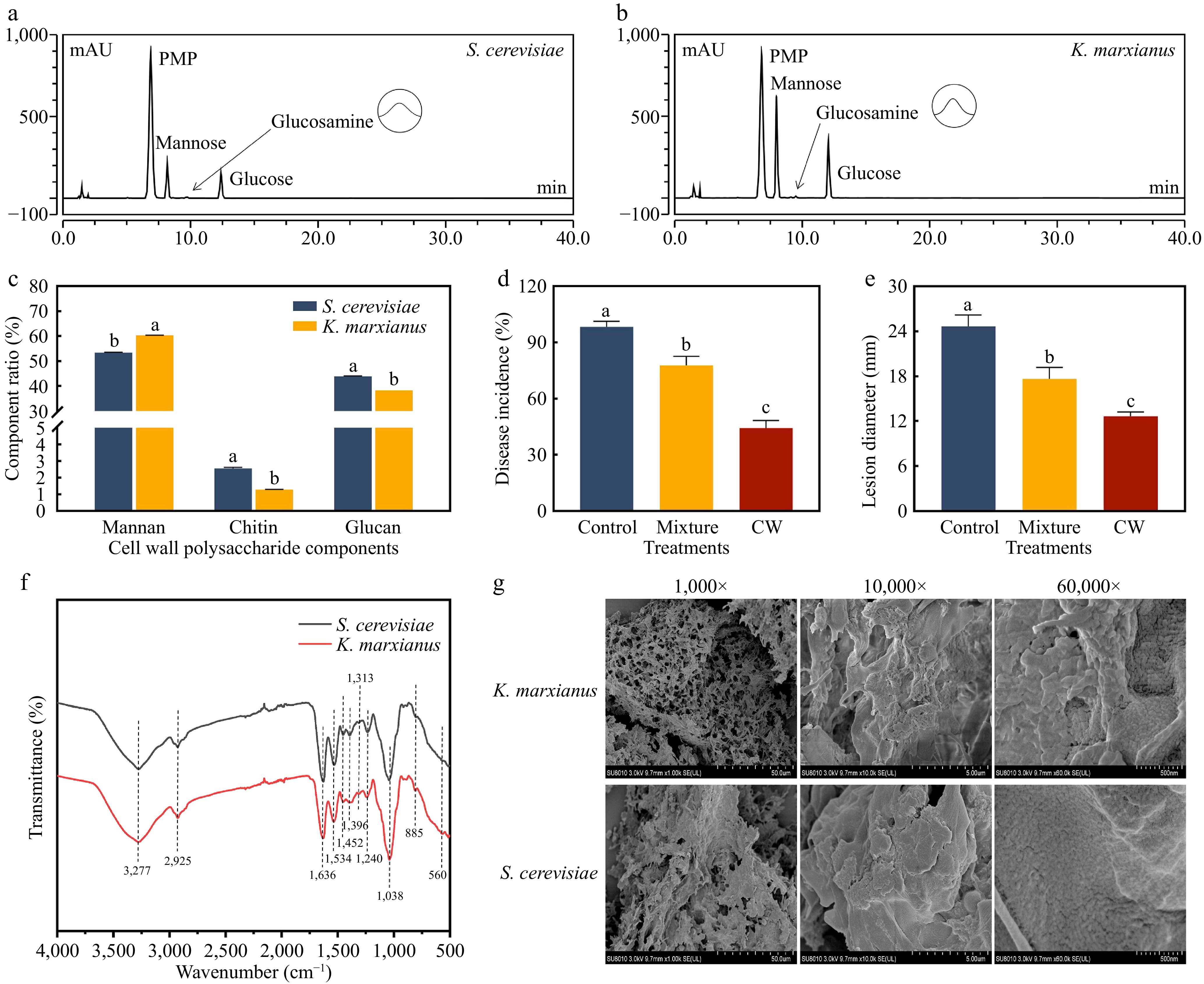
Figure 3.
HPLC chromatograms of cell wall monosaccharides from (a) S. cerevisiae and (b) K. marxianus. (c) Proportion of polysaccharides in the cell walls. On day 5, (d) disease incidence and (e) lesion diameter of 0.5% (w/v) K. marxianus cell wall and its equally proportional polysaccharide mixture induced resistance to pear blue mold. (f) FTIR spectra of the cell walls. (g) SEM images of the K. marxianus and S. cerevisiae cell walls. The letters indicate a significant difference (p < 0.05) by Duncan's multiple-range test. Bars represent the mean ± SEM of three replicates.
The FTIR spectra of the K. marxianus and S. cerevisiae cell walls are shown in Fig. 3f. The wide absorption peak at 3,277 cm−1 was mainly due to the O–H stretching vibration of the intermolecular hydrogen bonds. The absorption peak at 2,925 cm−1 was weaker, which was caused by the saturated C–H stretching vibration. The band at 1,636 cm−1 was mainly due to the C=O stretching vibration of the amide group. The amide II band emerged at 1,534 cm−1, which was the absorption peak for C–N stretching and N–H bending, suggesting that the yeast cell wall composition may contain amino sugars. The 1,452 cm−1 band was mostly driven by CH2 bending and CH3 deformation, whereas the 1,396 cm−1 absorption peak was caused by C–H bending and symmetric CH3 deformation. The signal at 1,313 cm−1 was indicative of amide III. The peak at 1,240 cm−1 was attributable to N–H deformation, while the peak appearing at 1,038 cm−1 was the sugar's C–OH stretching vibration[22]. The signal at 885 cm−1 indicated the existence of a β-glycosidic bond. The absorption band at 560 cm−1 was the stretching vibration of the C–C skeleton of the pyranose ring.
Figure 3g depicts the surface microstructure of the cell walls. After 1,000 times magnification, the K. marxianus cell wall had a more numerous and complex pore structure than the S. cerevisiae cell wall. After magnification of 10,000× and 60,000×, the cell wall surface of K. marxianus revealed a large number of layered folds, while the cell wall surface of S. cerevisiae was smoother. The cell wall is composed of many layers, the innermost of which is a conserved structure, while the other layers are layered on top with some variation[23]. A porous and rough K. marxianus cell wall may increase the contact area with the fruit, so that the outer layer of the cell wall has more receptors to stimulate the fruit and develop fruit resistance more effectively.
ROS levels, antioxidant enzyme activities, and defense-related enzyme activities in pear fruit
-
The green fluorescence intensity of the pear tissues in the spores + 0.5% CW group was the highest, as shown in Fig. 4a. The first and most direct reaction of plants to infections is called oxidative burst, which quickly generates a lot of ROS[24]. ROS is a signaling molecule found in plants that activates the host immune system, increasing plant resistance to infections[25]. The above results indicated that a significant oxidative burst occurred in the pear fruit treated with K. marxianus cell walls, and the ROS levels were noticeably greater than those in the spore group. Therefore, one of the ways that the K. marxianus cell wall may have produced resistance in pear fruit was by inducing an oxidative burst.
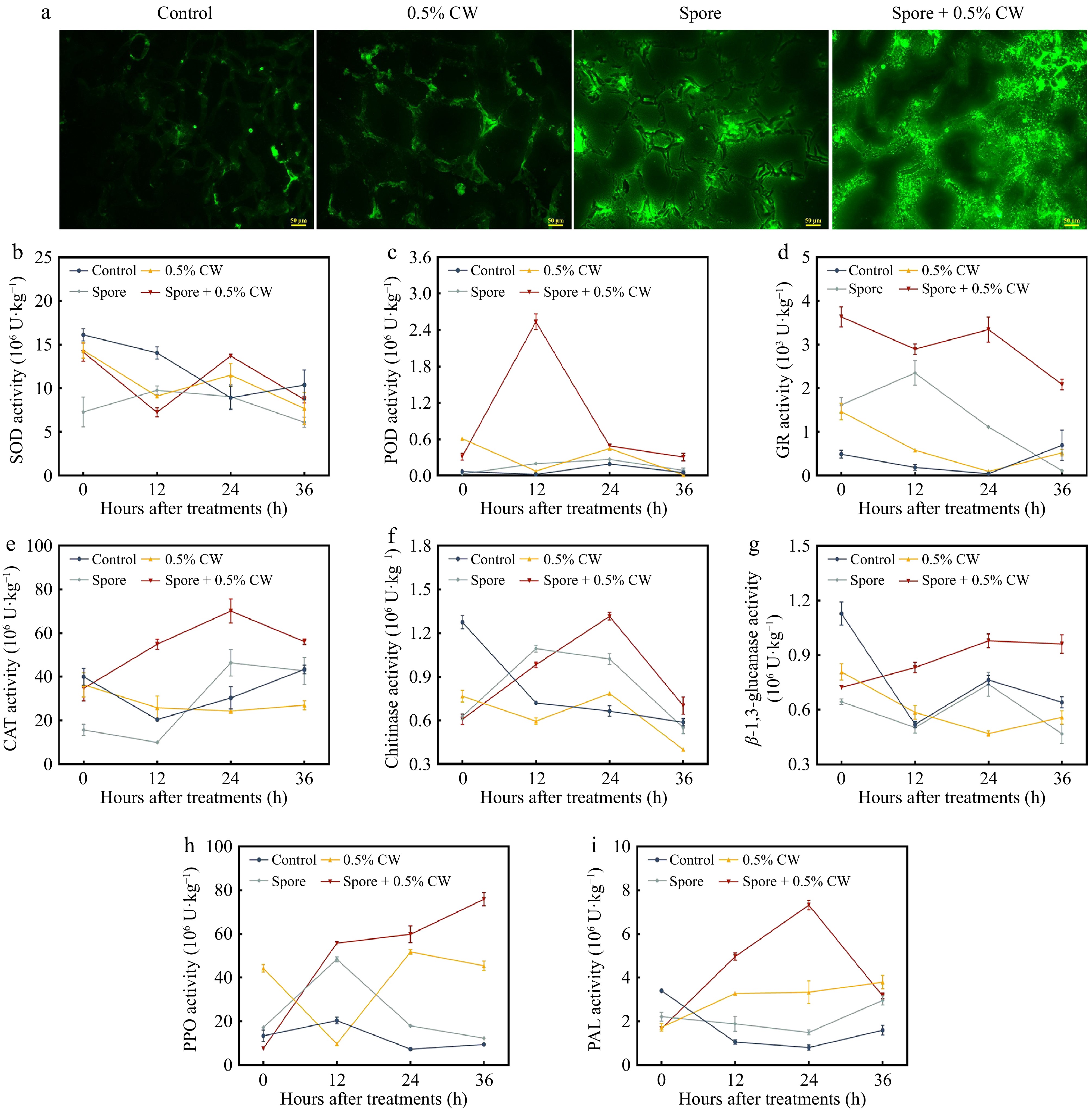
Figure 4.
(a) Reactive oxygen species accumulation in pear fruit after different treatments. Impact of the K. marxianus cell wall on the activities of (b) superoxide dismutase (SOD), (c) peroxidase (POD), (d) glutathione reductase (GR), (e) catalase (CAT), (f) chitinase (CHI), (g) β-1,3-glucanase (GLU), (h) polyphenol oxidase (PPO) and (i) phenylalanine ammonia lyase (PAL) in pear fruit. 0.5% CW: only adding 0.5% (w:v) K. marxianus cell wall-treated pear fruit for 24 h. Different letters indicate significant differences among samples (p < 0.05). Bars represent the mean ± SEM of three independent replicates.
Overproduction of ROS can cause lipid membrane peroxidation, which can harm or even kill cells[26]. In order to maintain a dynamic balance of ROS and mitigate the negative effects caused by excess ROS, plants contain a range of antioxidant enzymes to scavenge ROS[27]. SOD activity in the spore + 0.5% CW group was elevated at 24 h followed by a decrease, whereas POD activity peaked at 12 h and was significantly greater than that of the other groups (Fig. 4b and c). GR and CAT activities in the spore + 0.5% CW group were essentially at a high level during the whole procedure and reached a peak at 24 h (Fig. 4d and e). The antioxidant system's first line of defense is SOD, which catalyzes the conversion of superoxide anion to hydrogen peroxide. CAT then converts hydrogen peroxide to water and oxygen, scavenging ROS and minimizing oxidation-induced cellular damage[28]. POD is also involved in the catabolism of hydrogen peroxide, and an increase in POD activity is thought to be a significant physiological indicator of the development of fruit resistance. GR, a flavoenzyme, catalyzes the conversion of oxidized glutathione (GSSG) to reduced glutathione (GSH), which helps sustain the activity and reducibility of sulfhydryl proteins[29]. After 12 and 24 h of induction by the K. marxianus cell wall, the activity of antioxidant enzymes in fruit showed an obvious response to pathogen infection. Increasing the activities of antioxidant enzymes might be another mechanism by which the K. marxianus cell wall induces resistance in pear fruit.
In the spore + 0.5% CW group, chitinase, β-1,3-glucanase, and PAL activities first rose and subsequently dropped, peaking at 24 h (Fig. 4f, g, and i). PPO activity continued to increase in the spore + 0.5% CW group, while PPO activity in the spore group peaked at 12 h and then declined (Fig. 4h). Pathogenesis-related proteins (PR) indicate plants' defense-induced responses to stress. PR proteins may fight infections, break down cell walls and other macromolecules, and block the binding of pathogen shell proteins to plant receptors[30]. PR proteins, including chitinases and β-1,3-glucanase, may break down glucan and chitin in fungal cell walls and mycelia. The enhanced activity of chitinase and β-1,3-glucanase suggested that the K. marxianus cell wall might promote the formation of SAR in pear fruit. This was triggered to exhibit a greater resistance response when attacked by pathogens. The primary function of PPO is to oxidize phenolic compounds into more toxic quinones, which inhibit and kill pathogenic fungi[31]. PPO activity was dramatically boosted by the K. marxianus cell wall, indicating improved disease resistance. PAL is the key enzyme in the metabolic pathway of phenylpropane and contributes to the creation of resistant compounds such as lignin and phytoalexin[32]. In addition to the creation of resistant substances, the K. marxianus cell wall may also accumulate salicylic acid by boosting PAL activity as a way to increase fruit resistance[33]. To summarize, another way that the K. marxianus cell wall made pears resistant to P. expansum was by increasing the activity of defense-related enzymes.
Effect of K. marxianus cell wall on GABA shunt in pear fruit
-
In the spore + 0.5% CW group, GAD activity increased and reached the greatest level at 36 h (Fig. 5a). At 0 and 12 h, the relative expression of the GAD1 gene was considerably greater in the 0.5% CW and the spore + 0.5% CW groups compared to the control and spore groups (Fig. 5b). The expression of GAD1 in the spore + 0.5% CW group also rose steadily, which was compatible with GAD activation. The GABA-T gene expression in the K. marxianus cell wall group was significantly different from that in spore group at 0 and 12 h (Fig. 5c). The expression of SSADH gene in the K. marxianus cell wall treated groups was higher at 0 h and decreased at 12 h (Fig. 5d). At 36 h, Glu content in treatment groups rose compared to the control group, with the spore group increasing the most (Fig. 5e). However, only the endogenous GABA content in pear fruit after cell wall treatment was significantly increased compared to the control group (Fig. 5g). In the event of insignificant changes in GAD content across all groups (Fig. 5f), the considerable accumulation of GABA in the spore + 0.5% CW group might be attributed to increased GAD activity at 36 h. Furthermore, the lower GABA-T level in the spore + 0.5% CW group showed a reduction in GABA breakdown (Fig. 5h).
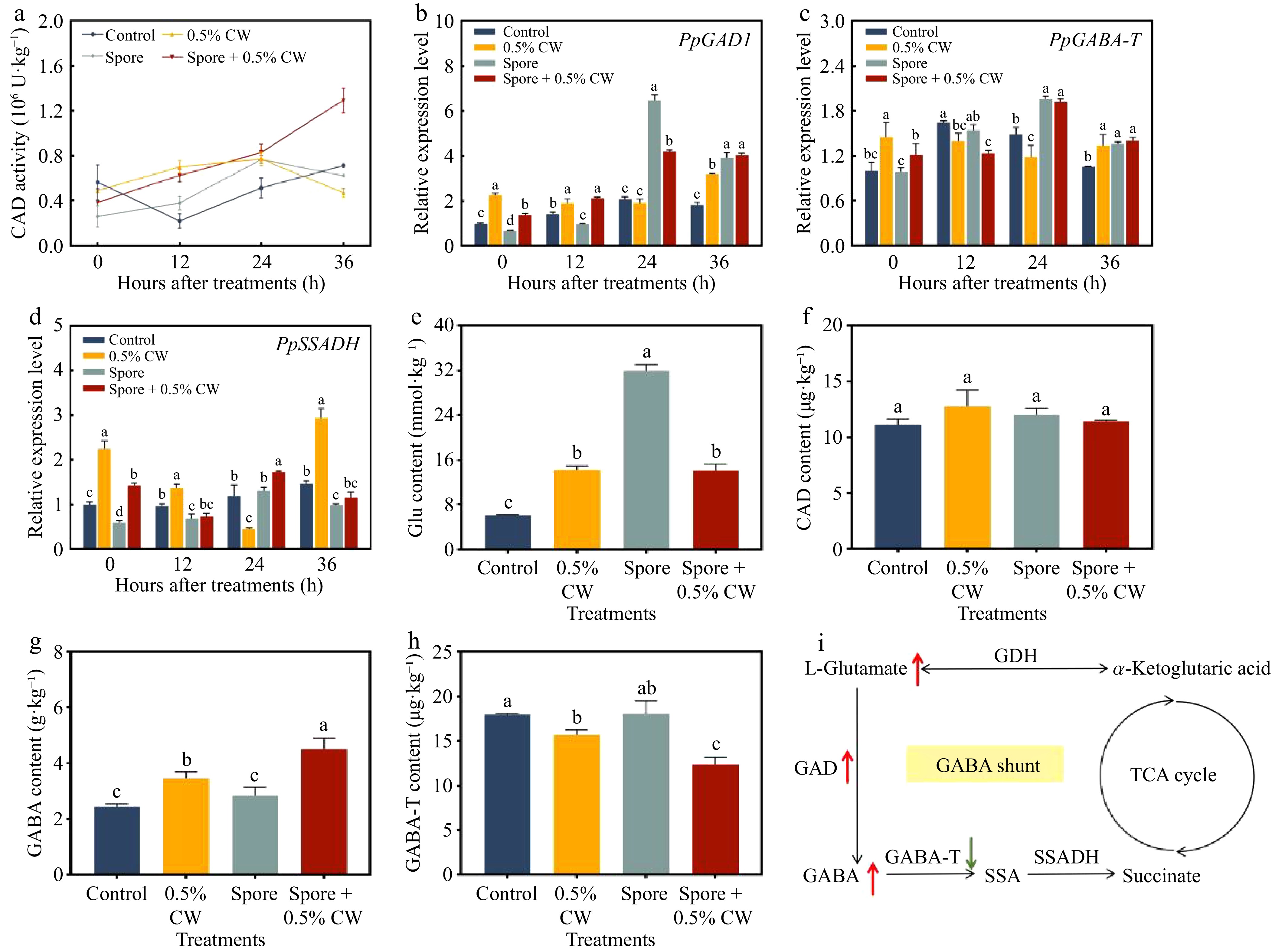
Figure 5.
(a) Activity of glutamate decarboxylase (GAD), (b) gene expression of GAD1, (c) GABA transaminase (GABA-T), and (d) succinic semialdehyde dehydrogenase (SSADH) in pears. (e) Content of L-glutamate (Glu), (f) GAD, (g) GABA, and (h) GABA-T in pear fruit after spore treatment for 36 h. (i) GABA shunt metabolic pathway in pear. GDH: glutamate dehydrogenase; SSA: succinic semialdehyde. Different letters represent significant differences (p < 0.05). Error bars represent mean ± SEM of three independent replicates.
GABA is a commonly accessible non-protein amino acid with important biological functions in plants. As shown in Fig. 5i, the major pathway for GABA synthesis in plants is the GABA shunt[34]. In this pathway, glutamate is irreversibly decarboxylated by glutamate decarboxylase (GAD) to produce GABA, which is subsequently converted into succinic semialdehyde (SSA) by GABA transaminase (GABA-T). SSA is further oxidized to succinate by succinic semialdehyde dehydrogenase (SSADH) in mitochondria, and the resulting succinate enters the tricarboxylic acid (TCA) cycle, which is closely related to cellular energy metabolism[35]. GABA often accumulates under stress conditions and contributes to plant adaptation to biotic and abiotic challenges by regulating cellular pH and carbon–nitrogen balance[36]. Moreover, GABA metabolism has been reported to interact with other stress-response pathways, thereby enhancing plant stress tolerance[37]. The increased GABA accumulation observed after cell wall treatment may therefore be associated with enhanced induced resistance, possibly through its roles in stress metabolism and redox regulation.
Based on the above results, it shows that the K. marxianus cell wall enhanced GABA buildup in pear by increasing glutamate content and GAD activity. Meanwhile, the downregulation of genes that catabolized GABA and decreasing of GABA-T content also allowed GABA to accumulate. Overall, the induction of endogenous GABA accumulation in pear fruit by the K. marxianus cell wall represents a plausible metabolic component of the induced resistance response.
Transcriptomic analysis of pear fruit resistance induced by cell wall-derived β-glucans
-
β-glucan isolated from the yeast cell wall was applied to pear fruit to induce resistance, and the resulting transcriptomic responses are shown in Fig. 6. In the principal component analysis (PCA) plot, glucan-treated pear fruit was clearly separated from the control group (Fig. 6a). Using a threshold of Fold Change ≥ 2 and FDR < 0.01, volcano plot analysis identified 460 differentially expressed genes (DEGs) in glucan-treated pear fruit compared with the control, including 209 upregulated and 251 downregulated genes (Fig. 6b). A total of 328 DEGs in the Control vs Glucan comparison were annotated to KEGG. KEGG pathway enrichment analysis was performed using the KEGG database[38]. As shown in Fig. 6c, among the top 20 pathways with the lowest Q values, plant hormone signal transduction and plant–pathogen interactions were significantly enriched (p < 0.05, q < 0.05) and were closely related to induced resistance in pear fruit. After β-glucan treatment, genes involved in plant hormone signaling in pear fruit were significantly altered (Fig. 6d). In the brassinosteroid (BR) signaling pathway, the BR receptor gene At5g66900 encoding BRI1 (log2FC = 1.3), the co-receptor gene LRK10 encoding BAK1 (log2FC = 2.6), and the BR signaling kinase gene BSK5 (log2FC = 1.4) were all significantly upregulated. In the jasmonic acid (JA) signaling pathway, the gene LOC103957049 encoding jasmonic acid–isoleucine synthetase (JAR1) was upregulated (log2FC = 1.7). In contrast, no differentially expressed genes were enriched in the salicylic acid (SA) signaling pathway. These results indicate that β-glucan enhances pear fruit resistance mainly through activation of the BR and JA signaling pathways, rather than the SA pathway. Plant hormones play central roles in mediating induced resistance in fruit by regulating defense-related signaling pathways. BRs are polyhydroxylated plant steroid hormones involved in plant growth, development, and stress responses[39]. In the BR signaling pathway, β-glucan treatment enhanced the expression of the BR receptors BAK1 and BRI1, which was accompanied by increased expression of the downstream signaling component BSK, suggesting activation of BR-mediated signaling. The induction of JAR1 by β-glucan is likely to enhance the conversion of JA to JA-Ile, contributing to the activation of JA-mediated signaling.
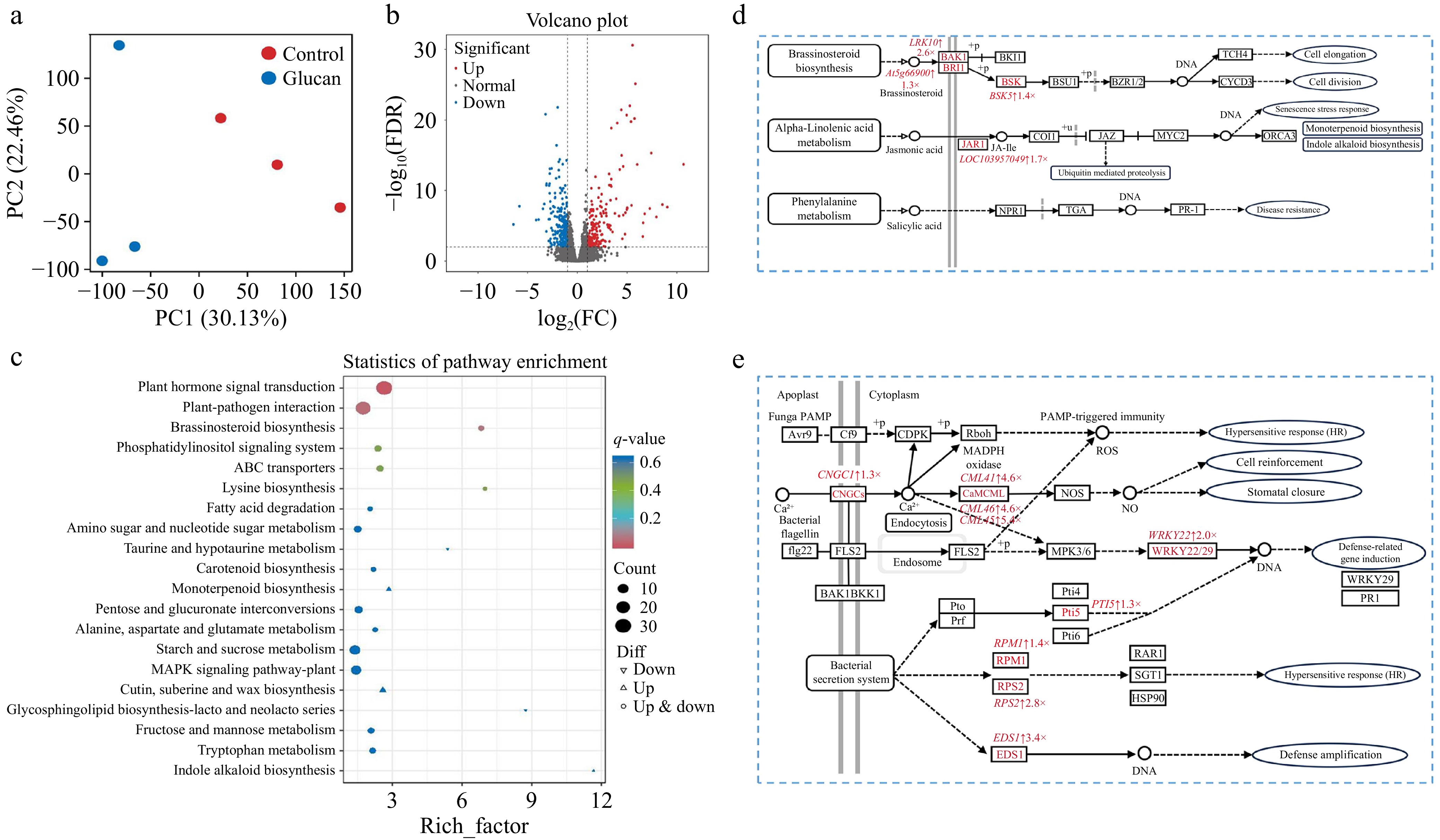
Figure 6.
Transcriptomic analysis of β-glucan-induced resistance in pear fruit. (a) PCA plot of all samples. (b) Volcano plot of differentially expressed genes. (c) KEGG pathway enrichment analysis of control vs. β-glucan. (d) Plant hormone signal transduction pathways. (e) Plant–pathogen interaction pathways. Red indicates upregulated genes. Light red represents genes with 1 ≤ log2FC < 2, while dark red represents genes with log2FC ≥ 2. Pathway maps were generated based on the KEGG database and reproduced with permission.
Calcium ions (Ca2+) function as key second messengers in plants and play essential roles in plant–pathogen interactions. Compared with the control, β-glucan treatment markedly upregulated immune-responsive genes in pear fruit (Fig. 6e). In the Ca2+ signaling pathway, the ion channel gene CNGC1 (log2FC = 1.3) was induced, likely facilitating Ca2+ influx and increasing cytosolic Ca2+ levels. Consistently, genes encoding Ca2+ sensors, including CML46 (log2FC = 4.6), CML41 (log2FC = 4.6), and CML45 (log2FC = 5.4), were strongly upregulated, indicating activation of Ca2+-dependent signaling. Upon pathogen recognition, dynamic changes in cytosolic Ca2+ levels act synergistically with ROS to trigger pattern-triggered immunity (PTI), which can subsequently initiate the hypersensitive response (HR). HR is characterized by localized programmed cell death that restricts pathogen colonization and further spread. Signals generated during HR can be transmitted to distal tissues, thereby activating systemic acquired resistance (SAR) and establishing a systemic defense network that enhances plant resistance to pathogens[40,41]. In agreement with this immune model, Ca2+ signals induced by β-glucan were further transduced through the MAPK cascade, leading to the upregulation of the transcription factor WRKY22 (log2FC = 2.0) and the transcriptional regulator PTI5 (log2FC = 1.3), which promotes PR protein accumulation and strengthens defense responses in pear fruit. In addition, β-glucan significantly induced the expression of resistance-related genes RPM1 (log2FC = 1.4), RPS2 (log2FC = 2.8), and EDS1 (log2FC = 3.4), suggesting reinforcement of immune signaling and HR activation. RPM1, as a representative resistance protein, recognizes pathogen effectors and triggers effector-triggered immunity (ETI). PTI is known to potentiate ETI responses, and together these two immune layers coordinately contribute to effective defense against diverse pathogens[42].
Transcriptomic analysis of cell wall-derived mannan-induced resistance in pear fruit
-
Figure 7 shows the results of transcriptomic analysis of pear fruit resistance induced by mannan isolated from the yeast cell wall. In the PCA plot, principal component 1 (PC1) accounted for 38.48% of the total variance and clearly distinguished mannan-treated pear fruit from the control group (Fig. 7a). The volcano plot in Fig. 7b shows that, compared with the control, 2,216 genes were differentially expressed in response to mannan treatment, with 1,276 upregulated and 940 downregulated genes. In the Control vs. Mannan comparison, a total of 1,560 DEGs were annotated to the KEGG database. Four pathways were significantly enriched and closely associated with induced resistance, ranked in descending order of significance: plant–pathogen interaction, plant hormone signal transduction, starch and sucrose metabolism, and the MAPK signaling pathway (Fig. 7c). Following induction with mannan derived from K. marxianus, pathogen-responsive genes were markedly upregulated in pear fruit (Fig. 7d). Representative genes encoding LRR receptor-like kinases, including XA21 (log2FC = 1.4) and the BAK1/BKK1-associated kinase LECRK3 (log2FC = 2.4), were induced, indicating enhanced immune perception. In parallel, the cation channel gene CNGC1 (log2FC = 2.7) was markedly upregulated, suggesting activation of Ca2+ influx. Consistent with downstream signal transduction, the MAPK cascade component MKK4 (log2FC = 1.2) and the transcription factor WRKY22 (log2FC = 1.7) were significantly induced. As a consequence, defense-related genes, including IOS1 (log2FC = 4.2) and the PR protein gene PRB1 (log2FC = 2.8), showed strong upregulation. In addition, key resistance-associated genes involved in effector-triggered immune signaling, such as PTI5 (log2FC = 1.9) and SAG101 (log2FC = 3.4), were also induced, suggesting reinforcement of localized hypersensitive defense responses. Notably, mannan-triggered immune responses followed a largely similar defense framework to those induced by β-glucan, including Ca2+ influx, activation of Ca2+–ROS signaling, PTI initiation, and HR-associated defenses. However, mannan displayed distinct immunomodulatory features by preferentially enhancing the expression of LRR-type receptor kinases (e.g., PK1, XA21, LRK10), Ca2+-dependent protein kinases (CDPKs), and the FLS2–MKK4/5–WRKY22/65 signaling module, leading to stronger induction of downstream defense genes such as IOS1 and PR proteins. These results suggest that, while β-glucan and mannan activate overlapping immune pathways, mannan may exert a more pronounced effect on receptor-mediated recognition and MAPK-dependent transcriptional reprogramming, thereby contributing to a partially distinct yet complementary immune activation profile in pear fruit. In addition, mannan treatment activated multiple phytohormone signaling pathways in pear fruit (Fig. 7e). In the ethylene (ETH) pathway, the kinase MKK4 (log2FC = 1.2) and the downstream transcription factor ERF1B (log2FC = 1.8) were significantly upregulated. In the BR pathway, the receptor BRI1 (log2FC = 2.8) and the co-receptor BAK1 (LECRK3, log2FC = 2.4) were induced, together with increased expression of the signaling kinase BSK5 (log2FC = 1.8) and the cell wall-modifying enzyme XTH22/TCH4 (log2FC = 1.5), suggesting reinforcement of cell wall integrity. In the JA pathway, the regulator TIFY10B (log2FC = 2.0) and the JA-responsive transcription factor MYC2 (BHLH35, log2FC = 2.4) were markedly upregulated. Notably, mannan uniquely activated the SA signaling pathway, as evidenced by the induction of NPR1 (log2FC = 1.1), TGA10 (log2FC = 2.9), and the downstream pathogenesis-related gene PRB1/PR1 (log2FC = 2.8), indicating the establishment of SAR. In contrast to β-glucan, which primarily activated BR- and JA-associated signaling without a pronounced SA response, mannan elicited a broader hormonal defense profile, characterized by the concurrent activation of the ETH, BR, JA, and SA pathways. Together, these findings indicate that although β-glucan and mannan activate partially overlapping hormone-regulated defense pathways, mannan confers a more comprehensive and multilayered immune response in pear fruit.
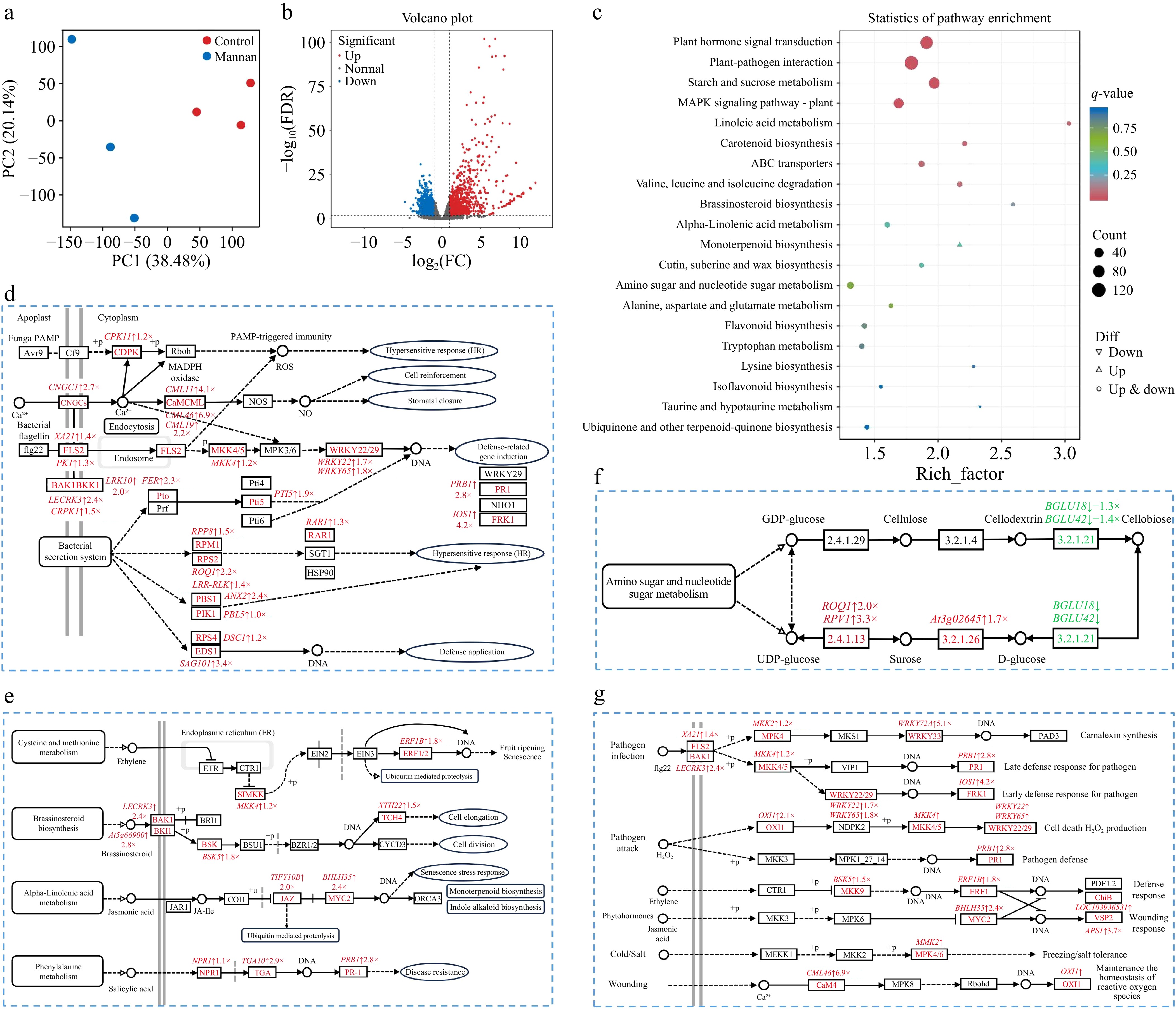
Figure 7.
Transcriptomic analysis of mannan-induced resistance in pear fruit. (a) PCA plot and (b) volcano plot of differentially expressed genes. (c) KEGG pathway enrichment analysis of control vs mannan. (d) Plant–pathogen interaction pathways. (e) Plant hormone signal transduction pathways. (f) Starch and sucrose metabolic pathways. (g) MAPK signalling pathways. Red indicates upregulated genes and green indicates downregulated genes. Pathway maps were generated using the KEGG database and modified with permission.
As shown in Fig. 7f, mannan treatment markedly altered carbohydrate metabolism in pear fruit. Compared with the control, mannan treatment significantly downregulated the genes encoding the rate-limiting β-glucosidases involved in cellulose hydrolysis, BGLU42 (log2FC = −1.3) and BGLU18 (log2FC = −1.4), in pear fruit. In contrast, genes encoding sucrose synthase (ROQ1, log2FC = 2.0; RPV1, log2FC = 3.3) and invertase (At3g02645, log2FC = 1.7), which catalyze the conversion of sucrose into glucose and fructose, were upregulated. These transcriptional changes are likely to favor glucose accumulation, thereby enhancing energy production through sugar metabolism to support defense responses in pear fruit[43].
Figure 7g shows that MAPK signaling was activated in pear fruit following mannan treatment. Following the induction of the FLS2–BAK1 receptor complex, downstream MAPKs, including MMK2 (log2FC = 1.2) and MKK4 (log2FC = 1.2), were upregulated. Signal amplification through the MAPK pathway led to strong induction of WRKY33 (WRKY72A, log2FC = 5.1), together with WRKY22 (log2FC = 1.7) and WRKY65 (log2FC = 1.8), resulting in increased expression of defense-related genes such as PRB1 and IOS1. These changes are associated with enhanced phytoalexin biosynthesis and accumulation of defense proteins, thereby strengthening both early- and late-stage immune responses. In addition, the upregulation of MMK2 suggests a potential contribution to improved stress tolerance, including cold and salt resistance. Consistent with pathogen and wound responses, mannan treatment also promoted ROS-associated signaling, as reflected by increased expression of OXI1 (log2FC = 2.1) and the Ca2+ sensor CML46 (log2FC = 6.9), which are involved in ROS production and redox homeostasis. Moreover, activation of the MAPK cascade in response to mannan treatment coincided with the induction of ethylene (ETH)- and jasmonic acid (JA)-responsive transcription factors, including ERF1B (log2FC = 1.8) and MYC2 (BHLH35, log2FC = 2.4). This was followed by increased expression of downstream defense effector genes such as ChiB (log2FC = 1.2) and VSP2/APS1 (log2FC = 3.7), which together contribute to enhanced pathogen defense and wound responses in pear fruit. Overall, mannan-induced resistance in pear fruit was orchestrated by MAPK signaling, which functioned as a central hub integrating phytoalexin-related defense metabolism, activation of resistance genes, ROS regulation, and phytohormone-mediated defense responses.
Although both mannan and glucan extracted from K. marxianus cell walls effectively induced resistance in fruit, transcriptomic analysis revealed that they activated distinct regulatory pathways, indicating differences in their underlying mechanisms. These differences are likely related to their structural characteristics and monosaccharide composition, which may influence how they are perceived by host receptors and how downstream defense signaling is triggered. Therefore, the resistance-inducing efficacy of yeast cell walls may depend on the relative abundance and structural organization of their major polysaccharide components, rather than a single universal mechanism.
-
This study demonstrates that the cell wall of K. marxianus exhibits a superior capacity to enhance resistance in pear fruit against P. expansum infection, thereby significantly reducing the incidence of blue mold, compared to that of S. cerevisiae. The enhanced biocontrol efficacy can be attributed to its distinct biochemical composition, characterized by a specific ratio of glucan, mannan, and chitin, coupled with a unique porous architecture. Application of K. marxianus cell wall elicits a coordinated defense response, initiating an oxidative burst, elevating the activity of key antioxidant and defense-related enzymes, and stimulating the accumulation of γ-aminobutyric acid via the GABA shunt pathway. Transcriptomic analyses further elucidated the underlying mechanisms, revealing that the constituent β-glucan and mannan synergistically activate core defense pathways, including phytohormone signaling and plant–pathogen interactions. Notably, mannan additionally modulates carbohydrate metabolism to fuel defense responses and amplifies resistance through the MAPK signaling cascade. These findings establish K. marxianus cell wall as a potent and scientifically grounded, food-grade elicitor for postharvest disease management. Future research should focus on optimizing its formulation and application protocols, and exploring its efficacy across a broader range of fruit-pathogen systems to facilitate its development into a practical and sustainable alternative to conventional fungicides.
We thank Huang Qiong from the Core Facilities, Zhejiang University School of Medicine for their technical support.
-
The authors confirm their contributions to the paper as follows: conceptualization: Wu Y, Yu T; methodology: Wu Y, Yan F; formal analysis, investigation, visualization, writing − original draft: Wu Y; resources: Chen XA; supervision, project administration, writing − review and editing: Yan F. All authors reviewed the results and approved the final version of the manuscript.
-
The datasets generated during and/or analyzed in the current study are available from the corresponding author on reasonable request.
-
The authors declare that they have no known competing financial interests or personal relationships that could have appeared to influence the work reported in this paper.
-
accompanies this paper online at: https://doi.org/10.48130/fia-0026-0037.
- Supplementary Table S1 The primers used in the pear fruit experiment.
- Supplementary Fig. S1 Lesion diameter (A) and disease incidence (B) of pear fruit after simultaneous treatment with inactivated yeast and pathogen inoculation or after inactivated yeast pre-treatment for 24 h before inoculation.
- Copyright: © 2026 by the author(s). Published by Maximum Academic Press on behalf of China Agricultural University, Zhejiang University and Shenyang Agricultural University. This article is an open access article distributed under Creative Commons Attribution License (CC BY 4.0), visit https://creativecommons.org/licenses/by/4.0/.
-
About this article
Cite this article
Wu Y, Chen XA, Yu T, Yan F. 2026. Efficacy and mechanistic insights of Kluyveromyces marxianus cell wall in enhancing pear fruit resistance against Penicillium expansum. Food Innovation and Advances 5(3): 444−456 doi: 10.48130/fia-0026-0037 

Efficacy and mechanistic insights of Kluyveromyces marxianus cell wall in enhancing pear fruit resistance against Penicillium expansum
- Received: 27 January 2026
- Revised: 04 June 2026
- Accepted: 10 June 2026
- Published online: 02 September 2026
Abstract: Postharvest fungal infestation poses a significant threat to fresh fruit, leading to substantial economic losses. This study investigated the efficacy and mechanism of yeast cell walls in inducing fruit resistance, with a focus on extracts from Kluyveromyces marxianus. Compared to the commonly referenced Saccharomyces cerevisiae cell wall, application of the K. marxianus cell wall provided superior protection in pear fruit against Penicillium expansum infection, markedly reducing disease incidence. This enhanced biocontrol activity is attributed to its distinct polysaccharide composition, characterized by 60.37% mannan, 38.34% glucan, and 1.29% chitin, and its unique structural properties. The treatment triggered a rapid wound oxidative burst, upregulated key antioxidant and defense-related enzymes, and promoted the accumulation of γ-aminobutyric acid in pear tissue. Transcriptomic analysis revealed that β-glucan and mannan components synergistically activated plant hormone signaling and plant–pathogen interaction pathways. Notably, mannan specifically modulated carbohydrate metabolism and MAPK-associated defense responses. These findings establish the K. marxianus cell wall as a potent and novel biocontrol agent, offering a sustainable strategy for managing postharvest decay through the targeted induction of fruit innate immunity.
-
Key words:
- Disease resistance /
- Blue mold /
- Transcriptomics /
- Glucan /
- Mannan