












-
Oxidative stress, characterized by an imbalance between reactive oxygen species (ROS) production and the cellular antioxidant capacity, arises when ROS levels exceed the system's ability to neutralize them[1]. Elevated ROS concentrations can cause nonspecific damage to proteins, lipids, and nucleic acids, and are considered critical in the pathogenesis of various diseases, including cancer, neurodegenerative disorders, cardiovascular conditions, metabolic syndromes, and inflammatory ailments[2−5]. It is widely recognized that dietary antioxidants, such as vitamins C and E, contribute to protection against ROS-mediated damage[6,7]. The United States Food and Drug Administration (FDA) classifies small-molecule antioxidants into two categories: direct and indirect antioxidants[8]. The direct antioxidant capacity of dietary extracts is commonly assessed using assays such as the oxygen radical absorbance capacity (ORAC)[9]. However, the extent to which these direct antioxidant activities function effectively in vitro and in vivo remains unclear. In contrast, indirect antioxidants exert their effects primarily by activating the Kelch-like ECH-associated protein 1-nuclear factor erythroid 2-related factor 2-antioxidant response element (Keap1-Nrf2-ARE) pathway[10,11]. This activation leads to the transcriptional upregulation of a suite of cytoprotective proteins, also known as phase II enzymes[12]. Under basal conditions, Nrf2 is kept inactive through its binding to the cytoskeletal anchor protein Keap1. Upon oxidative stress, Nrf2 dissociates from Keap1, translocates to the nucleus, and binds to AREs, thereby promoting the expression of phase II detoxifying enzymes such as heme oxygenase-1 (HO-1), glutathione S-transferase (GST), and Nicotinamide adenine dinucleotide (phosphate) hydrogen (NAD[P]H) quinone oxidoreductase 1 (NQO1)[13,14]. Therefore, when Nrf2 is activated, either directly or indirectly, it enhances the expression of a network of intrinsic cytoprotective genes. Such enhanced expression may help lower the risk of cancer and other chronic degenerative diseases that are closely associated with oxidative stress, electrophilic toxicity, and inflammation[14,15].
Numerous studies suggest that dietary components can act as indirect antioxidants. These compounds are believed to form covalent adducts with cysteine sulfhydryl groups in Keap1 through oxidation or alkylation reactions. Although electrophilicity, defined as the ability of a molecule to accept electrons and form covalent bonds with nucleophilic centers such as thiol groups in proteins, is common among most known ARE inducers, not all electrophiles can regulate ARE activity[16]. Representative indirect antioxidants from plants include sulforaphane in broccoli, curcumin in ginger, luteolin in celery, and chalcones from green perilla leaves[17−21]. Marine organisms, including fish, shellfish, and algae, also provide important sources of indirect antioxidants. For example, astaxanthin, a natural keto-carotenoid present in algae, shrimp, and salmon, has been demonstrated to enhance cellular resistance to oxidative stress via the Keap1-Nrf2-ARE signaling pathway[22,23]. In earlier work, our group isolated the antioxidant phenolic compound 3,5-dihydroxy-4-methoxybenzyl alcohol (DHMBA) from Pacific oyster (Crassostrea gigas), which showed protective effects against oxidation-related diseases[24,25]. Sea mussel extract, traditionally appreciated for its high marine lipid content, is likewise linked to several health benefits. Fish oil, known for its abundance of EPA and DHA, is commonly marketed as a functional food supporting health[26]. Meanwhile, other bioactive components from sea mussel, such as an antioxidant pentapeptide obtained from Mytilus edulis hydrolysate and the polysaccharide α-D-glucan from Mytilus coruscus, are being developed as functional ingredients[27−29]. Nevertheless, no Nrf2-ARE activators originating from sea mussels have been reported so far.
HepG2 cells are widely used as an in vitro model for studying hepatic oxidative stress and Nrf2 pathway activation due to their retained expression of key phase II enzymes and responsiveness to ARE inducers. However, it should be noted that HepG2 cells exhibit certain metabolic alterations compared to primary human hepatocytes, such as reduced cytochrome P450 activity and altered transporter expression, which may influence the extent of xenobiotic metabolism and cellular responses. Nevertheless, this model remains a well-accepted and informative platform for initial mechanistic screening of natural products with antioxidant potential.
In the present study, we demonstrated that the ethanol (EtOH) extract of sea mussel protects HepG2 cells against 2,2'-Azobis(2-amidinopropane) dihydrochloride (AAPH)-induced oxidative stress by activating the Keap1-Nrf2-ARE signaling pathway. These findings indicate that sea mussel EtOH extract and its subfractions may serve as a valuable food-derived material with potential for ameliorating liver-related diseases.
-
All chemicals were of analytical grade.
Extract preparation and bio-guided isolation of bioactive compounds
-
Fresh sea mussels (M. edulis), 15 kg, were purchased from Iwate Prefecture, Yamane Co. The edible part (5.5 kg) of the mussel was carefully obtained and mashed into pieces, then homogenized with a total of 14.5 L EtOH (99.5% purity) by shaking three times for 24 h. After filtration, the extracts were concentrated and evaporated to dryness using an evaporator and freeze-dryer. The sea mussel extract freeze-dried powder (373.4 g) was stored at −80 °C for further experiments. All bioactivity assays described in this study were performed within 2 years of extraction; prior to each experiment, the stored powder was freshly reconstituted, and the reproducibility of bioactivity was routinely confirmed by repeated ARE-driven luciferase reporter assays.
For bioactivity-guided fractionation, the extract was separated using silica gel (Kanto Chemical) open-column chromatography (8.5 cm × 67 cm) gradient mobile phase was used: hexane/ethyl acetate ratio = 100:0, 9:1, 8:2, 7:3, 6:4, 5:5, 4:6, 3:7, 2:8, 1:9, 0:10; ethyl acetate/methanol ratio = 5:5 and methanol 100%. Preliminary fractionation of the extract resulted in eight fractions (Fr. 1–8) based on bioactivity-guided fractionation.
For chemical profiling of the active subfractions, HPLC analysis was performed on an Agilent 1260 Infinity system equipped with a ZORBAX Eclipse XDB-C18 column (4.6 mm × 250 mm, 5 μm). The mobile phase consisted of acetonitrile (A) and 0.1% formic acid in water (B) at a flow rate of 1.0 mL/min with gradient elution as follows: 0–5 min, 5% A; 5–30 min, 5%–95% A; 30–35 min, 95% A; 35–40 min, 95%–5% A. The column temperature was maintained at 30 °C, and the eluate was monitored at 210 nm. Due to the limited sample quantity and the complexity of the subfractions, detailed chemical profiling was performed only for Fr. 5.
Cell culture
-
HepG2 cells were purchased from the RIKEN Bioresource Centre Cell Bank (Tsukuba, Japan). The cells were maintained in high-glucose Dulbecco's Modified Eagle's Medium (DMEM, Wako, Osaka, Japan) supplemented with 10% (v/v) fetal bovine serum (FBS, Thermo Fisher Scientific, Canada) and an antibiotic mixture of penicillin-streptomycin (Wako, Osaka, Japan), and incubated at 37 °C in a humidified 5% CO2 atmosphere.
Cell viability assay
-
HepG2 cells (4 × 10⁴ cells/well) were seeded into 96-well plates for 24 h. Cell viability was evaluated using the CCK-8 assay (Dojindo Molecular Technologies) according to the manufacturer's protocol. Samples at appropriate concentrations were dissolved in water and applied to cells for 24 h. After culturing, the cells were treated with 100 μL of DMEM and 10 μL of CCK-8 reagent for 2 h solubilized with isopropanol. The absorbance was measured at 450 nm with a Wallac 1420 ARVO Mx plate reader (PerkinElmer, Tokyo).
Cytoprotective assay
-
To measure cell viability, HepG2 cells (4 × 10⁴ cells/well) were seeded into a 96-well plate. Oxidative stress was induced by incubating cells with the free radical generator AAPH (16 mM) in cell medium for 24 h. Test samples at various concentrations were pre- or co-treated with AAPH. Cell viability was evaluated using the CCK-8 assay (Dojindo Molecular Technologies) as described above.
Measurement of intracellular ROS accumulation
-
Intracellular ROS accumulation in HepG2 cells (4 × 10⁴ cells/well) was measured using the fluorescent probe 2',7'-dichlorofluorescein diacetate (DCF-DA) (Sigma). Briefly, HepG2 cells (4 × 10⁴ cells/well) were seeded into a dark 96-well plate and pretreated with or without test extracts. Oxidative stress was induced by incubating the cell medium with 16 mM AAPH for 24 h. At the end of incubation, the cells were stained with DCF-DA (25 μM) for 45 min at 37 °C. The fluorescence intensity was measured at excitation and emission wavelengths of 485 and 535 nm, respectively.
ORAC assay
-
The oxygen radical scavenging capacity was measured using the ORAC assay. All procedures were performed according to a previously described protocol with minor modifications[29,30]. Briefly, the sea mussel EtOH extract was lyophilized to dryness, and the resulting powder was dissolved in 75 mM phosphate buffer (pH 7.4) at final concentrations of 1.0, 2.0, and 4.0 mg/mL for the determination of relative ORAC values. A reaction mixture consisting of 20 μL of sample solution or Trolox standard, 200 μL of fluorescein solution (94.4 nmol/L), and 75 μL of AAPH solution (31.7 mmol/L) was incubated at 37 °C for 1.5 h in a Wallac 1420 ARVO Mx plate reader (PerkinElmer, Tokyo). Fluorescence decay was recorded at 10-s intervals with excitation and emission wavelengths of 493 and 515 nm, respectively. Trolox solutions at concentrations of 6.25, 12.5, 25, and 50 μM were used to generate a standard curve. The absolute ORAC value at 1.0 mg/mL was expressed as micromoles of Trolox equivalent per gram of dry extract (μmol TE/g extract, dry weight), and the relative ORAC values at different concentrations were expressed as fold change relative to the control. The data are presented as mean ± SD from three independent experiments performed in triplicate.
Luciferase reporter gene assay
-
The assay for the reporter gene assay has been described previously[31]. In brief, HepG2 cells (4 × 10⁴ cells/well) were seeded into 96-well white plates for 24 h. We transfected pGL4.37 (luc2p/ARE/hygro) and pGL4.75 (hRluc/CMV) (an internal control for transfection efficiency) vectors at a 20:1 mass ratio using Lipofectamine 3000 (Life Technologies, Tokyo) according to the manufacturer's protocol. After 24 h of transfection, the transfection reagent was removed. Then, cells were treated with different concentrations of samples for another 24 h. Luciferase activity was assayed using the dual luciferase system (Promega, Madison, WI, USA) according to the manufacturer's protocol. DHMBA was used as a positive control (PC).
RT-PCR analysis
-
HepG2 cells at 6 × 10⁵ cells/well in DMEM were seeded into a 12-well plate for 24 h. Cells were harvested by scraping after samples were added to the cells for 24 h. Total RNA was extracted and purified using the RNeasy Mini Kit (Qiagen, Texas, USA) according to the manufacturer's protocol and quantified by measuring the absorbance at 260 nm. For reverse transcription-polymerase chain reaction (RT-PCR), first-strand cDNA was synthesized from 1.0 μg of total RNA using the ReverTra Ace qRT-PCR Master Mix kit (Toyobo, Tokyo) with random primers.
Real-time PCR reactions were performed on an ABI 7500 real-time PCR system (Applied Biosystems), using SYBR® Green PCR Master Mix (Applied Biosystems). The amplification reactions were performed as follows: one cycle of 30 sec at 95 °C, followed by 40 cycles at 94 °C for 5 s (denaturation), 60 °C for 30 s (annealing), and 72 °C for 30 s (extension). The target gene was then determined via normalization to that of GAPDH. GAPDH: forward primer 5′-GAA GGT GAA GGT CGG AGT C-3′, reverse primer 5′-GAA GAT GGT GAT GGG ATT TC-3′. Primers for the target gene were listed as below, HO-1: forward primer 5′-CAG GCA GAG GGT GAT AGA AGA G-3′, reverse primer 5′-GGA GCG GGT GTT GAG TGG-3′; NQO1: forward primer 5′-AAG GAT GGA AGA AAC GCC TGG AGA-3′, reverse primer 5′-GGC CCA CAG AAA GGC CAA ATT TCT-3′; glutamate-cysteine ligase catalytic subunit (GCLC): forward primer 5′-GTG GAT GTG GAC ACC AGA TG-3′, reverse primer 5′-GCG ATA AAC TCC CTC ATC CA-3′; glutamate-cysteine ligase modifier subunit (GCLM): forward primer 5′-GGC ACA GGT AAA ACC AAA TAG TAA C-3′, reverse primer 5′-CAA ATT GTT TAG CAA ATG CAG TCA-3′; glutathione peroxidase 1 (GPx1): forward primer 5′-CCC TCT GAG GCA CCA CGG T-3′, reverse primer 5′-TAA GCG CGG TGG CGT CGT-3′; glutathione reductase (GR): forward primer 5′-AAC AAC ATC CCA ACT GTG GTC-3′, reverse primer 5′-CCA TAT TTA TGA ATG GCT TCA TCT T-3′; superoxide dismutase 1 (SOD1): forward primer 5′-AGG GCA TCA TCA ATT TCG AG-3′, reverse primer 5′-TGC CTC TCT TCA TCC TCC TTT GG-3′. The primer sequences are according to Joko et al.[31].
Western blot
-
HepG2 cells (1.4 × 10⁶ cells/well) in DMEM supplemented with 10% FBS were seeded into 6-well plates. The cells were incubated with various concentrations of samples for 24 h. The cells were then washed with PBS. Cells were lysed in lysis buffer on ice for 1 h and homogenized by passing the cells through syringe needles. The homogenates were centrifuged at 16,100 × g for 20 min at 4 °C. Protein concentration was determined using a BCA protein assay kit (Thermo Fisher Scientific, Waltham, MA, USA). The protein extract from each sample was separated with 10% SDS-PAGE and transferred to Immobilon-P membranes (Millipore Corporation, Bedford, MA, USA). The membranes were soaked in blocking solution overnight with gentle shaking. After washing, the membranes were exposed to either anti-NQO1 antibody (1:50,000, Abcam, Cambridge, UK) or anti-GAPDH antibody (1:10, 000, Santa Cruz Biotechnology, Texas, USA) overnight. After washing three times with TBS-T (20 mM Tris-buffered saline with 0.1% Tween 20), membranes were incubated with anti-rabbit IgG antibody (1:10,000, KPL, Gaithersburg, MD, USA) for 2 h. Finally, the membranes were detected using Immunostar Basic (Wako, Osaka, Japan). The bands were analyzed by a Molecular Imager VersaDoc MP 5000 system (Bio-Rad Laboratories Inc.) and quantified with ImageJ software.
GSH assay
-
Intracellular GSH levels were measured using a commercial GSH assay kit (Beyotime, S0055, CN) according to the manufacturer's protocol. The assay is based on the reaction of reduced glutathione with DTNB (5,5'-dithiobis [2-nitrobenzoic acid]), which produces a yellow product with absorbance measured at 412 nm, and the GSH concentration was determined by interpolation from a standard curve.
Statistical analysis
-
All values are expressed as the mean ± SD. Student's t-test and one-way analysis of variance (ANOVA) with the Tukey–Kramer test were used for multiple comparisons. Differences between means at the 5% confidence level were considered statistically significant. * p < 0.05, ** p < 0.01, *** p < 0.001, compared with control.
-
To evaluate the protective effect of sea mussel extracts against oxidative stress, extracts prepared using different solvents (EtOH, water, and hot water) were tested. AAPH can induce cellular production of ROS, leading to reduced cell viability[32,33]. We found that as the concentration of AAPH increased (0–22 mM), cell viability decreased dramatically, with 50% viability observed at the 16 mM dose, confirming the successful establishment of the cytotoxicity model (Fig. 1a). Subsequently, the cytoprotective effects of EtOH, water, and hot water extracts from sea mussel were evaluated. Results showed that the EtOH extract exhibited a clear concentration-dependent protective effect against AAPH-induced cytotoxicity, while the hot-water extract required considerably higher concentrations to achieve comparable protection, and the water extract showed no significant dose-response relationship under the tested conditions (Fig. 1b). From a practical perspective, EtOH extraction offers advantages over hot-water extraction, including lower energy consumption, simpler processing steps, and greater suitability for industrial-scale production. Therefore, the EtOH extract was selected for subsequent mechanistic studies. Moreover, none of the extracts exhibited obvious cytotoxicity at any of the tested concentrations (Fig. 1c).
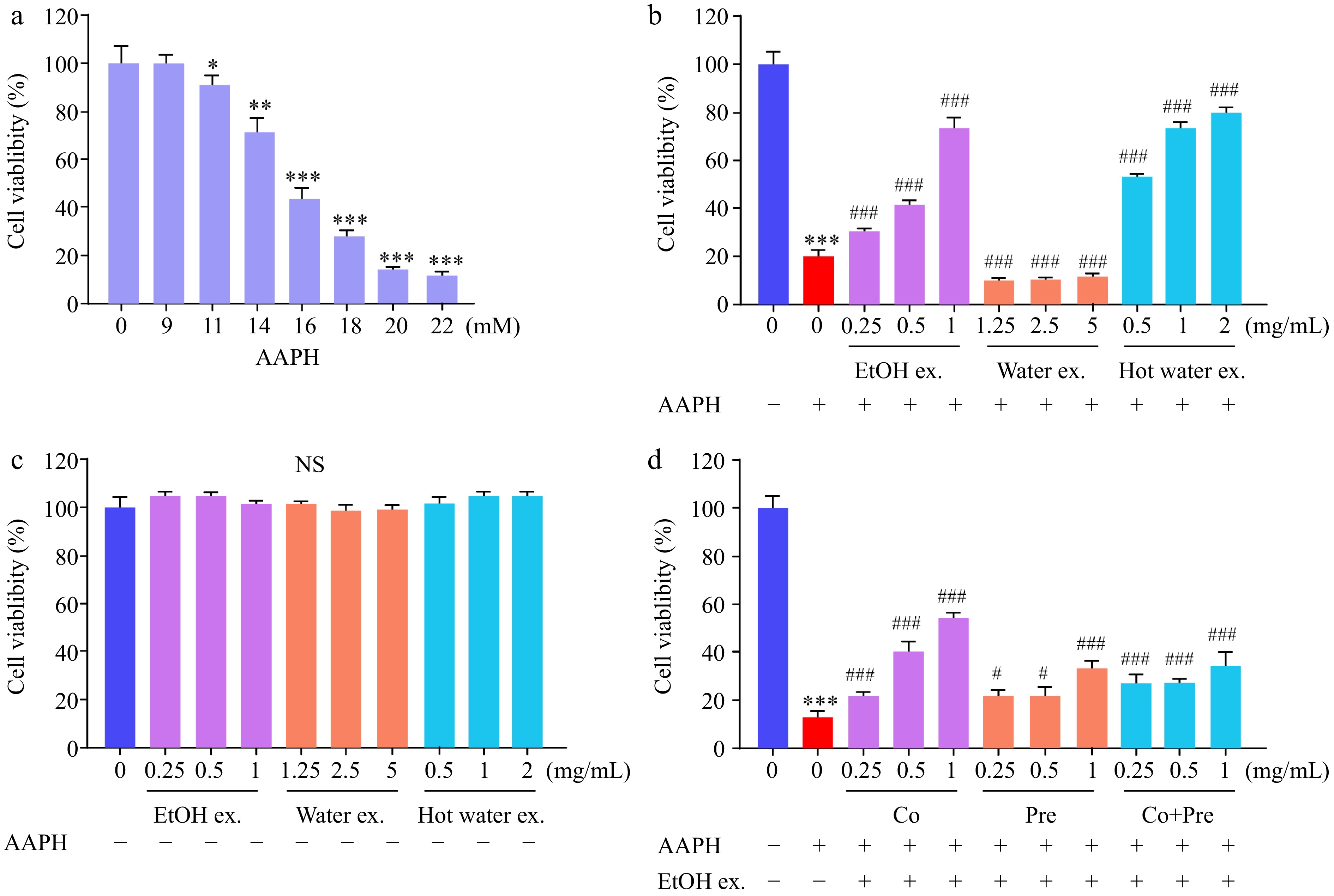
Figure 1.
Sea mussel extracts protected against AAPH-induced cytotoxicity. (a) AAPH exhibited dose-dependent cytotoxic effects on HepG2 cell viability. (b) Protective effects of EtOH, water, and hot water sea mussel extracts at various concentrations against AAPH-induced cytotoxicity. (c) EtOH, water, and hot water sea mussel extracts showed no cytotoxic effect on HepG2 cells. (d) Effects of EtOH extract at various concentrations on AAPH-induced cytotoxicity. Cells were treated with AAPH (16 mM) together with different concentrations of EtOH extract for 24 h.
Since the EtOH extract demonstrated the highest activity, further analysis was conducted on the effect of the order of addition of EtOH extract and AAPH on cell viability. Experiments were performed over 3 days. The 'Co' group involved simultaneous addition of EtOH extract and AAPH on the second day. The 'Pre' group received EtOH extract on the first day and AAPH on the second day. The 'Co + Pre' group was treated with EtOH extract on the first day, followed by a mixture of EtOH extract and AAPH on the second day. As shown in Fig. 1d, the 'Co' group exhibited the highest cell viability, indicating the strongest cytoprotection when EtOH extract was added together with AAPH. This protective effect was further enhanced with increasing concentrations of the EtOH extract.
While the present findings provide robust evidence for the cytoprotective effects of sea mussel extract in HepG2 cells, it is important to interpret these results within the context of an in vitro system. The HepG2 cell line, although widely used and mechanistically informative, does not fully recapitulate the complex metabolic and physiological environment of the intact human liver. Therefore, the observed activation of the Nrf2 pathway and upregulation of antioxidant enzymes should be considered as a mechanistic basis, rather than direct evidence of hepatic protection in vivo.
Direct antioxidant activity of sea mussel EtOH extract against AAPH-induced oxidative stress
-
ROS are generated as byproducts of the partial reduction of oxygen during cellular metabolism. Under physiological conditions, ROS are continuously produced in response to both endogenous metabolism and external stimuli, serving as key mediators of oxidative stress[34]. However, excessive accumulation of ROS can lead to oxidative damage of proteins, lipids, and DNA, resulting in significant cellular stress and contributing to the development of various diseases[1,35,36]. Therefore, quantifying intracellular ROS levels provides a direct means to evaluate whether the EtOH extract of sea mussel can effectively mitigate oxidative stress. To assess the antioxidant capacity of the extract, intracellular ROS levels were measured using the fluorescent probe DCF-DA. Our results demonstrated that pretreatment with varying concentrations of the EtOH extract significantly suppressed the AAPH-induced increase in intracellular ROS levels (Fig. 2a).
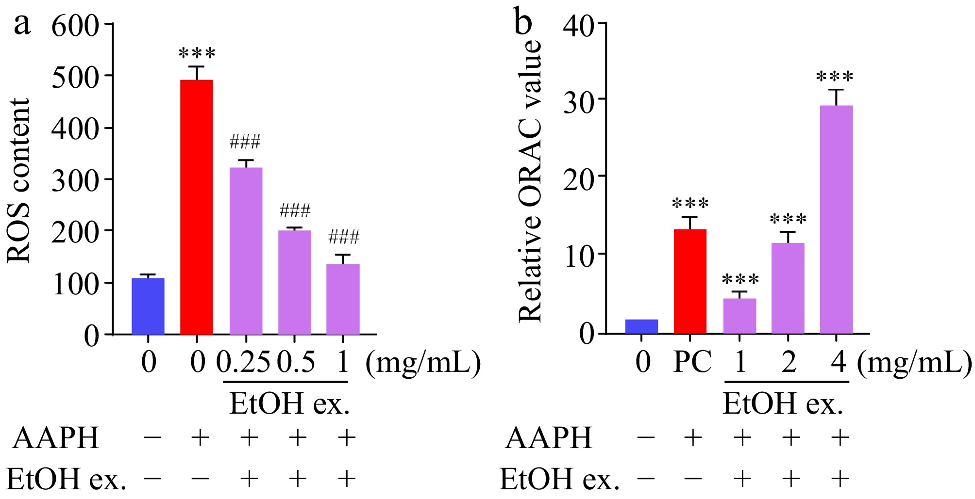
Figure 2.
Sea mussel EtOH extract reduced AAPH-induced ROS formation and exhibited ORAC activity. (a) Sea mussel EtOH extract reduced ROS formation induced by AAPH (16 mM). (b) Relative ORAC values of sea mussel EtOH extract at various concentrations: 1, 2, and 4 mg/mL. Data are expressed as fold change relative to the control (mean ± SD, n = 3). The absolute ORAC value at 1.0 mg/mL was 634.66 ± 39.51 μmol TE/g extract (dry weight). PC, DHMBA (500 μM).
This protective effect was attributed primarily to the direct antioxidant capacity of the extract, as direct antioxidants function by neutralizing free radicals through rapid chemical reactions, an activity commonly quantified by assays such as ORAC. In the present study, the absolute ORAC value of the sea mussel EtOH extract at 1.0 mg/mL was determined to be 634.66 ± 39.51 μmol TE/g dry extract. Furthermore, the relative ORAC values increased in a concentration-dependent manner (Fig. 2b), with the 4 mg/mL treatment exhibiting significantly higher activity compared to the control. Together, these results suggest that the sea mussel EtOH extract effectively combats oxidative stress primarily through direct free radical quenching mechanisms.
Indirect antioxidant capacity of sea mussel EtOH extract via the Keap1-Nrf2 pathway
-
While the direct radical-scavenging capacity described above contributes to the cytoprotective effects of the sea mussel EtOH extract, our subsequent experiments revealed that its antioxidant activity is also mediated through activation of the endogenous Keap1-Nrf2-ARE signaling pathway. The cellular response to oxidative stress is supported by direct and indirect antioxidant mechanisms[37]. Direct antioxidants, including chlorogenic acid, gallic acid, and rosmarinic acid, function by chemically scavenging ROS. Indirect antioxidants, represented by compounds like sulforaphane, quercetin, isoliquiritigenin, and curcumin, exert their effects through the activation of intrinsic cellular defense pathways, notably the Keap1-Nrf2 signalling cascade[38−40].
Under basal conditions, the transcription factor Nrf2 is held inactive in the cytoplasm through binding to its repressor protein Keap1, which promotes its proteasomal degradation. Upon exposure to oxidative stress or electrophilic stimuli, Nrf2 dissociates from Keap1, translocates into the nucleus, and binds to the ARE. This binding initiates the transcription of numerous cytoprotective genes, including phase II detoxifying enzymes and key antioxidant proteins such as HO-1, NQO1, GCLM, GPx1, GR, and SOD1[41−43]. Despite the well-established importance of this pathway, comparative studies on the indirect antioxidant potential of various food-derived extracts remain limited.
To determine its potential as an indirect antioxidant, we examined the ability of sea mussel EtOH extract to activate the Keap1-Nrf2 pathway. qRT-PCR analysis revealed that treatment with the sea mussel EtOH extract differentially upregulated the mRNA expression of seven canonical Nrf2 target genes, namely HO-1, NQO1, GCLC, GCLM, GPx1, GR, and SOD1 (Fig. 3). At the concentration of 2.0 mg/mL, all seven genes were significantly induced, whereas at 1.0 mg/mL, the upregulation of certain genes did not reach statistical significance. The upregulation of NQO1 was particularly pronounced, underscoring a robust activation of the associated cytoprotective gene program. Collectively, these findings support the conclusion that the extract acts as an effective indirect antioxidant through activation of the Keap1-Nrf2 signalling axis.
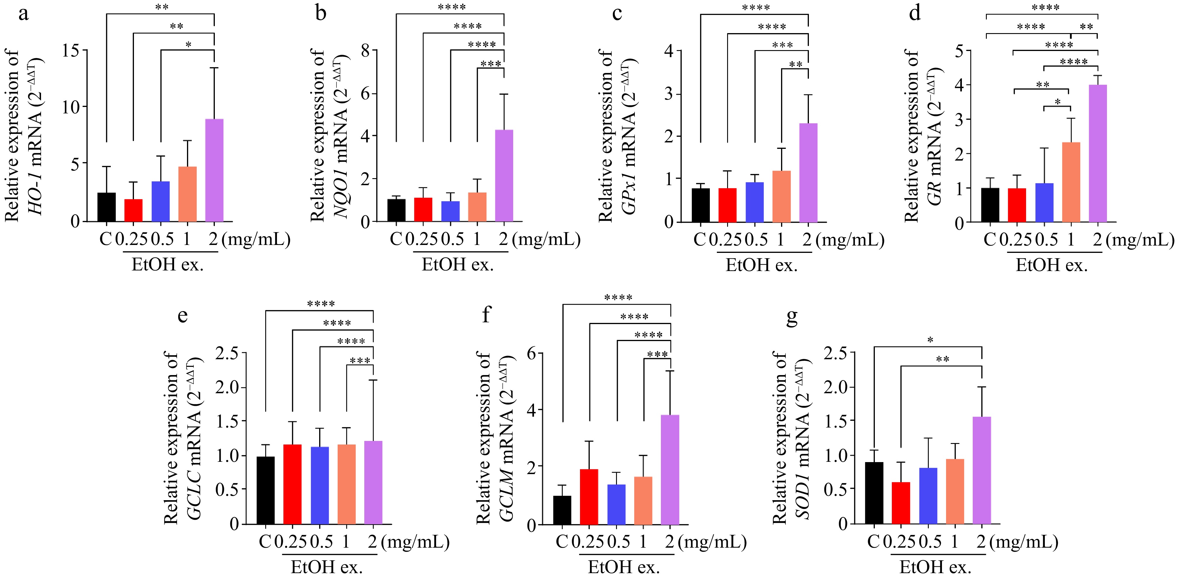
Figure 3.
Sea mussel EtOH extract induced expression of Nrf2 target genes in HepG2 cells. Cells were treated with the EtOH extract at 0.25, 0.5, 1.0, or 2.0 mg/mL for 24 h, and mRNA levels were measured by qRT-PCR. (a) HO-1 mRNA relative levels. (b) NQO1 mRNA relative levels. (c) GPx1 mRNA relative levels. (d) GR mRNA relative levels. (e) GCLC mRNA relative levels. (f) GCLM mRNA relative levels. (g) SOD1 mRNA relative levels. C, control, and untreated HepG2 cells.
Effect of sea mussel EtOH extract on ARE-regulated antioxidant protein NQO1 expression and intracellular glutathione levels
-
Among the Nrf2 target genes significantly upregulated by the EtOH extract, NQO1 is a well-characterized ARE-regulated gene that plays a critical role in cellular detoxification and antioxidant defense[44]. Its protein expression can be reliably detected by Western blot, enabling direct validation of the pathway activation observed at the mRNA level. Therefore, NQO1 was selected for further verification. In parallel, we also attempted to detect HO-1 protein expression by Western blot; however, HO-1 protein levels remained essentially unchanged over the concentration range tested (Supplementary Fig. S1). This observation is consistent with previous reports showing that HO-1 is subject to extensive post-transcriptional and translational regulation, often resulting in poor correlation between its mRNA and protein levels[45,46]. Accordingly, we focused on NQO1 as a more reliable protein marker for pathway activation in this experimental system. The protein expression of NQO1 was assessed by Western blot analysis, with DHMBA used as a PC. DHMBA is a phenolic compound recognized as an effective indirect antioxidant; it is known to induce the expression of antioxidant enzymes such as HO-1 and elevate intracellular glutathione (GSH) levels, representing a validated pharmacological approach to protective effects in a hepatic cell model[24,25]. As shown in Fig. 4a and b, treatment of HepG2 cells with sea mussel EtOH extract at concentrations ranging from 0 to 2 mg/mL resulted in a dose-dependent increase in NQO1 protein expression. The relative NQO1 levels approached those observed in the DHMBA-treated PC group, indicating a potent activation of this cytoprotective pathway.
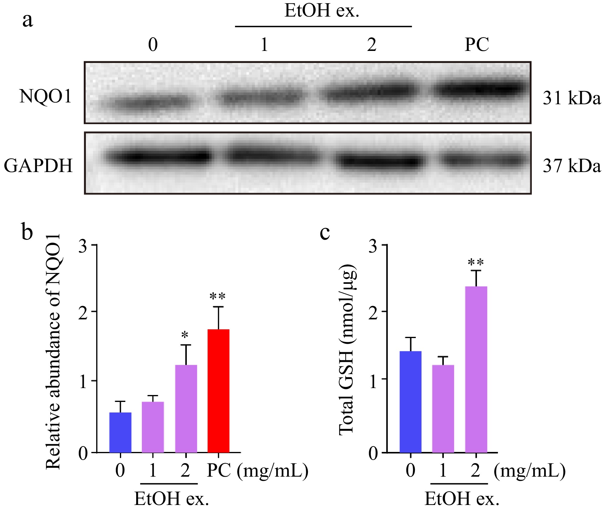
Figure 4.
Sea mussel EtOH extract upregulated the expression of the ARE-regulated antioxidant protein NQO1 and enhanced intracellular GSH levels in HepG2 cells. (a) Western blot analysis of NQO1 protein levels in HepG2 cells treated with or without sea mussel EtOH extract. (b) Relative quantification of NQO1 protein levels from panel (a). (c) Effect of sea mussel EtOH extract on intracellular GSH levels in HepG2 cells. PC, DHMBA, (500 μM).
In parallel, the effect of the extract on the cellular antioxidant status was evaluated by measuring intracellular GSH levels. GSH is a critical low-molecular-weight tripeptide present in all cells, essential for reducing hydrogen peroxide and protecting protein functional groups from oxidative damage[47,48]. Consequently, GSH content serves as a key indicator of overall cellular antioxidant capacity. Figure 4c demonstrates that increasing concentrations of the EtOH extract led to a progressive elevation in GSH levels, suggesting a marked enhancement of the intracellular antioxidant defense system.
Collectively, the data presented in Fig. 4 provide evidence that sea mussel EtOH extract induces the expression of the Nrf2-regulated protein NQO1 in a concentration-dependent manner and concurrently boosts cellular GSH levels, thereby substantiating its role in strengthening the antioxidant machinery of HepG2 cells.
Bio-guided isolation of active fractions from the sea mussel EtOH extract
-
Given the marked ability of the crude EtOH extract to activate the Keap1-Nrf2-ARE pathway and upregulate a panel of antioxidant enzymes, we next sought to identify the specific fractions responsible for this bioactivity. Marine organisms, including mussels, are known to produce a variety of bioactive constituents such as peptides, polysaccharides, and lipids, many of which have been reported to exert antioxidant effects through both direct radical scavenging and modulation of cellular signaling pathways[49−51]. However, the active components in M. edulis responsible for Nrf2 activation remain poorly characterized. To address this, we performed bioactivity-guided fractionation of the EtOH extract to isolate and evaluate the fractions most potent in inducing ARE-driven transcriptional activity. The EtOH extract of sea mussel was fractionated by silica gel column chromatography, yielding eight subfractions (Fr. 1–Fr. 8). Initial screening of these fractions for their ability to activate an ARE-driven luciferase reporter revealed that Fr. 5 exhibited the highest relative luciferase activity, while Fr. 6 also demonstrated a strong activating effect (Fig. 5a). To confirm these findings and assess concentration dependency, Fr. 5 and Fr. 6 were further tested across a range of concentrations. As shown in Fig. 5b, both fractions produced a concentration-dependent increase in relative luciferase activity within the tested range of 0.25 to 1 mg/mL. These results indicate that the bioactivity of the crude extract is primarily concentrated in Fr. 5 and Fr. 6. Although further compound identification was not pursued, it is plausible that multiple constituents within these subfractions may act in a synergistic manner to activate the Keap1-Nrf2-ARE pathway, highlighting the value of the whole fraction as a promising functional ingredient. While the cytotoxicity of the crude extract was confirmed to be negligible (Fig. 1c), the safety profiles of the purified subfractions Fr. 5 and Fr. 6 remain to be fully characterized in future studies.
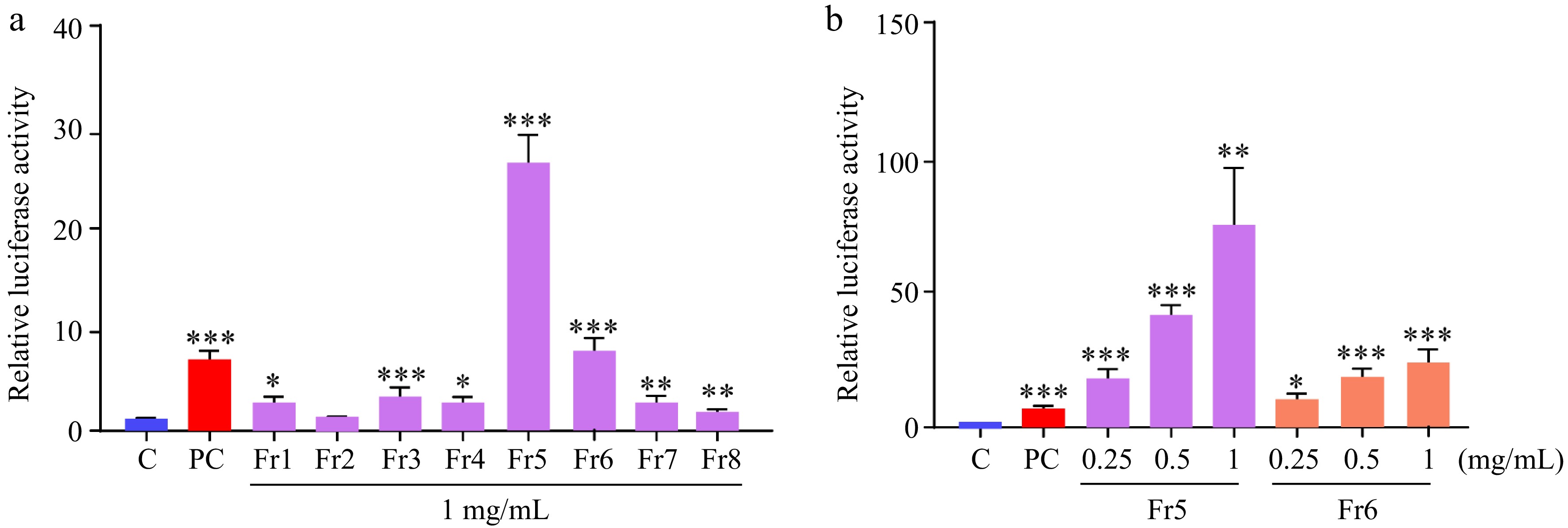
Figure 5.
Effect of sea mussel EtOH extract subfractions on ARE-driven reporter activity. (a) ARE-luciferase activity in HepG2 cells treated with different subfractions of the EtOH extract. (b) ARE-luciferase activity in response to Fr. 5 and Fr. 6 subfractions. C: control and untreated HepG2 cells. PC, DHMBA, (500 μM).
Bioactivity-guided fractionation identified Fr. 5 and Fr. 6 as the most active subfractions, with Fr. 5 showing the strongest ARE-driven luciferase activation. Preliminary chemical profiling of Fr. 5 was performed using HPLC (Supplementary Fig. S2), and the chromatographic profile indicated that this subfraction is a complex mixture predominantly composed of fatty acid derivatives. In contrast, Fr. 6 was not subjected to detailed chemical profiling due to the limited sample quantity and its lower bioactivity compared to Fr. 5. However, based on its chromatographic behavior on silica gel and similar elution profile, Fr. 6 appears to share comparable chemical characteristics. Due to the complexity of these subfractions and the limited quantities available, we were unable to isolate and identify individual active compounds at this stage. The observed ARE-activating activity may therefore arise from synergistic interactions among multiple constituents within these subfractions, rather than from a single bioactive molecule. This synergistic effect has been commonly reported for natural product extracts, where complex mixtures often exhibit greater biological activity than isolated pure compounds[52]. Further chemical characterization and compound identification of Fr. 5 and Fr. 6 are currently ongoing in our laboratory and will be reported in future studies.
In summary, bioactivity-guided fractionation of the sea mussel EtOH extract identified Fr. 5 and Fr. 6 as the most active subfractions in activating the Keap1-Nrf2-ARE pathway, with Fr. 5 exhibiting the strongest ARE-driven luciferase activation. As illustrated in Fig. 6, the extract and its bioactive fractions promote Nrf2 nuclear translocation and subsequent transcriptional upregulation of downstream antioxidant genes, thereby enhancing cellular defense against AAPH-induced oxidative stress. These results highlight the potential of sea mussel-derived fractions as natural modulators of the endogenous antioxidant system.
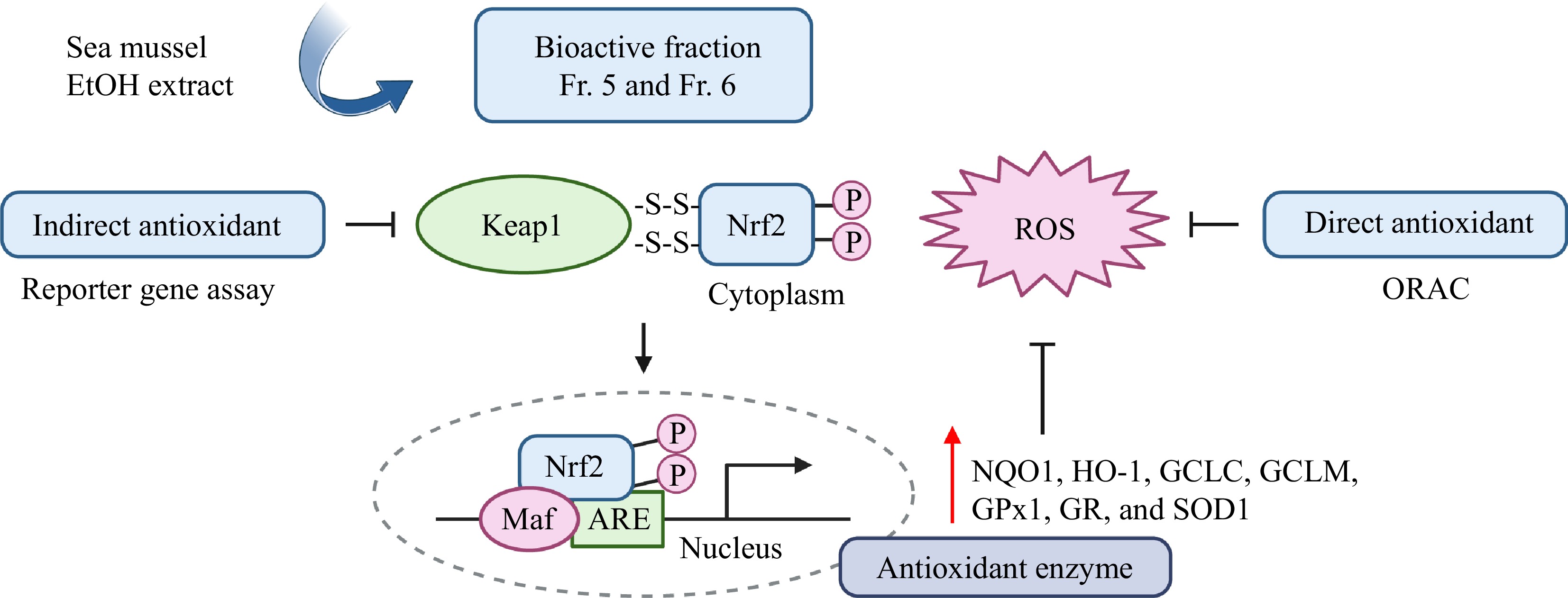
Figure 6.
Schematic illustration of the proposed mechanism by which sea mussel extract alleviates AAPH-induced oxidative stress through the Keap1-Nrf2-ARE pathway.
-
This study demonstrates that the EtOH extract of sea mussel (M. edulis) exerts cytoprotective effects against AAPH-induced oxidative stress in HepG2 cells by activating the Keap1-Nrf2-ARE signaling pathway, as evidenced by ARE-driven reporter activation and transcriptional upregulation of canonical Nrf2 target genes, including NQO1, HO-1, GCLC, GCLM, GPx1, GR, and SOD1. Bioactivity-guided fractionation further identified subfractions Fr. 5 and Fr. 6 as the primary active components. These findings position sea mussel as a promising source of natural Nrf2 activators for functional food applications. However, given the inherent limitations of the in vitro system, further isolation and structural characterization of the active compounds within Fr. 5 and Fr. 6, along with evaluation of their in vivo efficacy in suitable animal models, are warranted to fully assess their translational potential. In addition, direct evidence of Nrf2 nuclear translocation and the upstream molecular events governing this response remain to be elucidated in future studies.
-
The authors confirm their contributions to the paper as follows: conceptualization: Tan H, Zheng K; experiments and formal analysis: Li Z, Yang R; methodology and data curation: Zhao C, Wang X, Tan H; funding acquisition: Tan H, Li B; draft preparation: Zheng K, Chiba H, Hui S; finalizing the manuscript: Tan H; project administration and supervision: Tan H, Li B. All authors reviewed the results and approved the final version of the manuscript.
-
The data that support the findings of this study are available from the corresponding author upon reasonable request.
-
The authors declare that they have no conflict of interest.
-
accompanies this paper online at: https://doi.org/10.48130/fia-0026-0041.
- Supplementary Fig. S1 Sea mussel EtOH extract did not appreciably influence HO-1 protein expression in HepG2 cells.
- Supplementary Fig. S2 HPLC chromatogram of subfraction Fr. 5 from sea mussel EtOH extract.
- Copyright: © 2026 by the author(s). Published by Maximum Academic Press on behalf of China Agricultural University, Zhejiang University and Shenyang Agricultural University. This article is an open access article distributed under Creative Commons Attribution License (CC BY 4.0), visit https://creativecommons.org/licenses/by/4.0/.
-
About this article
Cite this article
Zheng K, Li Z, Yang R, Wang X, Zhao C, et al. 2026. Sea mussel (Mytilus edulis) ethanol extract and its subfractions ameliorate AAPH-induced oxidative stress via the Keap1-Nrf2-ARE pathway. Food Innovation and Advances 5(3): 483−491 doi: 10.48130/fia-0026-0041 

Sea mussel (Mytilus edulis) ethanol extract and its subfractions ameliorate AAPH-induced oxidative stress via the Keap1-Nrf2-ARE pathway
- Received: 10 April 2026
- Revised: 01 September 2026
- Accepted: 01 September 2026
- Published online: 29 September 2026
Abstract: Epidemiological evidence indicates that dietary antioxidants can mitigate oxidative stress and support immune regulation. The activation of nuclear factor erythroid 2-related factor 2 (Nrf2) represents a key cellular defense mechanism against oxidative damage. In this study, we investigated the protective effect of a sea mussel (Mytilus edulis) ethanol extract against 2,2'-Azobis(2-amidinopropane) dihydrochloride (AAPH)-induced cytotoxicity in HepG2 cells. The extract significantly attenuated cell damage and demonstrated a dose-dependent activation of the antioxidant response element (ARE). Mechanistically, the cytoprotection was mediated through the significant upregulation of multiple Nrf2-target genes at the transcriptional level, including HO-1, NQO1, GCLC, GCLM, GPx1, GR, and SOD1. Further analysis confirmed that the extract enhanced the protein expression of NQO1 and elevated intracellular GSH levels. Through bioactivity-guided fractionation, subfractions Fr. 5 and Fr. 6 were identified as the most active components, with Fr. 5 exhibiting the strongest ability to activate the Keap1-Nrf2-ARE pathway. These in vitro findings based on the HepG2 hepatocyte model indicate that sea mussel ethanol extract alleviates oxidative stress by directly scavenging radicals and by indirectly activating the endogenous Keap1-Nrf2-ARE antioxidant pathway, which provides fundamental cellular evidence supporting its further development as a promising nutraceutical candidate for follow-up in vivo investigations targeting oxidative stress-associated liver injury.
-
Key words:
- Mytilus edulis /
- Nrf2 /
- Antioxidant response element /
- Oxidative stress /
- HepG2 cells