













-
The basket clams in the genus Corbicula Megerle von Mühlfeld, 1811[1] (Bivalvia: Cyrenidae) represent one of the most evolutionarily successful and taxonomically challenging groups of freshwater bivalves. The genus contains a diverse group of brackish and freshwater clams that are natively distributed across Asia, Africa, Australia, and the Middle East, and invasive lineages that have established self-sustained populations in Europe, the New World, and New Zealand[2−4], as well as other non-native territories in Asia and Africa[3]. Some taxa have even successfully invaded areas already occupied by their congeners[5,6], where they may directly compete with the resident native populations.
Unlike most bivalves, Corbicula exhibits exceptional reproductive plasticity, containing lineages capable of either traditional sexual reproduction or a specialized form of asexuality known as 'androgenesis'[7]. In the sexual mode, individuals are distinctly either male or female, and males produce reduced, monoflagellate spermatozoa[8]. These sexual Corbicula lineages are found exclusively in their native distribution range in Asia[3,8−11]. In contrast, androgenetic lineages are hermaphroditic and produce unreduced, biflagellate spermatozoa[12]. The unreduced spermatozoa of these lineages are capable of fertilizing oocytes from other individuals of any congeneric species or even self-fertilizing[2,7]. Unlike the restricted sexual lineages, androgenetic lineages are widely distributed, having been reported in both their native range[13] and non-native regions across Europe and the Americas[14,15]. Corbicula clams also exhibit a variety of larval development strategies, ranging from free-swimming planktotrophic larvae[16] and benthic larvae[9,10] to maternal incubation of juveniles in the gills. This parental care system is commonly found in freshwater Corbicula lineages, including both dioecious and hermaphroditic species[8,11,17−19].
A recent systematic revision of Corbicula on a global scale utilizing molecular phylogenetic analyses classified the genus into two subgenera[3]. The first, nominotypical subgenus represents the most diverse clade, containing C. japonica Prime, 1864, from brackish water and other freshwater species-level taxa, both invasive and native species from Africa, Australia, and Asia[3]. Members of this subgenus exhibit the typical Corbicula shell morphology in terms of having an ovate-triangular shape and prominent concentric sculpture appearing as distinct ribs[20]. The other subgenus is Sphaerocorbicula Bolotov, Bespalaya & Aksenova, 2025, in Bespalaya et al.[3]. Members in this subgenus are characterized by their small and ovate shell and very weak expression of concentric sculpture. This subgenus is currently comprised of three freshwater species, all endemic to Myanmar[3]. However, this global assessment recovered only 27 species-level clades. This number contrasts sharply with the approximately 80 species currently accepted as valid for the genus in a global database[21]. Thus, there are a considerable number of nominal taxa whose taxonomic status remains unresolved using modern systematic approaches, particularly those native to Southeast Asia.
Corbicula regia Clessin, 1879, represents one of the enigmatic species within the genus. It was described from the Malay Peninsula[22], with only a few museum lots available from the vicinity of the type locality[20,23]. For over half a century, no new records or additional specimens have been reported. Our recent field surveys in Malaysia and Southern Thailand yielded sufficient fresh specimens of C. regia for detailed morphological comparison and molecular analysis. Utilizing these newly collected specimens, the primary goal of this study is to resolve the evolutionary history and taxonomic status of C. regia. Specifically, we aim to establish its phylogenetic placement, investigate the evolutionary origins of habitat transitions within Corbicula, and comprehensively redescribe the morpho-anatomy and reproductive mode of this elusive species.
-
Specimens of C. regia were newly collected from two locations on the Malay Peninsula (Fig. 1a). The first site was in the Perai River in Penang State, Malaysia (5°26'43.5" N, 100°27'38.8" E). This location is near the type locality[23−25]. At this site, specimens were collected from a slow-flowing river at a depth of 1 m where there was a sandy bottom. The second site was in the Trang River in Trang Province, Thailand (7°36'42.2" N, 99°31'05.5" E). At this location, C. regia was found in high abundance on a gently sloping sandy–muddy substrate at the water's edge (Fig. 1b).
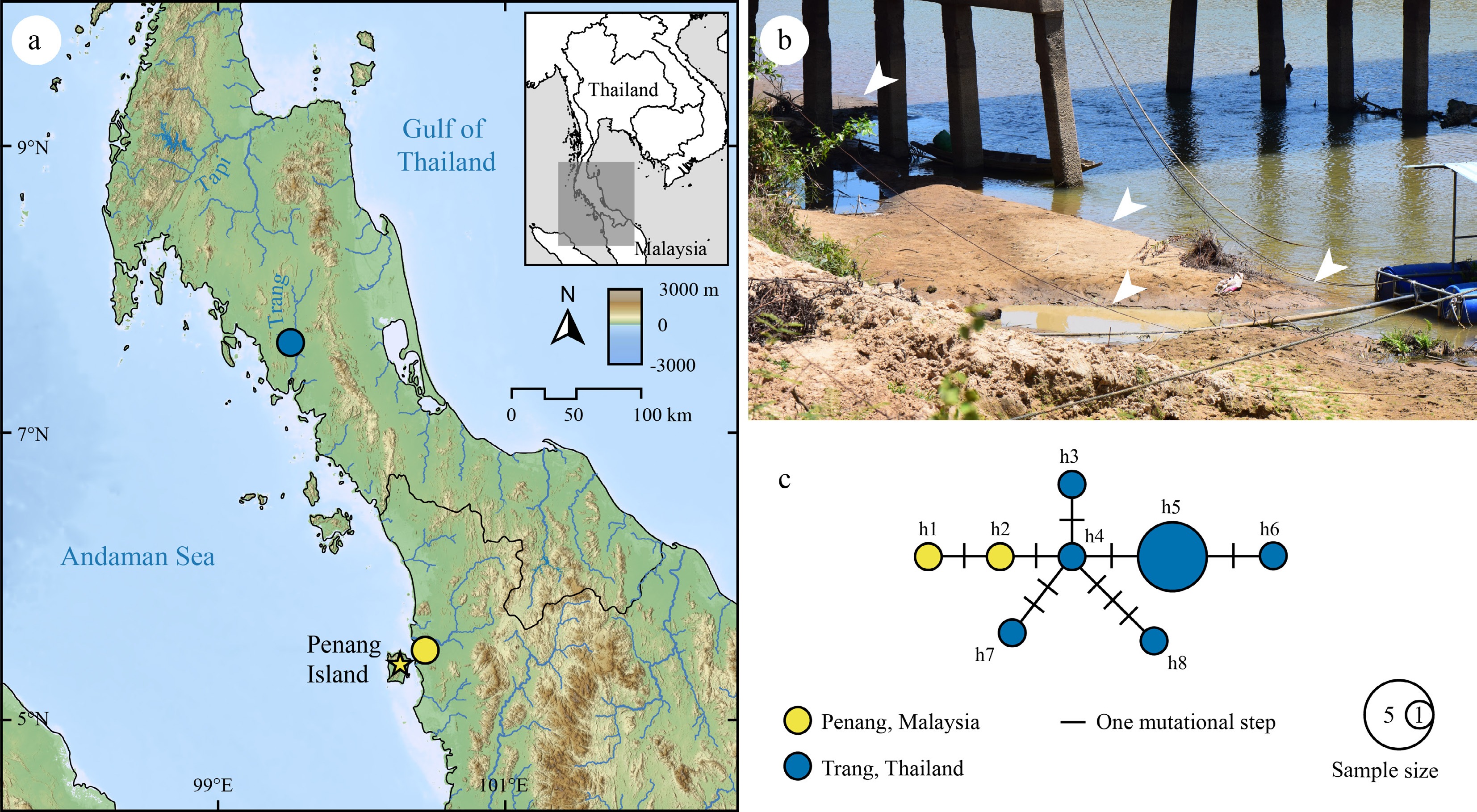
Figure 1.
(a) Map showing sampling sites (circles) and the type locality (yellow star) of Fasciocorbicula regia. The map was created in QGIS v3.36 using river layers derived from the HydroSHEDS dataset (
www.hydrosheds.org ), land topography raster imagery from NASA EARTHDATA (www.earthdata.nasa.gov ), and sea bathymetry data from GEBCO's global gridded bathymetric data (https://download.gebco.net ). (b) Habitat at Trang River, Thailand (7°36'42.2" N 99°31'05.5" E); white arrowheads indicate areas where F. regia individuals were observed or collected. (c) Median-joining network generated from a 663-bp sequence of the COI gene. Each circle represents a unique haplotype, with the size proportional to the number of individuals sharing that haplotype. Colors indicate the sampling sites (Penang, Malaysia in yellow and Trang, Thailand in blue). Lines connecting the haplotypes represent mutational steps, with each hash mark signifying a single mutation.Clams were collected by hand and euthanized using the two-step method under an animal use protocol approved by the Faculty of Science, Mahidol University Animal Care and Use Committee (SCMU-ACUC; permit no. MUSC63-017-525). Initially, specimens were placed in a container with 5% (v/v) ethanol for 30 min, or until the foot and adductor muscles were fully relaxed (indicated by a fully gaping shell). Once fully anesthetized, specimens were either preserved in 95% (v/v) ethanol for morphological and molecular analyses or carefully removed from their shells and fixed in Bouin's solution for histological study. The voucher specimens are deposited in the Mahidol University Museum of Natural History (MUMNH), Department of Biology, Faculty of Science, Mahidol University, Bangkok, Thailand.
Shell morphology was assessed by examining various characteristics, including the outline, size, thickness, concentric ribs, surface coloration, umbo shape and position, hinge dentition, and muscle scars. Shell length, height, and width were measured to the nearest 0.01 mm using a digital Vernier caliper. Soft body anatomy was dissected and investigated under a stereomicroscope (Stemi 508; Carl Zeiss, Germany). The gills were also investigated for potential juvenile incubation.
Nomenclatural acts
-
The new names contained in this publication are available under the International Code of Zoological Nomenclature. This work and the nomenclatural acts it contains have been registered in ZooBank. Zoobank Life Science Identifier (LSID) for this publication is: urn:lsid:zoobank.org:pub: 32A27D69-25EC-4A27-875D-75558D09B876. The LSID registration and any associated information can be viewed in a web browser by adding the LSID to the prefix
https://zoobank.org .Molecular analyses and sequencing
-
Genomic DNA was extracted from foot tissues using the NucleoSpin Tissue Extraction Kit (Macherey-Nagel, Germany). Target gene fragments were amplified through polymerase chain reaction (PCR) targeting two mitochondrial genes and one nuclear gene, including the cytochrome c oxidase subunit I gene (COI), the large ribosomal subunit rRNA gene (16S rRNA), and the 28S large ribosomal subunit rRNA gene (28S rRNA). The primers used, amplicon lengths, and PCR profiles are listed in Supplementary Table S1. PCR products were purified and bi-directionally sequenced using the same primers on an automated sequencer (ABI PRISM 3730XL). The resulting sequences were assembled and aligned using MEGA12 v12.0.11[26]. The consensus nucleotide sequences obtained in this study were deposited in the GenBank Nucleotide sequence database under accession numbers PZ097215–PZ097233 for COI, PZ111377–PZ111382 for 16S rRNA, and PZ097209–PZ097214 for 28S rRNA (Supplementary Table S2). One C. regia individual was sequenced for all three genes and used in the multi-locus phylogenetic analysis; the other 13 individuals were sequenced for the COI gene only and used in the haplotype network analysis.
Multi-locus phylogeny and genetic divergence
-
The multi-locus phylogeny was inferred from 27 selected taxa within the superfamily Cyrenoidea (Supplementary Table S3). The ingroup included 20 taxa from three extant genera within the family Cyrenidae. These comprised C. regia and 17 representatives of Corbicula species—13 freshwater species and one brackish water species (C. japonica) in the subgenus Corbicula s.s., and three recently described freshwater species in the subgenus Sphaerocorbicula from Myanmar[3]—alongside two closely related taxa, Batissa violacea (Lamarck, 1818) and Villorita cyprinoides (Gray, 1825)[27]. The outgroup consisted of three species from Glauconomidae and four species from Cyrenoididae, selected based on recent phylogenetic analyses suggesting their close evolutionary affinities to Cyrenidae[27,28].
Sequence alignment was performed using MAFFT v7.49[29] with the L-INS-i algorithm, and each gene was aligned independently. Sequences retrieved from GenBank that were longer than the amplicons generated in this study were manually trimmed to match the length of our amplified fragments. The individual gene alignments were then concatenated into a single data matrix. The final data matrix length was 1,980 bp, comprising 663 bp of COI, 528 bp of 16S rRNA, and 789 bp of 28S rRNA. The optimal partition scheme and the best-fit models of nucleotide substitution were selected using PartitionFinder2 v2.1.1[30] under the corrected Akaike Information Criterion (AICc), resulting in a five-partition scheme that treats the three codon positions of COI, 16S rRNA, and 28S rRNA as distinct partitions (Supplementary Table S4).
Phylogenetic trees were reconstructed by two independent approaches: maximum-likelihood (ML) and Bayesian inference (BI). The ML analysis was implemented in IQ-TREE v2.2.2.7[31], utilizing 10,000 ultrafast bootstrap replicates (UFBoot) to assess topological robustness[32]. The BI analysis was conducted with MrBayes v3.2.7[33]. Two simultaneous Markov chain Monte Carlo (MCMC) runs, each comprised of three heated chains and one cold chain, were executed for 10,000,000 generations with sampling every 1,000 generations. Convergence was confirmed as all estimated parameters achieved an effective sample size (ESS) exceeding 200. After discarding the initial 25% of samples as burn-in, the remaining trees were used to generate a consensus topology, node supports, and branch lengths. The resulting phylogenetic trees from both analyses were visualized and edited in FigTree v1.4.4 (
https://tree.bio.ed.ac.uk/software/figtree ). Clades were considered well-supported if they achieved bootstrap (BS) values ≥ 95% in the ML analysis and Bayesian posterior probabilities (BPP) ≥ 0.95 in the BI analysis[32,34]. The ML and BI phylogenetic analyses were executed via the CIPRES Science Gateway[35]. Additionally, pairwise genetic divergences for the COI dataset were determined using uncorrected p-distances as implemented in MEGA12.Divergence time estimation
-
Divergence times were estimated in BEAST v2.7.8[36] using the same dataset employed in the preceding phylogenetic analyses. To calibrate the molecular clock, two calibration points of a reliable fossil and a geological event were selected based on previous studies[3,37]. The first calibration point was based on a C. jilinensis Zhu & Zhou, 1988 fossil, which represents the most recent common ancestor of Corbicula. This fossil originated from the end of the Early Cretaceous, providing a minimum age of 100.5 million years ago (Mya) and a soft upper bound of 143.1 Mya[38]. The second calibration point utilized the estimated age of Lake Biwa, Japan, approximately 4 Mya, to represent the divergence point of the endemic species C. sandai Reinhardt, 1878[39].
Analyses were performed using a lognormal relaxed clock algorithm with the Yule speciation process as a tree prior[40]. A simplified HKY model was applied to all partitions to avoid overparameterization. Two independent MCMC chains were run for 50 million generations, with trees sampled every 1,000 generations. Convergence and ESS parameter sufficiency were verified using Tracer v1.7[41]. Results from both runs were combined with a 25% burn-in using LogCombiner v2.6.2, and the maximum clade credibility (MCC) tree was generated via TreeAnnotator v2.6.2[36]. All estimations were executed on the CIPRES Science Gateway[35].
Ancestral area reconstruction
-
Historical biogeography was inferred through ancestral area reconstruction as implemented in RASP v4.2[42,43] using three probabilistic algorithms: Statistical Dispersal-Vicariance Analysis (S-DIVA), Statistical Dispersal-Extinction-Cladogenesis (S-DEC), and Bayesian Binary MCMC (BBM). The input consisted of 75,002 trees derived from the combined BEAST analyses, with the MCC tree serving as the consensus tree. Eight native geographical areas were defined: East Asia (A), Southeast Asia (B), South Asia (i.e., India; C), Central and West Asia (D), Australia (E), Africa (F), North America (G), and South America (H). The native distribution area of each taxon is shown in Supplementary Table S3. Parameters were kept at default settings in all analyses, with the exception of the BBM analysis, which was run for 500,000 MCMC generations. To reduce model complexity, all analyses restricted the maximum number of ancestral areas per node to two. This constraint assumes that ancestral lineages did not historically occupy more than two of the defined regions (A)–(H) at any given time. Finally, to produce a unified biogeographical scenario for interpretation, the results from all three methods were integrated using the 'Combine Results' feature in RASP.
Ancestral state reconstruction
-
Ancestral state reconstruction for habitat type (i.e., freshwater or brackish water) was performed using stochastic character mapping[44] as implemented in the R package phytools v2.5.2[45]. The habitat trait assigned to each taxon is shown in Supplementary Table S3. The time-calibrated MCC tree, previously generated from BEAST analysis, was used as the fixed topology. Transitions between habitat states were modeled using an Equal Rates (ER) framework, with transition rates estimated via an ML method, and 100 stochastic character maps were simulated across the tree using the function make.simmap. The resulting simulations were summarized to provide marginal posterior probabilities for ancestral states at each internal node. This analysis was conducted in R v4.5.1.
Haplotype network analysis
-
A haplotype network was constructed using the median-joining algorithm[46] as implemented in PopART v1.7[47] to visualize genealogical relationships among mitochondrial COI haplotypes. The dataset included two sequences from the topotype, Penang, Malaysia, and 12 sequences from the Trang River, Thailand.
Histological analysis and sperm isolation
-
Ten mature individuals collected on 18 March 2023 from Trang River, Thailand, were selected from the largest specimens for histological analysis. Specimens fixed with Bouin's solution were washed, dehydrated through a graded alcohol series, and embedded in paraffin. Sections of 5 µm thickness were cut using a rotary microtome, placed on glass slides, and stained with hematoxylin and eosin. The gonadal slides were examined under a compound microscope (ZEISS Primostar 3, Carl Zeiss, Germany) and imaged with a benchtop fluorescence microscope (APX100; Olympus, Japan).
Sperm morphology was examined using a modified version of the isolation protocol from Pigneur et al.[48]. Approximately 0.5 cm3 of gonad tissue was isolated from each respective specimen and minced with scissors. The tissue was incubated in 1 mL of 0.1 mg/mL collagenase solution at 45 °C for 30 min. Following the digestion, the suspension was centrifuged at 3,000 rpm for 3 min. The resulting precipitate was dropped onto a glass slide, and sperm characteristics were examined using a compound microscope (ZEISS Primostar 3; Carl Zeiss, Germany). Detailed sperm morphology was examined and imaged using an inverted confocal laser scanning microscope (CLSM FV1000; Olympus, Japan). Sperm head size was measured using digital measurement tools in ImageJ v1.54g[49].
-
Phylogenetic trees recovered members in the family Cyrenidae as a monophyletic group (Fig. 2) with significant support in the BI analysis (BPP = 0.99) and strong support in the ML analysis (BS = 91%). Within Cyrenidae, V. cyprinoides occupies the basal position, followed by B. violacea, and then a maximally supported clade of the genus Corbicula (BS = 100%, BPP = 1). Most notably, C. regia appears in a distinct and divergent position, falling outside the primary Corbicula s.s. clade, which receives high support (BS = 99%, BPP = 1). The distinct lineage of C. regia is recovered as a sister relationship to the subgenus Sphaerocorbicula clade from Myanmar, with moderate to high support (BS = 78%, BPP = 0.97). The Corbicula s.s. clade exhibits a deep substructure. The brackish water species, C. japonica, is distinctly separated from the freshwater clade that includes both native and invasive species (BS = 99%, BPP = 0.99). While the relationship among the freshwater lineages is largely unresolved, the clade containing C. fluminea (Müller, 1774), C. tobae Martens, 1900, and C. largillierti (Philippi, 1844) is supported in the ML analysis (BS = 95%). In terms of genetic divergence, C. regia differs from the subgenus Corbicula s.s. by an average of 11.85% and from the subgenus Sphaerocorbicula by 11.44% based on COI gene sequence p-distances (Table 1).
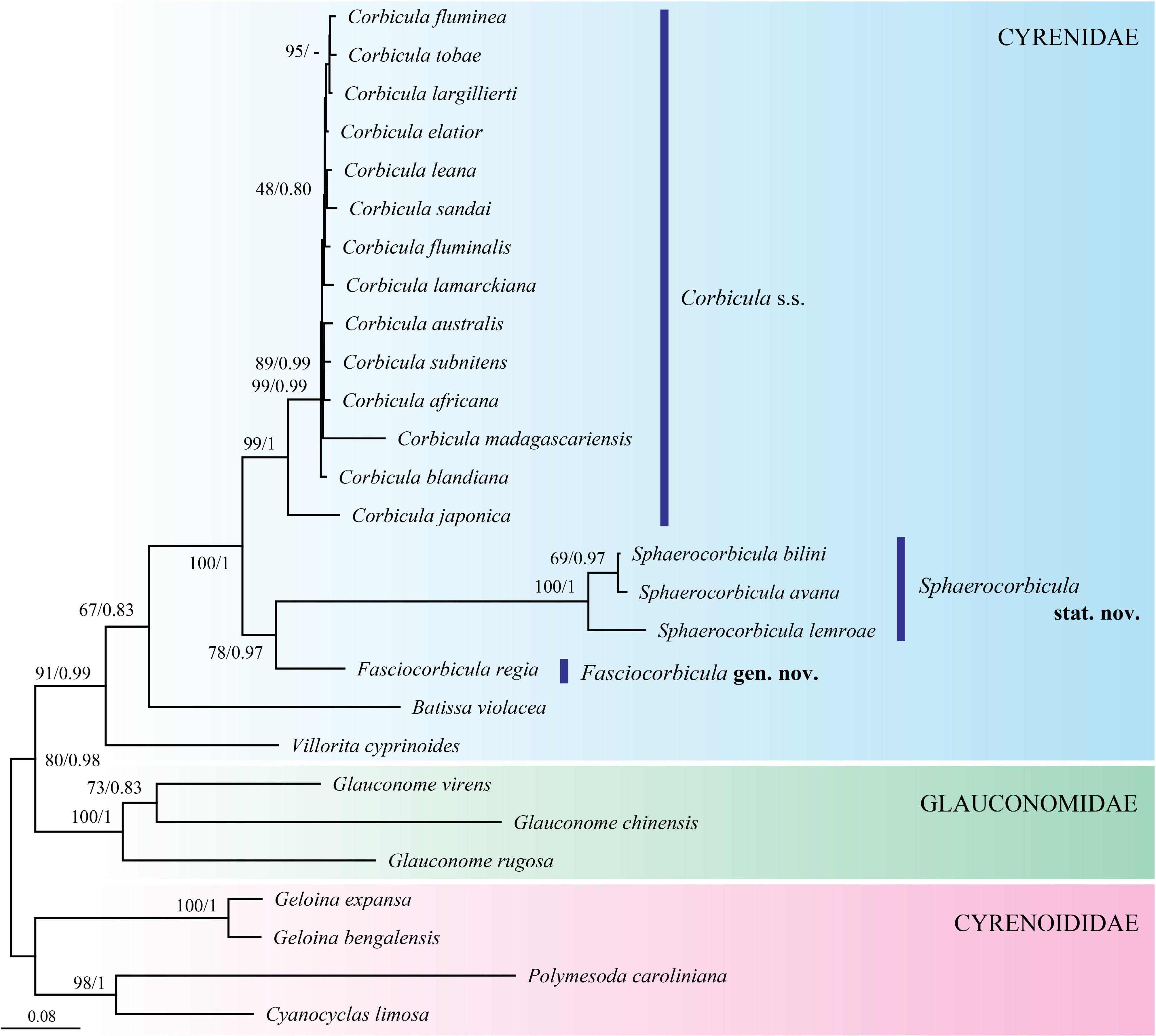
Figure 2.
Maximum likelihood (ML) phylogenetic tree constructed using a 1,980-bp multi-locus alignment of mitochondrial (COI and 16S rRNA) and nuclear (28S rRNA) sequences. Node support is indicated by ML bootstrap values (BS) and Bayesian posterior probabilities (BPP), formatted as ML/BI. Branch lengths are proportional to the scale bar provided.
Table 1. Average genetic divergence (%) based on uncorrected p-distances of a 539-bp COI gene fragment among genera within the family Cyrenidae, and average interspecific divergence among species within each genus (in bold).
Genera 1 2 3 4 5 1. Corbicula s.s. 4.13 2. Sphaerocorbicula stat. nov. 11.87 7.48 3. Fasciocorbicula gen. nov. 11.85 11.44 n/a 4. Batissa 17.12 15.77 16.33 n/a 5. Villorita 16.71 16.08 15.58 16.88 n/a Given its unique phylogenetic placement, substantial genetic divergence from established lineages, and distinct morphological characters (see the description below), the Fasciocorbicula gen. nov. is proposed and described herein to accommodate C. regia. Furthermore, to maintain the monophyly of Corbicula, we also elevate the subgenus Sphaerocorbicula Bolotov, Bespalaya & Aksenova, 2025[3] to the genus level.
Divergence time estimation and historical biogeographic analyses
-
The time-calibrated phylogeny in Fig. 3 yields a topology largely consistent with the multi-locus ML and BI analyses. However, the topological relationships within Corbicula s.s. differs. While the ML and BI trees group C. madagascariensis Smith, 1882 with other freshwater lineages, the MCC tree generated by BEAST resolves this African species as a significant early split from the remaining freshwater lineages, following the divergence of the estuarine species C. japonica. The relationships among freshwater lineages are also not significantly supported.
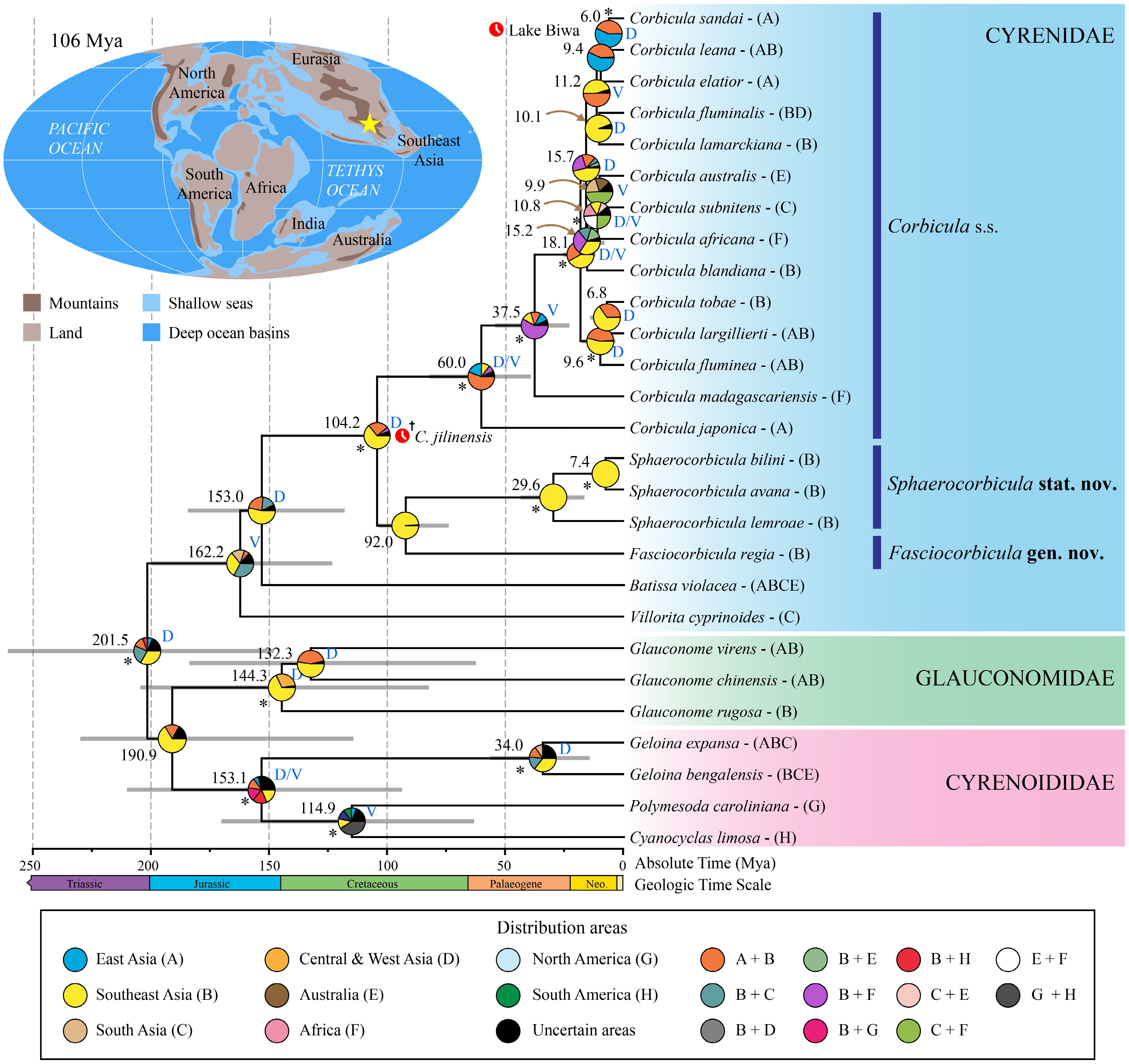
Figure 3.
Estimated divergence time and ancestral area reconstruction results. The phylogeny was generated in BEAST v2.7.8 using a concatenated dataset of mitochondrial (COI and 16S rRNA) and nuclear (28S rRNA) sequences. Significantly supported nodes (BPP ≥ 0.95) are indicated by asterisks*. Values above branches represent the estimated time since the most recent common ancestor (tMRCA) in Mya. Grey bars at nodes indicate the 95% highest posterior density (HPD) intervals for node ages. Pie charts at the internal nodes represent the marginal probabilities of ancestral area reconstructions. Blue letters adjacent to nodes indicate inferred biogeographic events: dispersal (D) and vicariance (V). Calibration points are indicated with red clock symbols. Native distributions of the examined taxa are shown in parentheses following the taxon names. The paleogeographic map (upper left) illustrates the hypothetical landmass during the Early Cretaceous (~106 Ma), with a yellow star indicating the hypothesized center of origin of the common ancestor of Corbicula s.s. + Sphaerocorbicula + Fasciocorbicula. The map was redrawn from Scotese[50].
The divergence of the Cyrenidae clade from the Glauconomidae is estimated to have occurred near the Triassic–Jurassic boundary (201.5 Mya; 95% HPD = 148.0–260.5 Mya; Fig. 3; Supplementary Table S5). Within the family, the lineage leading to the common ancestor of Corbicula s.s. + Sphaerocorbicula + Fasciocorbicula separated from Batissa Gray, 1853, during the Late Jurassic (153.0 Mya; 95% HPD = 119.4–194.7 Mya). This diversification likely occurred in Southeast Asia via a dispersal event (probability of ancestral areas = 53%; Supplementary Fig. S1, Supplementary Table S5). The diversification of the three genera then began in the Early Cretaceous (104.2 Mya; 95% HPD = 100.6–109.3 Mya), marked by the split between the genus Corbicula s.s. and the common ancestor of Sphaerocorbicula + Fasciocorbicula. Biogeographical analysis suggests this originated in Southeast Asia via a dispersal event (probability of ancestral areas = 64%). Subsequently, these two genera diverged within Southeast Asia around 92.0 Mya (95% HPD = 74.7–108.0 Mya) in the Late Cretaceous, although this node was not significantly supported by the MCC tree.
Within the genus Corbicula s.s., the brackish-water C. japonica diverged during the Paleocene (60.0 Mya; 95% HPD = 39.1–82.2 Mya). This event likely took place in an ancestral range encompassing East and Southeast Asia (probability of ancestral areas = 55%), driven by a combination of dispersal and vicariance. This was followed by the divergence of the African species C. madagascariensis around 37.5 Mya (95% HPD = 22.8–54.3 Mya). The majority of freshwater Corbicula species show more recent diversification events occurring throughout the Neogene (18.1–6.0 Mya). These events were mainly centered in East or Southeast Asia and involved multiple dispersal and vicariance events.
Reconstruction of the ancestral character state of habitat types
-
The ancestral state reconstruction of habitat types indicates that the common ancestor of the superfamily Cyrenoidea most likely inhabited a brackish environment (Fig. 4; Supplementary Table S5). This ancestral brackish state is conserved across the entire Glauconomidae clade and the majority of the Cyrenoididae, with Cyanocyclas limosa (Maton, 1811) representing a derived condition characterized by an independent shift to freshwater. Within the Cyrenidae, the basal taxon V. cyprinoides retains the brackish condition, while B. violacea occupies an intermediate brackish-freshwater niche. In contrast, Fasciocorbicula gen. nov., Sphaerocorbicula, and the majority of Corbicula s.s. have turned into freshwater species. This habitat variability within the family is reflected by mixed probabilities of brackish and freshwater ancestry at the corresponding ancestral nodes. Interestingly, the genus Corbicula s.s. shows a clear transition away from the ancestral brackish condition. The most recent common ancestor of the genus, as well as all internal nodes within the genus, exhibit a high probability of being in a freshwater state (probability of ancestral freshwater state = 90%–100%; Supplementary Table S5). The results also suggest that the presence of the brackish species, C. japonica, within the freshwater clade is probably an evolutionary reversal from freshwater back to brackish habitats, as suggested by a high probability of a freshwater state (90%) at the corresponding ancestor node.
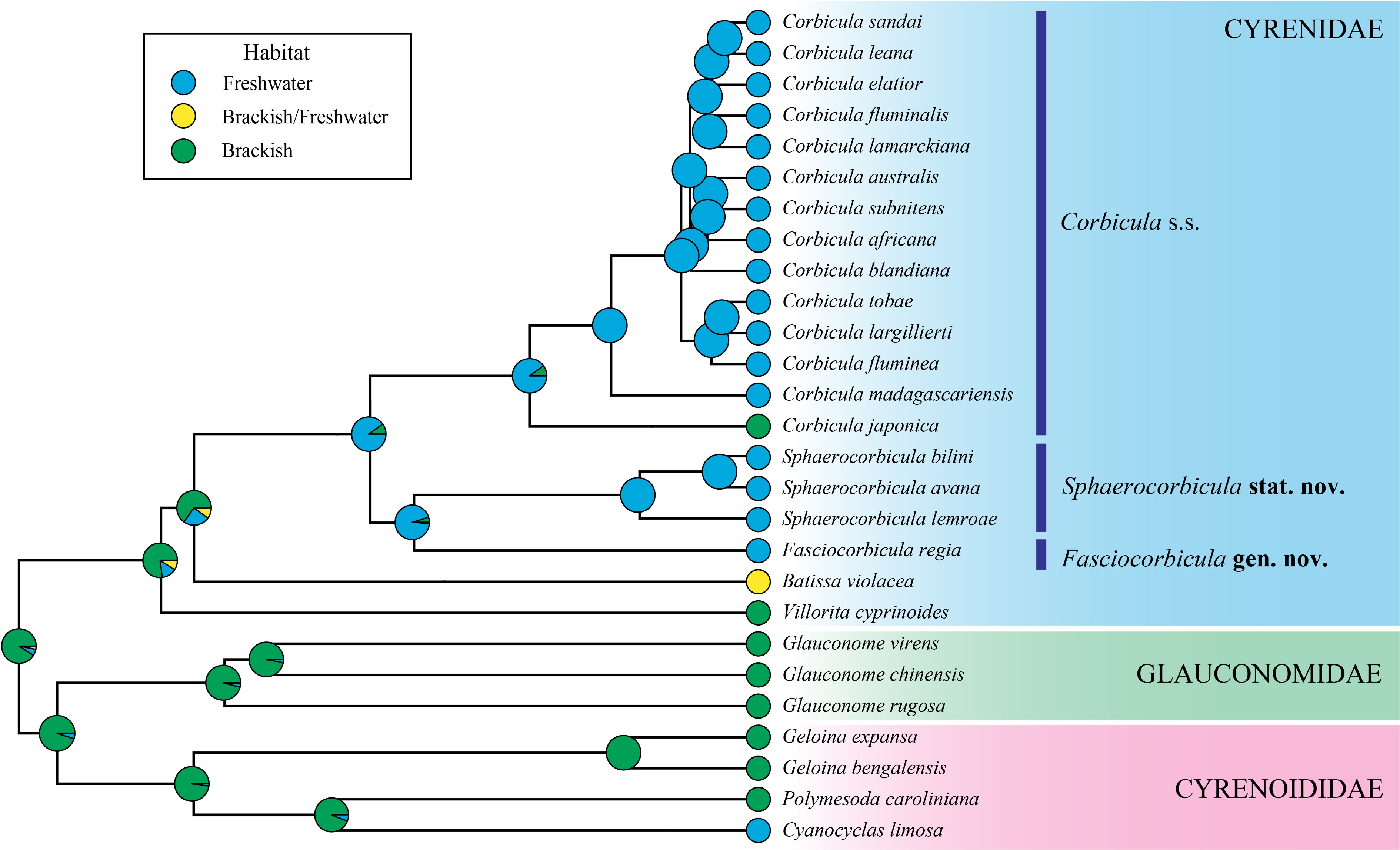
Figure 4.
Ancestral state reconstruction of habitat types mapped onto the Maximum Clade Credibility (MCC) tree derived from BEAST analysis. Pie charts at internal nodes indicate the marginal posterior probabilities for each ancestral state (blue for freshwater, green for brackish, and yellow for a wide distribution range covering both freshwater and brackish water). Observed states for extant taxa are shown at the tips.
Haplotype network analysis
-
The median-joining haplotype network generated from the 663-bp COI gene dataset reveals a clearly structured pattern comprising eight unique haplotypes from 14 examined F. regia (Fig. 1c). Haplotype h5 is the most common, represented by five individuals. The remaining haplotypes consist of one to two individuals and are connected to others by one to three mutational steps. The network also reveals a distinct phylogeographic pattern in which each haplotype corresponded exclusively to a single area, with no haplotypes shared between regions.
Taxonomy
-
Family Cyrenidae Grey, 1840
Fasciocorbicula
-
Jeratthitikul, gen. nov.
Zoobank:
https://zoobank.org/urn:lsid:zoobank.org:act:B2445A14-2428-4AEF-8EFB-35DA4DE150D5 Type species: Corbicula regia Clessin, 1879, by present designation.
Diagnosis: Shell small (maximum length 14.2 mm), inflated, highly inequilateral, and anteriorly elongated. Concentric ribs strong and widely spaced. Hinge with three cardinal teeth in each valve and two crenulated lateral teeth. Internal surface dull with white nacre, marked by five dark-purple radial stripes: three conspicuous stripes evenly distributed from umbo to ventral margin, and two narrow stripes extending from umbo towards lateral teeth.
Differential diagnosis: The new genus differs significantly from Corbicula s.s. and Sphaerocorbicula by its small shell and the five dark-purple radial stripes on the inner shell surface. The exterior of the new genus is sculptured with rather strong concentric ribs, resembling Corbicula s.s. However, this feature readily distinguishes it from Sphaerocorbicula, which possesses a very weak concentric sculpture.
Molecular diagnosis: The new genus is recovered as a distinct clade in the multi-locus phylogenetic analyses (Fig. 2) and exhibits a genetic divergence of approximately 11% to 16% in the COI gene from other genera in the family Cyrenidae (Table 1). It also differs from other genera by nine fixed nucleotide substitutions in the COI gene fragment (Table 2).
Table 2. Fixed nucleotide differences of COI sequences for genus-level diagnosis.
Genera Fixed nucleotide differences* Corbicula s.s. 2G, 8T, 15A, 24A, 86A Sphaerocorbicula stat. nov. 164T Fasciocorbicula gen. nov. 77G, 122G, 128G, 134A, 185G, 293A, 404A, 503A, 551A Batissa 36C, 38T, 49G, 59T, 68T, 81G, 92G, 134G, 215G, 216T, 217C, 218T, 201A, 273G, 274C, 275T, 276T, 368G, 383T, 392A, 461G, 474A, 530T, 531C Villorita 47A, 68G, 81C, 93G, 95T, 98C, 128A, 131A, 252A, 256A, 278G, 371A, 404G, 459C, 470A, 507G, 520C, 524G, 543G *Nucleotide position based on the sequence alignment in this study. Description: As in the description of the type species.
Etymology: The genus name Fasciocorbicula is derived from a combination of the Latin word 'fascia', meaning 'strip or band', and the original genus name of the type species, Corbicula. This refers to its characteristic fine stripes on the inner side of the shell. The gender is feminine.
Distribution: The Malay Peninsula (Southern Thailand and Malaysia).
Species included: There is currently only one species, Fasciocorbicula regia (Clessin, 1879), that belongs to this genus.
Fasciocorbicula regia (Clessin, 1879) comb. nov. (Figs 1−7)
-
Corbicula regia Clessin, 1879[22]: 267 (original description), pl. 43, fig. 5 (reproduced in Fig. 5a of this study); Preston, 1915[51]: 221–222 (description); Prashad, 1928[24]: 17 (mentioned); Prashad, 1928[23]: 34 (description), pl. 3, fig. 10–12; Brandt, 1974[20]: 320 (description), pl. 28, fig. 86; Glaubrecht et al. 2007[25]: 259 (lectotype designation), fig. 2Q.
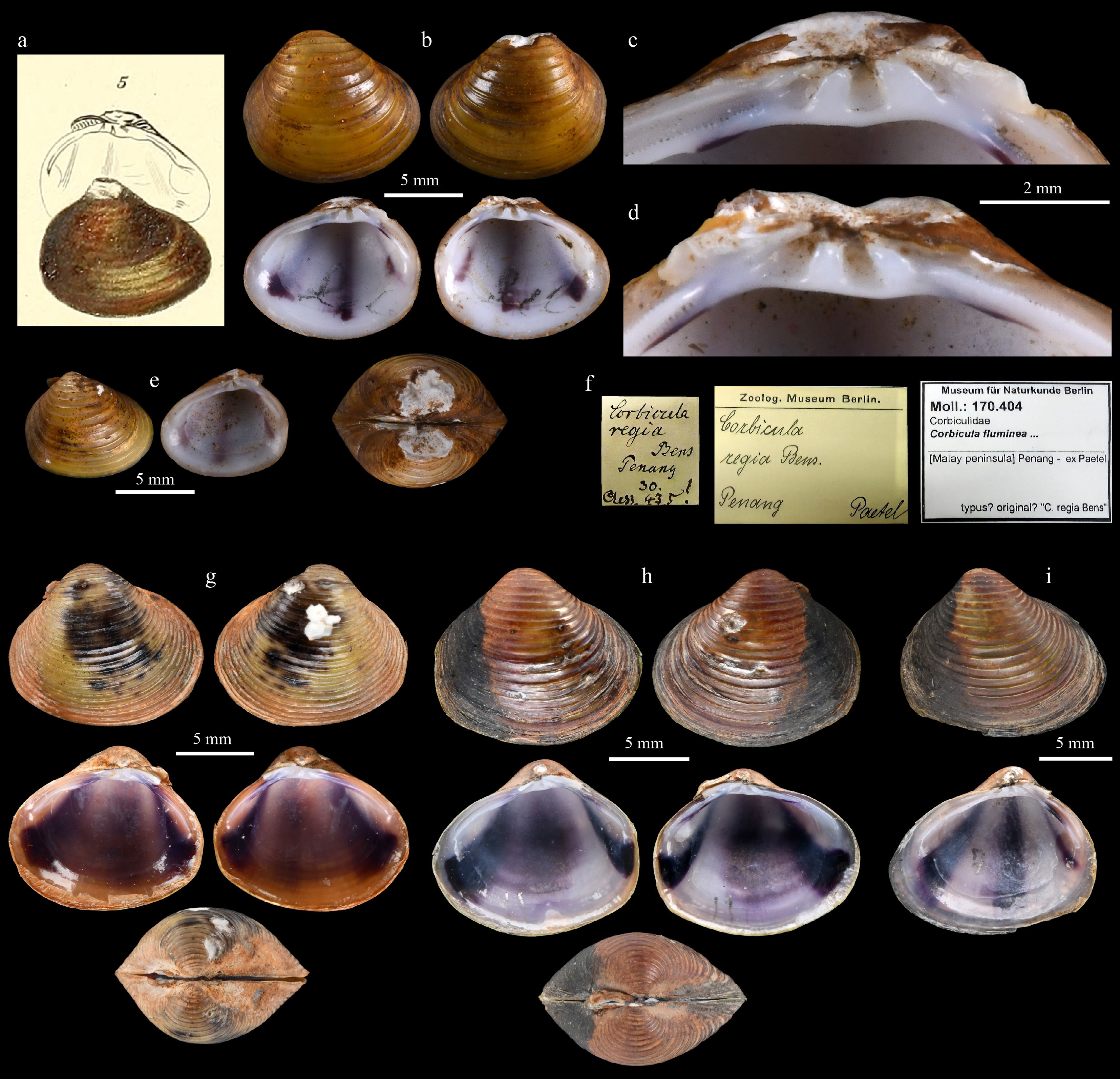
Figure 5.
Fasciocorbicula regia. (a) Original figure by Clessin (1879: pl. 43, fig 5[22]); (b) lectotype ZMB 170.404a from Penang, Malaysia; (c) hinge dentition of the right valve, and (d) left valve of the lectotype; (e) paralectotype ZMB 170.404b, right valve; (f) label of the type material; (g) topotype specimen MUMNH-COR0274-2 from Penang, Malaysia; (h) specimen MUMNH-COR0275-11 from Trang River, Thailand; (i) specimen MUMNH-COR0275-7, right valve, from Trang River, Thailand. Photo Credit: (a) Image digitized by the Biodiversity Heritage Library (Public domain;
www.biodiversitylibrary.org/page/34337663 ), (b)–(i) C. Sutcharit.Type locality: 'Wahrscheinlich Indien'[22] [= Probably India] (in an error for Penang, Malaysia[24,25])
Lectotype: 1 shell (ZMB 170.404a; Fig. 5b–d) MALAYSIA, Penang.
Paralectotype: Right valve (ZMB 170.404b; Fig. 5e), MALAYSIA, Penang.
Other examined material: Two shells (MUMNH-COR0274), MALAYSIA, Perai River, Kampung Baharu, Kubang Semang, Penang State; 5°26'43.5" N 100°27'38.8" E; 15 xii 2022; B. Ng, S. Panha, C. Sutcharit, and E. Jeratthitikul leg. One shell (SMF 228100a) THAILAND, Trang, South Thailand; R. Brandt leg. Two shells (MUMNH-COR0268) THAILAND, Trang River at Nong Trut, Mueang Trang District, Trang Province; 7°36'42.2" N 99°31'05.5" E; 20 xii 2021; E. Jeratthitikul leg. Eighteen shells (MUMNH-COR0275) same location data as previous; 18 iii 2023; E. Jeratthitikul and R. Boonyayon leg.
Re-description: Shell small (length 11.2 mm, height 9.9 mm, and width 8.6 mm in lectotype; Fig. 5b), thick, ovate anteriorly elongated, inflated, and highly inequilateral. Umbo broad, inflated, and prominent, oriented anteriorly, pointing inwards, and showing signs of erosion. Anterior margin rounded and elongated; ventral margin curved; posterior margin wider and obtuse. Periostracum yellowish-brown to brownish. Concentric ribs relatively strong and distantly spaced (4 ribs per 5 mm in lectotype). Ligament short and prominent. Internal surface dull with white nacre, marked by five dark-purple radial stripes: three primary stripes distributed evenly across shell surface, extending from umbo to ventral margin, and two shorter stripes extending from umbo towards lateral teeth; middle median radial stripe sometimes faint or very faint. Hinge margin moderately broad; three cardinal teeth on each valve and two crenulate lateral teeth; cardinal teeth small and non-bifid (Fig. 5c, d). Lateral teeth elongated, depressed, and slightly arched, with anterior lateral teeth longer than posterior ones. Adductor muscle scars small and oval.
Siphons conical, thin-walled, with narrow circular or oval apertures (Fig. 6a, f–i); apertures subequal in size. Outer surface of siphons white to semi-transparent; inner surface white to yellow with pigmentation often concentrated near apertural rings as yellow-brown to dark brown bands (Fig. 6a, g, i); in living specimens, inner surface of exhalant siphon occasionally mottled with yellow or brownish pigment pattern (Fig. 6f). Inhalant siphon aperture with 1–3 rows of long papillae; exhalant siphon aperture smooth (Fig. 6g) or with 1–2 rows of conical papillae (Fig. 6h). Siphonal muscles weak. Presiphonal suture relatively long, outer surface smooth (Fig. 6a) or with a single row of short papillae (Fig. 6f). Marginal mantle smooth (Fig. 6e) or with a row of widely spaced minute papillae. Radial mantle muscles weak, forming separate bundles (Fig. 6e). Gills elongated, slightly ribbed (Fig. 6d); outer demibranch narrower than inner demibranch. Labial palps narrow, triangulate, pointed at tip (Fig. 6a–c).
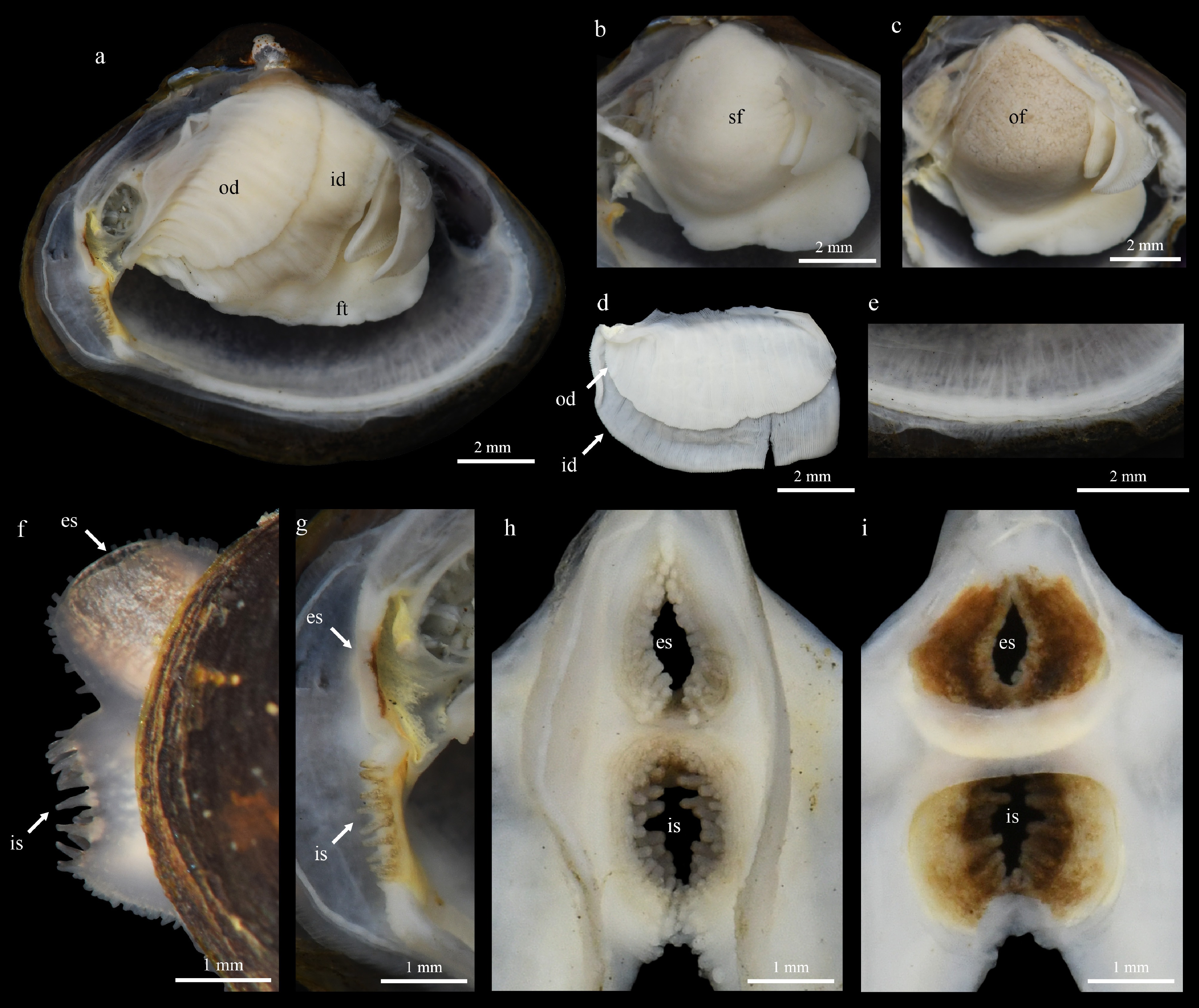
Figure 6.
Anatomy of Fasciocorbicula regia from Trang River, Thailand. (a) Anatomy of the soft body, with right mantle lobe removed; anatomy of (b) male and (c) female showing fully developed gonadal follicles, right mantle lobe and right gills removed; (d) right gills; (e) outgrowths on the mantle edge; (f) lateral view of extruded siphons in living specimen; (g) anatomy of siphons in preserved specimens, with the right part of the siphons removed; (h) outer and (i) inner views of the siphons. Photographs by A. Pholyotha. Abbreviations: es, exhalant siphon; ft, foot; id, inner demibranch; is, inhalant siphon; od, outer demibranch; of, oogenic follicles; sf, spermatogenic follicles.
Reproductive biology: Fasciocorbicula regia is dioecious, distinctly male or female, as evidenced by the gonads of dissected specimens (Fig. 6b, c) and histological sections (Fig. 7a, f), which contained either spermatozoa or oocytes within a single individual. In males, various stages of spermatogenic follicles were present (Fig. 7a), ranging from the early growing stage (Fig. 7b) to the maturing stage (Fig. 7c), in which aggregations of spermatozoa were observed in the follicle lumen (Fig. 7d). Spermatozoa were relatively small and monoflagellate (Fig. 7e). The sperm head was spherical in shape. The average (± SD) head length was 4.1 ± 0.2 µm (n = 10). The condition of the gonads indicates that the specimens were at the late mature to spawning stages. The sex ratio of the studied specimens was approximately 1:1 (n = 10). No evidence of larval brooding was found in the inner demibranchs of any examined specimens (n > 15 specimens) (Figs 6d, 7h).
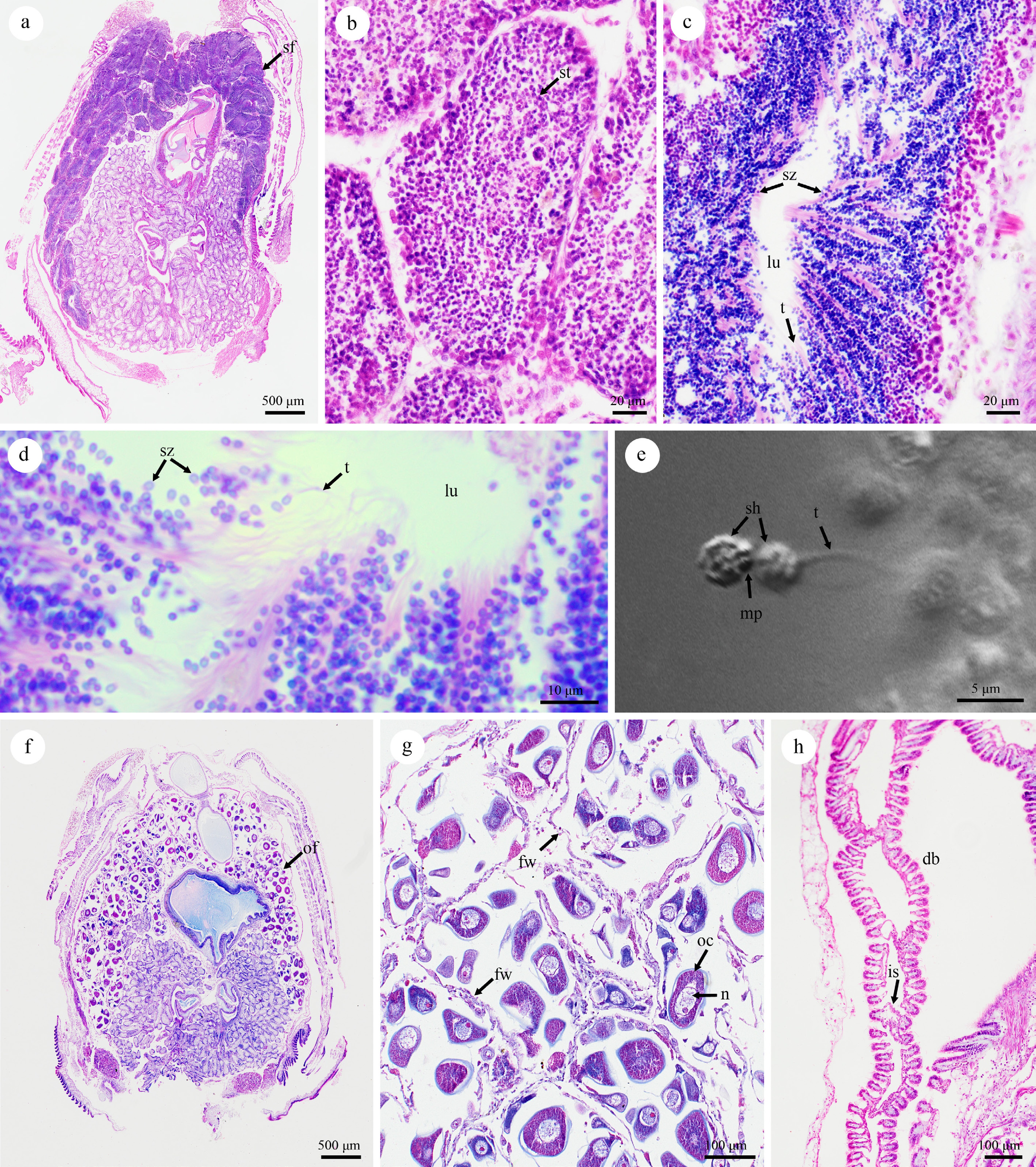
Figure 7.
Histological thin sections of Fasciocorbicula regia stained with Hematoxylin–Eosin showing gonadal developmental stages and related organs in (a)–(d) males, (f)–(h) females, and (e) sperm morphology examined by inverted confocal laser scanning microscopy; (a) general aspect of the male visceral mass showing spermatogenic follicles in various stages; (b) spermatogenic follicles in the early growing stage; (c) maturing stage showing fully developed spermatozoa; (d) aggregations of spermatozoa in the center of the follicle lumen; (e) detail of sperm morphology showing the head and monoflagellated tail; (f) general aspect of the female visceral mass showing a mature gonad containing mature oogenic follicles in a late developmental stage; (g) mature oogenic follicles, partially spawned; (h) section of the inner demibranch. Abbreviations: db, demibranch; fw, follicle wall; is, interlamellar septa; lu, lumen; mp, midpiece; n, nucleus; oc, oocytes; of, oogenic follicle; sf, spermatogenic follicle; sz, spermatozoa; sh, spermatozoa head; t, spermatozoa tail.
Variation: Shell dimensions of the newly collected specimens from the Penang and Trang populations ranged from 10.7–14.2 mm (mean 12.8 mm; n = 12) in shell length, 9.8–12.2 mm (mean 11.1 mm) in shell height, and 7.6–8.7 mm (mean 8.1 mm) in shell width. The overall size did not differ significantly between the two populations. However, the overall shell shape varies ontogenetically from ovate in juveniles to becoming more anteriorly elongated in fully grown adults.
The newly collected specimens from both populations align with the general morphology of the lectotype (Fig. 5b), though they exhibit some intraspecific variation. Specimens from both populations possess finer concentric ribs (6–8 ribs per 5 mm vs 4 ribs per 5 mm in the lectotype), a more obtuse posterior end, and a duller interior surface (Fig. 5g–i). Additionally, the middle dark purple stripe on the inner shell surface is often reduced, appearing only as a faint to very faint dark tinge in many individuals (Fig. 5g, h), although it is still quite clear in some individuals (Fig. 5i). The periostracum of specimens from the Trang River tends to be darker, ranging from brownish to dark brown (Fig. 5h, i). Finally, some topotype specimens from Penang feature a darker interior surface with an orange-brown hue near the shell edge (Fig. 5g).
Distribution: The Malay Peninsula (Southern Thailand and Malaysia; Fig. 1a).
Habitat: Slow-flowing rivers with a sandy or sandy-muddy bottom (Fig. 1b).
Comments: Clessin (1879) provided a description and figures of 'Corbicula regia' based on specimens from Paetel's collection housed in the Zoological Museum in Berlin, but attributed the authorship to 'Benson' (on page 267[22]). However, there is no evidence that 'Benson' provided either a description or figure of this taxon; therefore, Clessin (1879) is the sole valid authority for the name[23,25]. In the original description, the type locality was stated as 'Wahrscheinlich Indien' [= Probably India], and this was followed by Preston[51]. However, in a later work revising the Indian Corbicula, Prashad[24] stated that the type locality seemed erroneous, and he subsequently excluded this species from the Indian fauna. Prashad also had the opportunity to re-examine the specimens in the Zoological Museum in Berlin (ZMB), the Natural History Museum, London (NHM), and the Indian Museum in Calcutta and found that all of these specimen lots had probably come from the same donor, and the original labels clearly indicated the collection locality to 'Penang'[23]. Therefore, Prashad corrected the type locality to 'Penang, Malaysia'[23]. This correction is further supported by Glaubrecht et al.[25], who designated the unique name-bearing type based on a specimen from Paetel's collection (lectotype ZMB 170.404a) with the collection locality stated as 'Penang' (Fig. 5f). Moreover, our newly obtained specimens from the Perai River, Penang, Malaysia, confirm its presence in the Malay Peninsula, with its range extending further north to the Trang River in Southern Thailand (Fig. 1).
-
The phylogenetic analyses presented in this study provide the first molecular assessment of 'C. regia', a species that has remained enigmatic since its description over a century ago[22]. The multi-locus phylogenies (COI + 16S rRNA + 28S rRNA) consistently recovered it as a distinct evolutionary lineage from both the Corbicula s.s. and Sphaerocorbicula clades (Figs 2, 3), underpinning the generic-level revisions proposed herein. Corbicula s.l. is thus split into three genera corresponding to three divergent clades: Corbicula s.s., Sphaerocorbicula stat. nov., and Fasciocorbicula gen. nov. Sphaerocorbicula comprises three species endemic to Myanmar[3]: S. avana (Theobald, 1874) comb. nov., S. bilini (Bespalaya et al., 2025 in Bespalaya et al.[3]) comb. nov., and S. lemroae (Bespalaya et al., 2025 in Bespalaya et al.[3]) comb. nov. Fasciocorbicula gen. nov. is monotypic, accommodating F. regia (Clessin, 1879) comb. nov., while Corbicula s.s. contains all remaining Corbicula species.
The establishment of Fasciocorbicula gen. nov. is strongly supported by its distinct morphological characters. While Corbicula s.s. is characterized by ovate-triangular shells with strong concentric ribs[3,18], and Sphaerocorbicula by small, ovate shells with weak concentric sculpture[3], Fasciocorbicula gen. nov. possesses a unique combination of features. The type species, F. regia, is distinguished by its very small (shell length not exceed 15 mm), rather thick shell, highly inequilateral and elongate anteriorly, and inflated with strong, widely spaced concentric ribs; however, the most important character is the radial color patterns on the internal shell surface.
The presence of dark purple stripes on the inner surface of the valves (Fig. 5) stands out as a unique diagnostic feature, making the genus very distinguishable from other genera in the Cyrenidae. In most described members of Corbicula s.s., pigmentation of the internal shell surface is typically uniform, although some species/populations exhibit a concentrically banded pattern or a darker color marked on a specific area on the inner surface of the valves[3,10]. For example, in the widespread C. fluminea, the interior is generally characterized by a solid deep violet or white coloration, occasionally with a darker central blotch under the umbo, darker tinge along the lateral teeth, or darker band along a shell edge[10,52]. Pronounced radial stripes may be present on the periostracum of juveniles in some species, such as in C. japonica[10] and C. fluminalis[53]. These external markings rarely translate into distinct, permanent dark stripes on the internal surface in adult specimens. If internal markings are present, they typically appear as a central blotch near the umbo or a faint radial dark band, but never extend to the ventral margin of the shell[52,53]. However, these patterns are fundamentally different from the condition observed in the new genus. In F. regia, the pigmentation is not restricted to the hinge or umbonal area but presents as five continuous, sharp radial stripes extending from the umbo to the shell margin (Fig. 5). Although S. bilini, recently described from Myanmar, exhibits a dark stripe on the inner surface of the shell similar to that in F. regia, its stripe is dark grey to black, and there is only a single stripe near the posterior margin of the shell[3]. Phylogenetically, S. bilini is nested within the genus Sphaerocorbicula, distant from the position of F. regia (Figs 2, 3). Furthermore, S. bilini is readily distinguishable from F. regia by its triangular and slightly elongated shell shape and by the shell exterior, which has a very fine, weak concentric sculpture and a V-shaped dark marking pattern[3].
The evolutionary reversal from freshwater to brackish environments in C. japonica
-
Corbicula clams are of significant interest due to their evolutionary transition from saline to freshwater environments. The family Cyrenidae currently comprises five extant genera—Batissa, Corbicula, Fasciocorbicula gen. nov., Sphaerocorbicula, and Villorita Gray, 1833[27]—with diverse habitat preferences observed across them. Members of the Villorita are restricted to brackish water[54], while Batissa species are euryhaline, capable of inhabiting both freshwater and the freshwater–estuarine interface[28]. In contrast, Fasciocorbicula gen. nov., Sphaerocorbicula, and Corbicula have successfully radiated into diverse freshwater niches, with C. japonica remaining the only species that inhabits brackish water[2,10]. Phylogenetic evidence from previous studies consistently placed C. japonica at the base of the Corbicula clade, followed by a vast and diverse radiation of freshwater taxa[2,8,14,15,28]. This topology provides strong evidence to support that the common ancestor of Corbicula likely inhabited estuarine or coastal environments before lineages specialized into purely freshwater habitats[17], positioning C. japonica as a critical evolutionary link for the colonization of inland waters[28].
However, the recent discovery of a new freshwater clade from Myanmar (i.e., the genus Sphaerocorbicula[3]) and the phylogenetic placement of F. regia in this study challenge this hypothesis. Our phylogenetic reconstruction places the brackish C. japonica deeply nested among the freshwater lineages (Figs 2, 3), rather than as a basal sister lineage to freshwater taxa. Moreover, results from the ancestral state reconstruction analysis support this topology, identifying the freshwater habitat as the most likely ancestral habitat type for Corbicula species (90% posterior probability; Fig. 4; Supplementary Table S5). This indicates that C. japonica represents a case of evolutionary reversal, implying that the lineage did not simply retain an ancestral marine tolerance (i.e., as in a common ancestor shared with Batissa and Villorita), but rather secondarily re-colonized the brackish environment from a freshwater ancestral stock. The time-calibrated phylogeny and biogeographic analysis in this study suggest that this transition may have occurred in the Paleogene era (~37.5 Mya; Fig. 3) in an area spanning Southeast to East Asia, supported by the divergence timing of C. japonica from the common ancestor of the highly diverse freshwater clade.
Reproductive biology of F. regia
-
Histological examination revealed F. regia to be dioecious, with individuals possessing either distinctly male or female gonads (Fig. 7a for male and Fig. 7f for female). The male gonads contained small spermatozoa with a single flagellum and a spherical head (Fig. 7e). This sperm morphology is a reliable indicator of sexual reproduction mode, as all sexually reproducing Corbicula lineages possess reduced monoflagellate spermatozoa, whereas hermaphroditic lineages that reproduce through androgenesis produce unreduced biflagellate spermatozoa[7,12,17,55]. Sexual reproduction confers distinct evolutionary advantages over androgenesis, particularly in the long-term maintenance of genetic diversity[2]. This is clearly evidenced in the case of F. regia in this study. The median-joining haplotype network generated from the COI gene dataset (Fig. 1c) revealed a highly structured population comprised of eight unique haplotypes identified from 14 examined individuals across two populations. Significantly, the network displays a distinct phylogeographic structure, with haplotypes corresponding exclusively to single areas. Such high genetic divergence is also typically observed in sexually reproducing Corbicula populations within their native ranges[3,56]. In contrast, invasive androgenetic populations exhibit significantly lower genetic diversity in their introduced ranges compared to that observed in the native Asian populations[2].
The small spermatozoa with a single flagellum and a spherical head observed in F. regia align with the primitive or ect-aquasperm type of spermatozoa commonly found in invertebrates that release their gametes into the water and reproduce by external fertilization[57]. The morphology of this sperm type is characterized by a small size with a spherical or short-conical head, a short midpiece containing a ring of mitochondria, and a single flagellum[58]. This primitive sperm morphology stands in sharp contrast to Corbicula s.s. members that produce spermatozoa with an elongated or rod-shaped head, regardless of whether they are mono- or biflagellate[8,11−13]. Spermatozoa with an elongated nucleus are considered a modified type among bivalves[59], aligning with the definition of ent-aquasperm, in which sperm are released into the water and drawn in by the inhalant siphon of neighboring individuals. The fertilization then occurs inside the confines of the mantle cavity[57]. This modification of sperm morphology appears to be an evolutionary adaptation to their specialized reproductive mode of internal fertilization and inner demibranch brooding[12]. The modified sperm may serve as a derived character for the Corbicula s.s. clade, which evolved from spherical primitive spermatozoa as in F. regia, regardless of whether they use internal or external fertilization. Unfortunately, there is no information on sperm anatomy available for the genus Sphaerocorbicula, which also has sexual reproduction[3] and follows a phylogenetic placement between F. regia and the diverse clade of Corbicula s.s. This information would better resolve the evolutionary history of spermatozoa morphology in the family Cyrenidae.
The genus Corbicula s.s. exhibits a remarkable plasticity in life-history strategies, particularly regarding larval development[17]. This diversity ranges from the release of free-swimming planktotrophic larvae found in the estuarine species, such as C. japonica[16], to the release of benthic larvae, as seen in the Lake Biwa endemic C. sandai[9], and probably C. elatior Martens, 1905[10]. However, the most derived strategy is the parental care system, characterized by the maternal incubation of juveniles within the inner demibranchs. This brooding behavior is widely documented in various freshwater lineages, both dioecious and hermaphroditic species, and from across native and invasive ranges[8,11,17−19].
Brooding strategies in Corbicula s.s. have been linked to the colonization of freshwater habitats and are considered a derived character state that evolved from a non-brooding ancestor[28]. However, among the diverse lineages of this freshwater group, a few species retain a non-brooding strategy[3,9]. In the case of F. regia, no brooding individuals were found among any of the specimens examined in this study. In addition, histological examination of the gills revealed no larvae and the absence of any thickening of interlamellar septa (Fig. 7h), which supports a non-brooding reproductive strategy in F. regia. Based on phylogenetic placement and the evidence of sperm morphology, we assume that F. regia may have free-swimming planktotrophic larvae, similar to estuarine species like C. japonica[16] and V. cyprinoides[60]. Alternatively, as for adaptation to a freshwater environment, it may have larvae that are non-swimming and transform into benthic juveniles immediately after leaving the egg capsules, such as in C. sandai[9]. Further research is required to discriminate between and verify these hypotheses.
-
This study elucidates the systematics of F. regia, elevating it to the rank of a distinct genus based on concordant molecular, morphological, and reproductive biology evidence. The identification of a unique internal shell character and the confirmation of sexual reproduction in F. regia, and the uncovering of phylogenetic evidence of a habitat reversal in C. japonica significantly advance our understanding of evolution in the family Cyrenidae. These findings emphasize that the diversity of Southeast Asian Cyrenidae, and particularly Corbicula s.s., is vastly underestimated and that ancient, sexually reproducing lineages persist in native river systems despite the global expansion of invasive lineages. Future research should expand sampling in other areas of Southeast Asia to cover nearly 100 unstudied nominal Corbicula taxa[21] and further expand the multi-locus phylogeny to a phylogenomic study[37] or a comparative genomic study between F. regia and androgenetic Corbicula lineages, which would investigate the genomic architecture underlying the transition between sexual and asexual reproductive modes, or between C. japonica and other freshwater lineages to test the hypothesis of secondary recolonization of brackish environments from freshwater ancestors.
We thank Klomthong S and Boonyayon R for their assistance in collecting samples, and Pootanon P and Sapparojpattana P for laboratory assistance in sperm preparation. We also extend our gratitude to Anderson DJ for grammar checking and comments on an earlier version of this manuscript.
-
All procedures were reviewed and preapproved by the Faculty of Science, Mahidol University Animal Care and Use Committee (SCMU-ACUC), identification number: MUSC63-017-525, approval date: June 8, 2020. During the preparation of this work, the authors used Google Gemini (Date: March 25, 2026) for grammar correction and language refinement. The authors reviewed and edited all content produced with the assistance of this tool, verified its accuracy, and take full responsibility for the integrity and originality of the final manuscript. This work represents the authors' own intellectual contribution, and no AI tool is credited as an author.
-
The authors confirm their contributions to the paper as follows: study conception and design: Jeratthitikul E, Sutcharit C; specimen collection: Jeratthitikul E, Sutcharit C, Panha S, Ng B; analysis and interpretation of results: Jeratthitikul E, Singhakaew S; image preparation: Sutcharit C; and draft manuscript preparation: Jeratthitikul E. All authors reviewed the results and approved the final version of the manuscript.
-
The DNA sequence data underlying this article are available in the GenBank Nucleotide Database at www.ncbi.nlm.nih.gov/nucleotide/ and can be accessed using the accession numbers provided in this article and listed in Supplementary Tables S2 and S3.
-
Author Beewah Ng was employed by Sea Marine Products. The remaining authors declare that the research was conducted in the absence of any commercial or financial relationships that could be construed as a potential conflict of interest.
-
accompanies this paper online at: https://doi.org/10.48130/jzser-0026-0013.
- Supplementary Table S1 Summary of primers used, amplicon lengths, and PCR profiles.
- Supplementary Table S2 Collection data and GenBank accession numbers for the newly sequenced individuals in this study.
- Supplementary Table S3 List of taxa used in the phylogenetic analysis with associated habitat types, native distribution ranges, and GenBank accession numbers.
- Supplementary Table S4 Partition schemes with their respective quantities of base pairs and the best-fit models of nucleotide substitution.
- Supplementary Table S5 Results of divergence times, ancestral area estimates, and ancestral state reconstruction of habitat types for each node.
- Supplementary Fig. S1 Historical biogeographic events of the members in the family Cyrenidae and related taxa.
- Copyright © 2026 by the author(s). Journal of Zoological Systematics and Evolutionary Research published by Maximum Academic Press on behalf of John Wiley & Sons Ltd. This is an open access article under the terms of the Creative Commons Attribution License, which permits use, distribution and reproduction in any medium, provided the original work is properly cited.
-
About this article
Cite this article
Jeratthitikul E, Sutcharit C, Singhakaew S, Panha S, Ng B. 2026. Phylogenetic placement and reproductive mode of Corbicula regia (Venerida: Cyrenidae) bring new insights into systematics and evolution of Corbicula clams. Journal of Zoological Systematics and Evolutionary Research 2026: e014 doi: 10.48130/jzser-0026-0013 

Phylogenetic placement and reproductive mode of Corbicula regia (Venerida: Cyrenidae) bring new insights into systematics and evolution of Corbicula clams
- Received: 26 March 2026
- Revised: 23 July 2026
- Accepted: 19 August 2026
- Published online: 18 September 2026
Journal of Zoological Systematics and Evolutionary Research
2026,
Article number: e014 (2026)
|
Cite this article
Abstract: The genus Corbicula (Bivalvia: Cyrenidae) includes some of the most successful invasive freshwater species globally, yet its native diversity in Southeast Asia remains poorly understood. Corbicula regia is an enigmatic species originally described from Penang, Malaysia, with no confirmed records for over a century. In this study, C. regia was rediscovered from its type locality in Malaysia, and a new population in Southern Thailand was found. Multi-locus phylogenetic analysis (COI, 16S rRNA, and 28S rRNA) recovered C. regia as a distinct evolutionary lineage that is completely separated from both the Corbicula s.s. and Sphaerocorbicula clades, warranting the establishment of Fasciocorbicula Jeratthitikul, gen. nov., to accommodate the species, and the elevation of the genus Sphaerocorbicula stat. nov. Morphological examination supports the distinctness of the new genus, which is characterized by a unique dark purple radial stripe pattern on the inner shell surface. Histological examination and sperm morphology revealed that F. regia is strictly dioecious with non-brooding larval development, and possesses primitive, spherical-headed, monoflagellated spermatozoa. These traits contrast sharply with the hermaphroditic, androgenetic, and brooding strategies typical of widely invasive Corbicula s.s. lineages. Time-calibrated phylogeny and ancestral state reconstruction analysis suggested that the center of origin for these three genera was in the Southeast Asian region and its radiation occurred during the Early Cretaceous (~104.2 Mya), with all Corbicula s.s. lineages originating from a common freshwater ancestor. This challenges the widely held hypothesis of direct freshwater colonization from a brackish common ancestor. Instead, the results suggest that the brackish C. japonica represents a case of evolutionary reversal, having secondarily recolonized brackish environments from a freshwater ancestor.
-
Key words:
- Freshwater bivalves /
- New taxa /
- Reproductive biology /
- Secondary brackish species /
- Systematics /
- The Malay Peninsula