











-
Radiation enteritis, a common complication arising from abdominal or pelvic radiotherapy, can significantly impair the quality of life of cancer patients[1]. Clinically, radiation enteritis presents with a diverse array of gastrointestinal symptoms, such as abdominal pain, diarrhea, nausea, vomiting, and, in severe cases, intestinal bleeding and perforation. These symptoms not only exacerbate physical suffering but also complicate treatment plans, often requiring dose reductions or interruptions in radiotherapy, potentially compromising therapeutic outcomes.
The underlying pathogenesis of radiation enteritis is multifaceted, primarily involving intestinal barrier disruption, oxidative stress, inflammatory responses, and gut microbiota dysbiosis[2], forming a self-reinforcing loop: oxidative stress acts as the trigger, directly damaging intestinal epithelial cells (IECs) and disrupting barrier integrity while simultaneously triggering inflammation. Inflammation, in turn, amplifies oxidative injury and directly disrupts tight junction proteins, establishing a vicious cycle. Barrier disruption enables bacterial translocation and toxin release, which further fuel inflammation and oxidative stress. Concurrently, microbial dysbiosis exacerbates inflammation by compromising barrier integrity and activating the immune system. Thus, these intertwined pathological processes drive the initiation and progression of radiation enteritis.
Some plants have long been recognized for their medicinal properties, and numerous studies have explored their potential in treating various diseases. In the context of radiation enteritis, several representative plants and their extracts, including Sophora flavescens[3], Scutellaria baicalensis[4], Huanglian[5], Centella asiatica[6], and Paeonia lactiflora[7], have been well investigated for their therapeutic effects. These plants contain a diverse array of bioactive compounds, which can be broadly classified into flavonoids, terpenoids, phenylpropanoids, terpenophenolics, non-flavonoid polyphenols, polysaccharides, alkaloids, anthraquinones, and sterols. The active constituents exert their therapeutic effects on radiation enteritis primarily through three mechanisms—antioxidant, anti-inflammatory, and intestinal barrier repair[8−11]—by acting on canonical pathways, such as activating Nuclear factor erythroid 2-related factor 2 (Nrf2)/antioxidant response element (ARE) pathway to enhance endogenous antioxidant defense, and inhibiting the nuclear factor kappa-B (NF-κB) and NOD-like receptor family pyrin domain containing 3 (NLRP3) inflammasome to block the inflammatory cascade.
In recent years, the therapeutic potential of plant-based remedies has gained considerable attention. Clinical evidence indicates that of the diverse oral pharmaceuticals for radiation enteritis, traditional Chinese herbal decoctions demonstrate the most pronounced therapeutic efficacy[12]. Plants and their extracts, endowed with a rich repertoire of bioactive compounds, offer a promising alternative or complementary approach to mitigate radiation enteritis.
Therefore, this review examines the pathogenesis of radiation enteritis and synthesizes existing literature on the use of plants and their extracts for both prophylactic and therapeutic purposes. Additionally, it provides an overview of various plant-derived active factors used in the treatment of radiation enteritis, along with their primary mechanisms, highlighting their potential as therapeutic agents. By elucidating the underlying mechanisms and summarizing the current state of research, this review aims to provide a foundation for future investigations and the development of novel therapeutic strategies. Furthermore, it discusses future directions, including the application of synthetic biology, nanotechnology, and artificial intelligence (AI) to optimize the efficacy and delivery of plant-based therapies for radiation enteritis.
-
Radiation enteritis is a common complication in patients with abdominal and pelvic malignant tumors following radiotherapy, characterized by radiation-induced intestinal epithelial damage, which may include mild inflammation. During radiotherapy, approximately 75% of patients with abdominal and pelvic tumors experience side effects, with 50% reporting a significant decrease in quality of life. Radiation enteritis can be classified into acute and chronic forms based on the onset time and course of symptoms. Acute radiation enteritis usually occurs within 3 months after the start of radiotherapy, with an incidence rate exceeding 75%. Major clinical manifestations include diarrhea, intestinal hemorrhage, fecal incontinence, and others[13]. Chronic radiation enteritis has a delayed onset, with symptoms typically emerging 9−14 months after radiotherapy. However, they may occur at any time within 30 years post-treatment, with a peak incidence rate of 20%. Chronic radiation enteritis presents with symptoms such as bowel obstruction, malabsorption, malnutrition, and other complications, often featuring more severe bleeding compared to the acute form.
The mechanism of radiation-induced enteritis
-
Radiation enteritis is caused by the complex interplay of various pathophysiological mechanisms triggered by ionizing radiation. Ionizing radiation triggers deleterious effects in intestinal tissue, directly inducing mitochondrial oxidative stress and a burst of reactive oxygen species (ROS)[14], while also triggering acute inflammatory responses. Radiation targets and destroys tight connections between IECs, compromising barrier integrity and damaging endothelial cells. It can also alter the composition of the gut microbiota and impair the function of intestinal stem cells (ISCs). These events constitute an integrated network, driving disease progression from the acute to the chronic phase[14].
Disruption of intestinal barrier function
-
The intestinal barrier comprises a mucous layer, tightly connected IECs, and the underlying lamina propria, preventing the invasion of pathogens and toxins. However, radiation therapy can damage the integrity of the intestinal barrier and increase intestinal permeability. In the early stages of radiation enteritis, the proliferative capacity of IECs is impaired, accompanied by villous atrophy, crypt loss, and intestinal mucosal injury. These morphological alterations lead to abnormal expression of tight junction proteins such as Zonula occludens-1 (ZO-1) and claudin-1, thereby impairing intestinal barrier function, enhancing bacterial translocation, and exacerbating inflammatory responses[15]. The disruption of intestinal barrier function not only facilitates pathogen invasion and triggers systemic inflammatory response syndrome (SIRS) but also promotes gut microbiota dysbiosis and amplifies oxidative stress, further exacerbating the pathological process.
Oxidative stress and imbalance of antioxidant balance
-
Radiotherapy induces substantial production of ROS and reactive nitrogen species (RNS), triggering sustained oxidative stress and a continuous elevation of ROS. Mechanistically, the quinone (IQ) site of the mitochondrial respiratory chain complex I is a major source of superoxide and H2O2 after irradiation, accounting for up to two-thirds of intracellular ROS[14]. Under physiological conditions, ROS and RNS play pivotal roles in various metabolic, immune, growth, and differentiation processes, thereby maintaining normal cellular functions. The body possesses a free radical scavenging and antioxidant defense system; however, radiation therapy can disrupt its normal functioning. Concurrently, excessive ROS accumulation induced by radiation elicits oxidative stress, leading to damage to cellular proteins, lipids, and nucleic acids, which in turn triggers apoptosis and necrosis. Additionally, ROS serves as a key second signal for activating the NLRP3 inflammasome[14], thereby linking oxidative stress to downstream inflammatory responses. This cascade of events further damages the intestinal mucosal barrier, destroys crypt cells and intestinal villi, and precipitates severe mucosal inflammation[16].
Imbalance of gut microbiota
-
The gut microbiota maintains a dynamic equilibrium with the host. Under normal physiological conditions, the gut microbiota contributes to polysaccharide breakdown, regulates energy storage in adipocytes, metabolizes xenobiotics, facilitates the renewal of IECs, and maintains the integrity of the intestinal epithelial barrier. It also helps stabilize the mucous layer, decrease intestinal mucosal permeability, and maintain host immune homeostasis. However, radiotherapy can disrupt this equilibrium, inducing structural and diversity alterations in the gut microbiota, primarily characterized by a reduction in beneficial bacteria including Bifidobacterium and Lactobacillus, and an increase in opportunistic pathogens such as Escherichia coli and Klebsiella pneumoniae. Functionally, this dysbiosis reduces short-chain fatty acids (e.g., butyrate), which are essential for colonic epithelial energy and tight junction integrity, while depletion of mucin-degrading bacteria such as Akkermansia muciniphila weakens the mucus layer[17]. Dysbiosis of the gut microbiota affects the function of the intestinal barrier[18], compromising intestinal epithelial barrier integrity, disrupting the mucous layer, and increasing intestinal mucosal permeability. In the chronic phase, metabolites derived from dysbiosis can persistently activate DNA damage sensing pathways, contributing to chronic inflammation and fibrosis[19]. This cascade of events disrupts host immune homeostasis, activates immune cells, triggers the release of pro-inflammatory cytokines, exacerbates tissue damage, and elevates the risk of intestinal infections[20].
Inflammatory reaction
-
Radiotherapy upregulates pro-inflammatory cytokines, including Interleukin-1 beta (IL-1β), Interleukin-6 (IL-6), Interleukin-8 (IL-8), and tumor necrosis factor alpha (TNF-α). These cytokines regulate cytoskeletal protein reorganization, directly leading to the rearrangement of tight junction proteins and impaired intestinal barrier function. The inflammatory cascade is further amplified by bacterial translocation: lipopolysaccharide (LPS) from Gram-negative bacteria that leak across the damaged barrier binds to Toll-like receptor 4 (TLR4) on immune cells, further activating NF-κB and creating a self-reinforcing loop[14]. Consequently, the inflammatory response further exacerbates intestinal damage, forming a vicious cycle, and extends beyond the intestinal mucosa to affect other organs via blood circulation, potentially leading to multiple organ dysfunction syndrome (MODS).
Cell apoptosis and autophagy
-
Radiotherapy can induce apoptosis of IECs, leading to loss of mucosal epithelial cells and the compromise of intestinal barrier integrity. Meanwhile, autophagy plays a crucial role in the development of radiation enteritis by eliminating damaged organelles and proteins, thereby maintaining cellular homeostasis. Radiotherapy can inhibit autophagy, which exacerbates intestinal damage. Conversely, activation of autophagy alleviates oxidative stress, inflammatory responses, and cell apoptosis, thus helping to preserve intestinal barrier function[21]. Dysregulation of autophagy may result in the accumulation of intracellular damage, which further promotes cell apoptosis.
Abnormal activation or inhibition of signaling pathways
-
Multiple signaling pathways are involved in the occurrence and development of radiation enteritis. The stability of intestinal tissue is maintained by the self-renewal, regeneration, and reprogramming of ISCs, and the Wnt/β-catenin pathway plays a key regulatory role in maintaining intestinal tissue homeostasis. The pathway is mediated by β-catenin, which acts as a transcription co-activator to promote the expression of various target genes. Research has demonstrated that the Wnt/β-catenin pathway is crucial for the self-renewal and proliferation of ISCs following radiation injury; however, its precise mechanism remains unclear (Fig. 1a)[22]. The Notch pathway maintains the balance between proliferation and differentiation of IECs, affecting the fate of neighboring cells through intercellular signaling. After binding to its ligands, the Notch intracellular domain translocates to the nucleus to modulate gene expression (Fig. 1b). The bone morphogenetic protein (BMP) pathway has been shown to negatively regulate the self-renewal of ISCs by controlling gene expression and inhibiting BMP signaling (Fig. 1c)[23]. The Hedgehog pathway maintains the stability of the microenvironment of ISCs through paracrine secretion. Indian Hedgehog, secreted by IECs, acts on mesenchymal cells, which in turn produce signaling factors that negatively regulate ISC proliferation (Fig. 1d)[24]. Meanwhile, the signaling pathways regulating oxidative stress, inflammatory responses, and cell apoptosis are intricately interconnected, collectively influencing the occurrence and development of radiation enteritis. Abnormal activation or inhibition of these pathways may result in aberrant proliferation and differentiation of intestinal cells, further exacerbating intestinal damage.
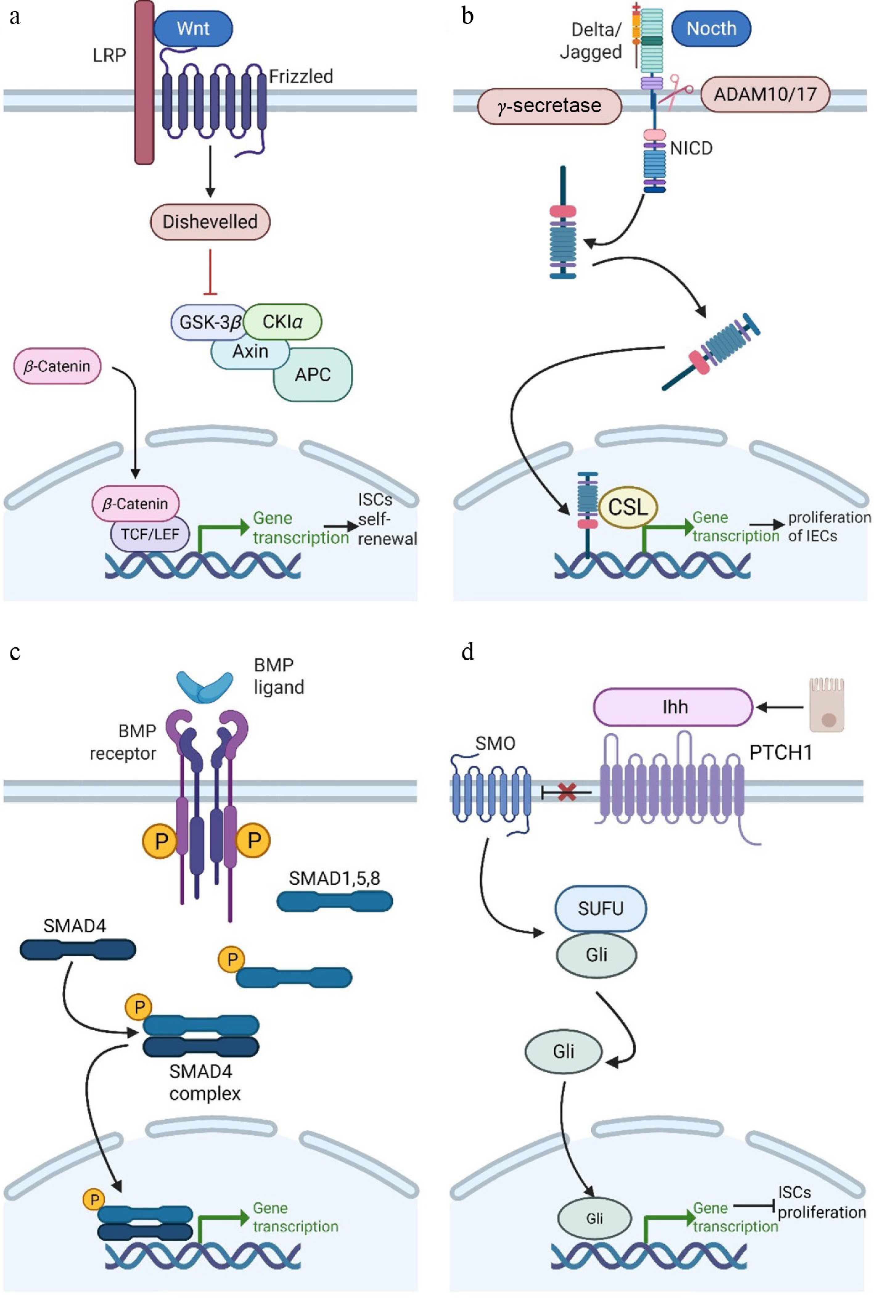
Figure 1.
Signal pathways associated with the occurrence of radiation enteritis. (a) Activation of the Wnt/β-catenin pathway promotes self-renewal and proliferation of ISCs. (b) Notch pathway activation promotes proliferation and differentiation of IECs. (c) BMP pathway activation negatively regulates self-renewal of ISCs. (d) Hedgehog pathway activation negatively regulates ISC proliferation. This figure was created with BioRender (
BioRender.com ). -
Various plant medicines and their extracts have demonstrated promising efficacy in the prevention and treatment of radiation enteritis. These plants and their extracts typically exhibit various biological activities, including antioxidant, anti-inflammatory, tissue repair-promoting properties, and immune regulation. The following section reviews some representative plants and plant extracts, along with research on their applications in radiation enteritis.
Sophora flavescens and its extracts
-
Sophora flavescens contains alkaloids and flavonoids, including matrine (Fig. 2a), oxymatrine, dihydroflavones, and isoflavones. It exhibits anti-inflammatory, antioxidant, antimicrobial, and immune-regulatory effects. Additionally, it has shown promising applications in the treatment of radiation enteritis. Sophora flavescens extract can alleviate symptoms of radiation enteritis by reducing oxidative stress, inhibiting the production of pro-inflammatory cytokines, and regulating immune function. Matrine can decrease the production of inflammatory factors such as IL-6, IL-1β, and TNF-α induced by LPS through inhibiting NF-κB pathway activation, thus alleviating the inflammatory response in radiation enteritis[3]. Oxymatrine can inhibit LPS-induced NF-κB activity, reduce the expression of pro-inflammatory cytokines, regulate gut microbiota by increasing beneficial bacteria, and decrease harmful bacteria, thereby reducing inflammation[25]. Beyond these alkaloids, the flavonoid kurarinone from Sophora flavescens has been identified as an uncompetitive inhibitor of soluble epoxide hydrolase (sEH), an enzyme that links oxidative stress and inflammation. By inhibiting sEH, kurarinone stabilizes anti-inflammatory epoxy fatty acids and alleviates neuroinflammation[9]. This sEH-targeting mechanism may also contribute to the protective effects of Sophora flavescens against radiation enteritis.
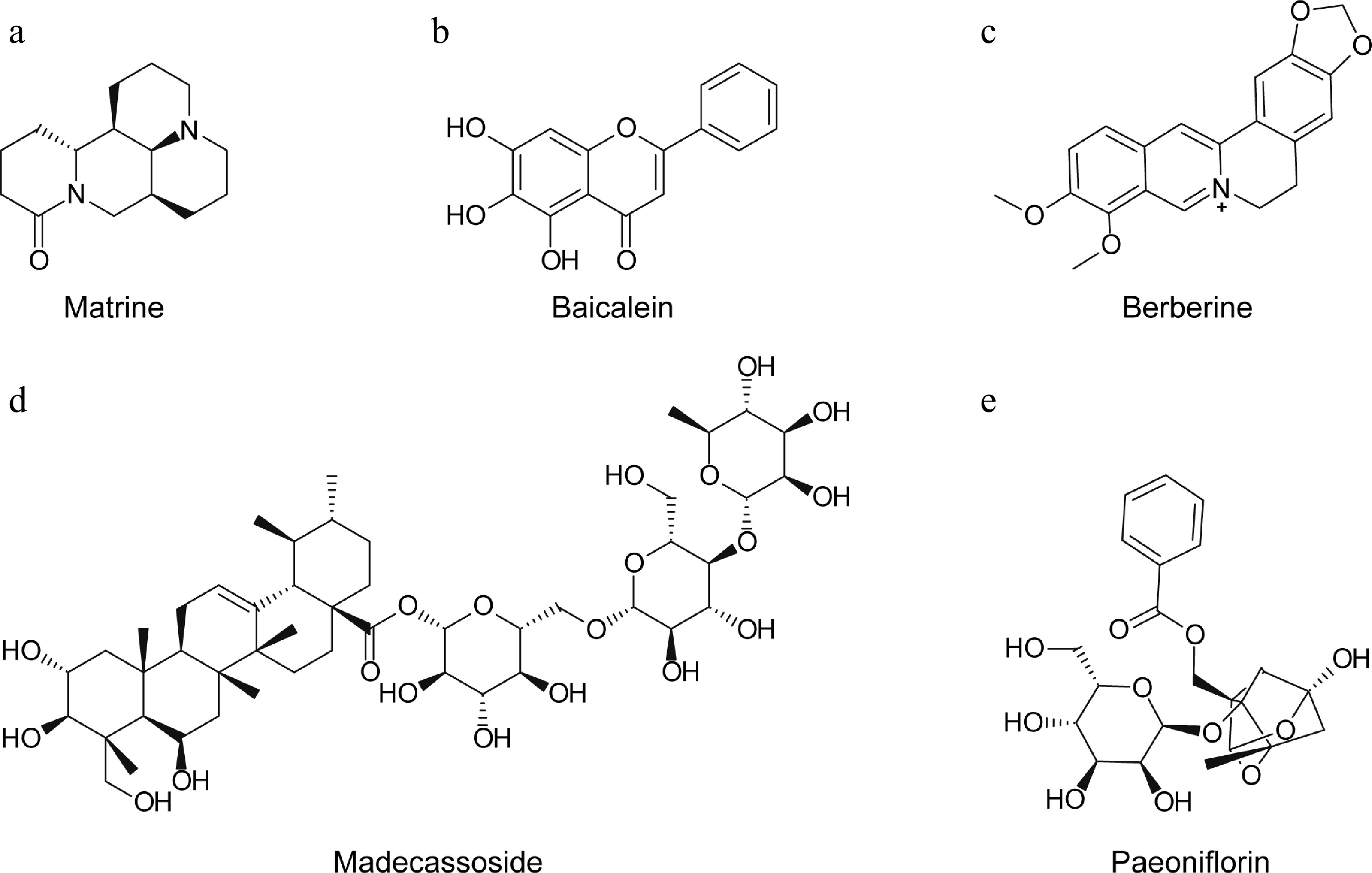
Figure 2.
The structural formula of (a) matrine, the main extract from Sophora flavescens roots, (b) baicalein, the main extract from Scutellaria baicalensis, (c) BBR, the main extract from Huanglian, (d) madecassoside, the main extract from Centella asiatica, and (e) paeoniflorin, the main extract from Paeonia lactiflora.
Scutellaria baicalensis and its extracts
-
Scutellaria baicalensis demonstrates therapeutic efficacy in treating radiation enteritis via its extract, baicalein (5,6,7-trihydroxyflavone) (Fig. 2b). Baicalein is one of the main flavonoids extracted from the dried roots of Scutellaria baicalensis. It possesses many beneficial properties, including antioxidant, antibacterial, anti-inflammatory, anticancer, hypolipidemic, anti-atherosclerotic, antithrombotic, and immune-regulatory effects[4]. Clinically, baicalein has been used to treat cerebrovascular diseases, including vasculitis and paralysis, and its diverse pharmacological effects provide a solid foundation for its application in the treatment of radiation enteritis[26].
Research has demonstrated that baicalein can significantly increase the length of intestinal villi and the number of crypts, reduce bacterial translocation to mesenteric lymph nodes, and effectively alleviate radiation-induced intestinal damage[27]. It can also improve intestinal barrier function and enhance intestinal defenses against external pathogens by upregulating the expression of tight junction molecules such as Claudin-3 (CLDN3) and ZO-1. Additionally, baicalein inhibits the expression of inflammatory cytokines and adhesion molecules such as P-selectin and Vascular cell adhesion protein 1 (VCAM-1), thereby reducing infiltration of neutrophils and eosinophils and alleviating inflammatory reactions. It also reduces the levels of pro-inflammatory cytokines, further inhibiting radiation-induced intestinal inflammation. Moreover, baicalein promotes the regeneration of intestinal crypts, significantly reducing crypt damage caused by radiation and contributing to intestinal functional recovery.
Huanglian and its extracts
-
Berberine (BBR), an isoquinoline alkaloid found in Huanglian extract (Fig. 2c), has shown potential as a therapeutic agent for treating or preventing radiation-induced enteritis. It can inhibit the production of inflammatory mediators and exert anti-inflammatory effects through regulation of macrophage polarization[5]. BBR also exhibits potent antioxidant capacity, effectively neutralizing free radicals to mitigate oxidative stress-induced damage to intestinal tissue. Furthermore, BBR promotes the secretion of goblet cells and stimulates the proliferation and regeneration of IECs, which not only helps repair damaged intestinal mucosa but also significantly enhances the barrier function of the intestine, preventing the invasion of harmful substances.
Additionally, BBR regulates the gut microbiota, helping to restore its balance, enhance intestinal defenses, and resist pathogen invasion[28]. It promotes the proliferation and differentiation of ISCs, accelerating the regeneration and repair of intestinal tissue by activating the mechanistic target of rapamycin complex 1 (mTORC1) and signal transducer and activator of transcription 3 (STAT3) signaling pathway[29]. With its wide availability from various Berberis plants and Chinese herbal medicines such as Huanglian, low cost, and multiple pharmacological activities, BBR emerges as a promising adjuvant for the treatment of radiation enteritis, offering patients a cost-effective therapeutic option.
Centella asiatica and its extracts
-
Centella asiatica, a traditional Asian herb, has been used for wound healing and skin diseases. Recent studies have demonstrated that Centella asiatica has a significant therapeutic effect in treating radiation-induced enteritis. Research indicates that Centella asiatica can alleviate symptoms of radiation enteritis by protecting vascular endothelial cells. Extracts from Centella asiatica can restore radiation-induced endothelial cell dysfunction and alleviate damage to the intestinal epithelial barrier[29]. Notably, epidermal growth factor (EGF), secreted by radiation-damaged endothelial cells treated with Centella asiatica, plays a key role in restoring intestinal epithelial barrier function. In a mouse model of radiation-induced enteritis, the paracrine effect induced by Centella asiatica can restore epithelial barrier integrity and alleviate symptoms of the condition[29].
The main active ingredients in Centella asiatica extract include triterpenoids such as madecassoside (Fig. 2d), asiaticoside, madecassic acid, and asiatic acid. These compounds exhibit significant anti-inflammatory, antioxidant, and wound-healing properties.
Paeonia lactiflora and its extracts
-
Paeonia lactiflora, a plant from the Ranunculaceae family, has a long history of medicinal use in China. Based on different medicinal parts and processing methods, it is classified into red peony and white peony. Red peony has effects such as clearing heat, cooling blood, dispersing blood stasis, and relieving pain. White peony can nourish the blood, promote blood circulation, balance body fluids to reduce sweating, alleviate liver-related discomfort and pain, and help stabilize liver energy.
The main active compound in Paeonia lactiflora is paeoniflorin (Fig. 2e), a monoterpenoid glycoside extracted from peony roots. It exhibits a wide range of pharmacological activities, including anti-inflammatory, analgesic, antipyretic, and antispasmodic effects, with low toxicity[7]. Recent studies have shown that paeoniflorin upregulates the expression of suppressor of cytokine signaling 3 (SOCS3), which acts as an endogenous 'inflammatory brake', and inhibits the apoptosis signal-regulated kinase 1 (ASK1)/tissue factor (TF) axis, thereby alleviating radiation-induced enteritis[30]. In mouse models of radiation enteritis, paeoniflorin significantly reduces intestinal inflammation and ischemic symptoms. Its mechanism may also involve inhibiting group 3 innate lymphoid cells (ILC3s) via the death receptor 3 (DR3), thereby promoting mucosal repair in chronic colitis[30].
In addition to the five representative plants described above, numerous other medicinal plants and their extracts have been examined for their protective effects against radiation enteritis, as summarized in Table 1. These include Bletilla striata, Glycyrrhiza uralensis, Salvia miltiorrhiza, Astragalus membranaceus, Taraxacum mongolicum, Carica papaya, Erigeron annuus, Garcinia mangostana, Alpinia officinarum, Portulaca oleracea, Agrimonia pilosa, and Psidium guajava. Although they originate from diverse botanical sources, these natural products share common mechanisms of action, primarily involving antioxidant activity, anti-inflammatory effects, and protection of the intestinal barrier. Collectively, these findings underscore the extensive therapeutic potential of plant-derived compounds in the management of radiation enteritis.
Table 1. The role of plants and plant extracts in the treatment of radiation enteritis.
Plant Plant extracts Role Ref. Bletilla striata Bletilla striata extract Bletilla striata polysaccharide, the main component extracted from Bletilla striata, can inhibit the NLRP3 inflammasome and exhibit anti-inflammatory effects. It also enhances immune function by activating the Mitogen-activated protein kinase (MAPK) and NF-κB signaling pathways. [31] Glycyrrhiza uralensis Glycyrrhiza uralensis extract It can inhibit LPS-induced secretion of pro-inflammatory cytokines in macrophages and whole blood; Glycyrrhetinic acid and 18β-glycyrrhetinic acid have been shown to regulate signaling pathways associated with inflammation, such as the Janus kinase (JAK) and signal transducer and activator of transcription (STAT) (JAK/STAT) pathway and NF-κB pathways. [32] Salvia miltiorrhiza Salvianic acid A It can alleviate intestinal inflammation by regulating immune response and the production of inflammatory factors; salvianic acid A in Salvia miltiorrhiza extract and other phenolic acid components have potent antioxidant activity, which can clear free radicals and reduce oxidative stress damage; It has the effect of promoting blood circulation and removing blood stasis, which can improve microcirculation, increase intestinal blood flow, alleviate ischemic injury, and promote intestinal repair. [33] Astragalus membranaceus Astragalus polysaccharides, Astragalus saponins, isoflavones Astragalus polysaccharides can regulate immunity by modulating gut microbiota, while maintaining intestinal barrier integrity and repairing damaged intestinal mucosa. The antioxidant and anti-inflammatory properties of Astragalus saponins and isoflavones help alleviate intestinal inflammation and tissue damage. Isoflavones with estrogen-like activity have a specific promoting effect on intestinal function recovery. [34] Taraxacum mongolicum Taraxacum mongolicum extract Taraxacum mongolicum has antioxidant, anti-inflammatory, and gastrointestinal protective effects. Its essential component polysaccharides have been proven to have antioxidant and antibacterial activities. Taraxacum mongolicum extract can alleviate cell damage caused by oxidative stress. [35] Carica papaya (Papaya) Papaya seed alcohol extract, papaya fruit extract Papaya seed alcohol extract exhibits antibacterial activity, while papaya fruit extract has significant antioxidant and anti-inflammatory properties. Phenolic and flavonoid compounds in papaya can scavenge free radicals and enhance the activity of antioxidant enzymes. [36] Erigeron annuus Erigeron annuus root extract Erigeron annuus root extract can alleviate acute inflammation by inhibiting the production of NF-κB-related NO and prostaglandin E2 (PGE2). Erigeron annuus root extract has antioxidant capacity and can clear free radicals. [37] Garcinia mangostana (Mangosteen) Mangosteen peel extract The skin of mangosteen is rich in xanthones, especially α-mangostin, which are effective antioxidants that can clear excess ROS and alleviate oxidative stress damage to intestinal tissue; Mangosteen peel extract has significant anti-inflammatory activity and can inhibit the production and release of inflammatory mediators. [38] Alpinia officinarum (Galangal) Galangal extract Galangal extract is rich in antioxidant components, which can eliminate free radicals and enhance the activity of antioxidant enzymes, thereby reducing radiation-induced oxidative damage. Galangal extract can downregulate the MAPK and JAK/STAT pathways, thereby exerting anti-inflammatory effects. [39] Portulaca oleracea (Purslane) Purslane extract Purslane extract can inhibit the production of pro-inflammatory cytokines, for example, by downregulating inflammatory factors to alleviate the damage to the intestinal barrier; Purslane is rich in various antioxidant components, such as polyphenols, flavonoids, and omega-3 fatty acids, which help to eliminate excessive free radicals in the body and reduce oxidative damage; Purslane extract can regulate the structure of intestinal microbiota and improve dysbiosis. [40] Agrimonia pilosa Agrimonia pilosa extract Agrimonia pilosa extract can inhibit the inflammatory response of macrophages induced by LPS; Agrimonia pilosa is traditionally used to treat acute and chronic enteritis and diarrhea. [41] Psidium guajava (Guava) Guava leaf extract Guava leaf extract exerts anti-inflammatory effects by inhibiting the production of pro-inflammatory cytokines (e.g., IL-6, IL-1β, and TNF-α), modulating the NF-κB and other inflammatory signaling pathways, inhibiting the activity of cyclooxygenase (COX) and inducible nitric oxide synthase (iNOS), and reducing the production of ROS. [42] -
Plant-derived bioactive compounds exhibit broad therapeutic potential against radiation enteritis, attributed to their diverse chemical structures and biological activities. Based on core structural scaffolds and functional groups, these compounds are categorized as follows (Table 2):
Table 2. Active factors in plant extracts and their mechanisms.
Class Subclass Specific compounds Mechanism Flavonoids Flavones Luteolin Antioxidant; blocks JAK1/JAK2 ATP binding site to inhibit STAT3[43] Apigenin Antioxidant; inhibits NLRP3 inflammasome via NIMA-related kinase 7 (NEK7) inhibition[44] Baicalein Antioxidant; suppresses TLR4 dimerization and MAPK phosphorylation[45] Flavonols Quercetin Antioxidant; blocks NF-κB nuclear translocation by disrupting p65-Importin-α interaction[46] Kaempferol Antioxidant; activates Nrf2 pathway; inhibits COX-2 expression[47] Isorhamnetin Antioxidant; actives Nrf2 pathway[48] Flavanols EGCG Antioxidant; binds c-Jun to inhibit Activator protein-1 (AP-1); repairs intestinal barrier[49] Anthocyanidins Delphinidin ROS scavenger; upregulates tight junction[50] Terpenoids Triterpene glycosides Asiaticoside Enhances epithelial regeneration; reduces oxidative stress[51] Pentacyclic triterpenoids Ursolic acid Activates Nrf2 pathway; inhibits NLRP3 inflammasome assembly[52] Celastrol Reprograms T-cell balance: inhibits T helper 17 cells (Th17) differentiation, promotes Regulatory T cells (Treg) expansion[53] Triterpene glycosides Ginsenosides Activates phosphatidylinositol 3-kinase (PI3K)/protein kinase B (Akt) signaling pathway to inhibit mitochondrial apoptosis; enhances barrier Astragaloside IV Modulates TLR4/NF-κB signaling; reduces pro-inflammatory cytokines[54] Gypenosides Attenuates oxidative stress; suppresses NF-κB[55] Diterpenoids Tanshinone Inhibits ROS; downregulates TNF-α/IL-6 expression[56] β-boswellic acid Inhibits 5-Lipoxygenase (5-LOX); reduces leukotriene-mediated inflammation[52] Andrographolide Blocks NF-κB nuclear translocation; enhances Nrf2-mediated antioxidant response[57] Tetraterpenoids β-Carotene Direct ROS scavenging; quenches ¹O2[56] Monoterpene glycoside Paeoniflorin Upregulates SOCS3; inhibits ASK1-TF axis; reduces IL-6/TNF-α and neutrophil infiltration[30] Phenylpropanoids Phenylethanoid glycosides Salidroside Activates Nrf2/ Heme oxygenase-1 (HO-1) pathway; suppresses MAPK/NF-κB signaling[48] Coumarins Esculetin Scavenges ·OH; inhibits xanthine oxidase. Umbelliferone Antioxidant; enhances Glutathione S-transferase (GST) activity[58] Scopoletin Suppresses iNOS; inhibits leukocyte migration[58] Lignans Liriodendrin Modulates pregnane X receptor (PXR) and constitutive androstane receptor (CAR) receptors to detoxify ROS[59] Terpenophenolics Cannabinoids Cannabidiol, CBD Activates CB2 receptors; reduces TNF-α/IL-6; inhibits NADPH oxidase Non-flavonoid polyphenols Polyphenolic dilactone Ellagic acid Chelates redox-active metals; upregulates Nrf2-dependent genes[46] Phenolic acids (Hydroxycinnamic acids) Chlorogenic acid ROS scavenger; inhibits NF-κB nuclear translocation[46] Rosmarinic acid, RA Suppresses COX-2/PGE2; inhibits complement activationv[60] Stilbenes Resveratrol Activates Sirtuin 1 (SIRT1) to deacetylate NF-κB p65 and Nrf2; upregulates SOCS3; inhibits Interleukin-1 receptor-associated kinase 4 (IRAK4)[61] Diarylheptanoids Curcumin Binds IκB kinase β(IKKβ) ATP-site to inhibit NF-κB; Blocks TNF receptor-associated factor 6 (TRAF6) ubiquitination; promotes M2 macrophage polarization via Signal transducer and activator of transcription 6 (STAT6)/Peroxisome proliferator-activated receptor gamma (PPARγ)[62] Condensed tannins Proanthocyanidins Antioxidant; stabilizes gut microbiota; reduces epithelial permeability[60] Polysaccharides Heteropolysaccharides Rheum tanguticum polysaccharides, RTP Enhance mucosal immunity; scavenge free radicals[63] Astragalus polysaccharides, APS competitively binds TLR4 LPS-domain; induces IL-10 secretion; inhibits MyD88 recruitment[47] Ganoderma lucidum polysaccharide Immunomodulatory; activates intestinal Dendritic cells (DCs) to promote Treg differentiation[47] Spirulina polysaccharides ROS scavenging; upregulates antioxidant enzymes[64] Jujube polysaccharide Prevents apoptosis; enhances goblet cell function[65] Alkaloids Quinolizidine Matrine Inhibits TLR4/MyD88 pathway; reduces IL-1β/IL-18 secretion[66] Isoquinoline BBR Suppresses Gram-negative bacteria growth; reduces LPS release; inhibits TLR4/NF-κB signaling Amide alkaloids Piperine Potentiates Nrf2 activation; inhibits P-glycoprotein to enhance bioavailability of other bioactives[45] Anthraquinones Hydroxyanthraquinone-carboxylic acid derivatives Rheinic acid Inhibits NF-κB pathway; activates PPARγ to downregulate COX-2/iNOS[67] Hydroxyanthraquinones Emodin Blocks NLRP3- Apoptosis-associated speck-like protein containing a CARD (ASC) interaction; inhibits inflammasome assembly[53] Sterols Phytosterols Stigmasterol stabilizes lipid membranes against oxidative damage[56] Flavonoids
-
Flavonoids, characterized by a core C6-C3-C6 carbon skeleton, include flavones, flavonols, flavanols, and anthocyanidins. Their primary therapeutic function in radiation enteritis is attributed to their antioxidant activity. Luteolin and apigenin (abundant in celery, bell pepper, and Lonicera japonica) effectively alleviate radiation-induced lipid peroxidation by scavenging ROS and inhibiting NADPH oxidase[43]. Baicalein, from Scutellaria baicalensis, exhibits potent antioxidant capacity due to its unique 7-hydroxy group. It concurrently inhibits the mitochondrial apoptosis pathway, thereby shielding IECs from radiation-induced DNA damage[45]. Among flavonols, quercetin activates the Nrf2/ARE pathway, upregulating Superoxide dismutase (SOD) and Glutathione peroxidase (GSH-Px)[46]. EGCG chelates metal ions and halts free-radical chain reactions[49].
Flavonoids also exert anti-inflammatory and tissue repair effects in the intestine. Luteolin downregulates the NF-κB pathway, reducing TNF-α and IL-6[43]. Kaempferol promotes intestinal mucosal barrier repair via phosphatidylinositol 3-kinase (PI3K)/protein kinase B (Akt) signaling pathway[47]. EGCG upregulates tight junction proteins such as ZO-1 and occludin[49]. Additionally, anthocyanidins, like delphinidin, can stabilize cell membranes and counteract radiation-induced increases[50].
Non-flavonoid polyphenols
-
Non-flavonoid polyphenols exhibit potent antioxidant, anti-inflammatory, and cytoprotective activities. Ellagic acid efficiently scavenges free radicals (e.g., ·OH, O2−) via its lactone ring structure[46], thereby decreasing malondialdehyde (MDA) levels in irradiated intestinal tissue. Chlorogenic acid (abundant in coffee and Lonicera japonica) activates the Nrf2/ARE pathway to boost antioxidant enzymes and inhibits apoptosis-related proteins (e.g., caspase-3) in IECs[46]. Rosmarinic acid (from rosemary and perilla) chelates metal ions to halt ROS generation[60]. Curcumin leverages its β-diketone moiety for potent free radical scavenging[62].
These compounds also exhibit anti-inflammatory and cytoprotective effects. Ellagic acid can suppress NF-κB, thereby reducing the release of IL-1β and TNF-α[46]. Rosmarinic acid promotes mucosal repair via the PI3K/Akt/mTOR pathway[60]. Curcumin inhibits COX-2 and iNOS expression to reduce PGE2 and NO levels and also modulates gut microbiota[62]. Additionally, stilbenes such as resveratrol (sourced from Polygonum cuspidatum) activate SIRT1 to suppress NF-κB and STAT3 signaling pathways, reducing pro-inflammatory cytokines like IL-6 and IL-8[61], and upregulate tight junction proteins such as claudin-1 and ZO-2 to restore radiation-compromised barrier function.
Terpenoids
-
Terpenoids are classified into monoterpenes, diterpenes, triterpenes, and tetraterpenoids. The monoterpene glycoside paeoniflorin (from Paeonia lactiflora) blocks JAK2/STAT3 signaling and suppresses NF-κB-mediated inflammation[7]. Tanshinone (derived from Salvia miltiorrhiza roots) alleviates radiation enteritis pain by inhibiting COX-2 and PGE2[56]. Andrographolide exerts anti-inflammatory effects by blocking the TLR4/MyD88 signaling pathway[57].
Triterpenes and tetraterpenes promote intestinal mucosal regeneration. Triterpenoid saponins such as ginsenosides and astragaloside IV mitigate intestinal fibrosis through suppression of the TGF-β/Smad pathway. Asiaticoside activates Wnt/β-catenin, stimulating the proliferation of Lgr5+ ISCs[51]. Ursolic acid (found in plants like Brownea grandiceps) induces ZO-1 expression and inhibits Matrix metalloproteinase-9 (MMP-9) activity, thus maintaining mucosal structural integrity. The tetraterpenoid β-carotene enhances goblet cell secretion, preserving the integrity of the mucus layer[52].
Phenylpropanoids
-
Phenylpropanoids, characterized by a C6-C3 backbone, include coumarins and lignans known for their anti-inflammatory and antioxidant properties. The coumarin derivative esculetin mitigates oxidative stress by suppressing iNOS expression, thereby reducing NO production. Another key coumarin, scopoletin, inhibits macrophage overactivation by modulating the MAPK pathway[58]. Liriodendrin, derived from Sargentodoxa cuneata, exhibits estrogenic activity that modulates the Th17/Treg balance, thereby ameliorating radiation-induced intestinal immune dysregulation[59].
Polysaccharides
-
Plant polysaccharides confer intestinal radioprotection through antioxidant regulation, immunomodulation, and anti-inflammatory actions. Astragalus polysaccharides promote the differentiation of intestinal lamina propria CD4+ T cells into Tregs, suppressing excessive inflammation[47]. Ganoderma lucidum polysaccharide enhances macrophage phagocytic capacity via the TLR4/NF-κB pathway[47]. RTP activates the Nrf2/HO-1 axis to scavenge ROS and increase SOD levels[63]. Additionally, jujube polysaccharide and spirulan exhibit comparable protective effects[65].
Other classes of plant bioactive compounds, such as alkaloids, sterols, and anthraquinones, also demonstrate therapeutic effects on radiation enteritis. The alkaloid BBR inhibits intestinal epithelial cell apoptosis via the AMP-activated protein kinase (AMPK)/mTOR pathway[46]. Stigmasterol (a phytosterol) competitively binds to cholesterol receptors to attenuate intestinal inflammation. The anthraquinone rhein reduces pro-inflammatory cytokine release by suppressing the NF-κB pathway[67].
-
Plant-derived bioactive compounds utilize their distinct chemical architectures to precisely modulate key pathological processes in radiation enteritis. According to our systematic phytochemical classification, we explain how they orchestrate radioprotection through an integrated regulatory network—the Antioxidant-Anti-Inflammatory-Repair Triad—placing particular emphasis on the underlying antioxidant mechanisms.
Antioxidant
Free radical scavenging
-
Plant bioactives directly neutralize O2−and ·OH via electron donation from phenolic hydroxyls or conjugated double bonds, thereby halting oxidative chain reactions[68]. Flavonoids (e.g., quercetin, luteolin) utilize ortho-dihydroxy structures for efficient scavenging and regenerate their antioxidant capacity through quinone-hydroquinone redox cycling[68]. Polyphenols such as curcumin (with a β-diketone moiety) and ellagic acid (with a lactone ring) trap free radicals, reducing MDA and 8-hydroxy-2'-deoxyguanosine (8-OHdG) levels. As a terpenoid, β-carotene physically quenches singlet oxygen (1O2) via its conjugated polyene chain, thereby preserving enterocyte membrane integrity[69]. Additionally, RTP significantly lowers intestinal ROS post-irradiation through β-glycosidic bond conformations that stabilize radical-phenolic hydroxyl resonance structures (Fig. 3a).
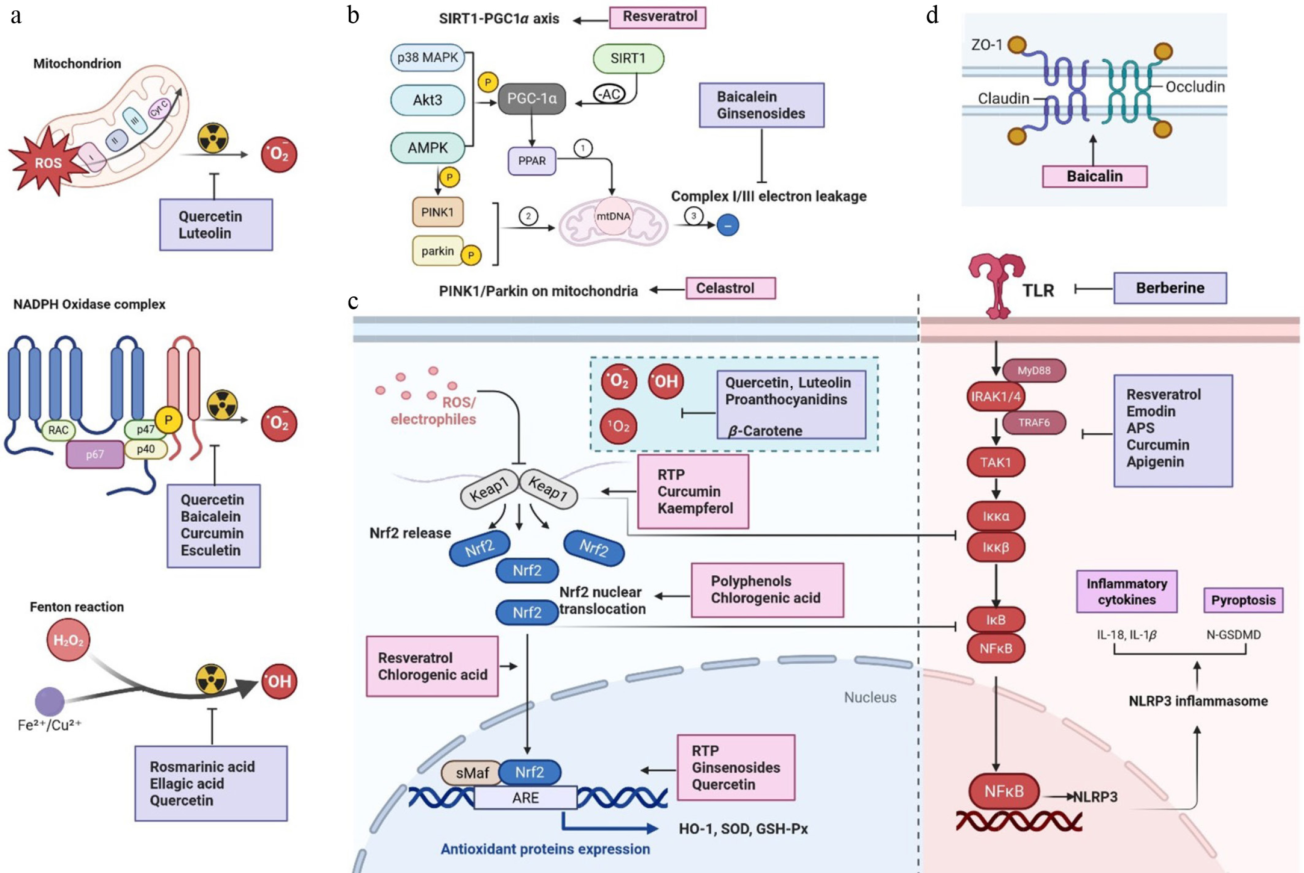
Figure 3.
Multitarget antioxidant defense network of phytochemicals against radiation-induced oxidative stress. (a) Radiation-triggered ROS sources. Ionizing radiation induces O2− generation via mitochondrial ETC leakage (Complex I/III) and NOX activation. Transition metals (Fe2+/Cu2+) catalyze ·OH formation through Fenton reactions. (b) Core antioxidant pathway and anti-inflammatory synergy. Direct scavenging, such as flavonoids (e.g., quercetin), donate electrons to neutralize O2−/·OH, while carotenoids (β-carotene) physically quench 1O2. Phytochemicals (curcumin/resveratrol) modify Keap1 cysteine residues (Cys151/Cys273), enabling Nrf2 nuclear translocation and transcriptional upregulation of antioxidant enzymes (HO-1, SOD, GSH-Px). (c) Anti-inflammatory synergy. Bioactives concurrently suppress NF-κB activation (via IKKβ inhibition) and NLRP3 inflammasome assembly, while modulating the TLR4/MyD88 and MAPK pathways to attenuate pro-inflammatory cytokine cascades. (d) Barrier repair. Tight junction reinforcement (ZO-1/occludin upregulation) further restores intestinal homeostasis. This figure was created with BioRender (
BioRender.com ).Transition metal ions (Fe2+, Cu2+) catalyze ·OH generation via Fenton reactions (Fig. 3a). Phytochemicals inhibit this process by metal chelation. Polyphenols sequester Fe2+ through catechol or galloyl groups, while flavonoids like quercetin bind Cu2+ to reduce metal-mediated DNA damage. Carotenoids further intercept lipid peroxidation intermediates. For instance, lycopene markedly decreases radiation-induced MDA in intestinal epithelium.
Activation of endogenous antioxidant pathways
-
Plant bioactives enhance cellular antioxidant defenses via the Nrf2/ARE pathway, which upregulates SOD, GSH-Px, catalase (CAT), and GSH to accelerate ROS decomposition (Fig. 3c). Under basal conditions, Nrf2 undergoes Kelch-like ECH-associated protein 1 (Keap1)-mediated ubiquitination and degradation[70]. By modifying Keap1 cysteine residues, Phytochemicals disrupt the Keap1-Nrf2 interaction, enabling Nrf2 nuclear translocation and ARE-driven transcription of HO-1, NAD(P)H:quinone oxidoreductase 1 (NQO1), and SOD[70]. Notably, HO-1 generates biliverdin, which can be converted to bilirubin for direct ROS scavenging, alongside CO and Fe2+[70]. RTP elevates Nrf2 and downstream HO-1/NQO1 expression. Flavonols and polyphenols inhibit oxidation of Keap1, promoting Nrf2 nuclear accumulation and induction of antioxidant enzymes[71]. Triterpenoids (e.g., Ginsenosides) activate the PI3K/Akt pathway, which phosphorylates Nrf2 and inhibits Keap1-mediated degradation, thereby extending antioxidant responses. (Fig. 3a, c).
Mitochondrial protection via ETC regulation and mitophagy activation pathways
-
Radiation-induced electron leakage from ETC Complex I (NADH dehydrogenase) and Complex III (cytochrome bc1 complex) generates O2− as a major ROS source (Fig. 3a). Plant bioactives preserve mitochondrial homeostasis by reducing ROS production, maintaining mitochondrial quality control, and promoting mitochondrial biogenesis[72]. Flavones (e.g., baicalein) inhibit mitochondrial permeability transition pore (mPTP) opening, thereby preventing cytochrome c release and subsequent caspase-3-mediated apoptosis[72]. Resveratrol activates the SIRT1/Peroxisome proliferator-activated receptor gamma coactivator 1-alpha (PGC-1α) axis to enhance mitochondrial biogenesis and oxidative phosphorylation[72]. Mechanistically, SIRT1 deacetylates PGC-1α in an NAD+-dependent manner, strengthening its binding to PPARα/γ and activating ETC component-encoding genes, thereby reducing electron leakage from Complexes I and III[73]. Ginsenosides stabilize Complex I to minimize electron leakage while activating PGC-1α to upregulate mitochondrial SOD (Mn-SOD), thereby reducing oxidative stress[73] (Fig. 3a, b).
Radiation-induced mitochondrial damage collapses membrane potential, triggering abnormal accumulation of PTEN-induced kinase 1 (PINK1) on the outer mitochondrial membrane. Plant bioactives exploit the PINK1/Parkin mitophagy pathway to promote programmed clearance[72]. Celastrol (a triterpenoid) amplifies Parkin's ubiquitin ligase activity, recruiting Parkin to damaged mitochondria, promoting outer membrane proteins ubiquitination, and accelerating autophagosome formation. Resveratrol operates through the SIRT1-Liver kinase B1 (LKB1)-AMPK axis, coordinating biogenesis via PGC-1α, whereas APS engages TLR4-mediated AMPK activation, promoting rapid elimination of damaged mitochondria through dual ULK1 phosphorylation and PINK1/Parkin amplification[73] (Fig. 3b).
Suppression of ROS-generating enzymes
-
NADPH Oxidase (NOX1/NOX4), highly expressed in the intestinal epithelium, serves as a primary ROS source. Ionizing radiation activates Rac1-GTPase, triggering the membrane translocation of NOX subunits to form active complexes with NOX2, which generates O2−. Concurrent activation of pro-oxidant enzymes, such as xanthine oxidase (XO), exacerbates the ROS burst[74] (Fig. 3a). Plant bioactives counteract this by targeted enzyme inhibition. Flavonoids (quercetin, luteolin, baicalein) block Protein Kinase C δ (PKCδ)-mediated p47phox phosphorylation, preventing NOX assembly. Curcumin and esculetin downregulate XO expression, reducing uric acid and ROS co-production, and impairing Rac1 membrane localization, thereby diminishing NOX activity.
Repair of oxidatively damaged biomacromolecules
-
Radiation-induced DNA single-strand breaks (SSBs) trigger the activation of poly ADP-ribose polymerase 1 (PARP1) hyperactivation, which leads to the synthesis of poly ADP-ribose (PAR) chains and the recruitment of repair proteins.
Plant bioactives facilitate macromolecular repair through chemical restitution and signaling pathway activation. They directly reduce oxidized protein carbonyls by covalently restoring cysteine residues, e.g., EGCG. They terminate lipid peroxidation via phenolic hydrogen transfer, e.g., proanthocyanidins quenching peroxyl radicals[75]. They also stabilize damaged DNA bases through intercalation, e.g., baicalein, facilitating 8-OHdG correction. These compounds concurrently orchestrate enzymatic repair systems, including PARP1/p53-mediated base excision and GPx4-driven phospholipid peroxidation resolution, thereby systemically restoring structural and functional integrity to radiation-compromised biomolecules[75].
Collectively, plant bioactives orchestrate a multi-layered antioxidant defense network that dynamically counteracts radiation-induced oxidative stress by controlling sources (suppressing ROS generation at enzymatic and mitochondrial levels), intercepting radicals efficiently (via direct scavenging and metal chelation), and reinforcing cellular defenses (by amplifying endogenous antioxidant capacity), ultimately restoring redox homeostasis[76]. By mitigating the initiating oxidative burden, these compounds establish a critical foundation for subsequent anti-inflammatory actions and barrier repair processes(Fig. 3d)[76].
Anti-inflammatory
Dual suppression of NF-κB and NLRP3 inflammasome
-
NF-κB and NLRP3 inflammasome are core regulators of radiation-induced enteritis. Radiation activates IKKβ via ROS or direct membrane damage, triggering nuclear translocation of the NF-κB p50/p65 and upregulating TNF-α, IL-6, and iNOS. Concurrently, radiation-induced ROS/K+ efflux activates the NLRP3 inflammasome, promoting IL-1β/IL-18 maturation, pyroptosis, and the release of Damage-associated molecular patterns (DAMPs)[77] (Fig. 3c).
Plant bioactives target both upstream NF-κB suppression and downstream NLRP3 blockade. Curcumin directly binds to the ATP-binding site of IKKβ, inhibiting its kinase activity and p65 nuclear translocation. Resveratrol activates SIRT1 to deacetylate the p65 subunit at Lys310, reducing its DNA-binding affinity[77]. Quercetin blocks the interaction between the NF-κB p65 subunit and Importin-α. Rhein activates PPAR-γ to inhibit NF-κB nuclear translocation and significantly downregulates COX-2 and iNOS[78] (Fig. 3c).
Additionally, plant bioactives also prevent NLRP3 oligomerization and caspase-1 activation, thereby inhibiting inflammasome assembly and the release of IL-1β and IL-18. Emodin inhibits the interaction between NLRP3 and ASC[78]. Apigenin suppresses the activity of NEK7, a key kinase involved in NLRP3 activation. Nicotiflorin (isolated from Morus alba roots) blocks inflammasome assembly, reduces caspase-1 activity, and decreases IL-1β secretion.
Modulation of the MAPK signaling pathway
-
Radiation activates TLR4 or generates ROS, initiating signaling through the rat sarcoma virus (RAS)-rapidly accelerated fibrosarcoma (RAF)-mitogen-activated protein kinase kinase (MEK)-extracellular signal-regulated kinase (ERK) cascade. Activated MAPK stimulates the AP-1 transcription factor complex, promoting the expression of inflammatory genes such as IL-1β and MMP-9 (Fig. 3c).
Plant bioactives modulate this pathway by inhibiting MAPK phosphorylation and regulating AP-1 activity. Specifically, baicalein inhibits ERK and c-Jun N-terminal kinase (JNK) phosphorylation by blocking MEK1/2 activity[79]. Curcumin downregulates p38 MAPK phosphorylation via MAPK kinase (MKK) 3/6 inhibition[79] while EGCG directly binds c-Jun, suppressing its DNA-binding activity.
Regulation of the JAK-STAT signaling pathway
-
The JAK-STAT pathway transduces signals from cytokines such as IL-6 and IFN-γ, sustaining chronic inflammation. Plant bioactives inhibit JAK kinase activity and promote SOCS protein expression. Luteolin competitively occupies the ATP-binding pocket of JAK1/JAK2, blocking STAT3 phosphorylation. Resveratrol activates SIRT1 to upregulate SOCS3, providing negative feedback on JAK-STAT signaling. Paeoniflorin upregulates SOCS3, inhibits the ASK1-TF axis, significantly reducing pro-inflammatory cytokine levels such as IL-6 and TNF-α, reducing neutrophil infiltration into intestinal tissues[80] (Fig. 3c).
Antagonism of the TLR4/MyD88 pathway
-
The TLR4/MyD88 pathway serves as a pivotal upstream regulator of radiation-induced intestinal inflammation. Radiation compromises intestinal barrier integrity, increasing mucosal permeability and activating the TLR4/MyD88, which in turn activates both NF-κB and MAPK pathways to induce pro-inflammatory cytokines (TNF-α, IL-6, IL-1β). Furthermore, TLR4 activation upregulates pro-IL-1β and NLRP3 expression, providing the priming signal for NLRP3 inflammasome assembly[81] (Fig. 3c).
Plant bioactives target this pathway: APS competitively binds the LPS-binding domain of TLR4, preventing LPS-TLR4 complex formation and MyD88 recruitment. Curcumin directly interacts with the TRAF6 RING domain, blocking its ubiquitination activity[81]. Resveratrol binds to the ATP-binding site of IRAK4, inhibiting its kinase activity and disrupting IRAK4-TRAF6 complex formation[82]. Glycyrrhizin inhibits TLR4 signal transduction, reducing the activity of the TLR4/MyD88/NF-κB pathway. Polyphenols directly suppress TLR4 dimerization, decreasing downstream IRAK1 phosphorylation[82] (Fig. 3c).
Crosstalk regulation between inflammatory pathways and oxidative stress
-
Plant bioactives modulate the intricate interplay between inflammation and oxidative stress. NF-κB inhibition reduces inflammation-associated ROS production (e.g., from macrophages), indirectly mitigating oxidative damage. Nrf2 activation promotes competition with NF-κB for the transcriptional coactivator CREB-binding protein (CBP), thus limiting NF-κB transactivation and creating synergistic antioxidant and anti-inflammatory effects. SIRT1 acts as a central hub in this reciprocal regulation. Through deacetylation, SIRT1 simultaneously inhibits NF-κB, activates Nrf2 and Forkhead box O3 (FOXO3), effectively integrating antioxidant defenses with anti-inflammatory mechanisms[83] (Fig. 3).
Intestinal barrier repair
Modulation of intestinal immune cell phenotypes
-
Radiation reprograms intestinal immunity toward pro-inflammatory phenotypes, polarizing macrophages to M1 and T cells toward inflammatory subsets. Plant bioactives counteract this by promoting macrophage repolarization to M2 and restoring T cell balance. Curcumin suppresses M1 markers (iNOS, CD86) and upregulates M2 markers via PPARγ activation. Celastrol blocks Th17 differentiation by inhibiting STAT3/ Retinoic acid receptor-related orphan receptor gamma t (RORγt) while promoting Treg expansion via TGF-β/Smad3 signaling[84].
Restoration of intestinal barrier function
-
Compromised intestinal barrier integrity exacerbates radiation enteritis. Plant compounds enhance tight junction proteins and suppress epithelial apoptosis. Flavonoids (e.g., baicalin) activate PKCζ to upregulate ZO-1 and occludin expression[85]. Ginsenoside inhibits Bax/Bak-mediated mitochondrial apoptosis through PI3K/Akt activation.
Regulation of gut microbiota
-
Radiation-induced dysbiosis promotes LPS release and inflammation[13]. Plant bioactives restore microbial-immune crosstalk. BBR suppresses the growth of Gram-negative bacteria, reducing LPS and inhibiting the TLR4/NF-κB pathway[86]. Inulin enriches Bifidobacterium spp., induces Treg differentiation, and stimulates IL-10 production[87] (Fig. 3c).
Regulation of developmental signaling pathways
-
Beyond their antioxidant and anti-inflammatory actions, plant-derived bioactives ameliorate radiation enteritis by modulating key developmental pathways (Wnt/β-catenin, Notch, Hedgehog, and BMP) that govern intestinal stem cell homeostasis and epithelial repair. Importantly, radiation-induced ROS and inflammatory cytokines directly impinge on these pathways, creating a vicious cycle of aberrant activation and persistent injury[88].
Plant bioactives interrupt this cycle through multi-tiered interventions. For the Wnt/β-catenin pathway, they act at four nodes: (i) at the receptor/ligand level, by upregulating SFRPs; (ii) at the destruction complex, restoring β-catenin degradation; (iii) directly on β-catenin, promoting its proteasomal degradation or blocking nuclear translocation; and (iv) at the transcriptional level, suppressing T-cell factor 4 (TCF4)/lymphoid enhancer-binding factor 1 (LEF1) synthesis to silence MYC proto-oncogene protein (c-Myc) and cyclin D1[89].
For the Notch pathway, plant compounds target γ-secretase or downregulate Jagged1, blocking Notch intracellular domain (NICD) release and subsequent HES/HEY transcription. For the Hedgehog pathway, they inhibit Smoothened or suppress Gli activity. BMP signaling, which crosstalks with Wnt via Smad-dependent mechanisms, is indirectly modulated through shared networks[90].
Critically, these pathways do not operate in isolation. Parafibromin serves as a shared transcriptional co-activator for Wnt, Notch, and Hh, while NICD integrates inputs from multiple pathways. This network architecture explains why plant bioactives simultaneously inhibit multiple pathways, restoring coordinated signaling homeostasis in the irradiated intestine[91].
In summary, plant bioactives combat radiation enteritis through a multi-layered network that establishes antioxidant defense by scavenging ROS and activating Nrf2, blocks inflammation by suppressing NF-κB/NLRP3 and MAPK/JAK-STAT cascades, repairs the intestinal barrier by restoring tight junctions and modulating microbiota, and concurrently targets the Wnt/β-catenin, Notch, Hedgehog, and BMP pathways via signaling crosstalk. These four dimensions work synergistically to form a protective network that transcends the limitations of single-target agents.
-
Although natural plant products are often labeled as safe and non-toxic, the implications of safety are far more complex in the specific population of cancer patients undergoing radiotherapy. These patients frequently receive concomitant radiotherapy, chemotherapy, and multiple supportive care agents, significantly increasing the risk of herb–drug interactions. Herbal supplements may interfere with cancer treatment in various ways: affecting the metabolic pathways of other drugs, altering blood pressure, increasing bleeding risk, interfering with radiotherapy efficacy, and modifying sedative or anesthetic responses. Furthermore, the antioxidant properties of plant extracts represent both a core advantage in the treatment of radiation enteritis and a fundamental dilemma. Theoretically, the mechanisms of free radical scavenging, inflammation suppression, and cell survival protection, while safeguarding normal intestinal tissues, may also exert similar protective effects on tumor cells, thereby reducing the radiotherapeutic efficacy against the tumor. However, some studies have indicated that certain natural products can alleviate multiple toxicities induced by chemoradiotherapy without compromising antitumor efficacy[92].
The quality consistency of plant extracts is a fundamental prerequisite for clinical safety, yet this issue remains inadequately addressed in current research and clinical practice. Natural and herbal products may exhibit variability in active ingredient content due to differences in source materials. Therefore, the clinical translation of plant extracts necessitates the establishment of a comprehensive quality control system throughout the entire process. Additionally, the low bioavailability of plant extracts constitutes a core bottleneck limiting clinical efficacy. Current research is actively exploring various drug delivery system solutions to enhance efficacy and reduce off-target tissue exposure via targeted delivery, offering potential value for improving safety in translational applications.
-
Recent advancements in AI, synthetic biology, and related technologies offer new opportunities to enhance the therapeutic potential of phytochemicals in radiation enteritis, covering multiple aspects, including drug discovery, synthesis, and delivery (Fig. 4).
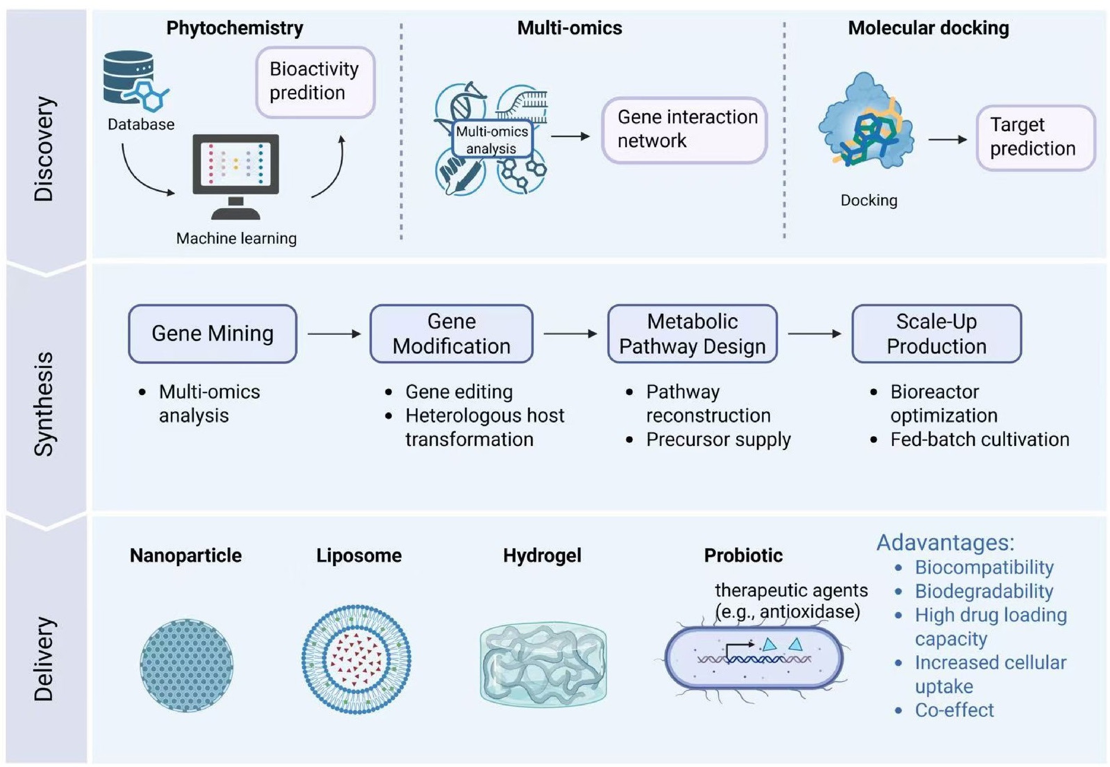
Figure 4.
Emerging technologies that facilitate the development of drugs from natural products. (a) Innovative drug discovery via AI-assisted phytochemistry, multi-omics analysis, and molecular docking. (b) Biosynthetic strategies for bioactive factors. (c) Drug delivery using nanoparticles, liposomes, hydrogels, or probiotic in situ secretion. This figure was created with BioRender (BioRender.com).
The identification and optimization of novel phytochemicals for radiation enteritis will remain a key focus in future research. Advances in high-throughput screening technologies, combined with AI algorithms, will facilitate the rapid identification of bioactive compounds from vast libraries of plant extracts. Here, we propose an integrative framework that synergizes phytochemistry, multi-omics analysis, and AI-assisted prediction of antioxidant targets. First, identification and activity screening of plant components are conducted to search for potential plant-derived bioactive factors. Notably, AI-driven approaches can currently analyze complex datasets to predict the bioactivities of phytochemicals (e.g., anti-inflammatory and antioxidant activities) based on their chemical structure and genomes[93]. For example, research has confirmed that machine learning (ML) models serve as a powerful tool for accelerating the discovery of novel antioxidant candidates[93]. Besides, multi-omics (genomics, transcriptomics, proteomics, and metabolomics) analysis can be conducted to identify effector gene candidates by comprehensively profiling the molecular changes in intestinal tissues under oxidative stress induced by radiation exposure[94]. In addition, AI technologies can significantly enhance the efficiency of target prediction in the drug development process. For instance, using AI-accelerated molecular docking platforms, such as the combination of AlphaFold2 and AutoDock, for rapid prediction of binding affinities between plant-derived compounds and targets associated with radiation enteritis (e.g., NF-κB, TNF-α). Promising studies in this area have already been reported[95].
Concerning the production and optimization of phytochemicals, synthetic biology holds great promise for the future. The construction of microbial cell factories through genetic engineering and metabolic pathway optimization will enable the efficient biosynthesis of phytochemicals with therapeutic potential. This topic has been thoroughly summarized in previous reviews. Recently, a study successfully achieved the de novo synthesis of the complex plant secondary metabolite lignans through the optimization of key enzymes and metabolic engineering approaches, providing new insights into the biosynthesis and large-scale production of natural products[96].
The bioavailability and targeted delivery of phytochemicals are recognized as key factors that limit their therapeutic efficacy in radiation enteritis. Advances in nanotechnology and biomaterials science, however, will play a crucial role in developing innovative delivery systems for phytochemicals in the future. For instance, the encapsulation of phytochemicals in biodegradable nanoparticles[97], liposomes[98], or hydrogels[99] can protect them from degradation in the gastrointestinal tract, control their release, and enhance their absorption and distribution to the affected tissues. Additionally, the design of stimuli-responsive delivery systems that release phytochemicals in response to specific physiological or pathological conditions (e.g., inflammation or oxidative stress) will enable more precise and effective treatment of radiation enteritis[100]. Furthermore, combining phytochemicals with other therapeutic agents, such as probiotics, within a single delivery system may provide synergistic therapeutic effects and improve overall treatment outcomes.
-
It is currently believed that the pathogenesis of radiation enteritis is primarily attributed to a combination of factors, including barrier disruption, oxidative stress, inflammatory responses, and dysbiosis of the gut microbiota. These pathological aspects also represent key targets through which plants and their extracts exert preventive and therapeutic effects against radiation enteritis, which correspond to the mechanisms of action of plant bioactive factors.
Various plant-derived bioactive compounds counteract radiation enteritis through the construction of the multi-dimensional Antioxidant-Anti-Inflammatory-Repair Triad network. At the antioxidant level, they establish the first line of defense by activating the Nrf2/ARE pathway, directly scavenging free radicals, preserving mitochondrial function, and repairing oxidatively damaged biomacromolecules to block initial oxidative stress. Concurrently, on the anti-inflammatory front, they synergistically suppress the NF-κB/NLRP3 inflammasome axis, modulate MAPK/JAK-STAT/TLR4 signaling pathways, and form reciprocal inhibitory loops with antioxidant mechanisms. Subsequently, at the barrier repair level, they restore tissue homeostasis by reshaping intestinal immune cell phenotypes, reconstituting tight junction integrity, and regulating gut microbiota-immune crosstalk. This multi-target synergistic network achieves cascade protection—spanning from source control of ROS generation, efficient free radical interception, and enhanced cellular defense to inflammation resolution and structural repair—through precise cross-talk between pathways, thereby providing a systematic molecular template and theoretical framework for developing low-toxicity, broad-spectrum natural intestinal radioprotectants.
Recently, progress in AI, synthetic biology, and nanotechnology has opened new avenues to harness phytochemicals for radiation enteritis therapy. AI-assisted phytochemistry, multi-omics analysis, and molecular docking enable rapid discovery and target prediction of bioactive compounds. Synthetic biology facilitates the efficient biosynthesis and optimization of phytochemicals through engineered microbial cell factories. Moreover, advanced delivery systems enhance the bioavailability, stability, and targeted release of phytochemicals, thereby improving their therapeutic efficacy. These emerging technologies offer an integrative framework to accelerate the translation of plant-derived natural products into effective treatments for radiation enteritis.
-
The authors confirm contribution to the paper as follows: conceptualization, resources: Zheng Y,Zhu Z; validation: Zheng Y, Yu Y, Pham TTH; formal analysis, visualization, investigation: Tian M, Gu Z, Zhao L; data curation: Zheng Y, Yin T, Yu Y, Pham TTH; writing − original draft preparation: Tian M, Gu Z, Zhao L, Yin T, Yu Y, Pham TTH, Li X, Zheng Y, Zhu Z, Nian W; writing − review and editing: Zheng Y, Huang B, Zhu Z, Tian M, Gu Z, Zhao L, Yin T, Yu Y, Pham TTH, Chuawong P, Li X, Nian W; supervision, project administration: Zheng Y, Huang B, Zhu Z; funding acquisition: Zhu Z. All authors reviewed the results and approved the final version of the manuscript.
-
Data sharing is not applicable to this article as no datasets were generated or analyzed during the current study.
-
This research was funded by the Chongqing Municipal Science and Health Joint Medical Research Project Key Project (2024ZYD009), the Open Project of Panxi Crops Research and Utilization Key Laboratory of Sichuan Province (No. SZKF2109), the Fundamental Research Funds for the Central Universities (2023CDJXY-009) and the National Natural Science Foundation of China (32202129, 32202563).
-
The authors declare that they have no conflict of interest.
-
# Authors contributed equally: Meng Tian, Ziyi Gu, Leiran Zhao
- Copyright: © 2026 by the author(s). Published by Maximum Academic Press, Fayetteville, GA. This article is an open access article distributed under Creative Commons Attribution License (CC BY 4.0), visit https://creativecommons.org/licenses/by/4.0/.
-
About this article
Cite this article
Tian M, Gu Z, Zhao L, Yin T, Yu Y, et al. 2026. Medicinal plants for radiation enteritis: key bioactive compounds and relevant antioxidant mechanisms. Medicinal Plant Biology 5: e016 doi: 10.48130/mpb-0026-0011 

Medicinal plants for radiation enteritis: key bioactive compounds and relevant antioxidant mechanisms
- Received: 03 February 2026
- Revised: 16 April 2026
- Accepted: 22 May 2026
- Published online: 30 June 2026
Abstract: Radiation enteritis, a complication following cancer radiotherapy, is caused by reactive oxygen species (ROS)-mediated oxidative damage, coupled with inflammatory dysregulation and disruption of the gut microbiota. Recent advancements in phytotherapy have highlighted the potential application of plant-derived bioactive compounds (e.g., curcumin from Curcuma longa, epigallocatechin gallate [EGCG], and resveratrol) as mitigators in treating radiation-induced intestinal injury. These compounds, characterized by potent ROS-scavenging activity, excellent biocompatibility, and minimal toxicity, exhibit promising attributes for therapeutic intervention. To better understand the botanical diversity and underlying mechanisms of radiation enteritis, this review systematically profiles the alkaloid-, flavonoid-, and polyphenol-rich medicinal plants, relevant bioactive constituents, and their associated therapeutic effects in synergistic modulation of oxidative, inflammatory, and apoptotic cascades. These natural compounds effectively leverage dual antioxidant mechanisms, including direct ROS scavenging via phenolic hydroxyl groups, indirect activation of the nuclear factor erythroid 2-related factor 2 (Nrf2)/Kelch-like ECH-associated protein 1 (Keap1), upregulation of endogenous antioxidants, suppression of pro-inflammatory cytokines, and enhancement of tight junction proteins for barrier repair. Despite their potential as adjuvant therapies, challenges remain in identifying exact antioxidant targets and translating these into clinical applications. To address this issue, we propose a framework that integrates phytochemistry, multi-omics profiling, and AI-enabled prediction of antioxidant targets. This review aims to facilitate the development of plant-based radioprotectants as next-generation adjuvant therapies.
-
Key words:
- Radiation enteritis /
- Medicinal plants /
- Antioxidant /
- Oxidative stress /
- Phytotherapy /
- Plant extracts /
- Plant-derived bioactives