











-
Artificial grassland habitats such as lawns in parks, sports fields, and pastures are familiar environments in human life and frequently produce conspicuous fungal fruit bodies during the rainy season and in autumn in Japan. Some fungi growing on lawns occasionally attract public attention through mass media reports, whereas others form fairy rings on lawns, a phenomenon often regarded as undesirable in turfgrass management. Although several studies have reported fungi occurring in lawns and other artificial grasslands in Japan[1−3], these reports are largely fragmentary, and comprehensive surveys of fungal diversity in artificial grasslands are still lacking. Consequently, the fungal communities inhabiting artificial grasslands remain poorly understood in Japan.
Compared with forest fungi, macrofungi occurring in lawns and related habitats have rarely been the focus of systematic mycological surveys[4]. As a result, the species composition and diversity of fungi inhabiting artificial grasslands are still largely unclear. Furthermore, recent studies have reported previously undescribed fungal taxa from lawn habitats[5,6], indicating that artificial grassland remains insufficiently explored from a mycological perspective.
To improve our understanding of macrofungal diversity in artificial grassland habitats, we conducted surveys of lawn-inhabiting fungi in warm-temperate regions of Japan from 2024 to 2025. During these surveys, we encountered two agaric species, Agrocybe broadwayi and Xuaniella urbica, which are newly recorded in Japan. In this study, we describe these species based on morphological observations and molecular phylogenetic analyses and discuss their ecology and distribution in Japanese lawn habitats.
-
Fresh fruiting bodies were photographed in the field using a compact digital camera (TG-6, Olympus, Tokyo, Japan), and habitat data were recorded. After collection, macromorphological features were promptly observed under LED light in the laboratory, and specimens were then dried using a food dehydrator at 45 °C for 48 h. Color descriptions of macromorphological features followed Kornerup and Wanscher[7], e.g., 'yellowish white (4A2)' and 'yellowish white (1–4A2)'. Micromorphological observations were conducted using 3% potassium hydroxide (KOH) to soften tissues and facilitate observation, and Melzer’s reagent was used to test for chemical reactions. Micromorphological structures were photographed using a microscope camera (TS-2500, J-SCOPE, Kawasaki, Japan) and measured using Piximètre (v5.10). Measurements followed Oguchi & Hosaka[6].
DNA extraction, PCR, and sequencing
-
DNA was extracted from dried specimens using a modified version of the microwave-based rapid extraction method of Izumitsu et al.[8], following Oguchi & Hosaka[6]. Polymerase Chain Reaction (PCR) was performed to obtain ITS and LSU sequence data from ribosomal DNA. The protocol and conditions are shown below. A reaction mixture containing 3.5 µL of sterile distilled water, 5 µL of EmeraldAmp MAX PCR Master Mix (TaKaRa, Kusatsu, Japan), 0.25 µL each of forward and reverse primers (10 µm), and 1 µL of template DNA was placed in 0.2 mL tubes on ice. PCR amplification was performed using a Veriti® 96-Well Thermal Cycler (Applied Biosystems Inc., Foster City, CA, USA). The ITS region was amplified using primer pairs ITS5/ITS4[9] and the LSU region using LROR/LR5[10]. The initial denaturation step was conducted at 95 °C for 3 min. This was followed by 35 cycles consisting of denaturation at 95 °C for 35 s, annealing at 51 °C for 30 s, and extension at 72 °C for 1 min (all primer sets). Final extension was carried out at 72 °C for 10 min. PCR products were confirmed by electrophoresis on a 1% agarose gel stained with ethidium bromide under UV light. If PCR products were successfully amplified, they were purified using ExoSAP-IT (Millipore, Molsheim, France). Sequencing was performed using a BigDye Terminator Cycle Sequencing Kit on an ABI 3500 Genetic Analyzer (Applied Biosystems Inc., Foster City, CA, USA), following the manufacturer's instructions.
Phylogenetic analyses
-
Raw sequences obtained in this study were visually inspected using BioEdit (v7.2.5[11]), and consensus sequences were assembled by aligning forward and reverse reads with Clustal W (v2.1[12]). Assembled ITS sequences were analyzed by blastn using default option settings (NCBI;
https://blast.ncbi.nlm.nih.gov/Blast.cgi?PROGRAM=blastn&BLAST_SPEC=GeoBlast&PAGE_TYPE=BlastSearch , accessed on 1 February 2026). Sequences with ≥ 97% identity in blastn searches (excluding sequences with query coverage < 70%) and sequences of related species deposited in GenBank datasets were used as a reference for phylogenetic tree construction (Table 1).Table 1. A list of specimens used in this study.
Species1 Voucher and/or strain numbers ITS2 LSU2 Locality Agrocybe arvalis TENN 073105 MH615058 – USA Agrocybe arvalis DSM 9710 MN306191 – GERMANY Agrocybe broadwayi TNS F-84789 PX977843 – JAPAN: Ibaraki Agrocybe broadwayi TNS F-110973 PX977844 PX963668 JAPAN: Mie Agrocybe broadwayi TNS F-110974 PX977845 PX963669 JAPAN: Ibaraki Agrocybe broadwayi OMDL iNat # 224511333 PV818605 – USA Agrocybe broadwayi ABS1-BAGD PQ898389 – INDIA Agrocybe broadwayi (T) NY 814876 OP162979 – GRENADA Agrocybe cubensis (T) NY 814881 OP162978 – CUBA Agrocybe dura CBS:157.63 MH858248 – unknown Agrocybe dura CBS 246.38 MH855957 – USA Agrocybe eduradii (T) LE 313652 OM524381 – RUSSIA Agrocybe firma F26774 MZ314314 – USA Agrocybe firma RMA 17 MG663239 – USA Agrocybe imaii (T) SAPA:10000036 MZ725547 – JAPAN Agrocybe molesta MO502967 OR336123 – USA Agrocybe pediades M. van der Walt:VDW1454 MT304652 – SOUTH AFRICA Agrocybe pediades HFJAU0368 MN258622 – CHINA Agrocybe pediades LE 262824 JN684785 – RUSSIA Agrocybe praecox OS387 KC842389 – NORWAY Agrocybe praecox OMDL iNat # 225665840 PQ678613 – USA Agrocybe praecox HMAS 297971 OR236782 – CHINA Agrocybe praecox PDD 86836 KM975410 – NEW ZEALAND Agrocybe putaminum PDD 96108 KM975434 – NEW ZEALAND Agrocybe retigera FLAS:60923 MH016951 – USA Agrocybe retigera SL1223 OR527351 – SINGAPORE Agrocybe retigera FAFU04 MW362215 – CHINA Agrocybe retigera ISBW36 PQ331056 – PAKISTAN Agrocybe rivulosa CCB160 KF830098 – USA Agrocybe rivulosa KUBOT_KRMK_2020_95 MW487609 – INDIA Agrocybe smithii PBM3793 MG663269 – USA Agrocybe striatipes (T) HMAS286942 OQ186168 – CHINA Agrocybe subpediades LE 217898 JN684795 – RUSSIA Agrocybe vervacti M60 MW425942 – HUNGARY Agrocybe vervacti GC 95058 OQ845960 – ITALY Cyclocybe erebia ANGE318 KM260145 – ITALY Atheniella flavida FFAAS0355 MW969654 – CHINA Atheniella flavida (T) FFAAS0350 MW969653 MW969665 CHINA Atheniella flavoalba 604 JF908464 – ITALY Atheniella flavoalba CBS 258.53 MH857185 MH868723 FRANCE Atheniella flavoalba H6032608 MW540661 – FINLAND Atheniella flavoalba HMJAU43764 MT497547 – CHINA Clitocybula fuscostriata FFAAS1029 OR238881 OR238893 CHINA Clitocybula fuscostriata (T) FFAAS1030 OR238882 – CHINA Clitocybula intervenosa (T) BAP 613 MH414561 MH385335 SÃO TOMÉ E PRINCIPE Clitocybula oculus F26468 MZ317354 – USA Clitocybula oculus AFTOL-ID 1554 DQ192178 DQ151452 USA Delicatula integrella KA12-1305 KR673538 – KOREA Gerronema kuruvense HTBM3056 PX308920 PX309038 CHINA Hemimycena lactea OULU:GAJ15636 OR863440 OR863509 FINLAND Hydropodia silvae-nipponicae TNS F-82594 OQ676557 OQ676555 JAPAN: Ibaraki Hydropodia silvae-nipponicae (T) TNS F-82592 OQ676556 NG_242114 JAPAN: Ibaraki Trogia delicata DED 8235 (SFSU) MH414567 MH385341 SÃO TOMÉ E PRINCIPE Xuaniella urbica TNS F-84752 PX977846 PX963670 JAPAN: Ibaraki Xuaniella urbica TNS F-84784 PX977847 PX963671 JAPAN: Ibaraki Xuaniella urbica TNS F-84786 PX977848 PX963672 JAPAN: Ibaraki Xuaniella urbica TNS F-84791 PX977849 PX963673 JAPAN: Chiba Xuaniella urbica TNS F-84841 PX977850 PX963674 JAPAN: Kanagawa Xuaniella urbica HTBM2719 PX308955 PX309075 CHINA Xuaniella urbica HTBM2910 PX308939 PX309056 CHINA Xuaniella urbica (T) HKAS150761 PX308937 PX309053 CHINA Xeromphalina enigmatica (T) TENN-F-055726 KM066052 KM066079 FINLAND 1 Holotype specimens are marked with (T). 2 GenBank accession numbers for each locus used in this study; PX963668 and PX963669 were excluded from the analyses. Bold indicates sequences obtained in this study. Phylogenetic analyses of A. broadwayi and X. urbica were conducted independently. For A. broadwayi, phylogenetic analyses were conducted using the ITS dataset only. For X. urbica, ITS and LSU datasets were first analyzed separately; because no significant conflict was detected between the two datasets, they were concatenated for combined analyses. Concatenation was performed manually by visual inspection using BioEdit.
Both ITS and ITS + LSU datasets were aligned using MUSCLE (v3.8[13]) in MEGA (v7.0.26[14]). Maximum-likelihood (ML) phylogenetic analyses were conducted using IQ-TREE (v2[15]). The best-fit substitution models were selected automatically. For the A. broadwayi ITS dataset, the HKY model with empirical base frequencies and gamma-distributed rate heterogeneity (four categories) was selected. For the X. urbica ITS + LSU dataset, the ITS partition was modeled with gamma-distributed rate heterogeneity (four categories), whereas the LSU partition used the TPM3 model with rate heterogeneity (two categories).
Branch support was assessed using ultrafast bootstrap approximation (UFBoot)[16] with 1,000 replicates, the SH-aLRT test with 1,000 replicates, and the approximate Bayes (aBayes) test. Xeromphalina enigmatica (TENN-F-055726) was used as the outgroup for the X. urbica analyses, whereas Cyclocybe erebia (ANGE318) was used as the outgroup for the A. broadwayi analyses.
-
The examined Japanese specimens of A. broadwayi (TNS F-84789, -110974, -110973) formed a clade with the holotype specimen of A. broadwayi from Grenada (NY 814876) and additional specimens from North America and India. They also formed a clade with A. retigera (Speg.) Singer collected from China, North America, Pakistan, and Singapore (Fig. 1). This clade was well supported (99.2/1/97).
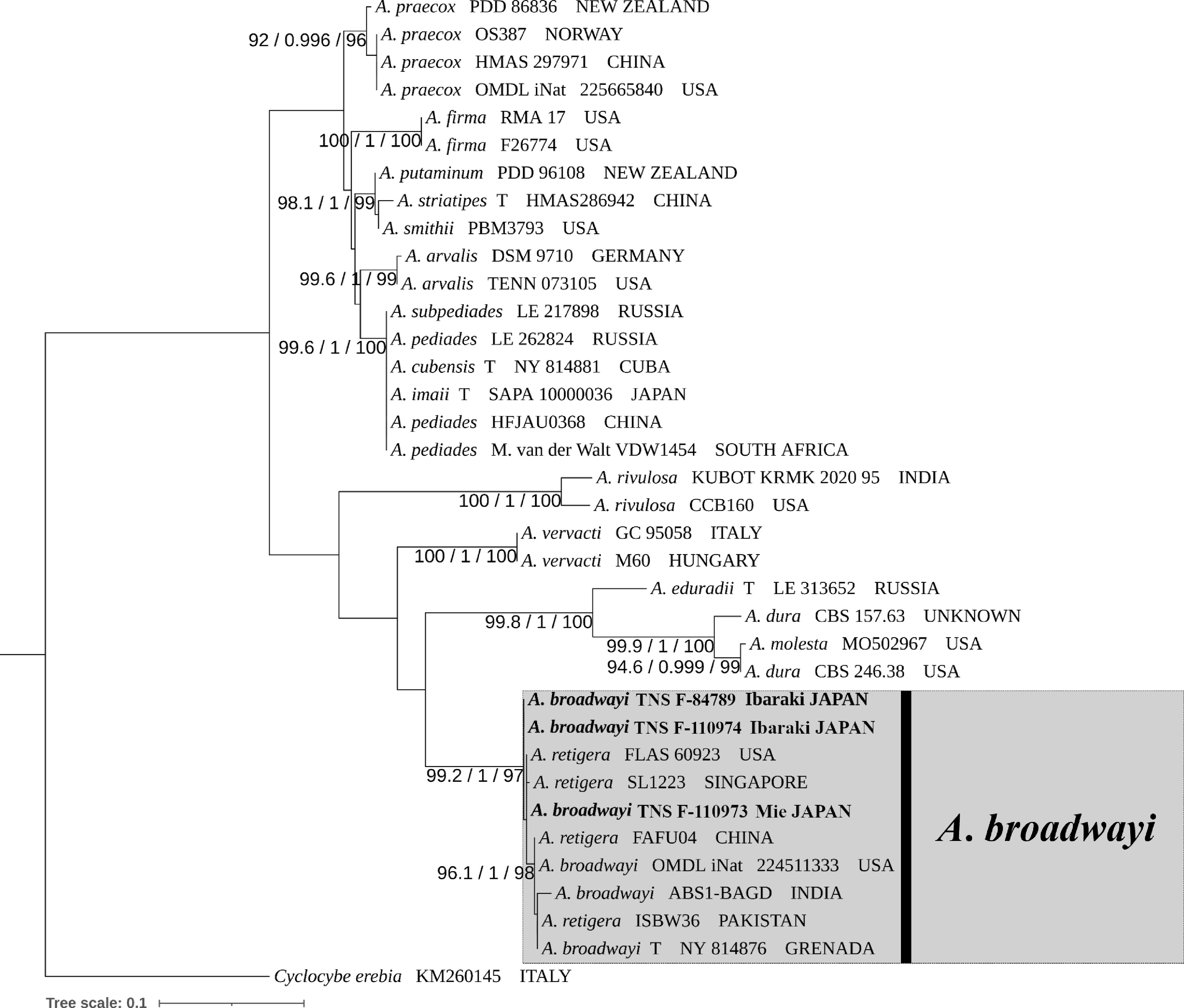
Figure 1.
Phylogenetic placement of Agrocybe broadwayi from an ITS using IQ-TREE. SH-aLRT support (X ≥ 90%), aBayes support (Y ≥ 0.95), and ultrafast bootstrap support (Z ≥ 90%) are indicated above or below the branches as X/Y/Z.
In addition, the Japanese specimens of X. urbica (TNS F-84752, -84791, -84786, -84784, -84841) formed a clade with Chinese specimens of X. urbica, including the holotype specimen (HKAS150761) (Fig. 2). This clade was well supported (99.5/1/99).
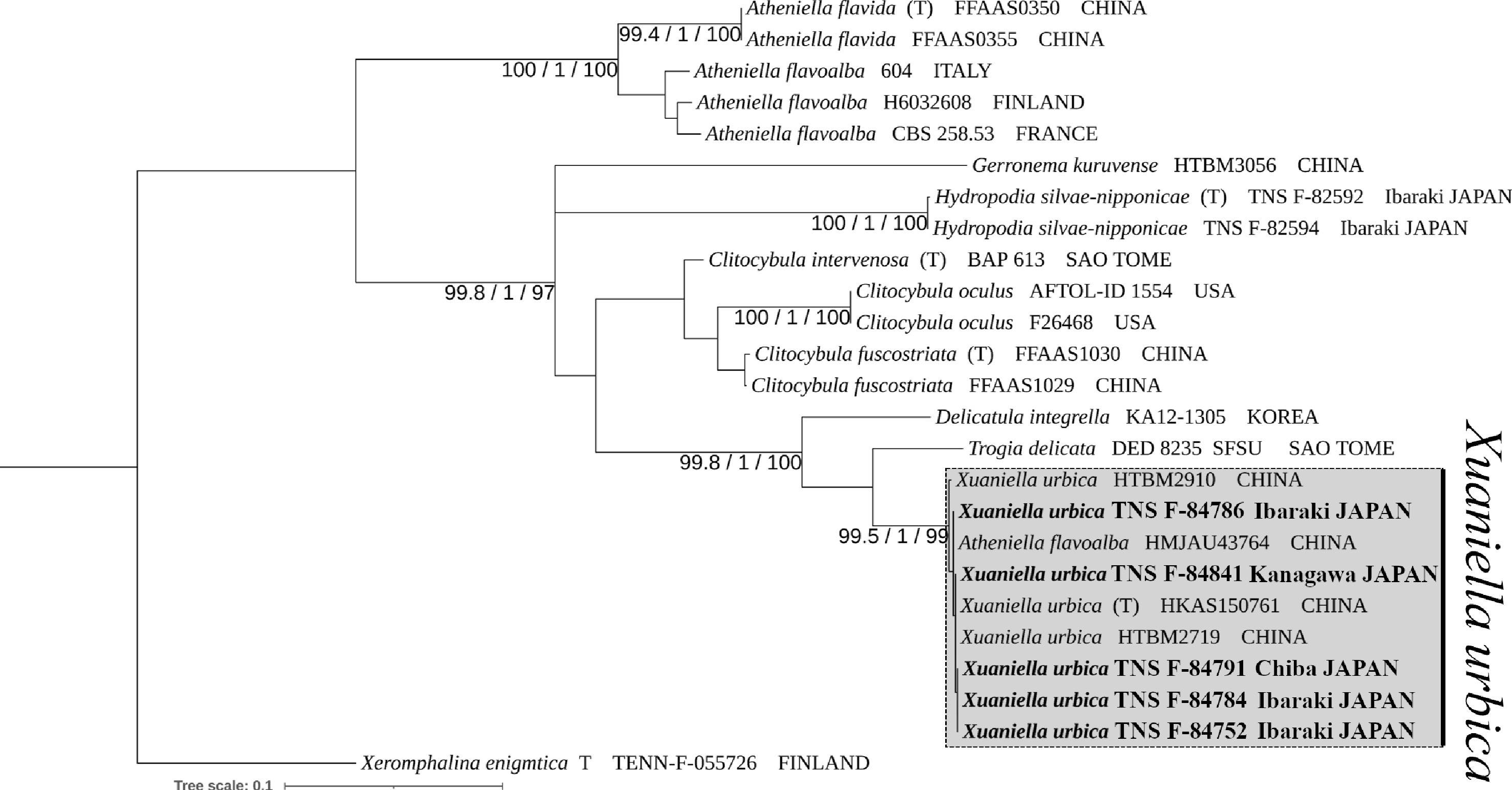
Figure 2.
Phylogenetic placement of Xuaniella urbica among phylogenetically related species based on the ITS and LSU dataset using IQ-TREE. SH-aLRT support (X ≥ 90%), aBayes support (Y ≥ 0.95) and ultrafast bootstrap support (Z ≥ 90%) are indicated above or below the branches as X/Y/Z.
Taxonomy
-
Agrocybe broadwayi (Murrill) Dennis, Bull. Soc. Mycol. France 69 (2): 179 (1953) [MB#292330] (Fig. 3a–l).
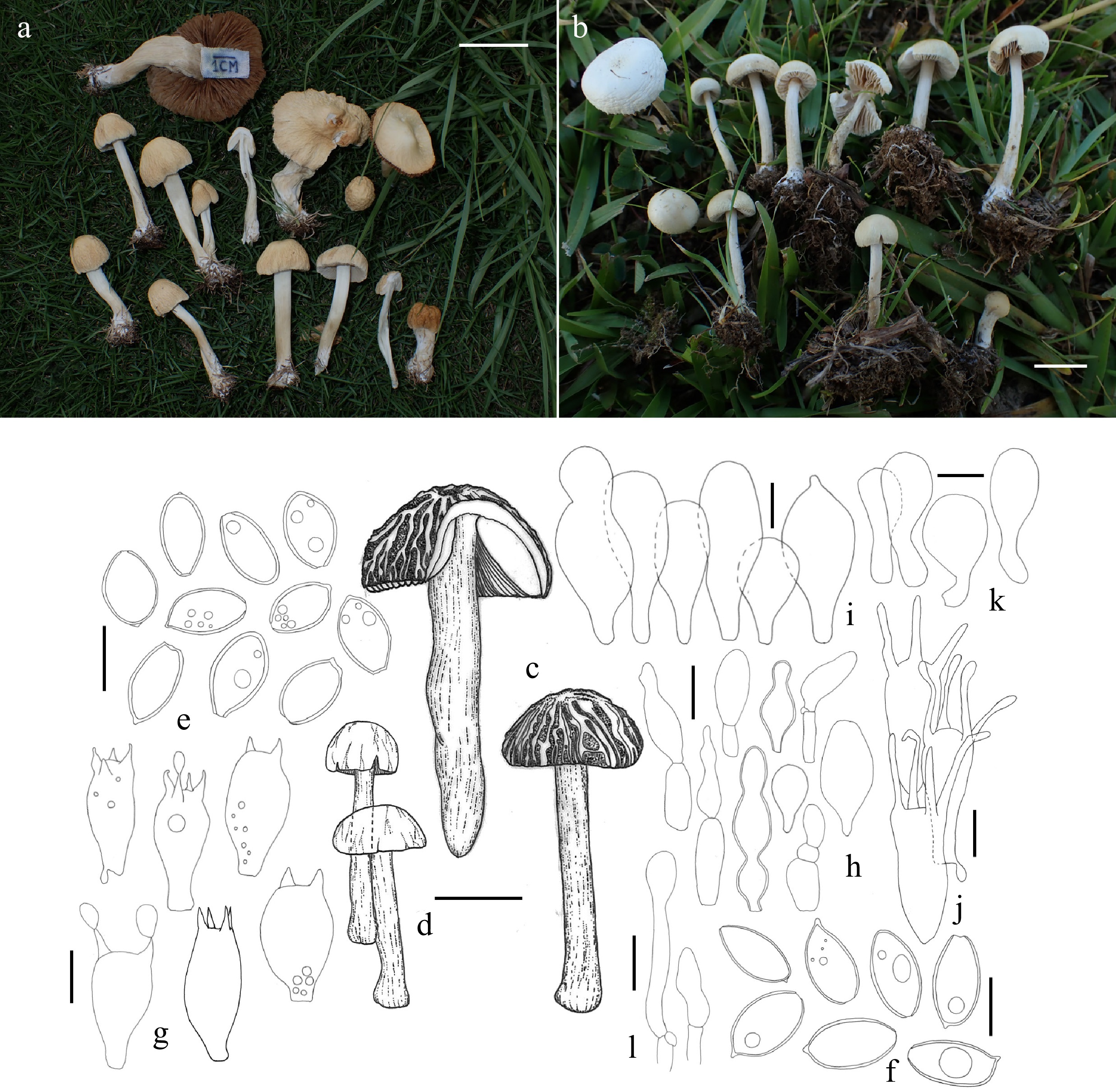
Figure 3.
Agrocybe broadwayi. (a)–(d) Basidiomata. (e), (f) Basidiospores. (g) Basidia. (h) Cheilocystidia. (i) Pleurocystidia. (j) Irregular elements. (k) Elements of pileipellis. (l) Elements on stipitipellis. Voucher specimens shown here: (a), (e), (g), (k) and (l) (TNS F-84789). (b), (f), (j) (TNS F-110973). (c) (TNS F-110974). Scale bars = 1 cm in (b); 2 cm in (c), (d); 3 cm in (a); 10 µm in (e)–(g), (i)–(l); 20 µm in (h).
≡ Hebeloma broadwayi Murrill, Mycologia 4 (2): 82 (1912) [MB#237868]
≡ Agrocybe broadwayi var. indica Natarajan & Raman, Biblioth. Mycol. 89: 70 (1983) [MB#116781]
Basidiomata (Fig. 3a–d) medium size, with a wrinkled pileus. Pileus 7–55 mm diam, at first convex, then plano-convex, finally flattening with a broad umbo, surface rugulose to rugose, sometimes almost smooth, sometimes the margin slightly wavy, yellowish white 4A2, pale yellow 4A3, orange white 5A2 or pale orange 5A3, rarely brownish orange 6C5–8 (6C5-sahara, 6C6-caramel, 6C7-reddish golden, 6C8-brownish orange) or light brown 6D5–8 (6D5-sunburn, 6D6-cinnamon, 6D7-raw sienna, 6D8-light brown). Gills 3–5 mm wide, adnexed and close, the edge smooth, relatively thin, non-marginate, sometimes wavy in age, white to yellowish white 4A2 or orange white
6A2 when young, then yellowish grey 4B2, orange grey 5–6B2 (5B2-alabaster, 6B2-birch dark), greyish orange 5B3–4 and 6B3 (6B3-flesh) with age, finally becoming brownish orange 6C3-6 (6C4-red-haired, 6C5-sahara, 6C6-caramel), light brown 6D4–6 (6D4-camel, 6D5-sunburn, 6D6-cinnamon) and 7D5–6 (7D6-titian red), brown 7E5–7 (7E5-somalis), dark brown 7F5–8 (7F6-eye brown) or reddish brown 8E4–6 (8E5-madeira) at mature. Stipe 20–80 × 2–16 mm, almost equal but becoming swollen toward the base, often twisted in large basidiomata, the surface glabrous or slightly fibrillose, white or off-white to concolor with pileus, especially brownish on the lower part, the base covered by white mycelium, nearly hollow. Flesh white and soft; odor and taste indistinct.
Basidiospores (Fig. 3e, f) (11.4–)12.2 – 15.5(–16.9) × (6.3–)7.3 – 9(–9.3) µm, average = 13.5 ± 1 × 8 ± 0.5 µm, Q = (1.4–)1.5 – 1.9(–2.2), Q average = 1.7 ± 0.1, (specimens = 3, n = 30), ellipsoid to elongate, thick-walled, with a germ pore, sometimes with oil droplets, brownish in KOH 3% aq., surface smooth. Basidia (Fig. 3g) 23–30.5 × 10.1–14.2 µm, average = 26.2 ± 2 × 11.9 ± 0.9 µm, (2–)4-spored, clavate to ovoid, hyaline but rarely yellowish in KOH 3% aq., seldom clamped at the base in these specimens. Cheilocystidia (Fig. 3h) present, 16–74 × 5–26 µm, variable, globose to pyriform, clavate, or lageniform, mostly thin-walled, but sometimes thick-walled, hyaline with a smooth surface. Pleurocystidia (Fig. 3i) present, 23–47.5 × 13–22 µm, globose, clavate to utriform; abnormally shaped elements sometimes present (Fig. 3j), finger- or basidium-like, hyaline with a smooth surface, 27–33 × 11–13.5 µm except atypical appendage, appendage cylindrical or somewhat swollen, 1–2–3–4–5 arising on the elements, 10–18.5 × 1.2–3.2 µm. Pileipellis hymeniform, elements (Fig. 3k) 19–46 × 7.5–23 µm, globose to pyriform or clavate, hyaline and thin-walled. Stipitipellis cutis, hyphae 3–9.5 µm wide; terminal elements (Fig. 3l) present, 15–42 × 4–7.5 µm, hypha-like to elongate, very rare in these specimens. Clamp connections present in many parts but rare at the base of basidia. Hymenial trama regular.
Habit, habitat and distribution: gregarious in lawns composed mainly of Zoysia Willd. spp., mixed with Trifolium repens L., Plantago asiatica L., etc. June to September. Honshu (Ibaraki and Mie Prefectures) in Japan.
General distribution: Asia: India (Watling R. and Abraham SP.[17]; from sequence based of ABS1-BAGD), Japan (in this study); The Caribbean: Grenada[18], Cuba[19], Trinidad and Tobago[18]; North America: USA (from sequence based of OMDL iNat 224511333); South America: Brazil[19,20], French Guiana[21].
Collections examined: Ibaraki: Ayumizaki-kōen, Kasumigaura-shi, 1 Sep. 2024, K. Oguchi (KO24-211NR; TNS F-84789); the same locality, 11 Aug. 2025, K. Oguchi & K. Fujii (KO25-1AD; TNS F-110974). Mie: Shichirimi hama fureai-beech, Atawa, Mihama-chō, Minami muro-gun, 28 Jun. 2025, K. Oguchi (KO25-129; TNS F-110973).
Japanese name. Shiwa fumizuki-take (newly proposed here).
Remarks: this species is characterized by a smooth to rugose, wrinkled pileus and its occurrence in turf habitats. The morphological characters observed in the present specimens agree well with those of the A. broadwayi holotype examined by Dennis[18] and Eberhardt et al.[22], and they also closely match the morphological features of A. retigera as described by Cortez & Silveira[23], and Niveiro et al.[24]. However, differences were observed when compared with the specimens identified as A. retigera by Akram et al.[25], which formed the same clade as the Japanese specimens (Fig. 1), including larger basidiospores (Fig. 3e, f), differences in the size of cheilocystidia (Fig. 3h), and the pileipellis forming a cutis (although composed of the same type of elements (Fig. 3k). This species is here reported as a new record in Japan.
Among the Japanese specimens examined, only specimen TNS F-110973 exhibited abnormal finger- or basidium-like elements with apical appendages (Fig. 3j). This type of element is comparable to those reported in A. arvalis (Fr.) Singer[3,26,27] and is morphologically noteworthy. It exhibits features resembling those of basidia, in that the tips of the appendages are similar to sterigmata and are somewhat swollen; however, it differs in that the number of appendages is variable (1–5) and that they are unusually long and thick compared with typical sterigmata. This feature may represent a newly observed morphological character of A. broadwayi.
Sequences of A. retigera obtained from GenBank formed a clade with sequences derived from the holotype specimen of A. broadwayi in the phylogenetic analyses (Fig. 1). In addition, branch lengths within this clade were relatively short. These results suggest that these taxa are phylogenetically very closely related and may represent the same species. As also noted by Eberhardt et al.[22], A. retigera, which shares similar habitats and morphological characteristics, may be conspecific with A. broadwayi, and because A. broadwayi has nomenclatural priority, A. retigera may eventually be treated as its synonym. Therefore, further examination of the type specimen of A. retigera is desirable to clarify their taxonomic relationship.
In Japan, morphologically similar species to A. broadwayi have been reported including A. farinacea Hongo, A. pediades (Fr.) Fayod, and A. molesta (Lasch) Singer (the third species is currently treated as a synonym of A. dura in Index Fungorum but was recognized as a distinct biological species by Flynn & Miller[28], although this species has not been formally reported from Japan). Agrocybe farinacea differs in lacking a rugose pileus surface, having larger basidiospores, and occurring on compost rather than lawns[3,29]. Agrocybe pediades has more slender basidiomata and lacks a rugose pileus surface[3,24,27]. Agrocybe molesta overlaps in habitat but differs in lacking a rugose pileus surface and in possessing an annulus on the stipe[24].
Agrocybe broadwayi has been reported from tropical to warm-temperate regions, including Asia[17], South America[19−21], the Caribbean[18,19], and Japan (in this study). Notably, records referred to A. retigera have also been reported from tropical to warm-temperate grassland habitats in Asia[17,25,30], North and South America[23,31−34], and the Pacific region[23]. Thus, the known geographic and ecological distributions of A. broadwayi and A. retigera show substantial overlap. Together with their phylogenetic affinity and morphological similarity, this distributional pattern further supports the hypothesis that the two taxa represent a single species. Among the previously known records, the Japanese collection represents the easternmost and northernmost occurrence of the species (TNS F-84789; approximately 36° N, 140° E). Furthermore, because this species has been recorded from artificial grasslands across a wide geographic range, it may have been dispersed through human activities associated with turfgrass. Further surveys are needed to better understand its distribution.
Xuaniella urbica K.L. Yang, Jia Y. Lin & Zhu L. Yang, J. Fungi 11 (10, no. 749): 45 (2025) [MB#572642] (Fig. 4a–j)
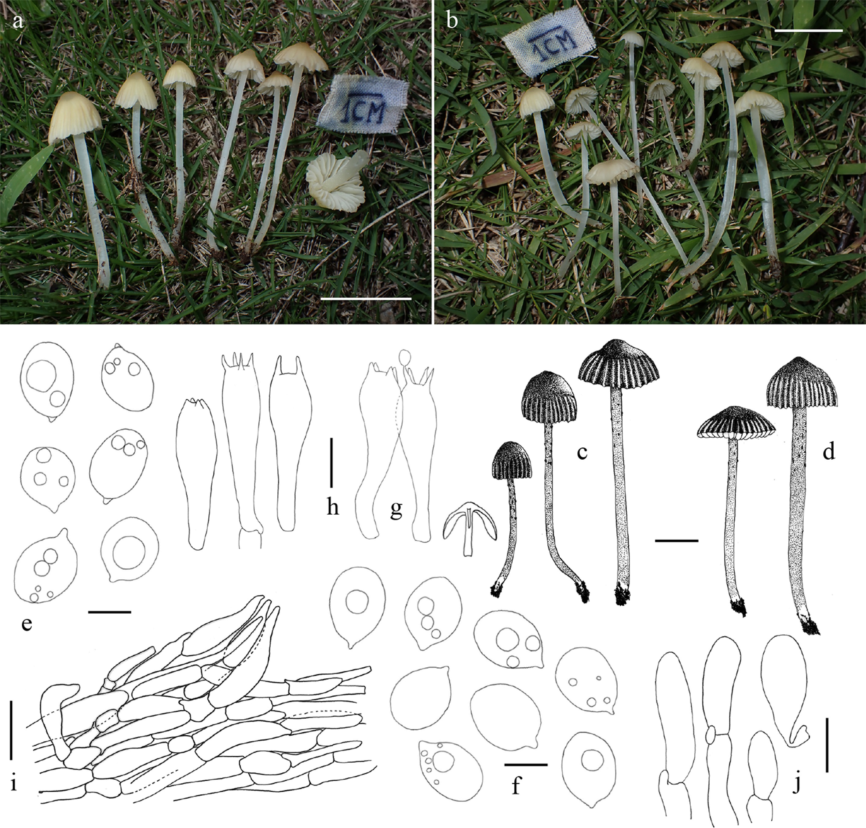
Figure 4.
Xuaniella urbica. (a)–(d) Basidiomata. (e), (f) Basidiospores. (g), (h) Basidia. (i) Pileipellis. (j) Elements on stipitipellis. Voucher specimens shown here: (a) (TNS F-84784). (b), (f) (TNS F-84791). (c), (g) (TNS F-84752). (d), (e), (j) (TNS F-84786). (h), (i) (TNS F-84841). Scale bars = 1 cm in (c), (d); 2 cm in (a), (b); 5 µm in (e), (f); 10 µm in (g), (h), (j); 40 µm in (i).
Basidiomata (Fig. 4a–d) mycenoid, white to yellowish, slender. Pileus 4–30 mm diam, parabolic to convex, then expanding to campanulate or becoming flattened, with an acute to broad umbo at the center, surface not viscid to faintly tacky when wet, lubricous, clearly striate to sulcate except the center, at first the margin yellowish white 1–4A2 (1A2-milk white, 2A2-pale), the center pale yellow 4A3 (4A3-cream), greyish yellow 3B4–6 (3B4-straw yellow, 3B5-wax yellow, 3B6-mustard yellow), 3C4–5 (3C5-absinthe yellow) and 4B3–4 (4B3-ivory, 4B4-champagne) or olive yellow 3C6, sometimes orange white 5A2, pale orange 5A3, orange grey 5B2 or greyish orange 5B3–4 overall, finally light brown 5D4–5 (5D4-dark blonde, 5D5-clay) or brown 6E6–7 (6E6-leather, 6E7-cognac) when old, pileus edge and gill edges often tinged with brownish. Gills 1.5–2.5 mm wide, adnate to slightly subdecurrent, subdistant, edges almost smooth to serrate, white when fresh, then yellowish white 4A2. Stipe 15–70 mm long, 1–4 mm wide, almost equal, surface with scattered fine white scales toward the apex, lower part glabrous, not viscid, pure-white to translucent. Flesh somewhat watery, soft, fragile and pure-white; odor and taste indistinct.
Basidiospores (Fig. 4e, f) (5.9–)6.8 – 8.6(–9.6) × (4.8–)5.2 – 6.6(–8.1) µm, average = 7.7 ± 0.6 × 5.9 ± 0.5 µm, Q = (1–)1.2 – 1.5(–1.6), Q average = 1.3 ± 0.1, (specimens = 5, n =30), subglobose to ellipsoid, thin-walled, smooth, hyaline in KOH 3% aq., with constantly present oily droplets, weakly bluish to inamyloid in Melzer’s reagent. Basidia (Fig. 4g, h) (17–)21 – 37 × (4.5–)5.8 – 9 µm, clavate, (2–)4-spored. Cheilo- and pleurocystidia absent. Pileipellis (Fig. 4i) a cutis, suprapellis composed of thin cylindrical elements 6–20 µm wide, sometimes containing brownish pigment, with terminal elements ventricose and sometimes erect; subpellis composed of thin tubular to swollen elements 50–70 µm wide. Stipitipellis a cutis to mixocutis, sometimes present terminal elements (Fig. 4j) 10–40 × 5.5–10 µm, erect, hyaline; hyphal aggregations frequent, 85–130 × 40–70 µm (parallel × perpendicular to the surface), composed of clavate to subovoid elements 6.5–17 × 3.5–9 µm. Clamp connections frequent. Hymenial trama regular.
Habit, habitat and distribution: gregarious on lawns composed mainly of Zoysia Willd. spp., and other plants, Trifolium pratense L., Trifolium repens L., etc. Early July to October. Honshu (Chiba, Ibaraki and Kanagawa Prefectures), Japan.
General distribution: Asia: Guangdong Province in China[5] and Japan (in this study).
Collections examined: Chiba: Shisui sōgō-kōen, Sumi, Shisui-machi, Inba-gun, 1 Sep. 2024, K. Oguchi & K. Fujii (KO24-214NR; TNS F-84791). Ibaraki: Ami-machi sōgō undo-kōen, Yoshiwara, Ami-machi, Inashiki-gun, 2 Jul. 2024, K. Oguchi (KO24-85; TNS F-84752); In Tsukuba Botanical Garden, Amakubo, Tsukuba-shi, 1 Sep. 2024, K. Oguchi & K. Fujii (KO24-205NR; TNS F-84784); Ayumizaki-kōen, Kasumigaura-shi, 1 Sep. 2024, K. Oguchi (KO24-208NR; TNS F-84786). Kanagawa: Toda, Atsugi-shi, 30 Oct. 2024, S. Tsuji (KO24-288NR; TNS F-84841).
Japanese name. Rōbai-take (newly proposed here).
Remarks: This species forms small, translucent white to yellow basidiomata on lawns dominated by Zoysia Willd. spp. It was described in 2025 from China as the type species of the genus Xuaniella Kun L. Yang, Jia Y. Lin & Zhu L. Yang, which is currently monotypic and comprises only X. urbica. Phylogenetic analyses based on multiple loci (ITS, LSU, rpb2, and tef-1α) placed this genus within Porotheleaceae Murrill[5], and the present study yielded the same result using ITS and LSU sequences (Fig. 2). This study additionally revealed that this species occurs in Japan, representing the second known distribution record following the original report from China.
The Japanese specimens showed minor differences from the original Chinese description, including somewhat larger basidiospores. However, other morphological characteristics generally agreed well. Moreover, the Japanese and Chinese specimens form a clade with extremely short branches in the ITS + LSU phylogenetic tree, suggesting that an expanded morphological interpretation of X. urbica is necessary. Furthermore, this indicates the distribution range of this species may extend to higher latitudes.
A species with very similar macroscopic features and habitat preferences is Atheniella flavoalba (Fr.) Redhead, Moncalvo, Vilgalys, Desjardin & B.A. Perry; however, it differs in possessing cystidia in the hymenium[35] (as Mycena flavoalba (Fr.) Quél.). Because X. urbica, together with the above-mentioned A. broadwayi, occurs on lawns in parks and other grassy areas in warm-temperate regions, it may be a common species in Japan. In addition, a report from China described children who had eaten this species and subsequently experienced symptoms such as abdominal pain and vomiting[5], suggesting that it may be a poisonous species. Therefore, caution is warranted in Japan as well.
-
In this study, A. broadwayi and X. urbica were recorded from Japan for the first time based on both phylogenetic and morphological evidence. Both species were collected from warm-temperate lawns in Japan, mainly dominated by Zoysia spp., in artificial environments such as parks. The habitats were characterized by short, densely growing turfgrass subjected to frequent trampling. In addition, members of Lycoperdaceae and Marasmius neooreades K. Oguchi & K. Hosaka were often observed in nearby areas. These fungi may share a preference for disturbed grassland environments maintained by human activities.
In the phylogenetic analyses, sequences labeled as A. retigera formed a clade with the Japanese specimens examined in this study and sequences derived from the holotype specimen of A. broadwayi. Furthermore, the morphological characters of A. retigera reported by Cortez & Silveira[23], and Niveiro et al.[24] closely matched those observed in the Japanese specimens. Eberhardt et al.[22] also suggested that these two taxa may be the same species, and the present results support this interpretation. Records identified as A. retigera have been reported from Asia[17,25,30], North and South America[23,31−34], and the Pacific region[23], suggesting that A. broadwayi may represent a grassland-associated species widely distributed in tropical to warm-temperate regions worldwide.
In contrast, X. urbica is currently known only from China and Japan. However, its confirmed distribution spans a broad latitudinal range, from Guangdong Province (approximately 23° N[5]) to Ibaraki Prefecture (approximately 36° N). This distribution pattern suggests that the species may occur widely in subtropical to warm-temperate lawns across East Asia. Accordingly, X. urbica may represent a more common and widespread species than previously recognized. The discovery of these two species in warm-temperate lawns suggests that artificial grassland habitats in Japan remain insufficiently explored from a mycological perspective.
-
Not applicable.
-
The authors confirm their contribution to the paper as follows: study conception and design: Oguchi K; data collection: Oguchi K, Hosaka K; analysis and interpretation of results: Oguchi K; draft manuscript preparation: Oguchi K. All authors reviewed the results and approved the final version of the manuscript.
-
The sequence data that support the findings of this study are available in the GenBank repository (www.ncbi.nlm.nih.gov/genbank), under the accession numbers listed in Table 1.
-
The authors declare that they have no conflict of interest.
- Copyright: © 2026 by the author(s). Published by Maximum Academic Press on behalf of Jilin Agricultural University. This article is an open access article distributed under Creative Commons Attribution License (CC BY 4.0), visit https://creativecommons.org/licenses/by/4.0/.
-
About this article
Cite this article
Oguchi K, Hosaka K. 2026. Newly recorded agarics growing on lawns in Japan. Panfungi 1: e011 doi: 10.48130/panfungi-0026-0005 

Newly recorded agarics growing on lawns in Japan
- Received: 01 July 2026
- Revised: 31 July 2026
- Accepted: 03 August 2026
- Published online: 28 August 2026
Abstract: Two species previously unrecorded from Japan are reported from warm-temperate lawns. Agrocybe broadwayi was originally described from Grenada and is known to occur in lawns and grassy roadsides. The species has previously been reported from Brazil, Cuba, French Guiana, India, and Trinidad and Tobago, but has not been recorded from East Asia. Morphological characteristics of Japanese specimens were consistent with previous descriptions of A. broadwayi, and molecular phylogenetic analyses showed that they formed a clade with the holotype specimen of the species. In addition, previously unreported micro-morphological features were observed in the Japanese specimens and are described herein. Xuaniella urbica was originally described from China and is known to occur in subtropical to warm-temperate lawns. Japanese specimens were collected from warm-temperate lawns and showed morphological characteristics consistent with those of X. urbica. In molecular phylogenetic analyses, they formed a clade with the holotype specimen of X. urbica. As neither species has previously been reported from Japan, these records represent the easternmost occurrences of both species and considerably extend their known distributions.
-
Key words:
- Agaricales /
- Agrocybe broadwayi /
- Grasslands /
- Molecular phylogeny /
- Taxonomy /
- Xuaniella urbica