












-
Osteoporosis (OP) is a systemic skeletal disorder characterized by low bone mass, microarchitectural deterioration, and increased bone fragility. It represents a prevalent yet often underdiagnosed chronic disease in aging societies[1]. According to the International Osteoporosis Foundation, approximately 200 million people worldwide are affected by osteoporosis, and nearly one-third of women and one-fifth of men over 50 years of age will experience an osteoporotic fracture during their lifetime[2]. With accelerating global population aging, the prevalence of osteoporosis is expected to increase further, leading to higher fracture-related mortality and a growing healthcare burden[3].
Although antiresorptive and osteoanabolic therapies have advanced, and clinical risk assessment and treatment algorithms have become increasingly refined, several challenges continue to limit therapeutic efficacy. These include residual fracture risk, poor adherence, rebound bone loss after discontinuation of treatment, and the reductionist paradigm of linking a single therapeutic target to a single bone cell process[4,5]. Recent studies indicate that bone marrow is not merely a hematopoietic site[6] but a dynamic, integrated microenvironment with features of both an immune organ and a bone-metabolic organ. Within this niche, immune cells, stromal cells, blood vessels, and nerves collectively regulate bone remodeling and repair[7]. Thus, beyond osteoblast-osteoclast imbalance, dysregulated bone-immune-metabolic coupling represents an important mechanistic dimension of osteoporosis, particularly in aging- and inflammation-associated disease contexts. In this review, immunosenescence is used as an organizing framework rather than a universal etiological definition of osteoporosis. Its relevance is most directly supported in age-related and postmenopausal osteoporosis and may extend to metabolic or chronic inflammation-associated bone loss. In glucocorticoid-induced, disuse-related, endocrine, genetic, and other secondary forms, immune niche dysfunction may function as a downstream modifier or amplifier rather than the primary initiating mechanism.
In this context, natural products have emerged as promising candidates for modulating the bone marrow immune niche owing to their multitarget activity, network-level regulatory capacity, and favorable biocompatibility[8]. Compared with conventional interventions, natural products may simultaneously target multiple pathological processes, including inflammatory amplification, immune cell dysfunction, oxidative stress, cellular senescence, metabolic dysregulation, and impaired osteogenesis[9]. Accordingly, natural products should be evaluated within a framework of osteoporotic immune microenvironment remodeling, particularly for their translational potential to alleviate immunosenescence and restore tissue regeneration[10].
Previous reviews have examined estrogen deficiency-mediated osteoimmunity in postmenopausal osteoporosis, immune aging and bone fragility, reciprocal skeletal-hematopoietic niche regulation, and multicellular immune reprogramming in the postmenopausal bone marrow[11]. Specifically, it links immunosenescence-associated changes across the hematopoietic, immune, stromal, adipose, and vascular compartments to regenerative failure; maps natural products to defined microenvironmental nodes and intercellular axes; and proposes experimental criteria for distinguishing genuine niche reprogramming from isolated improvements in bone remodeling endpoints. However, current studies have largely focused on the effects of natural products on bone mass, markers of bone turnover, and local signaling pathways; such evidence remains insufficient to establish their capacity to reprogram the bone marrow immune niche and restore regenerative potential[12]. On this basis, this review examines how immunosenescence may contribute to dysfunction of the bone marrow immune niche in age-related, postmenopausal, and selected metabolic or inflammation-associated forms of osteoporosis, and how natural products might modulate the resulting multicellular pathological networks. By incorporating emerging technologies and models, such as single-cell omics, spatial omics, and complex disease models, this review identifies key evidence gaps and outlines future translational directions (Fig. 1). Overall, this review extends the conceptual framework of osteoporosis research from conventional regulation of bone metabolism to immune microenvironmental reprogramming and provides a theoretical basis for mechanistic studies and precision translation of natural products.
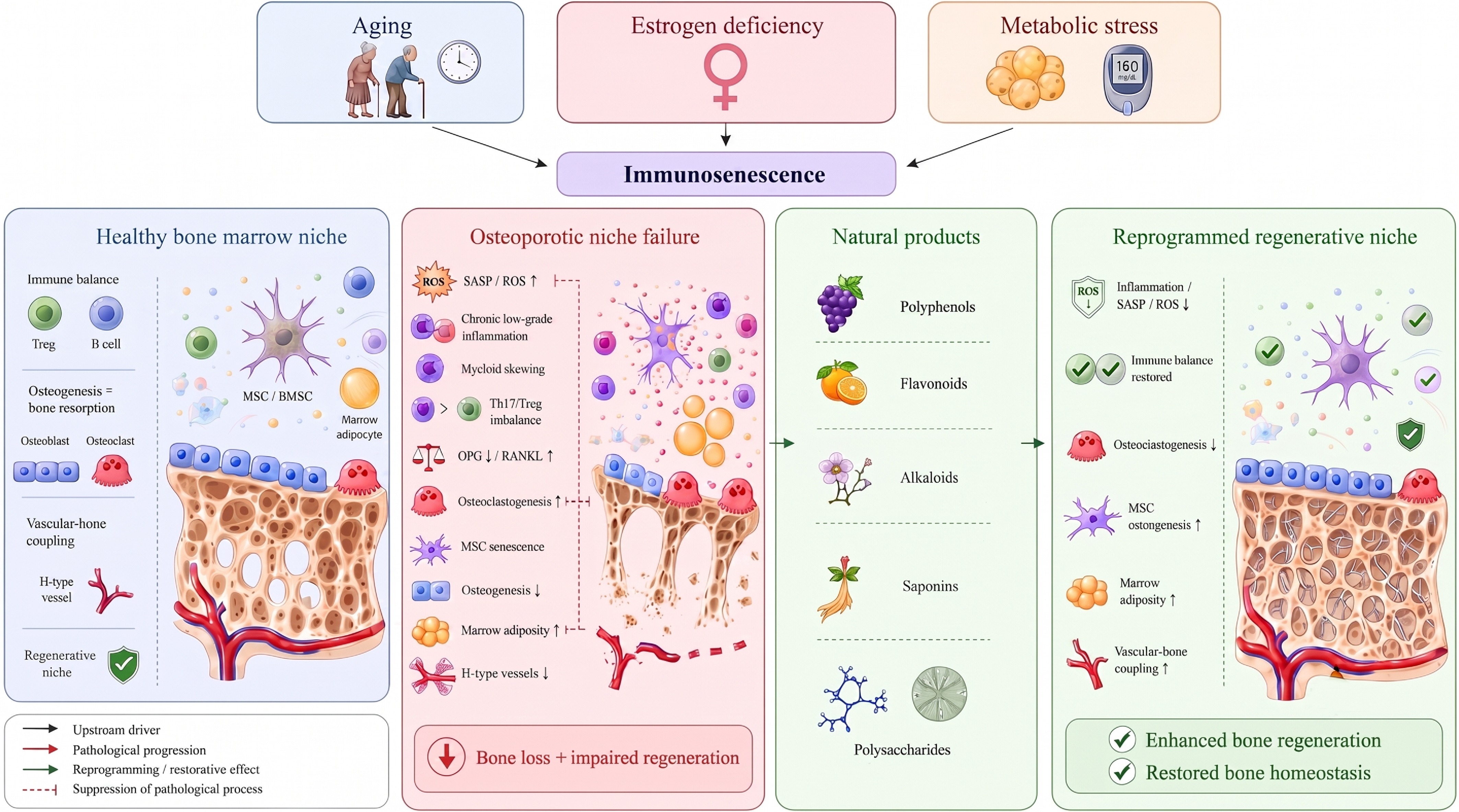
Figure 1.
Immunosenescence-associated dysfunction of the bone marrow niche in osteoporosis and potential natural product-mediated reprogramming. In a healthy bone marrow niche, immune cells, mesenchymal stem cells (MSC)/BMSC, osteoblasts, osteoclasts, H-type vessels, and appropriate levels of bone marrow adiposity collectively maintain immune homeostasis, vascular-bone coupling, and balanced bone remodeling. Aging, estrogen deficiency, and metabolic stress may converge on immunosenescence and dysfunction of the bone marrow immune niche, thereby contributing to chronic low-grade inflammation, elevated senescence-associated secretory phenotype (SASP)/reactive oxygen species (ROS), myeloid skewing, Th17/regulatory T-cell (Treg) imbalance, disruption of the B-cell osteoprotegerin (OPG)/RANKL axis, MSC senescence, increased bone marrow adiposity, and reduced H-type vessels, ultimately leading to bone loss and regenerative dysfunction. Natural products promote the reprogramming of the pathological bone marrow niche into a regeneration-supportive niche by modulating multiple nodes, including inflammatory responses, immune homeostasis, MSCs' osteogenic potential, bone marrow adiposity, and vascular-bone coupling.
-
Recent studies conceptualize the aging bone marrow niche as an interconnected hematopoietic, stromal, vascular, neural, and metabolic system rather than a collection of independently aging cell populations[13]. Immunosenescence can contribute to the bone marrow niche by promoting myeloid skewing, chronic inflammation, adaptive immune imbalance, mesenchymal stem cell (MSC) dysfunction, marrow adiposity, and impaired vascular-bone coupling. These interconnected cellular events collectively shift the local microenvironment toward enhanced bone resorption and impaired regeneration, as summarized in Fig. 2.
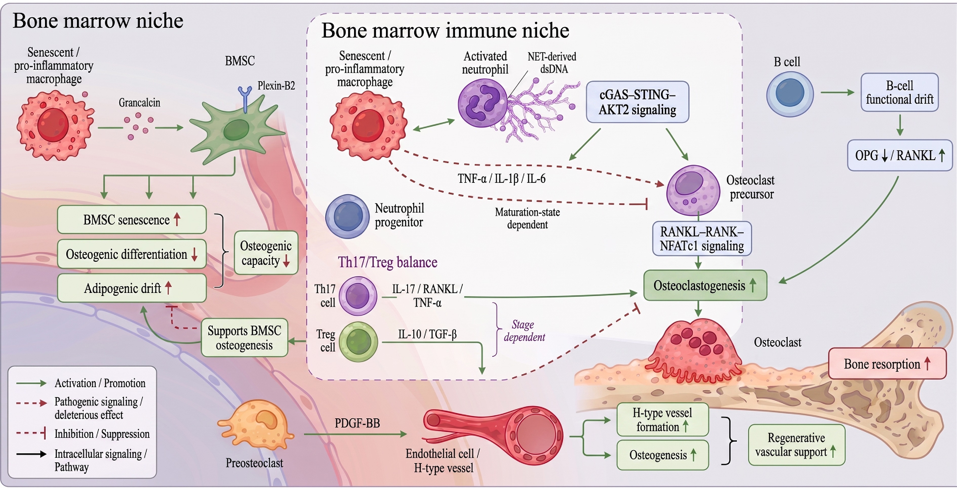
Figure 2.
Cellular interaction map of immunosenescence-associated bone marrow niche destabilization. Senescent or proinflammatory macrophages promote osteoclastogenesis through tumor necrosis factor alpha (TNF-α), interleukin (IL)-1β, and IL-6 signaling, while impairing BMSCs' osteogenic differentiation and promoting cellular senescence and adipogenic drift through the grancalcin-Plexin-B2 axis. Activated neutrophils release neutrophil extracellular trap (NET)-derived double-stranded DNA (dsDNA), which activates cGAS-STING-AKT2 signaling in macrophages and osteoclast precursors, thereby amplifying inflammatory macrophage activation and osteoclastogenesis; in contrast, neutrophil progenitors may restrain osteoclast formation in a maturation-state-dependent manner. Th17-derived IL-17, RANKL, and TNF-α, together with B-cell functional drift characterized by reduced OPG and increased RANKL, enhance osteoclast differentiation and bone resorption. Conversely, Treg-derived IL-10 and TGF-β suppress osteoclastogenesis and support BMSC-related osteogenesis, although these effects may vary with the stage of bone remodeling or repair. In parallel, preosteoclast-derived PDGF-BB promotes H-type vessel formation and osteogenesis, supporting regenerative vascular-bone coupling.
Normal bone marrow immune niche maintains bone homeostasis
-
Bone homeostasis is not governed solely by the balance between osteoblasts and osteoclasts. Instead, it depends on a highly organized bone marrow immune niche that provides structural support, trophic cues, and immune regulation[14]. The bone marrow niche comprises MSCs, osteoblasts, osteoclasts, and diverse immune cell populations, including macrophages, B-cells, and regulatory T-cells (Tregs)[15]. Within this system, immune cells are not merely inflammatory sources but key regulators of the direction, magnitude, and temporal dynamics of bone remodeling[16].
Bone lineage and immune cells jointly maintain bone homeostasis through coordinated functional modules, including the RANKL-RANK-osteoprotegerin (OPG) axis, chemokine networks, and paracrine cytokine signaling. Bone lineage cells directly regulate the formation and resorption of bone. For example, osteoblasts locally regulate bone remodeling by secreting OPG, sclerostin, and RANKL. RANKL binds RANK on osteoclast precursors and promotes osteoclast activation[17]. Conversely, OPG acts as a decoy receptor for RANKL, limiting excessive osteoclastogenesis and maintaining the balance between bone resorption and formation[18]. In parallel, immune cells translate cues from tissue injury, hormonal changes, and metabolic fluctuations into osteogenic or osteoclastogenic signals[19]. For instance, immune-cell-derived cytokines, including tumor necrosis factor alpha (TNF-α), interleukin (IL)-1β, and IL-6, modulate the RANKL-RANK-OPG axis and help maintain a controlled balance between pro-inflammatory and pro-reparative signals[20,21]. T- and B-cells exert bidirectional regulatory effects within this network. Under specific immune conditions, both T- and B-cells express RANKL and OPG and thereby participate in bone remodeling[22]. Certain T-cell subsets also regulate MSC differentiation, osteoblast activity, and local inflammatory thresholds through cytokine secretion, thereby maintaining immune tolerance, limiting low-grade bone marrow inflammation, and supporting an osteogenic environment[23]. Although pro-inflammatory subsets such as Th17 cells contribute to host defense and injury responses, their excessive activation disrupts bone remodeling through IL-17- and RANKL-mediated pathways[24].
Thus, a healthy bone marrow immune niche is defined not by low immune activity, but by a dynamic niche capable of switching between bone resorption and repair, and between immune surveillance and immune restraint. This dynamic adaptability enables bone tissue to maintain basal turnover and initiate effective regeneration after injury.
Immunosenescence remodels the bone marrow niche
-
With aging, the structure and function of the bone marrow immune system undergo systematic remodeling, a process known as immunosenescence[25]. One of the earliest changes in immunosenescence is hematopoietic myeloid skewing, characterized by increased production of monocyte/myeloid progenitors and a relative reduction in lymphoid progenitors, including T- and B-cell precursors. This shift provides a cellular basis for enhanced bone resorption[26]. During immunosenescence, the peripheral naïve T-cell pool contracts, responses to new antigens decline, and effector T-cells accumulate[27]. B-cell generation is also reduced, leading to decreased secretion of osteoprotective factors such as OPG[28]. Innate immune cells, particularly macrophages and neutrophils, also remain persistently activated at a low level and release inflammatory mediators[29]. Together, these processes establish a pro-inflammatory, pro-osteoclastogenic microenvironment. Aged macrophages tend to adopt an M1-like phenotype, thereby enhancing osteoclastogenic signals such as RANKL. Aged T-cells also produce elevated levels of TNF-α and IL-17, thereby disrupting osteogenic activity[30]. Aged B-cells and bone marrow adipocytes may secrete chemokines and lipid mediators, further promoting the accumulation of inflammatory cells and suppressing osteogenesis[31]. Notably, bone marrow immunosenescence is not merely a reflection of peripheral blood immunosenescence; it exhibits tissue-specific features. For example, aged macrophages secrete the calcium-binding protein grancalcin, which suppresses the osteogenic capacity of bone marrow MSCs through Plexin-B2 signaling, providing direct evidence linking immunosenescence to skeletal aging[32]. Overall, immunosenescence shifts the bone marrow niche away from homeostasis, creating a local niche that favors bone resorption and impairs bone formation.
Chronic low-grade inflammation under the background of inflammaging
-
Immunosenescence reshapes the cellular composition of the bone marrow niche, whereas inflammaging provides the inflammatory context that sustains this process[33]. During osteoporosis-associated aging, chronic low-grade inflammation is one of the most prominent pathological features[34]. Within the bone marrow niche, senescent immune cells, MSCs, and osteocytes continuously secrete IL-6, IL-1β, TNF-α, MCP-1, and multiple chemokines as part of the senescence-associated secretory phenotype (SASP)[35]. Even in the absence of infection, these mediators chronically activate immune cells and establish self-sustaining inflammatory circuits. This process leads to persistent activation of NF-κB, JAK-STAT, and NLRP3 inflammasome signaling in the local microenvironment[36], while suppressing pro-osteogenic Wnt/β-catenin and BMP pathways. IL-6 signaling in the bone marrow niche is highly context-dependent. In classical signaling, IL-6 binds membrane-bound IL-6R, whereas IL-6-soluble IL-6R complexes activate gp130 on a broader range of cells through trans-signaling. Both routes engage JAK-STAT3, whereas gp130-associated SHP2 can additionally activate MAPK/ERK[37]. Under defined osteogenic conditions, IL-6/IL-6R-STAT3 signaling supports BM-MSC differentiation; however, sustained IL-6 signaling in a SASP-rich niche may reinforce expression of RANKL and osteoclastogenic communication[38]. Bone-specific studies also indicate that trans-signaling can promote both osteoclastogenesis and bone formation, showing that receptors' availability, exposure duration, cell type, and disease stage determine the net skeletal effect[39]. Consequently, the bone marrow becomes increasingly biased toward pro-osteoclastogenic and anti-osteogenic signaling, causing bone turnover to progressively deviate from homeostasis[40]. Sustained TNF-α and IL-17 signaling promotes proliferation and differentiation of osteoclast precursors while inhibiting the osteogenic capacity of MSCs[41]. In parallel, SASP factors released by senescent bone marrow MSCs further disrupt neighboring cells' function, reinforcing this vicious cycle[42]. In osteoporosis, chronic inflammaging-associated low-grade inflammation may explain why many patients develop persistent bone loss, bone marrow fat accumulation, and delayed bone repair despite the absence of overt infection or acute inflammation. It may also explain why interventions that target terminal bone resorption alone often fail to fully restore bone's quality and regenerative potential.
Estrogen deficiency and bone marrow adiposity
-
Within the bone marrow niche, the pathogenic effects of estrogen deficiency are mediated largely by the immune system[43]. Postmenopausal women often exhibit a chronic low-grade inflammatory phenotype accompanied by remodeling of the immune cell composition. These changes increase T-cell activation and promote production of TNF-α and RANKL, thereby enhancing osteoclastogenesis. Concurrently, reduced OPG levels weaken the inhibition of bone resorption[44]. In addition, fatty acids, insulin resistance, and oxidative stress associated with obesity and metabolic syndrome can promote a pro-inflammatory milieu by altering macrophage and MSC metabolism[45,46]. Bone marrow adipose tissue (BMAT) expands markedly with aging and osteoporosis, and adipocyte-derived factors, including IL-6, RANKL, and adiponectin, can suppress osteogenesis while promoting osteoclastogenesis[47]. Furthermore, increased bone marrow fat can exacerbate ROS accumulation through fatty acid release, drive adipogenic differentiation of MSCs, and further deplete the osteogenic progenitor pool[48]. Thus, from a bone marrow niche perspective, osteoporosis, particularly postmenopausal and metabolism-related subtypes can be viewed as a niche disease shaped by endocrine alterations, immune dysregulation, and metabolic stress. Estrogen deficiency and bone marrow adiposity therefore represent key structural drivers of the disease's heterogeneity and progression.
Bone marrow niche dysfunction as a regenerative component of osteoporosis
-
Bone marrow niche dysfunction should be regarded as an important mechanistic component of osteoporosis rather than a universal disease definition[49]. In age-related and postmenopausal osteoporosis, chronic inflammation, immune cell remodeling, stromal senescence, and vascular-bone uncoupling may progressively reduce the niche's capacity to support osteogenesis and tissue repair[50]. Normal bone repair depends on coordinated sequential processes, including inflammatory initiation, inflammation resolution, vascular reconstruction, osteoblast recruitment, and bone remodeling. Under aging, estrogen deficiency, and metabolic dysregulation, however, the bone marrow niche remains in a state of chronic low-grade inflammation. Pro-inflammatory factors remain elevated, osteoclast precursors become more readily activated, and MSC lose osteogenic potential while shifting toward adipogenic differentiation. Together, these changes lead to concurrent bone loss and repair failure[51]. This process is driven not solely by intrinsic bone cell deterioration, but by the combined dysfunction of immune cells, stromal cells, and the vascular system. For example, aged myeloid cells suppress BMSCs' osteogenesis and promote adipogenic differentiation by secreting factors such as grancalcin[32]. In addition, multiple immune cell subsets in the bone marrow of patients with postmenopausal osteoporosis exhibit pro-inflammatory and pro-osteoclastogenic features, further compromising the regenerative niche[52]. Therefore, delayed osteoporotic fracture healing, incomplete restoration of bone quality, and reduced regenerative efficiency reflect a shift in the bone marrow niche from a repair-supportive state to one that sustains inflammation and osteoclastogenesis.
Accordingly, therapeutic evaluation in osteoporosis should not focus solely on bone mass and bone resorption indices, such as BMD, BV/TV, or TRAP. It should also assess whether interventions restore the resolution of inflammation, MSC osteogenic potential, vascular-bone coupling, and pro-reparative immune states[53]. For natural products, their potential value lies not merely in isolated anti-inflammatory or osteogenic effects, but in their ability to remodel the bone marrow immune niche across multiple nodes and restore its capacity to support bone homeostasis and tissue regeneration.
-
The transition from immunosenescence to impaired bone regeneration is driven by a coordinated network of myeloid skewing, adaptive immune imbalance, MSC senescence, marrow adiposity, and vascular-bone uncoupling. These processes collectively promote bone resorption, suppress bone formation, and impair repair capacity, as illustrated in Fig. 3.
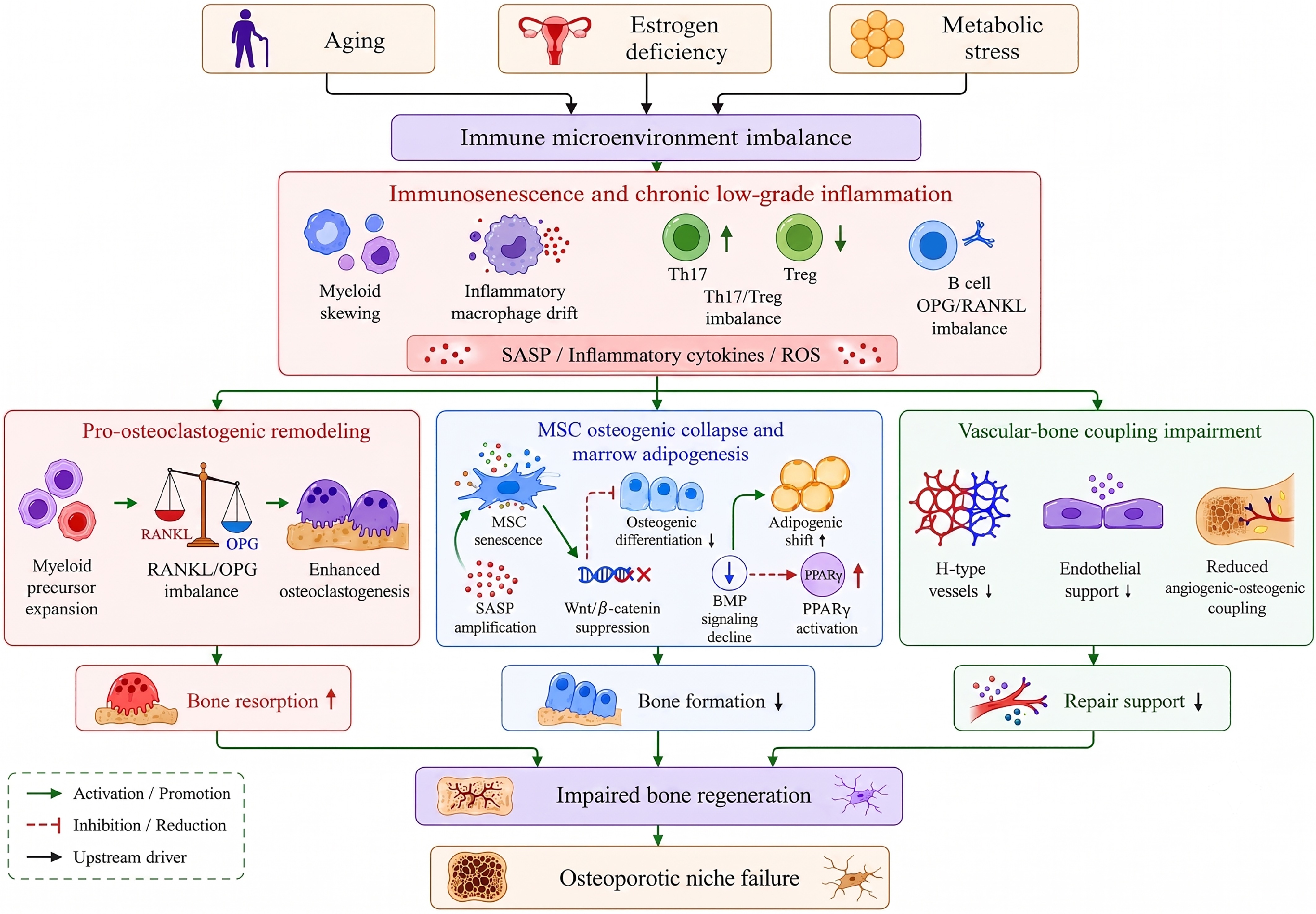
Figure 3.
Mechanistic cascade from immune microenvironmental imbalance to regenerative failure in osteoporosis. Aging, estrogen deficiency, and metabolic stress may converge on an imbalanced immune microenvironment, which further manifests as immunosenescence and chronic low-grade inflammation, accompanied by myeloid skewing, inflammatory macrophage drift, Th17/Treg imbalance, and disruption of the B-cell OPG/RANKL axis. Subsequently, pathological signals diverge into three major mechanistic modules: Pro-osteoclastogenic remodeling, MSCs' osteogenic collapse with bone marrow adiposity, and impaired vascular-bone coupling. Pro-osteoclastogenic remodeling leads to increased bone resorption. MSC senescence, Wnt/β-catenin suppression, BMP signaling decline, and PPARγ activation contribute to reduced bone formation, and the loss of H-type vessels together with impaired endothelial support results in insufficient reparative support.
The myeloid cell-osteoclast precursor axis: Myeloid skew and enhanced osteoclastogenesis
-
Immunosenescence-induced myeloid skewing markedly expands the pool of osteoclast precursors in the bone marrow. In response to RANKL, TNF-α, and other osteoclastogenic stimuli, these precursors undergo accelerated osteoclast differentiation[54]. In aged and inflammatory environments, macrophages tend to adopt a pro-inflammatory phenotype and secrete cytokines such as TNF-α and IL-1β, thereby directly promoting osteoclastogenesis[55]. Conversely, osteogenic macrophages, or osteomacs, which support skeletal homeostasis, become functionally impaired or numerically reduced. Together, these changes reflect disrupted immune-osteoclast coupling[56,57]. In osteoporotic mice, bone marrow macrophage-derived grancalcin suppresses MSCs' osteogenesis through Plexin-B2 signaling, whereas the same macrophage population exhibits enhanced osteoclastogenic potential[58]. Macrophage-MSC crosstalk is bidirectional and metabolically regulated. Persistent glycolytic M1-like states release TNF-α, IL-1β, and ROS, promoting MSCs' senescence and suppressing osteogenic commitment, whereas a timely transition toward oxidative phosphorylation- and fatty acid oxidation-associated reparative states supports osteogenesis through factors such as IL-10, TGF-β, and BMP-2[59]. Transient early inflammatory signals may nevertheless contribute to progenitor recruitment and osteogenic priming, indicating that the temporal dynamics are critical. MSCs reciprocally modulate macrophage states through paracrine mediators, including PGE2 and TGF-β, as well as extracellular vesicles[60]. Disruption of this feedback loop therefore links macrophages' immunometabolic dysfunction to regenerative failure.
In addition, neutrophil extracellular traps (NETs) released in aged bone marrow can induce local inflammation and indirectly promote osteoclastogenesis[61]. Polarization of macrophages in the aging bone marrow niche is partly regulated by the cGAS-STING pathway. Cytosolic DNA released from damaged nuclei or mitochondria, as well as NET-derived DNA, may activate cGAS-STING and downstream TBK1-IRF3/Type I interferon and NF-κB signaling, thereby shaping macrophages' inflammatory states[62]. Transient activation supports the innate immune defense, whereas persistent stimulation by self-DNA during aging or metabolic stress may sustain inflammaging and favor pro-inflammatory and pro-osteoclastogenic programs. Nevertheless, its effects are context-dependent. Thus, the M1/M2 terminology used here should be regarded as an operational simplification of a broader macrophage state continuum[63]. Thus, increased osteoclast activity does not simply arise from cell-intrinsic changes within the bone lineage[64], but reflects pathological remodeling of the myeloid immune compartment. Effective natural product-based interventions should therefore not only rebalance osteoblast-osteoclast dynamics but also reshape pro-osteoclastogenic myeloid subsets and restore reparative immune states[65].
T-cell imbalance: adaptive immune remodeling mediated by the Th17/Treg axis
-
T lymphocytes play a pivotal role in skeletal immune regulation, particularly through Th17/Treg axis-mediated adaptive immune remodeling. During aging, remodeling of the lymphocyte pool reduces the number and function of regulatory T-cells, but the proportion of pro-inflammatory Th17 cells often increases[11]. Aging-associated Treg insufficiency and Th17 expansion shift the bone marrow niche toward a pro-osteoclastogenic state. Th17-derived IL-17 is a potent osteoclastogenic factor that induces the production of RANKL and TNF, thereby enhancing bone resorption. In contrast, Tregs suppress osteoclastogenesis and support MSCs' osteogenic differentiation through the production of IL-10 and TGF-β[66]. In addition, aged T-cells may become exhausted or acquire a pro-inflammatory phenotype. A high proportion of senescent T-cells has been detected in the bone marrow of older patients with osteoporosis[67]. These cells continuously release IFN-γ, TNF, and other cytokines, further disrupting the balance of bone repair[68]. Overall, adaptive immune remodeling in osteoporosis is characterized by Th17/Treg imbalance and aberrant memory T-cell activation, which amplify inflammatory osteoclastogenic signals and suppress immune-tolerant reparative pathways.
B-cells' functional drift and OPG-RANKL imbalance
-
B-cells are key components of the bone marrow immune niche and regulate the RANKL-OPG axis. Under homeostatic conditions, B-cells serve as a source of OPG, which acts as a decoy receptor for RANKL, thereby limiting RANKL-RANK binding on osteoclast precursors, restraining osteoclastogenesis, and maintaining bone resorption within a physiological range[69]. During aging, however, the bone-protective function of B-cells may shift, causing them to amplify pro-osteoclastogenic signals. This functional shift appears particularly prominent in postmenopausal osteoporosis. Under estrogen-deficient conditions, B-cells regulate osteoclast formation through granulocyte colony-stimulating factor and the RANKL-OPG system, and cooperate with T-cell-derived TNF-α signaling to form a pro-osteoclastogenic cascade[70]. These findings suggest that B-cells are not merely passive targets but active nodes through which adaptive immunity mediates bone loss. Immunosenescence further impairs B-cell generation and homeostatic OPG support. Meanwhile, the pro-inflammatory microenvironment may drive a subset of B-cells toward increased RANKL expression and inflammatory cytokine secretion[71]. Single-cell analysis of bone marrow from patients with postmenopausal osteoporosis shows that B-cells, together with T-cells, monocytes, dendritic cells, and other immune populations, exhibit enhanced cytokine and chemokine signatures. Moreover, enhanced B-cell communication is associated with increased monocyte-to-osteoclast differentiation[72]. Thus, B-cell abnormalities are not defined solely by reduced OPG or increased RANKL, but by a broader functional transition from bone-protective regulators to inflammatory amplifiers and pro-osteoclastogenic collaborators.
MSC senescence and osteogenic lineage collapse
-
Bone marrow MSCs are a fundamental cellular source for bone regeneration and are highly responsive to changes in the immune microenvironment[73]. Aging and chronic inflammation induce MSC senescence and activate the SASP by promoting DNA damage, ROS accumulation, and mitochondrial dysfunction. Senescent MSCs exhibit reduced proliferative capacity, impaired osteogenic differentiation, and increased adipogenic potential[74]. Immunosenescence is an important extrinsic driver of this process. Macrophages and neutrophils accumulate in aged bone marrow and secrete grancalcin, which suppresses BMSCs' osteogenesis and promotes adipogenic differentiation through Plexin-B2 signaling[58]. In addition, IL-17 and ROS released by aged T-cells upregulate the senescence marker p16 in MSCs, inhibit Wnt/β-catenin signaling, and downregulate osteogenic gene expression[75]. Thus, MSC senescence represents a critical link that converts immune microenvironmental imbalance into osteogenic failure and regenerative dysfunction.
Bone marrow adiposity and amplification of metabolic inflammation
-
BMAT is a key pathological node linking metabolic dysfunction, immune dysregulation, and impaired osteogenesis[76]. BMAT expansion not only reflects functional decline in osteogenic progenitors but also actively contributes to bone marrow niche deterioration through the secretion of adipokines, free fatty acids, ROS-associated signals, and inflammatory mediators[77]. Senescent immune cells are major drivers of BMAT expansion. In diabetic osteoporosis, insulin deficiency or resistance can induce aberrant immune cell activation and excessive production of pro-inflammatory factors, ultimately disrupting the balance of bone remodeling[78]. In addition, adipocyte-derived IL-6, RANKL, fatty acids, and oxidative stress signals promote pro-inflammatory myeloid activation and osteoclastogenesis while suppressing MSCs' osteogenic differentiation. Single-cell studies have revealed enhanced cytokine and chemokine signatures across multiple bone marrow immune cell populations, together with the expansion of pro-osteoclastogenic monocyte subsets, further supporting the role of metabolic inflammation in amplifying bone resorption[79]. Thus, metabolic and immune dysregulation form a positive feedback loop within the bone marrow niche, further driving pathological microenvironmental remodeling.
Dysregulation of vascular-immune-bone coupling and regenerative failure
-
Bone regeneration depends on temporally coordinated interactions among vascular, immune, and osteogenic lineage cells. In healthy bone tissue, H-type vessels are closely associated with osteogenic MSCs, supporting trabecular bone formation and fracture repair[80]. However, with the progression of immunosenescence, angiogenesis is impaired and H-type vessels are reduced, limiting nutrient and oxygen supply to the osteoblasts and progenitor cells[81]. Inflammatory cytokines such as TNF-α can also inhibit angiogenesis and promote endothelial cell senescence, thereby reinforcing a vicious cycle[67]. In fracture repair models, aged and osteoporotic animals show insufficient vascularization at the healing site, reduced osteogenic activity, and aberrant immune cell infiltration[82]. These findings indicate that, under immunosenescent conditions, immune abnormalities disrupt vascular support, thereby restricting both the spatial niche and biological drive required for osteogenesis and ultimately reducing regenerative capacity.
In summary, osteoporosis-associated immune microenvironmental imbalance does not arise from the dysfunction of a single cell type or disruption of a single signaling pathway. Rather, it represents a network-level alteration driven by multiple pathological processes, including myeloid cell skewing, adaptive immune remodeling, stromal cell senescence, persistent metabolic inflammation, and insufficient vascular support. These interconnected abnormalities converge into pathological reprogramming of the bone marrow niche, characterized by sustained pro-inflammatory signaling, enhanced osteoclastogenesis, suppressed osteogenesis, and impaired tissue regeneration. Therefore, future evaluations of the antiosteoporotic effects of natural products should not be limited to their effects on osteoclast activity or the expression of osteogenesis-related markers. Instead, greater attention should be given to whether they can coordinately correct dysregulated pathological networks across multiple key microenvironmental nodes. Table 1 summarizes the key cellular components and potential directions for natural product-based interventions.
Table 1. The cellular axis of immune microenvironment disruption in osteoporosis.
Cell/structural unit Trends of change Molecule signaling axis Impact on bone remodeling Impact on tissue regeneration Intervention focus on natural products Hematopoietic stem/progenitor cells Myeloid shift occurs, with an increase in mononuclear/myeloid precursors and a decrease in lymphoid generation. Epigenetic drift, DNA damage, ROS, inflammatory factors, myeloid-biased transcriptional program. Enhance the sources of mononuclear macrophages and osteoclast precursors to promote intensified bone resorption. Impair the immune cell renewal capacity required for immune homeostasis and tissue repair. Amelioration of HSCs' aging through antioxidant, anti-inflammatory, and immunometabolic regulation. Monocytes/osteoclast precursors Myeloid progenitors undergo amplification and exhibit enhanced sensitivity to stimuli such as RANKL, TNF-α, and IL-1β, leading to intensified differentiation into osteoclasts. RANKL-RANK-NFATc1, TNF-α, IL-1β, NF-κB, MAPK, c-Fos Increased osteoclast formation, enhanced bone resorption, and a shift in bone turnover toward resorption. Excessive bone resorption disrupts the trabecular bone structure, reducing the stability of the callus after a fracture. Inhibition of the RANKL/RANK/NFATc1 axis regulates osteoclast differentiation. Macrophages/osteocytes Under aging and inflammatory conditions, cells exhibit a pro-inflammatory phenotype with an increase in M1-like macrophages, the number or function of osteocytes declines, hindering the resolution of inflammation, senescent macrophages secrete grancalcin, which inhibits osteogenesis in BMSCs. TNF-α, IL-1β, IL-6, NF-κB, NLRP3, p38 MAPK, grancalcin-Plexin-B2 Promote the activation of osteoclast precursors and enhance immune-osteoclast coupling; reduce the supportive role of osteogenic macrophages in bone formation. Inflammation fails to resolve promptly, leading to inhibition of MSCs' osteogenesis and a shift in fracture repair from a repair-promoting state to a persistent inflammatory state. Induce the transformation of macrophages from M1 to a M2/pro-focal repair phenotype. Neutrophils Excessive activation of neutrophils leads to increased release of NETs, amplifying sterile inflammation. NETs, extracellular DNA, histones, myeloperoxidase (MPO), elastase, ROS, NLRP3 Indirectly promotes osteoclast formation, exacerbates local inflammation and tissue damage. Injury to endothelial cells and MSC function, interfering with vascularization and bone repair processes. Antioxidant effects, inhibited NET formation, and reduced inflammatory microbody activation. T-cell/Th17-Treg axis The initial T-cell repertoire shrinks, while effector/memory T-cells and senescent T-cells accumulate, leading to an increase in Th17 cells and a decline in the quantity or function of Tregs. IL-17, TNF-α, IFN-γ, RANKL, IL-10, TGF-β, STAT3, Foxp3 Th17 cells promote osteolysis through IL-17, RANKL, and TNF-α, whereas Treg deficiency weakens antiosteolytic effects and immune tolerance. Immunological tolerance and inflammatory resolution are impaired, with deterioration of the MSCs' osteogenic microenvironment Restore the Th17/Treg balance and reduce T-cell-derived RANKL and inflammatory cytokines. B-cells B-cell production declines, resulting in reduced homeostatic OPG support, though some B-cells may transition to a RANKL-expressing and pro-inflammatory secretory state. OPG, RANKL, TNF-α,
G-CSFThe decline in OPG and the increase in RANKL jointly relieve the inhibition of osteoclastogenesis, thereby promoting bone resorption. From a bone protective regulator to an inflammatory amplifier and osteoclast synergizer, compromising the regenerative niche Regulate the B-cells' OPG/RANKL balance to suppress the pro-inflammatory B-cell state. Bone marrow mesenchymal stem/progenitor cells Signs include aging, decreased proliferation, inhibited osteogenic differentiation, enhanced adipogenic differentiation, and increased release of SASP. p16, p21, ROS, SASP, Wnt/β-catenin, BMP/Smad, PPARγ, C/EBPα, Plexin-B2 The reduction in osteoblast-derived cells leads to decreased bone formation, while the differentiation of MSCs toward the adipocyte lineage further depletes the pool of osteogenic progenitor cells. Delayed initiation of osteogenesis, impaired fracture healing and reduced capacity for bone defect regeneration. Promote MSCs' commitment to the osteogenic lineage and inhibit adipogenesis. Senescent osteocyte/osteoblast lineage Osteocytes and osteoblasts undergo aging and functional decline. Levels of RANKL, sclerostin, and SASP factors increase, leading to reduced osteogenic support capacity. RANKL, OPG, sclerostin, IL-6, TNF-α, damage-associate molecular patterns (DAMPs), Wnt/β-catenin, Nrf2/HO-1 Elevated RANKL promotes osteolysis, whereas sclerostin inhibits Wnt-mediated osteogenesis, leading to further imbalance between bone formation and bone resorption. Decreased deposition and mineralization quality of the bone matrix, with impaired mechanical properties and repair quality of bone tissue. Antioxidant, antiapoptotic, inhibits abnormal expression of sclerostin/RANKL and activates the osteogenic pathway. Bone marrow adipocytes The expansion of bone marrow adipose tissue leads to adipocytes secreting adipokines, free fatty acids, ROS, and inflammatory mediators, which subsequently form a metabolic-inflammatory positive feedback loop with immune cells. IL-6, RANKL, adiponectin, free fatty acids, ROS, PPARγ, C/EBPα Promote pro-inflammatory transformation and osteogenesis of myeloid cells, while inhibiting osteogenic differentiation of MSCs. The bone marrow cavity transitions from an osteogenesis-supportive environment to a fatty and inflammatory environment, reducing bone repair efficiency. Inhibit MSCs' adipogenic differentiation; reduce lipid toxicity and metabolic inflammation. Vascular endothelial cells/H-type vessels Inflammation and oxidative stress induce endothelial aging, leading to impaired angiogenesis and a reduction in H-type vessels. VEGF, HIF-1α, Notch, CD31, TNF-α, ROS Vascular-bone coupling decreases, resulting in reduced nutrient, oxygen supply, and paracrine support to osteoblasts and MSCs. Insufficient vascularization in the fracture healing area results in restricted osteogenic space and regenerative dynamics. Improve vascular-bone coupling and the local repair microenvironment -
As immune-related microenvironmental dysregulation is increasingly recognized as an important component of age-related, postmenopausal, and inflammation-associated osteoporosis, the value of natural products should be considered beyond their conventional anti-inflammatory, antioxidant, or pro-osteogenic effects. Compared with single-target drugs, natural products typically exhibit structural diversity, broad network-level regulatory capacity, and the ability to simultaneously influence inflammation, metabolism, senescence, and cell fate. Therefore, natural products should be evaluated within a mechanism-oriented framework rather than as isolated compounds with individual pharmacological effects. Structurally diverse natural products may converge on common microenvironmental processes, including inflammatory amplification, immunosenescence, immune cell imbalance, stromal dysfunction, and vascular-bone coupling. Therefore, representative compounds are discussed below according to the pathological nodes they regulate rather than solely their chemical classification (Fig. 4).
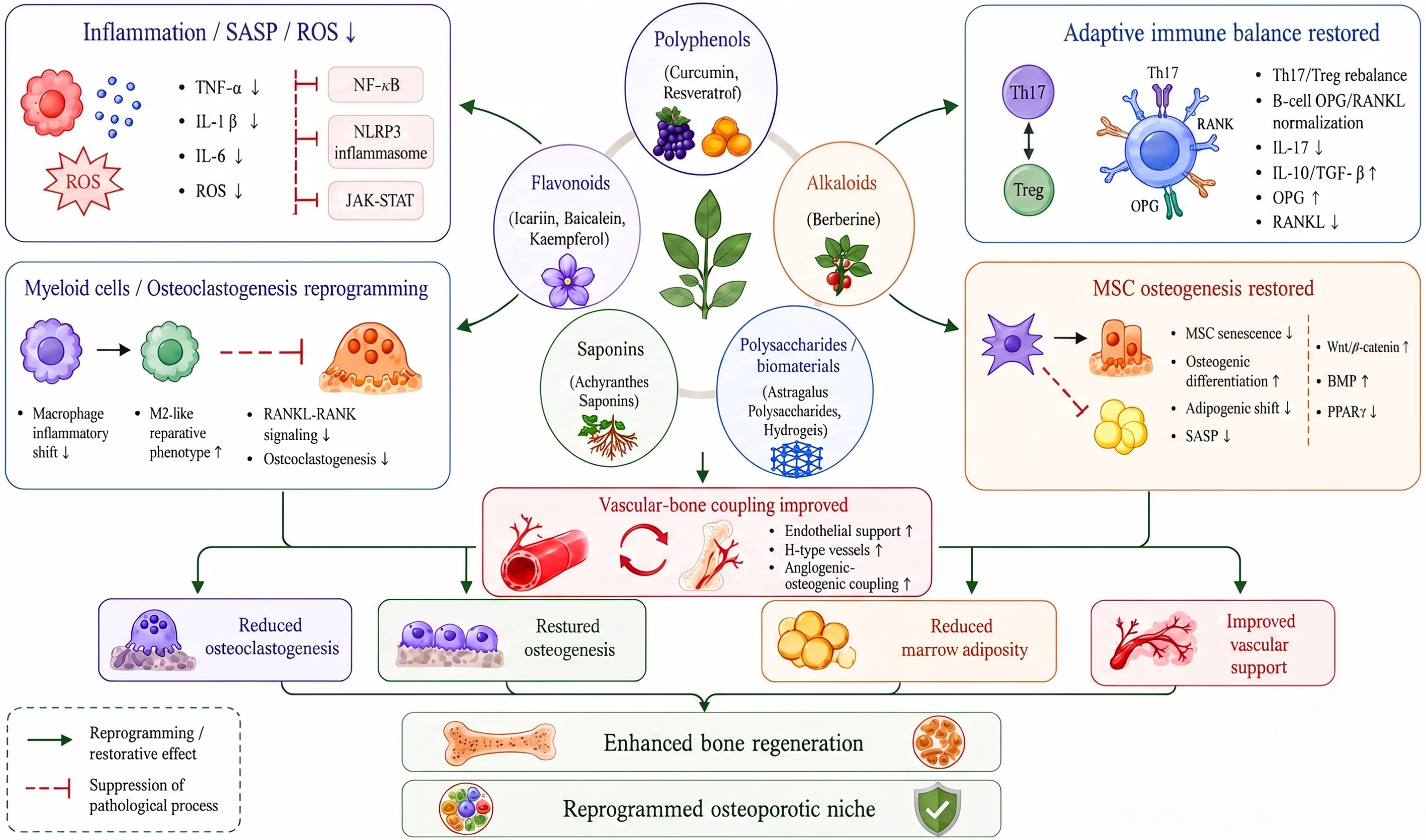
Figure 4.
Natural product-mediated reprogramming network of the bone marrow immune niche in osteoporosis. Natural products derived from diverse chemical classes converge on several microenvironmental regulatory modules, including inflammatory control, immune cell remodeling, restoration of MSCs, metabolic regulation, and vascular-bone coupling. Their major effects include suppressing chronic inflammation, SASP, and ROS; reducing pro-inflammatory mediators such as TNF-α, IL-1β, and IL-6; and modulating inflammatory amplification pathways, including NF-κB, the NLRP3 inflammasome, and JAK-STAT signaling. They may also reshape myeloid cell states, attenuate RANKL-RANK signaling and osteoclastogenesis, restore Th17/Treg balance and the B-cell OPG/RANKL axis, promote MSCs' osteogenic differentiation, inhibit MSCs' senescence and bone marrow adiposity, and improve endothelial support, H-type vessel formation, and vascular-bone coupling.
Inhibition of chronic inflammation and inflammatory amplification network
-
Chronic low-grade inflammation provides a fundamental context for immune microenvironmental imbalance in osteoporosis. Under chronic low-grade inflammatory conditions, TNF-α, IL-1β, and chemokines remain elevated in the bone marrow, shifting the RANKL-RANK-OPG axis toward a pro-osteoclastogenic state and activating inflammatory amplification networks involving NF-κB, STAT3, and NLRP3 signaling[83]. The role of natural products is not limited to reducing individual inflammatory mediators; instead, they may suppress chronic inflammation and thereby ameliorate imbalance in the immune microenvironment. Natural products may inhibit activation of the inflammasome, reduce pro-inflammatory output from macrophages and T-cells, and thereby alleviate suppressed MSC osteogenesis. For example, natural polyphenols may target the macrophage-MSC immunometabolic axis by suppressing persistent glycolytic inflammatory programs and promoting mitochondrial oxidative metabolism and appropriately timed reparative macrophage transitions, thereby relieving inflammatory inhibition of MSCs' osteogenesis[84]. These immunometabolic effects may converge on the AMPK/SIRT1/PGC-1α energy-sensing axis. AMPK responds to energy stress and promotes oxidized nicotinamide adenine dinucleotide (NAD+)-dependent SIRT1 activity, and SIRT1 subsequently deacetylates PGC-1α, supporting mitochondrial biogenesis and oxidative metabolism while limiting NF-κB/NLRP3-associated inflammatory signaling[85]. Within the bone marrow niche, this axis may restrain persistent glycolytic macrophage states and preserve MSCs' osteogenic competence over adipogenic drift[86]. Resveratrol and berberine may engage this regulatory node, although direct evidence for complete axis-mediated niche reprogramming in human osteoporosis remains limited[87]. Taken together, natural products may suppress chronic inflammation and inflammatory amplification networks, attenuate pro-osteoclastogenic communication, and restore an immune homeostatic state that supports osteogenesis and repair.
Alleviation of immunosenescence and cellular senescence phenotype
-
Immunosenescence and cellular senescence are upstream drivers of the progressive deterioration of the osteoporotic immune microenvironment. Key features include immune cells' functional drift, expansion of the SASP, ROS accumulation, mitochondrial dysfunction, and reduced osteogenic potential of MSCs[88]. Natural products may alleviate senescent phenotypes by simultaneously regulating oxidative stress, inflammatory amplification, and metabolic homeostasis. For example, some natural products may exert dual senolytic and SASP-suppressive effects by selectively eliminating senescent stromal and immune cells in the bone marrow and reducing the inflammatory burden[89]. Natural polyphenols may also neutralize excessive ROS, alleviate oxidative stress-driven immune dysfunction, improve the inflammatory state of the bone marrow niche, and modulate cellular senescence pathways through telomerase activity, epigenetic modifications, and related mechanisms[90]. In addition, natural products may modulate immune-stromal interactions in the bone marrow through multitarget actions, restore the hematopoietic and immune-regenerative capacity of HSCs, and alleviate bone marrow immunosuppression[91].
Remodeling of immune cells' function and lineage imbalance
-
Remodeling the osteoporotic immune microenvironment requires not only reducing inflammatory mediators but also correcting pathological shifts in immune cells' composition and functional states. In osteoporotic bone marrow, expansion of myeloid cells, Th17/Treg imbalance, B-cells' functional drift, and excessive neutrophil activation collectively shift the microenvironment toward a pro-inflammatory, pro-osteoclastogenic, and regeneration-impaired state[92]. Natural products may enhance splenic natural killer (NK)-cells' activity and lymphocytes' transformation capacity and increase the levels of leukocytes, erythrocytes, platelets, hemoglobin, and bone marrow cells, thereby improving a suppressed bone marrow niche[93]. Natural products may also stimulate neutrophil's immunoregulatory activity, promote the proliferation and activation of T/B lymphocytes and dendritic cells, regulate the secretion of immune mediators, and enhance bone marrow immune cells' function[94]. Gallic acid has been reported to promote the recovery of bone marrow cells, regulate the cell cycle, inhibit apoptosis, and improve immune organ function, thereby alleviating bone marrow suppression[95]. Taken together, natural products may provide a safe and effective strategy for correcting imbalance in the bone marrow's immune microenvironment by regulating immune cells' function and restoring stromal-immune interaction networks.
Restoration of MSCs' osteogenic potential and inhibition of bone marrow adiposity
-
Impaired osteogenesis in osteoporosis is not caused solely by the functional decline of mature osteoblasts; rather, it arises from depletion of the MSC progenitor pool, disruption of osteogenic programs, and a lineage shift toward adipogenesis. Aging and chronic inflammation upregulate the expression of PPARγ, shifting MSCs from an osteogenesis-supportive state toward adipogenic and pro-inflammatory secretory phenotypes[96]. The therapeutic potential of natural products lies in their ability to restore osteogenic programs while correcting adipogenic bias. Several plant-derived bioactive compounds have been reported to promote the differentiation of MSCs toward an osteogenic lineage. For example, icariin, a major active component of the traditional Chinese medicine Epimedium, promotes the osteogenic differentiation of BMSCs and enhances bone defect repair[97]. Rutin, a natural flavonoid involved in cellular proliferation and bone development, may enhance MSCs' osteogenic capacity[98]. In addition, natural products may inhibit MSCs' adipogenic differentiation through multiple mechanisms. Flavonoids and coumarin derivatives may block key steps in adipogenesis by suppressing PPARγ and C/EBPα, interfering with cell-cycle progression, and reducing intracellular triglyceride accumulation[99]. For instance, coumarin constituents derived from plants of the genus Libanotis have shown potential to regulate bone marrow adipogenesis[100].
Improvement of vascular-bone coupling and the tissue repair microenvironment
-
Osteoporosis-associated regenerative dysfunction arises not only from impaired osteoblast function but also from disrupted coupling among vascular support, immune status, and osteogenic programs[101]. Normal bone repair depends on sequential transitions involving angiogenesis, MSC recruitment, and callus maturation. However, under aging and chronic inflammatory conditions, persistent pro-inflammatory cytokines and oxidative stress lead to endothelial dysfunction, insufficient vascularization, and delayed osteogenic initiation[102]. Natural products may promote repair by regulating vascular-bone coupling, remodeling the local microenvironment, and modulating bone marrow's immune homeostasis. For example, under high-glucose conditions, curcumin may reverse osteogenic and pro-angiogenic dysfunction in BMSCs and restore their capacity to mediate vascular-bone coupling[103]. Ginsenosides may synergistically promote bone formation by regulating interactions between osteoblasts and the bone microenvironment and exerting vascular remodeling and immunomodulatory effects[104]. In addition, tannin-derived compounds may scavenge ROS, attenuate inflammatory injury, improve the local microenvironment at bone defect sites, and create favorable conditions for skeletal regeneration[105].
-
Within the framework of osteoporotic remodeling of the immune microenvironment, natural products should no longer be viewed simply as conventional pharmacological agents with anti-inflammatory, antioxidant, or pro-osteogenic activities. Instead, they should be evaluated according to the key microenvironmental nodes they target and their network-level regulatory properties. Current evidence indicates that natural bioactive compounds, including polyphenols, flavonoids, alkaloids, and glycosides, may regulate the bone marrow niche's homeostasis at multiple levels. These effects include suppressing chronic inflammatory signaling, attenuating SASP-mediated tissue damage, and adipogenic differentiation of MSCs, and promoting vascular-bone coupling and tissue repair to support functional reconstruction of the bone microenvironment. To distinguish mechanistic plausibility from translational readiness, the evidence summarized in this section is classified into four levels: In vitro mechanistic evidence, direct preclinical evidence from osteoporosis-related animal models, indirect preclinical evidence from other inflammatory or regenerative disease models, and human observational or interventional evidence. Cell-based studies identify potential targets but cannot establish in vivo efficacy, whereas animal studies provide a proof of concept but remain limited by species differences, supraphysiological dosing, and model-specific pathology. Among the representative natural products reviewed here, direct clinical evidence for bone marrow immune niche reprogramming remains scarce. Therefore, translational potential is evaluated according to the models' relevance, reproducibility, target engagement, pharmacokinetic exposure, safety, formulation dependence, and the availability of human validation (Table 2).
Table 2. Natural products redefining microenvironmental targets and effects.
Microenvironment target Representative compound Target cells Molecular pathway Evidence source Ref. Chronic inflammation, oxidative stress, and osteocyte-immune cell interaction Curcumin Macrophages, T-cells, osteoblasts, osteoclasts, BMSCs Improving abnormal interactions among macrophages, T-cells, and osteocytes through anti-inflammatory and antioxidant effects Cell and animal model [106] Macrophage polarization, metabolic inflammation, osteogenesis-osteoclast coupling Berberine Macrophages, BMSCs, osteoclasts, osteoblasts Reducing TNF-α and IL-6 levels, increasing IL-10 levels, and promoting the transformation of macrophages toward the anti-inflammatory M2 phenotype. Animal model [107] Macrophage polarization and chronic inflammatory amplification Isoeugenol Bone marrow macrophages, osteoclast precursors Regulation of bone marrow macrophage M1 polarization via p38 MAPK and arachidonic acid metabolic pathways ameliorates chronic inflammation-mediated immune dysregulation. Cell and animal model [108] STING-dependent inflammation, chronic inflammatory microenvironment RTA-408 Macrophages, BMSCs, osteoblasts, and osteoclast-related cells Inhibit STING-dependent NF-κB signaling to mitigate microenvironmental dysregulation induced by chronic inflammation. Cell and animal model [109] Inflammatory bodies, Th17/Treg balance, oxidative stress Baicalein, icariin T-cells, macrophages, BMSCs, osteoclast precursors Regulate NLRP3 inflammasome activation, Th17/Treg balance, and antioxidant pathways to enhance its targeted modulation of immune cells and microenvironment responsiveness. Cell model [110] Oxidative stress, ferroptosis, immune cell homeostasis Chlorogenic acid, protocatechuic acid Osteoblasts, BMSCs, immune cells, osteoclast-related cells Clear ROS, alleviate oxidative stress, and inhibit ferroptosis associated with immune dysregulation, thereby maintaining the homeostasis of immune cells and osteocytes in the bone microenvironment. Cell and animal model [111] T-cell-RANKL axis, osteogenesis Resveratrol Activated T-cells, osteoclast precursors, BMSCs Inhibit the secretion of RANKL by activated T-cells, thereby blocking the RANKL/RANK signaling pathway and reducing osteoclast differentiation and bone resorption. Animal model [112] Oxidative stress, cellular senescence, local inflammation Salvianolic acid A Osteoblasts, BMSCs, immune cells Eliminate free radicals to reduce intracellular oxidative stress and achieve microenvironment remodeling Cell model [113] Osteoblast apoptosis, oxidative stress, and impaired osteogenic differentiation Naringenin Osteoblasts, BMSCs Activation of the Nrf2/HO-1 signaling pathway inhibits osteoblast apoptosis and alleviates impaired osteoblast differentiation. Cell model [114] Osteoclast differentiation and inflammatory bone resorption Homoisoflavonoid derivative 5g Osteoclast precursor cells, osteoclasts Reduce the activation of the ERK1/2 and IκBα/NF-κB signaling pathways, thereby blocking bone loss caused by abnormal osteoclast activity. Cell model [115] Aging of BMSCs, mitochondrial autophagy, and restoration of osteogenic function Kaempferol BMSCs, osteoblasts Targeting Sp1 to activate FUNDC1-mediated mitochondrial autophagy for improving BMSCs' senescence in postmenopausal osteoporosis Cell and animal model [116] Th17 cells, systemic immune homeostasis, osteoclastogenesis Chloroquine Th17 cells, Tregs, osteoclast precursors, and bone marrow immune cells Reduce the proportion of Th17 cells promoting osteoclastogenesis in the bone marrow; enhance protective cytokines such as IFN-γ, IL-4, and IL-10; and restore systemic immune homeostasis. Cell and animal model [117] Th17/Treg balance, inflammatory bone erosion Nuciferine T-cells, synovial/inflammatory microenvironment cells, osteoclast-related cells Improve collagen-induced bone erosion; reduce pro-inflammatory cytokines; and serum immunoglobulins (Ig)G, IgG1, and IgG2a; and restore the Th17/Treg balance in rats. Cell and animal model [118] RANKL-induced osteoclastogenesis Kirenol Osteoclast precursor cells, osteoclasts Inhibition of the NFATc1 and Cav-1 signaling pathways suppresses RANKL-induced osteoclastogenesis and prevents ovarianectomy-induced osteoporosis Cell and animal model [119] RANKL triggering signal, osteoclast formation Corosolic acid Osteoclast precursor cells, osteoclasts Modulate RANKL triggering signals to inhibit osteoclast formation-induced bone loss Cell and animal model [120] Abnormal osteoclastogenesis and bone resorption Holothurin A, Echinoside A Osteoclast precursor cells, osteoclasts Targeting CB1 and MKP-1 to inhibit the expression of factors such as c-fos and NFATC1 improves abnormal bone resorption Animal model [121] ER/RANK/NFATc1 axis, OVX-associated immune microenvironment Achyranthes saponin Osteoclast precursors, osteoclasts, and bone marrow immune cells Regulation of the RER/RANK/NFATc1 signaling pathway improves the bone marrow immune niche, thereby inhibiting OVX-induced osteoporosis Cell and animal model [122] Vessel-bone coupling, H-type angiogenesis, estrogen-deficient bone loss Harmine Osteoclast precursors, endothelial cells, osteoblasts Promote the secretion of PDGF-BB by osteoclast precursors, induce H-type angiogenesis, improve bone vascular-immune coupling, and alleviate bone loss caused by estrogen deficiency. Cell and animal model [123] ROS, NF-κB/NFATc1, osteoclast differentiation Dictamnine Osteoclast precursor cells, osteoclasts Regulate the ROS, NF-κB, and NFATc1 signaling pathways to inhibit RANKL-induced osteoclast differentiation Cell and animal model [124] SIRT1, NF-κB acetylation, ROS activity Crebanine Osteoclasts, osteoblasts, BMSCs, inflammatory cells Targeting Sirt1 to interfere with NF-κB acetylation and ROS activity improves bone loss Cell and animal model [125] Intestinal-bone-immune axis, systemic inflammation, short-chain fatty acids Dendrobium officinale polysaccharides Gut microbiota, intestinal epithelial cells, osteoblast/osteoclast-related cells Reshapes short-chain fatty acid (SCFA)-producing microbiota, restores intestinal barrier integrity and butyrate/isovalerate levels, reduces systemic inflammation, and activates Wnt/β-catenin signaling; fecal microbiota transplantation reproduces the osteoprotective effect. Animal model [126] Osteogenesis, bone marrow fat accumulation, and immune microenvironment repair Dendrobium polysaccharides and Scutellaria baicalensis polysaccharides Osteoclasts, BMSCs, bone marrow adipocytes, immune cells Inhibits osteoclastogenesis in OVX rats, regulates lipid accumulation in the bone marrow cavity, and exerts a reparative effect on the bone marrow immune niche. Animal model [127] Local immune microenvironment, macrophage function, bone repair Astragalus polysaccharide hydrogels Macrophages, BMSCs, osteoblasts, vascular endothelial cells Regulate macrophages' function and optimize the local immune microenvironment to promote bone repair. Cell model [128] Bone-targeted delivery, aging metabolism, and the immune microenvironment Self-assembled nanoparticles with well-defined oligosaccharide CS4-NP BMSCs, bone marrow immune cells, osteoclasts, osteoblasts Achieve targeted delivery of active ingredients to significantly enhance bone mass in the OVX model while improving the local aging metabolism and immune microenvironment. Cell and animal model, human observational studies [129] Representative natural products targeting inflammatory amplification and immunometabolic regulation
-
Chronic inflammation and metabolic dysfunction represent key drivers of deterioration of the bone marrow niche. Representative natural products targeting inflammatory signaling, oxidative stress, and immunometabolic imbalance are discussed in this section. Curcumin, a natural polyphenol, may restore osteoblast-osteoclast balance and modulate abnormal immune-bone cell interactions in the bone marrow niche through its antioxidant and anti-inflammatory properties, thereby ameliorating immune reprogramming-driven bone loss[106]. Berberine is a representative alkaloid with immunometabolic regulatory potential. Berberine has been reported to reduce TNF-α and IL-6 levels, increase IL-10 levels, and promote macrophage polarization toward an anti-inflammatory M2 phenotype, thereby improving the osteoporotic bone microenvironment[107]. Isoeugenol has also been reported to modulate the osteoporotic immune microenvironment. It suppresses pro-inflammatory M1 macrophage activation, promotes anti-inflammatory M2 macrophage polarization, reduces TNF-α and IL-6 accumulation in the bone marrow niche, and thereby alleviates chronic inflammation-mediated immune imbalance[108]. RTA-408 may attenuate osteoclastogenesis and inflammatory microenvironmental disruption by inhibiting STING-dependent NF-κB signaling, highlighting the therapeutic potential of context-dependent cGAS-STING modulation[109].
Representative natural products regulating immune cells' states and osteogenic lineage commitment
-
Restoration of immune cells' homeostasis and osteogenic capacity is essential for niche recovery. This section summarizes representative natural products that regulate immune cells' states, immune-stromal interactions, and MSCs' lineage commitment. Naringenin has been reported to inhibit osteoblast apoptosis and alleviate osteoblast differentiation defects by activating Nrf2/HO-1 signaling[114]. The homoisoflavonoid derivative 5g binds FGFR1, thereby reducing the activation of ERK1/2 and IκBα/NF-κB and preventing ther bone loss associated with abnormal osteoclastogenesis[115]. Kaempferol, another representative flavonoid, may alleviate BMSCs' senescence in postmenopausal osteoporosis by targeting Sp1 and activating FUNDC1-mediated mitophagy[116]. Chloroquine may reduce the proportion of pro-osteoclastogenic Th17 cells in the bone marrow; increase protective cytokines such as IFN-γ, IL-4, and IL-10; and restore systemic immune homeostasis[117]. Nuciferine may ameliorate collagen-induced bone erosion, reduce pro-inflammatory cytokines and serum immunoglobulins, including IgG, IgG1, and IgG2a, and restore Th17/Treg balance in rats[118]. Terpenoid compounds such as kirenol inhibit RANKL-induced osteoclastogenesis and prevent ovariectomy-induced osteoporosis by suppressing NFATc1 and Cav-1 signaling[119]. Corosolic acid may modulate RANKL-triggered signaling to inhibit osteoclastogenesis-associated bone loss[120]. In addition, the sea cucumber-derived saponins Holothurin A and Echinoside A may improve abnormal bone resorption by targeting CB1 and MKP-1 and suppressing the expression of osteoclastogenic factors such as c-Fos and NFATc1[121]. Achyranthes bidentata saponins may improve the bone marrow immune niche by regulating ER/RANK/NFATc1 signaling, thereby attenuating ovariectomy-induced osteoporosis[122].
Representative natural products targeting stromal, vascular, and gut-bone microenvironments
-
Bone regeneration depends on coordinated regulation of stromal function, vascular support, and systemic communication. This section highlights representative natural products targeting MSCs' function, vascular-bone coupling, and the gut-bone axis. Natural polyphenols may not only directly modulate the bone marrow niche but also exhibit enhanced efficacy when formulated as metal ion-coordinated self-assembled nanoparticles. Mechanistically, these nanosystems may enable targeted delivery to the bone marrow niche, where they modulate macrophages' function and cytokine secretion, thereby improving osteoporosis-associated chronic inflammation[124]. Beyond their direct effects on bone cells, orally administered natural products may regulate the bone marrow immune niche through the gut-bone axis. By reshaping the gut microbiota, preserving intestinal barrier integrity, and increasing short-chain fatty acid (SCFA) production, they may reduce endotoxin-driven systemic inflammation, influence immune cells' states and trafficking, and thereby modulate osteoclastogenesis and MSCs' osteogenic capacity[125]. In ovariectomized mice, Dendrobium officinale polysaccharides, structurally characterized as an acetylated glucomannan, enriched SCFA-producing bacteria, restored butyrate and isovalerate levels, and transferred osteoprotection through fecal microbiota transplantation, supporting a causal microbiota contribution[126]. Polygonatum and Dipsacus polysaccharides have also been reported to inhibit osteoclastogenesis in ovariectomized rats, regulate lipid accumulation in the bone marrow cavity, and restore the bone marrow immune niche[127]. In addition, oxidized Astragalus polysaccharide-based composite hydrogels may optimize the local immune microenvironment by modulating macrophage function, thereby promoting bone repair[128]. Self-assembled chondroitin sulfate-mimetic tetrasaccharide nanoparticles, referred to as CS4-NPs, may enable targeted delivery of bioactive components, increase bone mass in ovariectomized models, and improve local aging-associated metabolic and immune microenvironments[129]. Moreover, magnesium ion-containing polysaccharide hydrogels may synergistically regulate angiogenesis, immune cell infiltration, and extracellular matrix remodeling, thereby providing an integrated structure-function platform for local osteoporosis therapy[130].
Natural products exhibit multicomponent, multitarget, and multipathway regulatory properties in the prevention and treatment of osteoporosis. Current evidence suggests that they may modulate classical signaling axes closely associated with bone remodeling, including RANKL/OPG, Wnt/β-catenin, BMP/Smad, MAPK, NF-κB, and Nrf2/HO-1, as well as broader mechanisms involving the suppression of inflammation, alleviation of oxidative injury, remodeling the immune microenvironment, promoting angiogenesis, regulating energy metabolism, and modulation of the gut-bone axis. Through these mechanisms, natural products may help restore bone's microenvironmental homeostasis. Compared with pharmacological interventions targeting a single molecule or pathway, the network-level regulatory properties of natural products may offer advantages for treating osteoporosis, a highly heterogeneous, dynamically evolving, and multifactorial disease.
However, several key limitations remain. Much of the available evidence is derived from in vitro cell experiments and animal models, whereas clinical studies remain limited in number and quality. Moreover, natural products are compositionally complex, their pharmacodynamic material basis remains incompletely defined, and their targets are widely distributed. Their in vivo absorption, distribution, metabolism, and excretion profiles, together with their effective doses, safety margins, and long-term intervention effects, require systematic evaluation. Future studies should clarify active compounds, key metabolites, and pharmacodynamic material bases while integrating advanced technologies, including single-cell omics, spatial transcriptomics, multi-omics integration, organoid models, bone microenvironment-mimetic platforms, and artificial intelligence-assisted prediction. These approaches may help delineate the key cell populations, core molecular nodes, and spatiotemporal mechanisms through which natural products regulate the bone microenvironment. This framework may facilitate the transition of natural product-based antiosteoporosis research from empirical mechanistic description to a modern paradigm characterized by mechanistic precision, systematic evidence, and clinical translational potential.
-
As life science research shifts from reductionist, single-molecule mechanistic analysis toward multiscale, dynamic, and systems-level interpretation of complex biological processes, traditional strategies based on endpoint assays, single-parameter evaluation, and two-dimensional cell culture models are increasingly insufficient for capturing the spatiotemporal actions of natural products within specific microenvironments, heterogeneous cellular responses, and remodeling of multilayered regulatory networks. Because the biological effects of natural products are often embedded in multidimensional interaction networks, including cell-cell, cell-matrix, and metabolic-immune interactions, experimental models that more closely recapitulate in vivo physiological and pathological states are essential. Accordingly, integrating high-throughput omics, spatial biology, single-cell analysis, organoid models, microfluidic chips, and artificial intelligence-driven multimodal data analysis has become a key technical strategy for elucidating how natural products mediate microenvironmental reprogramming (Fig. 5). This integrative framework helps overcome the limited physiological relevance and dynamic resolution of traditional models and provides a stronger evidence base for defining the multitarget, multipathway, and context-dependent actions of natural products.
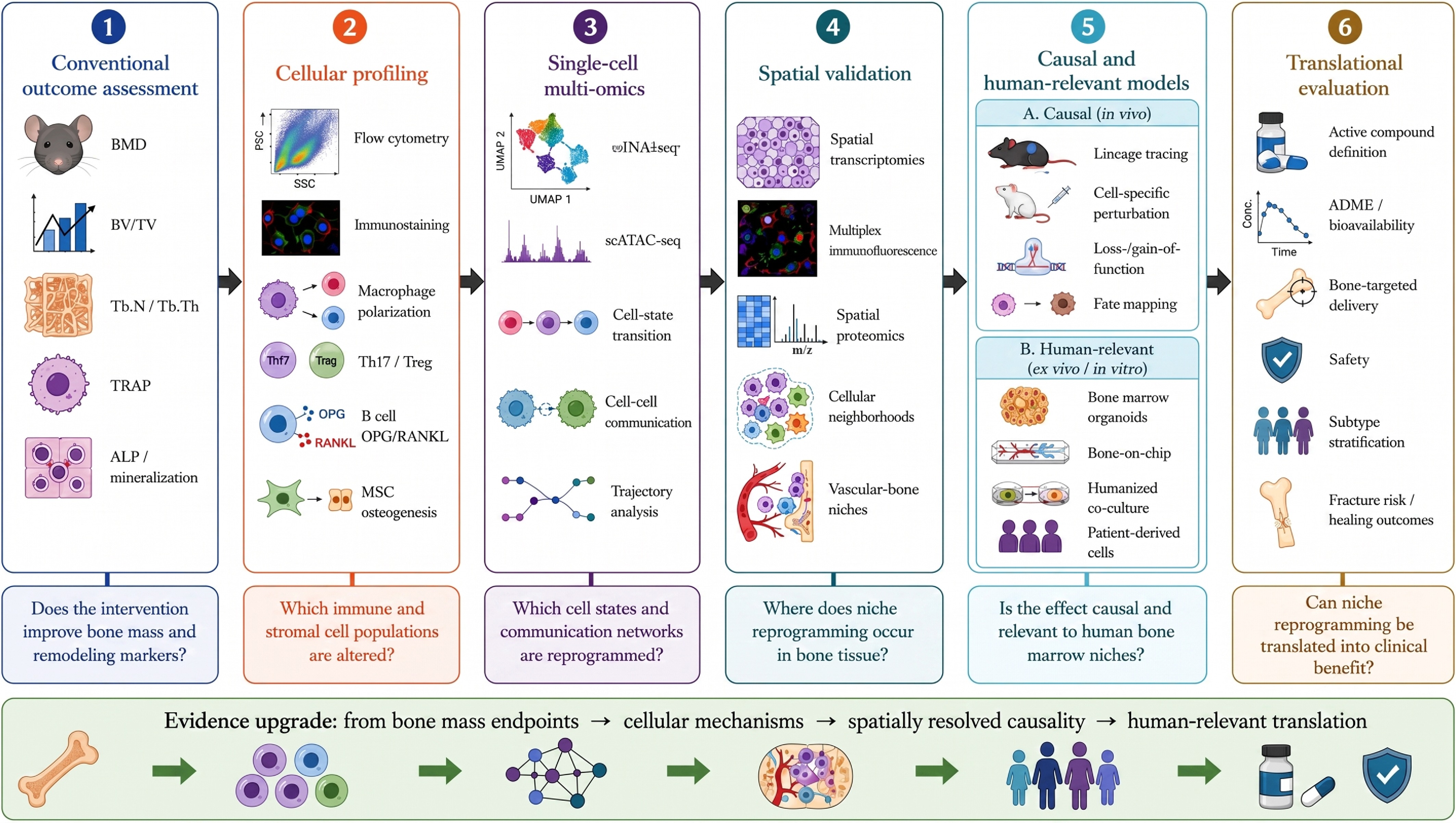
Figure 5.
Technical roadmap for validating natural product-mediated reprogramming of the bone marrow niche. Traditional studies usually assess endpoints such as bone mass, trabecular parameters, TRAP staining, ALP activity, and mineralized nodules to determine whether an intervention improves bone remodeling. Cellular profiling using flow cytometry, immunostaining, and functional assays can further evaluate macrophage polarization, Th17/Treg balance, the B-cell OPG/RANKL axis, and MSC osteogenesis. Single-cell multi-omics can reveal cell state transitions, lineage trajectories, and cell-cell communication, whereas spatial omics and high-dimensional imaging can localize microenvironmental reprogramming within tissue niches. Lineage tracing, functional perturbation, organoids, bone marrow chips, and humanized co-culture models provide causal and human-relevant evidence. Integrating active compound definition, ADME/bioavailability, bone-targeted delivery, safety, patient stratification, and fracture/healing outcomes may move natural product-based osteoporosis research from endpoint observation toward mechanistic precision and clinical translation.
Single-cell omics: redefining cellular heterogeneity in osteoporosis
-
Traditional osteoporosis research has largely relied on bulk tissue-level analyses or averaged measurements of selected cell populations, which may obscure cellular heterogeneity, lineage continuity, and dynamic changes in rare functional subsets within the bone marrow immune niche. The emergence of single-cell RNA sequencing (scRNA-seq) and single-cell multi-omics has provided powerful tools for systematically dissecting the cellular composition, state transitions, and intercellular interactions of the bone marrow niche at a single-cell resolution[131]. scRNA-seq studies have revealed the spatial heterogeneity of bone marrow endothelial cells (BMECs) and identified specific capillary subtypes, thereby clarifying distinct expression patterns of vascular endothelial cells and osteogenic progenitors in trabecular bone-adjacent regions[124]. Other studies have shown that early myelopoiesis in human bone marrow localizes to a high-oxygen arterial-endosteal microenvironment, that hematopoietic stem and progenitor cells are enriched in adipocyte-associated regions, and that MSC expansion occurs in patients with acute myeloidleukemia. In studies of natural product-mediated remodeling of the osteoporotic immune microenvironment, single-cell technologies can compare transcriptional states, compositional changes, and cell-cell communication patterns across cell subsets before and after an intervention. This approach enables an assessment of whether natural products can reverse pathological immune remodeling and restore homeostasis-associated cellular features. Furthermore, single-cell assay for transposase-accessible chromatin (ATAC) sequencing, single-cell epigenomics, and integrated multi-omics analyses can reveal changes in chromatin's accessibility and transcriptional regulatory networks that underlie the determination of cell fate, lineage skewing, and senescence programs. Therefore, single-cell omics enables not only the construction of high-resolution maps of the osteoporosis-associated bone marrow niche but also the definition of cellular targets, microenvironmental specificity, and mechanistic verifiability of natural products' actions. Nevertheless, dissociation bias, rare-cell loss, batch effects, and limited longitudinal sampling may distort inferred cell states and should be addressed through orthogonal imaging, flow cytometry, and functional validation.
Spatial omics and high-dimensional imaging: From changes to spatial localization
-
Bone tissue exhibits marked spatial heterogeneity. Osteoblasts, osteoclasts, and immune cells do not function in isolation; instead, they form highly organized spatial interaction networks within trabecular bone, the bone marrow cavity, and perivascular niches[132]. Whereas single-cell omics primarily reveals changes in cellular composition and functional states within the osteoporotic microenvironment, spatial omics and high-dimensional imaging technologies further define where these changes occur anatomically and how they influence neighboring cell populations and local tissue architecture[133]. Spatial transcriptomics maps the spatial distribution of genes' expression while preserving the tissue architecture, thereby identifying key molecular features in active bone formation regions, enhanced bone resorption sites, inflammation-enriched areas, and vascular-associated microdomains. When combined with high-dimensional imaging technologies, including multiplex immunofluorescence, imaging mass cytometry, and spatial proteomics, this approach can further resolve the spatial proximity, intercellular communication patterns, and signaling interaction networks of specific cell subsets[134]. For example, local co-localization of osteoclasts and inflammatory macrophages may indicate spatially restricted promotion of bone resorption by inflammatory mediators, whereas spatial coupling between osteogenic progenitors and vascular endothelial cells provides evidence for coordinated regulation of angiogenesis and bone formation[135]. In studies of natural product-based interventions for osteoporosis, spatial omics can determine whether therapeutic effects occur within specific bone-remodeling microregions and translate conventional molecular expression changes into mechanistic evidence with tissue localization and structural relevance. Future integration of spatial omics with single-cell sequencing, metabolomics, and high-dimensional imaging analysis may enable the construction of spatially resolved dynamic maps of osteoporotic microenvironmental reprogramming. Major limitations include RNA's degradation during bone decalcification, resolution-coverage trade-offs, high analytical costs, and the largely static nature of current spatial datasets.
Lineage tracing and cell interaction analysis: establishing causal chains
-
The effects of natural products on bone tissue are often not mediated by a single direct target. Instead, they depend on indirect modulation of multicellular interaction networks within the bone microenvironment. Therefore, lineage tracing and cell-cell interaction analyses have become critical technical approaches for distinguishing direct cellular targets from secondary downstream effects[136]. Genetic labeling, tracing cell fate, and inducible reporter systems can dynamically resolve the origins, differentiation trajectories, and functional outcomes of key cell populations, including MSCs, bone marrow macrophages, and osteoclast precursors, during fracture repair and bone remodeling[137]. For example, in studies of natural product-based interventions, macrophage-specific reporter animal models can determine whether natural products affect macrophages' transition toward the osteoclast lineage and whether this process contributes to their inhibition of bone resorption or promotion of tissue repair[138]. Accordingly, integrating lineage tracing, predictions of cell-cell communication, and functional validation can help define the direct cellular targets, key paracrine signals, and downstream effector cells of natural products. This integrated approach can also prevent broad changes in molecular expression from being misinterpreted as specific mechanisms.
Organoids, organ chips, and humanized platforms: bridging the gap between mechanism and translation
-
Traditional two-dimensional cell culture systems cannot fully recapitulate the three-dimensional architecture, cellular heterogeneity, extracellular matrix deposition, or dynamic signaling gradients of the bone marrow niche. Therefore, they have clear limitations for modeling bone's immune microenvironment and its pathological remodeling[139]. Organoid technology uses three-dimensional self-organizing systems to partially mimic the key features of bone tissue's development, bone regeneration, and the bone marrow niche, thereby providing a more physiologically relevant platform for evaluating how natural products regulate osteogenic differentiation, matrix mineralization, determination of cell fate, and intercellular communication[140]. In comparison, organ-on-a-chip technology incorporates dynamic fluid flow, mechanical stimulation, and multitissue interactions, enabling the construction of biomimetic models such as bone-vascular chips, bone marrow chips, and bone-immune microenvironment chips. These platforms can simulate bloodflow shear stress, nutrient exchange, inflammatory cytokine diffusion, and dynamic drug exposure[141]. In studies of natural product-mediated regulation of the bone marrow immune niche, organoids and organ-on-a-chip systems can enable more systematic evaluations of the effects on vascular-bone coupling, immune-inflammatory responses, and osteoblast-osteoclast balance. They may also facilitate active compound screening, dose optimization, and safety assessment. In addition, multicellular co-culture systems constructed from human bone marrow cells, vascular endothelial cells, and immune cells, as well as patient-derived cell models, may more accurately reflect human-specific pathological responses in postmenopausal osteoporosis, age-related osteoporosis, and inflammation-associated bone loss. These models may also inform individualized intervention strategies and clinical trial designs[142]. Overall, organoids, organ-on-a-chip platforms, and humanized models improve the physiological relevance and translational credibility of mechanistic studies on natural products and provide technical support for elucidating their multicellular, multitarget, and microenvironment-dependent regulatory effects. Current organoid and organ-on-a-chip platforms nevertheless incompletely reproduce biological aging, mechanical loading, endocrine inputs, and systemic immunity, and their standardization and interlaboratory reproducibility remain important challenges.
-
Although the proposed framework is biologically plausible, current evidence remains heterogeneous and sometimes conflicting. Immune cells' functions are highly dependent on developmental stage, activation state, and disease phase. For example, activated neutrophils and NETs may amplify inflammatory osteoclastogenesis, whereas bone marrow neutrophil progenitors can suppress osteoclast formation[60,62]. Similarly, chronic Th17/IL-17 activity is generally pro-osteoclastogenic, but IL-17-producing cells may support callus formation during acute fracture repair[28,63]. Macrophage states also extend beyond a simple M1/M2 dichotomy, whereas cGAS-STING and IL-6 signaling can exert divergent effects according to the downstream signaling branch, duration of exposure, and target cell. Moreover, most natural product studies rely on in vitro assays or short-term ovariectomized rodent models, and improvements in bone mass or selected molecular markers do not establish multicellular niche reprogramming. Some mechanisms are also extrapolated from nonosteoporotic disease models. Human evidence remains limited, is frequently cross-sectional, and is potentially confounded by age, medications, metabolic disorders, and fracture status. Therefore, convincing evidence of niche reprogramming should combine cell state profiling, spatial validation, altered intercellular communication, causal perturbation, target engagement, pharmacokinetic exposure, and confirmation in human-relevant models.
Natural products have shown multitarget, multipathway, and systems-level potential for regulating osteoporosis-associated microenvironments, but the systematic and causal mechanistic explanations remain limited regarding how they reprogram the bone microenvironment across osteoimmune, osteovascular, osteometabolic, and osteoneural regulatory networks. Their translation from basic research to clinical application also faces substantial barriers. First, natural products are compositionally complex and are influenced by the species' origin, geographic environment, extraction procedures, and batch-to-batch variation, which may compromise the stability of their chemical composition and pharmacological efficacy[143]. Many studies still use crude extracts or compound formulations as interventions. Although these approaches may reflect the holistic regulatory advantages of natural products, they also complicate the identification of pharmacodynamic material bases, core active constituents, and key targets. Future studies should strengthen the systematic identification of active ingredient profiles, quality markers, and efficacy-related components, and establish links among chemical characterization, in vivo exposure, target engagement, and biological effects. These efforts will improve the reproducibility, quality control, and clinical translatability of research findings.
Second, natural products commonly exhibit limited bioavailability, rapid in vivo metabolism, and insufficient tissue targeting[144]. Because bone tissue contains an abundant mineralized matrix, has a distinct vascular supply, and presents local microenvironmental barriers, active compounds often have difficulty entering the bone marrow niche and maintaining effective concentrations[145]. Therefore, conventional administration routes alone may be insufficient to fully realize their bone-protective potential. Future strategies may integrate nanodelivery systems, bone-targeting materials, hydrogels, exosome-like carriers, and local sustained release platforms to enhance the accumulation and sustained activity of natural products at bone defect sites, within the bone marrow niche, and in active bone remodeling regions, thereby enabling more precise microenvironmental regulation[146]. A further translational challenge is that ovariectomized rodents do not fully reproduce the heterogeneity of human osteoporosis in aspects such as the microbiota's composition, immune aging, comorbidities, and natural product metabolism. Delivery strategies should therefore be matched to the intended mechanism: Poorly absorbed compounds may still act locally through microbial metabolites, whereas direct modulation of the bone marrow niche requires adequate systemic exposure and bone-targeted delivery[147]. Humanized models, exposure-response analyses, microbiome-based stratification, and biomarker-guided clinical studies will be required to distinguish responders and verify causal gut-bone effects. In addition, the multitarget nature of natural products offers advantages for modulating complex disease networks but also complicates mechanistic interpretation and drug development. Conventional drug discovery typically emphasizes single targets and clearly defined mechanisms of action, whereas natural products often exert integrated effects by simultaneously regulating multiple cell populations, signaling pathways, metabolic nodes, and microenvironmental processes. Therefore, future research should not focus solely on identifying a single "key target". Instead, single-cell omics, spatial omics, network pharmacology, metabolomics, and artificial intelligence modeling should be applied to identify the core nodes, key cellular states, and major microenvironmental regulatory axes within their action networks, thereby establishing a mechanistic evaluation framework that better reflects the systems-level regulatory properties of natural products.
Finally, natural product-mediated reprogramming of the bone microenvironment should extend beyond preventing bone loss to promoting bone regeneration. The goal of osteoporosis interventions should not be limited to slowing bone loss but should also include improving bone tissue's quality, restoring bone remodeling balance, enhancing fracture healing, and reducing refracture risk[148]. Therefore, future studies should place greater emphasis on the integrated effects of natural products on bone marrow MSCs' senescence, osteogenic lineage commitment, immune-inflammatory status, vascular-bone coupling, bone matrix mineralization, and tissue repair capacity. Only by integrating mechanistic studies, delivery technologies, model refinement, and clinical evaluation can natural products be translated from laboratory-bioactive candidates into effective strategies for the prevention and treatment of osteoporosis.
Overall, the value of natural products in osteoporosis-related microenvironmental reprogramming should not be interpreted solely in terms of conventional anti-inflammatory, antioxidant, or pro-osteogenic effects. Instead, their value should be reconsidered within the complex ecosystem of bone tissue. Future research should focus on cellular heterogeneity, spatial localization, causal validation, and translational feasibility while establishing a continuous evidence chain from screening active compounds to mechanistic elucidation, from animal experiments to humanized validation, and from basic research to clinical application. Only through this approach can research on natural product-mediated regulation of the bone microenvironment support precision prevention and treatment of osteoporosis and provide strategies with greater mechanistic depth and clinical value for bone-regenerative medicine.
-
This review uses immunosenescence and dysfunction of the bone marrow niche as an organizing framework for understanding age-related, postmenopausal, and selected metabolic or inflammation-associated forms of osteoporosis. We do not propose immunosenescence as the universal initiating cause of all osteoporosis. Instead, immune aging may interact with estrogen deficiency, metabolic stress, mechanical unloading, glucocorticoid exposure, and other etiological factors to amplify osteoclastogenesis, impair stromal and vascular support, and reduce skeletal regenerative capacity. In parallel, this review summarizes the multilevel regulatory effects of natural products, including polyphenols, flavonoids, alkaloids, saponins, and polysaccharides, on inflammatory amplification, immune homeostasis, and tissue repair. Based on the network-level intervention properties of natural products, this review further proposes research strategies that integrate advanced technologies, including single-cell omics, spatial omics, lineage tracing, and humanized models. This review may help integrate immune microenvironmental imbalance into the established framework of bone-related metabolic regulation and provide a more explanatory theoretical framework for elucidating the mechanistic basis and precision translation of natural product-mediated reprogramming of the bone microenvironment.
-
Not applicable.
-
The authors confirm their contributions to this work as follows: conceptualization: Duan Y; visualization: Duan Y, Li LY, Liao YY; investigation: Duan Y, Luo QZ; funding acquisition: Li J, Li SX; supervision: Li SX; writing – original draft: Duan Y, Li LY, Luo QZ, Liao YY, Li J, Li SX; writing – review and editing: Duan Y, Li J, Li SX. All authors reviewed the results and approved the final version of the manuscript.
-
Data sharing is not applicable to this review as no datasets were generated or analyzed.
-
The authors declare that they have no conflict of interest.
- Copyright: © 2026 by the author(s). Published by Maximum Academic Press on behalf of China Pharmaceutical University. This article is an open access article distributed under Creative Commons Attribution License (CC BY 4.0), visit https://creativecommons.org/licenses/by/4.0/.
-
About this article
Cite this article
Duan Y, Li LY, Lou QZ, Liao YY, Li J, et al. 2026. Immunosenescence-driven bone marrow immune niche destabilization in osteoporosis: microenvironmental targets and natural product-mediated reprogramming. Targetome 2(4): e042 doi: 10.48130/targetome-0026-0040 

Immunosenescence-driven bone marrow immune niche destabilization in osteoporosis: microenvironmental targets and natural product-mediated reprogramming
- Received: 14 May 2026
- Revised: 29 July 2026
- Accepted: 04 August 2026
- Published online: 25 August 2026
Abstract: Osteoporosis is a heterogeneous skeletal disorder arising from diverse age-related, endocrine, metabolic, mechanical, and pharmacological factors. In age-related, postmenopausal, and selected metabolism-associated forms of osteoporosis, immunosenescence may contribute to dysfunction in the bone marrow immune niche, thereby amplifying bone resorption and regenerative decline. This review examines osteoporosis through the complementary perspective of the bone marrow immune niche and summarizes the core mechanisms by which immunosenescence contributes to destabilization of the bone marrow immune niche in osteoporosis. On this basis, we further discuss the molecular basis and regulatory potential of natural products in mediating bone marrow-related immune microenvironmental reprogramming. Emerging technologies, including single-cell omics, spatial omics, organoid models, and humanized research platforms, are also integrated to propose novel strategies for the precision prevention and treatment of osteoporosis. Overall, this review incorporates immune microenvironmental regulation into the conventional framework of bone metabolic dysfunction and provides a systematic theoretical basis for mechanistic elucidation, precision delivery, and clinical translation of natural products in prevention and treatment of osteoporosis.