













-
Virus-induced gene silencing (VIGS) is a technology exploiting the plant's RNAi-mediated antiviral defense mechanism to silence host mRNAs through post-transcriptional targeting. During virus invasion and spreading, the transcripts expressed from the virus genome are recognized by the plant RNAi machinery to produce small interfering RNAs (siRNAs). These siRNAs further guide the RNA-induced silencing complex (RISC) to degrade targeted viral transcripts[1]. By engineering viral vectors carrying host-derived gene sequences, VIGS redirects this machinery to silence corresponding host mRNAs[2]. As an efficient and rapid reverse genetics tool for high-throughput functional genomics in plants, VIGS has been widely applied in field and horticultural crops[3,4].
To date, over 50 plant viruses have been engineered as VIGS vectors for gene silencing in plants[5]. Commonly used systems include tobacco rattle virus (TRV), cucumber mosaic virus (CMV), turnip yellow mosaic virus (TYMV), barley stripe mosaic virus (BSMV), and cabbage leaf curl virus (CaLCuV)[6−9]. Each vector exhibits distinct host specificity and silencing efficiency. For instance, BSMV is widely used in monocots[10], whereas African cassava mosaic virus (ACMV) has a narrow host range, primarily limited to Nicotiana benthamiana and cassava (Manihot esculenta)[11]. Among these, the TRV-based system is one of the most versatile, functioning efficiently in both monocots and dicots. Nevertheless, no single VIGS vector is universally effective across all plant species.
CaLCuV, a bipartite begomovirus, exhibits broad host specificity within the Brassicaceae family, infecting crops including cabbage (Brassica oleracea var. capitata), cauliflower (B. oleracea var. botrytis), and the model plant Arabidopsis thaliana[12]. This DNA virus possesses two circular single-stranded DNA components (DNA-A and DNA-B). Since the DNA-A-encoded coat protein gene AR1 is dispensable for systemic infection, researchers developed the first CaLCuV-based VIGS vector by replacing AR1 with host-derived gene sequences to trigger siRNA-mediated silencing in Arabidopsis[13]. To eliminate the need for particle bombardment, Tang et al. subsequently cloned the viral DNA-A and DNA-B components into the pCAMBIA1301 binary vector, generating constructs pCVA and pCVB[14]. This modified system enables Agrobacterium tumefaciens-mediated delivery and expresses both artificial and endogenous miRNAs for efficient VIGS[14]. Using this optimized vector, Xiao et al. achieved significant downregulation of the PDS gene in several Brassicaceae species, including B. oleracea, B. rapa, and B. nigra[15].
Wutacai (Brassica rapa syn. Brassica campestris ssp. narinosa), a common vegetable in the Yangtze-Huaihe River Basin, is a variant of non-heading Chinese cabbage. It exhibits diverse cultivars and is prized as a winter delicacy. Similar to other A-genome B. rapa species, wutacai presents challenges for genetic transformation[16]. Consequently, VIGS serves as a valuable alternative for functional genomics. While TRV-based VIGS has been successfully applied in heading Chinese cabbage (B. rapa ssp. pekinensis)[17], this system remains ineffective in non-heading varieties[15]. The turnip yellow mosaic virus-derived vector pTY-S is widely used in Pakchoi, another variant of non-heading Chinese cabbage (NHCC)[18,19]. However, this VIGS system requires particle bombardment using gene-gun equipment—an expensive and technically demanding inoculation method.
In this study, we established a VIGS system in wutacai using the CaLCuV vector platform. This system achieved efficient downregulation of the PDS gene across diverse wutacai germplasms. Furthermore, we demonstrated its reliability for silencing functional genes by targeting the senescence-associated gene Stay-Green 1 (SGR1) and flowering regulator FLOWERING LOCUS C (FLC). This CaLCuV-based VIGS platform will facilitate functional genomics studies in wutacai.
-
The wutacai genotypes used in this study, W16-18, W19-24, and Xiaobaye, were provided by the Vegetable Genetics and Breeding Laboratory at Anhui Agricultural University. Seeds were sown in seedling trays containing a 2:1 (v/v) mixture of commercial potting substrate and vermiculite. Germinated seedlings were maintained in a growth chamber under controlled conditions: 22 °C (day)/18 °C (night) temperatures, 300 μmol·m−2·s−1 photosynthetic photon flux density, 70% relative humidity, and a 16-h light/8-h dark photoperiod. After three weeks, seedlings were transplanted into 10 cm × 10 cm × 8 cm (length × width × height) pots for further development.
Homolog searching and sequence analysis
-
Due to the unavailability of a wutacai reference genome, we cloned both genomic and coding sequences of BcPDS, BcSGR1, and BcFLC using primers designed against the Brassica rapa 'Chiifu' v4.0 genome[20], followed by Sanger sequencing. Amino acid sequences were predicted using the ExPASy Translate tool (
https://web.expasy.org/translate ). Multiple sequence alignments were performed using DNAMAN software for comparative analysis. The accession numbers of the PDS, SGR and FLC sequences used are listed in Supplementary Table S1.Construction of gene silencing vectors
-
cDNA inserts (~500 bp) corresponding to conserved amino acid regions (> 60% identity between wutacai and Brassica homologs, via multiple alignment) were selected as silencing targets for VIGS. These fragments were amplified from cDNA, ensuring that they correspond to the coding sequences of the target genes. Gene-specific primers containing 17-bp homologous arms were designed to amplify these fragments. pCVA::00, the empty pCVA backbone without any insert, served as the negative control in VIGS experiments, and pCVB, the helper vector harboring the CaLCuV DNA-B component, was co-infiltrated with pCVA-derived constructs to provide the necessary viral movement functions. The pCVA vector was linearized with KpnI (Takara Bio, Japan) at 37 °C. Both PCR products and the linearized vector were purified using the FastPure Gel DNA Extraction Mini Kit (Vazyme Biotech) and assembled via FastCloning methodology[21]. Constructs were verified by Sanger sequencing. All primers are listed in Supplementary Table S2, and the full cloned sequences of the three fragments (BcPDS, BcSGR1, and BcFLC) are provided in Supplementary Table S3.
VIGS assay
-
Agroinfiltration was performed according to a previously described method with modifications[14]. Constructs pCVB, pCVA, pCVA::BcPDS, pCVA::BcSGR1, and pCVA::BcFLC were individually transformed into Agrobacterium tumefaciens GV3101. Positive transformants were confirmed by PCR and cultured in 2 mL LB (Luria–Bertani) medium supplemented with 50 μg mL−1 rifampicin and 50 μg mL−1 kanamycin. Cultures were scaled up to 50 mL LB with identical antibiotics and incubated at 28 °C with 220 rpm shaking overnight. Bacterial cells were harvested by centrifugation, washed twice with infiltration buffer (10 mM MgCl2, 10 mM MES, 200 μM acetosyringone), and separately resuspended to OD600 of 1.0, 2.0, and 3.0. After incubation at room temperature (22 °C) in the dark for 3–4 h for virulence induction, equal volumes of the suspensions containing pCVB and pCVA-derived constructs were mixed (1:1, v/v) to obtain final OD600 values of 0.5, 1.0, and 1.5. The growth temperatures were set at 20, 22, and 24 °C. Each treatment combination was performed with three biological replicates, and each replicate contained 20 individual plants, giving a total of 60 plants per treatment combination. Prior to infiltration, plants were randomly assigned to treatment groups to minimize positional and environmental bias. The mixed Agrobacterium suspensions were infiltrated into the abaxial surface of cotyledons of one-week-old seedlings using a 1-mL syringe. After infiltration, plants were kept in darkness at room temperature for 24 h and then transferred to the corresponding temperature regimes. At 14 d post-infiltration, genomic DNA was extracted from plants in each treatment group, and PCR was performed with specific primers to detect viral DNA accumulation, thereby evaluating the infection efficiency of each treatment combination.
DNA and RNA isolation, PCR detection, and RT-qPCR analysis
-
Total genomic DNA was extracted from young leaves of wutacai and N. benthamiana by CTAB. Frozen tissue (100 mg) was ground in liquid N2 and incubated at 65 °C for 60 min in 600 μL extraction buffer (2% CTAB, 1.4 M NaCl, 20 mM EDTA, 100 mM Tris-HCl [pH 8.0], 0.2% β-mercaptoethanol), mixed every 10 min. After two 24:1 chloroform/isoamyl alcohol extractions, DNA was precipitated with 0.7 vol isopropanol (−20 °C, 30 min), washed, air-dried, and dissolved in TE. Purity and concentration were assessed by NanoDrop. Total RNA was extracted from 100 mg of leaf tissue with Plant RNA Kit (Omega) with DNase I, eluted in 50 μL, and quantified. cDNA was synthesized from 1 μg RNA using TransScript SuperMix (TransGen) in 20 μL (25 °C 10 min, 42 °C 30 min, 85 °C 5 s). RT-qPCR was performed using a CFX96™ (Bio-Rad) with ChamQ SYBR Master Mix (Vazyme) in 20-μL reactions containing 2 μL 10-fold diluted cDNA, 10 μL 2× Master Mix, and 0.4 μL each primer (final 0.2 μM). Cycling: 95 °C 30 s; 40 cycles of 95 °C 10 s and 60 °C 30 s (read at 60 °C); melt curve 60→95 °C, 0.5 °C/5 s. Relative expression (2−ΔΔCt) with Actin or UBQ10 as reference. Three technical and biological replicates. Primers in Supplementary Table S2.
Statistical analysis
-
Statistical analyses were conducted using GraphPad Prism 8.0 (GraphPad Software, San Diego, CA, USA). For multiple-group comparisons, one-way analysis of variance (ANOVA) followed by Tukey's HSD post hoc test was applied. For two-group comparisons, a two-tailed Student's t-test was used. A p-value < 0.05 was considered statistically significant. Different lowercase letters above the bars in figures indicate significant differences at p < 0.05.
-
Phytoene desaturase (PDS) is an essential enzyme in plant carotenoid biosynthesis[22]. PDS disruption consistently causes albinism[23,24], providing a visually distinct phenotypic marker. Consequently, PDS has become a widely adopted reporter gene in VIGS studies[25−27]. Given the absence of PDS sequence data in wutacai, we first identified orthologs by BLASTn analysis against the B. rapa 'Chiifu' reference genome using NbPDS as a query. Full-length genomic and coding sequences were subsequently amplified from wutacai gDNA and cDNA using primers designed against BrPDS (B. rapa PDS), with all clones validated by Sanger sequencing. The deduced amino acid sequence (designated BcPDS) was comprehensively aligned with PDS orthologs from 13 plant species. BcPDS consists of 563 amino acids and shares high sequence identity with PDS orthologs from other species: 83.8% with Arabidopsis thaliana (AtPDS, NP_001324464), 86.0% with heading Chinese cabbage (BrPDS, XP_009115636), and 85.5% with rice (OsPDS, XP_015648942) (Fig. 1a). These results indicate that BcPDS is highly conserved in wutacai. Phylogenetic analysis of 77 PDS proteins from diverse species revealed close evolutionary relationships among BcPDS, OsPDS, and AtPDS (Fig. 1b).
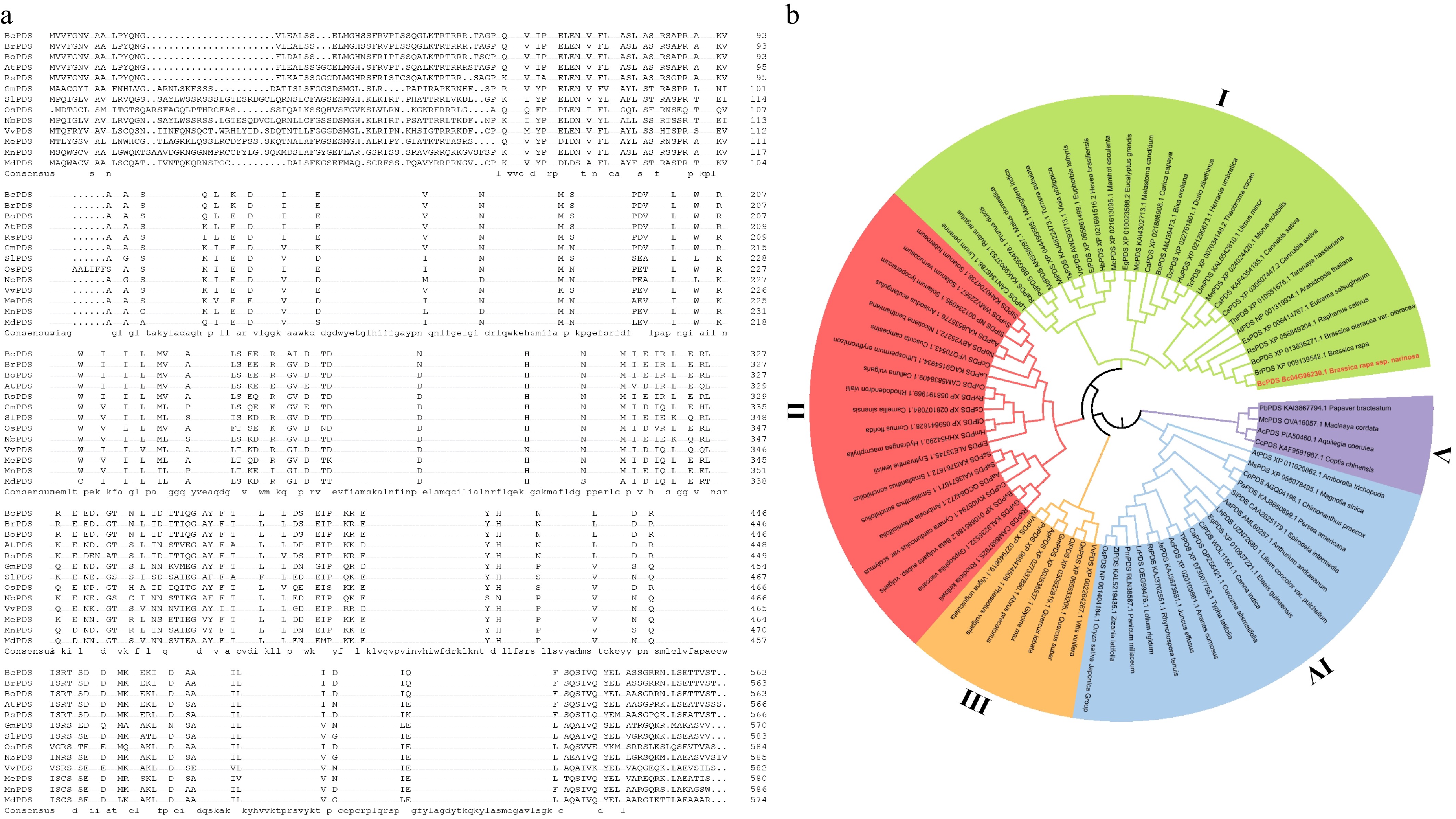
Figure 1.
Multiple sequence alignments and phylogenetic relationship of BcPDS and its orthologs. (a) Multiple sequence alignments of BcPDS and its orthologs from Chinese cabbage, Brassica oleracea, Arabidopsis, Radish, Soybean, Tomato, Rice, Tobacco, Grapevine, Cassava, Black mulberry, and Apple. The alignments were performed using full-length amino acid sequences of PDS. (b) Phylogenetic analysis of PDS proteins from 75 species. The unrooted phylogenetic tree was generated by the neighbour-joining method using the MEGA6 program with 1,000 bootstrap trials.
CaLCuV-based VIGS works in wutacai
-
Based on multiple sequence alignment (Fig. 1a), we targeted the highly conserved C-terminal domain of BcPDS to evaluate CaLCuV-based VIGS efficiency in wutacai. A 500-bp fragment of BcPDS was amplified and cloned into KpnI-digested pCVA (Fig. 2a, b). The resulting pCVA::BcPDS construct was transformed into Agrobacterium strain GV3101. Given the high sequence conservation between BcPDS and NbPDS, we first validated vector functionality in N. benthamiana. At 30 days post-infiltration (dpi), plants infiltrated with pCVA::BcPDS/pCVB exhibited pronounced leaf and stem photobleaching, whereas pCVA::00/pCVB controls remained phenotypically normal (Fig. 2c). PCR analysis confirmed viral vector accumulation of both constructs (Fig. 2d). RT-qPCR analysis demonstrated significantly reduced BcPDS transcript levels (> 50%) specifically in photobleached tissues of pCVA::BcPDS/pCVB-infiltrated plants (Fig. 2e). These results confirm the functional competence of the pCVA::BcPDS vector system.
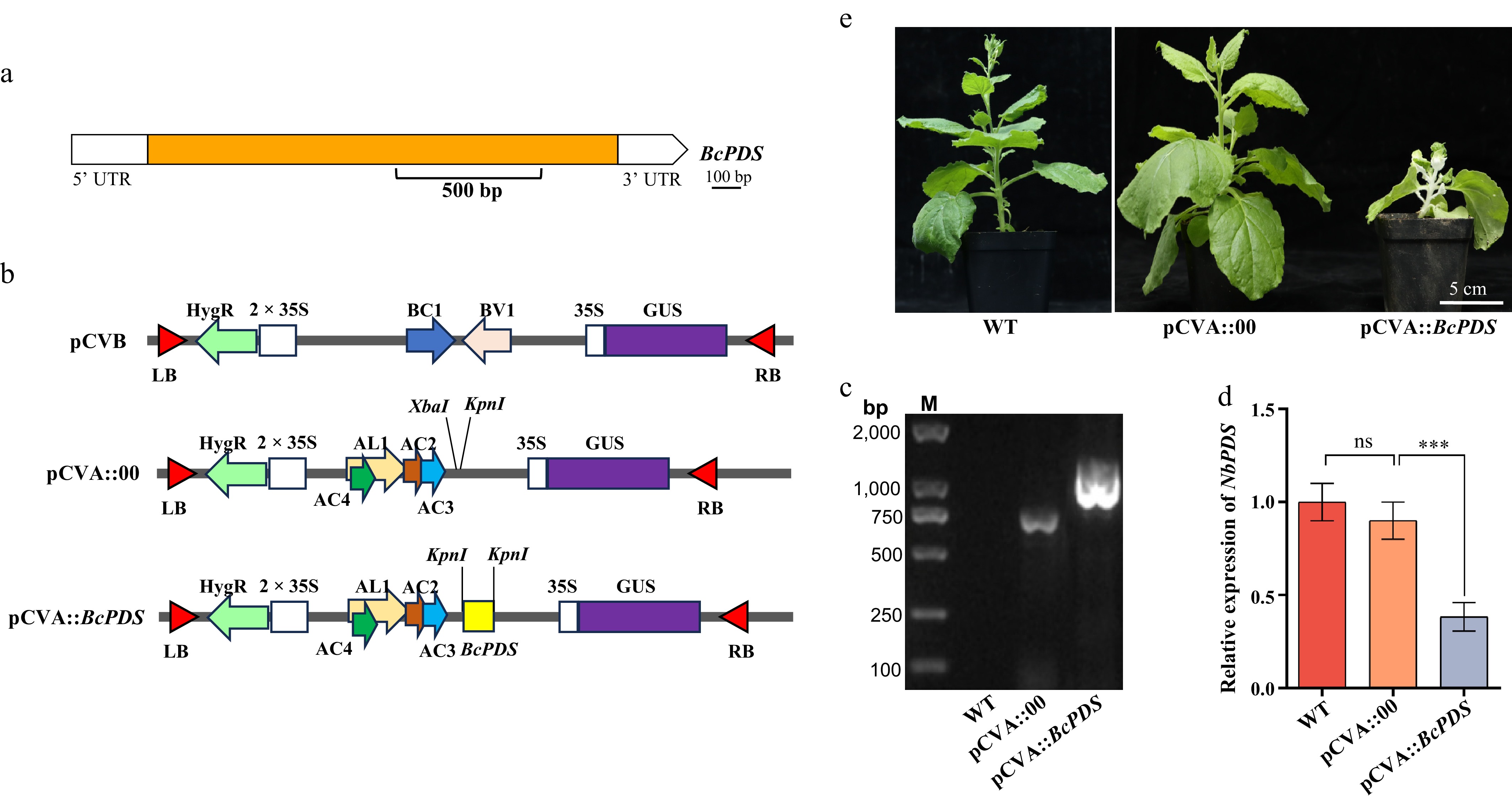
Figure 2.
Construction of the pCVA::BcPDS vector and silencing of PDS in N. benthamiana. (a) Structure of the BcPDS gene body; the fragment inserted in pCVA is indicated with a black underline. (b) Map of pCVB, pCVA::00, and pCVA::BcPDS vectors. LB, left border; RB, right border; HygR, hygromycin resistance gene; 2 × 35S, two consecutive 35S promoters; BC1, movement protein; BV1, nuclear shuttle movement protein; GUS, β-glucuronidase; AL1, replicase-associated protein; AC2, transactivator protein; AC3, replicase-associated protein; and AC4, putative pathogenesis-related protein. (c) CaLCuV-mediated VIGS phenotypes of N. benthamiana inoculated with empty pCVA vector (pCVA::00) and pCVA::BcPDS. Photos were captured after 30 d of infiltration, and the no-infiltration plant was also included as a control. The whole plants were shown in the upper panel, and the representative leaves were shown in the bottom panel. Silencing phenotype of BcPDS in tobacco. (d) Detection of CaLCuV fragments after VIGS treatments. Lane M shows DNA size markers. (e) Expression analysis of BcPDS in photobleached tissues by RT-qPCR of tobacco after infection. Data are presented as mean ± SD (n = 3). Statistical significance was assessed by one-way ANOVA followed by Tukey's HSD post hoc test (ns = not significant, *** p < 0.001).
Building on this validation, we applied the CaLCuV-VIGS system to wutacai alongside a TRV-based VIGS control targeting the identical BcPDS sequence. At 14 dpi, genomic DNA was isolated from infiltrated plants, with PCR analysis confirming successful pCVA accumulation in most specimens (Fig. 3a). Approximately 40% of infiltrated plants showed viral infection, significantly higher than the < 5% infection rate observed with TRV-based VIGS in parallel controls (Fig. 3b). Photobleaching phenotypes emerged by 21 dpi—notably faster than in N. benthamiana (Fig. 3c). Consistent with visual symptoms, RT-qPCR revealed that in photobleached wutacai tissues the BcPDS transcript levels was reduced by ~70% (Fig. 3d), representing significantly stronger silencing than achieved in N. benthamiana (Figs. 2e and 3d).
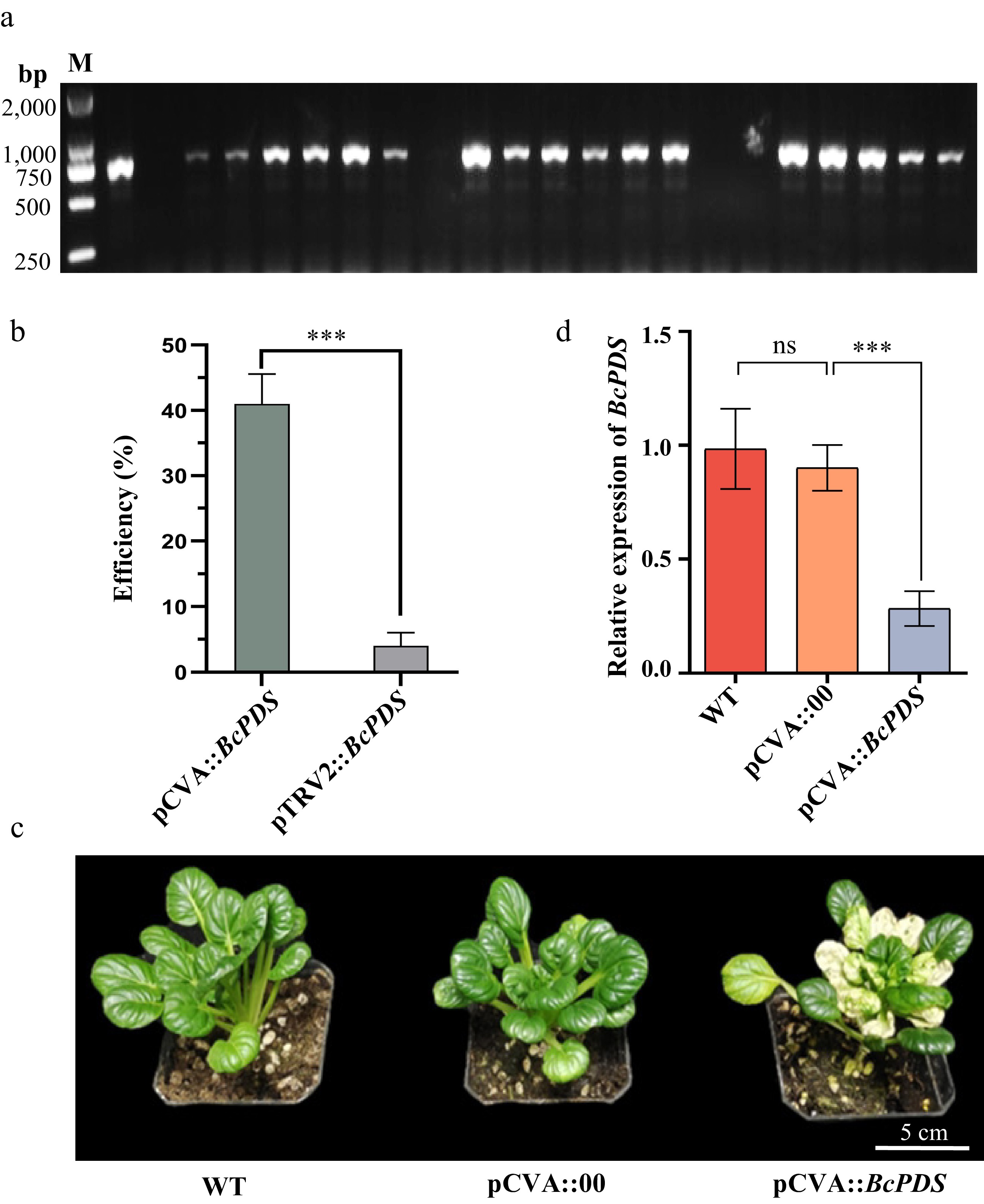
Figure 3.
Application of CaLCuV-VIGS to Xiaobaye using TRV-VIGS as a control targeting the same BcPDS sequence. (a) PCR detection of CaLCuV fragment accumulation in infiltrated plants at 14 dpi. (b) Infection efficiency of the CaLCuV-VIGS and TRV-VIGS system in Xiaobaye. Data are presented as mean ± SD (n = 3). Statistical significance was determined by Student's t-test (*** p < 0.001). (c) Silencing phenotype of BcPDS in Xiaobaye at 21 dpi. (d) Expression analysis of BcPDS in photobleached tissues by RT-qPCR of Xiaobaye after infection. Data are presented as mean ± SD (n = 3). Statistical significance was assessed by one-way ANOVA followed by Tukey's HSD post hoc test (ns = not significant, *** p < 0.001).
Optimizing conditions for CaLCuV-based VIGS in wutacai
-
To establish an efficient VIGS system, we optimized two key parameters known to influence the CaLCuV agroinfiltration process: growth temperature and Agrobacterium concentration[15,28]. A total of nine combined treatments were evaluated: Agrobacterium concentrations of OD600 = 0.5, 1.0, and 1.5; and growth temperatures of 20, 22, and 24 °C. We then assessed viral infection efficiency under these conditions. Genomic DNA was isolated from infiltrated plants, and PCR was performed to detect viral DNA accumulation. At each temperature tested, Agrobacterium concentration significantly influenced infection efficiency. The optimal concentration was OD600 = 1.0. Both lower (OD600 = 0.5) and higher (OD600 = 1.5) concentrations resulted in reduced efficiency (Fig. 4). Similarly, growth temperature significantly affected infection efficiency. Regardless of the Agrobacterium concentration, the highest efficiency was consistently achieved at 22 °C compared to 20 and 24 °C (Fig. 4). Overall, infection efficiency reached approximately 40% under the optimized condition of 22 °C and OD600 = 1.0. In contrast, efficiency was as low as 5% at 22 °C with OD600 = 0.5. Based on these findings, we determined the optimal conditions for CaLCuV-based VIGS in wutacai to be agroinfiltration with an Agrobacterium concentration of OD600 = 1.0, followed by growth at 22 °C.
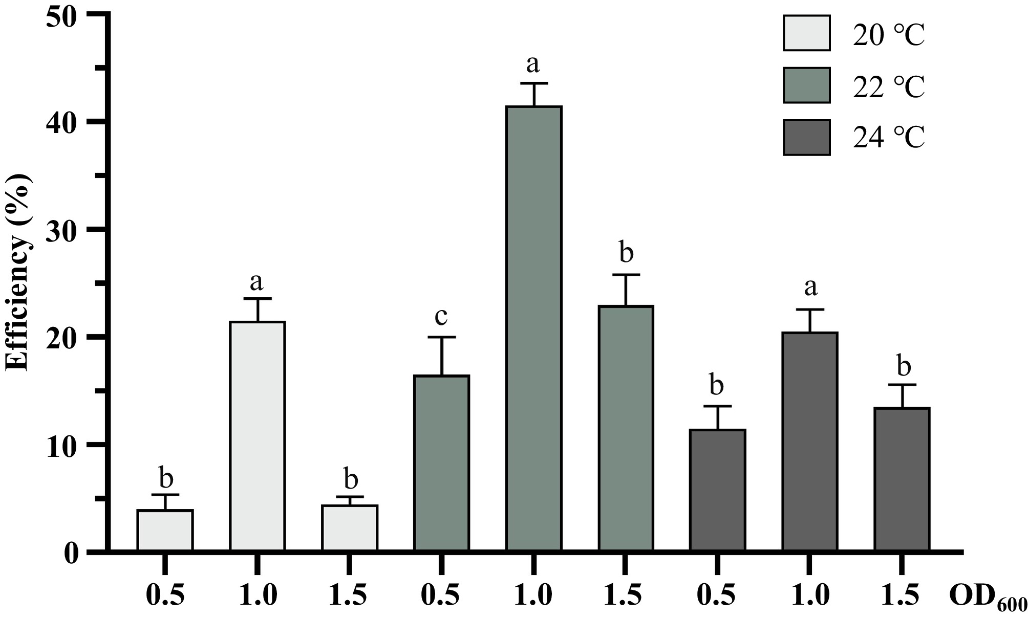
Figure 4.
Effects of Agrobacterium concentration (OD600 = 0.5, 1.0, 1.5) and growth temperature (20, 22, 24 °C) on CaLCuV-VIGS infection efficiency in Xiaobaye. Data are presented as mean ± SD (n = 3 biological replicates, each comprising 20 plants) (one-way ANOVA, Tukey's HSD test). Different lowercase letters indicate statistically significant differences at p < 0.05.
Analysis the efficiency of CaLCuV-based VIGS in various wutacai germplasms
-
Abundant germplasms represent valuable genomic resources for breeding programs. To fully exploit those genetic reservoirs, establishing versatile research methodologies applicable across diverse genetic backgrounds is essential. We therefore evaluated the portability of the CaLCuV-based VIGS system in multiple wutacai cultivars. Two core germplasms—W16-18, W19-24—were selected for VIGS experiments using pCVA::BcPDS. After infiltration as described above, photobleaching phenotypes developed in all tested germplasms by 30 dpi, albeit with variation in onset timing (Fig. 5a). The infection efficiencies of the two germplasms were statistically evaluated. Compared with that of Xiaobaye (approximately 40%, Fig. 4), the infection efficiencies of W16-18 and W19-24 were markedly lower, at about 30% and 18% (Fig. 5b), respectively, indicating that this system is influenced by genotype. RT-qPCR revealed approximately 40% and 60% reductions in BcPDS transcript levels in W16-18 (Fig. 5c) and W19-24 (Fig. 5d), respectively, relative to the controls (pCVA::00 and untreated plants).
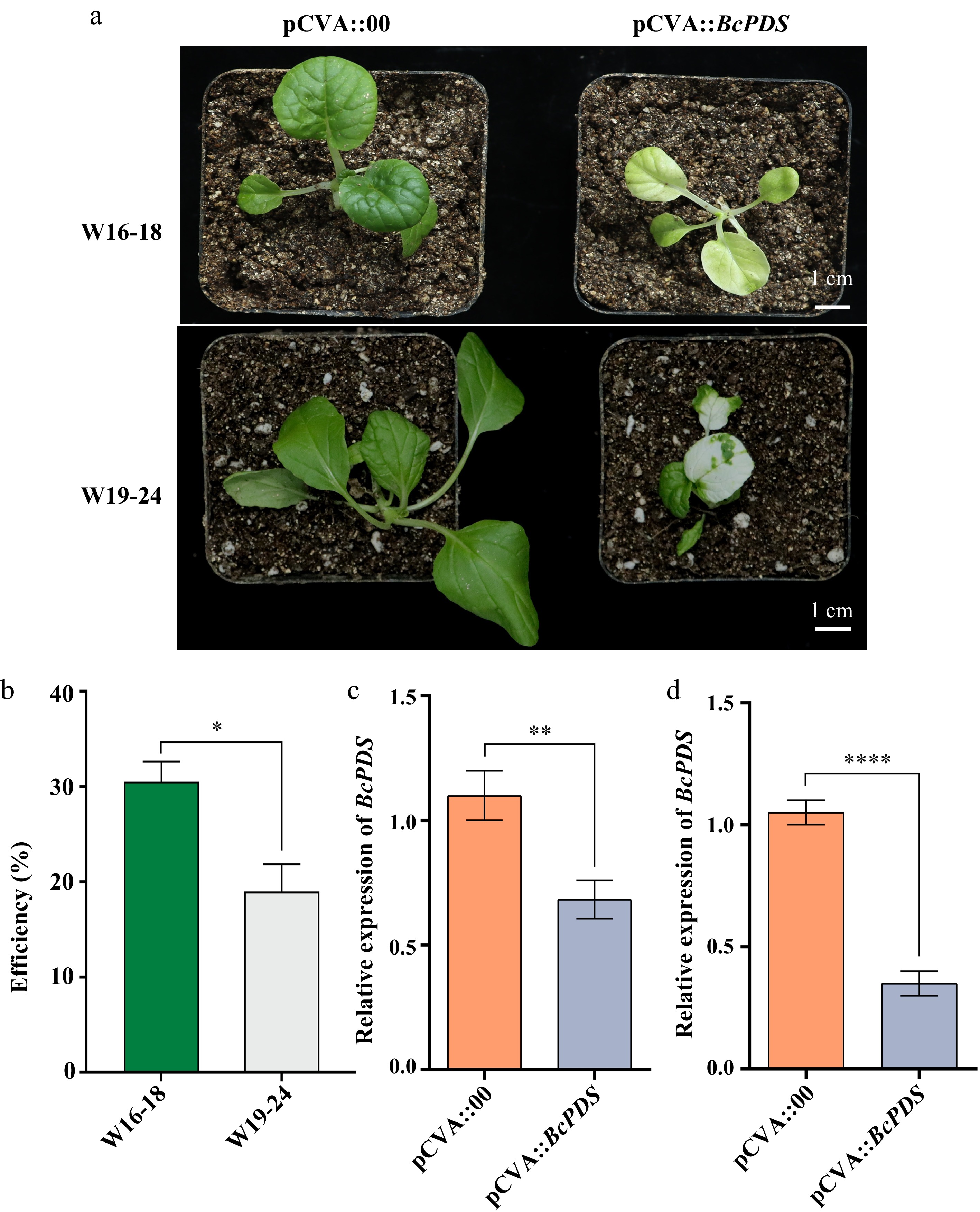
Figure 5.
Application of the CaLCuV-VIGS system targeting BcPDS in two representative wutacai core germplasms. (a) Photobleaching phenotypes were observed at 30 dpi in the tested germplasms (W16-18 and W19-24). All lines developed photobleaching by 30 dpi, albeit with variation in the onset timing of the phenotype. (b) Infection efficiency of W16-18 and W19-24 at optimized conditions (OD600 = 1.0, 22 °C). The plants showing a photobleached phenotype were counted. (c), (d) Expression analysis of BcPDS in photobleached tissues collected from (a) by RT-qPCR. (c) W16-18 plants. (d) W19-24 plants. Data are presented as mean ± SD (n = 3). Statistical significance was determined by Student's t-test (* p < 0.05, ** p < 0.01, *** p < 0.001).
Collectively, these results indicate broad applicability of the CaLCuV-VIGS system across major wutacai germplasms.
Functional characterization of BcSGR1 in leaf senescence
-
As a leafy vegetable crop, wutacai's economic and nutritional value depends critically on leaf quality, with senescence causing direct yield and quality losses. Deciphering the molecular mechanisms of leaf senescence in wutacai is therefore essential for breeding programs and represents an active research priority[29]. Stay-Green 1 (SGR1) interacts with light-harvesting complex II (LHCII) subunits to regulate chlorophyll degradation[30]. Mutations in SGR1 orthologs confer stay-green phenotypes across diverse species including Arabidopsis[30], rice[31], pea[32], tomato[33], Chinese cabbage[34] and citrus[35]. To evaluate the CaLCuV-VIGS system's utility for senescence and postharvest studies, we selected the wutacai SGR1 homolog showing the highest sequence identity to AtSGR1 for functional analysis. Sequence alignment showed high conservation across species (Fig. 6a), with BcSGR1 sharing 71.9% identity with AtSGR1 and 90% with BrSGR1. Phylogenetic analysis of 62 SGR proteins from diverse species revealed close evolutionary relationships, and BcSGR1, BrSGR1, and AtSGR1 are in the same group (Fig. 6b).
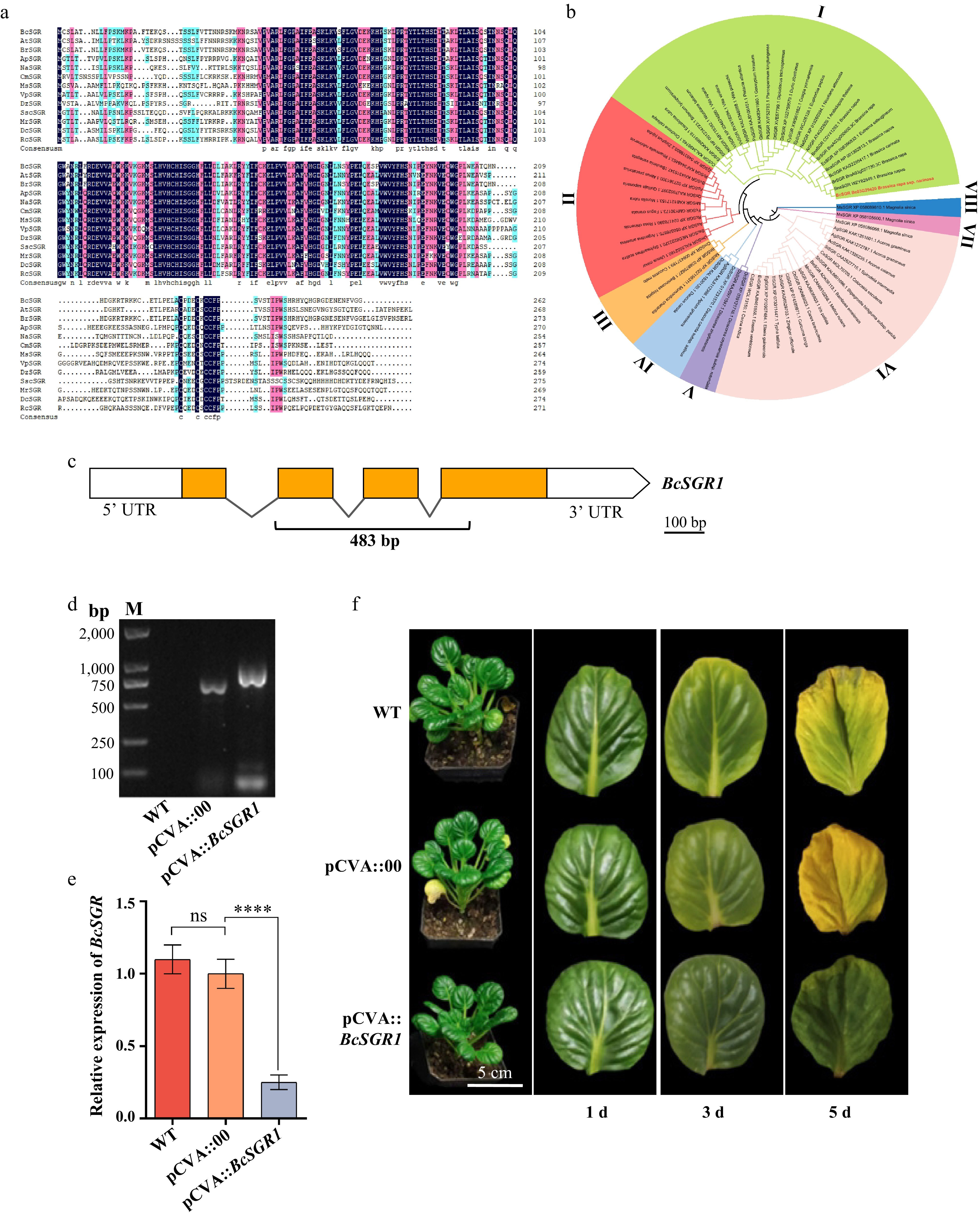
Figure 6.
Multiple sequence alignments and phylogenetic relationship of BcSGR1 and its orthologs, along with the construction of the pCVA::BcSGR1 vector and silencing of SGR in wutacai. (a) Multiple sequence alignments of BcSGR1 and its orthologs from Arabidopsis, Chinese cabbage, Rosary pea, Tobacco, Melon, Magnolia sinica, Grape, Ginger yam, Shrubby stylo, Chinese bayberry, Carrot, and China rose. The alignments were performed using full-length amino acid sequences of SGR. (b) Phylogenetic analysis of SGR proteins from 56 species. The unrooted phylogenetic tree was generated by the neighbour-joining method using the MEGA6 program with 1,000 bootstrap trials. (c) Structure of the BcSGR1 gene body; the fragment inserted in pCVA is indicated with a black underline. (d) Detection of CaLCuV fragments after VIGS treatments. Lane M shows DNA size markers. (e) Expression analysis of BcSGR1 by RT-qPCR after infection. Data are presented as mean ± SD (n = 3). Statistical significance was assessed by one-way ANOVA followed by Tukey's HSD post hoc test (ns = not significant, **** p < 0.0001). (f) Silencing phenotype of BcSGR1 in wutacai at 30 dpi.
We selected the most conserved protein domain to design the silencing fragment (Fig. 6c), generating the pCVA::BcSGR1 construct. Then, the construct was infiltrated into Xiaobaye; again, the pCVA empty construct was used as a control. At 30 dpi, PCR confirmed pCVA::BcSGR1 accumulation (Fig. 6d). RT-qPCR revealed ~80% reduction in BcSGR1 transcripts compared to controls (pCVA::00 and untreated plants; Fig. 6e). Leaves were then detached from plants and kept in moist condition at room temperature. Leaf color changes were documented at 1, 3, and 5 d. Leaves from the WT and pCVA::00 control plants showed obvious color fading by day 3 and turned nearly completely yellow by day 5. In contrast, leaves from pCVA::BcSGR1-infiltrated plants remained green throughout the observation period, despite showing signs of wilting (Fig. 6f). These data demonstrated that silencing BcSGR1 could significantly delay leaf senescence, and the CaLCuV-VIGS is useful for mechanism study in leaf senescence in wutacai.
Functional characterization of BcFLC in bolting
-
Premature bolting and flowering pose a significant threat to wutacai quality, severely limiting its marketable period. Consequently, breeding cultivars with delayed bolting-flowering time is a crucial objective for wutacai and other NHCC varieties. Understanding the molecular mechanisms governing flowering time in NHCC is therefore essential. While several flowering time-related genes have been cloned and functionally characterized in NHCC Pakchoi[19,36,37], research on wutacai has been limited[38]. FLC, a MADS-box transcription factor, acts as a central repressor of flowering. It controls the vegetative-to-reproductive transition by inhibiting the expression of key floral integrators such as FLOWERING LOCUS T (FT) and SUPPRESSOR OF OVEREXPRESSION OF CO 1 (SOC1)[39]. To elucidate the role of FLC in wutacai flowering, we isolated the homologs (BcFLC) with the highest sequence identity to the Arabidopsis ortholog sequence. Multiple amino acid sequence alignment revealed high conservation of FLC across analyzed plant species, with BcFLC sharing 43.6% identity with AtFLC (Fig. 7a). Phylogenetic analysis of 61 FLC proteins from diverse species revealed close evolutionary relationships, and BcFLC, BrFLC, and AtFLC are also in the same group (Fig. 7b). A 458-bp fragment covering a conserved region of the BcFLC coding sequence was cloned into the pCVA vector (Fig. 7c). Following agroinfiltration into Xiaobaye plants, accumulation of the pCVA construct was confirmed at 14 dpi (Fig. 7d). Quantitative analysis showed that BcFLC expression levels in pCVA::BcFLC-infiltrated plants dropped to less than 10% compared to control plants (Fig. 7e). Phenotypically, BcFLC-silenced plants initiated flowering at 30 dpi, approximately 15 d earlier than control plants (Fig. 7f).
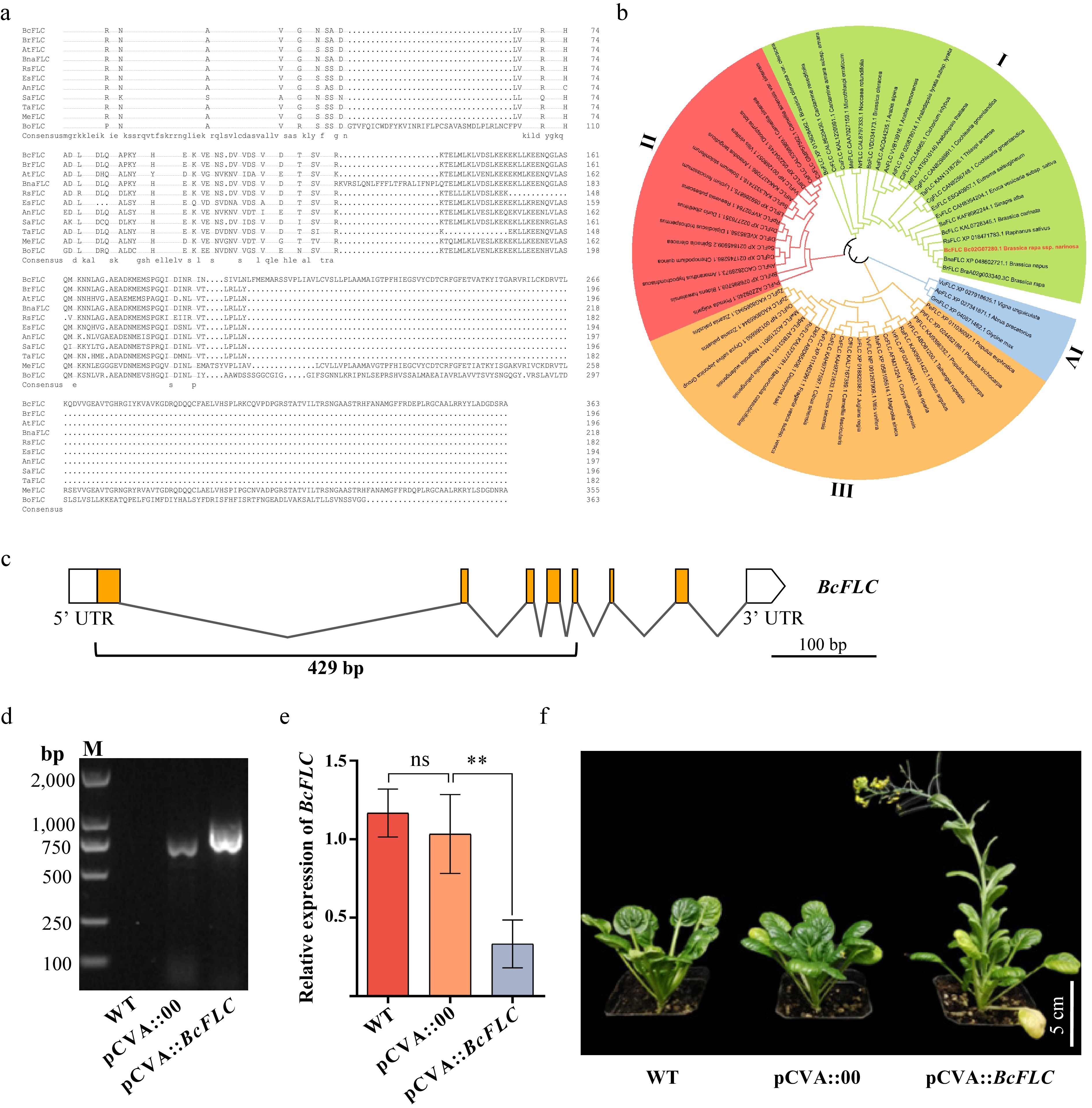
Figure 7.
Multiple sequence alignments and phylogenetic relationship of BcFLC and its orthologs, along with the construction of the pCVA::BcFLC vector and silencing of FLC in wutacai. (a) Multiple sequence alignments of BcFLC and its orthologs from Arabidopsis, Chinese cabbage, Rapeseed, Radish, Salt cress, Lyrate rockcress, White mustard, Field pennycress, Barrel medic, and Broccoli. The alignments were performed using full-length amino acid sequences of FLC. (b) Phylogenetic analysis of FLC proteins from 56 species. The unrooted phylogenetic tree was generated by the neighbour-joining method using the MEGA6 program with 1,000 bootstrap trials. (c) Structure of the BcFLC gene body; the fragment inserted in pCVA is indicated with a black underline. (d) Detection of CaLCuV fragments after VIGS treatments. Lane M shows DNA size markers. (e) Expression analysis of BcFLC by RT-qPCR after infection. Data are presented as mean ± SD (n = 3). Statistical significance was assessed by one-way ANOVA followed by Tukey's HSD post hoc test (ns = not significant, ** p < 0.01). (f) Silencing phenotype of BcFLC in wutacai at 30 dpi. Leaf color was recorded at 1, 3, and 5 d after detachment.
-
Since its initial development approximately 30 years ago, VIGS has proven to be a powerful technique for gene functional analysis, particularly in plants recalcitrant to stable genetic transformation[2]. To date, VIGS-based technologies have expanded beyond gene silencing to include virus-induced overexpression (VOX)[40] and virus-induced genome editing (VIGE)[41]. These systems provide rapid, convenient, and high-throughput research tools for functional genomics, greatly benefiting fundamental research.
Wutacai is rich in vitamin C, vitamin B1, and carotene, and is often referred to as a 'vitamin vegetable'. It is widely cultivated in the Yangtze-Huaihe River Basin[29]. To advance molecular breeding of wutacai, substantial efforts have been devoted to elucidating the genetic basis of key agronomic traits such as stress response[42], quality[43], and leaf shape[44]. However, the absence of a reliable genetic transformation system has considerably hampered such fundamental research. To facilitate functional genomics studies in this economically important vegetable crop, we established a robust and highly efficient CaLCuV-based VIGS system in wutacai seedlings by optimizing multiple influencing factors.
The selection of an appropriate VIGS vector is species-dependent and hinges on viral infectivity and silencing efficiency. Although TRV-derived VIGS vectors are widely used in various dicotyledonous plants and even some monocots[10], successful application in NHCC, which includes wutacai, has not been reported. In this study, we tested a TRV-based VIGS vector for gene silencing in wutacai but found infection ineffective (Fig. 3b), consistent with previous observations in B. oleracea[15]. However, a recent study did report successful TRV-VIGS application in heading Chinese cabbage[17]. Therefore, TRV-based VIGS may hold potential for NHCC, but further optimization is likely required. In contrast, CaLCuV, originally isolated from Brassicaceae crops, exhibits a broad host range within the Brassicaceae family[12]. This suggests CaLCuV-based VIGS may be particularly suitable for efficient infection and gene silencing in wutacai. Indeed, we achieved successful infection and silencing in the majority of our wutacai experiments (Figs. 3−7).
The method of delivering viral vectors into plant cells is a crucial factor impacting VIGS efficiency. Over the past two decades, various inoculation methods have been developed for different plant species, including biolistic bombardment and Agrobacterium-mediated infiltration[2]. For example, particle bombardment of a TYMV-based plasmid DNA effectively silenced the BrPDS gene in Brassica rapa[45], and biolistics delivered CaLCuV components to downregulate target genes in B. napus[46]. Notably, the widely used pTY-S VIGS vector for gene silencing in NHCC is also delivered via biolistic bombardment[18,19]. However, biolistic bombardment has drawbacks, including the requirement for expensive equipment and supplies, complex integration patterns, and relatively low throughput[47]. In comparison, Agrobacterium-mediated infiltration is a more cost-effective, convenient, and efficient method, especially in dicot species, making it the most frequently used approach for VIGS. Thus, we focused on optimizing leaf infiltration-based inoculation in this study.
Beyond the delivery method, Agrobacterium solution concentration and plant growth conditions significantly influence VIGS efficiency. High Agrobacterium density can increase infection efficiency but may cause necrosis on infiltrated leaves, subsequently inhibiting viral accumulation and spread[48]. Optimal Agrobacterium concentrations vary by species: OD600 = 1.0 for N. benthamiana, OD600 = 1.5 for tomato[49], and OD600 = 1.2 for tea plants[50]. We found that an A. tumefaciens solution at OD600 = 1.0 yielded the best infection efficiency in wutacai, consistent with findings in B. oleracea[15]. Previous studies reported higher infection ratios in plants maintained at 22 °C compared to 18 or 26 °C[15]. Similarly, we observed the highest silencing efficiency at 22 °C compared to 20 and 24 °C (Fig. 4). Furthermore, we confirmed that these optimized conditions are broadly applicable across different wutacai germplasms (Fig. 5).
It should be noted that the VIGS constructs used in the present study carry relatively large inserts. A recent study demonstrated that short RNA fragments (24–32 nt) can trigger robust gene silencing, offering several distinct advantages over the conventional long-fragment approaches[51]. Specifically, shorter inserts can substantially enhance scalability by dramatically reducing the size and complexity of VIGS constructs, thereby enabling faster, cheaper, and more simplified vector construction. They may also reduce the risk of off-target effects owing to their higher sequence specificity, allowing more precise targeting. Moreover, their compact nature facilitates multiplexed silencing, as multiple short fragments can be readily concatenated into a single vector. Thus, we propose that further optimization in the wutacai VIGS system could be achieved by systematically evaluating additional parameters, including silencing fragment length and GC content, seedling age, and humidity[48].
Leaf senescence and flowering time are crucial traits affecting wutacai yield and quality, whose underlying mechanisms require elucidation. In addition to the visual marker gene PDS commonly used to assess VIGS efficiency, we employed the BcSGR1 and BcFLC genes, which are proposed to be involved in senescence and flowering, to validate our system. Using our CaLCuV-mediated VIGS system, we successfully knocked down the expression of BcSGR1 and BcFLC. This resulted in significantly delayed leaf senescence in pCVA::BcSGR1-infiltrated plants (Fig. 6) and flowering advancement by approximately two weeks in pCVA::BcFLC-infiltrated plants (Fig. 7). These results demonstrate the successful application of the CaLCuV-mediated VIGS system for functional gene analysis in wutacai. The established VIGS system now provides a platform for high-throughput functional genomics studies in this crop.
-
In this study, we established a robust and efficient CaLCuV-based VIGS system for wutacai. By optimizing key factors influencing silencing efficiency, we achieved a maximum silencing efficiency of ~70% for BcPDS using an A. tumefaciens solution at OD600 = 1.0 and a growth temperature of 22 °C. Successful infiltration was demonstrated across all three cultivars tested, indicating broad applicability. The system's reliability was further confirmed by the successful knockdown of two functional genes and the observation of corresponding phenotypic changes. In summary, we developed a convenient and efficient CaLCuV-mediated VIGS system for transient gene functional analysis in wutacai. This system provides a valuable tool for reverse genetics and will significantly advance future functional genomics research in this important crop.
-
The authors confirm their contributions to the paper as follows: study conception and design: Wang W, Hou J, Wu J, Chen G, Tang X, Wang C, Yuan L; data collection: Yu M, Cao C, Tang X, Song L, Li Z; manuscript preparation and revision: Wang W, Chen G, Tang X. All authors reviewed the results and approved the final version of the manuscript.
-
All data generated or analyzed in this study are included in this published article and its supplemental files.
-
The authors declare that they have no conflict of interest.
-
accompanies this paper online at: https://doi.org/10.48130/vegres-0026-0025.
-
# Authors contributed equally: Mengling Yu, Changsheng Cao
- Supplementary Table S1 Accession numbers of PDS, SGR, and FLC.
- Supplementary Table S2 Experimental primer sequences.
- Supplementary Table S3 Complete nucleotide sequences of the cDNA-derived cloned fragments for VIGS.
- Copyright: © 2026 by the author(s). Published by Maximum Academic Press, Fayetteville, GA. This article is an open access article distributed under Creative Commons Attribution License (CC BY 4.0), visit https://creativecommons.org/licenses/by/4.0/.
-
About this article
Cite this article
Yu M, Cao C, Song L, Li Z, Hou J, et al. 2026. Establishment of a virus-induced gene silencing system in wutacai (Brassica rapa) using cabbage leaf curl virus-based vector. Vegetable Research 6: e033 doi: 10.48130/vegres-0026-0025 

Establishment of a virus-induced gene silencing system in wutacai (Brassica rapa) using cabbage leaf curl virus-based vector
- Received: 21 May 2026
- Revised: 01 August 2026
- Accepted: 28 August 2026
- Published online: 09 October 2026
Abstract: Virus-induced gene silencing (VIGS) is a powerful technology that exploits plants' natural antiviral defense mechanisms to achieve targeted gene silencing via viral RNA degradation. It has become an indispensable tool for rapid functional analysis in numerous species, particularly those recalcitrant to stable genetic transformation. In wutacai (Brassica rapa ssp. narinosa), no established protocol for generating stable transgenic plants is currently available, making VIGS particularly advantageous for gene functional studies. However, the application of VIGS in wutacai has rarely been reported. In this study, we successfully established a VIGS system in wutacai. By comparing the infection efficiency of two viral vectors, cabbage leaf curl virus (CaLCuV) and tobacco rattle virus (TRV), we found that the CaLCuV-based system was significantly more efficient than the TRV-based system. Furthermore, the established VIGS system proved applicable across diverse wutacai germplasms, indicating its broad applicability. In addition to silencing the widely used reporter gene phytoene desaturase (PDS), we also successfully silenced and functionally characterized the senescence-associated gene BcSGR1 and the flowering-regulating gene BcFLC, further validating the reliability of this system. Collectively, our findings demonstrate that the CaLCuV-mediated VIGS can be efficiently applied in wutacai, providing a robust tool for gene functional research and facilitating future molecular breeding efforts in this crop.