












-
Neovascular retinopathy is among the leading causes of blindness worldwide[1]. Despite differing causes, these diseases share ischemic, hypoxia-driven pathological neovascularization. Current anti-vascular endothelial growth factor (VEGF) therapy fails to address all clinical challenges, necessitating alternative pathogenic mechanisms and therapeutic strategies[2].
Accurate disease modeling requires systems that recapitulate retinal vascular pathophysiology. While animal models and 2D cultures have advanced biomedical research, they face inherent limitations—species differences or lack of native tissue complexity. Recently, 3D self-organizing retinal organoids (ROs) derived from pluripotent stem cells have emerged as powerful tools that closely mimic native retinal architecture and cellular composition[3,4]. However, non-vascularized ROs lack microvessels essential for oxygen, nutrient, and drug transport, and undergo atrophy under prolonged culture due to insufficient perfusion[5]. How to vascularize ROs remains a major challenge.
To address this, strategies including multicellular co-culture, 3D bioprinting, and microfluidic platforms have been developed to incorporate functional endothelial networks. These methods aim to incorporate endothelial networks with perfusion capacity into ROs, thereby improving their physiological relevance and expanding their applicability in biomedical research.
Although significant progress has been made in the research of ROs, studies on vascularized ROs (vROs) remain relatively limited. This review summarizes current construction methods for vascularized organoids, aiming to provide insights for the development of vROs and their applications in neovascular retinopathy studies.
-
Recent advances have enhanced the potential of vascularized organoids for disease modeling and translational research. Their generation is both a crucial tool for studying neovascular diseases and a prerequisite for constructing vROs. These organoids are mainly generated through cellular self-assembly or pre-formed vascular networks, both requiring suitable endothelial sources and hydrogels with tunable mechanics to support cell-matrix interactions.
Sources of materials in vascular engineering
Endothelial cells (ECs) and supporting cells
-
ECs are the primary constituents of vascular networks. Common sources include human umbilical vein ECs for screening, organ-specific ECs for barrier modeling, and pluripotent stem cell (PSC)-ECs for patient-specific applications[6,7]. Supporting cells such as pericytes and MSCs are often co-cultured to stabilize vessels and promote maturation[8].
Hydrogel materials in vascular engineering
-
Hydrogels provide a 3D biomimetic matrix for endothelial network formation. Natural hydrogels, such as collagen and Matrigel, support angiogenesis, while synthetic hydrogels offer tunable mechanical properties and batch consistency[9].
Vascularized organoid construction based on cellular self-assembly mechanisms
-
Based on cells and hydrogels, vascular organoids are developed through two approaches: cellular self-assembly and pre-formed vascular network integration. Self-assembly includes multicellular co-culture and organoid co-differentiation. In multicellular co-culture, vascular ECs are co-cultured with supporting cells including pericytes and mesenchymal stem cells (MSCs) within a 3D hydrogel matrix to form vascularized organoids, harnessing cell migration, organization, and differentiation in a supportive microenvironment. This approach avoids rigid scaffolding and instead relies on the natural interplay between cell types and extracellular matrix (ECM) components to form lumenized vascular networks[4]. While the multicellular co-culture strategy is effective in forming basic vascular structures, it may not fully replicate the complexity and functional integration of organ-specific vasculature. To address this, researchers have developed co-differentiation systems, which induce PSCs to simultaneously differentiate into vascular lineages and organ-specific cells. This approach draws inspiration from embryonic development, during which vascular networks co-evolve with surrounding tissues, forming structurally and functionally coordinated systems[10,11] (Fig. 1).
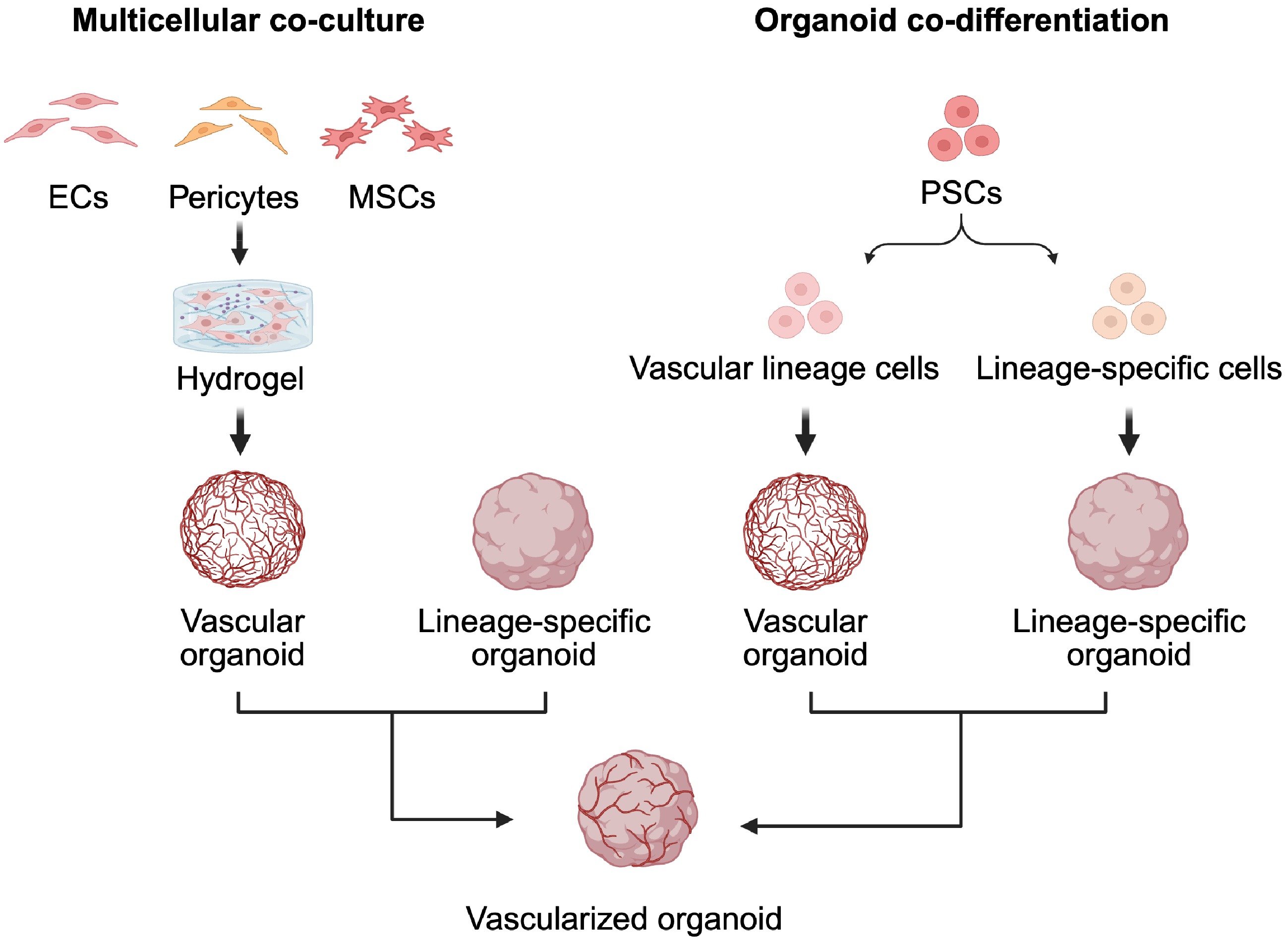
Figure 1.
Construction of vascularized organoids via cellular self-assembly. ECs and supporting cells are co-cultured in hydrogels to generate vascularized organoids, which can then be co-cultured with preformed organ-specific organoids. Alternatively, PSCs are induced to co-differentiate into vascular and organ-specific lineages within the same system, enabling intrinsic vascularization.
Co-differentiation strategies have also advanced kidney[12], brain[13], pancreatic[14], liver[15], and heart[16] organoid development, where vascular progenitors are co-induced with tissue-specific progenitors to form perfusable and physiologically relevant vasculature. These models improved not only the efficiency of vascular formation but also the fidelity of tissue maturation. However, coordinating multiple differentiating lineages is technically demanding, as minor deviations can compromise tissue function. Many current models lack functional perfusion systems, limiting in vivo simulation. Finally, the lack of standardized protocols hinders reproducibility and clinical translation[17].
Vascularized organoid construction via pre-formed vascular networks
-
While multicellular co-culture strategies and co-differentiation strategies have significantly advanced the development of vascular organoids, they often require extended culture times and may result in immature or poorly perfused vasculature. An alternative approach involves pre-formed vascular networks, which provide immediate perfusion capacity and structural support. This top-down strategy enhances organoid viability, improves nutrient delivery, and facilitates formation of more physiologically relevant tissues. Among the current methodologies for constructing vascular networks, the most widely used are 3D bioprinting technology and organ-on-a-chip (OoC) platforms, both of which enable precise spatial organization and dynamic microenvironmental control.
Three-dimensional bioprinting technology
-
Three-dimensional bioprinting has emerged as a powerful tool for constructing complex tissue architectures by precisely depositing bioinks, mixtures of cells, hydrogels, and bioactive molecules. This technique overcomes the spatial limitations of traditional tissue engineering and allows for the creation of anatomically and functionally accurate vascular structures.
Depending on specific experimental goals, different bioprinting techniques can be selected (Fig. 2). Extrusion-based bioprinting is suitable for printing large, centimeter-scale structures with high cell density and mechanical stability[18], while laser-assisted bioprinting offers micrometer-level resolution for producing fine vascular patterns[19]. Droplet-based bioprinting, by contrast, provides moderate resolution with relatively fast printing speeds, making it effective for rapid layer-by-layer construction of vascular features[20].
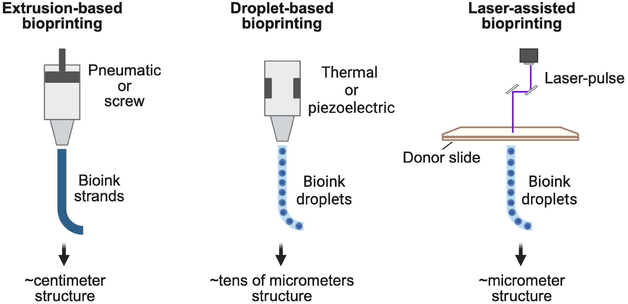
Figure 2.
Comparison of bioprinting techniques for vascular construction. Extrusion-based bioprinting fabricates large, centimeter-scale structures with high cell density and mechanical strength. Laser-assisted bioprinting provides micrometer-level resolution for fine and complex vascular patterns. Droplet-based bioprinting offers moderate resolution and faster speeds, enabling rapid layer-by-layer construction of vascular features.
Recent studies have demonstrated utility across multiple organ systems[21,22]. In retinal research, 3D bioprinting of glaucoma models revealed that ECs preferentially formed vascular tubes only in scaffolds containing both optic nerve head astrocytes and RGCs, demonstrating the potential of bioprinting for constructing retina-relevant vascularized models[23]. Other advanced approaches, such as electrohydrodynamic printing[24], multiphoton-guided printing[25], volumetric bioprinting[26,27], and embedded printing[28], have enabled the creation of intricate, perfusable, and hierarchically organized vascular networks.
Compared with conventional vascularization strategies, 3D bioprinting offers unparalleled spatial precision, compatibility with multiple bioinks, and centimeter-scale fabrication of hierarchically organized vascular networks that integrate into organoids and recapitulate native tissue architecture. However, current techniques face trade-offs among resolution, printing speed, and cell viability, along with challenges in achieving homogeneous cell distribution, limited real-time perfusion during printing, and difficulties in replicating dynamic physiological cues such as shear stress. The integration of bioprinting with real-time imaging and computational modeling continues to enhance its capacity to generate vascularized constructs tailored to specific anatomical and physiological requirements[29].
Vascularized organ-on-a-chip technology
-
In addition to bioprinting, OoC technology has become another cutting-edge platform for constructing vascularized organoids[30]. By combining stem cell-derived tissues with microfluidic systems, OoC platforms recreate a dynamic microenvironment that closely resembles in vivo physiological conditions[31]. These systems enable fine-tuned control over flow dynamics, nutrient and oxygen delivery, and spatiotemporal signaling, which are critical for the formation and maturation of functional microvasculature. A typical OoC device consists of a central culture chamber for tissue growth and adjacent microfluidic channels that provide continuous perfusion, mimicking blood flow and facilitating metabolic exchange[30].
OoC platforms are highly effective in modeling angiogenesis, tissue–tissue interface formation, and disease-specific microenvironments. For instance, Seiler et al. incorporated patient-derived intestinal myofibroblasts into an intestinal chip to induce endothelial cells to form perfusable capillary networks that replicated the submucosal vascular architecture[32]. Similarly, Rayner et al. co-cultured renal tubular epithelial cells and ECs in a kidney chip under continuous microfluidic flow to successfully recreate the vascular-tubular interface[33]. Akinbote et al. further extended these approaches to lung and heart tissues, where organ-specific microvascular chips revealed fibrotic responses unique to each tissue type under different perfusion conditions[34].
Tissue interface reconstruction represents another major advantage of OoC technology. For example, Liu et al. engineered a liver chip in which parenchymal hepatocytes and ECs were compartmentalized to establish stable nutrient exchange channels at their interface[35]. This model highlighted the essential role of vascular–parenchymal interactions in maintaining tissue functionality. Likewise, Hospodiuk-Karwowski et al. developed a pancreas chip by combining islet cells with vascular components in a biomimetic extracellular matrix, providing a new platform for studying islet–vascular cross-talk in diabetes[36].
Despite these advantages, OoC technology faces certain limitations. These include limited construct size, challenges in scaling up for high-throughput applications, the complexity of device fabrication, and difficulties in extracting cells for downstream analysis. Addressing these technical hurdles remains an active area of research.
Altogether, the integration of pre-formed vascular networks through 3D bioprinting and OoC technology offers powerful and highly controllable means to vascularize organoids. These approaches enable the generation of physiologically relevant tissue models with precise architecture and perfusion, thereby enhancing the applicability of organoids in regenerative medicine, drug screening, and personalized healthcare.
-
ROs are 3D self-organizing retinal tissues derived from human-induced PSCs that recapitulate layered architecture and generate major retinal cell types under optimized culture conditions[37−39]. Despite these advances, a major limitation of ROs is the complete absence of vasculature, which prevents the formation of either the inner BRB (iBRB) composed of endothelial tight junctions, or the outer BRB (oBRB) formed by RPE, Bruch’s membrane, and choroidal endothelium. As ROs enlarge, the lack of perfusion leads to hypoxia, nutrient deprivation, and central apoptosis, ultimately limiting maturation and long-term viability. To address this, several vascularization strategies have been developed to generate vROs.
Chen et al. developed a vRO model by co-culturing vascularized organoids and ROs on V-bottom polydimethylsiloxane microwell platforms, expressing tight junction protein claudin-5 and resembling the iBRB[40]. Xue et al. developed a perfusable retina-on-a-chip (RoC) bioreactor based on unidirectional microfluidics, showing comparable cell types, morphology, and retinal gene expression to static culture[41].
Meanwhile, vROs emerged as powerful tools to investigate complex metabolic retinopathy, particularly proliferative diabetic retinopathy (pDR). Under high-glucose culture conditions, vROs exhibit characteristic pDR-like phenotypes, including reduced organoid size and a significant loss of retinal ganglion cells (RGCs), suggesting their suitability for modeling the synergistic neurovascular damage that occurs under diabetic conditions[42]. High-glucose-stimulated ROs recapitulate multiple key early pathological changes of pDR, such as the loss of RGCs and amacrine cells, increased reactivity of astrocytes and microglia, and markedly elevated expression of inflammatory factors including VEGF and interleukin (IL)-1β[43]. These in vitro models offer highly controllable platforms to elucidate the molecular mechanisms underlying pDR and to screen anti-inflammatory and anti-angiogenic therapies.
Although oBRB modeling is typically not regarded as part of standard vRO systems, there is a growing development of experimental platforms that mimic the oBRB for studying choroidal neovascularization. One common approach involves co-culturing ECs to form capillary-like networks around the retinal pigment epithelium (RPE), simulating aspects of choroidal vasculature. The RPE itself plays crucial roles in guiding vascular patterning and angiogenesis. Further co-culture with vascularized organoid-derived ECs and pericytes enhances vascular integration and significantly upregulates neuronal maturation markers, indicating that neurovascular interactions support retinal development[44]. Leveraging advances in nanomaterials, a cryopreservable, ready-to-use OoC platform with nanofiber membrane integration has been validated for oBRB modeling[45]. Paek et al. developed a micropatterned hydrogel construct housed within an elastomeric microdevice to more realistically recapitulate the spatial organization of the RPE and the underlying choroidal vascular bed[46]. Increasing studies are applying oBRB modeling to disease contexts, such as wet age-related macular degeneration (wAMD), to recapitulate pathological states. Manian et al. integrated induced PSC-derived RPE, EC, and MSC from wAMD patients into an ECM platform that recapitulated key disease phenotypes[47]. Song et al. engineered a 3D oBRB tissue by bioprinting ECs, pericytes, and fibroblasts on the basal side of a biodegradable scaffold, followed by the establishment of an RPE monolayer on the apical side. Complement activation within this system induced wAMD-like phenotypes[48].
In summary, vROs represent promising tools for modeling neovascular retinopathy by providing physiologically relevant platforms for disease mechanism studies and drug screening. Despite current challenges such as limited vascular penetration, spatial resolution, and integration, emerging technologies like bioprinting, microfluidics, and stem cell programming continue to drive progress in this field.
-
With the continuous advancements in regenerative medicine and organoid technologies, organoid models have shown tremendous potential in the study of various diseases. For neovascular retinopathy, vascularized organoid models provide a controllable, reproducible, and functionally relevant in vitro platform, overcoming limitations of traditional cell culture and animal models. Despite progress, challenges remain in simulating the retinal vascular microenvironment and achieving functional vascular reconstruction. Future research should focus on the following areas.
The organ-specific construction of vascularized organoids remains underdeveloped, particularly for the retina, which possesses unique barrier structures and physiological functions. Current vascularization approaches often utilize non-retinal-derived cells, such as HUVECs[3], which fail to fully recapitulate the features of retinal microvascular endothelial cells. Future research should aim to efficiently induce retinal-specific ECs from induced PSCs or ASCs.
Current vascularized organoids often suffer from low maturity and instability. Without dynamic cues like shear stress, vasculature grows irregularly and loses barrier function[3]. Microfluidic systems can provide continuous perfusion and shear force to promote vascular maturation[49]. Multi-omics can monitor development and identify key signaling axes to enhance stability.
Finally, standardization and large-scale generation remain critical challenges, including defining cell sources, optimizing media, and designing perfusion channels, alongside quality control systems[4]. In the future, the integration of artificial intelligence-assisted image analysis, bioprinting, and 3D imaging technologies may drive the development of high-throughput organoid construction and evaluation systems, accelerating their application in retinopathy research.
In summary, vascularized organoids are emerging as powerful in vitro models to study neovascular retinopathy. With the continued convergence and optimization of multidisciplinary technologies, these models are expected to play increasingly important roles in disease mechanism research, drug development, and personalized medicine, ultimately advancing the progress of regenerative and precision ophthalmology.
-
vROs offer a transformative platform for neovascular retinopathy research by bridging in vitro systems and in vivo physiology. Using multicellular co-culture, stem cell co-differentiation, 3D bioprinting, and OoC technologies, vROs better recapitulate the retinal microenvironment, enabling mechanistic studies and therapeutic screening. Although challenges remain, particularly in achieving tissue-specific vascular identity, functional perfusion, and scalable standardization, ongoing innovations in biomaterials, stem cell biology, and microengineering. In the future, vROs are expected to serve as a vital bridge between basic research and clinical therapy, offering novel perspectives and technological support for the prevention and treatment of neovascular retinopathy.
-
Not applicable.
-
The authors confirm their contributions to the paper as follows: draft manuscript preparation: Ren A, Tamanna N; figure preparation: Ren A; manuscript revision, Deng X, Tamanna N, Ke M, Gong Y; supervision: Ke M, Gong Y. All authors reviewed the results and approved the final version of the manuscript.
-
Data sharing is not applicable to this article as no datasets were generated or analyzed during the current study.
-
This work was supported by the National Natural Science Foundation of China (82271088 to MK, 82471098 to YG) and the China Scholarship Council.
-
The authors have no relevant financial or non-financial interests to disclose.
- Copyright: © 2026 by the author(s). Published by Maximum Academic Press, Fayetteville, GA. This article is an open access article distributed under Creative Commons Attribution License (CC BY 4.0), visit https://creativecommons.org/licenses/by/4.0/.
-
About this article
Cite this article
Ren A, Deng X, Tamanna N, Gong Y, Ke M. 2026. Vascularized retinal organoids and vascularization strategies for retinopathy 3-dimensional modeling. Visual Neuroscience 43: e037 doi: 10.48130/vns-0026-0034 

Vascularized retinal organoids and vascularization strategies for retinopathy 3-dimensional modeling
- Received: 04 July 2025
- Revised: 19 June 2026
- Accepted: 26 June 2026
- Published online: 10 August 2026
Abstract: Neovascular retinopathy is characterized by pathological angiogenesis and remains one of the leading causes of vision loss. Vascularized retinal organoids (vROs) have emerged as advanced three-dimensional (3D) models that recapitulate retinal microenvironments by incorporating functional vasculature. This review highlights key vascularization strategies, including co-culture systems, stem cell differentiation, 3D bioprinting, and organ-on-a-chip technologies. These approaches enable disease modeling and drug screening, though challenges persist in vascular maturation and standardization. Future developments focusing on patient-specific models and perfusion systems will enhance the translational potential of vROs for neovascular retinopathy research and therapeutic development.
-
Key words:
- Neovascular retinopathy /
- Vascularized retinal organoids /
- 3D modeling