













-
Congenital cataracts are a major cause of childhood visual impairment worldwide. Although cataract extraction with intraocular lens (IOL) implantation and visual rehabilitation substantially improve visual outcomes, pseudophakic children remain in a period of active ocular growth and frequently experience postoperative axial elongation and progressive myopic drift[1,2]. Previous studies have demonstrated that a younger age at surgery is associated with a greater myopic shift during follow-up[3,4]. Li et al. reported that at 3 years postoperatively, the mean myopic shift ranged from 3.53 diopters (D) in children aged 1 to < 2 years to 1.99 D in those aged 6 years or older[5]. The mechanisms underlying this refractive change remain incompletely understood but may involve visual feedback-regulated ocular growth, as suggested by evidence linking light exposure to eye growth in children and experimental studies showing axial elongation under form deprivation. Despite intentional residual hyperopia at the time of IOL implantation to compensate for future ocular growth, the magnitude of postoperative myopic progression varies substantially among individuals, complicating long-term refractive management and highlighting the need for strategies to control myopic progression in pseudophakic children.
Low-concentration atropine eye drops have emerged as an effective intervention for slowing the progression of myopia in children[6]. Atropine is a nonselective muscarinic antagonist that can induce mydriasis and changes in the ciliary muscles' response[7]. Even at a concentration of 0.01%, mild and transient changes in pupil diameter (PD) and anterior segment parameters have been reported[8], although long-term studies indicate good tolerability with minimal visual side-effects[9,10]. However, pseudophakic children differ anatomically and functionally from phakic myopic children because the crystalline lens has been removed and accommodation is altered[11]. Whether low-concentration atropine induces measurable structural or hemodynamic changes in these eyes remains unclear.
In addition, the pharmacologic effects of atropine may extend beyond the anterior segment. Previous studies have demonstrated that atropine can be detected in the aqueous and vitreous humor after topical administration[12], and short-term alterations in the retinal or choroidal layers have been observed in myopic children[13,14]. However, systematic evaluation of both the anterior segment's structure and posterior segment neural and microvascular responses following atropine administration has rarely been performed in pseudophakic children.
The latest swept-source optical coherence tomography (SS-OCT) and SS-OCT angiography (SS-OCTA) techniques use long-wavelength light and ultrahigh-speed scanning to overcome the traditional imaging limits in depth and speed[15]. Consequently, it can resolve the ocular structures with low signal attenuation across the entire ocular axis. By enabling integrated imaging of the anterior and posterior segments in a single session, SS-OCT/OCTA minimizes systematic errors arising from repeated position changes during multidevice examinations, particularly for children with limited cooperation such as pseudophakic children. Additionally, its strong artifact suppression and artificial intelligence (AI)-driven automated analysis further improve the efficiency of acquiring the posterior segment's neural and vascular structures. The neural integrity of the macular region and its associated microvascular status may reflect ocular responses to atropine[16,17]. Parameters such as the thickness of the ganglion cell complex (GCC) and the retinal nerve fiber layer (RNFL) and macular vascular density may therefore provide additional insight into changes in the posterior segment following administration of atropine. However, systematic evaluation of these neural and vascular parameters remains limited in pseudophakic children.
In this study, we assessed the short-term safety of a single instillation of 0.01% atropine in pseudophakic children by evaluating the ocular biometric changes over 24 h. We evaluated the changes in short-term anterior segment OCT (AS-OCT)-derived biological parameters and retinal/choroidal neural and vascular parameters. These findings provide preliminary physiological and safety evidence for the use of low-concentration atropine in pseudophakic children with myopia.
-
This retrospective repeated measures study was conducted in accordance with the tenets of the Declaration of Helsinki and approved by the Zhongshan Ophthalmic Center Institutional Review Board (2026KYPJ057, dated June 8, 2026). The requirement for informed consent was waived by the Zhongshan Ophthalmic Center Institutional Review Board because of the retrospective nature of the study and the use of deidentified data. This study enrolled 29 children aged 6–12 years with a history of cataracts from August 2025 to February 2026. These children had undergone primary monofocal IOL implantation performed by two experienced cataract surgeons at the Zhongshan Ophthalmic Center. Pseudophakic eyes (n = 44) after congenital cataract surgery were included in the study. Inclusion criteria were a best-corrected visual acuity (BCVA) of 0.2 logarithm of the minimum angle of resolution (logMAR) or better, a spherical equivalent (SE) of ≤ −0.50 D, refractive astigmatism of < 2.00 D, intraocular pressure (IOP) < 21 mmHg, and an open anterior chamber angle. Exclusion criteria included prior use of low-concentration atropine, manifest strabismus, postoperative pupillary synechiae, glaucoma or narrow anterior chamber angles, any systemic disease, and known or suspected hypersensitivity to atropine. All children underwent a thorough evaluation that included a detailed medical and ocular history review, an assessment of visual acuity, slit-lamp examination, IOP measurement, noncycloplegic autorefraction, and ocular biometry measurements (IOLMaster 700; Carl Zeiss Meditec, Germany).
Atropine administration and examination schedule
-
Medical records of eligible children with IOL who had received a single dose of 0.01% low-concentration atropine eyedrops (Xingqi Pharmaceutical Co., Ltd.) were retrospectively reviewed. According to the clinical records, one drop of atropine was instilled into the conjunctival sac of the pseudophakic eye between 9:00 and 12:00. After instillation, the children were instructed to close their eyes and apply pressure to the lacrimal sac area to reduce systemic absorption through the nasolacrimal duct. Only a single dose was administered. Ocular examination data obtained before medication and at 1 and 24 h after the drug's administration were collected. For each child, the 1- and 24-h examinations were scheduled relative to the individual baseline examination time, with the 24-h assessment performed at approximately the same time on the following day. Anterior segment OCT and posterior OCTA examinations were performed using the latest SS-OCT/OCTA device (VG200C, SVision Imaging, China) by the same experienced ophthalmic technician. All children underwent SS-OCT/OCTA scans of both eyes following the same examination procedure at baseline (before medication), 1 and 24 h after administration of the drug. The SS-OCT/OCTA device uses a 1,050-nm laser with a 100,000 A-scans per s scan rate.
Anterior segment measurements
-
The AS-OCT examination was performed using the anterior segment imaging mode of the SS-OCT device. The operator acquired images after centering the image and ensuring stable scanning quality. Multiple acquisitions were repeated for each eye, and the image with the best signal quality and the fewest artifacts was selected for analysis. Anterior segment structural parameters included central corneal thickness (CCT), anterior chamber depth (ACD), PD, and anterior chamber angle area (Fig. 1). The ACD was defined as the axial distance from the central corneal endothelium to the anterior surface of the IOL. All boundary results automatically recognized by the built-in algorithm were reviewed by trained researchers. In cases of apparent boundary positioning deviations or local recognition failures, the built-in layer editing tool was used to manually correct the data before export. Scans with significant image distortion caused by inadequate cooperation, blinking, or eye movements were considered ineligible and excluded.
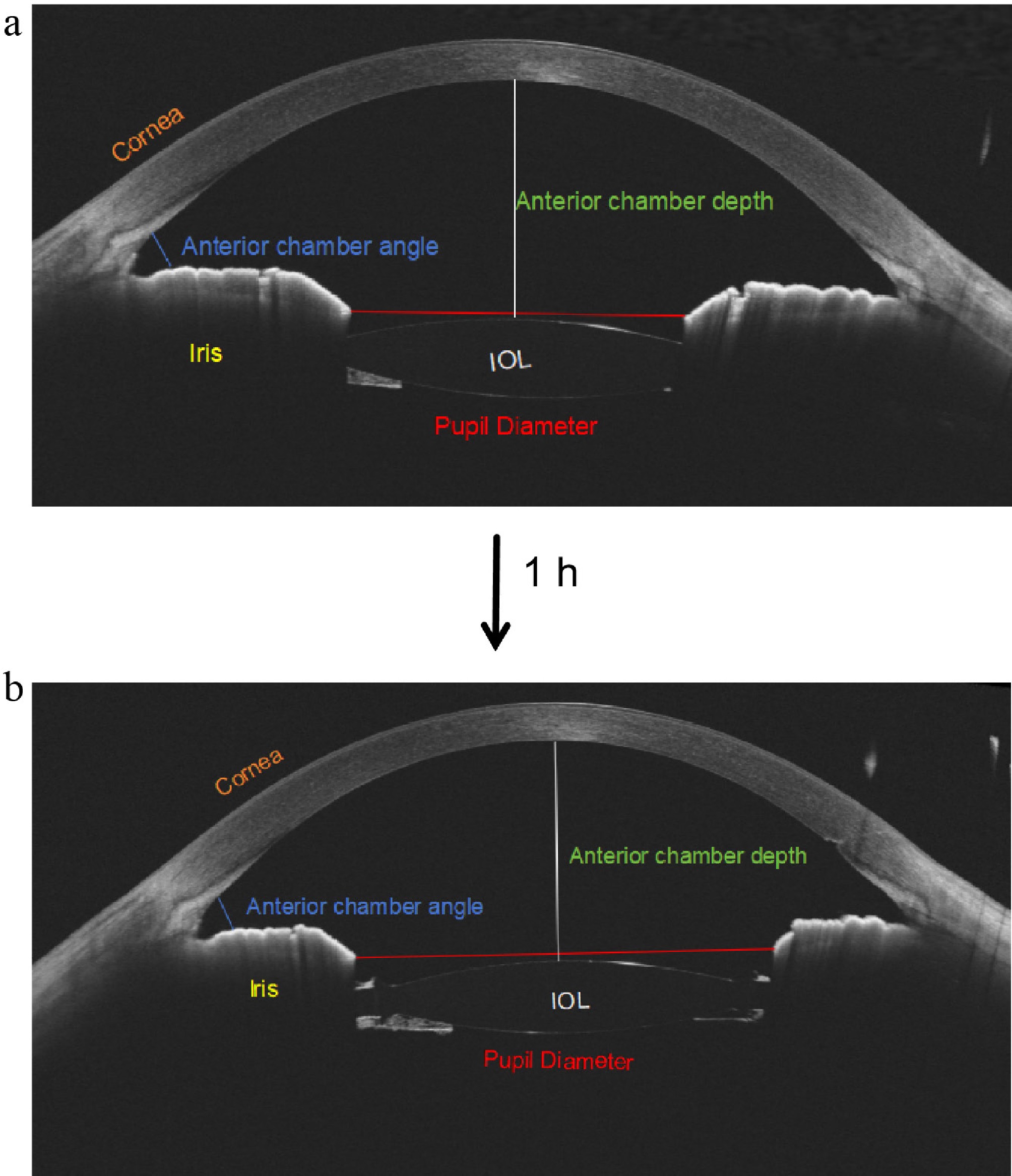
Figure 1.
Changes in the anterior structure of the AS-OCT image. (a) The AS-OCT parameters at baseline. (b) The pupil's diameter (red line) increased and the anterior chamber angle area (blue line) decreased slightly without changes in the other parameters after 1 h.
Posterior segment measurements
-
Posterior segment OCT and OCTA examinations were also performed using the same SS-OCT/OCTA device. The device has an axial resolution of approximately 5.0 μm, a transverse resolution of approximately 15 μm, and a scanning depth of approximately 3 mm. For the macular area, a 6 × 6-mm scanning range centered on the fovea was used with a raster scanning protocol of 1,024 × 1,024 B-scans. To reduce the impact of eye movement artifacts, the eye movement tracking function was enabled during the examination. Images with insufficient signal quality, noticeable motion artifacts, or those that could not be layered because of poor subject cooperation were not included in the analysis. Automatic layer segmentation and quantitative analysis of the retinal and choroidal layers were performed by the van Gogh analysis software provided with the device. After automatic segmentation using a deep learning algorithm, researchers manually rechecked the accuracy of the layers' boundaries. When the layer was poorly defined or deviated from the visible anatomical boundary, the built-in layer editing tool of the software was used for manual correction before calculating the indicators. Projection artifacts in the OCTA images were automatically processed by the built-in algorithm of the device's system to reduce the interference of superficial blood vessels on signals from deeper layers.
Before analysis, all images were individually corrected with the axial length (AL) of each subject. The magnification equation (magnification of the fundus measurement = 0.0492 × AL − 0.1818) was derived from the OCT system's optical configuration and the Navarro model[18]. All image assessments were conducted utilizing the integrated software. Because some children were young and still undergoing ocular growth, their AL fell below the fixed acquisition setting of the device. After correction with the individual AL, the effective imaging area captured by the 6 × 6-mm scan was reduced in some eyes. The actual image area was even smaller than 5 × 5-mm in a few cases because of a short AL (Fig. 2). To minimize edge effects and improve the accuracy of quantitative measurements, the early treatment diabetic retinopathy study (ETDRS)'s concentric ring partition (diameters of 1 and 3 mm, Fig. 2c) was used in the analysis of the macular area. The OCTA indicators included the foveal avascular zone (FAZ); retinal thickness (RT); macular GCC thickness; macular RNFL thickness; macular ganglion cell–inner plexiform layer (GCIPL) thickness; parameters related to the vessel density (VD) of the superficial, middle, and deep capillary plexus (SCP, MCP, and DCP); indicators related to perfusion defects in the choroidcapillary layer; and parameters related to choroidal structure. All parameters were exported for statistical analysis after quality control and necessary manual correction. Choroidal structures were extracted using adaptive threshold segmentation (the Niblack algorithm) with tailored pre- and postprocessing to accommodate varying vessel calibers and image contrast. Volumetric scan data were reconstructed in three dimensions to yield vascular lumens, after which the medium- and large-vessel layers (Sattler's and Haller's layers) were automatically identified to produce a choroidal vascularity map. From this map, the choroidal vascular volume (CVV) and the three-dimensional (3D) choroidal vascularity index (CVI, defined as CVV/choroidal total volume) were computed. The choroidal stromal volume (CSV) was derived as the total choroidal volume minus the CVV. Average choroidal thickness (ChT) was segmented automatically from structural OCT images with the basal boundary of the RPE to the choroid–sclera interface. All structural indicators were analyzed within the concentric ring zones of the ETDRS.
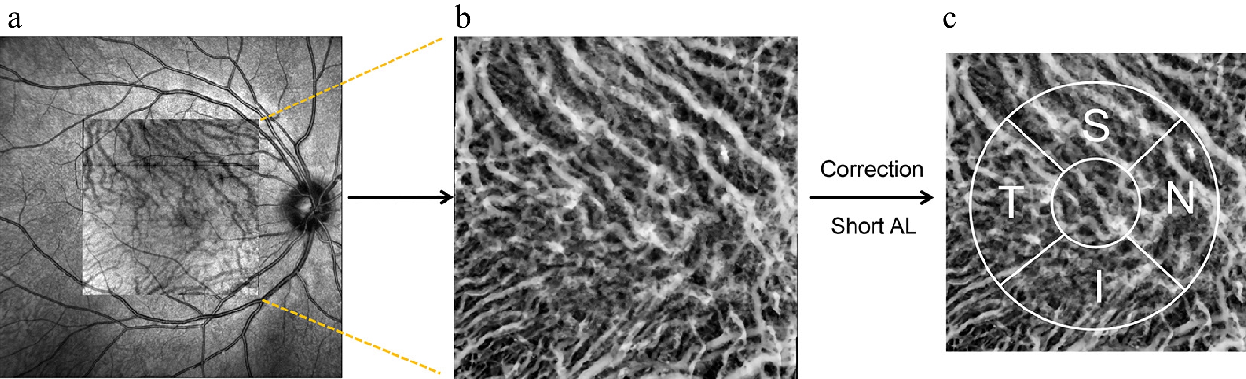
Figure 2.
Illustration of analyzing OCTA image parameters. (a) OCTA scan region of 6 × 6 mm. (b) Original en face OCTA image of the choroidal vascularity index image. (c) The 3-mm ETDRS grid on the magnified OCTA choroidal vascularity index image corrected with a short axial length (AL).
Statistical analysis
-
Statistical analysis utilized SPSS 26.0 software (IBM) and the results were illustrated using GraphPad Prism (GraphPad Software). Categorical data were evaluated using Fisher's exact test. Variables were expressed as the mean ± standard deviation, alongside the median and range. The data's normality was assessed with the Shapiro–Wilk test. Repeated analysis of variance (ANOVA) was used to compare continuous data among the three time points under normal distributions, whereas the Friedman test was used otherwise. Partial correlation analysis was also performed; p < 0.05 indicated statistical significance.
-
AI accurately segmented most images, and clinicians manually corrected the remaining details. This study comprised a total of 44 eyes (29 children; 20 males and 9 females) with a mean age of 10.52 ± 2.17 years, a mean SE of –3.47 ± 1.60 D, and a mean AL of 24.38 ± 1.68 mm. The demographic details are shown in Table 1.
Table 1. Demographic and clinical characteristics.
Variable Value (mean ± SD) Sex (%) Male (20, 68.96%) Female (9, 31.04%) Age (years) 10.52 ± 2.17 SE (D) −3.47 ± 1.60 AL (mm) 24.38 ± 1.68 IOP (mmHg) 15.88 ± 1.71 CCT (mm) 0.53 ± 0.14 ACD (mm) 4.24 ± 0.40 PD (mm) 4.38 ± 0.74 SE, spherical equivalent; AL, axial length; IOP, intraocular pressure; CCT, central corneal thickness; ACD, anterior chamber depth; PD, pupil diameter. Table 2 summarizes the AS-OCT parameters at baseline, 1 and 24 h after instillation of 0.01% atropine. The IOP increased from 15.88 ± 1.71 mmHg to 16.73 ± 2.03 mmHg at baseline and 1 h (p < 0.001, Table 2) but decreased to that of the baseline at 24 h. At 1 h, PD increased significantly (from 4.38 ± 0.74 mm to 5.12 ± 0.84 mm, p < 0.001, Table 2). At 24 h, PD decreased relative to the 1-h measurement but remained slightly smaller than that at the baseline (p = 0.032, Table 2). The ACD showed a small and nonsignificant decrease after administration (p > 0.05, Table 2), indicating no measurable shift in the IOL's position. Nasal and temporal anterior chamber angle areas showed significant changes over time (both p < 0.05, Table 2), reaching their lowest values at 1 h and returning to near-baseline levels at 24 h. Only the temporal values differed significantly between 1 and 24 h (p < 0.01, Table 2). The corneal thickness also showed no significant changes following administration of atropine (p > 0.05, Table 2).
Table 2. Comparative analysis of the anterior segment parameters across three time points.
Variable Mean ± SD P P1 P2 P3 Baseline 1 h 24 h IOP (mmHg) 15.88 ± 1.71 16.73 ± 2.03 15.70 ± 2.06 < 0.001*** 0.002** 0.614 0.004** CCT (mm) 0.53 ± 0.14 0.52 ± 0.17 0.53 ± 0.14 0.331 0.641 0.065 0.550 ACD (mm) 4.24 ± 0.40 4.20 ± 0.39 4.21 ± 0.40 0.526 0.448 0.567 0.875 PD (mm) 4.38 ± 0.74 5.12 ± 0.84 4.10 ± 0.87 < 0.001*** < 0.001*** 0.032* < 0.001*** TISA-N(mm2) 0.23 ± 0.08 0.20 ± 0.07 0.23 ± 0.09 0.021* 0.079 0.987 0.060 TISA-T(mm2) 0.29 ± 0.14 0.24 ± 0.06 0.28 ± 0.09 0.020* 0.071 0.583 < 0.01** IOP, intraocular pressure; CCT, central corneal thickness; ACD, anterior chamber depth; PD, pupil diameter; TISA-N, trabecular iris space area – nasal; TISA-T, trabecular iris space area – temporal. P, p-value among the three groups; P1, p-value for the comparison between baseline and 1 h after receiving medication; P2, for the baseline versus 24 h after the medication; P3, for 1 h versus 24 h after medication. * p < 0.05. ** p < 0.01, *** p < 0.001. Repeated-measures ANOVA or the Friedman test was used, as appropriate. Table 3 summarizes the retinal thickness and neural parameters at baseline, 1 h, and 24 h after instillation of 0.01% atropine. The RT showed its highest value in the superior region at baseline (343.76 ± 18.11 μm). The GCC's thickness peaked in the inferior region (121.56 ± 8.49 μm). The RNFL's thickness was highest in the inferior region (29.89 ± 3.77 μm). The GCIPL's thickness showed the highest value in the nasal region (92.68 ± 8.13 μm). After administration of the drug, RT showed no significant changes from baseline to 1 or 24 h in either the foveal or parafoveal regions (all p > 0.05, Table 3). Similarly, the GCC's thickness did not show significant differences at any time point in the foveal or parafoveal regions (all p > 0.05, Table 3). The RNFL's thickness also remained stable across all regions after administration of the drug, without significant differences from the baseline (all p > 0.05, Table 3). The GCIPL's thickness showed no significant changes in the foveal or parafoveal regions (all p > 0.05, Table 3). Overall, no significant short-term changes were observed in retinal neural structural parameters after 0.01% atropine instillation.
Table 3. Retinal thickness and retinal neural parameters at baseline, 1 and 24 h after atropine instillation.
Variable Mean ± SD P P1 P2 P3 Baseline 1 h 24 h RT (µm) Fovea 260.72 ± 19.85 261.09 ± 20.69 261.70 ± 21.51 0.653 0.887 0.746 0.894 Para-S 343.76 ± 18.11 344.30 ± 18.42 342.08 ± 22.59 0.551 > 0.999 > 0.999 0.907 Para-T 327.80 ± 15.58 328.15 ± 16.00 326.58 ± 19.58 0.328 0.723 0.497 > 0.999 Para-I 337.19 ± 16.13 337.29 ± 15.98 335.37 ± 20.92 0.649 > 0.999 > 0.999 > 0.999 Para-N 342.30 ± 21.70 342.47 ± 21.74 340.24 ± 25.72 0.978 > 0.999 > 0.999 > 0.999 GCC (µm) Fovea 43.14 ± 8.63 42.81 ± 8.64 44.00 ± 10.29 0.853 > 0.999 > 0.999 > 0.999 Para-S 121.49 ± 10.48 121.58 ± 11.16 120.37 ± 13.67 0.913 > 0.999 > 0.999 > 0.999 Para-T 107.04 ± 9.01 106.82 ± 9.01 107.45 ± 9.39 0.138 0.758 0.403 0.149 Para-I 121.56 ± 8.49 121.48 ± 8.06 121.76 ± 8.82 0.570 0.970 0.750 0.555 Para-N 117.85 ± 9.36 118.15 ± 9.46 118.26 ± 9.79 0.496 0.784 0.618 0.784 RNFL (µm) Fovea 14.74 ± 1.39 14.47± 1.38 14.21 ± 1.51 0.178 > 0.999 0.264 0.407 Para-S 29.21 ± 3.89 29.82 ± 3.44 29.48 ± 3.57 0.328 > 0.999 0.723 0.497 Para-T 19.60 ± 1.92 19.61 ± 1.62 19.66 ± 1.72 0.960 > 0.999 0.964 0.973 Para-I 29.89 ± 3.77 30.19 ± 2.82 30.30 ± 3.74 0.913 > 0.999 > 0.999 > 0.999 Para-N 24.91 ± 2.75 25.30 ± 2.34 25.31 ± 3.76 0.744 > 0.999 > 0.999 > 0.999 GCIPL (µm) Fovea 28.40 ± 8.06 28.34 ± 8.21 29.78 ± 9.72 0.136 0.972 0.303 0.269 Para-S 91.91 ± 7.98 91.76 ± 8.40 92.11 ± 8.55 0.084 > 0.999 0.210 0.129 Para-T 87.34 ± 7.85 87.21 ± 7.99 87.74 ± 8.09 0.394 0.951 0.660 0.268 Para-I 91.37 ± 6.51 91.28 ± 6.23 91.52 ± 6.97 0.808 0.959 0.945 0.873 Para-N 92.68 ± 8.13 92.85 ± 8.03 92.99 ± 8.23 0.494 > 0.999 > 0.999 0.723 RT, retinal thickness; GCC, ganglion cell complex; RNFL, retinal nerve fiber layer; GCIPL, ganglion cell–inner plexiform layer; Para-S, parafovea superior; Para-I, parafovea inferior; Para-N, parafovea nasal; Para-T, parafovea temporal. P, p-value among the three groups; P1, p-value for the comparison between the baseline and 1 h after receiving the medication; P2, p-value for the baseline versus 24 h after receiving the medication; P3, p-value for 1 h versus 24 h after receiving the medication. p < 0.05 was considered statistically significant. Repeated-measures ANOVA or the Friedman test was used, as appropriate. We subdivided macular retinal vascular density into three layers (superficial, middle, and deep) for detailed analysis. The SCP showed its highest VD in the superior region at baseline (46.24% ± 9.05%). The MCP's VD peaked in the temporal region (36.51% ± 6.32%). The DCP's VD showed the highest density in the temporal region (8.27% ± 5.66%). Before administration of the drug, superficial layers' vascular density exceeded deep layers' density across the superior, inferior, temporal, and nasal regions (all p < 0.05). After administration of the drug, SCP VD showed a slight decrease in the temporal region from the baseline to 1 h (41.10% ± 6.84% and 38.62% ± 7.04%, p = 0.031, Table 4); the other areas showed no significant changes (all p > 0.05, Table 4). The MCP's VD showed a slight decrease in the superior area from baseline to 24 h (31.29% ± 10.05% and 29.43% ± 10.53%, p = 0.046, Table 4), without significant differences in the other areas. The DCP's VD decreased from the baseline to 24 h only in the inferior area (5.65% ± 4.12% and 4.48% ± 3.20%, p < 0.05, Table 4). Additionally, there was no significant change in the FAZ area (p > 0.05, Table 4).
Table 4. Comparison of the vessel density of the superficial, middle, and deep capillary plexus at baseline, 1 and 24 h after receiving the medication.
Variable Mean ± SD P P1 P2 P3 Baseline 1 h 24 h SCP VD (%) Fovea 9.58 ± 3.57 9.18 ± 3.38 9.29 ± 4.06 0.605 0.632 0.683 0.974 Para-S 46.24 ± 9.05 45.05 ± 9.54 44.78 ± 10.34 0.695 > 0.999 > 0.999 > 0.999 Para-T 41.10 ± 6.84 38.62 ± 7.04 39.43 ± 7.50 0.090 0.031* 0.366 0.774 Para-I 45.54 ± 8.39 44.47 ± 7.47 44.33 ± 8.39 0.695 > 0.999 > 0.999 > 0.999 Para-N 42.78 ± 8.67 41.29 ± 8.35 41.84 ± 9.60 > 0.999 > 0.999 > 0.999 > 0.999 MCP VD (%) Fovea 19.66 ± 5.72 19.09 ± 6.00 19.06 ± 7.17 0.742 0.784 0.751 0.999 Para-S 31.29 ± 10.05 30.79 ± 10.55 29.43 ± 10.53 0.052 0.770 0.046* 0.592 Para-T 36.51 ± 6.32 35.25 ± 6.67 34.97 ± 7.33 0.595 > 0.999 > 0.999 > 0.999 Para-I 30.86 ± 7.47 28.78 ± 8.27 29.19 ± 9.24 0.468 0.723 > 0.999 > 0.999 Para-N 36.19 ± 7.25 34.42 ± 6.78 34.04 ± 10.46 0.270 0.723 0.367 > 0.999 DCP VD (%) Fovea 2.66 ± 2.41 2.41 ± 2.28 2.74 ± 3.04 0.928 > 0.999 > 0.999 > 0.999 Para-S 7.02 ± 4.44 7.45 ± 4.61 6.45 ± 4.42 0.533 > 0.999 > 0.999 0.836 Para-T 8.27 ± 5.66 7.87 ± 5.08 7.80 ± 4.82 0.740 0.834 0.775 0.993 Para-I 5.65 ± 4.12 4.93 ± 3.59 4.48 ± 3.20 0.014* 0.330 0.012* 0.603 Para-N 7.95 ± 4.79 7.00 ± 4.74 7.23 ± 4.38 0.408 0.548 > 0.999 > 0.999 FAZ (mm2) 0.31 ± 0.11 0.32 ± 0.11 0.32 ± 0.12 0.744 > 0.999 > 0.999 > 0.999 SCP VD, superficial capillary plexus vessel density; MCP VD, middle capillary plexus vessel density; DCP VD, deep capillary plexus vessel density; Para-S, parafovea superior; Para-I, parafovea inferior; Para-N, parafovea nasal; Para-T, parafovea temporal. FAZ, foveal avascular zone. P, p-value among the three groups; P1, p-value for the comparison between the baseline and 1 h after administration of the medication; P2, p-value for the baseline versus 24 h after administration of the medication; P3, p-value for 1 h versus 24 h after administration of the medication. * p < 0.05. Repeated measures ANOVA or the Friedman test was used, as appropriate. Table 5 shows the changes in the choroidal parameters at baseline, 1 and 24 h after medication. The average ChT decreased at 1 h and returned to baseline by 24 h in the inferior, temporal, and nasal areas (all p < 0.05, Table 5). There were no significant differences for the blood flow of the choriocapillaris at the three time points in all areas (all p > 0.05, Table 5). Before medication, the CVV showed the highest value in the temporal area, but there was no significant difference among all the areas (p > 0.05). The values of CSV were similar among the superior, inferior, temporal, and nasal areas. After administration of the drug, the CVV decreased at 1 h and then recovered at 24 h in the superior, inferior, and nasal areas (all p < 0.05, Table 5). There were no significant differences in the CSV at the three time points among most areas (all p > 0.05, Table 5), except in the temporal area. The CVI showed no significant differences in any of the areas (all p > 0.05, Table 5).
Table 5. Choroidal parameters at baseline, 1 and 24 h after atropine instillation.
Variable Mean ± SD P P1 P2 P3 Baseline 1 h 24 h ChT (µm) Fovea 325.00 ± 84.05 318.40 ± 77.40 323.10 ± 77.32 0.060 0.407 > 0.999 0.057 Para-S 305.00 ± 102.50 302.5 ± 101.60 302.80 ± 96.81 0.551 > 0.999 > 0.999 0.907 Para-T 334.30 ± 79.72 326.90 ± 73.14 331.70 ± 72.91 0.016* 0.129 > 0.999 0.017* Para-I 320.80 ± 88.28 310.50 ± 80.33 317.10 ± 83.66 0.032* 0.129 > 0.999 0.043* Para-N 299.70 ± 83.33 291.40 ± 78.54 294.10 ± 73.58 0.019* 0.023* > 0.999 0.099 CCF (mm2) Fovea 0.61 ± 0.05 0.60 ± 0.05 0.61 ± 0.03 0.529 > 0.999 0.859 > 0.999 Para-S 1.16 ± 0.12 1.14 ± 0.13 1.17 ± 0.10 0.542 > 0.831 > 0.872 > 0.534 Para-T 1.19 ± 0.09 1.16 ± 0.11 1.18 ± 0.08 0.815 > 0.999 > 0.999 > 0.999 Para-I 1.18 ± 0.11 1.18 ± 0.15 1.20 ± 0.25 0.983 > 0.999 > 0.999 > 0.999 Para-N 1.14± 0.10 1.11 ± 0.13 1.12 ± 0.16 0.431 > 0.999 0.723 0.859 CVV (mm3) Fovea 0.11 ± 0.04 0.10 ± 0.03 0.11 ± 0.04 0.073 0.087 > 0.999 0.295 Para-S 0.21 ± 0.06 0.21 ± 0.05 0.21 ± 0.06 0.049* 0.043* 0.859 0.497 Para-T 0.22 ± 0.07 0.21 ± 0.06 0.22 ± 0.07 0.060 0.407 > 0.999 0.057 Para-I 0.21 ± 0.08 0.20 ± 0.07 0.20 ± 0.08 0.029* 0.072 0.890 0.018* Para-N 0.19 ± 0.08 0.18 ± 0.07 0.19 ± 0.08 < 0.001*** 0.001** 0.493 0.011* CSV (mm3) Fovea 0.11 ± 0.03 0.11 ± 0.03 0.11 ± 0.03 0.420 0.661 > 0.999 0.934 Para-S 0.22 ± 0.05 0.22 ± 0.06 0.22 ± 0.04 0.649 > 0.999 > 0.999 > 0.999 Para-T 0.23 ± 0.06 0.23 ± 0.06 0.23 ± 0.05 0.049* 0.497 0.859 0.043* Para-I 0.22 ± 0.07 0.21 ± 0.06 0.22 ± 0.06 0.110 0.407 > 0.999 0.129 Para-N 0.20 ± 0.06 0.20 ± 0.06 0.19 ± 0.05 0.897 > 0.999 > 0.999 > 0.999 CVI Fovea 0.48 ± 0.05 0.48 ± 0.05 0.48 ± 0.05 0.788 0.839 0.995 0.831 Para-S 0.49 ± 0.05 0.48 ± 0.04 0.48 ± 0.05 0.443 0.388 0.768 > 0.839 Para-T 0.48 ± 0.05 0.48 ± 0.05 0.48 ± 0.04 0.728 0.758 0.988 0.812 Para-I 0.48 ± 0.06 0.48 ± 0.06 0.48 ± 0.05 > 0.999 > 0.999 > 0.999 > 0.999 Para-N 0.48 ± 0.06 0.48 ± 0.07 0.48 ± 0.07 0.090 0.081 0.992 0.208 ChT, choroidal thickness; CCF, choriocapillaris flow; CVV, choroidal vascular volume; CSV, choroidal stromal volume; CVI, choroidal vascularity index; Para-S, parafovea superior; Para-I, parafovea inferior; Para-N, parafovea nasal; Para-T, parafovea temporal. P, p-value among the three groups; P1, p-value for the comparison between baseline and 1 hour after receiving the medication; P2, p-value for baseline versus 24 h after receiving the medication; P3, p-value for 1 h versus 24 h after receiving the medication. * p < 0.05, ** p < 0.01,*** p < 0.001. Repeated measures ANOVA or the Friedman test was used, as appropriate. In the correlation analysis, AL was negatively correlated with the change in MCP VD between the baseline and 1 h and with the change in DCP VD between the baseline and 24 h both in the fovea (r = −0.318, p = 0.036, Fig. 3a and r = −0.314, p = 0.038, Fig. 3b, respectively). Regarding the changes in CVV, AL was only negatively correlated with that between the baseline and 1 h in the superior (r = −0.389, p = 0.009, Fig. 3c). The change in the PD between the baseline and 1 h were positively correlated with the change in CVV in the inferior and nasal areas and the CVI in the inferior area (r = 0.436, p = 0.004, Fig. 3d; r = 0.386, p = 0.012, Fig. 3e; and r = 0.440, p = 0.004, Fig.3f, respectively). There were no significant correlations between the changes in the PD and other parameters between the baseline and 24 h (all p > 0.05).
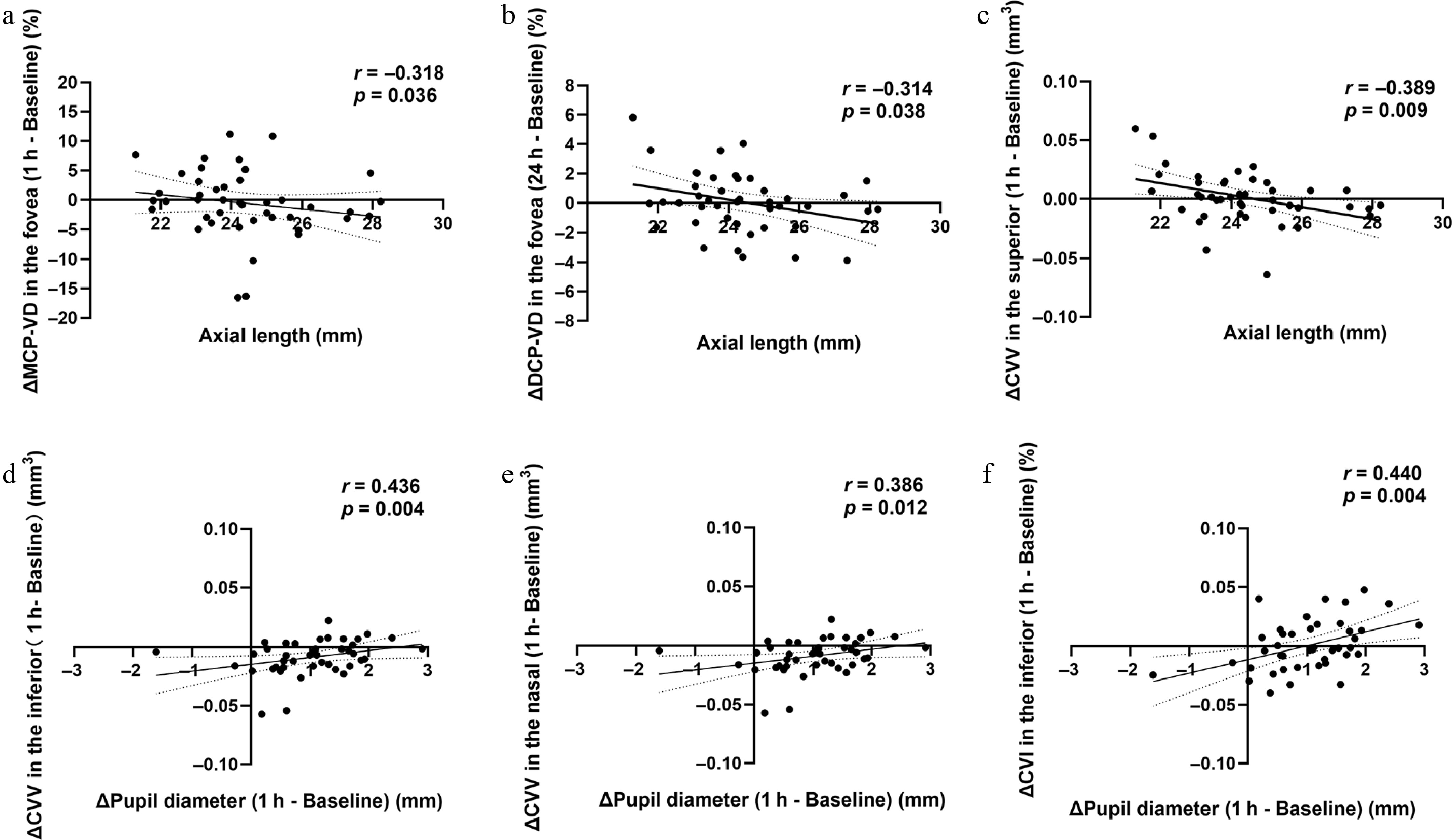
Figure 3.
The correlations of AL and the change in PD with the posterior parameters at 1 and 24 h. Correlations were assessed using partial correlation analysis, with p < 0.05 considered statistically significant.
-
The present study provides a comprehensive short-term evaluation of the anterior segment's structure and posterior neural and vascular parameters following a single instillation of 0.01% atropine in pseudophakic children using integrated AS-OCT and OCTA imaging. Overall, only mild and transient changes were observed, including a modest increase in PD and limited sector-specific fluctuations in several retinal and choroidal parameters, whereas most structural, neural and vascular indices remained stable within 24 h. These findings suggest that low-concentration atropine does not induce substantial short-term structural or hemodynamic disturbances in pseudophakic eyes and provide preliminary safety evidence supporting further investigations of atropine as a potential strategy for managing postoperative myopic progression in this special pediatric population.
Topical low-concentration atropine is widely regarded as one of the most effective pharmacologic interventions for slowing myopia's progression[19]. Establishing ocular safety is particularly important in pseudophakic children, in whom congenital cataract surgery alters the anterior segment's anatomy and long-term ocular development. In the present study, only mild and transient changes in the anterior segment were observed after the instillation of 0.01% atropine. Pupil diameter increased slightly at 1 h with a small reduction in the anterior chamber angle area, whereas other parameters such as the anterior chamber's depth and IOL position remained stable. A modest transient increase in intraocular pressure was also noted at 1 h but returned to baseline by 24 h. Mitsukawa et al.[8] used swept-source AS-OCT to evaluate healthy children and reported a significant increase in PD within 1 h after instillation of 0.01% atropine (0.83 mm), whereas other anterior segment parameters remained largely unchanged. In our cohort, the increase was smaller (0.74 mm), suggesting that the short-term mydriatic response is not enhanced in pseudophakic children. Together, these findings indicate that 0.01% atropine produces only limited short-term effects on the anterior segment's configuration in this population.
Anterior segment safety is particularly relevant because congenital cataract surgery is associated with an increased lifetime risk of secondary glaucoma[20]. Angle-closure mechanisms may occur early after surgery, whereas open-angle glaucoma typically develops later (approximately 7 years) and has been linked to abnormal angle development following early lens removal[21]. In phakic eyes, contraction of the longitudinal ciliary muscle fibers during accommodation facilitates aqueous outflow by exerting traction on the scleral spur[22]. Lens removal disrupts this biomechanical coupling, potentially modifying physiological outflow dynamics. Importantly, the modest pupil dilation induced by 0.01% atropine was not associated with iridotrabecular contact[23], and the intraocular pressure changes were transient, suggesting a low short-term risk of precipitating angle-closure glaucoma.
Consistent with previous reports, retinal and choroidal vascular parameters remained largely stable following instillation of atropine. Wang et al.[13] demonstrated that 0.01% atropine did not significantly alter the retinal vascular density, FAZ area, or choriocapillaris perfusion over a 3-month period, although a mild increase in choroidal thickness was observed. In contrast, we detected a slight, transient decrease in choroidal thickness at 1 h in the inferior, temporal, and nasal regions, which subsequently returned to the baseline by 24 h. Similarly, retinal vascular density showed only minor sector-specific reductions, including a decrease in temporal SCP VD at 1 h, and in superior MCP VD and the inferior DCP VD at 24 h. No significant differences were observed in the FAZ. Given their limited magnitude, regional restriction, and reversibility, these alterations are more likely to reflect transient physiological variability or mild pharmacologic responses rather than clinically meaningful effects[24].
Retinal neural structural parameters are closely associated with myopia. The progression of myopia is associated with thinning of the retinal nerve fiber layer and ganglion cell-related layers[25]. In the present study, the average macular GCC, RNFL, and GCIPL thickness remained unchanged at both 1 and 24 h after a single instillation of 0.01% atropine. These findings suggest that 0.01% atropine did not induce detectable acute structural alterations in the inner macular retina of pseudophakic children, supporting its short-term neurostructural safety in this special population. Compared with the mild transient changes observed in the PD, the stability of these neural measurements indicates that the early ocular response to atropine may be more physiological and hemodynamic. This interpretation is consistent with a previous posterior segment-focused study of low-dose atropine, in which retinal vascular parameters were generally stable without obvious structural retinal damage. The direct evidence regarding ganglion cell-related layers after administration of atropine remains limited. The absence of detectable changes in RT, GCC, RNFL, and GCIPL in our study supports the short-term neuroretinal safety of low-dose atropine in pseudophakic children.
The discrepancy between our short-term findings and the mild choroidal thickening reported in long-term studies suggests that atropine may exert time-dependent effects on choroidal structures[26−28]. In addition, choroidal thickness is subject to circadian variation, with prior studies reporting a diurnal amplitude of approximately 30 μm and a decrease from 372.2 μm at 9:00 to 364.9 μm at 11:00[29]. The modest reduction observed in our study therefore likely reflects, at least in part, physiological diurnal fluctuation rather than a direct pharmacologic effect. Notably, previous work suggests that a change of approximately 28–38 μm is required to reach clinical significance[30], and the variations observed in our study were well below this threshold. From a microstructural perspective, choroidal perfusion remained largely preserved. No significant changes were observed in the flow of the choriocapillaris, whereas CVV in some areas exhibited mild transient reductions with recovery at 24 h, indicating stable overall vascular integrity. This pattern suggests that the change in volume of the choroid also shows a similar trend to that of the ChT, and the change in the vascular lumen directly leads to that in the ChT in the early stage[31]. Previous studies reported that in children with myopia, CVV and CSV generally decline as myopia progresses or axial length increases[15,32]. In adults with high myopia, choroidal volume typically decreases and may coincide with matrix component retraction, suggesting that changes in volume reflect the cumulative effects of long-term structural remodeling rather than short-term responses over hours[33]. We characterized choroidal volume in children with congenital cataracts to provide a novel reference for future myopia research in this population.
Correlation analysis further indicated that the baseline ocular characteristics may influence the magnitude of short-term vascular responses. Longer AL was associated with greater reductions in MCP VD (r = −0.318) and DCP VD (r = −0.314) in the fovea, as well as with decreased CVV in the superior region (r = −0.389). In addition, changes in PD were correlated with regional CVV variations. The positive correlation between changes in PD and CVV may reflect a shared response to atropine rather than a direct effect of pupil dilation. Topically applied 0.01% atropine can reach the posterior segment, where muscarinic blockade may influence choroidal vascular tone through autonomic and cholinergic pathways[12,34]. Although a mechanical effect related to iris redistribution cannot be excluded, an autonomic mechanism may be more plausible. However, as these correlations were region-specific and observed only at 1 h, the underlying mechanism remains speculative. Although these associations were modest and region-specific, they suggest that eyes with greater axial elongation may exhibit altered vascular responsiveness to atropine. Given the propensity for progressive axial elongation in pseudophakic children[35], these findings highlight the potential relevance of choroidal biomarkers in this population. Retinal and choroidal perfusion are closely related to the metabolic support and functional integrity of neural tissues, and growing evidence in myopia suggests that neural and microvascular alterations may coexist[36−38]. Our findings add a neurovascular dimension to the interpretation of early atropine-related ocular responses in pseudophakic myopic children. Specifically, after a single instillation of 0.01% atropine, we did not detect significant acute changes in the thickness of the GCC, RNFL, or GCIPL, whereas only mild and transient changes were observed in selected anterior segment and choroidal or microcirculatory parameters. This pattern suggests that the short-term ocular response to low-dose atropine in this special population is more likely to reflect limited physiological or hemodynamic adjustment than overt neuroretinal structural disturbance. Clinically, these findings provide preliminary reassurance regarding short-term ocular tolerability in pseudophakic children, in whom altered ocular anatomy and parental concern often complicate decisions about the use of atropine. Nevertheless, given the retrospective design and short observation period, further longitudinal studies are needed to determine whether repeated exposure to 0.01% atropine may have cumulative neurovascular effects over time.
This study has several limitations. First, the sample size is relatively small, which may limit the ability to detect very subtle effects. Given the relatively small sample size of this special population and the large number of sector-based outcome measures assessed, the possibility of chance findings cannot be excluded, and isolated or marginally significant results should therefore be interpreted with caution. In addition, both pseudophakic eyes of each child were included without an untreated control group, limiting the ability to distinguish atropine-specific effects from natural physiological variation. Second, the recorded follow-up period was short, so we could not capture the long-term physiological effects. For practical reasons, we did not include the 12-h time point as an intermediate research time point to further observe changes in the parameters. Third, OCTA's quantitative indicators are sensitive to factors such as image quality, noise levels, and signal intensity, so the level of cooperation of the enrolled children may also affect the results. Therefore, future studies should incorporate larger sample sizes and a longer time to validate the findings of this study further.
-
In conclusion, this study provides a comprehensive short-term evaluation of the anterior segment's structure and posterior neural and vascular parameters following a single instillation of 0.01% atropine in pseudophakic children. Most structural, neural, and vascular parameters remained stable over the 24-h observation period, although mild transient sector-specific changes were observed in some retinal and choroidal indices. These findings support the short-term ocular safety of low-concentration atropine in this population and provide a quantitative basis for future studies exploring its potential role in managing postoperative myopic progression.
-
The study was approved by the Zhongshan Ophthalmic Center Institutional Review Board (Approval No. 2026KYPJ057, approval date June 8, 2026) and was conducted in accordance with the tenets of the Declaration of Helsinki. The requirement for informed consent was waived by the same ethics committee due to the retrospective nature of the study and the use of de-identified data.
-
The authors confirm their contributions to the paper as follows: contributed to the study's conception and design, commented on previous versions of the manuscript: Chen J, Ni Y; material preparation and data collection: Jiang Z, Qin Y, Li X, Chen W; data analysis: Dong X, Wang J; writing the first draft of the manuscript: Jiang Z, Qin Y. All authors reviewed the results and approved the final version of the manuscript.
-
The datasets generated and analysed during the current study are available from the corresponding author on reasonable request.
-
The authors declare that they have no conflict of interest.
-
# Authors contributed equally: Zhihao Jiang, Yingyan Qin, Xiaoyan Li
- Copyright: © 2026 by the author(s). Published by Maximum Academic Press, Fayetteville, GA. This article is an open access article distributed under Creative Commons Attribution License (CC BY 4.0), visit https://creativecommons.org/licenses/by/4.0/.
-
About this article
Cite this article
Jiang Z, Qin Y, Li X, Dong X, Chen W, et al. 2026. Short-term neurovascular and structural changes induced by single-dose 0.01% atropine in pseudophakic myopic children. Visual Neuroscience 43: e041 doi: 10.48130/vns-0026-0039 

Short-term neurovascular and structural changes induced by single-dose 0.01% atropine in pseudophakic myopic children
- Received: 29 June 2026
- Revised: 12 July 2026
- Accepted: 15 July 2026
- Published online: 31 August 2026
Abstract: To investigate the short-term neurovascular and structural effects of a single instillation of 0.01% atropine in pseudophakic eyes of myopic children. This retrospective repeated measures study enrolled 29 children aged 6–12 years with 44 pseudophakic eyes. All eyes received a single topical instillation of 0.01% atropine. Swept-source optical coherence tomography (OCT)/OCT angiography (OCTA) imaging of the anterior and posterior segments was performed at baseline, 1 and 24 h. Anterior segment parameters, macular neural thicknesses, retinal vascular density, foveal avascular zone (FAZ), and choroidal structural and vascular parameters were analyzed. Intraocular pressure and axial length were also measured. Intraocular pressure and pupil diameter (PD) increased slightly at 1 h, whereas the anterior chamber angle area changed over time (all p < 0.05), with the parameters returning to baseline by 24 h. No significant changes were observed in anterior chamber depth or corneal thickness (both p > 0.05). Retinal vascular density remained largely stable, with a few isolated sector-specific differences. The ganglion cell complex, retinal nerve fiber layer, ganglion cell–inner plexiform layer, and FAZ showed no significant changes (all p > 0.05). The average choroidal thickness in the inferior, temporal, and nasal regions demonstrated a transient decrease at 1 h, with recovery by 24 h. Choroidal vascular volume (CVV) and stromal volume exhibited similar trends in certain regions, whereas the choroidal vascularity index (CVI) remained stable. The change in PD was positively correlated with changes in CVV and CVI in some regions (both p < 0.05). In conclusion, a single instillation of 0.01% atropine did not induce clinically significant adverse changes in the anterior segment's anatomy or posterior circulation parameters in pseudophakic children with myopia, suggesting favorable short-term safety in this population.
-
Key words:
- Pseudophakic eye /
- Low-concentration atropine /
- Neurovascular changes