


-
Primary retinitis pigmentosa (RP), otherwise referred to as blanket retinal degeneration, is a hereditary degenerative disease marked by a progressive loss of photoreceptors and pigment epithelium[1], which can eventually lead to decreased vision. There are more than 1.5 million RP patients in the world. RP is the most common hereditary retinal dystrophy. The global prevalence rate is about 1/4,000, and the domestic prevalence rate is 1/4,016 to 1/3,467[2]. The precise etiology and pathophysiology are however still unknown. As of right now, no authorized therapy exists. The effectiveness of new RP techniques, including cell transplantation, gene therapy, cytokine treatment, nutritional therapy, and hyperbaric oxygen therapy for retinal degenerative illnesses, is still limited[3].
The clinical diagnosis of this condition is currently based on the presence of night blindness and peripheral visual field defects, fundus lesions, traces of low-voltage electroretinogram, and progressive deterioration of these signs. Although certain genes can be utilized for molecular diagnosis, molecular diagnosis is typically not feasible due to the disease's extreme genetic variability[3].
The production of vision is comprised of three distinct phases: light propagation, light signal conversion, and nerve signal transduction. Light rays traverse the refractive medium of the eye, reaching the retina. There, they are received by receptor cells and converted into nerve signals. These signals are then transmitted to the visual center of the brain, where they are processed to form visual perception. In general, RP exhibits clear or mild vitreous opacity within the refractive medium. It has been demonstrated that RP itself does not directly cause corneal or aqueous humor abnormalities, and sometimes concurrent cataracts and vitreous opacities occur. Given the severity of the visual impairment experienced by patients with RP, in addition to the opacity of the lens and vitreous, which results in blurred vision and decreased vision, the consideration of nerve conduction obstruction for visual field defects, night blindness, abnormal color vision, decreased visual contrast, and nystagmus is imperative.
Recently, functional magnetic resonance imaging (fMRI) in the resting state has garnered increasing attention from the research community for its potential to elucidate ongoing neuronal processes[4]. As a reliable method for measuring correlation amplitude, the amplitude of low-frequency fluctuation (ALFF) has been proven to be a valuable technique for studying the intensity of spontaneous neural activity[5], and has been applied to the study of neurophysiological activity and diseases in different brain regions. This methodology facilitates the direct representation of blood oxygen level dependence (BOLD) through the calculation of the low-frequency amplitude of each unit. For instance, a reduction in the ALFF may indicate a decrease in BOLD signals within specific brain regions. From an energetic perspective, this approach is indicative of the degree of spontaneous neural activity across different areas of the brain during a resting state[6].
Research conducted to date has employed methodologies such as regional homogeneity (ReHo) and functional connectivity (FC) to examine alterations in both intra- and inter-regional functional connectivity among individuals diagnosed with RP. It is imperative to acknowledge that the ALFF method, and the ReHo method are founded on disparate mathematical principles. To date, there has been a paucity of research investigating the relationship between ALFF values and RP across different brain regions. In summary, can the visual impairment of RP patients reveal the underlying mechanism of neurological injury by studying the difference in ALFF values in different brain regions of the brain with healthy people? The objective of this study was to utilize the ALFF method to compare the intrinsic brain activities of patients with RP to those of healthy subjects. This approach was taken to explore the potential neural mechanisms underlying RP and to investigate the potential applications of the ALFF method in clinical diagnosis[7].
-
A total of 20 patients diagnosed with retinitis pigmentosa (12 males and eight females) were recruited from the Ophthalmology Department of the First Affiliated Hospital of Nanchang University (Nanchang, China). The inclusion criteria of RP patients were: (1) The patients in question were diagnosed with idiopathic RP and exhibited the following characteristics: osteoid pigmentation, retinal vascular constriction, and waxy pallor of the optic disc; (2) the range of RP was limited to no more than two quadrants; and (3) without any concomitant ophthalmological diseases (such as maculopathy, cataracts, optic neuritis, etc.). Exclusion criteria of RP in the study were: (1) recurrent after RP repairment operation; (2) RP secondary to trauma; (3) RP patients who had critical complications (such as posterior polar cataract, open angle glaucoma and so on); (4) RP patients had laser treatment or surgery previously; (5) RP patients with severe physical or mental diseases[8].
Twenty healthy controls (HCs, 12 males and eight females) were also enrolled, and were matched in sex, status, and age. The HCs were in accordance with the following criteria: (1) uncorrected visual acuity better than 1.0, and without any ophthalmological diseases; (2) no history of mental disorders and organic encephalopathy such as bipolar disorder and depression [for more information about the mental disorders program, please refer to the Chinese Mental Disorders and Diagnostic Criteria, Third Edition (CCMD-3); (3) ability to finish MRI scanning.
The study was conducted in accordance with the principles of the First Affiliated Hospital of Nanchang University's medical ethics committee. The research methods employed adhered to the principles outlined in the Declaration of Helsinki. All participants were provided with the informed consent form and gave their consent to participate in the study.
MRI parameters
-
The 3-Tesla MR scanner (Trio, Siemens, Munich, Germany) was utilized to perform MRI scanning. The acquisition of images was conducted using a spoiled gradient recalled echo pulse sequence, with the parameters previously delineated in a preceding study[9]. At last, 240 functional volumes were also acquired.
fMRI data analysis
-
The data was pre-filtered with DPARSFA (
http://rfmri.org/DPARSF )[10], Statistical Parametric Mapping software (SPM8,www.fil.ion.ucl.ac.uk/spm ), and MATLAB 2013a (Math Works, Natick, MA, USA). Initially, the first 10 volumes of each participant were deleted to obtain a balanced signal. Subsequently, the head motion was rectified and the linear regression method was employed to eradicate the interference effect. Subsequently, the data were standardized to align with the Montreal Neurological Institute (MNI) space criteria[11].ALFF analysis
-
To begin with, the data was smoothed with a Gaussian kernel of 6 mm × 6 mm × 6 mm full-width at half-maximum (FWHM). Secondly, the fMRI volumes were thoroughly detrended and band pass-filtered (0.01–0.08 Hz) to achieve more accurate data. Subsequently, the power spectrum of each voxel was obtained by means of a fast Fourier transform (FFT). In the final analysis, the mean ALFF values of the distinct regions were obtained[12].
Statistical analysis
-
To analyze the fMRI data, a two-sample t-test was conducted to evaluate the voxel-wise discrepancy between the RP and HCs employing the REST toolbox. This study was conducted at the State Key Laboratory of Cognitive Neuroscience and Learning, Beijing Normal University, Beijing, China (The statistical threshold was set at the voxel level with p < 0.05 for multiple comparisons using Gaussian random field theory voxels with p < 0.01 and cluster size > 40 voxels, GRF corrected)[13]. These voxels were identified as regions of interest, exhibiting substantial disparities between the two groups.
The receiver operating characteristic (ROC) curve was used to evaluate the mean ALFF of different brain regions of subjects, and the AUC was used to evaluate the accuracy of using the mean ALFF of a certain brain region to distinguish RP and HCs. The correlation between HADS and ALFF values in emotion-related brain regions in RP patients was evaluated by Pearson correlation analysis.
-
There was no significant difference in age (p = 0.90), or weight (p = 0.81). And the two groups showed significant differences in best-corrected VA-left eye (p = 0.009) and best-corrected VA-right eye (p = 0.007). Meanwhile, the mean duration of RP was 14.76 ± 4.87 years (Table 1).
Table 1. Demographics and clinical measurements between RP and HCs.
Condition RP HCs t p-value Male/female 12/8 12/8 N/A > 0.99 Age (years) 50.67 ± 6.22 50.46 ± 5.87 0.089 0.90 Weight (kg) 61.76 ± 6.43 60.21 ± 5.76 0.065 0.81 Handedness 20 R 20 R N/A > 0.99 Duration of RP (years) 14.76 ± 4.87 N/A N/A N/A Best-corrected Va-right eye 0.35 ± 0.15 1.05 ± 0.15 −0.532 0.007* Best-corrected Va-left eye 0.50 ± 0.20 1.05 ± 0.10 −0.672 0.009* IOP-R (mmHg) 17.76 ± 1.64 16.16 ± 1.56 0.079 0.72 IOP-L (mmHg) 16.28 ± 1.87 16.45 ± 2.05 0.067 0.80 * p < 0.05 Independent t-tests comparing two groups. Abbreviations: RP, retinitis pigmentosa; HCs, healthy controls; N/A, not applicable; R, right, L, left. ALFF differences
-
Brain partitioning was performed according to the anatomical automatic labeling (AAL) template. The MNI coordinate system was developed with the anterior commissure as the origin to delineate the positional relationships of each brain region. The X-axis, ranging from left to right, is designated as positive; the Y-axis, extending from back to front, is assigned as positive; and the Z-axis, from bottom to top, is labeled as positive. The highest ALFF value for each brain region was recorded, expressed as voxel peaks. In contrast to the findings in the HCs, the ALFF values of the RP were significantly higher in the cerebellum, right and left frontal gyrus, left precuneus, and left thalamus, but lower ALFF values were found in the left caudate and left lingual (see Fig. 1 and Table 2).
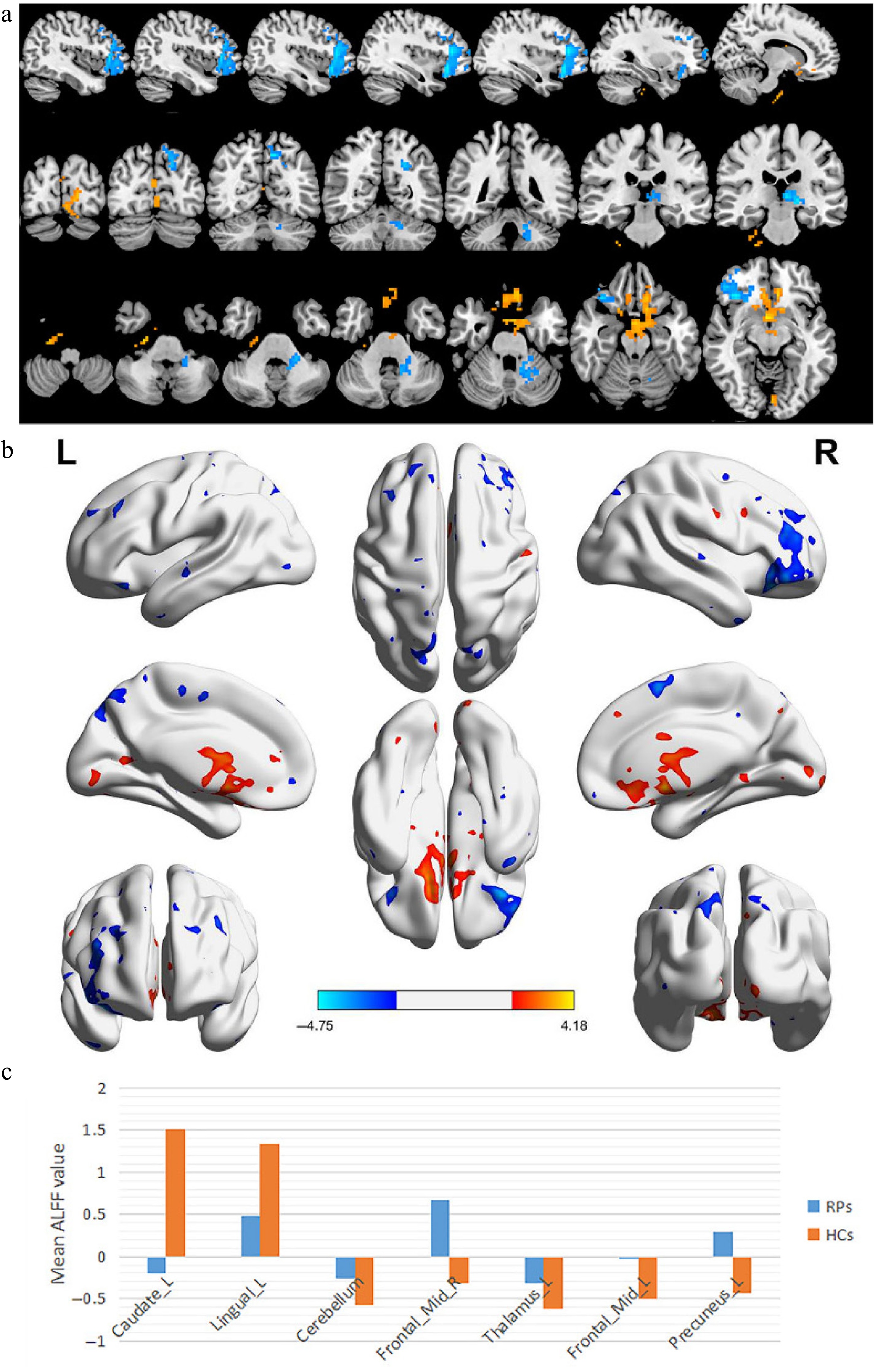
Figure 1.
(a), (b) Spontaneous brain activity in the RP and the HCs. Distinct differences of activity were detected in caudate L, lingual L, cerebellum, frontal mid R, thalamus L, frontal. Mid L, precuneus L. The red or yellow indicates higher ALFF values, while the blue indicates lower ALFF values. The significance level for multiple comparisons using Gaussian Random Field (GRF) theory is p < 0.01 (corrected by AlphaSim for clusters > 40 voxels, p < 0.01, z > 2.3). (c) The mean values of the altered ALFF values between the two groups. Abbreviations: ALFF, amplitude of low-frequency fluctuation; RP, retinitis pigmentosa; HCs, healthy controls; L, left; R, right.
Table 2. Brain regions with statistically significant ALFF values between RP and HCs.
Brain areas (AAL template) MNI coordinates BA Peak voxels T value ROI cluster number X Y Z HCs > RP (comparison of ALFF values) Caudate_L 0 3 −15 11 935 4.18 Cluster 3 Lingual_L −3 −81 −6 18 146 3.53 Cluster 5 HCs < RP (comparison of ALFF values) Cerebellum −21 −36 −36 129 −3.32 Cluster 2 Frontal_Mid_R 36 36 12 47 615 −4.75 Cluster 4 Thalamus_L −18 −24 0 80 −4.26 Cluster 6 Frontal_Mid_L −21 42 27 9 105 −3.53 Cluster 7 Precuneus_L −12 −63 48 7 135 −4.48 Cluster 8 ALFF, amplitude of low-frequency fluctuation; AAL, anatomical automatic labeling; BA, Brodmann area; RP, retinitis pigmentosa; HCs, healthy controls; MNI, Montreal Neurological Institute; L, left; R, right; B, bilateral. ROC curve
-
ROCs were employed to analyze the mean ALFF values of the different brain areas. The area under the ROC curve (AUC) demonstrated that the utilization of ALFF mean values in these brain regions to distinguish between RP and HCs exhibited a high degree of accuracy. The AUC was 0.784 (p = 0.003; 95% CI: 0.636−0.933) for Cerebellum, Frontal_Mid_R 0.893 (p < 0.0001; 95% CI: 0.791−0.995), Thalamus_L 0.866 (p < 0.0001; 95% CI: 0.753−0.979), Frontal_Mid_L 0.836 (p = 0.001; 95% CI: 0.709−0.963), Precuneus_L 0.872 (p < 0.0001; 95% CI: 0.755−0.989). The AUC was 0.869 (p < 0.0001; 95% CI: 0.743−0.995) for Caudate_L, Lingual_L 0.824 (p = 0.001; 95% CI: 0.676−0.973) (Fig. 2).
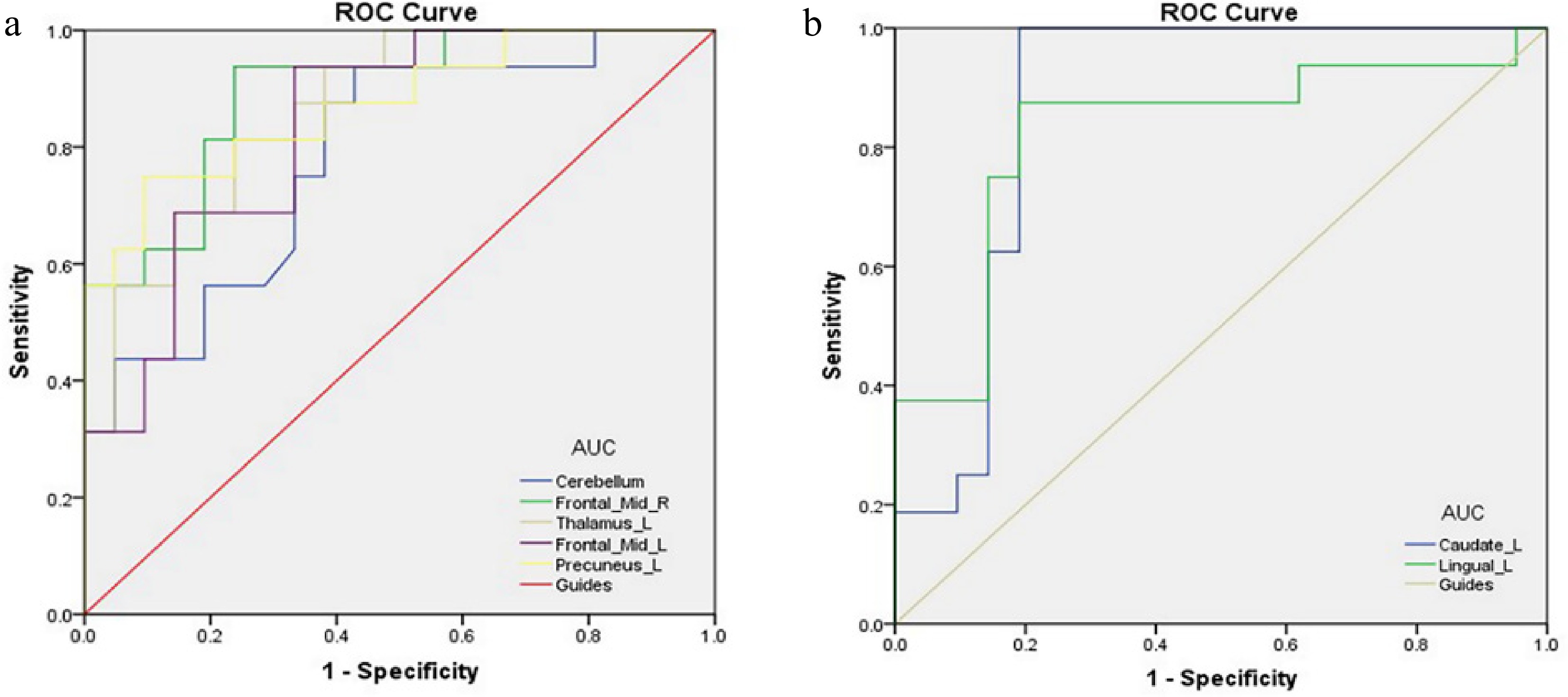
Figure 2.
ROC curve analysis of the mean ALFF values in the altered brain areas. Note: Only the average of ALFF for each brain region was included in this model. (a) ROC curve analysis of the mean ALFF value in brain areas where the mean ALFF value of the RP group was higher than the control group. The area under the ROC curve was 0.784 (p = 0.003; 95% CI: 0.636−0.933) for Cerebellum, Frontal_Mid_R 0.893 (p < 0.0001; 95% CI: 0.791−0.995), Thalamus_L 0.866 (p < 0.0001; 95% CI: 0.753−0.979), Frontal_Mid_L 0.836 (p = 0.001; 95% CI: 0.709−0.963), Precuneus_L 0.872 (p < 0.0001; 95% CI: 0.755−0.989). (b) ROC curve analysis of the mean ALFF value in brain areas where the mean ALFF value of the RP group was lower than the control group. The area under the ROC curve was 0.869 (p < 0.0001; 95% CI: 0.743−0.995) for Caudate_L, Lingual_L 0.824 (p = 0.001; 95% CI: 0.676−0.973). Abbreviations: ROC, receiver operating characteristic; ALFF, amplitude of low-frequency fluctuation; CI, confidence interval; HCs, healthy controls; RP, retinitis pigmentosa.
-
To date, there exists a limited number of studies examining the impact of RP on cerebral activity utilizing the ALFF methodology. In the current study, RP demonstrated significantly reduced ALFF values in the caudate nucleus and the lingual gyrus when compared to HCs. Conversely, there was a notable increase in ALFF values in the left thalamus, left precuneus, bilateral middle frontal gyrus, and the cerebellum. The functional roles of the brain regions exhibiting these signal alterations are detailed in Table 3. The fundus photography of RP is shown in Fig. 3. The majority of physiological activities within the human body are facilitated through the coordinated interaction of various brain regions, each designated for specific functions. While the occipital lobe plays a critical role in visual processing, it is important to note that regions of the parietal, temporal, and frontal lobes in both hemispheres also contribute to the intricate processing of visual stimuli. The functions of the cerebral hemispheres on both sides are asymmetrical. The right hemisphere predominantly governs the visual, tactile, and motor functions associated with the left side of the body, while the left hemisphere is responsible for similar functions on the right side. In instances where both hemispheres engage in executing complex tasks, one hemisphere, referred to as the dominant hemisphere, typically assumes a leading role. For example, the left hemisphere is the dominant hemisphere of language function, while the right hemisphere is dominant in spatial imagination. This may explain why the results of this study show certain brain regions on the left or right[14].
Table 3. Brain region alternation and its potential impact.
Brain regions Experimental result Brain function Anticipated results Caudate_L RP < HCs The part of the extrapyramidal system Dystonia, chorea Lingual_L RP < HCs Visual processing, word processing Visual impairment, aphasia Cerebellum RP > HCs Regulate body balance, regulate muscle tension, coordinate random movement Cerebellar ataxia, rotational movement disorder, nystagmus, imbalance and standing instability Frontal_Mid_R RP > HCs Controls spontaneous eye movement, part of the default model network Mental disorders, including depression and anxiety Thalamus_L RP > HCs Conduction pathways of various senses in the whole body Disturbance of consciousness and sleep, disturbance of thermoregulation, disturbance of endocrine metabolism, disturbance of circulation and respiration, disturbance of digestive system, and local neurological signs Frontal_Mid_L RP > HCs Controls spontaneous eye movement, part of the default model network Mental disorders Precuneus_L RP > HCs Cognitive function, episodic memory, self-related information processing Cognitive and affective disorders RP, retinitis pigmentosa; HCs, healthy controls. 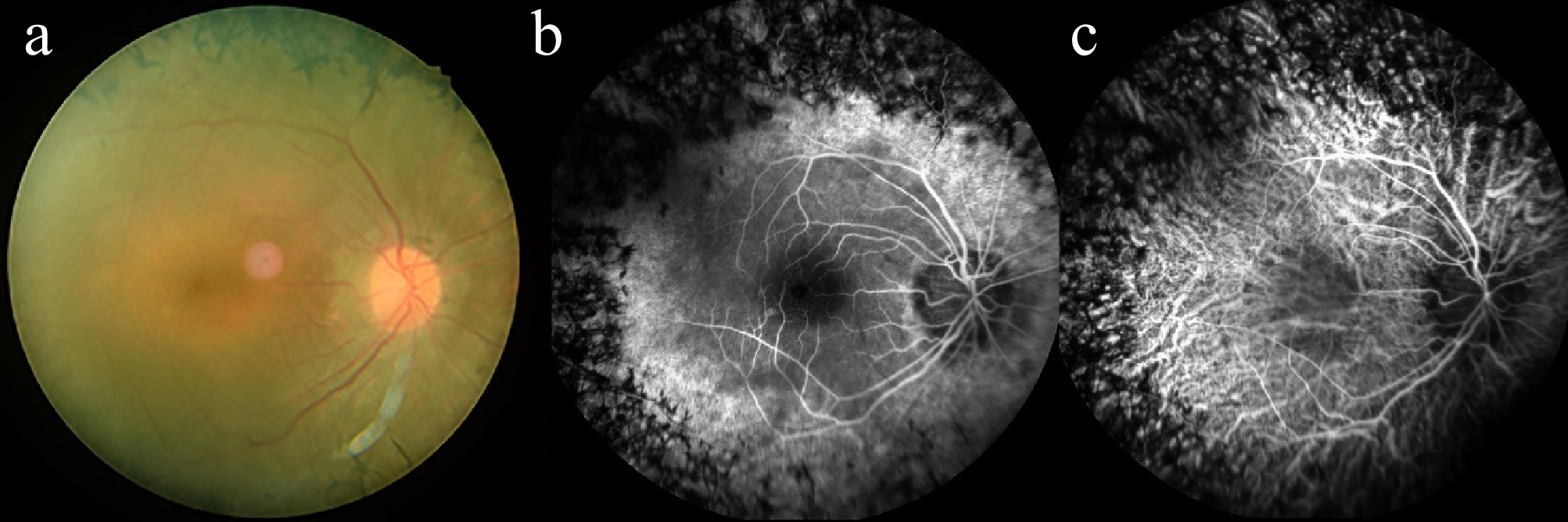
Figure 3.
Example of retinitis pigmentosa seen on (a) fundus camera, (b) fluorescence fundus angiography, and (c) indocyanine green angiography.
Analysis of the decreased ALFF values in RP
-
The caudate nucleus constitutes a component of the striatum and is characterized by its horseshoe shape, extending along the entire length of the lateral ventricle[15]. The caudate nucleus is an important part of the learning and memory system. It not only acts as the whip chain outside the cone, which regulates the body movement, but also has an impact on the sensory conduction process. Anatomical data also confirmed that there were direct projections from the head of the caudate nucleus to the neocortex, cingulate diencephalon, and brainstem[15], which means that the caudate nucleus may play an important role in the regulation of sensory conduction.
In this study, it was found that RP exhibited a marked reduction in ALFF within the caudate nucleus. Given the caudate's critical role in motor function and sensory processing, a decrease in ALFF may indicate a disruption in sensory conduction pathways. Consequently, we hypothesize that RP may experience deficits in sensory conduction as a result of these alterations.
The lingual gyrus is a bilateral brain structure, with one located in the left hemisphere and the other in the right hemisphere. It plays a crucial role in visual processing, particularly in the interpretation of letters[16]. In addition, it is believed that the lingual gyrus may also participate in the processing of logical analysis and visual memory[16]. In the present research, RP exhibited reduced ALFF in the left lingual gyrus. This finding suggests a potential correlation between RP and impairments in visual processing functions.
Analysis of the increased ALFF values in RP
-
The thalamus, also known as the dorsal thalamus, is the largest oval gray matter nucleus in the diencephalon, which is located in both sides of the third ventricle. The left and right thalamus are connected by gray matter mass (called intermediate mass)[17]. The thalamus serves as the most sophisticated sensory center and functions as a critical relay station for sensory conduction. It is the site where all sensory pathways, with the exception of olfactory pathways, synapse before transmitting information to the cerebral cortex. Additionally, there are neural connections established between the thalamus, hypothalamus, and striatum, which contribute to the formation of intricate subcortical centers responsible for unconditioned reflexes. The thalamus serves as a crucial area for the integration of neural activities, receiving and transmitting information from various cortical inputs and outputs[18]. The thalamus plays a pivotal role in regulating the transmission of information to the cortex. It is involved in the process of visual perception and the dynamic processing of visual information to the motor center via the retinothalamocortical pathways[19]. The findings of the current study indicate that the observed increase in ALFF suggests that RP may contribute to thalamic dysfunction. This observation suggests a potential association between the presence of these resting-state patterns and challenges in visual perception and information processing. Furthermore, research findings indicated the presence of intrinsic visual network disconnection and reorganization of the retino-thalamocortical pathway in the subject. This observation suggests the potential for impairment in visuospatial and stereoscopic vision[20].
The precuneus, located in the front of the cuneus, is involved in many functions, including visuospatial imagery[21], attention[22], episodic memory[23], and the default-mode network[24]. A prior study indicated that the precuneus is significantly involved in the encoding of spatial locations[25]. In addition, the precuneus was also involved in motor management, which transfers the motor signal to supplementary motor area[26]. Meanwhile, the precuneus was in charge of spatial working memory[27]. In our study, we observed that RP showed higher ALFF in the left precuneus, which may reflect the dysfunction of visuospatial imagery in RP.
The middle frontal gyrus is defined as a distinct anatomical region of the cerebral cortex. This specific brain location of the brain is responsible for the processing of higher information, a characteristic that is generally specific to cortical matter[28]. Furthermore, the middle frontal gyrus is thought to be associated with mental and physical balance. Carter et al.[29] employed fMRI to simultaneously monitor and postpone the acquisition of explicit knowledge within a conditioned reflex framework. Their experiments indicated that activity in the frontal gyrus was associated with the accuracy of clear emergency awareness in each trial[30]. In the current study, it was observed that the ALFF values were elevated in both the left and right middle frontal gyri. This finding suggests that as the duration and progression of the disease advance, the impairment of the left middle frontal gyrus in RP becomes more pronounced. Additionally, a positive correlation was identified between the signal values of the right middle frontal gyrus and anxiety scores. The substantial rise in signal values of the right middle frontal gyrus among RP subjects suggests a heightened propensity for irritability and anxiety. Thus, these results may provide some insight into the emotional fluctuations experienced by individuals with RP, see Fig. 4 for details.
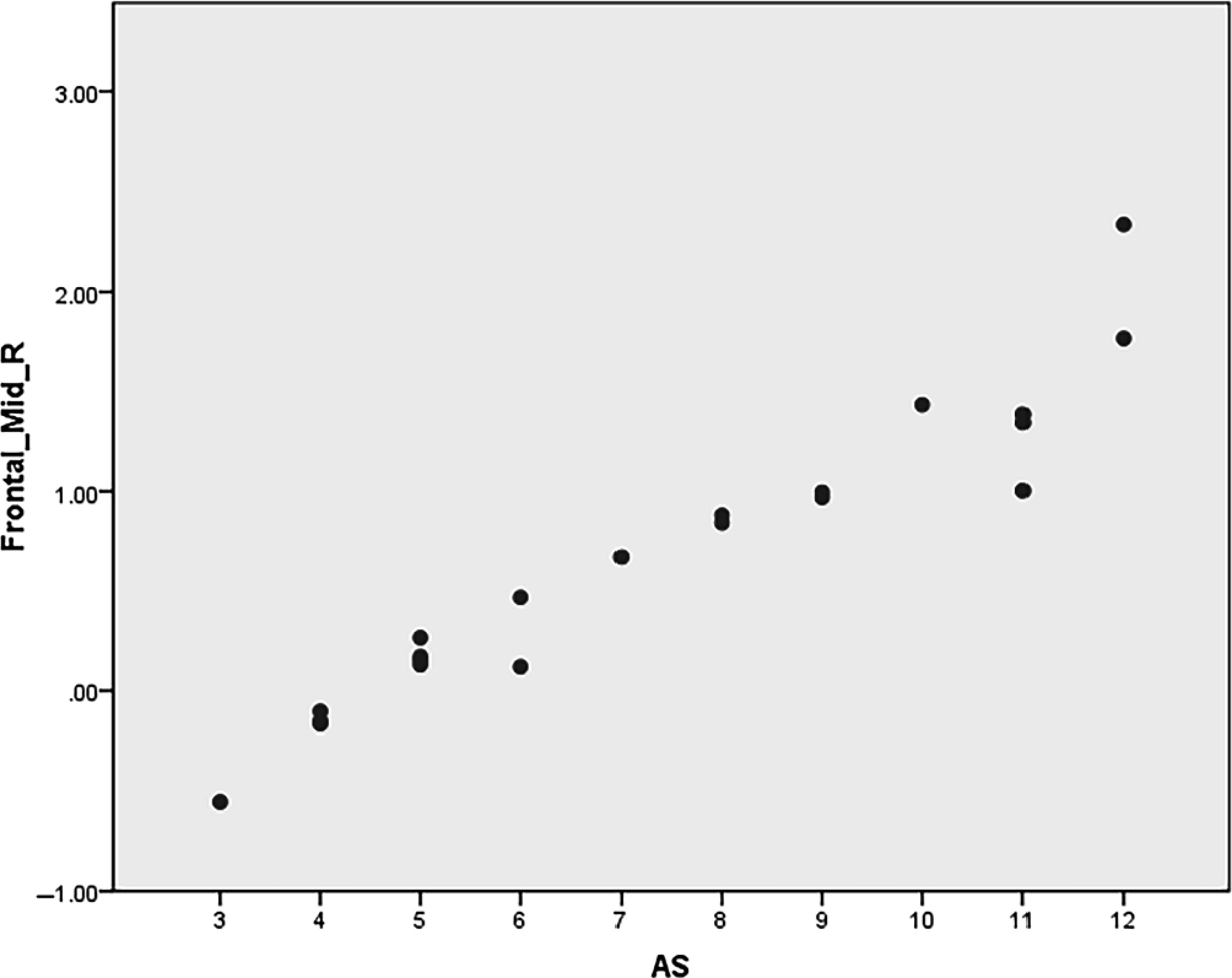
Figure 4.
The signal value of right middle frontal gyrus in RP was positively correlated with anxiety score. RP, retinitis pigmentosa; AS, anxiety score.
The cerebellum is an important center for motor regulation, with a large number of afferent and efferent connections. The movement of information from the cerebral cortex to the muscles and from the muscles and joints can be transmitted to the cerebellum. The cerebellum frequently synthesizes these two types of neural impulses and modulates the activity of associated muscles via efferent fibers, thereby ensuring the coordination of involuntary movements[31]. A prior study has examined alterations in both intra- and inter-regional FC among RP, employing ReHo and functional connectivity methodologies[32]. The ALFF and ReHo methods have been demonstrated to be effective tools for investigating intrinsic brain activity through BOLD signals. However, it is important to note that they exhibit distinct mathematical characteristics. ALFF quantifies the aggregate power of BOLD signal fluctuation within a designated low frequency range at a specific voxel resolution. ReHo is a method of detecting the similarity of the BOLD signal time series of a given voxel and its neighborhood. This provides important information for regional time synchronization[10]. In the previous study, RP exhibited markedly lower ReHo values in the bilateral lingual gyrus and the posterior lobe of the cerebellum (LGG/CPL) when compared to HCs. This finding indicates a diminished synchronicity of neural activity fluctuations within the primary visual cortex among individuals with RP. In the present study, a higher ALFF value has been observed in the cerebellum. The increase in ALFF in the cerebellum of patients with RP, as demonstrated in the present study, may serve as an indicator of functional impairment in this region. The cerebellum plays an important role in motor control and perception. Consequently, we hypothesized that this might result in motor control impairment in RP.
In our previous research, we explored the amplification of low-frequency fluctuations method applied in ophthalmological diseases[10,12,13,20,33]. Details are shown in Table 4. The results of brain activity in RP are shown in Fig. 5.
Table 4. Amplitude of low-frequency fluctuations method applied in ophthalmological diseases.
Author Year Disease Brain areas UDs > NCs UDs < NCs Kang et al.[33] 2019 Retinal detachment FSO, ITG OL, MFG Shi et al.[10] 2020 Vitreous hemorrhage RCPL, LCPL RMFG, RIFG, LMGF, RSFG, RMFG, LMFG Lin et al.[12] 2020 Retinitis pigmentosa RCPL, LITG, RFG RMFG, BC, BP, BSFG Ling et al.[13] 2021 Hyperthyroidism exophthalmos No brain region LCFSC Peng et al.[20] 2021 Neovascular glaucoma RSFG, LMFG RC, RMOG, LCG, RP, LMFG UD, unusual disease; NCs, normal controls; LCPL, left cerebellum posterior lobe; LCG, left cingulate gyrus; LCFSC, left calcarine fissure and surrounding cortex; LMFG, left medial frontal gyrus; RC, right cuneus; RMOG, right middle occipital gyrus; RSFG, right superior frontal gyrus; RIFG, right inferior frontal gyrus; BP, bilateral precuneus; RMFG, right middle frontal gyrus; RSFG, right superior frontal gyrus; RP, right precuneus; RC, right cuneus; FSO, frontal superior orbital; ITG, inferior temporal gyrus; OL, occipital lobe; MFG, medial frontal gyrus; LITG, left inferior temporal gyrus; BC, bilateral cuneus; BSFG, bilateral superior frontal gyrus. 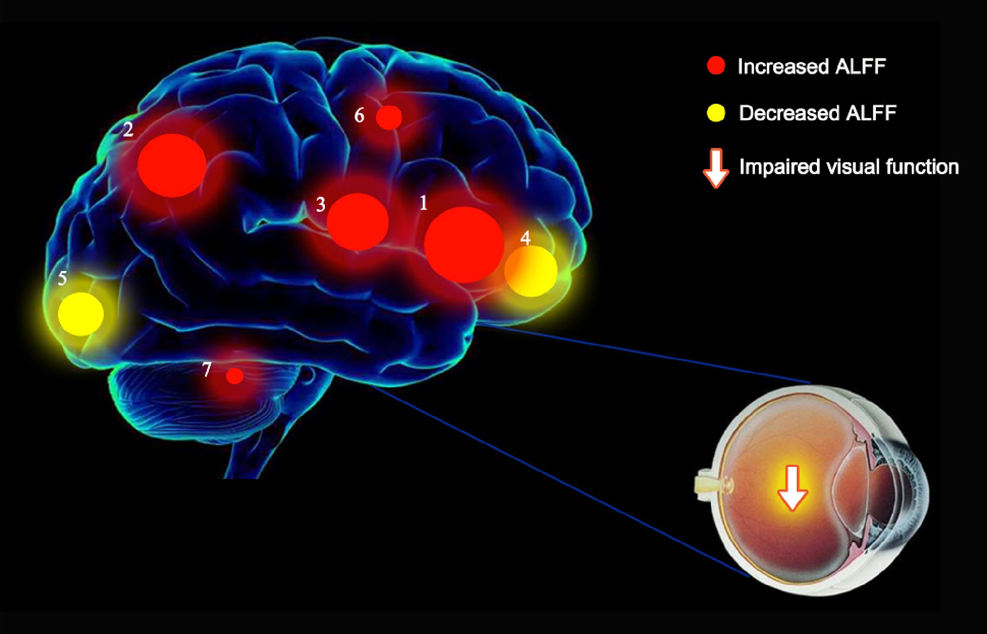
Figure 5.
The retinitis pigmentosa results of brain activity. Compared with the HCs, the ALFF values of the following regions were decreased to varying extents: 4: Caudate_L (BA 11, t = 4.18), 5: Lingual_L (BA 18, t = 3.53). Compared with the HCs, the ALFF values of the following regions were increased to varying extents: 1: Frontal_Mid_R (BA 47, t = −4.75), 2: Precuneus_L (BA 7, t = −4.48), 3: Thalamus_L (t = −4.26), 6: Frontal_Mid_L (BA 9, t = −3.53), 7: Cerebellum (t = −3.32).
In summary, the ALFF values of the left thalamus, left precuneus, left middle frontal gyrus, right middle frontal gyrus, and cerebellum in RP increased, and the ALFF values of the caudate nucleus and lingual gyrus decreased, suggesting that the body was experiencing visual impairment, mood swings, impaired motor control function, and perceptual impairment. Numerous studies have demonstrated that RP patients, due to prolonged visual input deprivation, experience functional and structural remodeling in their cerebral visual cortex and higher cognitive regions associated with vision[32,34]. This includes reduced neural activity, cortical thinning, and decreased gray matter volume. These findings suggest that the duration of visual deprivation may correlate with both the extent and severity of brain activity alterations. Consequently, we identified an informative measurement (ALFF) to quantify this reduction in neural activity. Our research results indicate that ALFF demonstrates excellent accuracy in assessing neural activity changes in RP patients. Meanwhile, ALFF is mostly used in neurology, such as cognitive impairment, impaired consciousness, localization of epileptic foci, prediction of Parkinson's disease, Tourette's syndrome, and psychiatric disorders (e.g., autism, bipolar disorder, postpartum depression, etc.) in amyotrophic lateral sclerosis patients. It is also used for non-neuropsychiatric systemic lupus erythematosus, congenital cortical cataract amblyopia, psychogenic erectile dysfunction, and altered brain function in lumbar disc herniation. This suggests that ALFF has the potential to serve as a bridge between other disciplines and neurology.
Despite the important findings of this study, it is undeniable that there are shortcomings in the present research. The present findings are statistically robust, but the small sample size is evident. It is conceivable that larger sample sizes would increase the generalizability of the findings, and in the future, more samples should be collected for in-depth study. The differences in different brain regions of all participants were only studied at a single time node, and longitudinal studies over time were not conducted, which makes this study informative and should be remedied in the future. Similarly, the combination of ALFF with other neuroimaging techniques allows for a more complete understanding of the structural and functional changes in RP.
-
In summary, the results of this study demonstrated that RP was characterized by abnormal spontaneous activities in various brain regions, which provide important information that demonstratethe underlying neural mechanisms of RP. The abnormal spontaneous activity of the left thalamus, left precuneus, left middle frontal gyrus, right middle frontal gyrus, cerebellum, caudate, and lingual gyrus has been shown to have high accuracy in the auxiliary diagnosis of RP.
-
The study methods and protocols were approved by the Medical Ethics Committee of the First Affiliated Hospital of Nanchang University (cdyfy2021039), and followed the principles of the Declaration of Helsinki. All subjects were notified of the objectives and content of the study and latent risks, and then provided written informed consent to participate.
The study was supported by the National Natural Science Foundation of China (Grant Nos 82160195, 82201223, 82303221, and 82460203).
-
The authors confirm their contributions to the paper as follows: study conception and design: Zhu XY, Shao Y, Chen X, Wang YX; data collection: Kang M, Hu JY; analysis and interpretation of results: Chen C, Xiang ZY, Xu SH, Zhong L, Wu JL, Hong Q, Wei H, Zou J, He LQ; draft manuscript preparation: Chen C, Xiang ZY, Xu SH, Zhong L, Wu JL, Hong Q, Wei H, Zou J, He LQ, Kang M, Hu JY. All authors reviewed the results and approved the final version of the manuscript.
-
The datasets used and/or analyzed during the present study are available from the corresponding author upon reasonable request.
-
The authors declare that they have no conflict of interest.
-
Authors contributed equally: Cheng Chen, Zhao-Yu Xiang, San-Hua Xu
- Copyright: © 2025 by the author(s). Published by Maximum Academic Press, Fayetteville, GA. This article is an open access article distributed under Creative Commons Attribution License (CC BY 4.0), visit https://creativecommons.org/licenses/by/4.0/.
-
About this article
Cite this article
Chen C, Xiang ZY, Xu SH, Zhong L, Wu JL, et al. 2025. Measuring abnormal intrinsic brain activities in patients with retinitis pigmentosa using amplitude of low-frequency fluctuation. Visual Neuroscience 42: e021 doi: 10.48130/vns-0025-0019 

Measuring abnormal intrinsic brain activities in patients with retinitis pigmentosa using amplitude of low-frequency fluctuation
- Received: 11 March 2025
- Revised: 05 June 2025
- Accepted: 04 August 2025
- Published online: 11 October 2025
Abstract: The targets of this study were to measure abnormal intrinsic brain activities in patients with retinitis pigmentosa (RP) using the amplitude of low-frequency fluctuation (ALFF) method, and to explore its potential neural mechanisms and clinical applications in the diagnosis of RP. A total of 20 patients with RP (12 males and eight females), and 20 healthy controls (HCs) (12 males and eight females) were recruited and were matched for sex and age. All participants finished the functional magnetic resonance imaging (fMRI) scanning. The ALFF method was applied to detect spontaneous brain activity. The receiver operating characteristic (ROC) curve was applied to distinguish RP patients from HCs. RP patients showed increased ALFF values in the left thalamus, left precuneus, left middle frontal gyrus, right middle frontal gyrus, and cerebellum, and decreased ALFF values in the caudate and lingual gyrus when compared with HCs. The ROC curve was used to evaluate the accuracy of using the ALFF mean in the above brain regions to distinguish RP and HC, and the results showed high accuracy. The area under the ROC curve was 0.784 (p = 0.003; 95% CI: 0.636-0.933) for Cerebellum, Frontal_Mid_R 0.893 (p < 0.0001; 95% CI: 0.791−0.995), Thalamus_L 0.866 (p < 0.0001; 95% CI: 0.753−0.979), Frontal_Mid_L 0.836 (p = 0.001; 95% CI: 0.709−0.963), Precuneus_L 0.872 (p < 0.0001; 95% CI: 0.755−0.989). The area under the ROC curve was 0.869 (p < 0.0001; 95% CI: 0.743−0.995) for Caudate_L, Lingual_L 0.824 (p = 0.001; 95% CI: 0.676−0.973). The RP patients exhibited abnormal spontaneous brain activity in vision and vision-related brain regions, which provides important information that demonstrates the underlying neural mechanisms of RP and indicates that the changes in ALFF values in the above brain regions may become one of the criteria used to assist in the diagnosis of RP.