


-
Originally from the Andean region of South America, arracacha (Arracacia xanthorrhiza Bancroft)[1] is currently mainly cultivated in Peru, Venezuela, Colombia, Ecuador, Bolivia, Chile, Brazil, and, to a lesser extent, in Central America and the Caribbean[2]. In Brazil, its cultivation is restricted to a few varieties and is a great alternative for small and medium-sized farmers, since, in addition being a traditional crop, it requires high labor inputs[3]. In addition to the social and economic issues, this vegetable deserves attention because of its high nutritional content; its roots are rich in carbohydrates, minerals, and vitamins, and they are easily digestible[4].
Commercially, arracacha is propagated vegetatively, with the shoots extracted from clumps and used immediately after the roots are harvested[5]. However, this technique has the disadvantage of spreading crop diseases[6]. Propagation via botanical seeds is not used commercially, as they present low germination and vigor, in addition to producing uneven plant development[2]. Therefore, an alternative for obtaining quality plant material is the use of in vitro micropropagation of plant tissue[7]. This technique has great potential to produce phytosanitary plants within a short period of time[8−10].
The culture medium plays a determining role in the proper development of in vitro shoots by providing the necessary complement of macronutrients, micronutrients, and other organic compounds that are essential for their successful germination and growth[11]. Success in tissue culture depends on several factors, including the effectiveness of explant sterilization methods, the culture medium used, and the origin and concentrations of plant growth regulators (PGRs), as well as rooting efficiency and ex vitro establishment. In vitro culture studies of arracacha have been mostly limited to callus culture investigations[4], evaluations of PGRs[7,12−18], and shoot induction studies[7,17,18]. The predominant saline media formulations reported are Murashige and Skoog (MS) and B5, with a cytokinin (6-benzy-laminopurine [BAP]) and an auxin (naphthaleneacetic acid [NAA]) being the main growth regulators used. Therefore, further studies are needed to validate a suitable culture medium for the establishment and development of arracacha in vitro. This technique holds significant potential to enhance the propagation and field cultivation conditions of arracacha. The primary objective is to maximize the number of shoots produced per explant while ensuring their phytosanitary quality. Accordingly, the present study investigated several stages of in vitro arracacha production, including trials of the multiplication medium (utilizing the apical stem meristem), the influence of photoperiod, and in vitro acclimatization (hardening), culminating in the transfer of the plantlets to ex vitro conditions.
This study details the sequential methodology used for the micropropagation of arracacha plantlets, including isolation of the apical meristems, subsequent shoot and root induction, in vitro hardening, and ex vitro development of different arracacha cultivars. For this purpose, different combinations of cytokinin (BAP) and auxin (NAA) concentrations were evaluated with a view towards initial plant development. In addition, an assay was conducted to determine the most suitable photoperiod for establishing the microplants. In vitro rooting was also assessed using different concentrations of IBA (indolebutyric acid), with and without activated charcoal, followed by ex vitro establishment under greenhouse conditions.
-
Mother plants of different arracacha cultivars were used. 'BRS Acarijó 56', 'BRS Rubia 41', and 'BRS Catarina 64' were supplied by Embrapa; the cultivar 'Amarela de Senador Amaral' (ASA) was purchased from a seedling producer. Mother plants showing no visible disease symptoms were selected and collected and were separated by size. After harvest, they were kept in trays in a greenhouse to allow clump healing. For inoculation into the culture media, the shoots were separated, cleaned, and cut to lengths of approximately 3 cm. Subsequently, they were subjected to a disinfestation step by immersion in 70% ethanol for 3 min, and followed by immersion in 4% sodium hypochlorite with the addition of 3 drops·L−1 of Tween 20 for approximately 20 min under agitation. The shoots were reduced to 1−1.5 cm and transferred to the laminar flow chamber to complete the disinfestation procedure. Inside the laminar flow chamber, the shoots were immersed in 70% alcohol for 1 minute followed by immersion in 2% sodium hypochlorite with 3 drops·L−1 of Tween 20% for 20 min, followed by triple washing in sterile ultrapure water (Supplementary Fig. S1), following the protocol described by Madeira et al.[2].
In both experiments, the basal culture medium used was the saline and vitamin B5 formulation[19], with the addition of 3% (w/v) sucrose, 0.7% (w/v) agar, and different concentrations of PGRs. The pH of all culture media was adjusted to 5.8. All materials used were autoclaved at 120 °C for 20 minutes.
Shoot induction
-
The treatments tested were B5 without the addition of PGRs (control culture medium); B5 + 0.2 mg·L−1 of BAP + 0.01 mg·L−1 of NAA, and B5 + 0.3 mg·L−1 of BAP + 0.1 mg·L−1 of NAA. After preparation of the culture medium, they were placed separately in test tubes (10 mL each). Explantation of the apical bud material was performed in a laminar flow chamber with the aid of a stereomicroscope.
The explants were placed in a growth chamber with a temperature of 20 ± 2 °C (the temperature closest to their place of origin), a photoperiod of 16 h, and a light intensity of approximately 20 μmol·m−2·s−1 provided by cool white, fluorescent lamps.
Effect of photoperiod on shoot induction
-
The following arracacha cultivars were evaluated: ASA, 'BRS Catarina 64' and 'BRS Rubia 41'. The basal culture medium used was the saline formulation and vitamin B5[19], supplemented with 0.3 mg·L−1 of BAP and 0.1 mg·L−1 of NAA, and the pH was adjusted to 5.8[17]. The test tubes were sealed and kept in a growth chamber with a temperature of 22 ± 2 °C and a light intensity of approximately 20 μmol·m−2·s−1, provided by cool white fluorescent lamps. The treatments consisted of 10, 12, and 14 h.
In vitro rooting and hardening
-
To evaluate in vitro rooting, different concentrations of IBA (0.0, 3.0, 6.0, 9.0, and 12.0 mg·L−1) were tested with and without the addition of activated charcoal, the latter when present at a concentration of 1 g·L−1 in B5 and ½ B5 media. Activated charcoal was incorporated into the culture medium together with salts, vitamins, IBA, and sucrose before the agar solidified.
In order to meet the need for in vitro rooting of the culture and ex vitro establishment, the ASA cultivar was used in the B5 medium and the B5 culture medium at half the concentration (½ B5), both supplemented with 30 g·L−1 of sucrose, 7 g·L−1 of agar, 0.3 mg·L−1 of BAP, and 0.1 mg·L−1 of IBA with different types of sealing for hardening: "M" parafilm (Bemis®), 13-thread gauze (Cicatrisan®) in four layers, and polyvinyl chloride (PVC). After in vitro establishment, the microplants were transferred to plastic cups with the substrate + coconut fiber (1:1) and kept in a growth chamber for four weeks.
Data analysis
-
The variables evaluated in the in vitro establishment experiment included the percentage of shoot regeneration, shoot number and height (cm), and callus diameter (cm). Regarding the photoperiod, the variables analyzed were the percentage of regeneration; the number of shoots, leaves and roots; and the percentage of callus.
For in vitro rooting and hardening, the length of the largest root (cm), the number of roots, plant height (cm), the number of shoots, petiole length (cm), and survival percentage were determined. When these plants were transferred to ex vitro conditions, the survival percentage, plant height (cm), the number of leaves, petiole size (cm), length (cm), and the number of roots were evaluated.
To examine rooting with different doses of IBA and the presence or absence of activated charcoal, the number of roots, the length of the largest root (cm), callus formation, and ex vitro survival percentage were evaluated.
Statistical analysis
-
The normality of errors was verified using the Shapiro–Wilk test, and the homogeneity of variances was tested using Bartlett's test. Data were subjected to analysis of variance (ANOVA). When the F-value was significant, Tukey's test was used at the 5% significance level. When these assumptions were not met, the data were subjected to nonparametric analysis using the Kruskal–Wallis test. In cases of a significant interaction between factors, relevant breakdowns were performed. The data were analyzed with the aid of R® software[20].
-
The ANOVA for the percentage of shoot regeneration at 14, 21, and 31 days after inoculation (DAI) revealed a significant difference between the culture medium at 14 DAI. After 21 DAI, a statistical difference in the percentage of shoot regeneration was not observed (data not shown). At 14 DAI, the culture medium that showed the greatest shoot regeneration was the one without PGR (Fig. 1), which did not differ from the culture medium supplemented with 0.3 mg·L−1 BAP + 0.1 mg·L−1 NAA; these presented 81.67% and 65% shoot regeneration, respectively (Table 1).
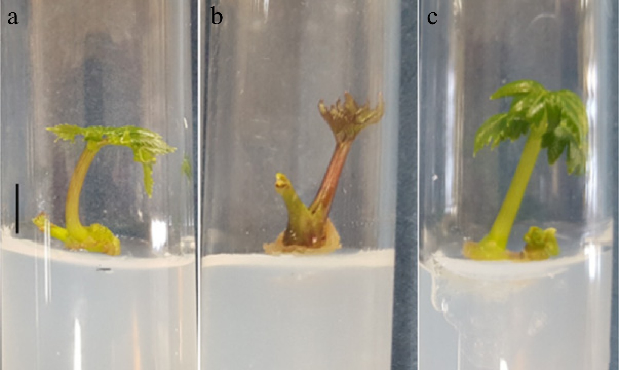
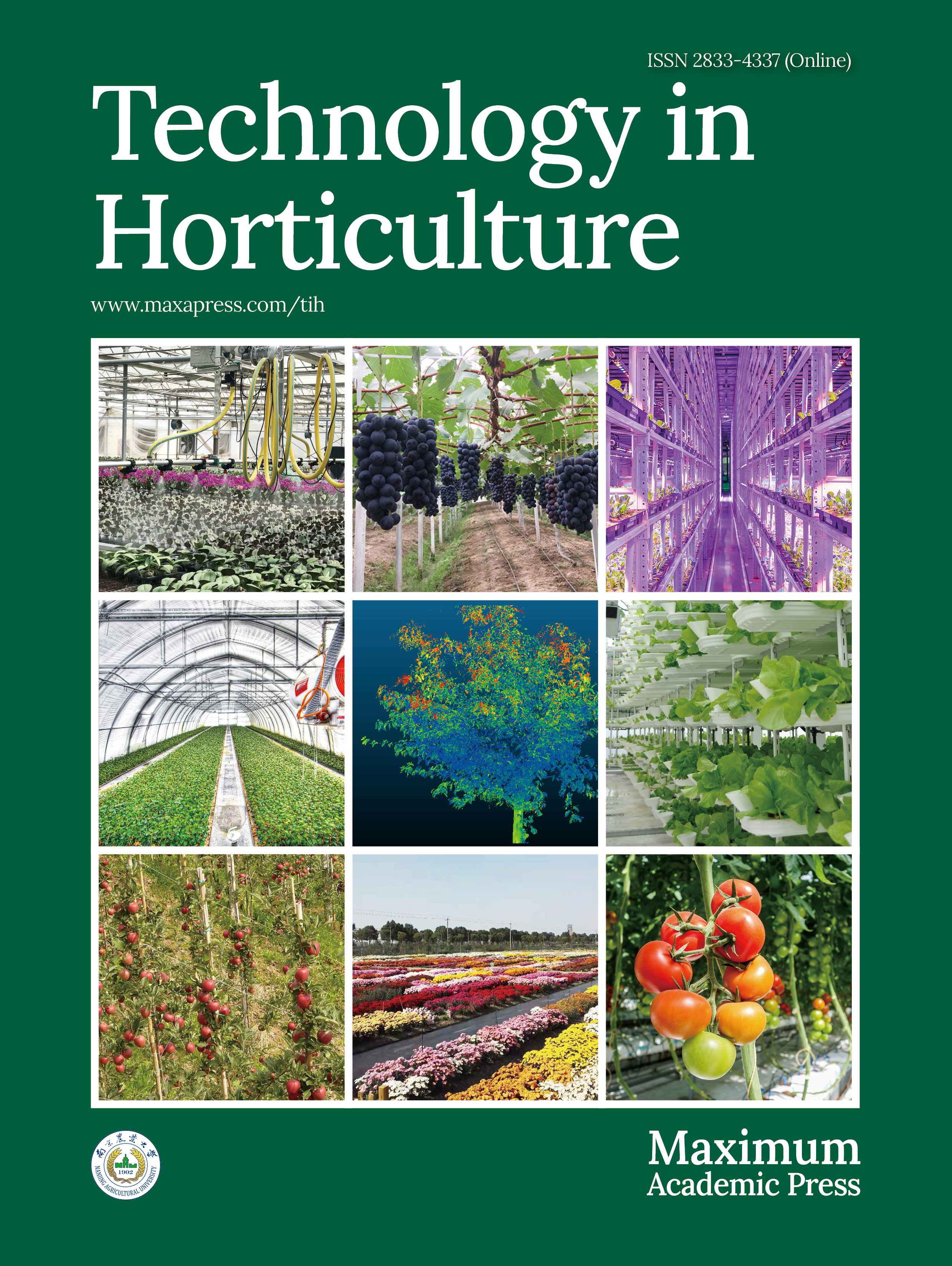
Figure 1.
Seedlings of arracacha (Arracacia xanthorrhiza Bancroft) obtained in vitro from shoot tips grown in B5 medium supplemented with 30 g·L−1 sucrose, 0.3 mg·L−1 BAP, and 0.1 mg·L−1 NAA. (a) Seedlings of ASA. (b) Seedlings of 'BRS Catarina 64'. (c) Seedlings of 'BRS Rubia 41'. Bar = 1 cm.
Table 1. Average shoot regeneration rate of apical buds of arracacha grown on different culture media at 14 DAI.
Culture media Shoot regeneration (%) B5 with no PGR 81.67a B5 + 0.2 mg L−1 BAP + 0.01 mg L−1 NAA 61.67b B5 + 0.3 mg L−1 BAP + 0.1 mg L−1 NAA 65.00ab Means followed by the same letter do not differ from each other by the Tukey test at 5% significance. At 21 DAI, a statistical difference was only observed among the cultivars evaluated for this characteristic. The highest percentage of establishment was for 'BRS Acarijó 56', followed by 'BRS Rubia 41', which achieved 71.67% and 58.33%, respectively, without differing from each other (Table 2).
Table 2. Average values of shoot regeneration rate of apical buds of arracacha of the cultivars at 21 DAI.
Cultivars Shoot regeneration (%) 'BRS Rubia 41' 58.33ab 'BRS Acarijó 56' 71.67a ASA 43.33b Means followed by the same letter do not differ from each other by the Tukey test at 5% significance. For the traits of shoot height, number of shoots, and callus diameter evaluated at 30 DAI, no significant interactions were observed; however, differences were observed between culture media and among cultivars for these traits (data not shown). For shoot height and callus diameter, when the culture media were evaluated in isolation, it was revealed that the culture medium B5 + 0.3 mg·L−1 BAP + 0.1 mg·L−1 NAA was the one that led to greater development of shoots and greater callus diameter. This medium did not differ from the control culture medium for shoot height but was statistically different from the other two media tested for callus diameter (Supplementary Table S1).
The cultivars presented differences in shoot height, number of shoots, and callus diameter at 30 DAI. The ASA cultivar showed the highest shoots, followed by 'BRS Acarijó 56', but in terms of the number of shoots and callus diameter, ASA had the lowest values for both traits, together with 'BRS Acarijó 56'. However, 'BRS Rubia 41' had more shoots and a larger callus diameter (Supplementary Table S2).
Effect of photoperiod on shoot induction
-
For the photoperiod combinations, the arracacha cultivars, and their possible interactions, the results presented in Table 3 were obtained.
Table 3. Summary of the ANOVA for the regeneration rate, number of shoots, number of leaves, number of roots, and percentage of callus in arracacha seedlings according to the combination of different cultivars and photoperiods.
SV Mean square DF Regeneration rate Number of shoots Number of leaves Callus (%) Cultivar 2 101.6292ns 0.1337ns 0.1433ns 5056.4815* Photoperiod 2 15.1070ns 0.0181ns 0.4300ns 584.2593ns Cultivar × photoperiod 4 49.1237ns 0.0515ns 0.1433ns 547.2037ns Residue 18 83.4774 0.1285 0.1948 236.2222 Total 26 CV (%) 10.3 19.2 41.8 34.6 Overall average 88.6 1.8 1.1 44.4 SV, source of variation; DF, degrees of freedom; CV, coefficient of variation; ns, nonsignificant; *, significant at 5% probability. According to Table 3, there was no significant interaction between the sources of variation for the variables evaluated or the isolated effect of the factors, except for the percentage of calluses. The average was 1.8 shoots, 1.1 leaves per shoot, and 0.1 roots per plant (Fig. 1).
Regarding callus formation, as shown in Table 3, there was a significant effect of cultivar. It can be observed that the lowest percentage of callus was verified in ASA, followed by the cultivars 'BRS Rubia 41' and 'BRS Catarina 64' (Supplementary Table S3).
Callus development (Fig. 2) probably occurred as a result of the genotype × environment interaction. In this case, the cultivars responded differently to the culture medium and PGRs used, since there was no effect of photoperiod.
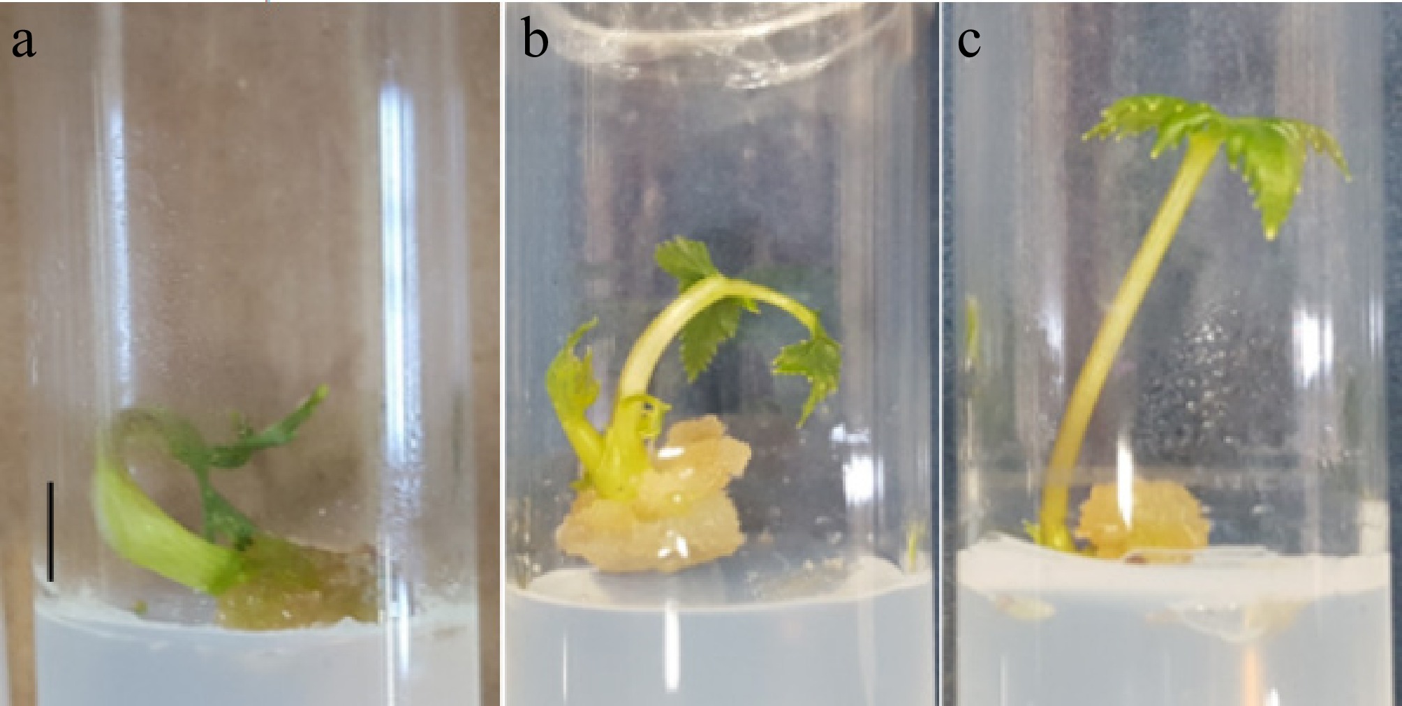
Figure 2.
Presence of callus at the base of arracacha (A. xanthorrhiza) seedlings obtained in vitro from shoot tips grown in B5 medium supplemented with 30 g·L−1 sucrose, 0.3 mg·L−1 BAP, and 0.1 mg·L−1 NAA. (a) Callus on a seedling of ASA. (b) Callus on a seedling of 'BRS Catarina 64'. (c) Callus on a seedling of 'BRS Rubia 41'. Bar = 1 cm.
In vitro rooting and hardening
-
Regarding in vitro rooting, the ANOVA showed significant effects (p < 0.05) for the interaction between IBA doses (0.0; 3.0; 6.0; 9.0; 12.0 mg·L−1) and the use of activated charcoal for all evaluated traits (Table 4).
Table 4. Summary of the ANOVA for the number of roots and the length of roots as a function of doses of IBA with and without the addition of activated charcoal, in culture media for in vitro cultivation of the A. xanthorrhiza cultivar 'BRS Rubia 41'.
SV Mean square DF Number of roots Length of roots Dose 1 5.67* 43.94* Charcoal 4 4.66* 10.60* Dose × charcoal 4 3.87* 2.86* Residue 60 2.43 1.10 CV (%) 28.06 27.11 * Significant at 5% probability of error by the F-test. In the absence of activated charcoal, the lowest technical efficiency was at a concentration of 4.84 mg·L−1 of IBA, which resulted in the lowest number of roots (4.49 roots·seedling−1). Thus, IBA concentrations above 4.84 mg·L−1 increased the number of roots in arracacha. Regarding root length, the use of activated charcoal resulted in a reduction of 0.0945 mm in root length for each increase in IBA of 1.0 mg·L−1. Once again, it was confirmed that for the cultivar studied, activated charcoal has an antagonistic effect on the development and, consequently, the growth of roots, probably through the adsorption of compounds from the culture medium.
In the absence of activated charcoal, the lowest root growth (2.19 mm) was obtained at a dose of 5.64 mg·L−1 of IBA. Therefore, it is estimated that there is an intrinsic relationship between the levels of endogenous and exogenous auxin for initiating the induction, initiation, and elongation of the root system.
Callus formation was observed only in the treatments without activated charcoal + IBA (9) and the absence of charcoal + IBA (12) (Table 5). The addition of activated charcoal did not promote callus formation, likely because of the characteristics described above. In the absence of activated charcoal, IBA concentrations higher than 9 mg·L−1 resulted in the formation of undifferentiated cells. The application of auxins favors root induction and initiation but inhibits elongation, whereas high concentrations can lead to callus formation[21].
Table 5. Percentage of callus versus the presence or absence of activated charcoal as a function of different doses of IBA in the in vitro cultivation of A. xanthorrhiza cultivar 'BRS Rubia 41'.
Treatment Presence of callus (%) Absence of activated charcoal + 0 mg·L−1 IBA 0b Absence of activated charcoal + 3 mg·L−1 IBA 0b Absence of activated charcoal + 6 mg·L−1 IBA 0b Absence of activated charcoal + 9 mg·L−1 IBA 100a Absence of activated charcoal + 12 mg·L−1 IBA 100a Presence of activated charcoal + 0 mg·L−1 IBA 0b Presence of activated charcoal + 3 mg·L−1 IBA 0b Presence of activated charcoal + 6 mg·L−1 IBA 0b Presence of activated charcoal + 9 mg·L−1 IBA 0b Presence of activated charcoal + 12 mg·L−1 IBA 0b CV (%) 21 Means followed by different letters, ranked by the nonparametric Kruskal–Wallis test, differ from each other at a 5% probability level. Regarding in vitro rooting and hardening, for the variables evaluated in vitro, significant differences were observed (Supplementary Table S4) between the two culture media (B5 and ½ B5) combined with different sealing materials (parafilm, gauze, and PVC) for length of the longest root, number of roots, number of shoots, plant height, and petiole length.
The survival rate of arracacha microplants cultivated in vitro, considering all treatments over a 30-d period, was 95.21%, representing the highest value obtained. This result is significant, as cultivation in flasks generally favors contamination and, consequently, plant mortality, which was not observed in this experiment. This value was considered sufficient to allow the hardening and acclimatization of a large number of plants. However, greater microplant losses were observed in flasks sealed with gauze. Both treatments containing gauze (Treatment 2: 1B5 + gauze; Treatment 5: ½ B5 + gauze) presented water loss, leading to plant dehydration caused by gas exchange with the environment.
Figure 3 shows the arracacha plants when they were removed from the cultivation medium and evaluated, for later placement in plastic cups with a substrate + coconut fiber (1:1).
Figure 3.
Arracacha (A. xanthorrhiza) plants rooted and hardened in vitro under the different treatments. (a) Treatment 1: B5 + parafilm; (b) Treatment 2: B5 + gauze; (c) Treatment 3: B5 + PVC; (d) T4: ½ B5 + parafilm; (e) Treatment 5: ½ B5 + gauze; (f) Treatment 6: ½ B5 + PVC.
As can be seen in Table 6, the highest values for the length of the largest root were found for Treatment 6 (½ B5 + PVC), which did not differ significantly from Treatment 3 (3.20 cm). The lowest values were found for Treatment 2 (B5 + gauze), which did not differ significantly from Treatment 5 (½ B5 + gauze), both of which were culture media using gauze-type sealing, where water loss caused dehydration in the plants through gas exchange with the environment.
Table 6. Average values of the different treatments for traits, including length of the largest root, number of roots, number of shoots, plant height, and petiole length, for the variables evaluated in vitro.
Treatments Length of the largest root (cm) Number of roots Number of shoots Plant height (cm) Petiole length (cm) Treatment 1: B5 + parafilm 1.64c 2.54ab 2.35bc 5.05b 4.30a Treatment 2: B5 + gauze 0.40d 0.35b 1.86d 0.00d 2.15c Treatment 3: B5 + PVC 3.20ab 6.33ab 2.24a 5.30b 3.95b Treatment 4: ½ B5 + parafilm 1.84bc 2.01a 2.72c 6.15a 4.45a Treatment 5: ½ B5 + gauze 1.48cd 2.96b 1.78bc 3.50c 2.55c Treatment 6: ½ B5 + PVC 4.67a 3.98ab 2.20ab 9.25a 4.45a Means followed by the same letter in the column do not differ from each other by the Tukey test at 5% significance. This pattern continued for the other evaluated traits, with the lowest averages found for the number of roots in Treatment 2 (B5 + gauze) and the lowest values found for the number of shoots in Treatment 5 (½ B5 + gauze).
Root and shoot growth are essential for subsequent hardening, acclimatization, and field cultivation. For the number of roots, higher results were also found under Treatment 3 (B5 + PVC), with an average of 6.33, not differing significantly from Treatment 6 (½ B5 + PVC), with an average of 3.98, and Treatment 1 (B5 + parafilm), with an average of 2.54. For the number of shoots, the best results were found in Treatment 4 (½ B5 + parafilm), with an average of 2.72 shoots per plant, which did not differ significantly from Treatment 1 (B5 + parafilm) and Treatment 5 (½ B5 + gauze), with an average of 2.35 and 1.78, respectively.
The plant height variable showed a significant difference when comparing Treatment 1 (B5 + parafilm) and Treatment 2 (B5 + gauze), Treatment 4 (½ B5 + parafilm) and Treatment 2 (B5 + gauze), Treatment 4 (½ B5 + parafilm) and Treatment 5 (½ B5 + gauze), Treatment 4 (½ B5 + parafilm) and Treatment 6 (½ B5 + PVC), and between Treatment 2 (B5 + gauze) and Treatment 3 (B5 + PVC). The highest values were observed in Treatment 6 (½ B5 + PVC) and Treatment 4 (½ B5 + parafilm). The PVC and parafilm sealants contain microlayers that allow the passage of light and thus stimulate the growth of microplants. Both treatments used half-strength B5 to supplement the culture medium.
For the petiole length trait, significant differences were observed when comparing the following pairs of treatments: Treatment 1 (B5 + parafilm) and Treatment 2 (B5 + gauze), Treatment 1 (B5 + parafilm) and Treatment 5 (½ B5 + gauze), Treatment 4 (½ B5 + parafilm) and Treatment 2 (B5 + gauze), Treatment 4 (½ B5 + parafilm) and Treatment 5 (½ B5 + gauze), Treatment 2 (B5 + gauze) and Treatment 3 (B5 + PVC), Treatment 2 (B5 + gauze) and Treatment 6 (½ B5 + PVC), Treatment 5 (½ B5 + gauze) and Treatment 3 (B5 + PVC), and between Treatment 5 (½ B5 + gauze) and Treatment 6 (½ B5 + PVC). Therefore, we found the same results in Treatments 4 (½ B5 + parafilm) and 6 (½ B5 + PVC).
It is worth highlighting the use of PVC and parafilm, materials that have microlayers and allow the passage of light, favoring the growth of microplants and possibly causing their etiolation[22].
For the variables evaluated ex vitro, after the acclimatization process, significant differences were observed (Supplementary Table S5) among the different culture media (B5 and ½ B5) and seals (parafilm, gauze, and PVC) for plant height, petiole length, number of roots, and number of leaves; only the length of the largest root did not present a level of significance.
The survival rate of plants after 30 days in cups with a substrate + coconut fiber (1:1) was 78% (Supplementary Fig. S2). Following the same trends as plants grown in vitro with gauze sealing, the treatment that presented the most losses during acclimatization was Treatment 2 (B5 + gauze).
In Table 7, it is possible to observe the values referring to the length of the largest root, plant height, number of roots, number of leaves, and length of the petiole of plants grown in the substrate + coconut fiber (1:1).
Table 7. Average values of the treatments for length of the largest root, number of roots, number of shoots, plant height, and petiole length for the variables evaluated ex vitro.
Treatments Length of the largest root (cm) Number of roots Number of leaves Plant height (cm) Petiole length (cm) Treatment 1: B5 + parafilm 6.60b 6.30a 2.30c 15.32b 7.87b Treatment 2: B5 + gauze 9.30a 4.15c 2.00c 15.96b 7.71b Treatment 3: B5 + PVC 10.20a 7.85a 3.00b 22.55a 11.81a Treatment 4: ½ B5 + parafilm 6.50b 5.70b 2.60c 16.005b 8.78b Treatement 5: ½ B5 + gauze 6.20b 5.90b 2.30c 13.98b 7.77b Treatment 6: ½ B5 + PVC 11.90a 6.50a 4.50a 23.015a 11.68a Means followed by the same lowercase letter in the column do not differ from each other according to the Tukey test at 5% significance. The highest average plant height was found in Treatment 6 (½ B5 + PVC), namely 23.02 cm, which did not differ significantly from Treatment 3 (B5 + PVC), with an average of 22.55 cm. For petiole length, Treatment 3 (B5 + PCV) obtained the highest average (11.81 cm), not differing significantly from Treatment 6 (½ B5 + PVC) with an average of 11.68 cm. This is because plants grown with PVC presented better development in vitro and, later, ex vitro.
For the length of the largest root, there was no significant difference when comparing all treatments with each other. The highest values were found in Treatment 6 (½ B5 + PVC) and Treatment 3 (B5 + PVC).
For the number of roots, a significant difference was found only between Treatment 2 (B5 + gauze) and Treatment 6 (½ B5 + PVC). The highest value was found in Treatment 3 (B5 + PVC), followed by Treatment 6 (½ B5 + PVC) and Treatment 1 (B5 + parafilm).
For the number of leaves, there was a significant difference when comparing Treatment 1 (B5 + parafilm), Treatment 2 (B5 + gauze), Treatment 4 (½ B5 + parafilm), Treatment 5 (½ B5 + gauze), and Treatment 6 (½ B5 + PVC). The highest values were found in Treatment 6 (½ B5 + PVC) and Treatment 3 (B5 + PVC).
The performance of the plants grown ex vitro can also be attributed to the substrate used (Plantmax®), together with coconut fiber, as both provided a favorable environment for the cultivation of the arracacha plants.
After the ex vitro evaluation phase, the plants were transferred to seedling trays and maintained in a growth chamber (Fitotron®) with controlled the temperature, photoperiod, and humidity for 30 days (Supplementary Fig. S1). Subsequently, the plants were transferred to a greenhouse and placed in 30-L pots containing soil and the substrate in a 1:1 ratio (Supplementary Fig. S1).
-
The results obtained in this experiment showed that the tested culture media favored the in vitro development of the evaluated cultivars, enabling shoot formation, especially when the B5 culture medium was added to + 0.3 mg·L−1 BAP + 0.1 mg·L−1 NAA. PGRs are natural or synthetic compounds applied in varying amounts directly to a target plant to alter its physiological processes[23].
The effect of PGRs, especially cytokinins, is crucial for in vitro shoot multiplication[24]. Conversely, balanced combinations of cytokinins and auxins can promote not only a higher sprouting rate but also the vigor and better morphological quality of the explants[25]. Concentrations of BAP 4[26], 6[27], and 10[12] times greater than that of NAA induced a greater shoot numbers in garlic, quinoa, and arracacha, respectively. The results obtained in this study indicate that a 1:3 concentration of cytokinin and auxin (0.1 mg L−1 NAA + 0.3 mg L−1 BAP) tends to produce greater shoot emission and greater in vitro development of arracacha plants, corroborating previous findings.
The fact that the culture medium yielding the highest percentage of shoot regeneration was the one without PGRs may be explained by its lower salt concentration, as arracacha is considered to be a hardy species whose field development often occurs in soils with average fertility[2]. However, in liquid medium, increasing the strength of the MS resulted in better growth results. Therefore, the growth response was affected by the amount of MS salts and the presence of agar in the medium[28].
The positive response of the cultivars evaluated in vitro may be related to the greater vigor of this species in the field. During genotype selection, field yield was considered, and the cultivar 'BRS Acarijó 56' is characterized by robust growth and high productivity[5]. As the concentration of BAP in the culture medium increases, shoot height decreases, but the number of shoots per explant increases[17]. Furthermore, explants in the medium supplemented with PGR exhibit more vigorous growth[12]. In this experiment, basal callus formation exceeding expectations was also observed, making it unsatisfactory as a potential promoter of genetic modifications. Large basal callus formations have been described[12,29,30]. This effect can be reduced by increasing the BAP concentration[31]. Care must be taken not to increase it excessively, as it is a cytokinin that can inhibit shoot formation and induce the formation of a larger basal callus than expected[17]. Callus formation occurs when there is a balance between the auxin and cytokinin concentrations[31]. Although initial root emergence often occurs from callus tissue, callus formation is not necessarily a prerequisite for rooting, as these processes are independent in most plant species[32]. In this experiment, excessive callus formation may have impaired root formation, since no root formation was observed in the cultivars and environments evaluated. In unsuccessful in vitro rooting tests on arracacha, Slíva et al.[12] suggested that rooting occurs spontaneously during acclimatization.
Effect of photoperiod on shoot induction
-
Photoperiod is known to influence plant development, although it did not have a significant effect under the conditions tested.
In general, cultivated plants experience the well-known influence of photoperiod on growth, photosynthetic capacity, and metabolism[33−35] through the regulation of flowering and tuberization. Photoperiod may also influence the growth or morphogenesis of explants cultured in vitro[36,37]. Murashige[38] pointed out that plants requiring specific photoperiods for vegetative growth and development are very likely to express this requirement under in vitro culture. Nevertheless, the photoperiod required for flowering does not necessarily correspond to that required for optimal shoot and root regeneration or for shoot proliferation[36].
In contrast to the results obtained in this experiment, Figueiredo et al.[39] observed significant photoperiod effects in physalis (Physalis peruviana), a species of Andean origin, similar to arracacha. The authors observed that the length of the aerial part was greater under a 16-h photoperiod, whereas the roots developed better with a shorter photoperiod (8 h). The number of leaves, however, was not influenced by the photoperiod.
Working with nodal segments of English potato (Solanum tuberosum) in MS medium, Kozai et al.[40] observed that root growth was suppressed under a short photoperiod (8 h), but under a longer photoperiod (16 h) root growth was enhanced, producing seedlings with short and thick stems with a similar number and a larger leaf area, obtaining the highest fresh and dry weights without loss of dry matter percentage.
In yam species (Dioscorea spp.), the number of leaves formed from nodal segments was higher under a 8-h photoperiod compared with others (10, 12, and 24 h)[41]. On the other hand, in nodal segments of Brazilian ginseng (Pfaffia glomerata), longer photoperiods improved the growth parameters of the explants[42].
In this experiment, it was observed that the photoperiod did not exert a significant influence on the in vitro development of arracacha cultivars. This result can be attributed to the culture medium being supplemented with sucrose, which acts as an exogenous carbon source, reducing the explants' dependence on photosynthesis for growth[43]. Consequently, variations in daylength do not directly impact shoot elongation or induction. Under these conditions, factors such as the quality of incident light and, notably, the composition of PGRs in the medium become more determining than the duration of light exposure[44].
Callus development (Fig. 2) occurred as a function of the genotype × environment interaction. In this case, the cultivars responded differently to the culture medium and PGRs used, since there was no effect for the photoperiod.
It is known that many genotype-dependent effects are caused by interactions between the plant genotype and the cultivation environment[36,45].
According to Grattapaglia and Machado[46], basal callus formation may result from high auxin concentrations in the culture medium, compromising shoot and root development. Madeira et al.[17] emphasized that callus formation in explants can be reduced by increasing the BAP concentration in the culture medium. However, the concentration of BAP in relation to NAA cannot be increased too much, because as the BAP/NAA ratio increases, there is less development of the shoot[47].
The results of this experiment suggest that the in vitro development of the evaluated arracacha cultivars is independent of the photoperiod.
In vitro rooting and hardening
-
From the evaluations performed, it was possible to observe that the initial root induction treatments promoted distinct responses in the formation of root primordia and in the induction of in vitro rooting using different concentrations of IBA. The root primordium is the result of gene activation resulting from the plant's endogenous indole-3-acetic acid (IAA). The action of auxins occurs initially at the cellular level in the primary and secondary meristems, stimulating cell division and subsequent cell elongation, and this initial action of auxins culminates in root formation[48].
Synthetic auxins favor the conjugation between endogenous IAA and amino acids that promote the synthesis of specific proteins that are necessary for the initial formation of roots[49−51]. In general, after the application of synthetic auxin, there is an immediate increase in the endogenous level of natural auxin; consequently, the formation of primordial roots begins[25].
IBA is among the most widely used auxins for in vitro rhizogenesis. Normally, the number of roots formed increases with higher concentrations of this phytoregulator, until its concentration becomes excessively high, inhibiting root formation and, consequently, callus formation[18]. According to Grattapaglia & Machado[46], when an excessive concentration of auxin is used, rhizogenesis is compromised by phytotoxicity. However, the unavailability of this phytoregulator compromises rhizogenesis, since, according to Fogaça et al.[52], auxins are responsible for the induction of root primordia.
In the in vitro cultivation of orchids, Nguyen[53] observed that doses of activated carbon greater than 0.14 g·L−1 inhibited the development of microplants. This diversity of results is commonly observed in tissue culture, as it is mainly related to the interaction between the genotype and other factors related to the rooting of plants grown in vitro, including endogenous auxin levels, conditions inherent to the mother plant such as juvenility, the culture medium, the presence of other PGRs and carbohydrates, mineral nutrition, and the presence of polyamines and substances such as activated carbon and phenolic compounds, in addition to the environmental growth conditions[54].
Regarding activated carbon, as this compound adsorbs compounds that are toxic to plant growth, it also adsorbs nutrients that are essential for growth, making their release slower or even unsatisfactory[55,56]. Similar results were found by Schwalbert et al.[57], where the authors observed an inhibitory effect on the number of roots of Desmodium incanum (Sw.) DC. in the presence of activated carbon in the nutrient medium.
In vitro root induction is an important step in the organogenesis process that ensures the seedlings' survival during acclimation[58]. Abdella et al.[59], when analyzing the in vitro rooting of apple (Malus spp.) trees, observed greater root formation when using half-strength MS medium, regardless of the PGRS used, reaching an average of 6.25 roots per explant. The same was found for the length of the largest root.
Alatar et al.[60], evaluating the in vitro rooting of Solanum lycopersicum, obtained a greater number of roots after 30 d in half-strength MS medium supplemented with 0.5 µM IBA, resulting in approximately 14 roots per plant. Therefore, it is worth noting that the media of the basic formulations diluted to 50% of their concentration enabled better in vitro rooting of several cultures[61].
It is also worth mentioning studies conducted by the research group, which showed that the use of a culture medium supplemented with vitamin B5, at full or half concentration, was effective for the multiplication, rooting, and maintenance of arracacha plants[62−64].
Regarding shoot number, the best results were found under Treatment 4 (½ B5 + film), with an average of 2.72 shoots per plant, not differing significantly from Treatment 1 (B5 + film) and Treatment 5 (½ B5 + gauze), which presented averages of 2.35 and 1.78 shoots per plant, respectively. These results indicate that both the culture medium's formulation and the type of flask seal can influence sprout induction. The sealing material can modify gas exchange, relative humidity, and ethylene accumulation, which directly affect sprouts' proliferation[65].
Chaves et al.[66], studying Physalis plants, also reported that reducing the culture medium's concentration (¾) resulted in a linear increase in the number of shoots, from 1 shoot per explant in the control to 1.75 shoots at 0.3 mg·L−1 of BAP. Therefore, although the medium's formulation plays a fundamental role, the type of flask seal should also be considered as a contributing factor in in vitro shoot development.
These results are consistent with Bansal et al.[67], who stated that media based on diluted formulations yielded better results for the multiplication of several species. Other studies have obtained similar results using BAP for regeneration and multiplication of arracacha[63].
Regarding the different types of sealants used for rooting and hardening microplants in vitro, Rodrigues et al.[68], studying the in vitro regeneration of Solidago chilensis, observed that closing the culture tubes with a cotton cap apparently reduces the in vitro ethylene concentration but does not prevent water loss. This facilitates gas exchange and causes the culture medium to evaporate more quickly, leaving the plants dehydrated, requiring shorter subcultures. In this study, we expected less senescence to occur when sealing methods that provided greater air circulation in the culture flask were used, as this would prevent the accumulation of gases such as ethylene, which can induce senescence. Over the 30 d, gauze was not adequate for hardening the arracacha plants. Therefore, for future tests, we recommend using gauze for a shorter period.
Plants grown in vitro are maintained under artificial development conditions with a controlled photoperiod, temperature, and growth environment, depending on the heterotrophic mode of nutrition. Therefore, they can develop certain structural physiological abnormalities[69], inducing morphological, anatomical, and physiological changes. These factors are responsible for inducing dehydration and high mortality during the transfer from the in vitro to the ex vitro environment. Therefore, the acclimatization process after the in vitro rooting phase is essential to promote gradual adaptation and minimize plant mortality, improving their physiological performance through the development of a functional root system and the control of stomatal and cuticular transpiration[70].
Ribeiro et al.[71], when studying different methods of in vitro rooting of blueberry plants, used the same concentration of IBA (20 mg·L−1) to optimize the acclimation process, combined with different vial sealing systems: Standard caps, plastic film, and filter caps. No significant differences were observed in the morphometric parameters during the in vitro proliferation phase. However, the use of filter caps increased the efficiency of micropropagation and acclimatization, in addition to improving plant survival rates. These results highlight the importance of optimizing culture vial sealing systems to develop more efficient acclimation protocols while maintaining the phytosanitary quality of the plants.
For the variables of plant height and petiole length, similar behavior was observed in the acclimation of Orthophytum mucugense, where the plants grown in flasks sealed with PVC film and filled with a culture medium containing 15 and 30 g·L−1 of sucrose showed greater survival than plants grown in the absence of sucrose.
According to Silveira et al.[72], who studied caroá plants and evaluated the number of roots, the sealing type factor did not have a significant influence, as they obtained averages of 7.17 cm for PVC and 10.12 cm for cotton, with no statistical difference between them.
Furthermore, Brito et al.[73], studying the acclimation of microplants of Comanthera mucugensis Giul. ssp. mucugensisa, found that after 90 d of acclimatization, the average survival percentage of plants grown in tubes sealed with PVC (70.32%) and a lid (66.73%) was higher than that of the treatment with a cotton plug (38.69%). The authors attribute the reduction in transpiration to the pre-existing water deficit of plants grown in tubes sealed with cotton, which can be compared to the use of gauze, which allows greater gas exchange with the medium. Its permeability allows the evaporation of water from the culture medium, which reduces the water potential of both the culture medium and the plant.
Unlike the studies by Silveira et al.[72], who studied the preacclimation of caroá plants with different types of seal, the best results were obtained with the cotton plug for shoot length (cm), with a gain of approximately 30%–40%. According to studies carried out by Rodrigues et al.[68], who observed greater average heights for cotton (9.26 cm) than for PVC (7.55 cm), these variables have a positive influence on the hardening of arracacha plants.
-
All cultivars showed in vitro shoot regeneration. All culture media favored the in vitro development of the genotypes, enabling the shoots formation. The medium that showed the best in vitro establishment was the culture composed of B5 + 0.3 mg·L−1 BAP + 0.1 mg·L−1 NAA. The B5 culture medium with no PGR favors the development of shoots and provides smaller callus diameters.
For in vitro rooting of arracacha microplants, the use of activated charcoal in the concentrations evaluated in the culture medium does not favor the formation of roots, nor their growth. However, it prevents the formation of callus in microplants. Therefore, the presence of auxin stimulates the formation of roots when in low concentrations (0.0−3.0 mg·L−1).
Regarding rooting and hardening, micropropagated plants of the 'Amarela de Senador Amaral' cultivar developed well, depending on the culture medium and sealing method. For rooting and hardening, we recommend using a ½ B5 + PVC culture medium with the standard dosage of the PGRs BAP and IBA (0.3 mg·L−1 BAP and 0.1 mg·L−1 IBA), as already used for cultivation. Therefore, PVC sealing is highlighted, as it is cheaper and more accessible than parafilm, in addition to showing greater efficiency during the hardening and acclimatization phases of arracacha plants.
-
The authors confirm their contributions to the paper as follows: all authors contributed equally to the conception and writing of the manuscript; all authors critically revised the manuscript and approved the final version; conceptualization: Finatto T, de Oliveira Vargas T; methodology: Abatti L, Gonçalves TJ, Mafessoni LIW, Candido DF, Daboit BN; analyses and interpretation of the data: Scariotto S, Abatti L; writing – original draft: Abatti L, Gonçalves TJ; writing – review and editing: Finatto T. All authors reviewed the results and approved the final version of the manuscript.
-
The datasets generated during and/or analyzed during the current study are available from the corresponding author on reasonable request.
-
The authors thank Universidade Tecnológica Federal do Paraná and Conselho Nacional de Desenvolvimento Científico e Tecnológico (CNPq grants - 421821/2021-3 and 132337/2024-0) for their financial support. The authors also thank Patrick Dias Marques from the Universidade Federal de Santa Catarina for training on in vitro establishment of arracacha, and Nuno Rodrigo Madeira from Embrapa for his support in conducting the experiment and providing the mother plants of arracacha.
-
The authors declare that they have no conflict of interest.
-
accompanies this paper online at: https://doi.org/10.48130/tihort-0026-0001.
- Supplementary Table S1 Average values of shoot height and callus diameter of the arracacha cultivars at 30 days after inoculation of the apical bud.
- Supplementary Table S2 Average values of the shoot height (cm), number of shoots and callus diameter (cm) of the arracacha cultivars at 30 days after inoculation of the apical bud.
- Supplementary Table S3 Average data on the regeneration rate, number of shoots, number of leaves, number of roots, and percentage of calluses of the 'Amarela de Senador Amaral (ASA)', 'BRS Catarina 64', and 'BRS Rubia 41' cultivars of arracacha.
- Supplementary Table S4 Analysis of variance for the traits length of the largest root, number of roots, number of shoots, plant height, and petiole length, within the respective treatments in the analyses performed in vitro.
- Supplementary Table S5 Analysis of variance for the traits plant height and petiole length, number of roots, petiole length, and number of leaves within the treatments.
- Supplementary Fig. S1 Plant material used to obtain explants of arracacha (Arracacia xanthorrhiza Bancroft).
- Supplementary Fig. S2 In vitro rooted and hardened arracacha (Arracacia xanthorrhiza Bancroft) plants from their respective culture media after 30 days of acclimatization in substrate + coconut fiber (1:1).
- Copyright: © 2026 by the author(s). Published by Maximum Academic Press, Fayetteville, GA. This article is an open access article distributed under Creative Commons Attribution License (CC BY 4.0), visit https://creativecommons.org/licenses/by/4.0/.
-
About this article
Cite this article
Abatti L, Gonçalves TJ, Mafessoni LIW, Candido DF, Daboit BN, et al. 2026. Micropropagation of arracacha: from meristem isolation to plant acclimatization. Technology in Horticulture 6: e011 doi: 10.48130/tihort-0026-0001 

Micropropagation of arracacha: from meristem isolation to plant acclimatization
- Received: 30 June 2025
- Revised: 26 September 2025
- Accepted: 11 December 2025
- Published online: 09 April 2026
Abstract: Arracacha is vegetatively propagated; thus, in vitro tissue culture combined with rooting and acclimatization is an alternative for acquiring high-quality disease-free plantlets. For in vitro propagation studies, three experiments were conducted. The first evaluated different combinations of plant growth regulators (PGRs): B5 medium formulation without a PGR, B5 + 0.2 mg·L−1 6-benzylaminopurine + 0.01 mg·L−1 1-naphthaleneacetic acid, and B5 + 0.3 mg·L−1 6-benzylaminopurine + 0.1 mg·L−1 1-naphthaleneacetic acid; this assay used three arracacha cultivars ('BRS Rubia 41', 'BRS Acarijó 56', and 'Amarela de Senador Amaral'). The second experiment evaluated the effect of photoperiod (10, 12, and 14 h) on three cultivars ('Amarela de Senador Amaral,' 'BRS Catarina 64', and 'BRS Rubia 41'). The third tested different concentrations of indolebutyric acid (IBA) (0.0, 3.0, 6.0, 9.0, 12.0 mg·L−1) with and without activated charcoal (1.0 g·L−1), and different formulations (B5 and ½ B5) supplemented with 0.3 mg·L−1 6-benzylaminopurine and 0.1 mg·L−1 indolebutyric acid sealed with one of three different materials (parafilm, gauze, polyvinyl chloride). The culture medium with the highest shoot regeneration rate that was most favorable for in vitro establishment was B5 + 0.3 mg·L−1 6-benzylaminopurine + 0.1 mg·L−1 1-naphthaleneacetic acid. Photoperiod did not affect cultivar development, and the responses varied among cultivars. Activated charcoal did not improve the rooting rates. The culture medium with ½ B5 sealed with PVC promoted better in vitro rooting and hardening of arracacha.
-
Key words:
- Arracacia xanthorrhiza Bancroft /
- In vitro hardening /
- Shoot induction /
- In vitro rooting /
- Photoperiod