











-
Feeding is the way that fish acquire nutrients from the environment which are essential for growth, development, and reproduction. The regulation of appetite in fish is governed by a sophisticated network of interacting hormones that integrate signals originating from both the central nervous system and peripheral tissues[1,2]. In the intricate hormonal cascade that modulates appetite in fish, neurohormones secreted by the brain, particularly the hypothalamic area, certain peptides such as neuropeptide Y (NPY)[3,4], ghrelin[5], and orexin[6] function as orexigenic factors in fish. Others function as anorexigenic factors, such as cocaine and amphetamine-regulated transcript (CART), cholecystokinin (CCK)[7], and leptin[8]. These appetite-related peptides are synthesized and secreted by feeding-related centers within the brain or peripheral tissues such as the liver, stomach, and intestine, thereby modulating food intake in fish.
Melanin-concentrating hormone (MCH), a 17-amino-acid cyclic peptide, encoded by the pro-melanin-concentrating hormone (pmch) gene, was originally identified in chum salmon (Oncorhynchus keta) and was found to be involved in the change of skin color[9]. To date, pmch has been identified in several mammals such as humans[10], rats[11], cattle[12], and pigs[13], as well as avian species such as chickens[14]. In fish, several studies have only identified one type of pmch gene in species such as ya-fish (Schizothorax prenanti)[15], common carp (Cyprinus carpio)[16], and Atlantic cod (Gadus morhua)[17], but more studies have identified two or more variants of pmch genes in species such as starry flounder (Platichthys stellatus)[18] and winter flounder (Pseudopleuronectes americanus)[19]. In mammals, it has been reported that MCH can act as orexigenic factor in appetite regulation. For example, the mRNA expression of pmch in the brain was inhibited by fasting in rodents[20]. Intracerebroventricular (icv) injection of MCH peptide promoted food intake of rats[21]. However, the role of pmch in appetite regulation in fish is controversial among species. Fasting promoted the expression of pmch in the hypothalamus of Schizothorax prenanti, starry flounder, and winter flounder, suggesting the orexigenic function of pmch. However, fasting inhibited the expression of pmch in the hypothalamus of common carp (Cyprinus carpio) and goldfish (Carassius auratus), and ICV injection of MCH inhibited food intake, suggesting the anorexigenic role of pmch[22]. Further studies are required to explore the appetite regulation function of pmch in different fish species.
Siberian sturgeon (Acipenser baerii), a primitive nonteleost member of the ray-finned fish lineage, is extensively cultivated globally, primarily for its caviar and as a source of meat[23]. The genetic and functional characterization of pmch in Siberian sturgeon remains poorly understood. In this research, we cloned the Siberian sturgeon pmch cDNA sequence, providing a foundational information for further investigation into its biological functions. We examined the expression profile of pmch genes in the brain and peripheral tissues. Furthermore, the expression patterns of pmch under different feeding statuses, including feeding, fasting, and refeeding, were detected to investigate the potential involvement of pmch in appetite regulation within Siberian sturgeon.
-
All Siberian sturgeon juveniles were obtained from Runzhao Fisheries (Sichuan, China). Fish were kept at indoor tanks (120 L) at the Sichuan Agricultural University farm supplied with a continuous flow of fresh water (20.4 ± 1.2 °C). Fish were provided with commercial sinking aquafeed (containing 42% protein, 6% fat, and 16% as; Unif, Zhongshan, China) at a rate of 3% of body weight once a day at 14:00, and kept under a 12-h light–12-h dark cycle for two weeks. For cloning and tissue distribution experiments, six fish (214.20 ± 29.54 g) were anesthetized with MS-222 (0.01%) and then their tissues were quickly sampled and frozen in liquid nitrogen. All animal handling procedures were approved by the Animal Care and Use Committee of Sichuan Agricultural University, and followed the guidelines on animal experiments of Sichuan Agricultural University.
RNA isolation and cDNA synthesis
-
Total RNA was isolated from tissues using an RNA extraction kit (Foregene, Chengdu, China) according to the manufacturer's protocol. The quality of the RNA was detected with 1% agarose gel after electrophoresis. Nanodrop 2000 was used to detect the concentration (A260/A280 > 1.8) and purity (A260/A230 > 1.8) of RNA. For this, 1 μg of RNA was reverse-transcribed into cDNA with the PrimeScript RT reagent kit containing gDNA Eraser (Takara, Dalian, China).
Molecular cloning and sequence analysis
-
The cloning primers of pmcha and pmchb (Table 1) were designed according to the unpublished hypothalamus transcriptome of Siberian sturgeon. Polymerase chain reaction (PCR) amplification was conducted as follows: 95 °C for 5 min; 95 °C for 30 s, 56 °C for 30 s, and 72 °C for 1 min (39 cycles); and 72 °C for 5 min. The PCR product was purified with the DNA purification kit (Tiangen, China) and inserted into the pMD19-T vector. The ligation products were transformed into Escherichia coli DH5α cells (TIANGEN, Beijing, China). Positive clones were selected and subjected to sequencing by Sangon (Shanghai, China). DNAMAN was used to analyze the nucleotide and deduced protein sequences. ORF Finder was used to analyze the open reading frames (ORFs). The SignalP 4.1 server was used to predict the presence of signal peptide domains. ClustalW was conducted to align the multiple sequences of PMCH amino acids (aa) (
www.ebi.ac.uk/clustalw ). Mega 7.0 software was used to construct the neighbor-joining phylogenetic tree.Table 1. Primer sequences of the clone and those used for quantitative real-time PCR (qRT-PCR).
Primer name Sequence (5'–3') Usage pmcha-f GATCGGTTTCTACAAGAACTGC Clone pmcha-r GCTTTGTTCATTGGAGGACTG pmchb-f GATAGTTTTTACAAGAACCTCAA pmchb-r CGTAACACACAGGTGAATAAC pmcha-qf AACAGACACCTGCCTTAC qRT-PCR pmcha-qr TCCCAGCATACATCTTAGC pmchb-qf CTTCCTTATTTCTGCCAACTCG pmchb-qr TAGACTCTTCCCAGCATACATC β-actin-qf GTTGGTATGGGACAGAAGGACA β-actin-qr CCAGTTGGTAACAATGCCGT ef-1α-qf AACCTTCAACACCCCAGCC ef-1α-qr CACCAGAGTCCATCACAATACC Periprandial experiment
-
To investigate the periprandial changes in the mRNA expression of pmcha and pmchb, seven groups of juvenile sturgeons (71.86 ± 20.44 g, n = 9 per group) were fed at the scheduled time (14:00, 0 h) for two weeks. Tissues samples including the hypothalamus and liver were sampled at various time points, namely 3 and 1 h prior to the scheduled feeding time (−3 h, −1 h), at the commencement of feeding time (0 h), and 1 and 3 h after feeding (+1 h, +3 h). In addition, two groups of unfed fish were sampled at 1 and 3 h after the feeding time as a control for the fed groups.
Fasting and refeeding experiment
-
In the fasting experiments, five groups of juvenile Siberian sturgeon ( 75.62 ± 15.42 g, n = 9 per group) were acclimated for two weeks and fed once daily at 14:00. Two groups of fish were fed every day; the other three groups of fish were fasted during the experiment. Six fish per group were sampled from the feeding and fasted groups on Day 1 and Day 10, and six fish in the refed group were sampled at 1 h after refeeding on Day 10. The hypothalamus and liver of Siberian sturgeon were sampled and then stored at −80 °C until RNA isolation.
Real-time quantitative PCR analysis
-
The quantitative PCR primers (Table 1) were based on the sequence of pmch1 and pmch2 through cloning. Total RNA (1 μg) was reverse-transcribed into cDNA by using the Prime Script RT reagent Kit (TaKaRa, Dalian, China) as described above. The CFX Real Time PCR Detection System (Bio-Rad, USA) was used to conduct real-time quantitative PCR (qRT-PCR). The reaction volume was 10 μL, including Taq SYBR Green qPCR Mix (5 μL, Innovagene, China), sterile distilled water (3.2 μL), 0.4 μL of each primer, and cDNA (1 μL). The reaction mixture without a template (cDNA) was used as a negative control. The qRT-PCR procedures included initial denaturation at 95 °C for 3 min, 40 cycles of denaturation at 95 °C for 15 s, and annealing at the appropriate temperature (pmcha, 52 °C; pmchb, 53°C) for 30 s. The target genes were normalized to the houskeeping genes (geometric average of the Ct value of β-actin and ef-1α), for with the expression levels are stable. The data were analyzed using the relative Ct method[24].
Statistical analysis
-
The data are shown as the mean ± standard error of the mean (SEM). SPSS 23 statistical software was used to conduct the statistical analyses (IBM Inc, USA). Student's t-tests were used to compare differences between two groups in the periprandial experiments and fasting experiment. One-way analysis of variance (ANOVA) followed by Tukey's honestly significant difference (HSD) test was conducted to compare the differences among different time points in the periprandial experiment. Here, p < 0.05 was considered to be a significant difference.
-
This study cloned two transcript of pmch genes, namely pmcha and pmchb, in Siberian sturgeon. One fragment of 604 bp, namely pmcha (PQ768526), containing an ORF of 540 bp encoding 179 aa, and another cDNA fragment of pmchb (PQ768527) containing an ORF of 474 bp encoding 158 aa, were obtained by qRT-PCR. The amino acids of PMCHa (Fig. 1a) and PMCHb (Fig. 1b) of Siberian sturgeon contained a signal peptide at the N-terminus (27 aa), and a MCH gene-related peptide (MGRP; 12 aa, DSGRDDSKIPVG) and a mature peptide, namely MCH (19 aa, DFDMLRCMLGRVYRPCWQV) at the C-terminus.
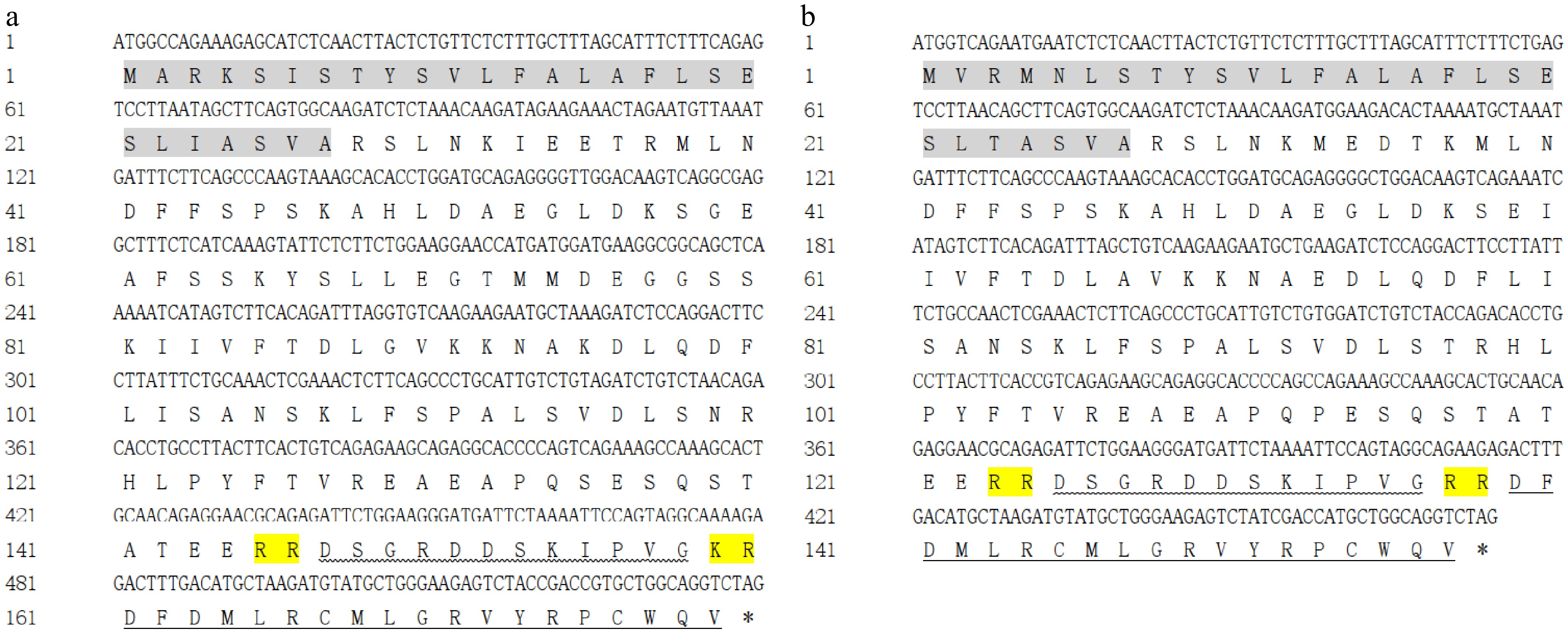
Figure 1.
Nucleotides and deduced amino acid sequences of pmcha and pmchb from Siberian sturgeon. The grey highlight indicates the signal peptide domains of pmcha and pmchb. The horizontal line represents the mature peptide region of MCH. The wavy line represents the MGRP region in PMCHa and PMCHb.
The amino acid sequence of the PMCHa and PMCHb prepropeptides of Siberian sturgeon showed low identity when compared with PMCH sequences from mammals, birds, reptiles and other teleost fishes, as shown in Fig. 2 (18%–27%). Siberian sturgeons' pmcha amino acids showed high identity with other PMCH sequences of other sturgeons, such as Acipenser ruthenus (XP_033880593.1, 96%; XP_058882986.1, 76%) and Polyodon spathula (XP_041109963.1, 77%; XP_041113265.1, 84%), as well as PMCHb from Siberian sturgeon (77%). Siberian sturgeons' PMCHb displayed relative low identity with those of other sturgeons (61%–77%). The phylogenetic tree showed that PMCHa and PMCHb from Siberian sturgeon were included in a clade consisting of Acipenseriformes but separated from other teleost fishes (Fig. 3).
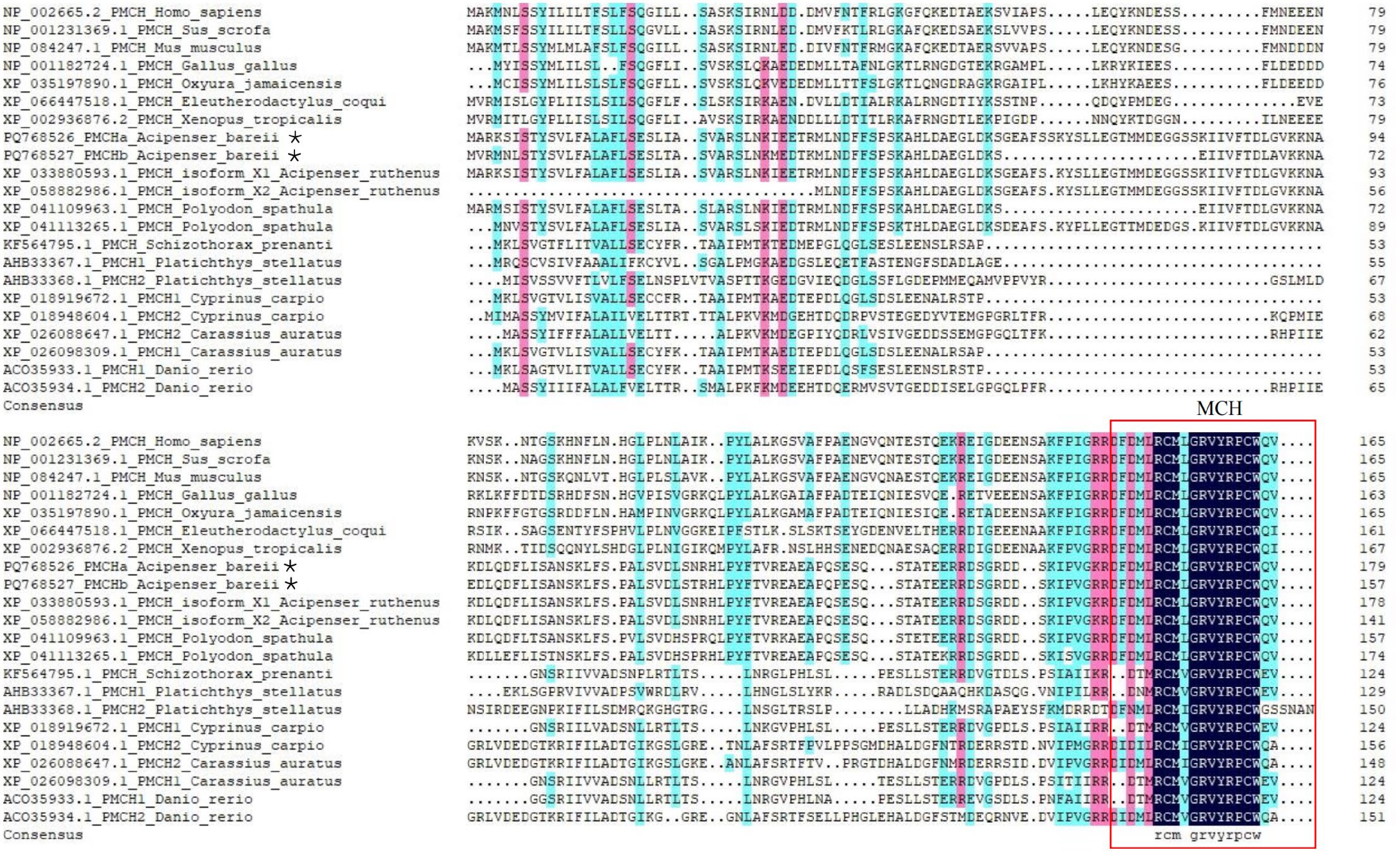
Figure 2.
The alignment of the deduced amino acid sequences of Siberian sturgeon pmch and other vertebrates. Black represents an amino acid consistency level of 100, pink represents 70, and blue represents 50.
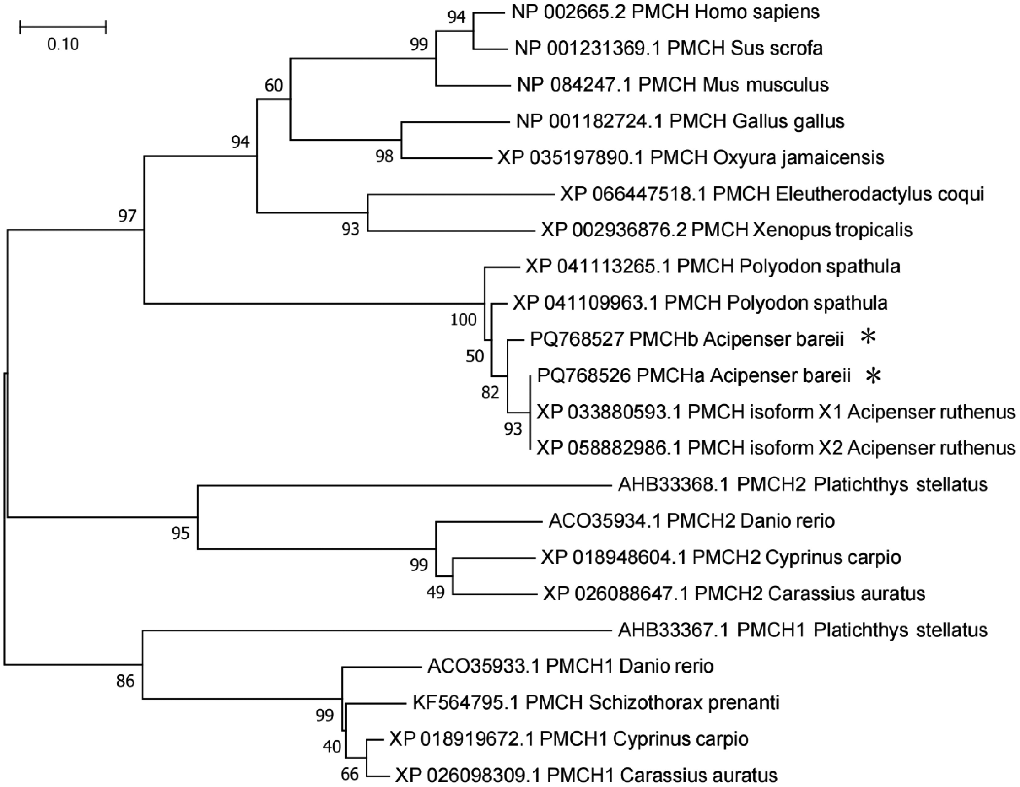
Figure 3.
The phylogenetic analysis of pmch amino acid sequences. MEGA 7.0 was used to construct the neighbor-joining phylogenetic tree and the confidence level of each branch was calculated by bootstrapping repeated 1,000 times.
Tissue distribution of pmcha and pmchb mRNA in Siberian sturgeon
-
Both pmcha and pmchb mRNA were widely distributed in different brain regions and peripheral tissues in juvenile Siberian sturgeon. The mRNA of pmcha was highly expressed in the liver, cerebellum, and hypothalamus (Fig. 4a). In addition, pmchb mRNA was highly expressed in the liver and cerebellum of Siberian sturgeon (Fig. 4b).
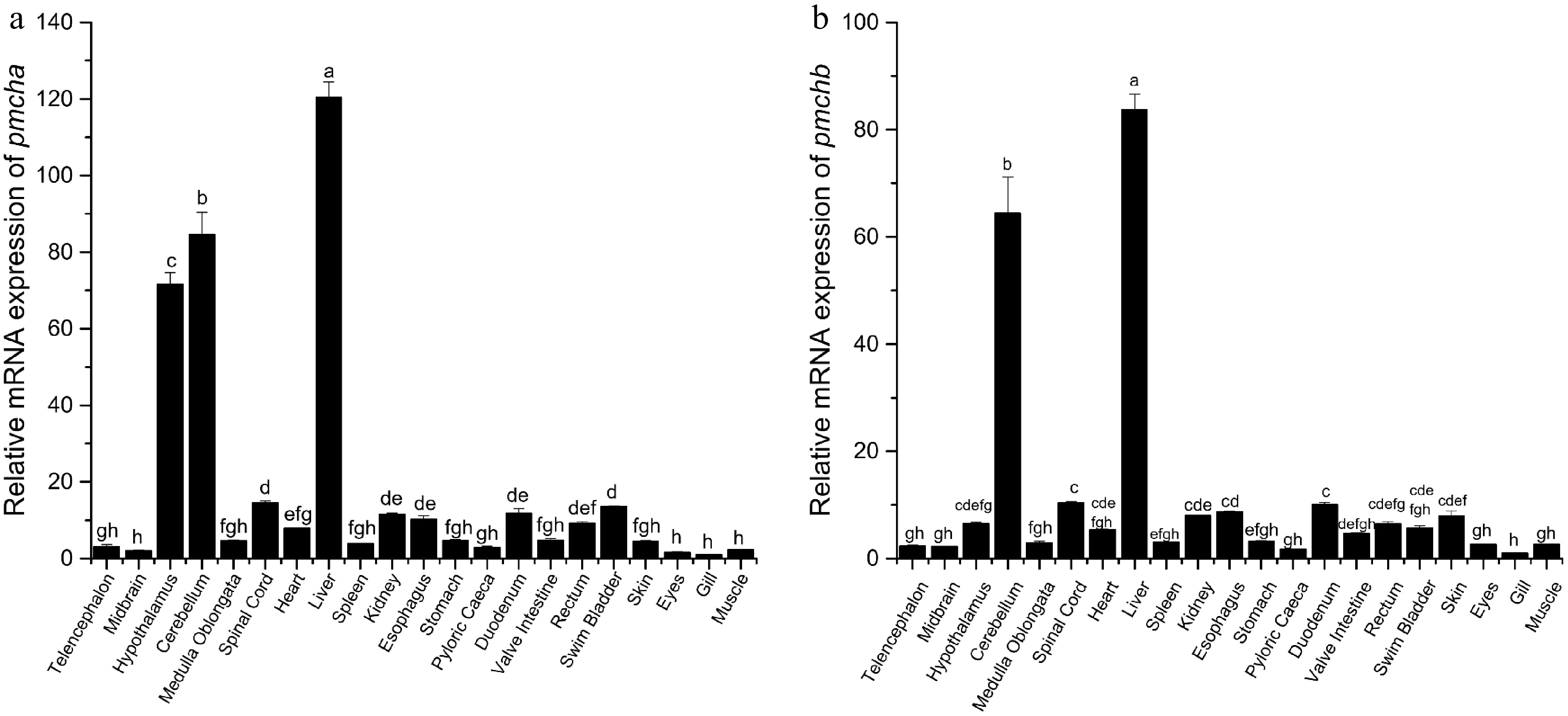
Figure 4.
Tissue distribution of (a) pmcha and (b) pmchb in Siberian sturgeon (n = 6). The mRNA expression of target genes was normalized to β-actin. Different letters indicate significant differences. p < 0.05 represents statistically significant.
Periprandial changes of pmcha and pmchb in Siberian sturgeon
-
Hypothalamic pmcha mRNA in the fed fish was increased in relative to that in unfed fish at +1 h after feeding but was not changed at +3 h after feeding (Fig. 5a). The expression of pmchb in the hypothalamus of the fed fish was promoted at +1 h after feeding and +3 h after feeding when compared with the unfed fish (p < 0.001, p < 0.01, Fig. 5b). Compared with the unfed groups, the mRNA expressions of pmcha and pmchb in the liver of Siberian sturgeon was inhibited at +1 h after feeding time (p < 0.001, p < 0.01) but increased at +3 h after feeding time (p < 0.001, p < 0.01) (Fig. 5c, d).
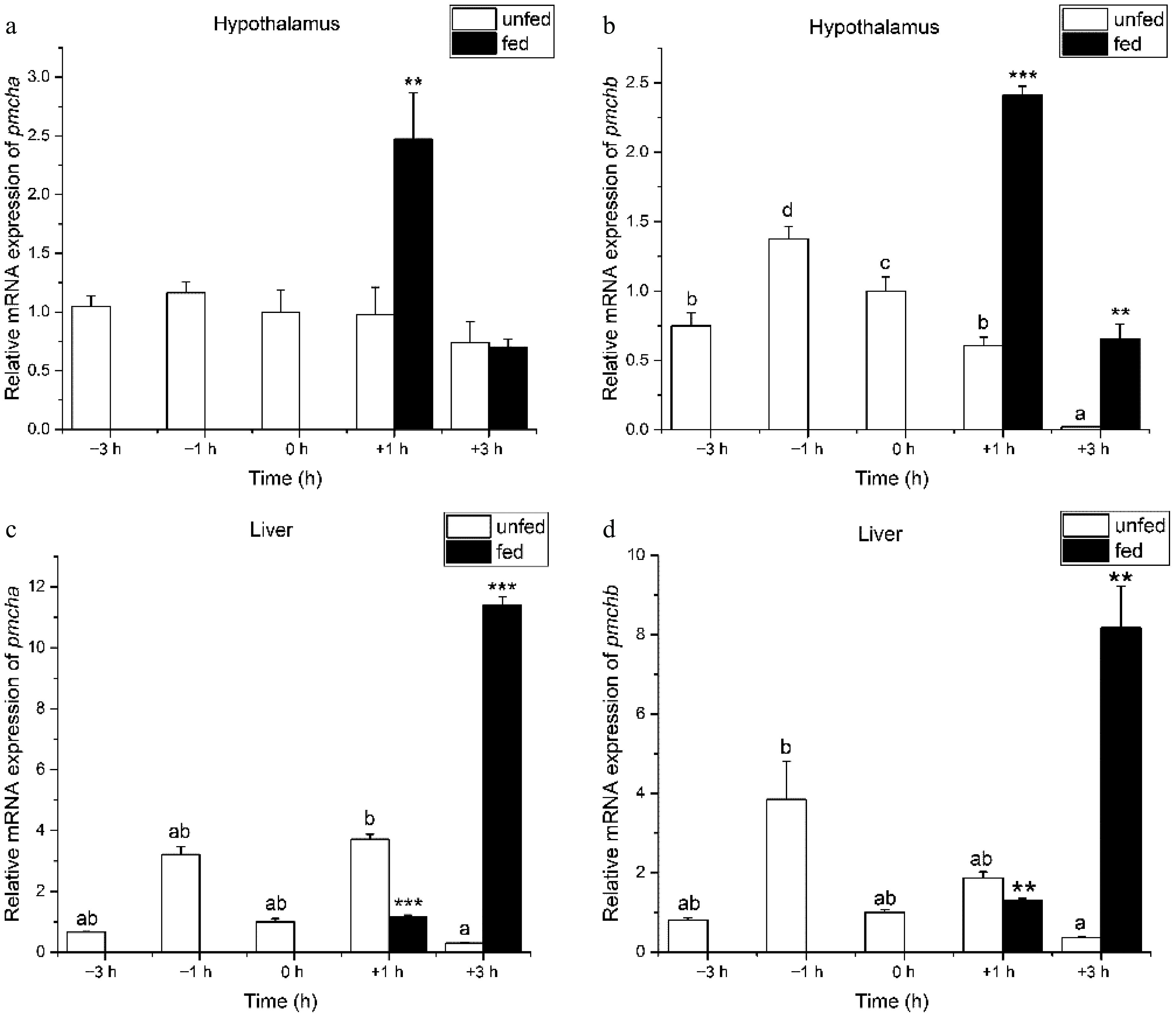
Figure 5.
Preprandial and postprandial changes in the mRNA expression of pmcha and pmchb in (a), (b) the hypothalamus and (c), (d) liver. The mRNA expression of target genes was normalized to β-actin and ef-1α. n = 9 fish per group. One-way ANOVA followed by Tukey's HSD test was conducted to compare the difference among different time points in the periprandial experiment. The asterisk represents significant differences between unfed fish and fed fish at the same time point (Student's t-test). * means p < 0.05, ** means p < 0.01, *** means p < 0.001 in the comparison between the fed and unfed groups at the same time point.
Fasting changes in the mRNA expression of pmcha and pmchb in Siberian sturgeon
-
After fasting for one day, the mRNA expression levels of both pmcha and pmchb in the hypothalamus were significantly inhibited when compared with the fed fish (p < 0.001, Fig. 6a; p < 0.01, Fig. 6b). The inhibitory effect was observed in the mRNA expressions of pmchb in the hypothalamus after 10 days of fasting relative to the fed groups (p < 0.05, Fig. 6b). Refeeding after 10 days of fasting increased the mRNA expression of pmcha and pmchb in the hypothalamus of Siberian sturgeon (p < 0.01, Fig. 6a; p < 0.05, Fig. 6b). There were no significant differences in the mRNA expression levels of pmcha and pmchb in the liver between fed fish and fasted fish on the first day. However, fasting for 10 days significantly reduced the mRNA expression of pmcha and pmchb in the liver of Siberian sturgeon (p < 0.001, Fig. 6c; p < 0.05, Fig. 6d).
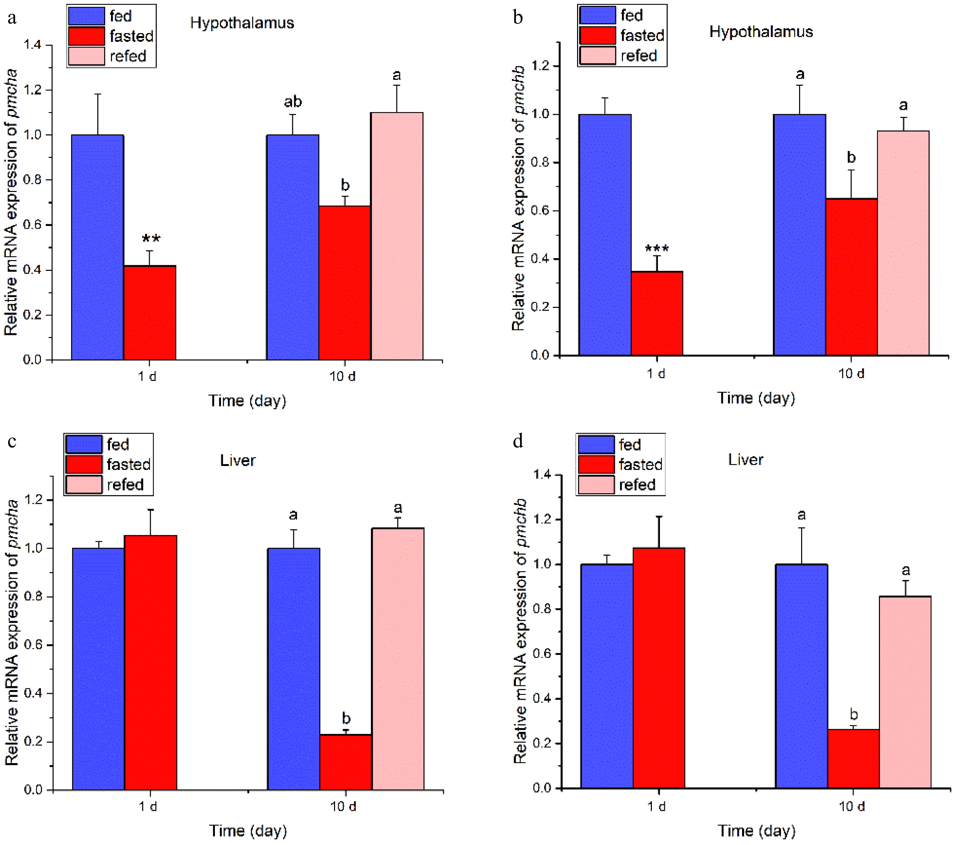
Figure 6.
Effect of fasting and refeeding on mRNA expression levels of pmcha and pmchb in the hypothalamus and liver of juvenile Siberian sturgeon. The mRNA expression levels were normalized to β-actin and ef-1α. n = 9 fish per group; one-way ANOVA followed by Tukey's HSD test was conducted to compare the difference among fed, fasted and refed groups. * represents significant differences between the fed group and the fasted group (Student's t-test), * means p < 0.05, ** means p < 0.01, *** means p < 0.001. Different letters indicate significant differences among the fed group, fasted group, and refed group.
-
The present investigation marks the initial cloning of two transcript variants of pmch cDNA sequences, pmcha and pmchb, from the Siberian sturgeon, thereby expanding our understanding of the pmch gene repertoire in fish. The variability in the number of pmch genes across different fish species is intriguing, with a single pmch sequence identified in ya-fish[15] and common carp[16], and two pmch genes in species such as zebrafish[25] and starry flounder[18]. Notably, the proteins PMCHa and PMCHb encoded by pmcha and pmchb in Siberian sturgeon diverged from those of other teleosts, which may reflect the unique evolutionary position of sturgeons. Indeed, the paleontological record indicates that sturgeon fossils date back to the Triassic period, underscoring their ancient lineage[26]. The teleost lineage, from which sturgeons diverged, experienced a pivotal whole-genome duplication event 320–350 million years ago[27], followed by additional whole-genome duplications in other fish lineages, such as cyprinids, 5.6–11.3 million years ago[28]. These evolutionary events likely contribute to the distinctive genetic makeup of Siberian sturgeon pmch genes when compared with other fish species. Furthermore, the conservation of the Siberian sturgeon's mature MCH peptide at the C-terminal in mammals and birds suggests that this region may be pivotal to its physiological functions.
Until now, little has been known about the distribution of pmch genes in fish species. The result of this study showed that pmcha was highly expressed in the hypothalamus of Siberian sturgeon, which is consistent with those in mammals such as humans[10] and rats[11], as well as chickens[14]. In addition, this finding is in accordance with reports in several teleosts. For example, pmch was highly expressed in the hypothalamus of ya-fish[15] and common carp[16], and MCH2-ir cell bodies were localized in the lateral tuberal nucleus (NLT) of the hypothalamus of zebrafish[25]. As well as the hypothalamus, pmcha and/or pmchb mRNA was highly expressed in other regions of brain, such as in the midbrain of ya-fish[15], the optic tectum/thalamus of winter flounder[19]. Interestingly, both pmcha and pmchb were highly expressed in the cerebellum of Siberian sturgeon. The cerebellum is an important structure that is involved in motor control and cognitive and emotional functions in fish[29,30]. This suggests that the high expression of pmch in the Siberian sturgeon cerebellum may be related to its role in adapting to environmental changes and behavioral regulation. Notably, pmcha and pmchb were highly expressed in the liver of Siberian sturgeon, a finding not previously observed in other teleosts. It could be postulated that pmcha and pmchb might play important roles in the regulation of energy metabolism in Siberian sturgeon. The distinct expression patterns of pmcha and pmchb suggest potential functional divergence between the two gene variants. Although pmcha shows high expression in the hypothalamus, pmchb does not, indicating that pmcha may be the primary isoform involved in central appetite regulation. In contrast, both isoforms are highly expressed in the liver and cerebellum, suggesting overlapping functions in peripheral energy metabolism and behavioral regulation. This functional divergence may have arisen from gene duplication events during evolution, allowing for subfunctionalization of the two pmch paralogs in sturgeon. Previous studies have reported that pmch1 and pmch2 were highly expressed in the pituitary of starry flounder[18], winter flounder[19], and goldfish[31]. Although this study did not detect the expression of pmcha and pmchb mRNA in the pituitary of Siberian sturgeon, a previous study observed that MCH immunoreactivity fibers were not abundant in the hypophysis of the Acipenseriform starry sturgeon (Acipenser stellatus)[32]. The results indicate that the tissue expression of pmch genes varies among species, and pmcha and pmchb might play crucial roles in the behavioral regulation and energy metabolism of Siberian sturgeon. Given that the mature MCH peptides encoded by pmcha and pmchb were 100% identical in Siberian sturgeon, their physiological functions were expected to be largely redundant at the protein level. However, in other teleosts, such as zebrafish, the two MCH paralogs have been shown to exhibit distinct tissue-specific expression patterns with little or no overlap[25], suggesting regulatory subfunctionalization following gene duplication. The differential hypothalamic expression of pmcha and pmchb observed in this study supports this notion, implying that the two paralogs may have evolved distinct regulatory mechanisms while retaining identical coding sequences for the mature peptide. This regulatory divergence may confer a selective advantage by allowing fine-tuned, context-dependent control of MCH signaling in different tissues and physiological states, as has been proposed for other duplicated neuropeptide genes in vertebrates[33,34].
Many studies have indicated that expression levels of appetite factors could be affected by feeding status[35]. Since pmcha and pmchb mRNA was highly expressed in the hypothalamus and liver of juvenile Siberian sturgeon, which are important sites involved in feeding regulation and energy metabolism, these tissues were chosen for periprandial and fasting experiments to explore the possible role of pmch in appetite regulation. In this study, mRNA expression of pmcha in the hypothalamus of Siberian sturgeon was promoted at 1 h after feeding, and mRNA expression of pmchb was promoted at 1 h and 3 h after feeding. The result suggests that pmcha and pmchb might have an anorexigenic effect in Siberian sturgeon, which is similar to the findings in common carp[16], but inconsistent with mammals and other fishes. In common carp, the level of pmch1 mRNA in the hypothalamus was increased at 1 h and 3 h after feeding[16]. However, mRNA expression of pmch in the hypothalamus of S. prenanti decreased at 1 h and 3 h after feeding[15]. These results suggest that in Siberian sturgeon, pmcha and pmchb might act as short-term satiety indicators in the hypothalamus.
To further explore the role of pmch on long-term appetite regulation in Siberian sturgeon, fasting and refeeding experiments were conducted. In this study, fasting for 10 days inhibited mRNA expression of mRNA expression but did not significantly affected mRNA expression of pmcha in the hypothalamus of Siberian sturgeon. This result suggests that pmcha may undergo adaptive tolerance during chronic energy deficiency, whereas pmchb might serve as a more persistent sensor of long-term fasting in Siberian sturgeon. Similar observations were reported in common carp[16] and goldfish[36]. Fasting for 3–7 days inhibited pmch expression in the hypothalamus of common carp, and refeeding promoted its expression[16]. In goldfish, fasting inhibited MCH-like immunoreactivity in the dorsal part of the nucleus recessus lateralis (NRLd) region of the hypothalamus[36], and an icv injection of mature MCH peptide decreased food intake[22]. These results suggest that pmchb in the hypothalamus might exert an anorexigenic role during long-term starvation in Siberian sturgeon. However, the stimulatory effect of fasting on pmch expression in the hypothalamus was observed in mice, chickens[14], and several fish species such as starry flounder[18] and Atlantic cod[17]. In addition, another study in winter flounder showed that fasting did not affect mRNA expression of pmch1 and pmch2 in the hypothalamus[19]. These observations suggest that fasting might induce the change of pmch expression in the hypothalamus in a species-related manner. Furthermore, refeeding after 10 days of fasting induced a increase in the expression of both pmcha and pmchb above fed levels, suggesting an emergency anorexigenic brake to prevent overconsumption after starvation. These findings suggest distinct temporal roles for pmcha (short-term) and pmchb (long-term) in hypothalamic appetite regulation. The liver is the main organ for lipid storage, and starvation lead to a decrease in lipid content in the liver of sturgeons[37]. It is noteworthy that fasting for 10 days inhibited mRNA expression of pmcha and pmchb in the liver. This result suggest that pmcha and pmchb might be involved in the regulation of lipid metabolism in the liver of juvenile Siberian sturgeon during starvation periods. Although little is known about the function of pmch genes in the liver of fish, a previous study revealed that icv injection of MCH triggered lipid accumulation and lipid uptake in the liver of rats[38]. These results suggest that pmcha and pmchb might be involved in the regulation of appetite and also participate in the regulation of lipid metabolism in the liver of juvenile Siberian sturgeon during starvation periods.
-
This study successfully cloned two transcript isoforms of the pmch gene from Siberian sturgeon, namely pmcha and pmchb, encoding proteins of 179 and 158 aa, respecively. Our study revealed that the mature MCH peptide encoded by Siberian sturgeon pmcha and pmchb showed high identity with mammals' MCH, but low identity with other teleost fish species' MCH. The pmcha and pmchb mRNA were highly expressed in the liver and cerebellum, and pmcha was also highly expressed in the hypothalamus of Siberian sturgeon, indicating potential functional divergence between the two paralogs. The expression patterns of pmcha and pmchb in the hypothalamus and liver in response to feeding and fasting suggest their potential roles as anorexigenic factors in appetite regulation. These findings provide important insights into the regulatory mechanisms of feeding control and energy homeostasis in Siberian sturgeon. Furthermore, the results have practical implications for sturgeon aquaculture. For example, the anorexigenic role of pmcha and pmchb suggests that feeding strategies avoiding prolonged fasting could prevent the downregulation of these satiety signals, potentially improving feed efficiency. Further studies are needed to investigate the role of mature MCH peptides and MGRP peptide for feeding and energy metabolism in aquaculture of Siberian sturgeon.
Thanks to Sichuan Agricultural University and Runzhao Fisheries (Sichuan, China) for providing us with the experimental fish.
-
These animal experiments were approved by the Animal Care and Use Committee of Sichuan Agricultural University (20250628).
-
The authors confirm their contributions to the paper as follows: experimental design and supervision: Li Z, Tang N; experimental implementation: Wu H, Jiang K, Luo Y, Li J, Li Y, Xiong Y, Wang M; data analysis and visualization: Wu H, Li J; supervision: Zhang X; resources and reagents: Li Z, Tang N, Wu H; writing-original draft: Wu H; writing-review and editing: Luo Y, Tang N. All authors reviewed the results and approved the final version of the manuscript.
-
The data used to support the findings of this study are available from the corresponding author upon reasonable request.
-
The authors declare that they have no conflict of interest.
- Copyright © 2026 by the author(s). Journal of Applied Ichthyology published by Maximum Academic Press on behalf of John Wiley & Sons Ltd. This is an open access article under the terms of the Creative Commons Attribution License, which permits use, distribution and reproduction in any medium, provided the original work is properly cited.
-
About this article
Cite this article
Wu H, Li J, Luo Y, Jiang K, Li Y, et al. 2026. Characterization, tissue distribution, and periprandial, fasting, and refeeding changes of pmch mRNA in Siberian sturgeon (Acipenser baerii). Journal of Applied Ichthyology 2026: e002 doi: 10.48130/jai-0026-0004 

Characterization, tissue distribution, and periprandial, fasting, and refeeding changes of pmch mRNA in Siberian sturgeon (Acipenser baerii)
- Received: 06 May 2026
- Revised: 23 June 2026
- Accepted: 13 July 2026
- Published online: 30 July 2026
Abstract: Melanin-concentrating hormone (MCH) is known to be involved in the regulation of feeding and energy balance in mammals. However, the role of MCH in feeding by fish remains largely unknown. In this study, two transcript variants of pro-melanin-concentrating hormone (pmch) named pmcha and pmchb were cloned from Siberian sturgeon (Acipenser baerii). PMCHa encoded a protein of 179 amino acids, and PMCHb encoded a protein of 158 amino acid, both of which showed low identity (nearly 18%−27%) when compared with those of mammals, birds and teleost fish species. Both PMCHa and PMCHb contained a conserved MCH-related mature peptide located in the C-terminal region. Quantitative real-time polymerase chain reaction analysis showed that although pmcha was highly expressed in the hypothalamus, both pmcha and pmchb were highly expressed in the liver and cerebellum. Feeding promoted the mRNA expression of pmcha and pmchb in the hypothalamus at +1 h after feeding, and fasting for 1 d inhibited hypothalamic expression of pmcha and pmchb, suggesting that pmcha and pmchb might act as anorexigenic genes in feeding control. In addition, feeding inhibited the expression of pmcha and pmchb at +1 h after feeding but increased their expression levels at +3 h, and fasting for 10 d inhibited the mRNA expression of pmcha and pmchb in the liver, suggesting that pmcha and pmchb might play important roles in regulating energy metabolism in the liver. Taken together, pmcha and pmchb might participate in inhibiting food intake and energy metabolism in juvenile Siberian sturgeon. These findings provide molecular insights into the feeding regulation of Siberian sturgeon and may inform the development of optimized feeding strategies in sturgeon aquaculture.
-
Key words:
- PMCH /
- Fasting /
- Cloning /
- Siberian sturgeon /
- Gene expression