













-
Pancreatic cancer is the most lethal malignant tumor in the gastrointestinal system. According to the latest GLOBOCAN statistics, the incidence of pancreatic cancer in China has been rising annually, with associated mortality ranking seventh globally[1]. Pancreatic cancer remains a therapeutic challenge, with five-year survival rates less than 10%[2] despite various innovative advances in treatment, such as cellular immunotherapy[3], antigen-based cancer vaccines[4], boron neutron capture therapy[5], and oncolytic virus therapy[6]. Although these novel therapies represent breakthroughs in cancer treatment, most of them are still in clinical trials and may cause unknown or severe toxic reactions; consequently, dedicated nursing management strategies are required.
Chimeric antigen receptor (CAR)-T cell therapy is one of the most promising innovative anti-tumor therapies. It utilizes genetically modified T cells to express tumor-specific CARs and has achieved significant results in patients with hematologic malignancies; related progress has also been reported in patients with solid tumors[7]. The classic CAR-T cell therapy targets for pancreatic cancer include claudin18.2 (CLDN18.2), Mesothelin (MSLN), Epidermal Growth Factor Receptor (EGFR), Human Epidermal Growth Factor Receptor 2 (HER2), Carcinoembryonic Antigen (CEA), Mucin-1 (MUC-1), prostate stem cell antigen (PSCA), and CD133[8]. In addition, trophoblast cell surface antigen 2 (TROP2) is a potential therapeutic target for pancreatic cancer. TROP2 is a transmembrane glycoprotein expressed on the surface of epithelial cells, which is overexpressed in various solid tumors and involved in tumorigenesis, invasion, and metastasis. Previous research data also indicate that CAR-T cell therapy targeting TROP2 can significantly inhibit tumor growth[9].
Multiple CAR-T cell therapies targeting MSLN, CLDN18.2, and EGFR are currently in clinical trials[8], with the highest overall response rate (ORR) reaching 16.7% and the highest disease control rate (DCR) at 70.8%[10]. However, CAR-T therapy may lead to a series of side effects, including cytokine release syndrome (CRS), immune effector cell-associated neurotoxicity syndrome (ICANS), immune effector cell-associated hematologic toxicity (ICAHT), and dermatologic adverse events (DAEs). Among these, CRS has received the maximum clinical and research attention, with recent phase 2 and phase 3 clinical trials of newer Bispecific T-cell Engagers (BiTEs) reporting incidence rates as high as 51% and 89%, respectively[11]. In contrast, although DAEs are not uncommon in CAR-T therapy, with the reported incidence ranging from 4% to 36% and severe cases being potentially life-threatening, they have been relatively underreported in the literature and have received limited attention[12,13]. CAR-T cell therapy-related DAEs primarily manifest as maculopapular rashes, erythematous rashes, purpura, macules, and bullous rashes[13].
More notably, research on the nursing management of CAR-T-related DAEs remains insufficient. To date, no standardized management protocols have been established for these events. In the context of dual-targeted CAR-T therapy, evidence-based nursing guidelines are virtually absent. Moreover, the relationship between CAR-T cell dose and DAE severity, as well as the corresponding nursing strategies tailored to different severity grades, has yet to be systematically characterized. All of these factors pose considerable challenges to clinical nursing practice. In this case study, we summarized and implemented evidence-based nursing strategies for DAEs in five patients with advanced pancreatic cancer treated with TROP2/CD133 dual-targeted CAR-T cell therapy. Through this case report, we aim to provide a feasible and replicable supportive care protocol for CAR-T-related DAEs using these evidence-based strategies.
-
Five patients with histologically confirmed advanced pancreatic cancer were enrolled in a clinical trial involving TROP2/CD133 dual-targeted CAR-T cell therapy between June 13 and November 1, 2024 (Table 1). The eligibility criteria were as follows: (1) age between 18 and 75 years; (2) survival period ≥ 3 months; (3) detection of biopsy specimens or pathological paraffin block sections: positive for TROP2 and CD133 detection; (4) advanced pancreatic cancer confirmed by histology or cytology; (5) availability of venous access for single apheresis or venous blood collection, and no other contraindications for blood cell separation. The exclusion criteria were the following: (1) active central nervous system metastases or severe autoimmune diseases; (2) diagnosis of any unstable systemic diseases (including but not limited to coronary heart disease, cerebrovascular diseases, etc.); (3) received treatment with CAR-T cells within the past six months; and (4) having mental or psychological disorders and unable to cooperate with treatment and efficacy evaluation. All five enrolled patients met the inclusion and exclusion criteria of this clinical trial. The patients had no significant comorbidities at baseline. Prior to leukapheresis, all patients had not received any chemotherapy, immunotherapy, or targeted therapy within 1–2 weeks or five half-lives (whichever was shorter).
Table 1. DAE characteristics by dose group.
Name Age Gender Dose group Rash percentage
(involved body
surface areas)Maculopapular
rash gradingPruritus grading Associated symptoms QRME 53 Female Low 3% (Chest, back, arms) Grade 1 Grade 1 / CHMI 53 Male Low 27% (Chest, back, arms) Grade 2 Grade 1 / NXME 46 Female High 90% (see Fig. 1−1) Grade 3 Grade 3 OM (Grade 2)
CRS (Grade 1)
Fever (D3–D11, highest: 40.5 °C)CWRO 69 Male High 90% (see Fig. 1−2) Grade 3 Grade 2 CRS (Grade 1)
Fever (D0/D4/D11~D12/D14,
highest: 38.5 °C)CHZH 69 Male High 90% (see Fig. 1−3) Grade 3 Grade 3 BP (Grade 4)
OM (Grade 2)
CRS (Grade 1)
Fever (D3~D7, highest: 40.2 °C)Note: The day of reinfusion is designated as D0; CRS: cytokine release syndrome; BP: bullous pemphigoid; OM: oral mucositis; low/high-dose group: 1.0 × 10⁸ cells / 2.0 × 10⁸. The DAE grading reference follows the Chinese Expert Consensus on Diagnosis and Treatment of Immune Checkpoint Inhibitor (ICIs)-Related Adverse Cutaneous Reactions (2024 Edition)[14], the Expert Consensus on Prevention and Treatment of Radiation (Chemotherapy)-Induced Oral Mucositis[15], and the American Society for Transplantation and Cell Therapy (ASTCT) Consensus Grading Criteria[16]. Following leukapheresis using the Spectra Optia® system, patients underwent lymphodepletion with fludarabine (25–30 mg/m2/day) and cyclophosphamide (250–350 mg/m2/day) infused intravenously for three days. On day 0, CAR T-cell products were administered at dose levels of 1.0 × 10⁸ cells (low-dose group) or 2.0 × 10⁸ cells (high-dose group).
All five patients developed rashes with pruritus on D2–D4 post-CAR-T cell infusion. Among them, two patients developed oral mucositis on D6 (Table 1). The severity of DAEs was more pronounced in the three patients from the high-dose group (Fig. 1), all presenting with vesicles. One patient developed bullae by D7, accompanied by fluid imbalance and lower limb edema. All patients developed grade ≥ 1 dermatologic toxicities, characterized by pruritus and maculopapular rashes, with a median onset on D3 post-infusion. Compared with those in the low-dose group, patients in the high-dose group experienced grade 4 drug-related adverse events (DAEs). One patient developed bullous pemphigoid, affecting 90% of the body's skin. Following blister rupture, significant exudate was observed, leading to erosion of the back skin. Additionally, complete blood count, blood biochemistry, coagulation function, serum cytokines, lymphocyte subsets, and pharmacokinetics (PK) were collected. The correlation between DAE occurrence and C-reactive protein (CRP) and interleukin-6 (IL-6) levels, and body temperature in the high-dose group is illustrated in Fig. 2. As illustrated in the figure, in the high-dose group, CRP and IL-6 levels showed a marked elevation coinciding with the onset of DAEs, and gradually declined as DAEs resolved following nursing interventions and symptomatic management. Body temperature changes followed a similar temporal pattern.
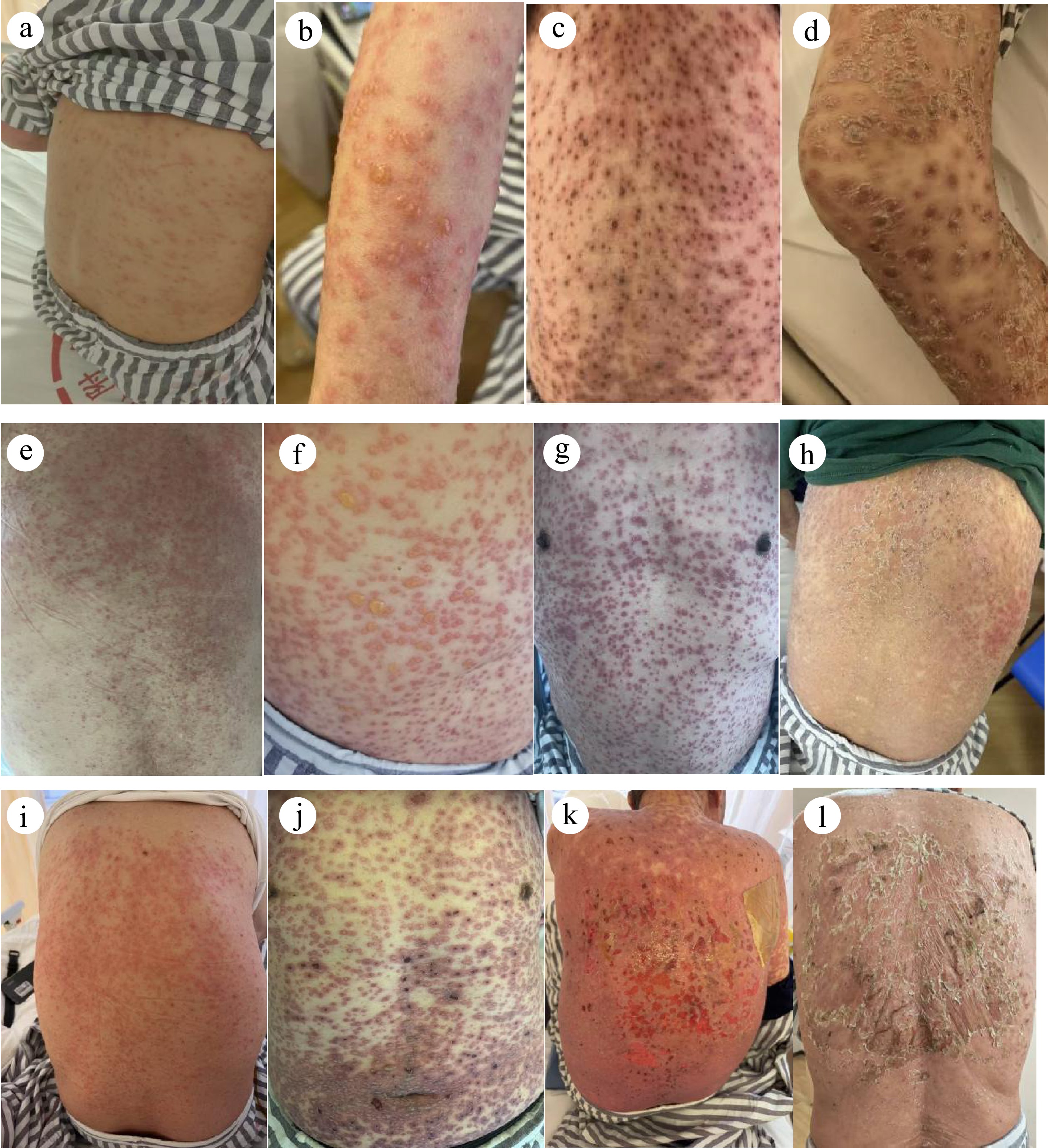
Figure 1.
Rash occurrence and outcome in the high-dose group. (a)–(d) NXME; (e)–(h) CWRO; (i)–(l) CHZH. (a) D3: rashes were spread across the entire body with mild pruritus; (b) D5: vesicles appeared, with a maximum diameter of 3 cm, accompanied by intense pruritus affecting sleep; (c) D10: vesicles partially ruptured and formed crusts; (d) D13: rusted lesions gradually shed, revealing new epidermal tissue at the affected sites; (e) D3: rashes were spread across the entire body, coalescing into patches with mild pruritus; (f) D4: dense vesicles appeared, with a maximum diameter of 2 cm and widespread pruritus; (g) D6: vesicles partially ruptured and the rashes darkened in color; (h) D14: systemic rashes crusted over and shed, with new skin emerging locally; (i) D3: rashes were spread across the entire body, accompanied by small vesicles and widespread pruritus; (j) D6: vesicles progressed to bullae, accompanied by pain and persistent pruritus affecting sleep; (k) D9: bullae ruptured, resulting in copious exudate and back erosion; (l) D21: wounds dried, fresh epithelial tissue appeared, and scabs fell, with no pigmentation or scarring.
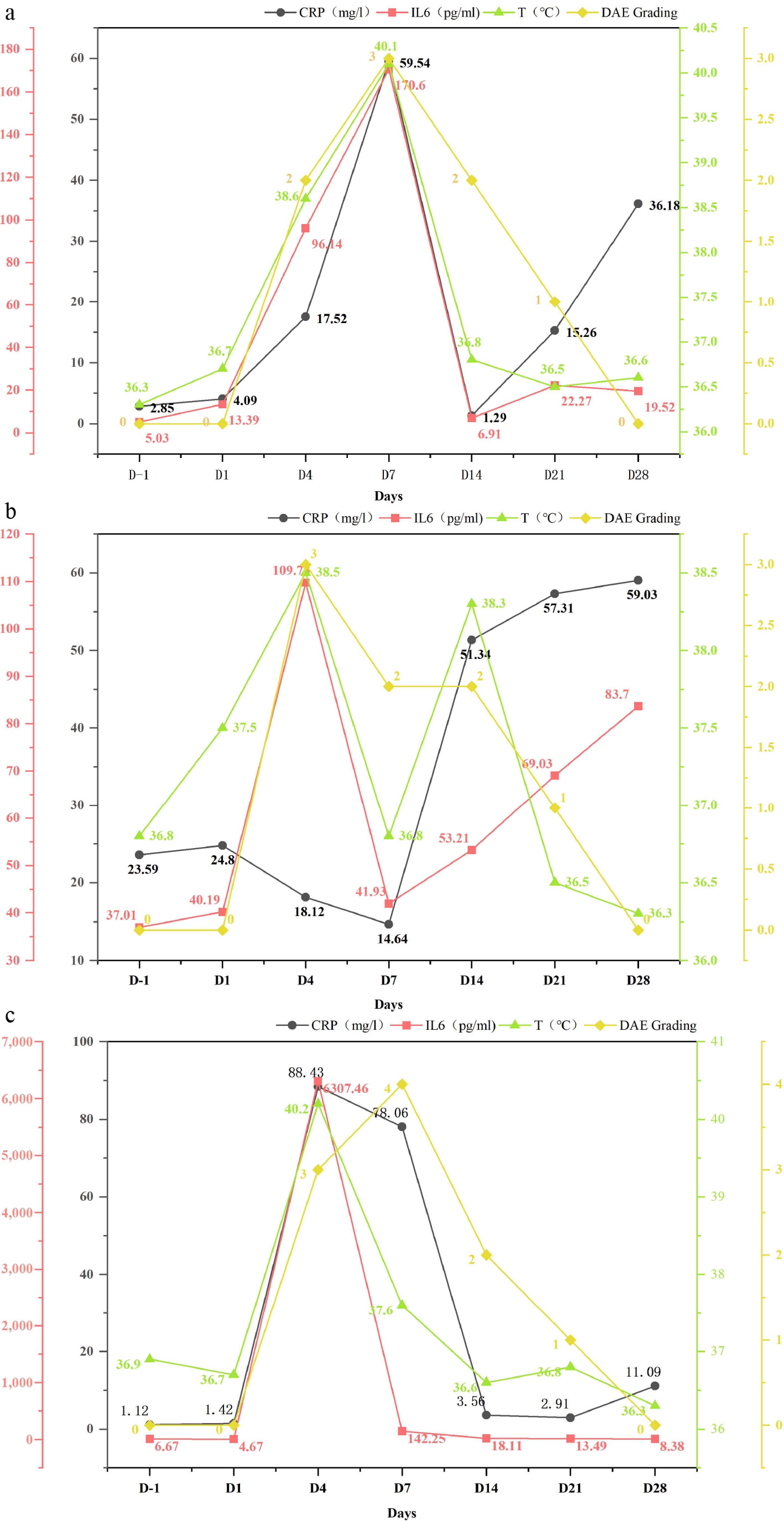
Figure 2.
Changes of CRP and IL-6 levels, body temperature, and DAEs in the high-dose group. (a) NXME; (b) CWRO; (c) CHZH.
-
Evidence-based questions were constructed and search terms were selected according to PIPOST (Population, Intervention, Professional, Outcome, Setting, Type of evidence). The search terms were "cancer/tumor/oncology", "immune checkpoint inhibitors/immunotherapy/ICIs/PD - 1/CAR-T", "cutaneous toxicities/immune-related cutaneous toxicities/cutaneous/dermatological adverse event/Pruritus/Oral Mucositis", "guideline/standard/consensus/statement/evidence summary". Computer searches were conducted from top to bottom based on the "6S" evidence pyramid model. The databases searched included the National Comprehensive Cancer Network (NCCN), European Society of Medical Oncology (ESMO), American Society of Clinical Oncology (ASCO), Chinese Anti-Cancer Association (CACA), Chinese Society of Clinical Oncology (CSCO), United Kingdom Oral Mucositis in Cancer Group (UKOMIC), the Multinational Association of Supportive Care in Cancer (MASCC), the European Academy of Dermatology and Venereology (EADV), the Society for Immunotherapy of Cancer (SITC), SciELO Brazil, CNKI, Wanfang Database, Chinese Medical Journal Full-Text Database, Web of Science, and PubMed. The search time limits ranged from the establishment of the databases to November 6, 2025.
A total of 310 records were identified across all databases. After deduplication, screening, and full-text review, 25 sources were ultimately included, comprising 12 clinical practice guidelines, 1 group standard, 9 expert consensus statements, and 3 evidence summaries (Fig. 3). The sources of the evidence are presented in Table 2.
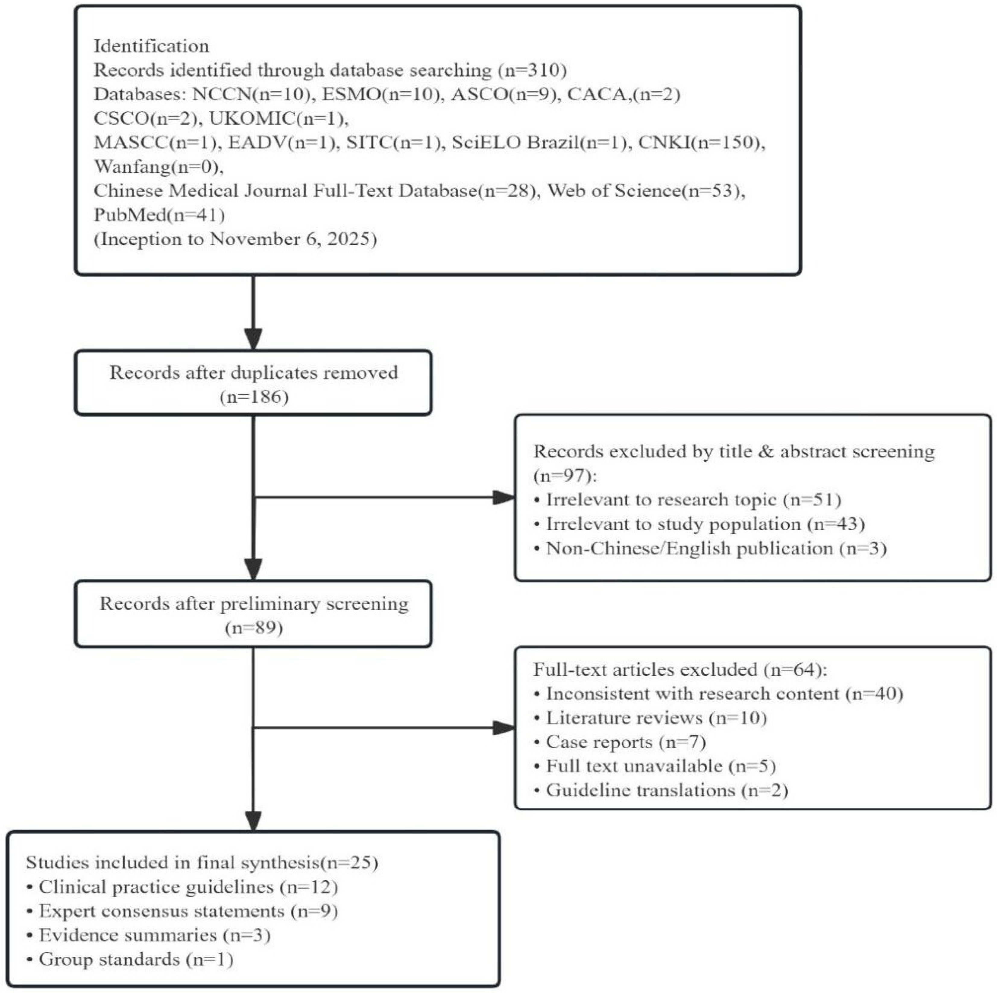
Figure 3.
Literature screening flowchart.
Table 2. Sources of evidence.
Title Author Type Year Diagnosis and treatment of ICI-related cutaneous adverse reactions Chinese Society of Dermatology (CSD) et al.[14] Expert consensus 2024 Prevention and treatment of oral mucositis Chinese Stomatological Association (CSA)[15] Expert consensus 2024 Immunotherapy-related toxicity management Haanen et al.[21] Guideline 2022 Cancer treatment cutaneous adverse event management Cury-Martins et al.[22] Expert consensus 2020 Prevention and nursing care of ICI-related cutaneous toxicity in lung cancer patients Zheng et al.[23] Evidence summary 2023 Immunotherapy-related toxicity management National Comprehensive Cancer Network (NCCN)[24] Guideline 2025 Cancer treatment and cutaneous-related adverse event management Zhu et al.[25] Guideline 2021 Diagnosis and treatment of Stevens–Johnson syndrome and
toxic epidermal necrolysisMurillo-Casas et al.[26] Guideline 2025 Management of cutaneous toxicity related to ICIs Yang et al.[27] Evidence summary 2024 Management of severe cutaneous toxicity caused by ICIs Choi et al.[28] Guideline 2025 Diagnosis and treatment of bullous pemphigoid China Dermatologist Association (CDA) et al.[29] Expert consensus 2025 Prevention and management of cutaneous adverse reactions in patients with tumor-targeted therapy Hu et al.[30] Evidence summary 2022 Management of chronic pruritus CDA[31] Guideline 2024 Application guidelines for moisturizing and emollient products CDA[32] Expert consensus 2023 Management of ICI-induced cutaneous adverse reactions Apalla et al.[33] Expert consensus 2022 Guidelines for CAR-T cell therapy of malignant hematological diseases Chinese Society of Clinical Oncology (CSCO)[34] Guideline 2024 Diagnosis, prevention, and treatment of acute oral mucositis caused by antitumor therapy CSCO[35] Expert consensus 2021 Oral care in cancer and palliative care United Kingdom Oral Mucositis in Cancer Group (UKOMIC)[36] Guideline 2019 Clinical practice management of ICI-related adverse events Brahmer et al.[37] Guideline 2021 Management of adverse events associated with ICIS. Schneider et al.[38] Guideline 2021 Glucocorticoid treatment for immune-related cutaneous conditions Subspecialty Committee on Autoimmune Diseases, Chinese Dermatologist Association[39] Expert consensus 2018 Nursing care for CAR-T cell therapy Ellard et al.[40] Guideline 2022 Management of psychosomatic symptoms in cancer patients Yin et al.[41] Expert consensus 2025 Management of CAR-T cell therapy for adults and children Hayden et al.[42] Guideline 2021 Long-term follow-up of CAR-T therapy products Shanghai Pharmaceutical Industry Association (SPIA)[43] Group standard 2023 Quality appraisal was independently conducted by two researchers trained in evidence-based nursing methodology, with disagreements resolved by a third researcher. The 12 clinical practice guidelines and 1 group standard were evaluated using the AGREE II[17] instrument; ten sources achieved Grade A recommendation and three achieved Grade B (Table 3). The 9 expert consensus statements were appraised using the JBI Critical Appraisal Checklist[18] for Text and Expert Opinion; six received "Yes" across all criteria and three received "Unclear" on one criterion each (Table 4). The 3 evidence summaries were assessed using the CASE tool[19]; two achieved "Yes" on all 10 items and one received "Not completely" on 2 items (Table 5). All 25 sources were retained for evidence synthesis. The evidence level of each source was graded using the Joanna Briggs Institute (JBI) 2014 Evidence Pre-ranking System and Grades of Recommendation[20].
Table 3. Quality appraisal results of clinical practice guidelines.
Included literature Standardized domain percentage (%) Domains
≥ 60%Domains
≥ 30%Recommendation grade Scope &
purposeStakeholder involvement Rigor of development Clarity of presentation Applicability Editorial independence Haanen et al.[21] 86.11 75.00 79.17 80.56 89.58 87.50 6 6 A NCCN[24] 97.22 77.78 66.67 91.67 70.83 70.83 6 6 A Zhu et al.[25] 86.11 55.56 62.50 86.11 64.58 70.83 5 6 B Murillo-Casas et al.[26] 88.89 77.78 80.21 88.89 68.75 79.17 6 6 A choi et al.[28] 86.11 77.78 77.08 86.11 66.67 70.83 6 6 A CDA et al.[31] 80.56 72.22 70.83 80.56 64.58 70.83 6 6 A CSCO[34] 91.67 86.11 84.38 94.44 77.08 79.17 6 6 A UKOMIC[36] 77.78 66.67 63.54 80.56 56.25 66.67 5 6 B Brahmer et al.[37] 88.89 77.78 78.13 77.78 68.75 83.33 6 6 A Schneider et al.[38] 86.11 80.56 86.46 80.56 64.58 66.67 6 6 A Ellard et al.[40] 86.11 77.78 73.96 86.11 66.67 70.83 6 6 A Hayden et al.[42] 86.11 80.56 77.08 88.89 68.75 75.00 6 6 A SPIA[43] 75.00 63.89 62.50 75.00 52.08 58.33 4 6 B NCCN: National Comprehensive Cancer Network; CDA: China Dermatologist Association; CSCO: Chinese Society of Clinical Oncology; SPIA: Shanghai Pharmaceutical Industry Association. Table 4. Quality appraisal results of expert consensus.
Author Q1 Q2 Q3 Q4 Q5 Q6 CSD et al.[14] Y Y Y Y Y Y Cury-Martins et al.[22] Y Y Y Y Y Y CDA et al.[29] Y Y Y Y Y Y CDA[32] Y Y Y Y Y Unclear Apalla et al.[33] Y Y Y Unclear Y Y CSA[15] Y Y Y Y Y Y CSCO[35] Y Y Y Y Y Y Subspecialty Committee on Autoimmune Diseases, China
Dermatologist Association[39]Y Y Y Y Y Unclear Yin et al.[41] Y Y Y Unclear Y Y CSD: Chinese Society of Dermatology; CDA: China Dermatologist Association; CSA: Chinese Stomatological Association; CSCO: Chinese Society of Clinical Oncology; Q1: Is the source of opinion clearly identified? Q2: Does the source have standing in the field of expertise? Q3: Are the interests of the relevant population the central focus? Q4: Are conclusions drawn based on analytical results and is the reasoning logical? Q5: Is reference made to existing literature? Q6: Are any inconsistencies with existing literature addressed? Table 5. Quality appraisal results of evidence summaries.
Domain Item Appraisal question Zheng et al.[23] Yang et al.[27] Hu et al.[30] Summary topic Q1 Is the summary specific in scope and application? Yes Yes Yes Summary methods Q2 Is the authorship of the summary transparent? Yes Yes Yes Q3 Are the reviewer(s)/editor(s) of the summary transparent? Yes Yes Not completely Q4 Are the search methods transparent and comprehensive? Yes Yes Yes Q5 Is the evidence grading system transparent and translatable? Yes Yes Yes Summary content Q6 Are the recommendations clear? Yes Yes Yes Q7 Are the recommendations appropriately cited? Yes Yes Yes Q8 Are the recommendations current? Yes Yes Not completely Q9 Is the summary unbiased? Yes Yes Yes Summary application Q10 Can this summary be applied to your patient(s)? Yes Yes Yes Overall inclusion Include Include Include Following appraisal, two researchers independently extracted the nursing interventions from all 25 sources and mapped them to five predefined care dimensions: (1) dynamic monitoring; (2) evidence-based management of DAEs; (3) drug use; (4) psychological support; and (5) discharge follow-up. Discrepancies were resolved through discussion or arbitration by a third researcher. The complete evidence-to-intervention mapping is presented in Table 6.
Table 6. Evidence-based nursing proposal for CAR-T cell therapy-related DAEs.
Step Content Level of evidence Recommendation grade Dynamic monitoring Vital signs monitoring 1. For acute hypersensitivity reactions, monitor vital signs and administer corticosteroids and antihistamines[22]. 5 A Hematology biomarker
Monitoring2. The incidence of DAEs is associated with an increased deviation of cytokine levels from the normal range, e.g., IL-6[21]. 1 A 3. Laboratory tests should be conducted before initiating immunotherapy[23,45]. 1 A Evidence-based management of DAEs Rash assessment and interventions 4. If skin symptoms occur, conduct a detailed medical history inquiry and immediately perform a full-body skin examination, including medication history[24,25]. 1 A 5. The severity of DAEs is typically evaluated using the CTCAE version 5.0 of the US National Cancer Institute[15,21,46] . 1 A 6 The Wallace Rule of Nines is used to assess the patient's rash area[26]. 1 A 7. Patients are advised to avoid skin irritants and sunlight[22,27]. 5 A 8. Avoid scratching and scalding with hot water, and limit the bathing time[21,27]. 1 A Nursing care of blisters 9. Exclude other causes of skin blisters. Suspect bullous pemphigoid and perform a biopsy[28]. 1 A 10. Maintain the structural integrity of the blister wall by preventing rupture, thereby establishing a biological dressing[26]. 1 A 11. Regularly clean the lesion with sterile water/saline and change the dressing. Completely cover the affected area with non-adherent or gauze dressings[29]. 5 A 12. The ruptured areas must be disinfected using an iodine solution[30]. 1 B Assessment and nursing care of pruritus 13. The severity of pruritus is evaluated using the Numerical Rating Scale method and the Visual Analog Scale for sleep disturbance[31,52]. 1 A 14. The principle of treatment and management is to actively treat the primary disease[31]. 1 A 15. Apply the moisturizing emollient and then apply the topical medication 30 min later[32]. 5 B 16. First- or second-generation antihistamines and corticosteroids may be used to relieve pruritus. For refractory pruritus, gabapentin may be considered[25,33]. 1 B 17. Grade 1 CRS is treated with symptomatic supportive therapy using non-steroidal anti-inflammatory drugs[34]. 1 A Assessment and nursing care of oral mucositis 18. The clinical grading criteria for oral mucositis follow the World Health Organization's oral toxicity scale[15]. 5 A 19. Treatment principles: relieve symptoms, prevent infections, and avoid complications[35]. 5 A 20. Conduct professional oral hygiene management[35,36]. 1 A 21. Various gargles (such as chlorhexidine), KangFuXinYe, glucocorticoids, lidocaine, and protective gels are recommended as local treatment drugs[15]. 5 A Drug use 22. Corticosteroids are used as the first-line treatment for immune-related adverse events[37]. 1 A 23. For patients with mild cutaneous toxicity, topical corticosteroids, emollients, and oral antihistamines may be used to manage symptoms. Patients with severe cutaneous toxicity unresponsive to topical therapy should receive systemic corticosteroids (initial dose 0.5–1 mg/kg/day; increase to 1–2 mg/kg/day if ineffective)[25,28,38]. 1 A 24. Use hormones in sufficient doses and for an adequate duration. Monitor for complications and consider prophylactic use of relevant medications[39]. 5 B Psychological support 25. The overall prevalence of depression among patients with malignant tumors in China is as high as 44.63%. Attention should be paid to patients' emotional well-being[40]. 1 A 26. Inform the patients; identify the physical and psychological symptoms of anti-tumor treatment, screen patients with these symptoms, and conduct scale assessment promptly[41]. 5 B Discharge follow-up 27. Long-term follow-up should be conducted by a multidisciplinary team[42]. 1 A 28. Patients receiving CAR-T cell therapy are required to undergo regular follow-up, and evaluation and management should be conducted well[43]. 1 B Dynamic monitoring
Monitoring of vital signs
-
The systemic inflammatory response following CAR-T cell infusion manifests clinically through hemodynamic alterations that serve as early indicators of potential cutaneous toxicity reactions[12]. Continuous electrocardiographic (ECG) monitoring is initiated at the beginning of cell infusion and maintained for a minimum of 6 h after the completion of the infusion. Subsequently, vital signs are measured every 6 h throughout the hospitalization. Prophylactic administration of antiallergic medications (e.g., promethazine, paracetamol) is done before infusion to mitigate the hypersensitivity risks. During infusion, clinicians and nurses routinely evaluate the patient's neurological status using the Glasgow Coma Scale (GCS) score, while monitoring for immediate signs of allergic reactions such as urticaria, angioedema, or bronchospasm. An emergency cart and airway management equipment are positioned at the bedside throughout the infusion period[22].
Monitoring of hematology biomarkers
-
Evidence implicates cytokine-mediated immune dysregulation as the pathophysiological basis for CAR-T-related DAEs[44]. Studies indicate that IL-6 and CRP levels are correlated with the onset and resolution of cutaneous toxicity, functioning as predictive biomarkers for monitoring disease activity, treatment response, and disease progression[21,23,45]. Blood samples for determination of complete blood count, CRP, biochemical parameters, coagulation function, and cytokines were collected 24 h before reinfusion, 6 h post-reinfusion, and on Days 1, 4, 7, 14, 21, and 28.
Evidence-based management of DAEs
Assessment of rashes and interventions
-
Rash is the most frequently observed symptom associated with CAR-T cell therapy-related DAEs, with the incidence rate reaching up to 36%[12]. The comprehensive evaluation begins with a detailed inquiry of the patient's history, including prior cutaneous disorders, medication exposure, and potential contact allergens. Objective documentation includes the time of onset, location, morphology, size, and accompanying symptoms[24,25]. Within the 3-day post-infusion window[13], nurses perform shift-wise evaluations using the Common Terminology Criteria for Adverse Events (CTCAE) version 5.0. The severity of DAEs is graded based on the corresponding skin lesion characteristics, presence of accompanying symptoms, and limitations in self-care ability[14,46]. The Wallace Rule of Nines is used to assess the patient's rash area[26].
Suggested management[22,27] includes the following: (1) avoid skin irritants and solar exposure; prioritize physical sun protection and avoid going out during peak sunlight hours; (2) use lukewarm water for washing the face and bathing, and apply pH-neutral skin cleansers; (3) wear soft, loose-fitting 100% cotton clothing to prevent skin friction with the areas affected by enlargement; (4) trim the nails regularly to avoid skin breaks caused by overgrown nails, and provide basic care guidance and health education; and (5) for patients requiring maintenance of venous access, select unaffected or minimally affected skin sites for peripheral catheter placement.
For Grade 3 or above, specialized interventions include the following: (1) encourage patients to wear soft, sleeveless tops and, if necessary, follow exposure therapy; instruct patients to use bed frame supports to minimize friction between the rash and bedding[47]; (2) for maculopapular rash skin lesions, gauze dressings or hydrocolloid dressings may be used; and (3) for epidermal detachment, covering with sterile gauze is the first choice[48].
Nursing care of blisters
-
Bullous reactions associated with CAR-T therapy may resemble other pathological conditions, presenting either as localized tension bullae or, more extensively, as potentially life-threatening blistering lesions. When patients present with blisters, it is suggested to first exclude other causes of cutaneous blisters, including herpes simplex, herpes zoster, and bullous pemphigoid. If bullous pemphigoid (BP) is considered, a skin biopsy is recommended[28]. In each nursing shift, it is essential to document the size of blisters, occurrence of rupture, and characteristics of the exudate, with emphasis on these observations during handover.
The management of blister formation includes the following[26]: (1) maintaining the structural integrity of the blister wall and preventing rupture, thereby establishing a biological dressing and providing resistance against infection, and (2) (for blisters ≥ 2 cm) employing hydrocolloid dressings[49] for protection. In instances where the blisters have ruptured, the management is as follows[29]: (1) implementing protective isolation[50], e.g., using laminar flow beds or wards, rigorously enforcing hand hygiene and aseptic techniques, and restricting visits; (2) exposing smaller erosive areas after daily debridement and dressing changes with normal saline or sterile water, but covering larger erosive areas with non-adhesive dressings or wet dressings; (3) disinfecting the ruptured areas using an iodine solution[30], and using recombinant bovine basic fibroblast growth factor (rb-bFGF)[51] after drying to facilitate wound healing; and (4) using protein preparations to strengthen systemic supportive treatment.
Assessment and nursing care of pruritus
-
Pruritus is also a common feature of CAR-T-associated skin toxicity, documented in two trials with an incidence of 15%[12]. Upon the onset of pruritus in patients, it is suggested to ascertain both the extent and the frequency of pruritus episodes, while also assessing the impact of pruritus on daily functioning. The severity of pruritus is assessed using the Numerical Rating Scale (NRS)[31] and the Visual Analog Scale for Sleep Disturbance[52]: both the intensity of pruritus and its impact on sleep are rated on a scale ranging from 0 to 10, wherein 0 indicates minimal discomfort and 10 indicates maximal severity.
The principle of pruritus treatment and management is the proactive intervention of the primary disease[31]. It is recommended to administer moisturizing emollients approximately 30 min before the application of topical medications[32], such as glucocorticoid creams, to optimize their efficacy in the management of rashes and pruritus. Use of oral antihistamines and topical calamine lotion may relieve pruritus. Patients should be instructed to apply medication using cotton balls or gauze for extensive areas of pruritus[33]. For patients with severe sleep disturbances, first-generation antihistamines with sedative effects may be considered[53]. Furthermore, patients should be educated to avoid scratching; instead, a gently tapping of the skin with fingertips may help them to alleviate the discomfort associated with pruritus.
Additionally, pruritus tends to intensify during episodes of high fever. Studies have indicated a significant correlation between CRS and the manifestation of rash. Thus, the management of high fever potentially alleviate pruritus[13]. The last three cases in this cohort developed fever, which were managed in accordance with the 2024 CSCO Clinical Application Guidelines for CAR-T Cell Therapy in Hematologic and Lymphatic System Tumors[34]: (1) collection of blood samples via peripheral and central venous catheters to exclude infection; (2) administration of nonsteroidal anti-inflammatory drugs (NSAIDs) supplemented with physical cooling measures; and (3) prompt replenishment of fluid balance, recording of 24-h fluid balance, and maintenance of a positive fluid balance.
Assessment and nursing care of oral mucositis
-
CAR-T cell therapy-related oral mucositis occurs in approximately 6% of patients, yet it has a significant impact on the quality of life[12]. According to the World Health Organization (WHO) oral toxicity scale[15], the grading of oral mucositis encompasses three dimensions: ulcers, pain, and the eating situation.
The primary management of oral mucositis includes the following[35]: (1) control oral pain and use mucosal protectants to enhance the healing process of oral tissues; (2) keep the oral cavity clean to reduce the risk of multiple infections; (3) prevent the progression of oral mucositis to higher grades of mucositis (WHO grade 3 or 4); and (4) avoid complications such as ulcer bleeding, oral multiple infections, malnutrition, dehydration, and electrolyte imbalance.
Non-pharmacological treatment methods[36] mainly include the following: (1) routine observation of the oral mucosa in every shift, adherence to oral hygiene, use of a soft-bristled toothbrush for brushing after meals and before sleep, and rinsing the mouth with saline after meals; (2) encouraging increased water drinking, with a daily fluid intake of approximately 1,500–2,000 ml; and (3) guiding the patients to consume a warm, cool, high-protein, high-calorie diet enriched with vitamin C and free of irritation, recommending small and frequent meals, and monitoring the nutritional status and body weight.
Local symptomatic interventions may be administered using pharmacological agents, including chlorhexidine, KangFuXinYe (which refers to a traditional Chinese medicine clinically used for the treatment of oral ulcers), and glucocorticoids. Existing evidence suggests that chlorhexidine can prevent and alleviate oral mucositis[54]. Patients should be instructed to increase the frequency of its use to 2–3 times daily, using approximately 10–15 ml each time. They should be advised to gargle it vigorously for a duration of 30 s to 1 min before spitting it out. Furthermore, patients should refrain from eating, drinking, or rinsing with clean water for at least 30 min after gargling to ensure the efficacy of chlorhexidine within the oral cavity. Both KangFuXinYe and glucocorticoids have demonstrated efficacy in alleviating the severity of oral mucositis; patients should be educated to use it rationally[15]. In addition, in instances of pain, the application of a protective oral gel is recommended[55], which has demonstrated to be effective in pain alleviation.
Drug use
-
Systemic corticosteroids are commonly used as the first-line treatment for immune-related adverse events[37]. According to the guidelines, patients with grade I/II DAEs are recommended to take oral antihistamines, apply glucocorticoid ointments topically, or take oral glucocorticoids at a dose of 0.5–1.0 mg/(kg·d). Patients with grade III/IV DAEs are treated with intravenous methylprednisolone at a dose of 1–2 mg/(kg·d)[25,28,38]. When using hormones for a long time, a gastric mucosa protector should be used simultaneously to prevent gastrointestinal reactions. Glucocorticoids can increase the risk of infection, so relevant infection indicators should be monitored regularly, and antibacterial drugs should be used prophylactically. Drugs should be administered strictly according to the doctor's advice in terms of timing and dosage, and the dosage should not be changed casually. The hormone dosage should be gradually reduced during medication. Patients should be instructed not to stop taking the drugs without permission. The patient's blood glucose, blood pressure, and night sleep should be monitored, and symptomatic treatment should be provided[39].
Psychological support
-
Cancer can cause anxiety in patients; the incidence of depression among patients with malignant tumors can reach as high as 44.63%[56]. At the same time, the long duration of CAR-T cell therapy and the uncertain of treatment outcomes may further exacerbate patient anxiety[40,57]. In this regard, proactive preventive strategies include the following[41]: (1) providing patients with a comprehensive explanation of CAR-T cell therapy protocol, including potential adverse reactions and coping strategies; (2) conducting regular ward visits every morning and evening to continuously assess the patient's clinical status and emotional well-being, utilizing the patient health questionnaire-9 (PHQ-9) or generalized anxiety disorder 7-item scale (GAD-7) for screening purposes; (3) promptly identifying and addressing symptomatic adverse reactions, such as DAEs, which may impact the patient's appearance, sensory perceptions, and subjective feelings, thereby affecting their daily activities, including eating and sleeping; (4) reducing frequent medical procedures in the ward; and (5) implementing mindfulness therapy and presenting successful cases from previous treatment, thereby enhancing the patient's confidence and effectively alleviating anxiety.
Discharge follow-up
-
Uncertain rare or long-term adverse reactions associated with CAR-T cell therapy, such as late-onset cytopenia and secondary malignancies, remain inadequately investigated[58]. Studies have indicated that some skin events may occur more than one year after CAR-T cell infusion[59]. Therefore, the implementation of long-term follow-up and monitoring of patients is crucial. The United States Food and Drug Administration (FDA) recommends extending the follow-up duration of post-CAR-T cell infusion to a lifetime, facilitating the timely and comprehensive documentation and identification of potential late-onset or delayed adverse reactions[60]. Long-term follow-up should be executed by a multidisciplinary team, comprising physicians, disease-related specialists, long-term follow-up nursing staff, data administrators, and clinical trial staff, in order to collect data regarding disease status and long-term effects[42]. Continuity of care should be provided for patients, while both patients and their families should be fully informed of possible complications and educated on effective self-management strategies. After discharge, patients are required to maintain regular hospital visits. A series of evaluations and management strategies should be undertaken during follow-up visits. Innovative remote intelligent medical service should be developed to foster information connectivity among patients for precise guidance and therapeutic interventions[43].
Nursing outcomes
-
Following implementation of the evidence-based nursing protocol, all five patients completed the management of DAEs without secondary skin infection or interruption of CAR-T cell therapy.
In the low-dose group, rashes and pruritus gradually resolved within approximately two weeks after CAR-T cell infusion. Blister formation and oral mucositis were not observed.
In the high-dose group, DAEs were more severe. The most severe case (patient CHZH) progressed from diffuse rash with vesicles on Day 3, to bullae with pain and sleep-disturbing pruritus on Day 6, and to ruptured bullae with copious exudate and back erosion on Day 9. By Day 21, wounds were dry, fresh epithelial tissue appeared, and scabs fell off without pigmentation or scarring (Fig. 1). As shown in Fig. 2, CRP and IL-6 levels and body temperature in the high-dose group showed an increasing trend during the period of worsening symptoms and gradually declined as the skin lesions improved. Given the very small sample size, these changes are presented descriptively rather than as formal statistical correlations.
During follow-up, no recurrent severe dermatologic adverse events were observed in any of the five patients.
-
Pancreatic cancer presents particular therapeutic challenges due to its immunosuppressive tumor microenvironment, which contributes to poor immunotherapy outcomes and prognosis. Given that pancreatic cancer is often diagnosed at advanced stages when curative treatment options are limited, there is an urgent need for early diagnosis to improve patient survival and therapeutic efficacy. Early detection enables timely intervention, facilitates surgical resection, and significantly enhances the effectiveness of subsequent systemic therapies[61]. Although CAR-T cell therapy offers a promising novel treatment option for pancreatic cancer, its clinical application still faces obstacles, such as stromal desmoplasia, heterogeneous antigen expression, and immunosuppressive tumor microenvironment[62]. Nevertheless, multiple CAR-T products have entered clinical trials. Systemic inflammatory responses following CAR-T cell infusion may manifest clinically through cutaneous toxicity reactions that demand vigilant monitoring and evidence-based interventions.
The underlying mechanism of CAR-T-induced epidermal toxicity remains unclear. One possible explanation is related to the attack of target antigens expressed on normal epithelial cells and vascular endothelial cells by CAR-T cells, namely the cross-reactivity of CAR-T cells with target antigens expressed on malignant cells and similar antigens expressed on normal tissues[12,63,64]. Another consideration, given the known immunomodulatory effects of CAR-T therapy, is that the possible mechanisms include cytokine-mediated inflammation and immune dysregulation. Studies have shown that CAR-T therapy can lead to the release of pro-inflammatory cytokines, which may contribute to the development of skin manifestations[13,45].
Although existing literature primarily consists of case reports, available clinical data indicate that the incidence of DAEs varies greatly, ranging from 4% to 36%[12,65]. Most reactions are mild and self-limiting, but severe symptoms also occur. In this case study, all 5 patients developed DAEs, mainly manifested as maculopapular rashes, BP, and oral mucositis. Severity was correlated with established risk factors, especially the CAR-T cell infusion dose[66], which is consistent with prior reports. It is important to note, however, that other potential contributing factors, including advanced age[67,68], female gender[69], and expression of CAR-T cell targets in the skin[70,71], could not be verified in this study. In future, a study with a larger and more diverse patient population is needed to validate the influence of these additional parameters. Importantly, associated symptoms (e.g., pruritus, pain) and comorbid conditions (e.g., CRS) were also present, which seriously impacted the treatment tolerance and quality of life, posing challenges to nursing practice. In this study, nursing interventions were adjusted according to the severity of DAEs observed in each dose group. However, due to the limited sample size and the case report design, the current evidence is insufficient to establish differentiated nursing protocols specific to each dose group. Future studies with larger cohorts are needed to further investigate whether dose-specific nursing protocols can be developed and standardized to optimize the management of DAEs across different CAR-T cell infusion doses. Therefore, clinical medical staff should attach importance to early identification of high-risk populations, evidence-based and personalized management protocols, and multidisciplinary long-term follow-up of CAR-T cell therapy-related DAEs.
Comprehensive dynamic assessment and risk prediction
-
The unpredictability of CAR-T cell therapy-associated toxic reactions presents significant clinical challenges. Previous studies have demonstrated significant individual variability in the incubation period of toxic reactions after the first infusion, with the fluctuation range ranging from 24 h to 19 months[64,65]. Therefore, it is crucial to establish early detection systems incorporating dynamic monitoring for high-risk patients. For this, predictive biomarkers emerge as a particular promising approach[45].
This study integrated the multi-parameter joint monitoring of IL-6 and CRP levels, and body temperature. The observed correlations between these parameters and the onset, progression, and resolution process of DAEs further validate the research conclusions of Valpione[69] and Dimitriou[72]. However, as non-specific inflammatory markers, the elevated IL-6 and CRP levels may also reflect various pathological states such as infection, autoimmune disorders, or advanced tumors[73]. Therefore, the clinical interpretation of these biomarkers requires further consideration. To enhance the predictive accuracy, it is suggested to establish a risk prediction model using artificial intelligence technology and incorporating multi-source heterogeneous data: (1) patient demographics, e.g., age, gender; (2) treatment parameters, e.g., CAR-T cell infusion dose; (3) biological markers, e.g., cutaneous target expression; (4) physiological measures, e.g., body temperature; and (5) inflammatory indices, e.g., IL-6 and CRP. Such an integrative predictive framework is of great value for achieving precise prevention and evidence-based management of DAEs and improving patients' safety. Future research may also explore the integration of advanced imaging modalities, such as near-infrared fluorescence and Cerenkov luminescence dual-modality imaging, to enhance real-time monitoring of changes in the tumor microenvironment during immunotherapy[74].
Multidisciplinary team collaboration involving nursing
-
The management of CAR-T cell therapy-related DAEs presents clinical challenges that demand a multidisciplinary collaboration. On one hand, there are limited clinical safety data and complex immune toxicity mechanisms with diverse symptom manifestations. On the other hand, treatment decisions need to balance between anti-tumor efficacy and toxicity control, coupled with comprehensive support and long-term management. All these highlight the necessity of multidisciplinary team collaboration. Current guidelines[75] also advocate that nurses should be actively present in the multidisciplinary team for toxicity monitoring, preventive interventions, and patient education. Meanwhile, its effectiveness has been corroborated by multiple studies. Research indicates that preventive interventions involving nurses can reduce the incidence of grade 3 skin toxicity from 21% to 8%, and the proportion of patients requiring systemic cortisol treatment can also decrease significantly from 36% to 10%[76]. Moreover, a nurse-led follow-up program has been shown to successfully reduce the incidence of high-grade adverse events by 25%[77].
In this case study, the severity of rashes was effectively controlled through multidisciplinary consultations involving clinicians of oncology, dermatology, rheumatology, infectious diseases, stomatology, nutriology, and psychology. Within the multidisciplinary team, the nursing team should play the following essential roles: (1) as risk assessors for developing and implementing multi-dimensional toxicity evaluations; (2) as communication coordinators for establishing communications among the multidisciplinary stakeholders; and (3) as care providers for implementing supportive care. This case study underscores that optimal DAE management requires integrating nursing expertise throughout the care continuum, ranging from risk assessment to longitudinal symptom management. The nurse-involved multidisciplinary collaboration model needs to be validated and broadly implemented.
Limitations
-
Although this case study developed and implemented an evidence-based nursing protocol for CAR-T cell therapy-related DAEs in 5 patients with pancreatic cancer, several important limitations need to be considered. Firstly, as it is a single-center phase I clinical trial with only five patients, the case study inherently limits a broad applicability of the findings. The successful results obtained in this case may not translate to all patients undergoing CAR-T cell therapy. The effectiveness of this evidence-based nursing protocol still needs to be verified through controlled clinical trials.
Secondly, this study did not have a control group, which precludes causal inference between the nursing interventions and the observed outcomes. It remains unclear whether the resolution or improvement of DAEs was attributable to the evidence-based nursing protocol, the natural course of the dermatologic events, concurrent pharmacological treatments, or a combination of these factors. Furthermore, due to the complexity of the real-world settings, the nursing outcomes may be influenced by multiple confounders, such as individual characteristics, heterogeneity of support systems, implementation, hardware resource, or regulations. These factors underline the importance of real-world data and patient-centered approaches when translating evidence into practice.
Thirdly, the associations between DAEs and biomarkers (CRP, IL-6), as well as body temperature, reported in this study were based on a descriptive observation of temporal trends rather than formal statistical analysis. Owing to the small sample size (n = 5, with only 3 patients in the high-dose group), deriving quantitative correlation metrics such as correlation coefficients or regression analyses were not feasible. These findings should therefore be interpreted as preliminary observations that warrant further validation in adequately powered studies.
Fourthly, for the sake of protecting patients' privacy, some contextual details had to be anonymized, which may have potentially affected the completeness of the case description and reported outcomes. Future multi-center controlled studies with larger samples and longitudinal studies to verify the effectiveness of the evidence-based nursing interventions are highly recommended.
-
The current absence of standardized CAR-T cell therapy-related DAE management guidelines presents both opportunities and challenges. This case study has demonstrated the preliminary feasibility of implementing an evidence-based nursing protocol for managing CAR-T cell therapy-related DAEs in patients with pancreatic cancer. The findings highlight that effective management of DAEs requires a comprehensive, individualized care continuum spanning from pretreatment assessment to long-term follow-up. To advance CAR-T cell therapy-related DAE management and research, structured identification and monitoring models incorporating multi-source data, including patient demographics, treatment parameters, biological markers, physiological measures, and inflammatory indices is recommended. Future research to develop and validate the nursing-involved multidisciplinary collaboration and long-term follow-up guidance of CAR-T cell therapy-related DAEs is also needed to maximize patients' benefits.
This study was sponsored by the Fundamental Research Funds for the Central Universities (Project No. YG2025QNA03) and Shanghai Jiao Tong University School of Medicine: Nursing Development Program.
-
This study was performed in line with the principles of the Declaration of Helsinki. Approval was granted by the Ethics Committee of the Ren Ji Hospital, School of Medicine, Shanghai Jiao Tong University (No. -LY [2024]067B). Written informed consent was obtained from all patients for participation in the study and for the publication of their clinical data and images. All patient-identifying information has been removed to protect privacy.
-
The authors confirm their contributions to this study as follows: conceived and designed the study: Ji M, Liu X; collected the case information: Zhou T, Ding S; conducted the literature review: Liu X, Gu S; interpreted the results: Liu X, Gu S, Ji M, Zhou T; drafted the initial manuscript: Cui J, Liu X; revised: Ji M. All authors reviewed the results and approved the final version of the manuscript.
-
Not applicable.
-
The authors declare no conflict of interest.
-
# Authors contributed equally: Xiaoqian Liu, Shiyun Gu
- Copyright: © 2026 by the author(s). Published by Maximum Academic Press, Fayetteville, GA. This article is an open access article distributed under Creative Commons Attribution License (CC BY 4.0), visit https://creativecommons.org/licenses/by/4.0/.
-
About this article
Cite this article
Liu X, Gu S, Zhou T, Ding S, Cui J, et al. 2026. Evidence-based nursing management of CAR-T cell therapy-related dermatologic adverse events in pancreatic cancer: case report and literature review. Gastrointestinal Tumors 13: e011 doi: 10.48130/git-0026-0010 

Evidence-based nursing management of CAR-T cell therapy-related dermatologic adverse events in pancreatic cancer: case report and literature review
- Received: 30 November 2025
- Revised: 17 May 2026
- Accepted: 26 May 2026
- Published online: 31 July 2026
Abstract: In this case report, we summarize and implement an evidence-based nursing protocol for dermatologic adverse events (DAEs) in five patients with advanced pancreatic cancer undergoing TROP-2/CD133-targeted chimeric antigen receptor (CAR)-T cell therapy (2 low-dose and 3 high-dose recipients). A systematic review of the current evidence, including 12 clinical practice guidelines, 9 expert consensus documents, 3 evidence summaries, and 1 group standard, was conducted to formulate the nursing protocol. The protocol consists of dynamic monitoring, toxicity assessment and symptom intervention, psychological support, medication management, and post-discharge follow-up. All patients developed DAEs within 48-96 h post-infusion, with higher severity observed in the high-dose group. The correlation between the occurrence of DAEs and C-reactive protein (CRP) and interleukin-6 (IL-6) levels and body temperature in the high-dose group was identified. With the implementation of the evidence-based nursing protocol, skin lesions were resolved in 4 cases by Day 14 and in one severe case by Day 21. This case study demonstrated that the nursing protocol was feasible, yet its efficacy needs to be validated through multi-center randomized controlled studies.