













-
Globally, food allergies represent a prevalent and serious health condition, primarily caused by abnormal immune responses to dietary allergens[1]. Milk contains many proteins that benefit human health and act as a major source of bioactive peptides[2]. However, milk also represents one of the most widely reported food allergens in infants and young children. Notably, over 77.5% of cow's milk protein allergy (CMA) reactions are due to casein (CN)[3]. This fact highlights casein's key role in triggering CMA responses. Therefore, strategies to reduce the allergenicity of CN while maintaining its nutritional value are essential to ensure consumer food safety.
Currently, technical strategies for mitigating CN allergenicity are classified into three main types: physical methods (e.g., ultrasound), chemical methods (e.g., enzymatic hydrolysis, glycosylation), and biological methods (e.g., fermentation)[4]. Among them, enzymatic hydrolysis has become the most widely applied approach, owing to its high efficiency and simple operation. For food-grade proteases, alkaline protease exhibits superior performance in reducing CN allergenicity compared with trypsin, pepsin, and papain[5]. Alkaline protease can recognize the carboxyl terminus of aromatic, acidic, and sulfur-containing amino acid residues and cleave peptide bonds, thereby disrupting the integrity of antigenic epitopes and reducing allergenicity[6]. Studies show that alkaline protease has strong hydrolytic activity on milk proteins[7]. Alkaline protease hydrolysis can significantly reduce the allergenicity of milk proteins and improve the antioxidant-related properties of the resulting hydrolysates. However, its application is still limited by the pronounced bitterness of the hydrolysates and the possible persistence of residual antigenic epitopes under different hydrolysis conditions[8]. Therefore, developing a strategy that can simultaneously reduce allergenicity and alleviate bitterness remains an important challenge in the preparation of casein hydrolysates.
Cold plasma (CP) is a non-thermal, gas-phase technology that has been studied for food processing applications[4]. Plasma is generated by ionizing gases and contains various reactive species[9], which can interact with proteins and peptides, leading to structural modification, peptide bond cleavage, and changes in functional properties[10]. Based on our previous research[11,12], cold plasma (75 W, 12 min) treatment of a 2 mg/mL casein solution reduced antigenicity by 80.46%. This reduction was associated with hydroxyl radicals generated by argon feed gas. These radicals cleaved peptide bonds and changed casein structure, disrupting both linear and conformational epitopes. The hypoallergenic effect was further confirmed in sensitized KU812 cells and BALB/c mice. However, the use of CP as an assisting treatment to modulate both allergenicity and bitterness in protein hydrolysates has rarely been explored[9,13]. Therefore, in the present study, alkaline protease hydrolysis followed by CP treatment was applied to prepare casein hydrolysates (CNHs), and the resulting products were evaluated in terms of structure, antigenicity, antioxidant activity, and bitterness. The novelty of this study lies in applying CP treatment after enzymatic hydrolysis and determining whether this sequential strategy offers advantages over individual treatments in reducing residual antigenicity and bitterness while maintaining antioxidant potential. The findings may provide experimental evidence for the future development of CNHs with reduced residual antigenicity and lower bitterness.
-
Casein (S12002) and alkaline protease (200 U/mg) were purchased from Yuanye Biotechnology Co., Ltd (Shanghai, China). Ultra-low molecular weight protein marker (3.3–20.1 kDa) and total antioxidant capacity (T-AOC) assay kits were purchased from Beijing Solarbio Science & Technology Co., Ltd (Beijing, China). Goat anti-human Immunoglobulin E (IgE) secondary antibody was obtained from Abnova (Taipei, Taiwan, China). HRP-labeled streptavidin, 1,1-diphenyl-2-picrylhydrazyl (DPPH), and 2,2′-azino-bis (3-ethylbenzothiazoline-6-sulfonic acid) diammonium salt (ABTS-[NH4]2) were purchased from Aladdin Reagent Co., Ltd (Shanghai, China). Ferrozine monosodium salt hydrate (CAS No. 63451-29-6) and iron (II) chloride tetrahydrate were purchased from Macklin Biochemical Co., Ltd (Shanghai, China). All chemicals were of analytical grade.
Preparation of CNHs
Enzymatic treatment
-
The hydrolysis conditions for alkaline protease were adopted from previous studies that significantly reduced casein antigenicity[5,14]. A 10 mg/mL solution was prepared by dissolving CN in a 0.02 M phosphate-buffered saline solution (pH 7.4). The solution was adjusted to pH 9.0 and incubated at 45 °C with an enzyme-to-substrate ratio of 5,000 U/g. After 30, 60, and 120 min of digestion, the reactions were terminated by heating at 90 °C for 15 min, followed by rapid cooling in an ice bath.
Cold plasma treatment
-
CP treatment conditions were adopted from previous work[11], showing reduced casein antigenicity. The dielectric barrier discharge (DBD) CP generator (Suman Electronics Co., Ltd, China) consisted of a power supply and a reactor (Supplementary Fig. S1). A 5 mL sample was placed in the reactor vessel positioned between the two dielectric plates. Upon activation of the generator, argon was employed as the carrier gas, with a flow rate of 1 L/min. Plasma was subsequently generated the moment a high-voltage current disrupted the carrier gas between the upper and lower dielectric plates of the reactor. The sample was treated with DBD plasma at 75 W for 10 min. After plasma treatment, the mixtures were centrifuged. Centrifugation conditions were 4,000 r/min for 15 min. The resulting supernatants were collected and freeze-dried. Untreated CN was used as the control. Samples treated only with alkaline protease were named ECN30, ECN60, and ECN120. These correspond to 30, 60, and 120 min of enzymatic digestion, respectively. Samples treated with DBD alone were designated CP, whereas those subjected to both enzymatic digestion and CP treatment were designated ECN30-CP, ECN60-CP, and ECN120-CP.
Determination of the degree of hydrolysis (DH)
-
The degree of hydrolysis (DH) was determined using the ortho-phthaldialdehyde (OPA) method. This method quantifies peptide bonds. A 400 µL aliquot of the diluted sample was prepared. The sample was diluted 1:20 in 0.02 M PBS. The mixture was incubated in the dark for 5 min. Then, its absorbance at 340 nm was measured. The concentration of primary amino groups in CN was quantified. An L-leucine standard curve (0−200 μg/mL) was used[15]. DH was calculated using the following equations:
$ Y=A\times \dfrac{N}{131.17\times c} $ (1) where, A is the amount of L-leucine equivalent determined from the standard curve (mg/mL); N is the dilution factor;
$ \text{DH}\left(\text{%}\right)=\dfrac{{Y}_{1}-{Y}_{0}}{{\text{h}}_{\text{tot}}}\times 100 $ (2) where, Y1 and Y0 are the primary amino group contents of the hydrolyzed and unhydrolyzed samples; htot is the total number of peptide bonds per gram of protein, taken as 8.2 for CN.
Gel electrophoresis
-
For analysis of the molecular weight distribution of CNHs, the SDS-PAGE protocol was performed according to the method described by Tang et al.[16]. CN samples were mixed with loading buffer and incubated at 100 °C for 5 min. Then, 10 μL of the sample was loaded onto precast gels and electrophoresed at 160 V for 40 min. The gels were stained with Coomassie Brilliant Blue R-250 and analyzed using a Gel Doc™ XR Imaging System.
Tricine-SDS-PAGE of CNHs was carried out according to the method of Ye et al.[5]. Samples were mixed with sample buffer and heated at 95 °C for 5 min. A 4% stacking gel and a 12% separating gel were prepared and electrophoresed under appropriate voltage conditions.
Enzyme-linked immunosorbent assay analysis
Human sera and ethics statement
-
Milk allergy serum was obtained from PlasmaLab International, a company based in Everett, WA, USA, with its clinical history meticulously verified (Supplementary Table S1). The utilization of human serum samples was approved by the Ningbo University Ethics Committee (approval ID: NBU2025001), in accordance with the Declaration of the International Ethical Guidelines for Biomedical Research Involving Human Subjects.
Sensitization assessment
-
The enzyme-linked immunosorbent assay (ELISA) was conducted according to Li et al.[17]. CN or its hydrolysates were added to 96-well plates and incubated overnight at 4 °C. After washing, 3% gelatin was added, and the plates were blocked at 37 °C for 1 h. After another wash, diluted serum samples were added and incubated at 37 °C for 1 h. This was followed by the sequential addition of biotin-labeled goat anti-human IgE and horseradish peroxidase (HRP)-labeled streptavidin, with each being incubated at 37 °C for 1 h. The TMB substrate was added, and the reaction was allowed to proceed at 37 °C for 15 min. Finally, sulfuric acid was added to terminate the reaction, and absorbance was measured at 450 nm.
Structural characteristics
Fourier transform infrared spectroscopy (FTIR)
-
FTIR spectra of CN samples were obtained following the method of Bing et al.[18]. Freeze-dried CN was ground with potassium bromide (KBr) at a ratio of 100:1 (w/w) and pressed into thin pellets. Spectra were recorded on a Nicolet Apex FTIR spectrometer (Thermo Fisher Scientific, USA). Spectra were collected over the range of 400–4,000 cm−1 with a resolution of 4 cm−1 and averaged over 32 scans. Second-derivative spectra and curve fitting of the amide I band (1,600–1,700 cm−1) were analyzed using Peakfit 4.12 software.
Tertiary structure analysis
-
UV absorbance spectra of CN samples were recorded using a UV–Vis–NIR spectrophotometer (UV-330, Shanghai Meppan Instruments Co., Ltd, China). The intrinsic fluorescence spectra of CN samples were recorded using an F-4700 fluorescence spectrophotometer (Hitachi, Japan) at an excitation wavelength of 280 nm, with a scanning range of 250–350 nm[19,20].
Surface hydrophobicity
-
Surface hydrophobicity of CN was determined using a modified method with the fluorescent hydrophobic probe 8-anilino-1-naphthalenesulfonic acid (ANS)[21]. The samples were prepared in serial concentrations. Two millilitres of each sample were mixed with 20 µL of 8.0 mM ANS solution and incubated for 15 min in the dark. Fluorescence intensity was measured at an excitation wavelength of 390 nm and emission at 470 nm. Finally, linear regression was conducted with protein concentration as the x-axis and fluorescence intensity as the y-axis, and the slope was taken as the surface hydrophobicity (H0) of the sample.
Distribution of particle size and zeta potential
-
Particle size and ζ-potential of CN and its hydrolysates were determined according to the method of Du et al.[22]. Each sample was dissolved in 0.02 M PBS to a final concentration of 0.1 mg/mL and analyzed using a Zetasizer Nano system (Malvern Instruments, Malvern, UK).
LC–MS/MS analysis
-
Peptide identification of hydrolysates was conducted following a previously established protocol[3]. Desalted samples were separated by nano-flow liquid chromatography using an Easy-nLC 1200 System (Thermo Scientific). The column was equilibrated with 100% mobile phase A (0.1% aqueous formic acid). Samples were first loaded onto a trap column (100 µm × 20 mm, 5 µm, C18) and then separated on an analytical column (75 µm × 150 mm, 3 µm, C18) using a gradient at 300 nL/min. After peptide separation, data-dependent acquisition (DDA) was carried out on a Q Exactive HF mass spectrometer (Thermo Scientific).
The LC–MS/MS raw data were processed with MaxQuant (v2.4.14.0). The identified peptide sequences were visualized as heatmaps using Peptigram (
https://bioware.ucd.ie/peptigram/ ). Peptide bioactivity was predicted using the PeptideRanker server (http://distilldeep.ucd.ie/PeptideRanker/ ), and antioxidant activity was assessed with the AnOxPePred web server[23]. Bitterness of peptides was predicted using the BFBP platform (www.cqudfbp.net )[24].Determination of antioxidant activities
DPPH radical scavenging activity
-
Samples for antioxidant assays were ultrafiltered using Pall ultrafiltration units (1 kDa) to remove molecules below 1 kDa. The DPPH radical scavenging activity was determined according to the method of Wang et al.[6]. First, 50 μL of the sample was combined with 150 μL of DPPH ethanol solution. The solution was then incubated at room temperature in the dark for 30 min, after which time the absorbance was measured at 517 nm. Ascorbic acid at the same concentration as the samples was used as a positive control. The DPPH radical scavenging rate (%) was calculated using the following formula:
$ \text{DPPH radical scavenging activity}\; (\text{%})=\left(\dfrac{{A}_{1}-{A}_{s}+{A}_{0}}{{A}_{1}}\right)\,\times 100 $ (3) where, A1 is the absorbance of water and DPPH, As is the absorbance of the sample and DPPH, and A0 is the absorbance of the sample and ethanol.
ABTS scavenging capacity
-
The ABTS radical scavenging activity was determined using the method of Li et al.[25]. A mixture of 200 mg ABTS, 34.4 mg potassium persulfate, and 50 mL distilled water was prepared and incubated for 14 h. The solution was subsequently diluted with 95% ethanol to yield a stock solution with an absorbance of 0.70 ± 0.02 at 734 nm. Then, 20 μL of sample and 180 μL of stock solution were added to a 96-well plate and incubated at room temperature for 5 min. The absorbance was measured at 734 nm, and the ABTS radical scavenging rate was calculated using the following formula:
$ \text{ABTS radical scavenging activity}\; (\text{%})=\left(1-\dfrac{{A}_{1}}{{A}_{0}}\right)\,\times 100 $ (4) where, A0 is the absorbance of ethanol with ABTS radicals, and A1 is the absorbance of the sample with ABTS radicals.
Ferrous iron chelating capacity
-
The ferrous ion-chelating capacity was determined as follows[25]: 0.5 mL of sample solution was mixed with 50 μL of 2.0 mM FeCl2 solution. After standing at room temperature for 5 min, 0.1 mL of 5 mM ferrozine solution was added. The mixture was incubated at room temperature for 10 min, after which absorbance was measured at 562 nm. Distilled water was used as the blank, and ethylenediaminetetraacetic acid (EDTA) at the same concentration served as a positive control:
$ \text{Chelating activity}\left(\text{%}\right)\,=\dfrac{{A}_{0}-\left({A}_{1}-{A}_{2}\right)}{{A}_{0}}\,\times\, 100 $ (5) where, A0 represents the absorbance of the water-substituted sample, A1 the absorbance of the sample, and A2 is the absorbance of the water-substituted ferrozine.
Total antioxidant capacity
-
The total antioxidant capacity (T-AOC) was determined using a commercial assay kit, following the manufacturer's instructions for both the assay procedure and calculation. A 10 mg amount of the sample was dissolved in 0.02 M PBS, and the absorbance was measured at 593 nm. The total antioxidant capacity of the sample was calculated using the calibration standard curve of ferrous sulfate heptahydrate (0−0.05 μmol/mL).
Free amino acids (FAA) analysis
-
For FAA analysis, 50 mg of CN was dissolved in distilled water in a 10 mL volumetric flask, diluted to volume, mixed thoroughly, and stored at 4 °C for 24 h. Subsequently, 1 mL of the supernatant was transferred to a 10 mL centrifuge tube, mixed with 1 mL of 5% sulfosalicylic acid solution, and centrifuged at 6,000 × g for 10 min. One milliliter of the resulting supernatant was then filtered through a 0.22 μm membrane. The contents of 17 free amino acids were determined using an amino acid analyzer (Biochrom 30+, Biochrom Ltd., UK).
Electronic Tongue measurements
-
The change in bitterness of the samples was evaluated according to the following method[26]. Thirty milligrams of the sample was dissolved in 30 mL of distilled water, and an electronic tongue was used to then detect and quantify the bitterness of the sample (SA402B, Insent Co., Japan).
Statistical analysis
-
Data are presented as mean ± standard deviation. Differences among groups were analyzed by one-way analysis of variance (ANOVA) using SPSS software, followed by Duncan's test. A value of p < 0.05 was considered statistically significant. Graphs were generated using Origin 2021 software (OriginLab Corporation, Northampton, MA, USA). All measurements were performed in triplicate using the same batch of sample.
-
DH reflects how many peptide bonds are cleaved during hydrolysis[14]. As shown in Fig. 1a, the DH of CNHs ranged from 1.4% to 20.8%. Samples treated only with alkaline protease had significantly higher DH than those treated only with CP. DH increased gradually as enzymatic hydrolysis time lengthened. After sequential treatment, DH values were significantly higher than those of single enzymatic hydrolysis. Specifically, the ECN60-CP group exhibited a 16.8% higher DH than the ECN60 group, with a value comparable to the ECN120 group. This suggests that CP treatment may exert effects similar to enzymatic hydrolysis[27]. Thus, CP-assisted enzymatic hydrolysis may further cleave the peptide bonds of hydrolysates, thereby significantly improving the hydrolysis efficiency of CN.
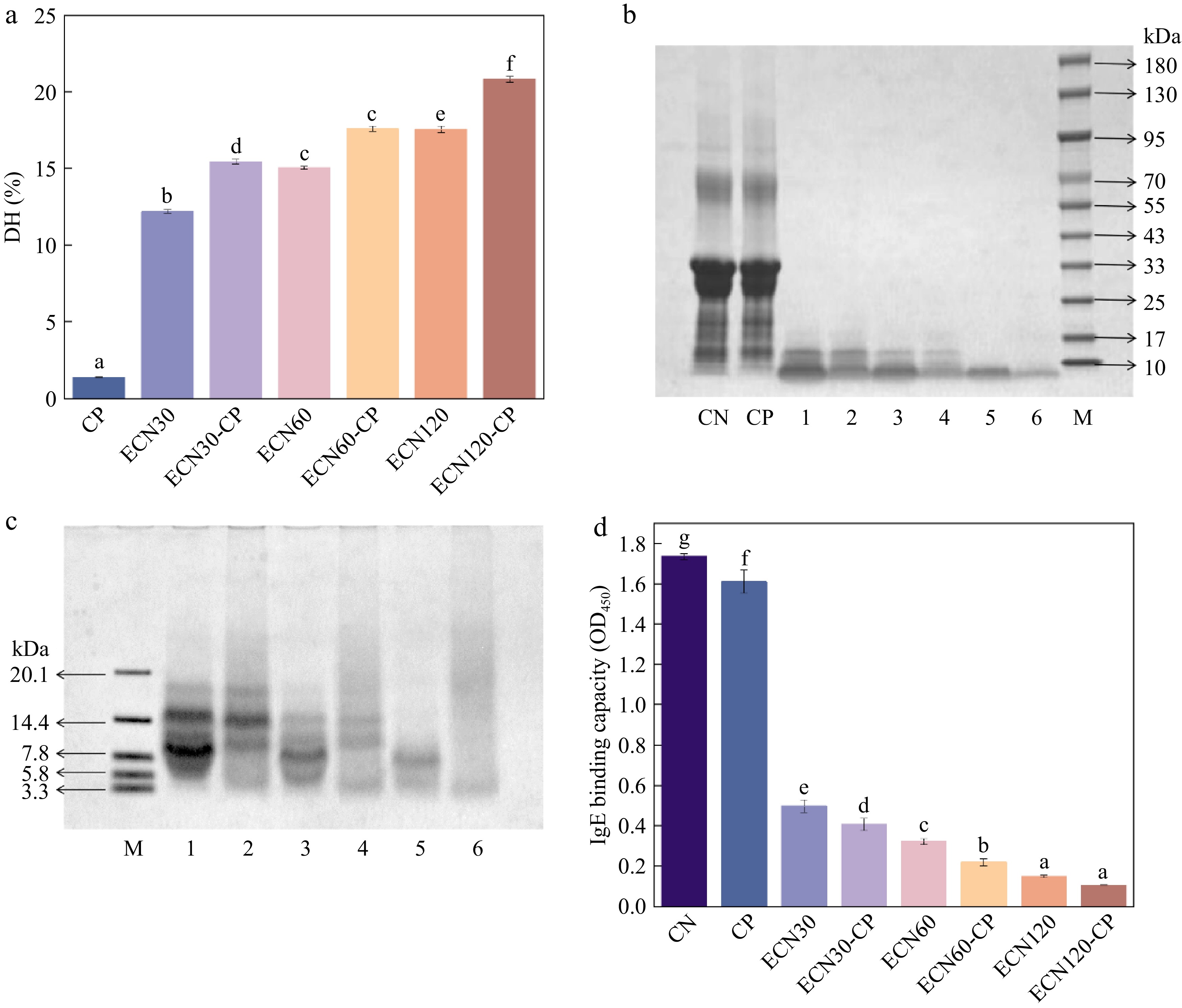
Figure 1.
Effects of different treatments on (a) hydrolysis degree, (b) SDS-PAGE, (c) Tricine-SDS-PAGE, and (d) IgE binding capacity of CN. Lane M: molecular weight marker (10−180 kDa, 3.3−20.1 kDa); Lanes 1–6: ECN30, ECN30-CP, ECN60, ECN60-CP, ECN120, and ECN120-CP.
The MW distribution of CN and its hydrolysates was determined via SDS-PAGE. As shown in Fig. 1b, compared with untreated CN, CP-treated casein showed no obvious changes in the electrophoretic band pattern, indicating that the protein remained largely intact, with no obvious cross-linking or backbone degradation. In our previous study, clear degradation and aggregation were observed after 9 min of CP treatment[11]. This may be related to the structural characteristics of casein at higher concentrations, with CP having a relatively limited effect on the peptide backbone. In contrast, alkaline protease hydrolysis reduced molecular weights to below 17 kDa. This indicated cleavage of peptide bonds to produce low-molecular-weight proteins or peptides. Tricine-SDS-PAGE analysis (Fig. 1c) provided further insights. The alkaline protease hydrolysates showed clear bands at approximately 15 and 8 kDa. Conversely, the corresponding bands in the ECN60-CP and ECN120-CP groups of the sequential treatment became markedly weaker or disappeared. This result was consistent with the DH measurement data. It indicated that sequential treatment may have promoted further degradation or fragmentation of the enzyme-hydrolyzed products into smaller peptides, which may affect the allergenicity of CN[28].
IgE binding capacity
-
IgE-mediated allergies are the most prevalent type of hypersensitivity disorder[29]. The IgE-binding capacity of the samples was determined using indirect ELISA. The results are presented in Fig. 1d. Compared with CN, IgE-binding capacity decreased by only 7.2% after CP treatment, whereas Cai et al.[11] reported an approximately 54% reduction in antigenicity after 9 min of CP treatment. This discrepancy may be due to differences in casein concentrations, which is why CP-assisted enzymatic hydrolysis was considered in this study. In contrast, alkaline protease reduced casein antigenicity by 91.5%, effectively hydrolyzing the IgE-binding epitopes[14]. After the sequential treatment, the antigenicity of ECN120-CP was reduced by 29.1% compared to ECN120. However, in terms of overall hydrolysis effects, the reduction in antigenicity for ECN60-CP was still less than that of ECN120. Nonetheless, the sequential treatment has the potential to maintain the sensory quality of the hydrolysates[10,30]. Additionally, a decrease in IgE binding does not directly correlate with reduced allergenic potential, and further analysis is needed to assess the impact of sequential treatment on antigen epitopes and structure.
Conformational changes of CN
Secondary structure
-
As shown in Fig. 2a, a prominent absorption peak was observed in the amide A region, ranging from 3,500 to 3,000 cm−1. This peak corresponds to N–H and O–H stretching vibrations, with these signals strongly linked to hydrogen bonds in the peptide backbone[31]. The peak area of CN was larger than that of CNHs, suggesting that the hydrolytic effect of sequential treatment may have altered hydrogen bond interactions in casein[22].
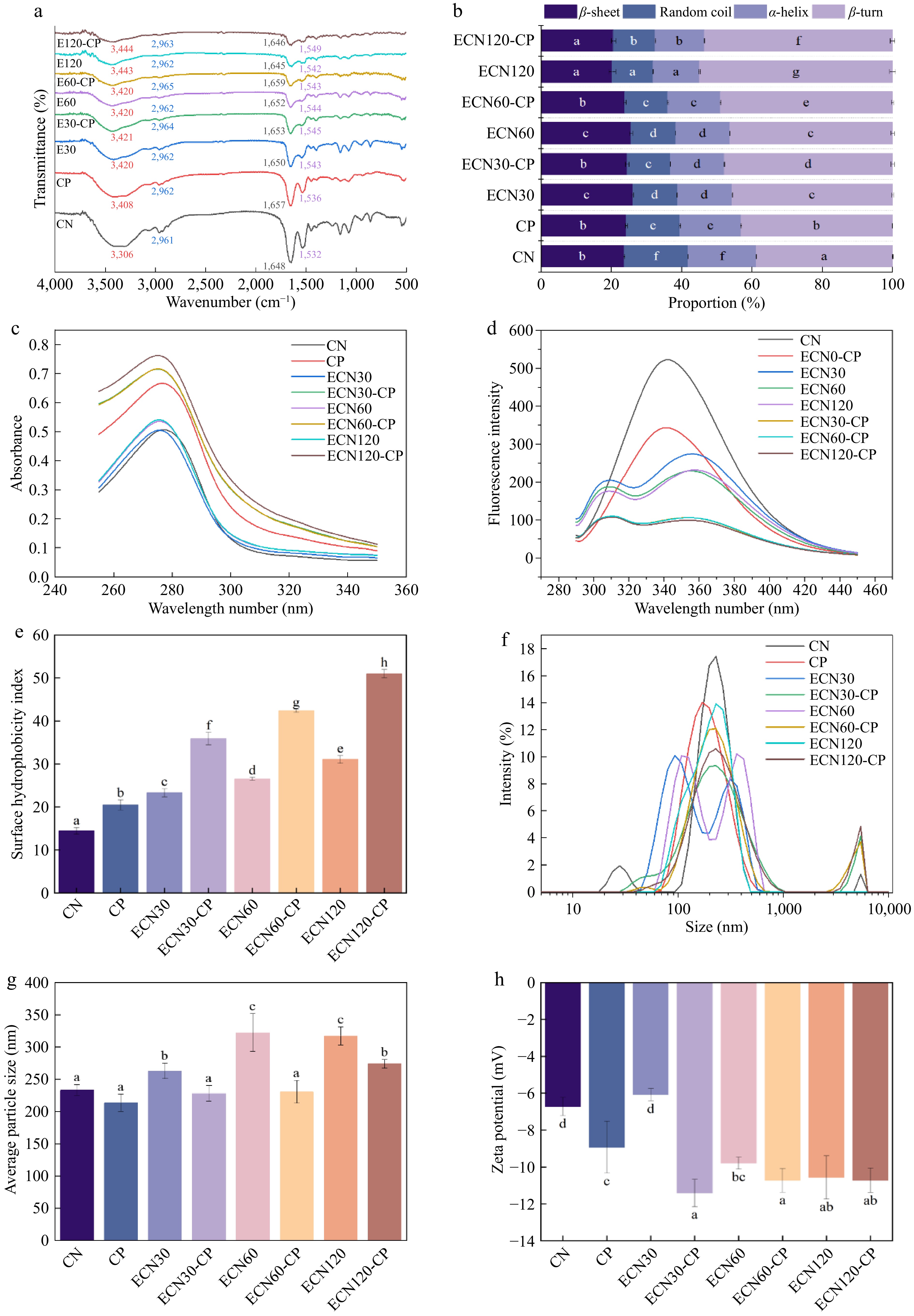
Figure 2.
Impact of various treatments on the structural characteristics of CN: (a) infrared spectroscopy; (b) secondary structure; (c) UV spectra; (d) fluorescence spectra; (e) surface hydrophobicity; (f) particle size distribution; (g) average particle size; and (h) potentials. Different letters indicate significant differences between groups (p < 0.05).
Changes in protein and peptide secondary structures are reflected in amide peak spectra[18]. Following different treatments, the amide I band (1,600–1,700 cm−1) and amide II band (1,500–1,600 cm−1) shifted in wavenumber with decreased intensities, suggesting that hydrolysis and CP treatment altered the conformation of CN and its hydrolysates. Accordingly, peak fitting of the amide I band was used to determine the relative contents of α-helix, β-sheet, β-turn, and random coil in CN and its hydrolysis products. As shown in Fig. 2b, compared with CN, the α-helix content of the ECN60 group decreased by 21.0% and its β-turn content increased by 19.5%, indicating that enzymatic hydrolysis disrupted the original ordered secondary structure of casein[32]. Relative to the ECN60 group, the ECN60-CP group showed a 5.7% rise in β-turn content and a 2.8% drop in α-helix content, suggesting enhanced local unfolding and structural rearrangement[33]. In contrast, compared with ECN120, ECN120-CP showed a slight decrease in β-turn content, along with slight increases in α-helix and random coil. This suggests that at a greater extent of hydrolysis, CP did not simply continue to favor β-turn formation, but instead induced a redistribution of secondary-structure elements. According to previous reports[19], this change may be associated with CP-induced oxidation of amino acid side chains and perturbation of non-covalent interactions in casein. These effects may weaken the conformational stability of casein and promote partial unfolding and structural rearrangement.
Tertiary structure
-
Ultraviolet absorption spectroscopy reflects protein structural changes[34]. As shown in Fig. 2c, CN's UV absorbance increased after different treatments. This finding is consistent with the DH results. It suggests that hydrolysis weakens hydrogen bonds within CN molecules. This leads to peptide chain stretching. Notably, combined enzymatic hydrolysis and CP treatment had a stronger effect. It significantly enhanced CN absorbance compared with single enzymatic hydrolysis. Specifically, ECN120-CP exhibited a 39.8% increase in UV absorbance compared with ECN120. The significant increase in UV absorption intensity is related to the distribution of characteristic amino acids. CP treatment may further cleave peptide bonds, expose aromatic amino acids, and thus lead to an increase in UV absorption intensity[35].
Intrinsic fluorescence emission spectra reveal changes in the microenvironment of aromatic amino acids[36]. As shown in Fig. 2d, at an excitation wavelength of 280 nm, the maximum fluorescence emission (λmax) of CN appeared at 342 nm. This corresponds to the characteristic emission of tryptophan residues. After enzymatic treatment, a new peak appeared at 309 nm, corresponding to tyrosine fluorescence. In native casein, tyrosine fluorescence is usually weak because energy is transferred from tyrosine to tryptophan within the molecule. Enzymatic hydrolysis may increase the distance between these two residues and reduce the efficiency of this transfer, allowing tyrosine fluorescence to become detectable[37]. After different treatments, CN's λmax shifted slightly toward the red. Specifically, the λmax of ECN120 shifted to 357 nm, with its fluorescence intensity decreasing significantly by 55.5%. This indicates changes in the microenvironment of tryptophan residues. Tryptophan residues were exposed from the hydrophobic core to a polar environment, leading to fluorescence quenching[22]. Furthermore, compared with ECN120, the maximum fluorescence intensity of ECN120-CP decreased by 57.2%. This trend is consistent with previous reports that cold plasma may further expose tryptophan residues and enhance fluorescence quenching through oxidation of aromatic amino acids[19,35].
Hydrophobicity
-
H0 indicates the degree of interaction between hydrophobic amino acid residues on the protein surface and polar water domains[16]. As shown in Fig. 2e, the surface hydrophobicity index of CN increased to varying degrees after different treatments. Compared with untreated CN, the ECN120-CP group showed the most significant increase in surface hydrophobicity, with a 2.5-fold enhancement. Furthermore, surface hydrophobicity was 64.2% higher in ECN120-CP than in ECN120. This result is consistent with the intrinsic fluorescence analysis and suggests that sequential treatment may have further loosened the conformation of casein, thereby exposing more hydrophobic sites to the solvent. In addition, studies have suggested that plasma-generated reactive species may contribute to peptide fragmentation, which may further increase the number of hydrophobic sites available for ANS binding[35].
Particle size and zeta potential analysis
-
Particle size strongly influences the functional properties of proteins. As shown in Fig. 2f, g, significant differences were observed in the particle size intensity distribution and average particle size among the different CNHs. Compared with the untreated sample, the diameter of ECN120 increased by 35.9% (Fig. 2g) after single enzymatic hydrolysis. This finding is consistent with whey protein hydrolysis[38]. This phenomenon is likely linked to the self-assembly behavior of short peptides. In detail, enzymatic cleavage of peptide chains exposes hydrophobic groups, which aggregate through hydrophobic interactions, thereby increasing CN particle size. In contrast, following CP-assisted enzymatic hydrolysis, the particle size of ECN60-CP was reduced by 28.6% compared with that of ECN60. This reduction may result from the disruption of electrostatic interactions and other non-covalent forces stabilizing protein structures by CP treatment, which reduces overall size and results in a more uniform distribution[39].
The ζ potential is a key indicator of the dispersibility of colloidal solutions[31]. As shown in Fig. 2h, the absolute ζ-potential values of all hydrolysis products, except for the ECN30 group, were significantly higher than those of the CN group, stabilizing after ECN120. Specifically, the absolute ζ-potential of ECN60 was 45.6% higher than that of CN, which indicates that alkaline protease cleaves peptide bonds to liberate negatively charged amino acids[40]. Meanwhile, the absolute ζ-potential of ECN60-CP was 9.6% higher than that of ECN60; this suggests that CP treatment may alter surface charge distribution through oxidation of amino acid side chains and rearrangement/exposure of ionizable groups, thereby increasing the absolute ζ-potential[31,41].
In summary, CP-assisted enzymatic hydrolysis induces multiple structural modifications in casein hydrolysates. Firstly, alkaline protease hydrolysis cleaves peptide bonds and disrupts the native molecular organization of casein, leading to a looser and more disordered structure. Secondly, CP may further modify amino acid side chains, disturb non-covalent interactions, and promote additional fragmentation of hydrolysates after enzymatic hydrolysis. These changes may further loosen the conformation and increase surface hydrophobicity, negative charge, and dispersibility. Such structural changes may contribute to subsequent changes in antigenicity, antioxidant activity, and bitterness.
Mass spectrometry analysis
Peptide distribution and epitope analysis
-
Peptide length and molecular weight are key indicators of residual antigenicity in protein hydrolysates. High-molecular-weight peptides are the main contributors to residual antigenicity[42]. As shown in Fig. 3a, in all the groups, the peptide mass distribution ranged from 0.5 to 3.5 kDa. Over 80% of the peptides were below 2 kDa. Compared with single-enzyme digestion, the sequential treatment increased the proportion of 0.5–1.2 kDa peptides and decreased the proportion of 2–3.5 kDa peptides. This suggests that CP may have promoted further breakdown of higher-molecular-weight peptide fragments. Antigenic epitopes recognized by specific IgE typically consist of 6–15 amino acids[43]. As shown in Fig. 3b, the proportion of peptides shorter than 12 residues increased after sequential treatment compared with single-enzyme hydrolysis. Notably, ECN120-CP treatment produced a 6.3% increase relative to single-enzyme hydrolysis. The result was consistent with the molecular weight data, suggesting that sequential treatment may promote the formation of low-molecular-weight peptides.
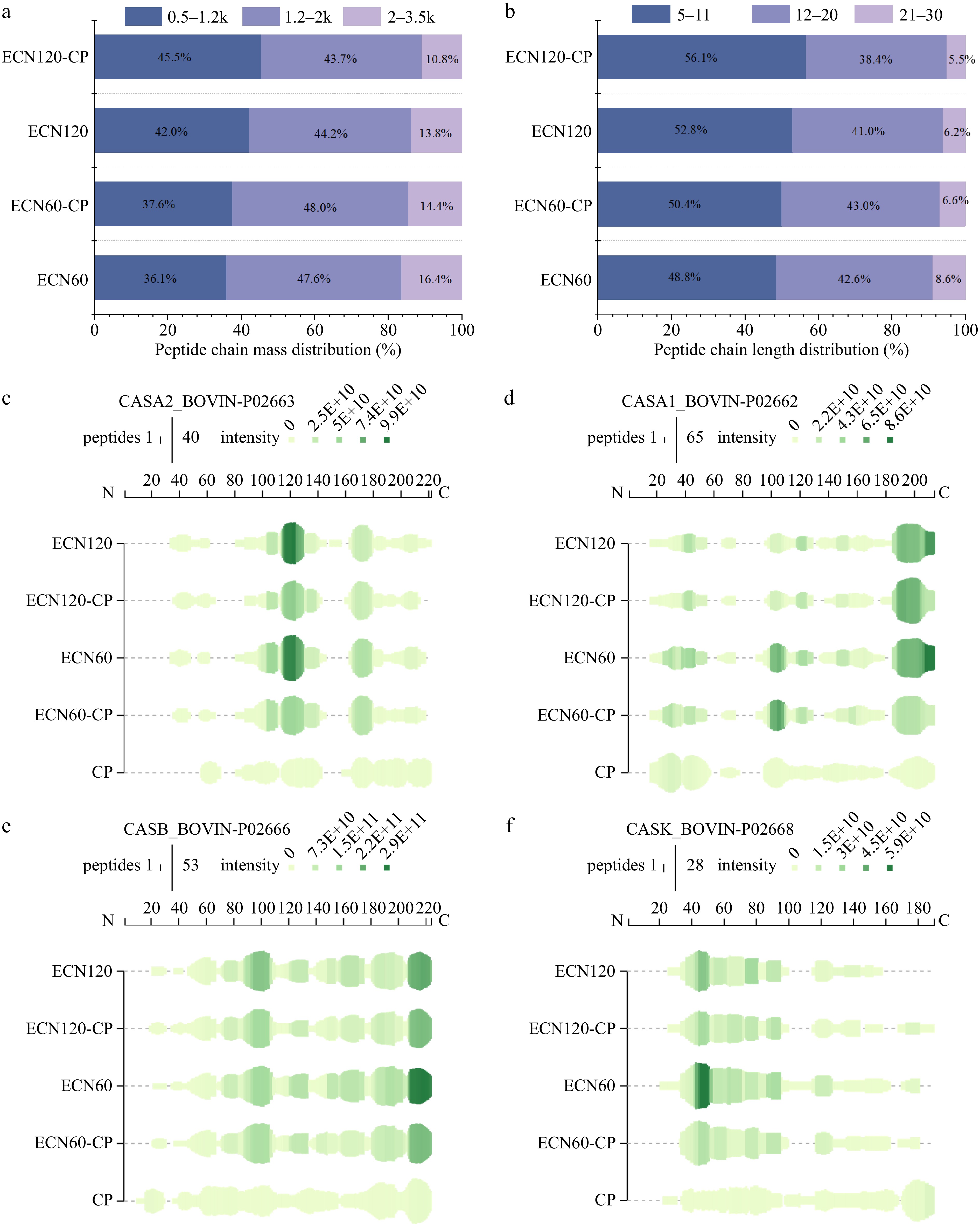
Figure 3.
Mass spectrometry analysis of hydrolysates of CNHs: (a) peptide chain mass distribution; (b) peptide chain length distribution; (c) bovine αS2-casein (P02663); (d) bovine αS1-casein (P02662); (e) bovine β-casein (P02666); (f) bovine κ-casein (P02668). The green bar height indicates the number of peptides, while the color intensity reflects the summed intensities of peptides overlapping this position.
Figure 3c−f illustrates the peptide composition and relative abundance in each hydrolysate. Following CP treatment, a spectrum with broad but shallow coverage and no distinct hotspots was observed. This phenomenon likely reflects the low cleavage specificity of CP[27]. In contrast, single-enzyme digestion produced distinct hotspot distributions. They were especially evident at the C-terminus of αS1-casein (Fig. 3d) and β-casein (Fig. 3e). Hotspots also appeared around residue 120 of αS2-casein (Fig. 3c). After sequential treatment, hotspot regions exhibited reduced intensity alongside more uniform sequence coverage. This indicates that CP treatment may have further fragmented alkaline protease-derived peptides, potentially contributing to the cleavage of residual antigenic epitopes. Based on our prior studies, CN's high allergenicity is mainly due to 22 antigenic epitopes (Supplementary Table S2)[11]. To further analyze the sequential treatment's effect on linear epitopes, the intensities of epitope-overlapping peptides longer than eight residues were quantified[44]. As shown in Table 1, after enzymatic hydrolysis alone, 10 out of 22 antigenic epitopes were cleaved, while the remaining epitopes exhibited further reduced intensity or complete disappearance following sequential treatment. Specifically, compared with the ECN120 group, the epitope intensities of V101-L111 (αS1), K180-Q187 (αS2), and K206-V215 (αS2) in the ECN120-CP group completely disappeared. Meanwhile, the epitope coverage rates of L21-S30 (β), P124-P133 (β), F39-P48 (κ), and P131-K137 (κ) decreased by 91.7%, 19.7%, 96.1%, and 79.2%, respectively. This result was consistent with the ELISA data, suggesting that sequential treatment may further disrupt the antigenic epitopes of CN after alkaline protease hydrolysis.
Table 1. Changes in epitope intensity of CNHs.
Protein Start End Intensity ECN60 ECN60-CP ECN120 ECN120-CP αs1-CN 101 113 2.24 × 10⁸ 1.45 × 10⁸ 3.96 × 10⁷ 0 αs2-CN 132 143 2.24 × 10⁸ 3.14 × 10⁸ 1.48 × 10⁷ 1.97 × 10⁷ 180 187 3.90 × 10⁸ 3.39 × 10⁸ 2.99 × 10⁷ 0 206 215 1.86 × 10⁹ 9.20 × 10⁷ 2.53 × 10⁷ 0 β-CN 21 30 0 0 5.24 × 10⁷ 4.35 × 10⁶ 60 66 1.00 × 10¹⁰ 1.01 × 10¹⁰ 1.39 × 10¹⁰ 1.28 × 10¹⁰ 72 83 2.48 × 10¹⁰ 2.00 × 10¹⁰ 2.08 × 10¹⁰ 1.51 × 10¹⁰ 124 133 1.03 × 10¹¹ 7.84 × 10¹⁰ 7.42 × 10¹⁰ 5.96 × 10¹⁰ κ-CN 39 48 2.48 × 10⁹ 1.24 × 10⁹ 5.65 × 10⁸ 2.19 × 10⁷ 81 99 0 4.33 × 10⁶ 0 0 118 122 1.18 × 10¹⁰ 7.09 × 10⁹ 3.27 × 10⁹ 1.43 × 10⁹ 131 137 2.61 × 10⁸ 3.59 × 10⁸ 3.23 × 10⁹ 6.71 × 10⁸ In silico prediction of peptide bioactivity and bitterness
-
PeptideRanker was applied to predict biological activity, whereas AnOxPePred was used to assess free radical scavenging (FRS). To improve reliability, only predictions with scores > 0.5 were considered[45]. CNH products showed > 25% predicted bioactivity, with FRS activity exceeding 14% (Fig. 4a). Moreover, single-enzyme hydrolysis yielded higher scores than single CP treatment. Notably, the scores for sequential treatment were comparable to, or lower than, those of single-enzyme hydrolysis. Because antioxidant prediction models rely solely on peptide sequences and neglect modifications of amino acid residues, predictions for sequential treatment may underestimate actual activity[46]. Furthermore, sequential treatment cleaved CN peptides into smaller fragments or free amino acids. Low-molecular-weight components containing fewer than five amino acids were frequently undetectable via LC–MS/MS, thereby necessitating further analysis of CNHs' antioxidant activities.
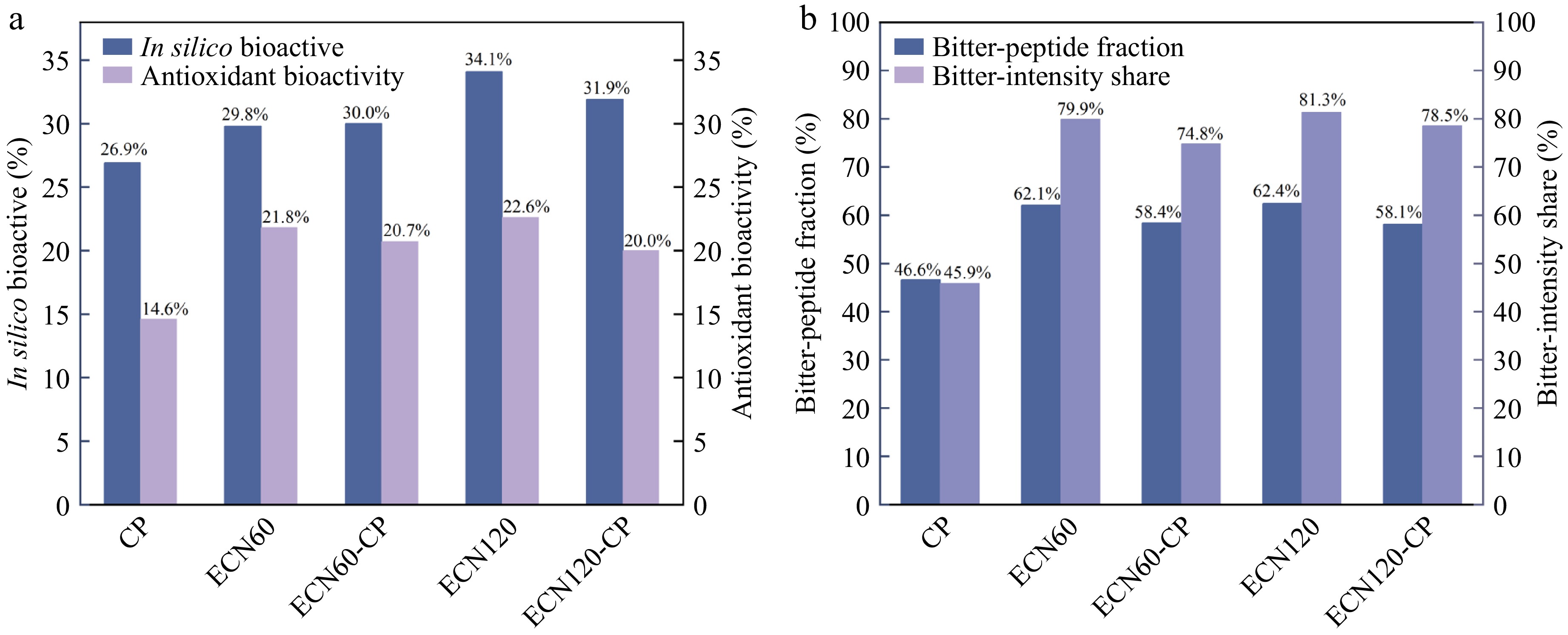
Figure 4.
Results of in silico prediction on CN: (a) bioactive analysis of peptide sequences; (b) bitter peptide analysis.
Hydrophobic peptides are considered the primary contributors to bitterness in protein hydrolysates[47]. The Q-rule, proposed by Ney, is a classical framework in food chemistry for explaining the relationship between peptide hydrophobicity and bitterness[48]. In this study, amino acid sequences were determined by mass spectrometry, and peptide bitterness was subsequently predicted based on Q-value using the BFBP platform[24]. Further comparisons were made between the bitter-peptide fraction (BPF) and bitter-intensity share (BIS), which represent the proportion of bitter peptides by quantity and intensity, respectively[49]. As shown in Fig. 4b, for both BPF and BIS, the proportion of bitter peptides from single-enzyme hydrolysis was significantly higher than that from single CP treatment, whereas both indices decreased after sequential treatment. Specifically, the BPF of ECN120 was 33.9% higher than that of the single CP treatment group; in contrast, the BPF of ECN120-CP was reduced by 6.9% relative to ECN120. It is generally accepted that bitterness intensity follows a bell-shaped relationship with the DH[8]. Thus, alkaline protease releases bitter peptides during CN hydrolysis, whereas sequential treatment may help reduce the bitterness of the hydrolysate by increasing DH and promoting further degradation of these peptides.
Antioxidant activities
DPPH radical scavenging activity
-
As shown in Fig. 5a, DPPH radical scavenging capacity showed no significant change after CP treatment alone, whereas enzymatic hydrolysis significantly increased the activity relative to untreated CN in a time-dependent manner. This observation was consistent with the DH results, indicating that a higher DH of alkaline protease generated more low-molecular-weight peptides and may have released additional amino acids or peptide fragments with antioxidant activity[50]. After sequential treatment, the DPPH scavenging capacity of ECN120-CP further increased by 1.9 times that of ECN120. This increase may be associated with changes in the physicochemical properties of the hydrolysates after CP treatment, particularly the increase in surface hydrophobicity, which may favor interactions with DPPH radicals and facilitate hydrogen atom transfer[51].
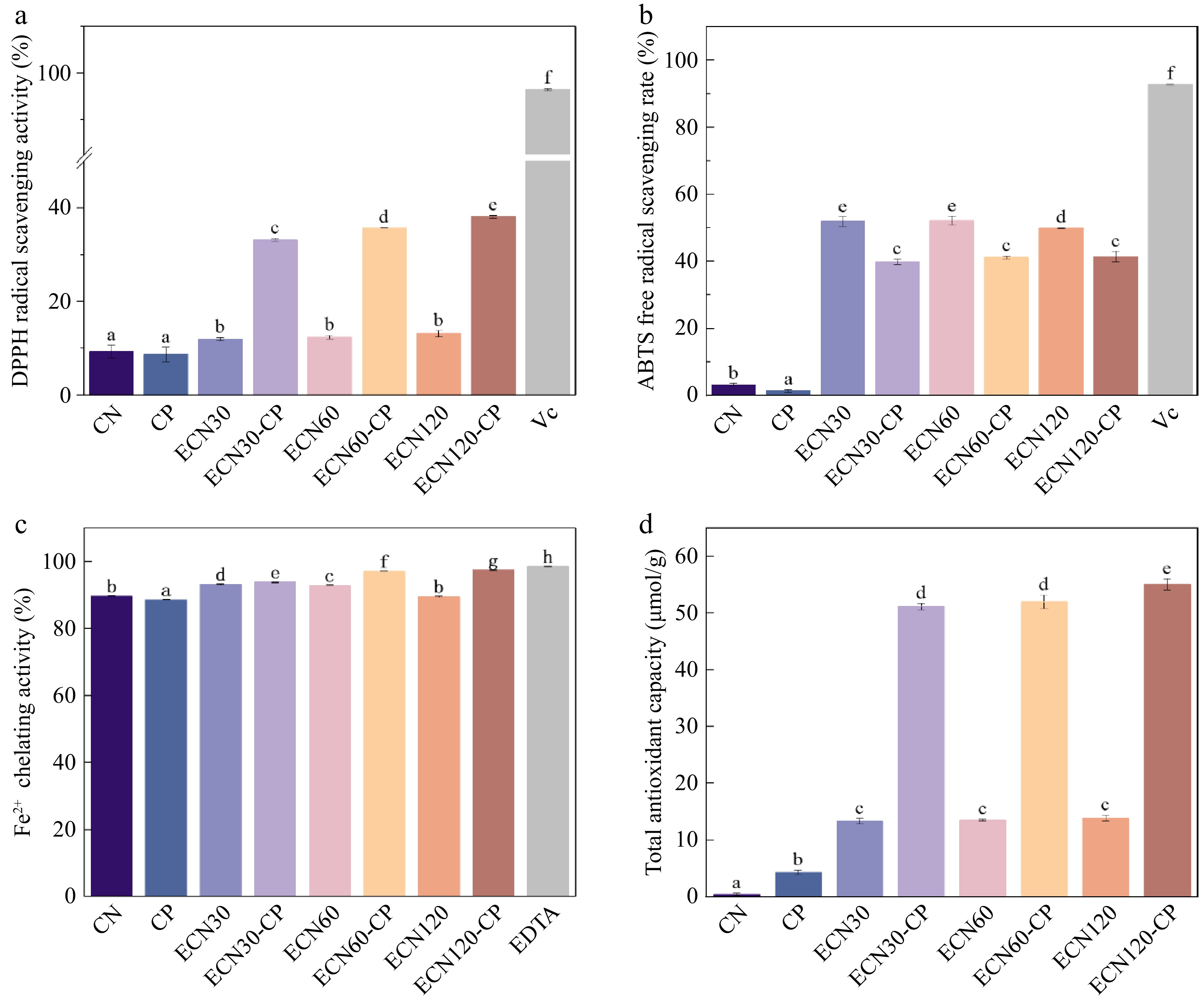
Figure 5.
Effects of different treatments on the antioxidant activity of CN: (a) DPPH radical scavenging activity; (b) ABTS radical scavenging activity; (c) ferrous ion chelation activity; (d) total antioxidant capacity. Different letters indicate significant differences between groups (p < 0.05).
ABTS radical scavenging activity
-
The ABTS radical scavenging capacity (Fig. 5b) was also significantly enhanced after enzymatic hydrolysis. However, after sequential treatment, the ABTS scavenging capacity of ECN120-CP was 17.1% lower than that of ECN120. This difference may be related to the different reaction mechanisms of the two assays. The DPPH assay is mainly associated with hydrogen atom transfer, whereas ABTS scavenging relies more on electron transfer[52]. Therefore, although the increased hydrophobicity after sequential treatment may favor DPPH radical scavenging, possible oxidation of amino acid residues may have affected electron transfer-related scavenging, which may partly explain the lower ABTS scavenging activity[53,54].
Ferrous chelating activity
-
As shown in Fig. 5c, both untreated and treated CN exhibited strong ferrous ion chelating ability. Sequential treatment further enhanced chelating ability, with ECN120-CP showing the highest activity, on par with the positive control. This indicates that CP treatment may oxidatively modify small-molecule peptides from alkaline protease hydrolysis, promoting the formation of negatively charged amino acids. These amino acids enhance the antioxidant activity of CNHs via free radical scavenging and metal ion chelation, which aligns well with the experimentally observed shift toward a more negative ζ-potential[6,55].
Total antioxidant capacity
-
As shown in Fig. 5d, all hydrolysates showed significantly higher antioxidant capacity than untreated casein. This is consistent with previous reports that alkaline protease hydrolysates have relatively high antioxidant potential[25,56]. After sequential treatment, the T-AOC value of ECN120-CP was approximately threefold higher than that of ECN120. The increases in solubility (Supplementary Fig. S2), net negative charge, and hydrophobicity suggest that sequential treatment altered the physicochemical properties of the hydrolysates, which may be related to the measured antioxidant response[57]. However, this result should be interpreted with caution, as interference from plasma-derived oxidation products cannot be ruled out. Overall, sequential treatment may improve the antioxidant-related properties of casein hydrolysates.
Free amino acid composition
-
The amino acid composition of CNHs is presented in Table 2. The total FAA content in the single enzymatic hydrolysis group was over 10 times higher than that in the CP treatment group, which demonstrates the strong proteolytic activity of alkaline protease[18]. The FAA composition may influence the antioxidant activity of casein hydrolysates. Negatively charged amino acids such as glutamic acid and aspartic acid may contribute to antioxidant activity through electron transfer and metal ion chelation[40]. In the present study, the combined content of glutamic acid and aspartic acid was 18.9% higher in ECN120-CP than in ECN120, which may be associated with the increased antioxidant activity. After sequential treatment (ECN120-CP), the total free amino acid content was 8.1% lower than that after enzymatic hydrolysis alone, suggesting oxidative modification of amino acid residues by CP. Previous studies have shown that the total content of eight bitter or weakly bitter free amino acids is positively correlated with the bitterness intensity of hydrolysates[58]. As shown in Fig. 6a, the total content of these amino acids decreased after sequential treatment. In particular, the ECN120-CP group showed an 11.1% lower level than the ECN120 group. These results suggest that sequential treatment may reduce the bitterness of casein hydrolysates through oxidative modification of amino acid residues.
Table 2. Effect of different treatments on the free amino acid composition of CN (mg/g); the last eight amino acids in the table belong to the category of bitter amino acids[58].
Amino acid CP ECN60 ECN60-CP ECN120 ECN120-CP Asp 0.016 ± 0.004ab 0.019 ± 0.003ab 0.027 ± 0.005b 0.007 ± 0.003a 0.020 ± 0.013b Glu 0.037 ± 0.006a 0.198 ± 0.006b 0.240 ± 0.016c 0.226 ± 0.039bc 0.257 ± 0.017c Thr 0.020 ± 0.003a 1.364 ± 0.034bc 1.325 ± 0.010b 1.435 ± 0.005c 1.421 ± 0.082c Ser 0.026 ± 0.003a 0.031 ± 0.002a 0.023 ± 0.009a 0.036 ± 0.009a 0.036 ± 0.010a Gly 0.056 ± 0.007a 0.128 ± 0.01b 0.170 ± 0.019c 0.132 ± 0.019b 0.193 ± 0.024c Ala 0.012 ± 0.001a 0.162 ± 0.019c 0.145 ± 0.003bc 0.114 ± 0.029b 0.112 ± 0.031b Pro 0.080 ± 0.013a 0.138 ± 0.013b 0.141 ± 0.023b 0.106 ± 0.002a 0.135 ± 0.013b Lys 0.031 ± 0.011a 0.071 ± 0.006b 0.095 ± 0.004c 0.096 ± 0.002c 0.120 ± 0.007d Cys 0.422 ± 0.015a 0.653 ± 0.016b 0.775 ± 0.012d 0.706 ± 0.023c 0.865 ± 0.025e Val 0.091 ± 0.015a 0.376 ± 0.017b 0.445 ± 0.020c 0.456 ± 0.009c 0.536 ± 0.021d Met 0.060 ± 0.017a 0.421 ± 0.032d 0.354 ± 0.006c 0.408 ± 0.009d 0.315 ± 0.011b Ile 0.088 ± 0.013b 0.042 ± 0.009a 0.036 ± 0.006a 0.073 ± 0.009b 0.043 ± 0.006a Leu 0.053 ± 0.007a 0.318 ± 0.032c 0.266 ± 0.016b 0.337 ± 0.011c 0.323 ± 0.014c Tyr 0.075 ± 0.013a 5.074 ± 0.060c 4.744 ± 0.082b 5.728 ± 0.088d 5.072 ± 0.118c Phe 0.048 ± 0.015a 7.135 ± 0.028c 6.275 ± 0.006b 7.944 ± 0.002d 7.153 ± 0.021c His 0.021 ± 0.008a 1.611 ± 0.024c 1.228 ± 0.030b 2.105 ± 0.028d 1.635 ± 0.020c Arg 0.071 ± 0.012a 0.557 ± 0.014d 0.490 ± 0.014c 0.456 ± 0.007b 0.481 ± 0.005c Total 1.208 ± 0.027a 18.300 ± 0.025c 16.776 ± 0.036b 20.365 ± 0.032e 18.717 ± 0.023d Different letters indicate significant differences between groups (p < 0.05). 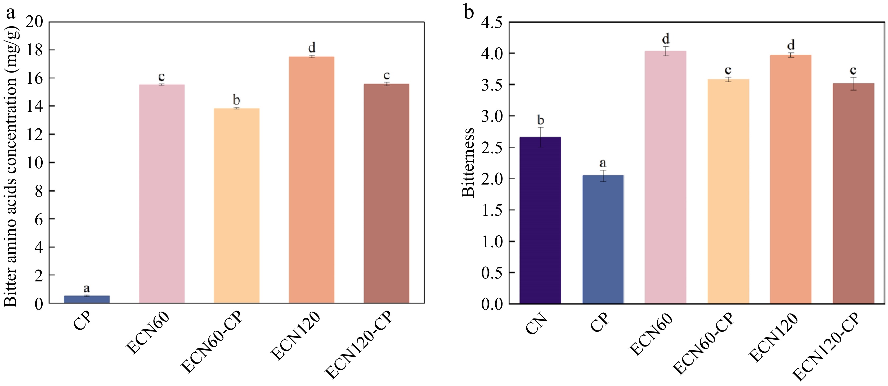
Figure 6.
Bitterness change of CN under different treatments: (a) total content of bitter free amino acids; (b) bitter taste value measured by electronic tongue. Different letters indicate significant differences between groups (p < 0.05).
Electronic tongue analysis
-
As shown in Fig. 6b, the bitterness value of the single enzymatic hydrolysate was significantly higher than that of CN; additionally, that of ECN60-CP was 11.3% and 9.8% lower than that of ECN60 and ECN120, respectively. Liu et al.[8] suggest that hydrophobic amino acids and bitter peptides released during enzymatic hydrolysis contribute to the bitterness of hydrolysates. Considering the bitter peptide and FAA results, sequential treatment may contribute to the reduced bitterness of the hydrolysates, possibly through further degradation of bitter peptide fragments and oxidative modification of some bitter amino acids. Thus, although the DH of ECN60-CP was comparable to that of ECN120, the CP-assisted enzymatic hydrolysis group exhibited a lower bitterness intensity.
-
In this study, CN was hydrolyzed by sequential alkaline protease hydrolysis followed by CP treatment, and the structural characteristics, antigenicity, antioxidant activity, and bitter taste properties of the hydrolysates were systematically investigated. This sequential treatment strategy integrates the selective cleavage effect of enzymatic hydrolysis with the protein oxidative modification function of cold plasma, thereby improving CN hydrolysis efficiency. As a consequence, the resulting hydrolysates exhibited a higher degree of hydrolysis, lower antigenicity, and reduced bitterness, while also showing promising antioxidant potential. However, several limitations should be acknowledged. Detailed plasma characterization was not performed, limiting interpretation of CP-related effects. Reduced IgE binding does not guarantee clinical safety and requires further evaluation. The lack of quantitative oxidative marker analysis may have contributed to an overestimation of antioxidant activity. Processing conditions, economic feasibility, and scalability also remain challenges for future studies.
-
ChatGPT (GPT-5) was used for language refinement in this work. All AI-generated content was reviewed, edited, and verified, with full responsibility assumed for the manuscript's integrity and originality. No AI tool is listed as an author.
-
The authors confirm their contributions to the paper as follows: conceptualization: Zhong C, Pan D, Du L; methodology, visualization: Zhong C, Shen W; investigation: Yang X, Wu Y, Shen W; formal analysis: Zhong C, Yang X, Wu Y; data curation: Zhong C, Yang X; validation: Wu Y; Writing-original draft: Zhong C; writing-review & editing: Roopesh MS, Zeng X, Du L; resources: Pan D; project administration: Pan D, Du L; supervision, funding acquisition: Du L. All authors reviewed the results and approved the final version of the manuscript.
-
All data produced or analyzed during the course of this study are included in this article and its accompanying supplementary information.
-
The authors declare that they have no conflict of interest.
-
accompanies this paper online at: https://doi.org/10.48130/fia-0026-0034.
- Supplementary Table S1 Allergic serum used in the study.
- Supplementary Table S2 Antigenic epitopes of CN.
- Supplementary Fig. S1 DBD cold plasma.
- Supplementary Fig. S2 Solubility of casein under different treatments.
- Copyright: © 2026 by the author(s). Published by Maximum Academic Press on behalf of China Agricultural University, Zhejiang University and Shenyang Agricultural University. This article is an open access article distributed under Creative Commons Attribution License (CC BY 4.0), visit https://creativecommons.org/licenses/by/4.0/.
-
About this article
Cite this article
Zhong C, Yang X, Wu Y, Shen W, Roopesh MS, et al. 2026. Cold plasma-assisted protease hydrolysis modulates antigenicity, antioxidant properties, and bitterness of bovine casein. Food Innovation and Advances 5(3): 406−418 doi: 10.48130/fia-0026-0034 

Cold plasma-assisted protease hydrolysis modulates antigenicity, antioxidant properties, and bitterness of bovine casein
- Received: 04 January 2026
- Revised: 19 May 2026
- Accepted: 16 June 2026
- Published online: 20 August 2026
Abstract: Enzymatic hydrolysis has been proven to be an effective approach to reduce the allergenicity of casein; however, its hydrolysates are usually associated with a strong bitter taste. Thus, this study adopted a two-step approach to prepare casein hydrolysates, involving initial alkaline protease hydrolysis followed by cold plasma (CP) treatment. This CP-assisted enzymatic treatment achieves higher hydrolysis efficiency, along with improved palatability and functional properties of casein hydrolysates. Spectroscopic and ELISA data revealed that CP-assisted enzymatic hydrolysates exhibited reduced endogenous fluorescence intensity, enhanced surface hydrophobicity, and a 29.1% additional reduction in IgE-binding activity relative to sole enzymatic hydrolysis. Moreover, in vitro antioxidant assays demonstrated that sequential treatment has the potential to improve the antioxidant activity of casein hydrolysates. Meanwhile, electronic tongue analysis indicated an 11.3% lower bitterness intensity in CP-assisted enzymatic hydrolysates compared with the sole enzymatic hydrolysis group. These results were further validated by mass spectrometry and free amino acid analyses. Overall, this sequential treatment may provide a useful reference for developing casein hydrolysates with reduced residual antigenic reactivity and bitterness.