












-
Nonalcoholic fatty liver disease (NAFLD) has become the most prevalent chronic liver disease globally, reaching pandemic levels. Its occurrence is primarily linked to the global rise in obesity, with an estimated prevalence of up to 75% among obese individuals[1]. NAFLD is involved in a series of liver abnormalities, ranging from simple hepatic steatosis to advanced stages that may deteriorate into nonalcoholic steatohepatitis (NASH) and occasionally evolve into cirrhosis[2]. Moreover, NAFLD also strongly affects the functions of extrahepatic organs. The pathogenesis of NAFLD has remained incompletely elucidated, but several contributing factors have been identified, including a high-fat diet, genetic factors, insulin resistance, liver fat accumulation, and inflammatory stimulation[3,4]. Although several antiobesity medications are available, their efficacy is limited and is accompanied by adverse effects, such as oil stains and diarrhea[5]. Therefore, identifying effective and safe therapeutic treatments for NAFLD has become critically important.
The gut microbiota has emerged as an essential contributor to the development and progression of metabolic disorders, which are closely linked to the pathophysiology of obesity and NAFLD, as evidenced by gut dysbiosis, increased intestinal permeability, and altered microbiota composition[6]. It has been established that most dietary interventions exert their effects through modulation of the gut microbiota, which may, in turn, exert beneficial effects on NAFLD via microbial-derived metabolites[7]. Short-chain fatty acids (SCFAs) are widely recognized as secondary metabolites and are generated during intestinal fermentation[8]. Increased levels of SCFAs in the intestine are positively associated with reducing the liver's inflammatory response and improving hepatic lipid metabolism, which are considered to be potential therapeutic targets for alleviating obesity-related NAFLD[9]. However, excessive production of SCFAs may enhance hepatic lipogenesis, as the liver can utilize them as a substrate for lipogenesis[10]. The gut–liver axis constitutes a bidirectional communication network connecting the gut and liver via the portal vein, biliary tract, and systemic circulation, in which SCFAs play a central role in improving NAFLD[11,12]. Further studies are required to elucidate how SCFAs derived from dietary interventions ameliorate NAFLD.
Laminaria japonica is a commercially significant seaweed in east Asia, valued for its rich content of nutrients and bioactive compounds, including algin, polysaccharides, and minerals[13]. The brown seaweed L. japonica contains 50.7% total dietary fiber (DF), including 32.8% water-soluble DF and 17.9% insoluble DF[14]. A previous study has shown that these active substances play an important role in improving NAFLD through modulating the gut microbiota[15]. Notably, as an indigestible polysaccharide, soluble dietary fiber is fermented by the gut microbiota to generate SCFAs as secondary metabolites, which have a positive influence on lipid and sugar metabolism[16]. A similar result demonstrated that L. japonica polysaccharide reshaped the gut microbiota in mice with Type 2 diabetes mellitus; specifically, it increased the abundances of Candidatus_Saccharimonas, Enterorhabdus, and Bifidobacterium, which correlated positively with cecal SCFA levels and bile acid secretion[17]. Interestingly, conventional processing of L. japonica typically leads to a substantial loss of bioactive components to meet the sensory standards. Microbial fermentation offers a potential solution to enhance both the sensory properties and the digestibility and bioavailability of L. japonica's bioactive properties[18]. Probiotics, as relatively safe and beneficial microorganisms, have gained widespread attention for their health-promoting effects beyond basic nutrition[19]. Pediococcus acidilactici is a probiotic strain known to enhance functional properties when cultivated in media containing milk and cereal bran[20], and it has robust fermentation capacity and strong intestinal tolerance[21]. Meanwhile, fermentation of black beans with P. acidilactici L19 increased the soluble dietary fiber content, which was attributed to the β-glucosidase produced by this strain, as this enzyme can release various bound molecules from their conjugated forms[22]. Significantly, previous work from our group also indicated that P. acidilactici significantly reduced the disturbance of lipid metabolism in high-fat diet-induced hyperlipidemic rats[23]. However, it remains unclear whether P. acidilactici-fermented L. japonica (FLJ) exerts hypolipidemic effects on the lipid metabolism in NAFLD rats.
The present study aimed to research the hypolipidemic mechanism of FLJ in a high-fat diet-induced NAFLD rat model. Our particular focus was on determining whether the hypolipidemic effects are mediated through the structural composition and functional modifications of the gut microbiota, thereby clarifying the link between the microbial ecology and the fermented product’s lipid-lowering activity.
-
P. acidilactici FZU106, isolated from Hongqu rice wine (Fuzhou University, Fuzhou, China), was cultured in broth for 24 h at 37 °C. After three generations, the bacterial culture was then harvested by centrifugation, washed with sterile saline, and resuspended to achieve a live bacteria count of 1.0 × 10⁹ colony-forming-units (CFU)/mL for subsequent animal administration and fermentation inoculation.
Fermentation of L. japonica was performed as described previously with modifications[24]. Fresh L. japonica was cleaned, dried, and ground to pass a 80-mesh sieve. Then 3 g of the powder was dissolved in 100 mL of distilled water and autoclaved at 110 °C for 15 min. After cooling, the substrate was inoculated with 1 mL of the bacterial suspension (1.0 × 10⁹ CFU/mL) and incubated at 37 °C for 24 h, with sampling every 2 h. The viable count reached ~1.0 × 10⁹ CFU/mL after 16–20 h, and the fermentation broth was directly used for oral gavage in rats.
Chemical compositional changes in response to fermentation
-
Dietary fiber content was determined using the standard food fiber method. Crude protein, crude lipid, moisture, protein, and ash were analyzed by AOAC International's methods[14]. The nutrient composition of unfermented and fermented groups is presented in Supplementary Table S1.
Animal experiment
-
Thirty-six 6-week-old male SPF Sprague–Dawley rats (150 ± 5 g) were obtained from Shanghai Laboratory Animal Research Center. After one week of acclimation with a normal-fat diet (NFD) and water ad libitum, the animals were randomly divided into six groups (n = 6): (1) NFD, normal diet + saline; (2) HFD, high-fat diet + saline; (3) HFD_Sim, HFD + simvastatin at 20 mg/kg/day; (4) HFD_Pa, HFD + live P. acidilactici FZU106 at 1.0 × 10⁹ CFU/mL/rat/day; (5) HFD_LJ, HFD + unfermented L. japonica suspension at 10 mL/kg/day; and (6) HFD_FLJ, HFD + FLJ suspension at 10 mL/kg/day and 1.0 × 10⁹ CFU/mL. The dosage was based on previous studies and solubility limitations[24]. The dietary energy profiles of the NFD and HFD are shown in Supplementary Table S2. After 8 weeks of the intervention, all rats were fasted overnight, anesthetized with isoflurane, and euthanized for sample collection. All procedures were approved by the Animal Care and Use Committee of Fujian Agriculture and Forestry University (FAFU 2025-017).
Biochemical analysis
-
Blood from the abdominal aorta was kept at room temperature in 5.0-mL tubes. Serum was separated by centrifugation (1,500 × g, 10 min) and stored at −80 °C. For analysis of total cholesterol (TC), triglycerides (TG), low-density lipoprotein cholesterol (LDL-C), and high-density lipoprotein cholesterol (HDL-C) in the serum, commercial kits were used. Protein content, TC, TG, total bile acid (TBA), nonesterified fatty acids (NEFA), glutathione peroxidase (GSH-PX), malondialdehyde (MDA), and total superoxide dismutase (T-SOD) in the livers were also measured using assay kits. Further, fecal lipids were extracted according to a modified protocol[25]. All kits were supplied by Jiancheng Institute of Biological Engineering (Nanjing, China).
Histopathologic evaluation
-
After fixation for 24 h in 4% paraformaldehyde, the collected liver and adipose tissue specimens were embedded in paraffin. The sections were cut at 5 μm, deparaffinized, and stained with hematoxylin and eosin (H&E) as per established protocols. Visualization and photomicrography were performed with an optical microscope (Leica RM2016, Germany) coupled with a digital camera (Leica DM2700 M, Germany).
Total lipid levels in the liver
-
Hepatic total lipids were extracted using a modified method[26]. Liver tissues were homogenized and incubated with a chloroform–methanol mixture (2:1, v/v) at 4 °C for 24 h. The extract was then mixed with 0.6% KCl and centrifuged (2,000 × g, 20 min). The organic phase was collected, dried under nitrogen, and redissolved in 200 μL of isopropanol. Total lipid content was determined using a commercial kit (Labtest Diagnostica S.A., MG, Brazil).
Quantitative reverse transcription polymerase chain reaction
-
Total RNA was extracted from liver tissues using a commercial kit (Dalian, China). Following DNA digestion, cDNA was synthesized with the PrimeScript RT reagent kit with a gDNA eraser. Quantitative reverse transcription–polymerase chain reaction (qRT-PCR) amplification was performed in triplicate using SYBR Premix Ex Taq II (Takara). Target genes' expression were normalized to β-actin. The primer sequences are listed in Supplementary Table S3.
Hepatic immunohistochemical analysis
-
Liver samples were fixed in 10% formalin for 48 h, then sectioned at 4 µm for immunohistochemistry. Histological examination results showed that the sections were stained with H&E. Hepatic immunohistochemical (IHC) analysis was performed using primary antibodies against SREBP-1C, CYP7A1, and CD36 (Sangon Biotech Co., Ltd., China). Immunohistochemical staining was carried out using appropriate secondary antibodies and detection systems. Brown-yellow positive staining was quantified with ImageJ 6.0 (Media Cybernetics).
Determination of fecal SCFAs
-
Fecal SCFAs were measured by gas chromatography (GC) using a modified protocol[27]. Samples (50 mg) were homogenized in saturated NaCl (500 μL, 3 min), acidified with H2SO4 (20 μL), and extracted with diethyl ether (800 μL). After centrifugation (7,600 × g, 15 min, 4 °C), the organic phase was dried with Na2SO4, re-centrifuged, and filtered (0.22 μm) for GC analysis.
Analysis of the gut microbiome via high-throughput sequencing
-
Cecal DNA was extracted (MoBio kit, USA). The 16S rRNA V3–V4 region was amplified (primers 338F/806R) and sequenced on an Illumina MiSeq platform (Majorbio, Shanghai). Operational taxonomic units (OTUs) were clustered at 97% similarity using MOTHUR (
https://mothur.org ). Genus-level differences among groups were analyzed with STAMP (v2.1.3) and are shown as extended error bar plots. Spearman's correlation analysis between key microbial phylotypes and lipid metabolic parameters was carried out using Metware Cloud, an online platform, with the results visualized as a heatmap. Furthermore, the network diagram integrating lipid parameters and the gut microbiota were constructed and visualized via the Metware Cloud platform.Functional predictions of intestinal microbiota
-
Based ongene sequencing data of 16S rRNA, the functional potential of the cecal microbiota was predicted via phylogenetic investigation of communities by reconstruction of unobserved states (PICRUSt). The reconstruction of Kyoto Encyclopedia of Genes and Genomes (KEGG) ortholog abundances facilitated the subsequent functional annotation of the prevalent metabolic pathways. Finally, the significantly different pathways among three groups were analyzed using Student’s t-test, and a diagram of the correlated network between microbial communities and potential functional pathways was constructed using the Metware Cloud platform.
Statistical analysis
-
The results are shown as the mean ± standard error of the mean (SEM). One-way analysis of variance (ANOVA) followed by Duncan's test was used for comparisons. Graphs were created with GraphPad Prism 11.0.
-
As displayed in Fig. 1, rats fed the HFD showed a significant increase in body weight in comparison with rats fed the normal-fat diet (NFD group). However, the intervention with FLJremarkedly attenuated this HFD-induced weight gain, resulting in a 23.57% reduction compared with the HFD-fed group (p < 0.05) (Fig. 1a, b). Consistent with body weight changes, HFD-fed NAFLD rats exhibited significantly higher liver, perirenal fat, and epididymal fat indexes comparison with the NFD-fed rats. In contrast, the kidney index remained unaffected by the dietary intervention across all groups. (Fig. 1c–f). Notably, administration of both P. acidilactici (Pa) and FLJ showed a sharp decrease with the accumulation of perirenal fat (by 34.27% and 40.16%, respectively) (p < 0.01) and epididymal fat (by 21.80 % and 34.64%, respectively) (p < 0.05) induced by the HFD. Histological examination of the adipose tissues via H&E staining revealed that both the Pa and FLJ interventions markedly reduced perirenal and epididymal adipocytes' size in HFD-fed NAFLD rats compared with those consuming the HFD (Fig. 1g, h).
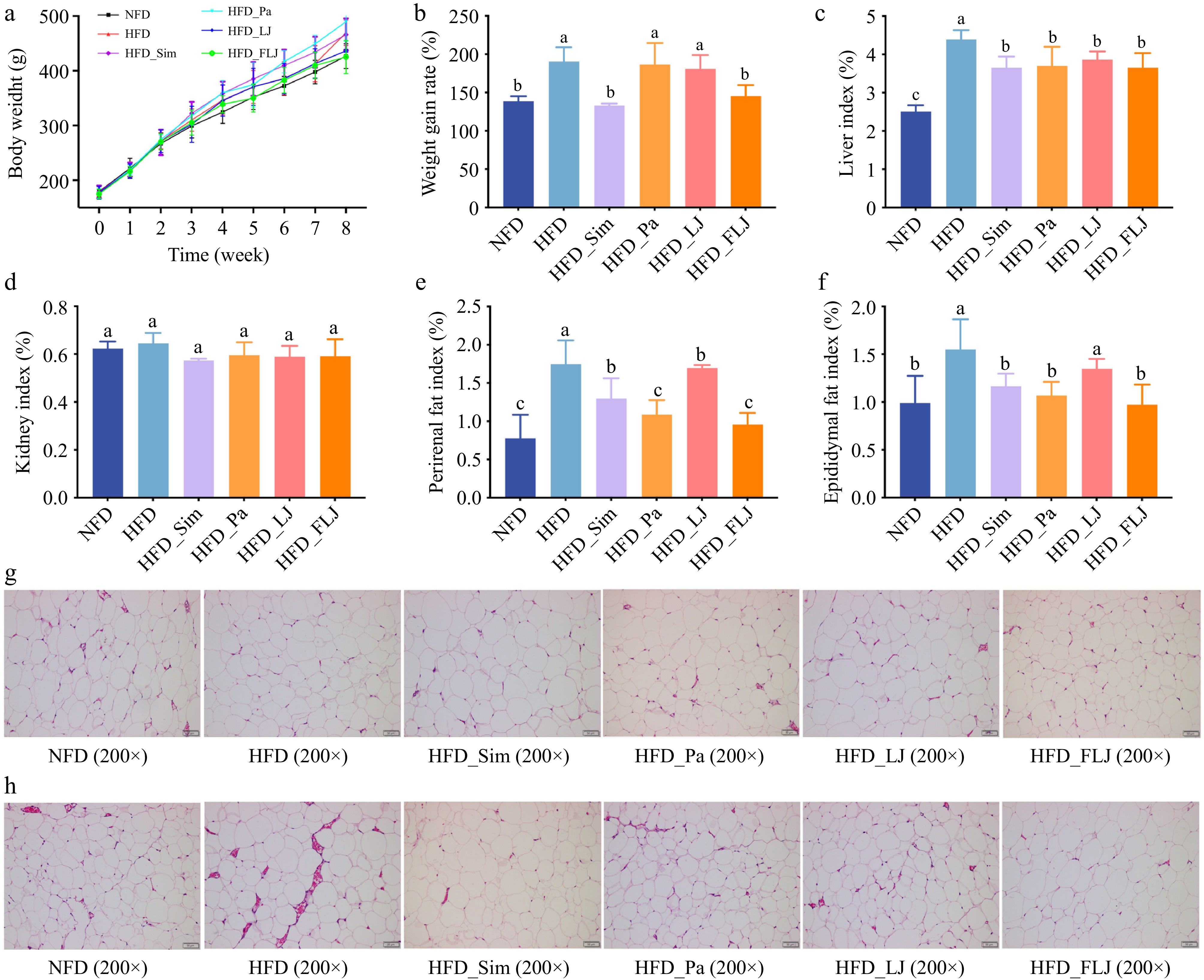
Figure 1.
The effect of the P. acidilactici FZU106-fermented L. japonica intervention on the physiological index and fat accumulation in NAFLD rats (n = 6). (a) Body weight growth, (b) rate of weight gain, (c) liver index, (d) kidney index, (e) perirenal fat index, (f) epididymal fat index, (g) H&E staining of perirenal adipocytes, and (h) epididymal adipocytes. Values are expressed as the mean ± SEM in each group. Different letters indicate significant differences among experimental groups (p < 0.05).
FLJ improved serum biochemistry in NAFLD rats treated with HFD
-
The HFD apparently increased serum total TC, TG, and LDL-C levels when compared with the NFD group, while concomitantly decreasing HDL-C levels (Fig. 2). However, the FLJ intervention obviously counteracted these HFD-induced alterations, significantly decreasing serum TC, TG, and LDL-C levels by 22.04%, 36.54%, and 33.77%, respectively, while enhancing HDL-C levels by 21.86% (p < 0.05). Notably, the FLJ intervention demonstrated superior lipid-modulating effects compared with both the HFD_Pa and HFD_LJ treatment groups.
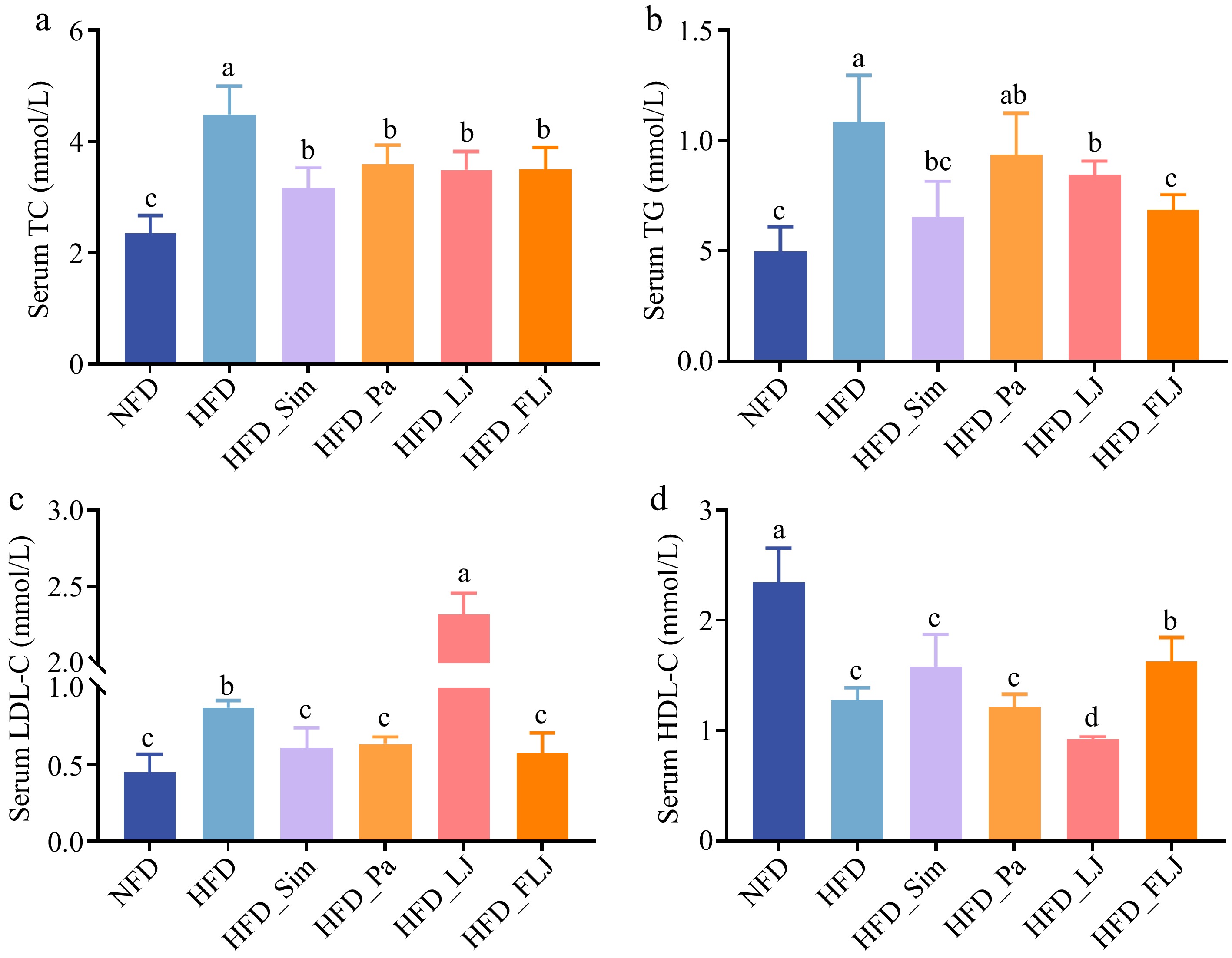
Figure 2.
The effect of the FLJ intervention on serum parameters in NAFLD rats (n = 6). (a) Serum TC, (b) serum TG, (c) serum LDL-C, and (d) serum HDL-C. Values are expressed as the mean ± SEM in each group. Different letters indicate significant differences among experimental groups (p < 0.05).
FLJ alleviated hepatic lipid accumulation and oxidative stress in NAFLD rats treated with the HFD
-
To further evaluate the lipid levels in the liver, hepatic TC, TG, TBA, NEFA, and total lipids (fat) levels were characterized, as shown in Fig. 3a–e. Hepatic lipid analysis revealed that, compared with the NFD controls, HFD-fed NAFLD rats had significantly elevated hepatic TC, TG, TBA, NEFA, and fat. However, the FLJ intervention dramatically decreased hepatic TC, TG, and fat by 21.88%, 42.52%, and 39.96%, respectively (p < 0.01), and significantly decreased hepatic TBA and NEFA by 20.49% and 41.78%, respectively (p < 0.05). Similarly, the HFD-fed NAFLD rats administered Pa and LJ also had a significant decrease in hepatic TC, TG, NEFA, and fat levels, but had no statistically significant difference in hepatic TBA levels. Histological examination via H&E staining confirmed substantial lipid droplet accumulation in the livers of HFD-fed NAFLD rats (Fig. 3i). In contrast, FLJ supplementation resulted in markedly smaller and fewer fat vacuoles, indicating effective amelioration of HFD-induced hepatic steatosis. As shown in Fig. 3f–h, the FLJ treatment significantly decreased hepatic MDA levels while increasing glutathione peroxidase (GSH-PX) and superoxide dismutase (SOD) activities. However, treatment with Pa and LJ showed less pronounced effects on oxidative stress markers.
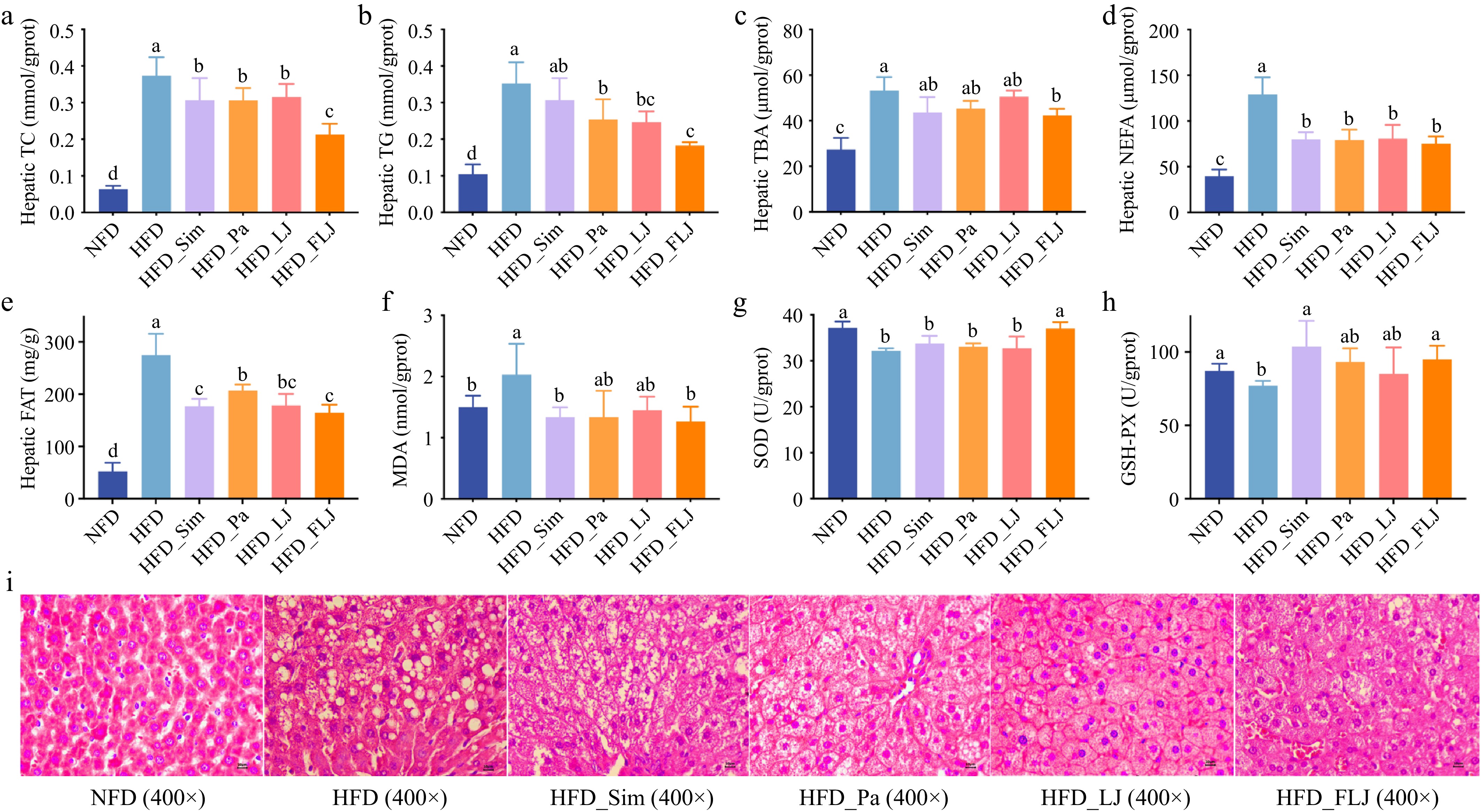
Figure 3.
The effect of the FLJ intervention on the liver parameters in NAFLD rats (n = 6). (a) Hepatic TC, (b) hepatic TG, (c) hepatic TBA, (d) hepatic NEFA, (e) hepatic FAT, (f) MDA, (g) SOD, (h) GSH-PX, and (i) H&E staining of liver tissues. Values are expressed as the mean ± SEM in each group. Different letters indicate significant differences among experimental groups (p < 0.05).
FLJ promoted fecal SCFA production in NAFLD rats treated with the HFD
-
SCFAs, key microbial metabolites derived from dietary polysaccharide fermentation, are beneficial to health. As presented in Fig. 4a–f, the concentrations of six major SCFAs (acetate, propionate, butyrate, isobutyrate, valerate and isovalerate) were quantified in fecal samples. The FLJ intervention significantly elevated the six fecal SCFAs' concentrations compared with the HFD control group (p < 0.05). In contrast, administration of either Pa or LJ significantly increased only the fecal isobutyrate concentration (p < 0.05), with no significantly statistical effects observed for the other SCFAs.
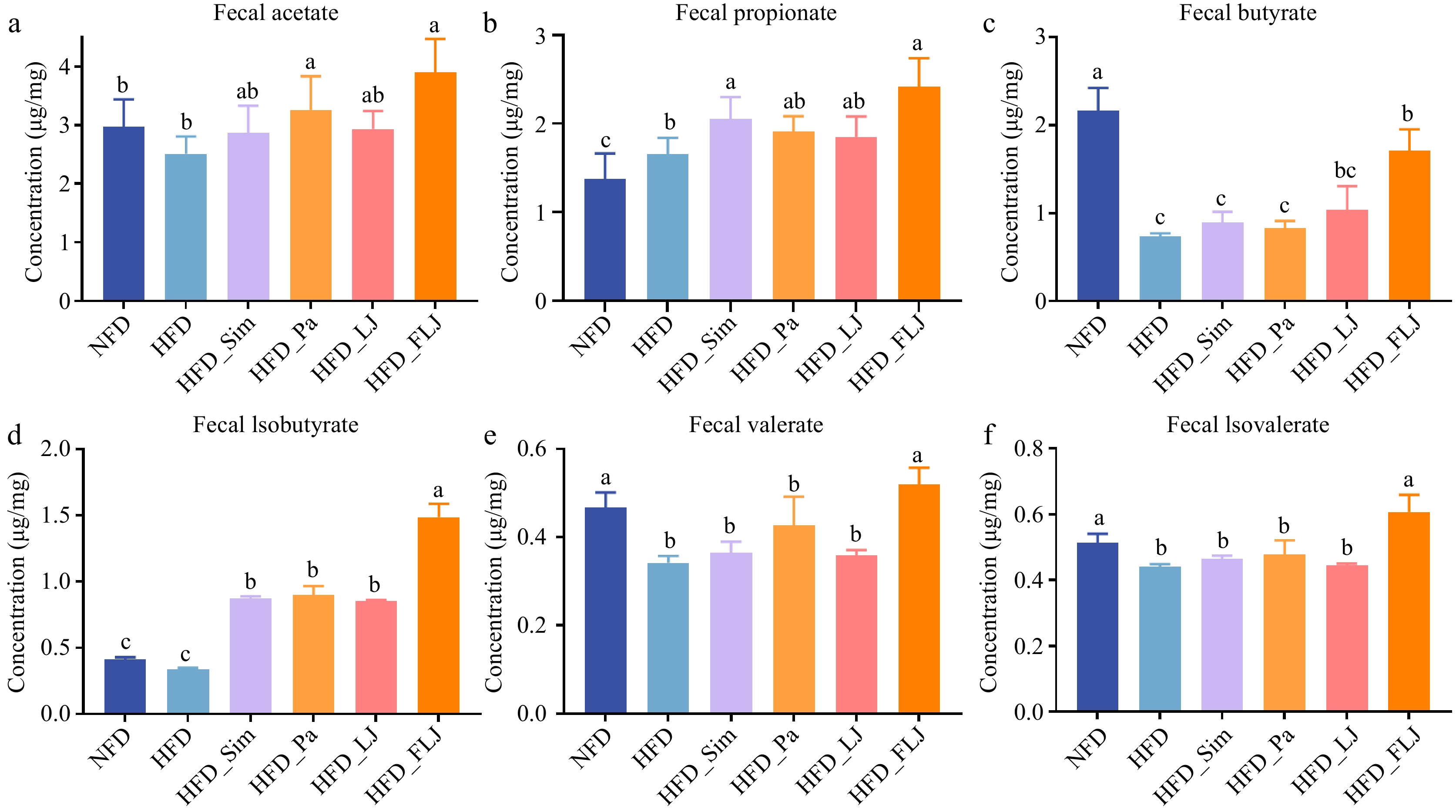
Figure 4.
The effect of the FLJ intervention on fecal SCFAs in NAFLD rats (n = 6). (a) Fecal acetate, (b) fecal propionate, (c) fecal butyrate, (d) fecal isobutyrate, (e) fecal valerate, and (f) fecal isovalerate. The values are expressed as the mean ± SEM in each group. Different letters indicate significant differences among experimental groups (p < 0.05).
FLJ regulated liver lipid metabolism-related pathways in NAFLD rats treated with the HFD
-
To investigate the molecular mechanisms of FLJ's lipid-lowering effect, the hepatic mRNA expression of key lipid metabolism-related genes was analyzed (Fig. 5). Compared with the NFD group, administration of the HFD significantly upregulated the mRNA expression levels of Cd36, Srebp-1c, Acat2, and Hmgcr, but markedly downregulated the mRNA expression levels of Cyp7a1 and Fas. The intervention with FLJ significantly normalized the expression of these dysregulated genes. Additionally, immunofluorescence detection presented a similar result in terms of gene expression (Fig. 6). Consistent with the transcriptional findings, immunofluorescence analysis revealed parallel changes at the protein level. FLJ supplementation significantly reduced the protein abundance of SREBP-1C and CD36, but enhanced CYP7A1's expression, aligning with the observed gene expression patterns.
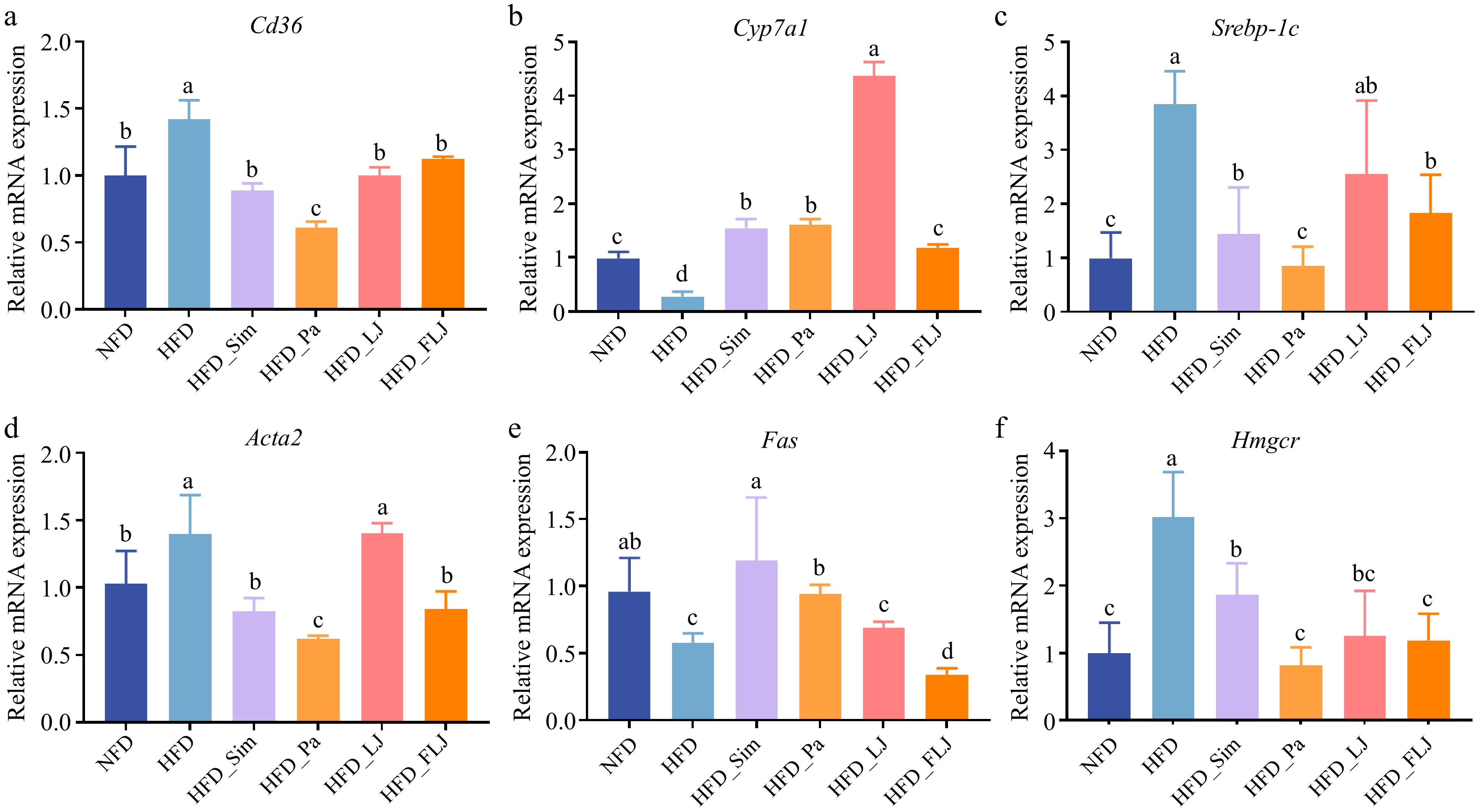
Figure 5.
The effect of the FLJ intervention on the mRNA expression levels of (a) Cd36, (b) Cyp7a1, (c) Srebp-1c, (d) Acat2, (e) Fas, and (f) Hmgcr in NAFLD rats (n = 3). Values are expressed as the mean ± SEM in each group. Different letters indicate significant differences among experimental groups (p < 0.05).
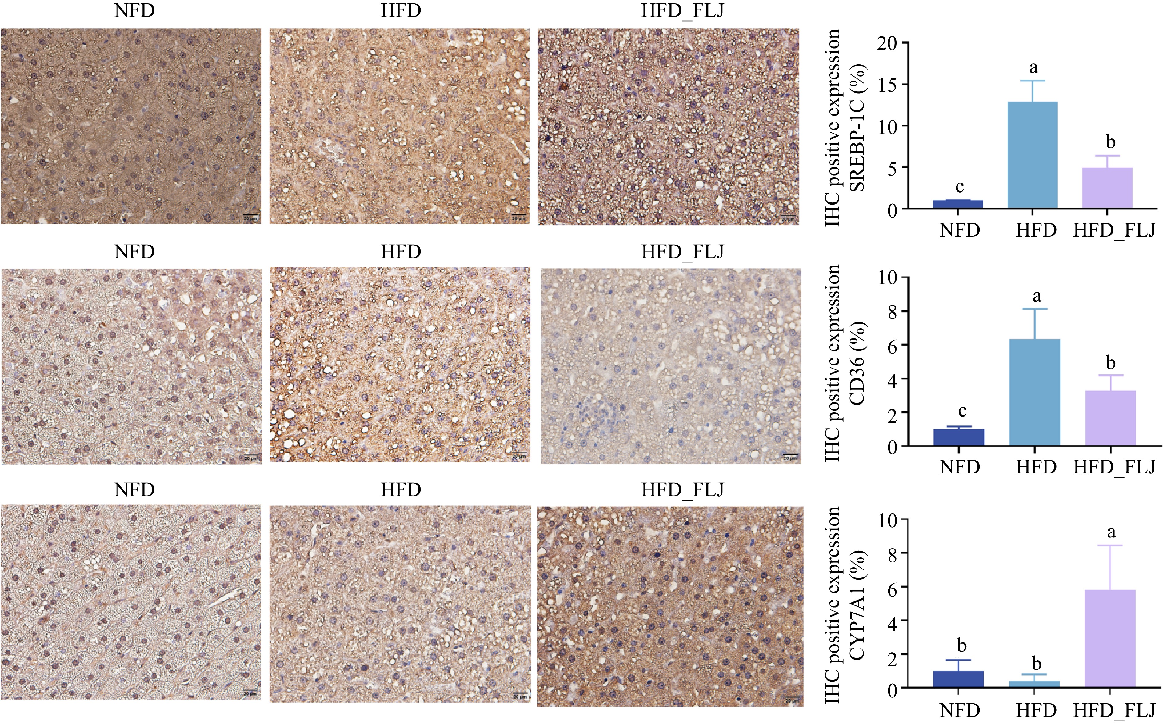
Figure 6.
Immunohistochemical staining for key proteins: (a) SREBP-1C, (b) CD36, and (c) CYP7A1 expression levels in the liver. Brown color (positive) shows the expression of the proteins SREBP-1C, CD36, and CYP7A1 in the cells. Quantification of the expression levels of SREBP-1C, CD36, and CYP7A1 by IHC are also shown on the right (n = 3). Values are expressed as the mean ± SEM in each group. Different letters indicate significant differences among experimental groups (p < 0.05).
FLJ reshaped structural composition of gut microbiota
-
To evaluate FLJ’s impact on the gut microbiota, we profiled the relative abundances of the top 15 genera (Fig. 7a, b). Compared with the NFD group, the HFD significantly altered the microbial composition, characterized by increasing abundances of Unclassified_f_Lachnospiraceae, Lachnoclostridium, Blautia, Collinsella, and Ruminococcus]_torques_group, alongside decreasing abundances of Ruminococcaceae, Candidatus_Saccharimonas, Christensenellaceae_R7_group, norank_o_Molicutes_RF39, Ruminococcus]_torques_group, Turicibacter, Clostridium_sensu stricto_1, Lachnospiraceae_NK4A136_group, and Nosocomiicoccus. Similarly, a comparative analysis between the HFD and HFD_FLJ groups revealed that the FLJ intervention significantly enriched beneficial taxa, including Akkermansia, Roseburia, Desulfovibrio, Ruminococcaceae_UCG-005, GCA-900066575, Family_Xlll_UCG001, Pediococcus, Defluvitaleaceae_UCG.011, Anaerotruncus, and Alistipes. Conversely, Romboutsia and [Eubacterium]_nodatum_group were significantly more abundant in the HFD group. Notably, Ruminococcaceae_UCG-005 was consistently enriched in both the NFD and HFD_FLJ groups (Supplementary Fig. S1), suggesting a potential role in the protective effects of the FLJ treatment.
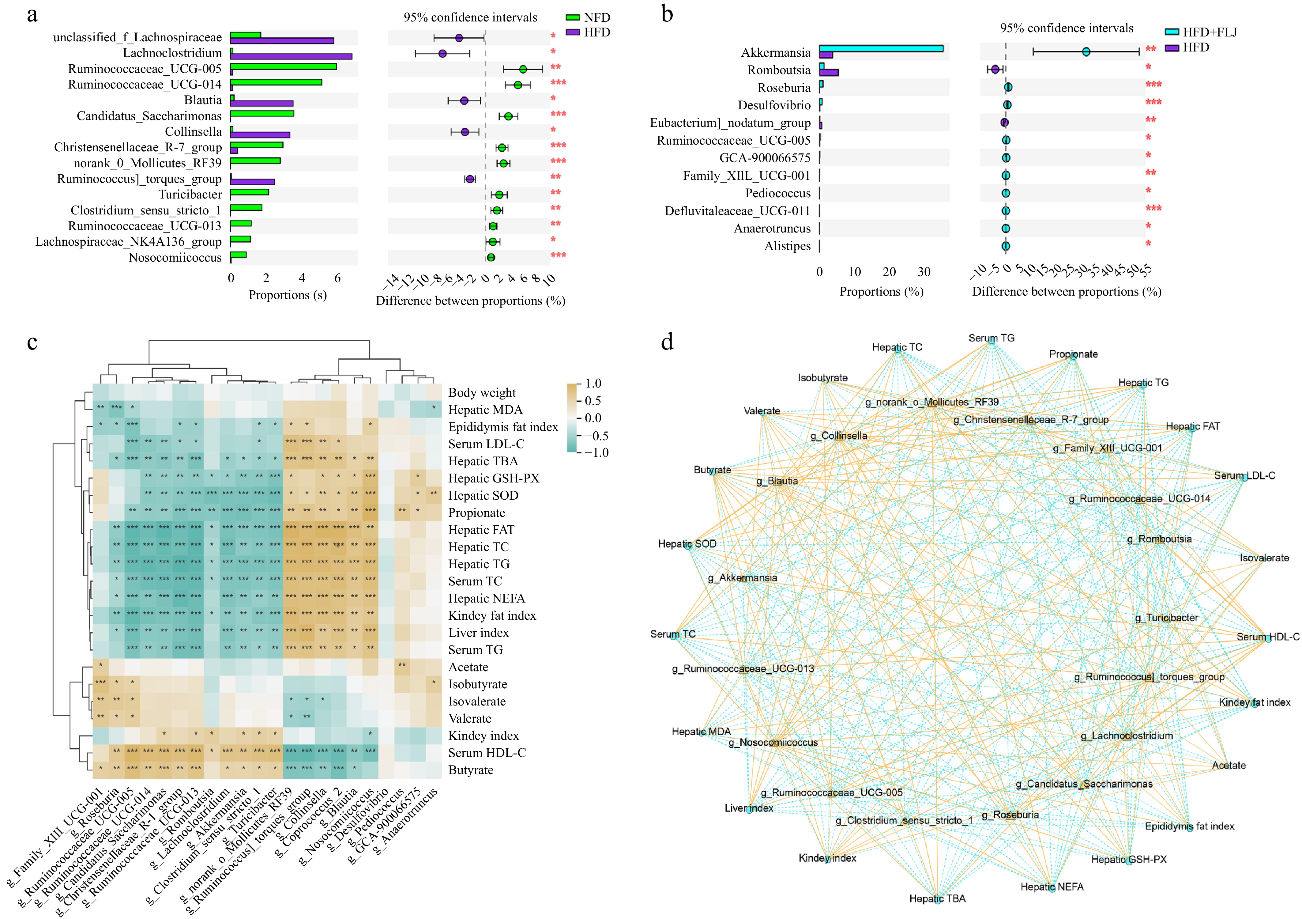
Figure 7.
Extended error bar plot showing differences in the mean proportions of significantly altered genera (Student's t-test) and Spearman correlations between altered fecal microbial phylotypes and lipid metabolic parameters. (a) NFD group vs. HFD group; (b) HFD group vs. HFD_FLJ group. (c) Spearman's correlation heatmap. (d) Visualization of the correlation network between the key intestinal microbial species and lipid metabolism-related parameters. Values are expressed as the mean ± SEM (n = 6). * p < 0.05, ** p < 0.01 and *** p < 0.001.
Spearman's correlation analysis
-
Spearman's correlation analysis was carried out to assess potential correlations between differentially abundant gut microbiota and lipid metabolic parameters. As shown in Fig. 7c, d, the abundance of Ruminococcaceae, Candidatus_Saccharimonas, Christensenellaceae_R7_group, Turicibacter, Clostridium_sensu stricto_1, Lachnoclostridium, and Akkermansia was significantly negatively correlated with serum TC, TG and LDL-C; hepatic TC, TG, TBA, NEFA, fat, GSH-PX, and SOD; and the liver and kidney fat indices, but were significantly positively related to the kidney index, serum HDL-C, and fecal butyrate. However, the relative abundance of norank_o_Molicutes_RF39, Ruminococcus]_torques_group, Collinsella, Blautia, and Nosocomiicoccus had the opposite outcome, suggesting that these microbiota were positively related to serum TC, TG, and LDL-C; hepatic TC, TG, TBA, NEFA, fat, GSH-PX, and SOD; and the liver and kidney fat indices, but significantly negatively correlated with serum HDL-C and fecal butyrate. In addition, Pediococcus and Anaerotruncus exhibited significant negative correlations wtih hepatic MDA content, but demonstrated a positive relationship with hepatic antioxidant enzymes (GSH-PX and SOD) and SCFA production (acetate, propionate, and isobutyrate).
Potential function of the gut microbiota
-
Functional metagenomic profiling using PICRUSt analysis revealed significant alterations in the microbial metabolic pathways among the experimental groups (Fig. 8a, b). The results demonstrated that administration of the HFD was predicted to alter several functional modules (14 enriched, 16 depleted) compared with the NFD group, Similarly, treatment with FLJ showed a marked trend toward functional modules (3 enriched, 27 depleted) compared wituh the HFD group. It is worth noting that four key metabolic pathways were consistently identified across all three groups: Glycosphingolipid biosynthesis – globo and isoglobo series, other glycan degradation, African trypanosomiasis, and steroid degradation.
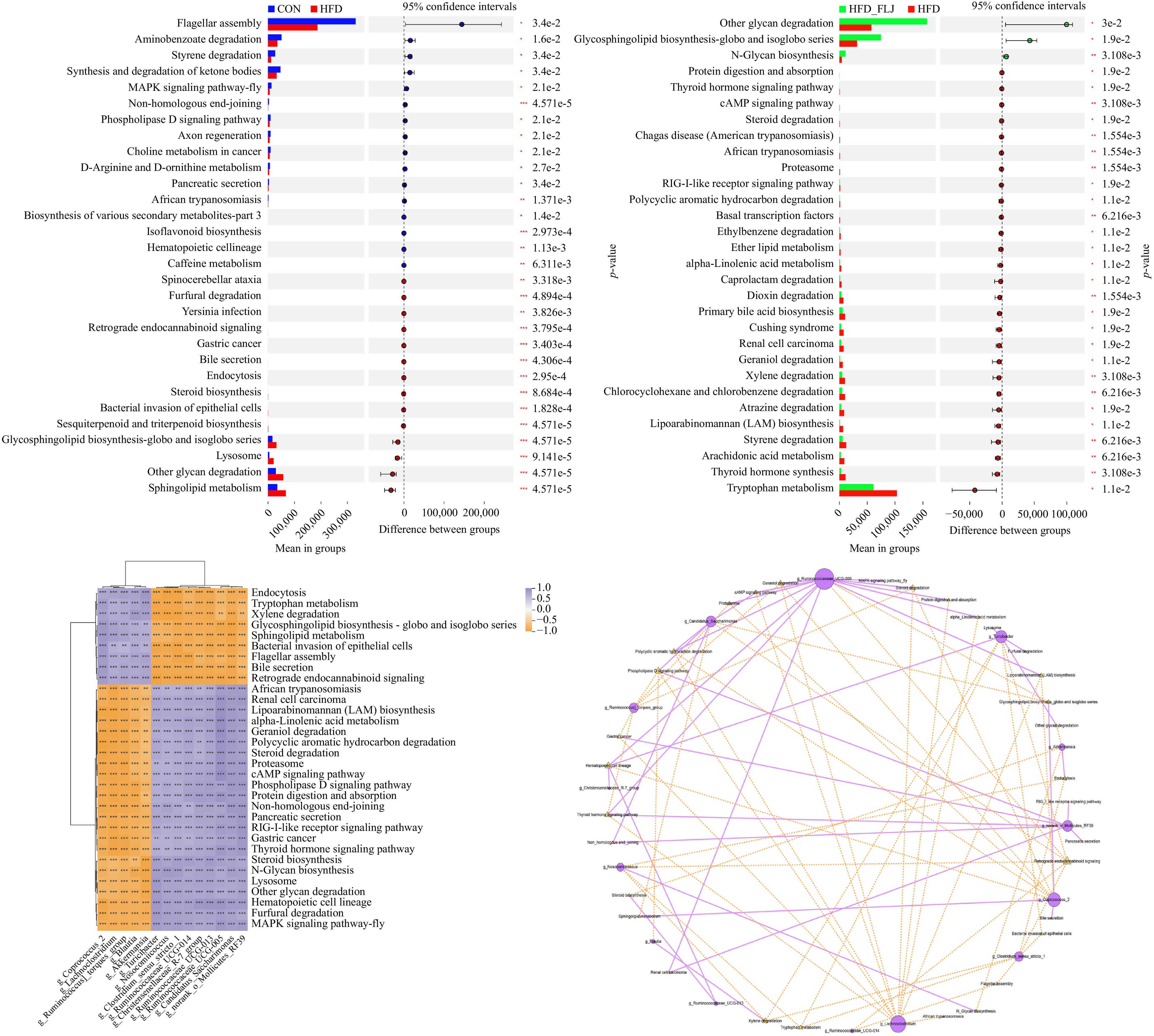
Figure 8.
Extended error bar plot showing differences in significantly and potentially altered metabolic functions of the gut microbia predicted by PICRUSt 3 (Student's t-test) and Spearman correlations between altered fecal microbial phylotypes and predicted metabolic pathways. (a) NFD group vs. HFD group; (b) HFD group vs. HFD_FLJ group. (c) Spearman’s correlation heatmap. (d) Co-occurrence network between the key intestinal microbial species and potentially predicted metabolic pathways. Values are expressed as the mean ± SEM in each group. * p < 0.05, ** p < 0.01 and *** p < 0.001.
Further analysis of the microbiota–function relationships demonstrated specific correlations between key bacterial taxa and metabolic pathways (Fig. 8c, d). The abundance of Akkermansia had a negative correlation with steroid biosynthesis, N-Glycan biosynthesis, and other degradation of other glycans, while showing a positive correlation with endocytosis. Conversely, the abundance of Ruminococcaceae_UCG-005 was positively correlated with steroid degradation, the mitogen-activated protein kinase (MAPK) signaling pathway, protein digestion and absorption, alpha-linolenic acid metabolism, polycyclic aromatic hydrocarbon degradation, the phospholipase D signaling pathway, and the thyroid hormone signaling pathway, but negatively correlated with glycosphingolipid biosynthesis – globo and isoglobo series,and retrograde endocannabinoid signaling.
-
NAFLD is a prevalent chronic liver condition characterized by hepatic steatosis in the absence of excessive alcohol consumption, primarily driven by prolonged HFD intake[28]. Accumulating evidence indicates that HFD-induced gut microbiota dysbiosis plays a crucial role in exacerbating NAFLD's progression[4,29]. The gut–liver axis serves as a key regulatory pathway through which the gut microbiota influence NAFLD's pathogenesis[30,31]. Consequently, maintaining the intestinal microecological balance has emerged as a promising therapeutic strategy for managing NAFLD. Our findings demonstrated that intervention with FLJ effectively maintained intestinal homeostasis and reduced hepatic lipid accumulation in HFD-fed NAFLD rats.
Consistent with established NAFLD models, feeding the HFD for 8 weeks in our study resulted in significant increase in body weight, liver index, and adipose tissue accumulation, accompanied by elevated serum levels of TC, TG, and LDL-C. These metabolic alterations aligned with previous reports on HFD-induced Type 3 diabetes mellitus models, suggesting elevated TC, TG, and LDL-C and decreased levels of HDL-C[24]. However, intervention with FLJ in HFD group significantly decreased body weight and the accumulation of perirenal and epididymal fat; moreover, it also significantly decreased serum lipid levels. Simultaneously, an analysis of the main nutrients before and after fermentation with P. acidilactici FZU106 revealed no significant differences between the unfermented and fermented groups, as shown in Supplementary Table S1. It was speculated that the β-glucosidase derived from P. acidilactici hydrolyzes cellulose, releasing β-D-glucose, which contributed to lowering lipids[22]. Moreover, P. acidilactici itself may also promote the reduction of lipids[32]. This hypolipidemic effect was also supported by Zhang et al., who showed that fucoidan from L. japonica significantly reduced plasma TC and TG, and improved HDL-c levels[24].
As the primary organ responsible for lipid metabolism, the liver exhibits particular vulnerability to HFD-induced damage[33,34]. It was previously reported that a persistent HFS in animals could cause abnormal lipid levels in the liver, further inducing the occurrence of NAFLD[35]. Our study showed a similar result, indicating that administration of the HFD for 8 weeks significantly increased hepatic TC, TG, TBA, NEFA, and fat levels. However, treatment with FLJ remarkedly ameliorated these alterations. Another published study has certified that sulfated polysaccharides from L. japonica could effectively alleviate fat accumulation and the formation of lipid lesions in the liver[36]. According to the histopathology revealed by H&E straining, treatment with FLJ significantly improved lipid droplet accumulation in HFD rats. Zhu et al. also found that treatment with L. japonica polysaccharides for 4 weeks in NAFLD rats sharply decreased lipid droplets in the liver and inhibited the formation of fatty liver disease[37]. In addition, this study found that the FLJ intervention significantly ameliorated the hepatic oxidative stress level, as evidenced by reduced MDA levels and enhanced activities of GSH-Px and SOD. Oxidative stress levels are the most important pathological event during NAFLD's development[38]. Collectively, our results indicate that FLJ represents a promising dietary intervention for the management or prevention of NAFLD.
The regulation of bile acid metabolism represents a crucial pathway in lipid homeostasis. As the primary metabolites of cholesterol, bile acids are synthesized in the liver via a multistep enzymatic cascade[23]. Cholesterol synthesis is an energy-consuming process in which cholesterol is generated from acetyl-coenzyme A through the regulation of a series of enzymes, and 3-hydroxy-3-methylglutaryl coenzyme a reductase (HMGCR) is the key rate-limiting enzyme[39]. Our results showed that treatment with FLJ downregulated the expression of HMGCR at the protein and mRNA levels, which effectively inhibited cholesterol synthesis in the liver. Importantly, the FLJ intervention upregulated CYP7A1, the key enzyme initiating bile acid synthesis, and downregulated ACAT2, which facilitates cholesterol esterification[15,40]. Our results demonstrated that the FLJ intervention significantly upregulated CYP7A1 at both the protein and gene levels and downregulated Acat2 at the mRNA level. This phenomenon further confirmed that treatment with FLJ significantly decreased hepatic TC and increased hepatic TBA. Furthermore, FLJ supplementation downregulated the expression of SREBP-1c at both the gene and protein levels; this is a master regulator of lipogenesis, potentially contributing to the decreased hepatic TG and NEFA accumulation. This finding aligns with Zhang et al.'s report demonstrating that treatment with 200 mg/kg fucoidan extracted from L. japonica significantly reduced the gene expression of SREBP-1c[24]. In addition, CD36, a principal transporter mediating free fatty acid uptake, exhibited expression levels that were strongly associated with the development of NAFLD and insulin resistance[41]. The FLJ treatment suppressed NEFA uptake by downregulating CD36's expression at the protein and mRNA levels.
The gut microbiota is critical to remaining intestinal homeostasis and influencing NAFLD's progression[42]. Increasing evidence has confirmed a close connection between the gut microbiota and NAFLD[43]. The FLJ intervention was likely poorly absorbed by the intestinal tract and was further subjected to microbial fermentation, being converted into secondary metabolites, particularly SCFAs. SCFAs, as gut microbiota metabolites, have a beneficial influence on improving NAFLD. Relative to the HFD controls, FLJ administration markedly increased fecal concentrations of SCFAs, including acetate, propionate, butyrate, isobutyrate, valerate and isovalerate, which was in agreement with other studies, suggesting that L. japonica polysaccharides enhanced SCFA production by changing the composition of gut bacteria in mice with Type 2 diabetes[17]. Many studies suggest that Ruminococcaceae and Lachnospiraceae are beneficial for improving intestinal health because they transform carbohydrates into SCFAs in the intestine, especially acetic acid and propionic acid[15]. This aligns with our results, as treatment with FLJ significantly upregulated the abundance of Ruminococcaceae_UCG-005, which was positively significantly related to the butyrate, isobutyrate, valerate and isovalerate levels. Additionally, the Ruminococcaceae family has also been certified to decrease levels of triacylglycerols, phospholipids, and cholesteryl esters[44]. Similarly, the abundance of Akkermansia, Roseburia, Desulfovibrio, GCA-900066575, Family_Xlll_UCG001, Pediococcus, Defluvitaleaceae_UCG.011, Anaerotruncus, and Alistipes showed a marked upregulation in the HFD_FLJ group. Akkermansia is a prevalent commensal in the mammalian gut and is often regarded as a beneficial microbe. Its abundance showed a positive association with pathways involved in SCFA metabolism, particularly those related to pyruvate and propanoate. Notably, the FLJ intervention significantly increased fecal propanoate[45]. Notably, the FLJ intervention significantly increased fecal propanoate levels, suggesting a potential mechanism for ameliorating NAFLD. Alistipes is a major contributor to SCFAs among the intestinal metabolites, but are also associated with microbiota dysbiosis and related diseases[46]. Desulfovibrio belongs to the family Desulfovibrionaceae, which is positively related to butyric acid and propionic acid in mice feces[47]. In addition, oral administration of Pediococcus in human clinical trials could decrease serum TG, TC, and LDL-C levels[48]. Furthermore, our study found that the abundance of Pediococcus had a positive correlation with acetate and propionate contents in the feces. Numerous studies have confirmed that Akkermansia muciniphila can promote intestinal barrier function, and its abundance is negatively correlated with atherosclerosis and HFD-induced obesity[14]. This result aligns with the findings in the FLJ group in the present study, showing that the abundance of Akkermansia is significantly negatively correlated with serum TC, TG and LDL-C levels; hepatic TC, TG, TBA, NEFA, and fat; and liver and kidney fat indices, but significantly positively corrrelated with the kidney index, serum HDL-C, and fecal butyrate. Insoluble dietary fiber from brown seaweed dose-dependently boosted the abundance of Akkermansia, as previously reported[49]. The study results demonstrated that treatment with FLJ significantly enhanced the composition of beneficial bacteria in HFD rats, and these bacteria could secrete unique enzymes such as fucosidase and galactosidase, which further degraded the polysaccharides of L. japonica to lower molecular substances for better utilization.
In order to understand the driving factors of the gut microbiota's community dynamics, PICRUSt analysis was used to predict the metagenomics of the intestinal microbiota. The potentially different metabolic pathways among all groups were involved in glycosphingolipid biosynthesis – globo and isoglobo series, other glycan degradation, African trypanosomiasis, steroid degradation, and bile secretion. The 8-week FLJ intervention showed a reduced trend toward primary bile acid biosynthesis and arachidonic acid metabolism, but an increased trend toward glycosphingolipid biosynthesis – globo and isoglobo series, other glycan degradation, and N-glycan biosynthesis. Spearman's correlation analysis demonstrated that Akkermansia's abundance may be positively associated with glycosphingolipid biosynthesis – globo, and bile secretion. Bile secretion plays an essential role in cholesterol metabolism, bile acid synthesis, and cholesterol excretion[50]. Bile acids act as signaling molecules in the gut–liver axis and are critical for regulating systemic lipid metabolism homeostasis. Therefore, bile acid metabolism is closely related to NAFLD and may cause cholesterol metabolism disorders, further raising the risk of lipid metabolism disorders and even cardiovascular disease[51]. In agreement with our results, Li et al. also found that bile secretion had a positive correlation with Akkermansia's abundance in the gut microbiota, as predicted by PICRUSt[52]. Additionally, the abundance of Ruminococcaceae UCG-005 demonstrated a strong association with bile acid metabolism and tryptophan metabolism, which was consistent with Sun et al.'s results, suggesting that Ruminococcaceae UCG-005 may be related to the deconjugation reaction from conjugated bile acids to free bile acids in rats with HFD-induced hepatic steatosis[53].
-
In summary, the present study illustrated that treatment with FLJ exhibited a positive effect against NAFLD in HFD rats. On the one hand, oral administration of FLJ significantly reduced body weight gain, lipid levels in the serum and liver, and fat accumulation in the liver. Further investigation has verified that treatment with FLJ modulated lipid metabolism-related genes' expression levels, which contributed to the process of transforming cholesterol into bile acids. On the other hand, the FLJ intervention can regulate lipid metabolism disorders by altering the gut microbiota's composition, suggesting significantly upregulated abundance of Ruminococcaceae_UCG-005 Akkermansia, Roseburia, Desulfovibrio, Pediococcus, and Defluvitaleaceae_UCG.011. These microbial alterations promoted SCFA production, further contributing to the improvement of systemic lipid metabolism. Thereby, these findings provide compelling evidence for the development of FLJ as a novel function food ingredient for managing NAFLD.
-
The experimental procedures were approved by the Institutional Animal Care and Use Committee of College of Food Science, Fujian Agriculture and Forestry University (FAFU 2025-017).
-
The authors confirm their contributions to the paper as follows: methodology: Fan X, Shi M; investigation, formal analysis: Fan X, Ye R; writing – original draft: Fan X; validation, data curation: Xie Z; conceptualization: Shi M, Zhang Q; supervision: Chen L, Zhang Q; funding: Chen L; project administration: Zhang Q. All authors reviewed the results and approved the final version of the manuscript.
-
All data generated or analyzed during this study are included in this published article and its supplementary information files.
-
The authors declare that they have no known competing financial interests or personal relationships that could have appeared to influence the work reported in this paper.
-
This accompanies this paper online at https://doi.org/10.48130/fia-0026-0036.
- Supplementary Table S1 Concentrations of main nutrients in Laminaria japonica fermented with Pediococcus acidilactici FZU106.
- Supplementary Table S2 Compositions of normal diet and high fat diet used in the animal experiment.
- Supplementary Table S3 Primer sequence for quantitative real-time PCR.
- Supplementary Fig. S1 Representative genus the relative abundance (%) of Pediococcus acidilactici FZU106-fermented Laminaria japonica intervention in NAFLD rat.
- Copyright: © 2026 by the author(s). Published by Maximum Academic Press on behalf of China Agricultural University, Zhejiang University and Shenyang Agricultural University. This article is an open access article distributed under Creative Commons Attribution License (CC BY 4.0), visit https://creativecommons.org/licenses/by/4.0/.
-
About this article
Cite this article
Fan X, Ye R, Xie Z, Shi M, Chen L, et al. 2026. Pediococcus acidilactici FZU106-fermented Laminaria japonica alleviates high-fat diet-induced NAFLD rats by modulating the gut microbiota and lipid metabolism. Food Innovation and Advances 5(3): 432−443 doi: 10.48130/fia-0026-0036 

Pediococcus acidilactici FZU106-fermented Laminaria japonica alleviates high-fat diet-induced NAFLD rats by modulating the gut microbiota and lipid metabolism
- Received: 05 January 2026
- Revised: 24 May 2026
- Accepted: 30 May 2026
- Published online: 02 September 2026
Abstract: This study aimed to investigate the hypolipidemic mechanism of Pediococcus acidilactici FZU106-fermented Laminaria japonica (FLJ) and evaluate its therapeutic potential against high-fat diet-induced nonalcoholic fatty liver disease (NAFLD) in rats. Our results demonstrated that the FLJ intervention significantly ameliorated NAFLD's progression through dual regulatory mechanisms. First, the intervention with FLJ significantly reduced body weight gain (−23.57%), perirenal fat and epididymal fat indexes (−40.16% and −34.64%, respectively), and serum and liver lipid levels, suggesting decreased hepatic fat accumulation (−39.96%) in NAFLD rats. Further mechanistic analysis demonstrated that FLJ modulated lipid metabolism-related pathways by downregulating the expression of key genes involved in cholesterol synthesis (HMGCR) and fatty acid uptake (CD36), while upregulating the expression of bile acid conversion enzymes (CYP7A1). Second, FLJ reshaped the gut microbiota's composition by enriching the abundance of beneficial bacteria, including Akkermansia, Ruminococcaceae_UCG-005, and Pediococcus, which were positively correlated with the short-chain fatty acid level (butyrate) and negatively correlated with lipid parameters in the serum and liver (total cholesterol and triglycerides). These bacteria exhibited a strong correlation with the bile secretion pathway, as revealed by functional metagenomic profiling through phylogenetic investigation of communities by reconstruction of unobserved states (PICRUSt) analysis. These findings suggested that FLJ attenuated NAFLD through coordination of the gut–liver axis, positioning it as a promising dietary intervention for metabolic liver disorders.