












-
The strawberry (Fragaria × ananassa L.) is a perennial herbaceous species in the Rosaceae family and represents a globally valued fruit crop recognized for its characteristic flavor, vivid pigmentation, and nutritional attributes[1,2]. Increasing consumer demand for high-quality fruit has intensified the need for reliable production of elite planting material, which has become a central limitation for industry expansion. Traditional propagation depends on stolon-mediated clonal reproduction, yet this approach often facilitates the accumulation and spread of viruses. Such pathogen transmission contributes to cultivar decline, reduced yield, and inconsistency in fruit quality. In vitro plant tissue culture is recognized as an effective approach for virus elimination and rapid micropropagation. This method supports large-scale production of virus-free plantlets while maintaining the genetic characteristics of cultivars, making it a core technique for stabilizing and advancing the strawberry nursery sector[3]. Despite these benefits, issues such as soil sickness associated with consecutive monoculture systems continue to hinder the post-acclimatization performance and management of micropropagated plantlets. Therefore, identifying innovative strategies to increase the environmental stress tolerance of strawberry plantlets holds considerable practical value.
Carbon nanotubes (CNTs), first reported by Iijima & Ichihashi, have emerged as important one-dimensional nanomaterials due to their outstanding mechanical, electrical, and thermal characteristics, supporting broad applicability across multiple disciplines[4]. Their use in plant science and biotechnology has advanced rapidly, with studies indicating that CNTs can act as effective molecular carriers capable of traversing the plant cell wall to promote the internalization and movement of exogenous molecules, influencing plant physiological and metabolic pathways[5−7]. Carboxylated multi-walled CNTs (FCNTs) possess favorable water dispersibility and biocompatibility, along with a reduced propensity to trigger intense immune activation in plants. These properties position FCNTs as promising candidates for applications in plant tissue culture and metabolic regulation[8,9].
Despite the emerging evidence supporting the biological relevance of FCNTs, the detailed molecular mechanisms through which they affect plant systems, particularly protein expression patterns and metabolic network regulation, remain insufficiently characterized. To address this limitation, this study applied an integrated proteomic and metabolomic strategy. Adventitious roots of strawberry were exposed to 500 mg/L FCNTs, followed by tandem mass tag (TMT)-based quantitative proteomic analysis and untargeted global metabolomic profiling to identify differentially accumulated proteins (DAPs) and differentially accumulated metabolites (DAMs) between treated and control samples.
This study aims to elucidate the molecular mechanisms underlying FCNT-mediated responses in strawberry adventitious roots through integrated quantitative proteomics and comprehensive metabolomics. The study also characterizes FCNT-responsive proteins and metabolites, maps the associated signaling and metabolic pathways, and identifies key molecular components potentially linked to enhanced stress tolerance and improved performance under tissue culture conditions.
-
Virus-free tissue-cultured plantlets of 'Hongyan' strawberry were obtained from the Shanghai Key Laboratory of Protected Horticultural Technology (Shanghai, China). The plantlets were sub-cultured on solid Murashige and Skoog (MS) medium containing 0.1 mg/L naphthaleneacetic acid (NAA) and 0.1 mg/L indole-3-butyric acid (IBA) to promote adventitious root formation. MS medium, NAA, and IBA were purchased from Aladdin Biochemical Tech Co., Ltd. (Shanghai, China). Cultures were maintained at 25 ± 2 °C under a 12-h photoperiod, with illumination at 50 μmol m-2 s-1 provided by cool-white fluorescent lamps.
Uniform and healthy adventitious roots were selected for treatment. A 500 mg/L FCNTs (purity > 95 wt%; inner diameter 5−15 nm; outer diameter < 50 nm; length < 10 μm; Aladdin, Shanghai, China) stock suspension was prepared in pure water and sonicated for 30 min before use. Roots were immersed either in the FCNT suspension (treatment group) or in pure water (control group). To improve surface contact and potential uptake, both groups were placed in an ultrasonic bath (800 W; Xianou, Nanjing, China) and exposed to pulsed sonication (on 10 s, off 10 s) for 20 min. Following treatment, root samples were blotted dry, rapidly frozen in liquid nitrogen, and stored at –80 °C until analysis. Proteomic and metabolomic analyses were conducted using three and six biological replicates per group, respectively.
Proteomic analysis
Protein extraction
-
Frozen root samples were finely ground in liquid nitrogen, and the resulting powder was transferred to pre-chilled centrifuge tubes containing phosphate-buffered saline (PBS). The sample was thoroughly homogenized in ice-cold lysis buffer. The homogenate was centrifuged at 14,000 × g for 20 min at 4 °C, and the supernatant was collected as the total protein extract. Protein concentration was quantified using the Bradford assay[10].
For mass spectrometry analysis, 100 μg of protein from each sample was reduced with dithiothreitol, alkylated with iodoacetamide, and digested with trypsin at an enzyme-to-substrate ratio of 1:50 overnight at 37 °C. The resulting peptides were desalted using C18 solid-phase extraction cartridges and then dried by vacuum centrifugation.
TMT labeling and LC–MS/MS analysis
-
The dried peptide samples were reconstituted and labeled using the TMT 16plex™ Isobaric Label Reagent Set (Thermo Fisher Scientific, Massachusetts, USA) following the manufacturer's protocol. Briefly, peptides from each group were incubated with distinct TMT tags at room temperature for 1 h, after which the labeling reaction was quenched with hydroxylamine. The labeled peptides were then pooled in equal amounts and desalted.
Liquid chromatography–tandem mass spectrometry (LC–MS/MS) analysis was performed using an Orbitrap Eclipse™ 480 mass spectrometer (Thermo Fisher Scientific, Massachusetts, USA) coupled to a nanoflow ultra-high-performance liquid chromatography (UHPLC) system. Peptides were separated on a C18 reversed-phase analytical column (75 μm × 25 cm, 2 μm) using a linear gradient from 5% to 35% of mobile phase B (0.1% formic acid in 80% acetonitrile) against mobile phase A (0.1% formic acid in water) over 120 min at a flow rate of 300 nL/min. The mass spectrometer was operated in data-dependent acquisition mode, with full MS scans acquired at 120,000 resolution across an m/z range of 350−1,500. The most intense precursor ions were subjected to high-energy collisional dissociation with a normalized collision energy of 30%, and MS/MS spectra were recorded at a resolution of 60,000.
Protein identification and bioinformatics analysis
-
Raw LC-MS/MS data were processed and quantified using Proteome Discoverer v2.4.1.15 (Thermo Fisher Scientific, Massachusetts, USA). Database searches applied a reversed sequence decoy strategy to control peptide false discovery rates (FDR), and identifications were validated with Percolator software, setting the false discovery rate at 0.01 for both proteins and peptide-spectrum matches (PSMs)[11].
Principal Component Analysis (PCA) was performed to assess separation among biological conditions. DAPs were identified using the t-test. Enrichment analysis of Gene Ontology (GO) terms and Kyoto Encyclopedia of Genes and Genomes (KEGG) pathways was conducted via Fisher's exact test[12,13]. Protein–protein interaction networks were analyzed, and network visualizations were generated with igraph[14,15].
Metabolomics analysis
Metabolite extraction
-
Frozen root powder (50 mg) was weighed and extracted with 600 μL of MeOH containing 2-Amino-3-(2-chloro-phenyl)-propionic acid (4 μg/mL), followed by vortexing for 30 s. The mixture was homogenized at 55 Hz for 60 s and then centrifuged at 12,000 × g for 10 min at 4 °C. The resulting supernatant was filtered through a 0.22 μm nylon membrane and collected in a vial for LC–MS analysis[16].
LC–MS analysis
-
Chromatographic separation was carried out on a Vanquish UHPLC system (Thermo Fisher Scientific, Massachusetts, USA) fitted with an ACQUITY UPLC HSS T3 column (1.8 μm, 2.1 mm × 100 mm; Waters, Milford, MA, USA) at 40 °C. The mobile phase for positive ion mode consisted of (A) 0.1% formic acid in water and (B) 0.1% formic acid in acetonitrile, while for negative ion mode, it consisted of (A) 5 mM ammonium formate in water and (B) acetonitrile. A linear gradient from 5% to 95% B over 14 min was applied at a flow rate of 0.3 mL/min[17].
Mass spectrometry analysis was performed using an Orbitrap Exploris 120 mass spectrometer (Thermo Fisher Scientific, Massachusetts, USA) equipped with an ESI ion source. Data were acquired in both positive and negative ion modes with the following parameters: spray voltage ± 3.50 kV, sheath gas 40 arb, auxiliary gas 10 arb, capillary temperature 325 °C, Orbitrap resolution of 60,000 for full MS and 15,000 for MS/MS, and stepped normalized collision energies of 20%, 30%, and 40%[18].
Data preprocessing and statistical analysis
-
Raw data files were converted to mzML format using MSConvert (ProteoWizard). Subsequent preprocessing, comprising peak picking, alignment, and retention time correction, was conducted with the XCMS package in R. Metabolites showing an RSD greater than 30% in quality control (QC) samples were excluded.
Multivariate statistical analyses were conducted using the ropls R package[19]. Unit-variance scaling was applied before PCA and Orthogonal Projections to Latent Structures-Discriminant Analysis (OPLS-DA). Model performance was evaluated using the cross-validated correlation coefficient (Q2). Metabolites with a Variable Importance in Projection (VIP) > 1.0 in the OPLS-DA model and a p-value < 0.05 from a two-tailed Student's t-test were considered DAMs. Metabolite identification was performed by matching accurate mass and MS/MS spectra against the HMDB, KEGG, and MassBank databases[20−22]. Pathway enrichment analysis was carried out using MetaboAnalyst 5.0[23].
-
A rigorous QC workflow was applied to ensure data reliability. The mass error distribution of all identified peptides was centered near zero, with most values within ± 20 ppm, indicating high mass accuracy during acquisition. The majority of identified peptides ranged from eight to 35 amino acids in length, consistent with complete tryptic digestion and confirming effective sample preparation. Applying the criteria of at least one unique peptide and quantifiable signals in over 50% of replicates, a total of 206 high-confidence proteins were consistently identified across all samples (Fig. 1a). Among the DAPs, only two were upregulated, whereas the top 20 most significantly downregulated DAPs are listed in Table 1.
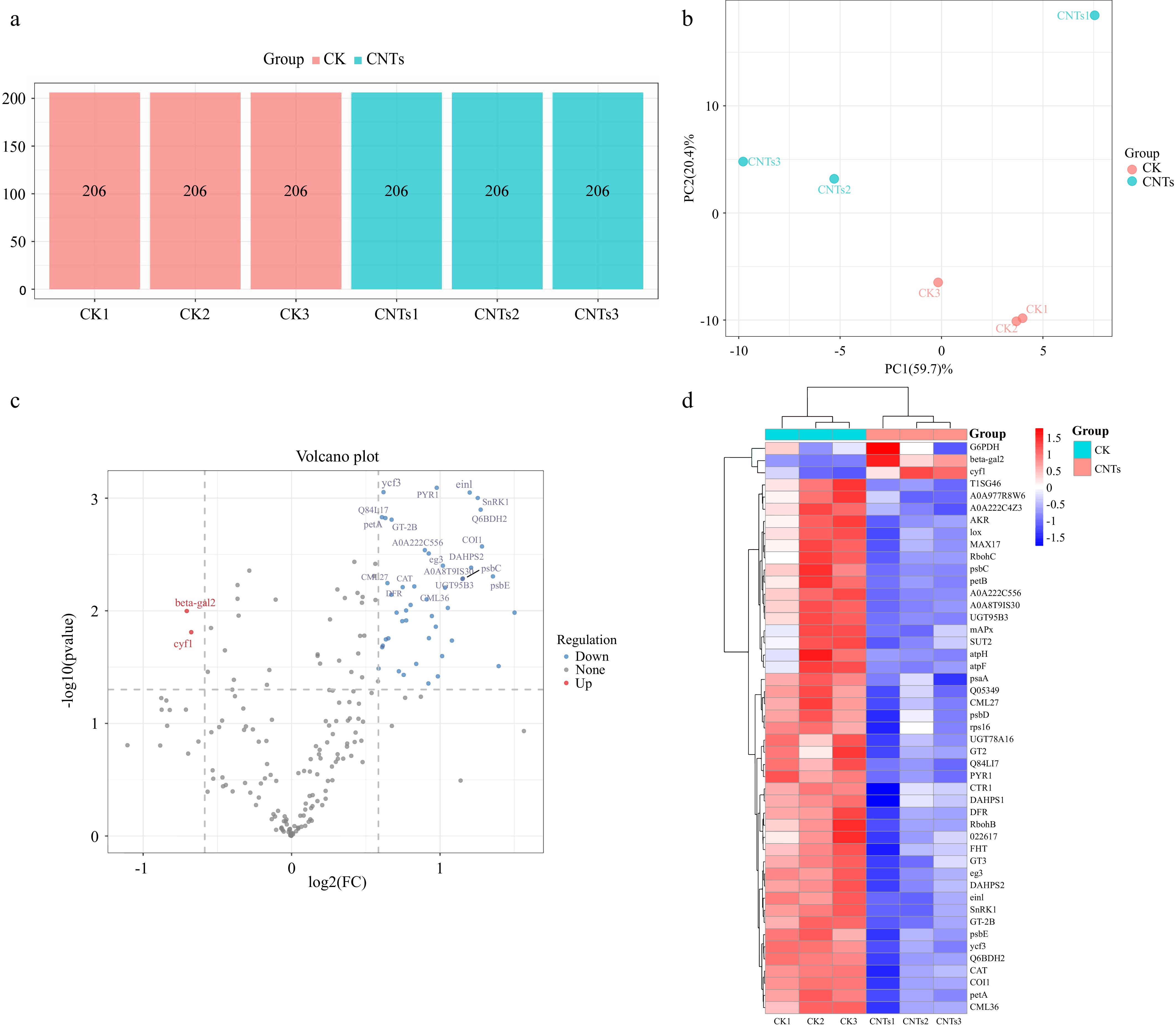
Figure 1.
Protein quantitative analysis of strawberry adventitious roots treated with FCNTs. (a) Number of identified proteins in each sample in the CK and CNTs groups; (b) PCA score plot showing the distribution of global proteomic profiles between CK and FCNT-treated groups; (c) Volcano plot of DAPs. The x-axis represents the log2 fold change of protein abundance, while the y-axis represents statistical significance (−log10 p-value). Red dots indicate significantly downregulated proteins, and blue dots indicate significantly upregulated proteins (p < 0.05, t-test); and (d) Heatmap showing the expression patterns of all DAPs between the CK and CNTs groups. Proteomic analyses were performed using three biological replicates per group (n = 3).
Table 1. Top 20 most significantly downregulated DAPs in strawberry adventitious roots following CNT treatment.
Protein_ID Protein_name CK_average CNTs_average log2(FC) A0A8K1H7Q9 Cytochrome b6 305.03 107.70 1.50 A0A288VIJ2 ATP synthase subunit b, chloroplastic (ATP synthase F[0] sector subunit b) (ATPase subunit I) 589.53 224.20 1.39 J7IKT1 Cytochrome b559 subunit alpha (PSII reaction center subunit V) 2,246.03 877.27 1.36 A0A290U3W5 Coronatine insensitive 1 protein 1,656.00 680.67 1.28 Q6BDH2 Aldo-keto reductase 1,125.93 465.67 1.27 A0A7S7YNU4 Non-specific serine/threonine protein kinase (EC 2.7.11.1) 340.90 142.93 1.25 A0A1J0CL90 Phospho-2-dehydro-3-deoxyheptonate aldolase (EC 2.5.1.54) 4,669.27 2,019.10 1.21 Q949H5 Ethylene receptor 503.33 219.13 1.20 A0A288VIK4 Photosystem II CP43 reaction center protein (PSII 43 kDa protein) (Protein CP-43) 14,642.80 6,581.93 1.15 A0A387IZT7 UDP-glucose glucosyltransferase 363.33 163.60 1.15 A0A1L4A1Q8 Respiratory burst oxidase-like protein C 1,659.03 784.77 1.08 T1SG46 Flavonoid 3'-hydroxylase 1,029.57 496.43 1.05 A0A6B9IJV4 Putative calcium-binding protein 1,202.87 591.10 1.03 A0A8T9IS30 L-galactose-1,4-lactone dehydrogenase 1,716.90 847.00 1.02 A0A288VIB6 30S ribosomal protein S16, chloroplastic 559.57 277.07 1.01 M9R5M8 L-ascorbate peroxidase (EC 1.11.1.11) 1,061.97 536.43 0.99 F2YHU9 Abscisic acid receptor PYR1 1129.13 573.33 0.98 Q5Y381 Aldo/keto reductase 457.13 233.13 0.97 A0A1L4A1N8 Respiratory burst oxidase-like protein B 404.30 210.03 0.94 Protein quantification and differential expression analysis
-
PCA of the proteomic data showed distinct clustering between the FCNTs-treated (CNTs) and control (CK) groups, with clear separation between groups and tight aggregation within groups (Fig. 1b), indicating a strong treatment effect and high reproducibility. Comparative analysis identified 47 DAPs between the groups, of which 45 were downregulated, and two were upregulated (Fig. 1c). Hierarchical clustering further highlighted the significant differences in protein expression profiles between the CK and CNTs groups (Fig. 1d).
Enrichment analysis and network interaction of DAPs
-
GO enrichment analysis classified the DAPs into Biological Process (BP), Cellular Component (CC), and Molecular Function (MF). Within BP, DAPs were significantly enriched in photosynthesis, the amino acid biosynthetic process, and the aromatic amino acid family biosynthetic process (Fig. 2a). In CC, prominent terms included chloroplast thylakoid membrane, membrane part, and proton-transporting ATP synthase complex (Fig. 2b). Enriched MF terms comprised iron ion binding, heme binding, electron transfer activity, calcium ion binding, and oxidoreductase activity (Fig. 2c).
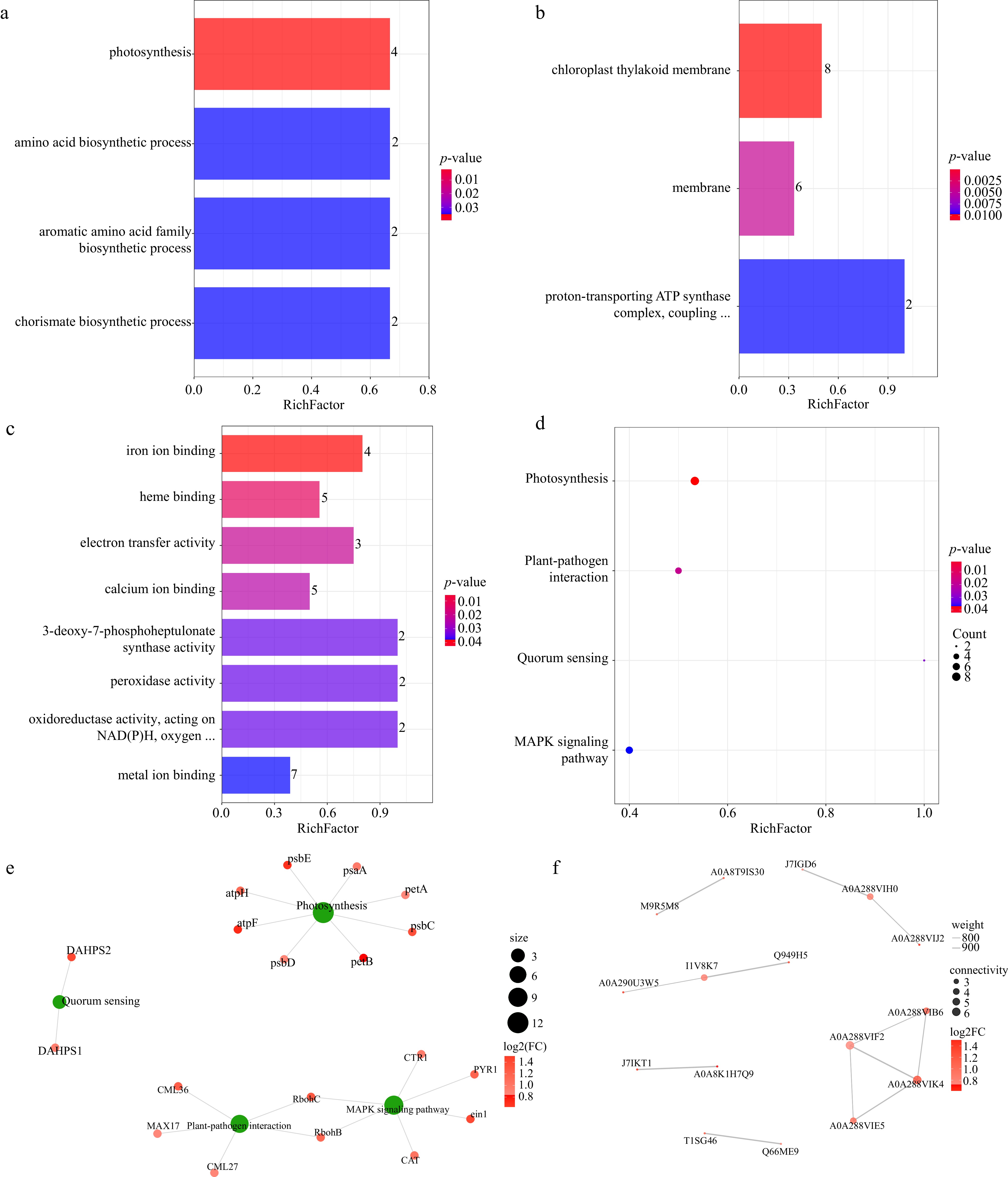
Figure 2.
Functional enrichment and interaction network analysis of DAPs in strawberry adventitious roots following FCNT treatment. (a) Bar chart for GO enrichment analysis of biological processes enrichment; (b) GO enrichment analysis of cellular component categories; (c) GO enrichment analysis of molecular function categories; (d) bubble chart for KEGG pathway enrichment. The x-axis represents the enrichment factor, and the y-axis shows the enriched KEGG pathways. Color intensity reflects the level of statistical significance, while bubble size corresponds to the number of DAPs associated with each pathway; (e) KEGG pathway enrichment and the DAPs network plot. Red nodes represent DAPs, whereas green nodes denote significantly enriched KEGG pathways; and (f) protein–protein interaction network of DAPs. Each node represents a protein, and red-colored nodes indicate DAPs. The intensity of the red color corresponds to the magnitude of the log2FC in protein abundance. Proteomic analyses were performed using three biological replicates per group (n = 3).
KEGG pathway analysis identified four significantly enriched pathways: photosynthesis (map00195), plant–pathogen interaction (map04626), quorum sensing (map02024), and the MAPK signaling pathway in plants (map04016) (Fig. 2d). An interaction network was constructed to depict the relationships between these key pathways and the associated DAPs (Fig. 2e). Furthermore, protein–protein interaction (PPI) network analysis showed that the DAPs formed a tightly connected functional module (Fig. 2f).
Metabolomic analysis
Data QC
-
The quality of the metabolomic data was initially evaluated using base peak chromatograms (BPCs), which displayed normal peak shapes and effective separation. Following the removal of metabolic features with an RSD > 30% in QC samples, 87.9% of the remaining features had an RSD below 30%, confirming high reproducibility and stability of the dataset for downstream analysis.
Multivariate statistical analysis
-
PCA of data acquired in both positive and negative ionization modes revealed clear separation between the CNTs and CK groups, with samples from the same condition clustering tightly, indicating significant metabolic profile changes induced by FCNTs treatment (Fig. 3a, e). The model explanatory parameters (R2X) were 0.561 and 0.564 for the positive and negative modes, respectively.
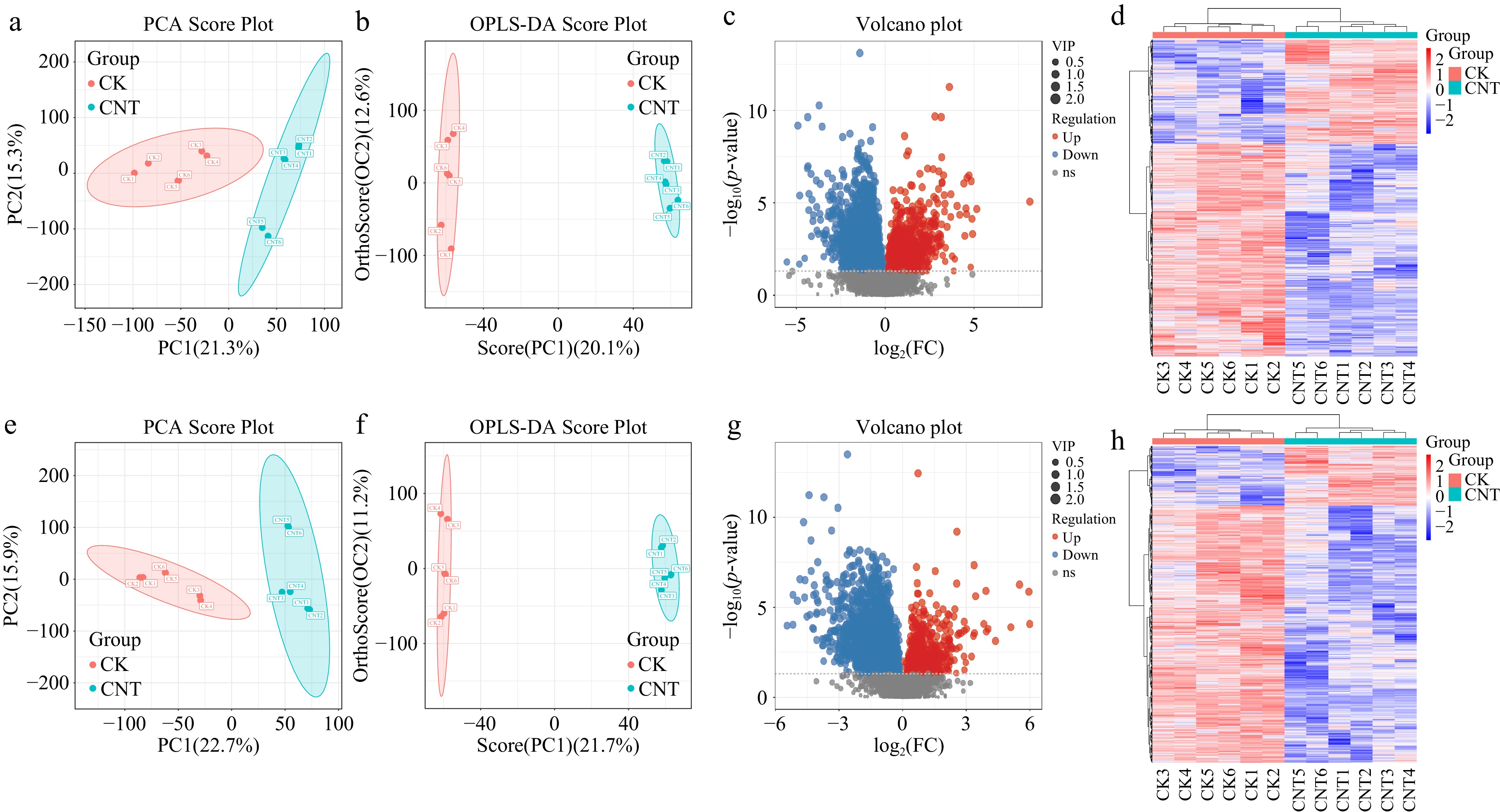
Figure 3.
Metabolomic profiling and differential metabolite analysis of strawberry adventitious roots following FCNT treatment. PCA score plots generated from metabolomic data acquired in (a) positive and (e) negative ionization modes. Each point represents an individual sample; ellipses indicate the 95% confidence intervals, and colors distinguish the experimental groups. OPLS-DA score plots in (b) positive and (f) negative ionization modes. The horizontal axis represents intergroup variation, whereas the vertical axis reflects intragroup variation. Greater clustering of samples within groups and clearer separation between groups indicate stronger model discrimination. Volcano plots of differential metabolite features identified in (c) positive and (g) negative ionization modes. Red dots represent significantly upregulated metabolite features, while blue dots represent significantly downregulated metabolite features (VIP > 1.0, p < 0.05, t-test). Heatmaps illustrating the abundance patterns of all differential metabolite features detected in (d) positive and (h) negative ionization modes. Note: Metabolomic analyses were performed using six biological replicates per group (n = 6).
To enhance group discrimination, supervised OPLS-DA was applied. The OPLS-DA score plots showed clear separation between the CNTs and CK groups (Fig. 3b, f). High predictive ability values (Q2 = 0.830 and 0.853 for positive and negative modes, respectively) confirmed the robustness and reliability of the models.
Identification of DAMs
-
Differential metabolites were identified using a combination of VIP > 1.0 from the OPLS-DA model and a p-value < 0.05 from Student's t-test. In positive ion mode, 4,105 of 18,733 features were classified as differential metabolites (1,353 upregulated, 2,752 downregulated). In negative ion mode, 3,580 of 18,005 features were differential (834 upregulated, 2,746 downregulated). Volcano plots depicted the distribution and fold changes of these metabolites (Fig. 3c, g), while clustering heatmaps highlighted extensive metabolic reprogramming, with a predominant downregulation trend in the CNTs group (Fig. 3d, h).
Identification and analysis of differential metabolites
-
By matching MS/MS spectra against public metabolite databases, 421 compounds were confidently identified. Among these, 86 were classified as DAMs, including 28 upregulated and 58 downregulated metabolites (Fig. 4a). A heatmap of these DAMs clearly differentiated the metabolic profiles of the control and treated groups (Fig. 4b). The top 20 identified DAMs are listed in Table 2.
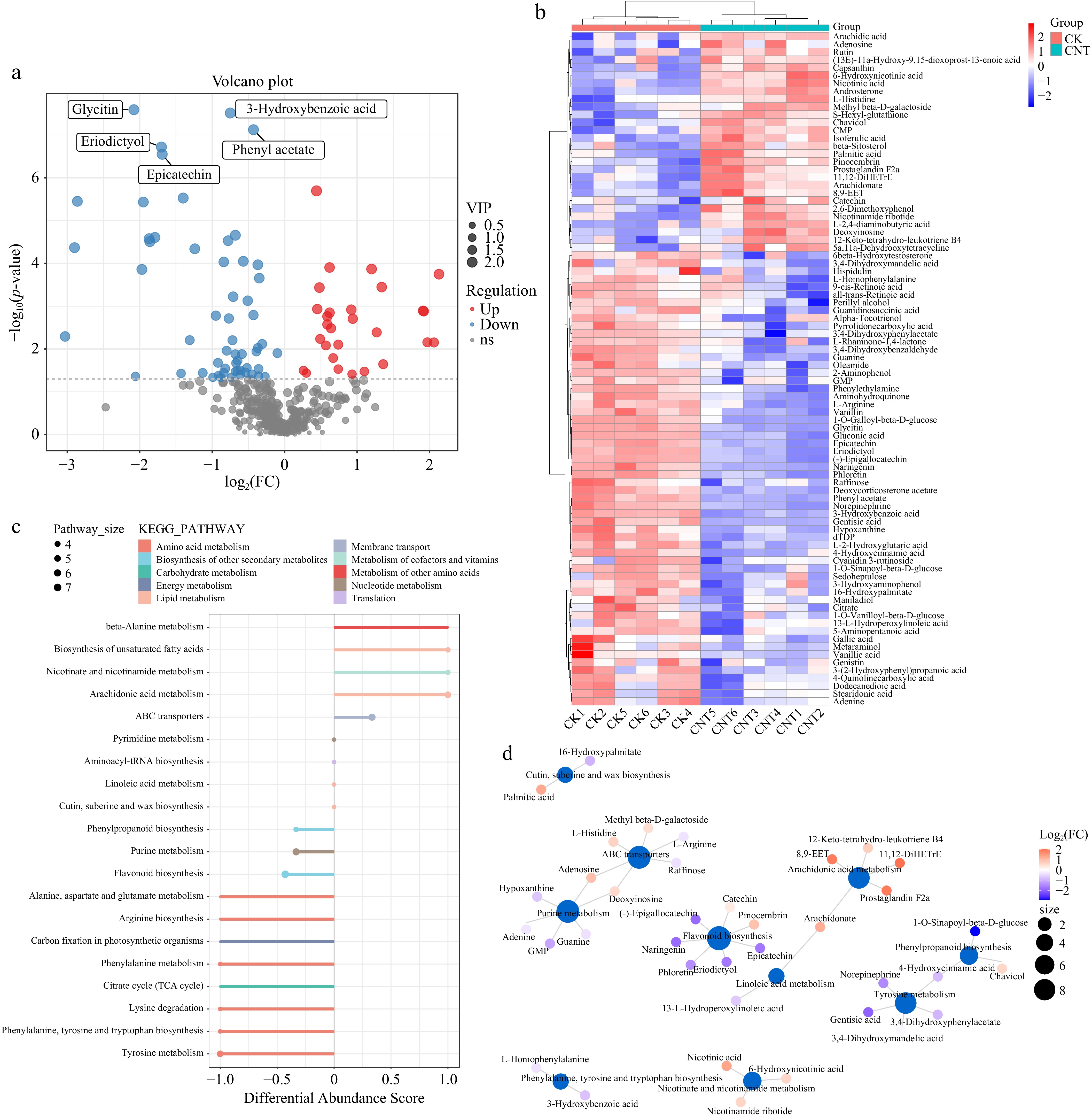
Figure 4.
Functional annotation and pathway analysis of DAMs in strawberry adventitious roots treated with FCNTs. (a) Volcano plot of identified DAMs. Red dots indicate significantly upregulated metabolites, while blue dots indicate significantly downregulated metabolites; (b) Heatmap of the 86 identified DAMs. Columns represent samples and rows represent metabolites. The dendrogram on the left illustrates hierarchical clustering of metabolites, while the top dendrogram represents clustering of samples; (c) Differential enrichment score map of KEGG pathway. The x-axis represents DA-score values, calculated as (number of upregulated metabolites − number of downregulated metabolites) divided by the total number of different metabolites within a pathway. The y-axis represents the corresponding metabolic pathways. The size of the points indicates the number of DAMs enriched in each pathway; and (d) Network diagram of the KEGG pathway. Blue nodes represent KEGG pathways, whereas other nodes correspond to metabolites. The size of pathway nodes reflects the number of connected metabolites, with larger nodes indicating higher metabolite connectivity. Metabolite node color represents the magnitude of log2 fold change (log2FC), where red indicates upregulation and blue indicates downregulation, with increasing color intensity corresponding to greater magnitude of change. Metabolites associated with multiple group comparisons do not contain log2FC information. (Metabolomic analyses were performed using six biological replicates per group).
Table 2. Top 20 identified DAMs in strawberry adventitious roots after CNTs treatment.
ID Name CK_average CNTs_average CNTs / CK M119T70 L-2,4-diaminobutyric acid 1,007,703.00 4,401,371.01 4.37 M321T592 11,12-DiHETrE 7,664,427.76 32,035,611.99 4.18 M397T679_1 beta-Sitosterol 10,801,962.03 42,345,514.14 3.92 M337T499 Prostaglandin F2a 1,261,350.64 4,769,852.20 3.78 M321T496 8,9-EET 1,069,183.37 4,029,598.42 3.77 M312T607 Arachidic acid 13,417,653.53 34,554,717.71 2.58 M124T638 Nicotinic acid 519,275,804.12 1,315,273,210.08 2.53 M255T652 Palmitic acid 309,187,712.76 746,766,562.00 2.42 M101T667 N-Nitroso-pyrrolidine 3,144,017.89 7,461,655.43 2.37 M305T576 Arachidonate 9,696,915.33 4,208,295.92 2.30 M273T317 Naringenin 16,287,012.19 4,493,655.45 0.28 M287T247 (-)-Epigallocatechin 12,874,873.03 3,523,501.98 0.27 M371T539_1 Deoxycorticosterone acetate 16,631,141.99 4,303,953.59 0.26 M153T240 Gentisic acid 18,999,244.36 4,846,269.29 0.26 M425T545 Alpha-Tocotrienol 14,465,691.80 3,473,684.87 0.24 M445T342 Glycitin 2,293,867.67 543,753.59 0.24 M150T72_1 L-Methionine 40501046.53 7304786.26 0.18 M331T55 1-O-Galloyl-beta-D-glucose 22159229.12 3042351.85 0.14 M369T258 1-O-Sinapoyl-beta-D-glucose 18050656.08 2413092.93 0.13 M110T31 2-Aminophenol 88731260.23 10892561.07 0.12 Note: The dataset includes both upregulated and downregulated DAMs identified under FCNT treatment compared with the CK, with 10 metabolites showing increased and 10 showing decreased abundance. KEGG pathway enrichment analysis
-
Pathway analysis of the 86 DAMs indicated significant enrichment in 45 metabolic pathways. The most affected pathways were grouped into five functional categories: (1) Central Energy Metabolism (e.g., citrate cycle, aminoacyl-tRNA biosynthesis); (2) Amino Acid Metabolism (e.g., biosynthesis of phenylalanine, tyrosine and tryptophan, flavonoid biosynthesis); (3) Nucleotide Metabolism (e.g., pyrimidine metabolism); (4) Lipid Metabolism (e.g., biosynthesis of cutin, suberine and wax, beta-alanine metabolism); and (5) Co-factor and Transport Systems (e.g., nicotinate and nicotinamide metabolism, ABC transporters) (Fig. 4c). An integrated pathway interaction network was constructed to illustrate the interconnections among these altered metabolic pathways (Fig. 4d).
-
The integration of TMT-based quantitative proteomics and global untargeted metabolomics offers a systems-level view of the molecular response of strawberry adventitious roots to FCNTs treatment. The multi-omics analysis indicates that FCNTs function not only as a physical stimulus but also as a strong elicitor, initiating a cascade of molecular events involving stress defense activation, metabolic resource redistribution, and reprogramming of metabolism[24].
Proteomic analysis revealed that DAPs were significantly enriched in the plant–pathogen interaction (map04626) and MAPK signaling (map04016) pathways. The MAPK cascade serves as a central signaling hub, mediating diverse environmental and developmental cues in plants and regulating stress-responsive genes and metabolic processes[25]. Simultaneous activation of the plant–pathogen interaction pathway, together with the upregulation of proteins involved in calcium ion binding, peroxidase, and oxidoreductase activities (Fig. 2c), indicates that FCNTs are recognized by strawberry roots as an exogenous stress stimulus. This recognition establishes a defense-primed state, consistent with reports that nanoparticles can function as abiotic elicitors[26,27]. These proteomic signals are strongly supported at the metabolic level, with significant alterations observed in the phenylpropanoid pathway and flavonoid biosynthesis (Fig. 4c). Flavonoids, as key metabolites produced in response to stress, play essential roles in antioxidant defense, UV protection, and antimicrobial activity[28,29]. The altered levels of precursor aromatic amino acids and their derivatives indicate a targeted metabolic flux toward the production of these defensive compounds. This coordinated response, from signal perception (MAPK activation) to defense signaling (plant–pathogen interaction pathway) and ultimately to metabolic execution (flavonoid biosynthesis), demonstrates a systematic strategy employed by strawberry roots to mitigate FCNTs-induced stress[30].
A significant and apparently paradoxical observation was the significant downregulation of proteins associated with photosynthesis and the chloroplast thylakoid membrane in root tissues (Fig. 2a, b). Although leaves are the primary site of photosynthesis, previous studies have reported the presence of these proteins in roots, potentially reflecting low-level expression, involvement in non-photosynthetic functions, or systemic metabolic coordination across the plantlet[31−33]. The downregulation of these proteins following FCNTs treatment suggests a strategic reallocation of resources: the plant appears to prioritize immediate stress defense over basal growth, redirecting energy and carbon skeletons away from anabolic processes such as photosynthesis. This concept of 'energy reallocation' is supported by metabolomic evidence. The TCA cycle, a central hub for ATP production and a source of carbon skeletons for biosynthesis, was significantly altered (Fig. 4c). Remodeling of core energy metabolism likely supports the increased demand for ATP and biosynthetic precursors necessary for the production of defense-related compounds, such as flavonoids[34]. This remodeling also helps sustain cellular homeostasis under stress, as indicated by the enrichment of pathways including ABC transporters.
This coordinated metabolic reprogramming offers a plausible explanation for another apparently paradoxical observation in this study: the concurrent activation of defense responses and enhancement of root growth. In most plant systems, defense activation is associated with growth suppression due to competition for limited metabolic resources, a classical growth–defense trade-off. One possible interpretation is that FCNTs induce a mild stress response resembling a hormesis-like effect; however, rigorous validation of this hypothesis would require detailed dose–response analyses and quantitative growth assessments. Within a hormesis framework, the 500 mg/L FCNT treatment may function as a low-intensity stressor that elicits adaptive responses without exceeding the plant's metabolic buffering capacity. This interpretation is consistent with the suppression of photosynthesis-related proteins, which may reduce energetic expenditure, alongside the alteration of TCA cycle components, which can sustain ATP generation and provide biosynthetic intermediates. Such metabolic adjustments could support simultaneous maintenance of root growth and establishment of a primed defensive state. Comparable responses have been reported in plants exposed to other nanomaterials, where low-dose treatments enhance both biomass accumulation and stress resilience across different species[35,36]. Therefore, FCNTs may act as a nano-elicitor that reorganizes metabolic priorities, attenuating the conventional coupling between growth and defense programs in strawberry roots.
A consistent pattern across both omics layers was the predominance of downregulated molecules, with 45 of 47 DAPs and 58 of 86 DAMs showing decreased levels. This trend indicates a broad 'slowdown' or re-prioritization of metabolic processes. Downregulation of rate-limiting enzymes in key biosynthetic pathways, such as 3-deoxy-7-phosphoheptulonate synthase in the aromatic amino acid pathway, likely generates metabolic bottlenecks, resulting in reduced accumulation of downstream metabolites, including tyrosine and tryptophan[37,38]. The alignment between protein expression and metabolite levels at these critical nodes reinforces the biological significance of these observations and highlights potential key regulatory targets of FCNTs action.
These results indicate that FCNTs function not as a nutrient but as a 'metabolic reprogramming elicitor'. The molecular alterations observed reflect a physiological state of 'defense priming', in which basal growth is temporarily moderated to pre-activate defense mechanisms. This primed state may increase plant resilience to subsequent biotic or abiotic stresses, such as soil-borne pathogens encountered in consecutive monoculture systems[39]. Previous studies on CNTs in plant tissue culture, such as increased proliferation in GF677 rootstock and increased growth and secondary metabolite production in Jojoba, underscore the biological activity of CNTs[40,41]. Thus, FCNTs' application in tissue culture presents a promising approach for generating 'primed plantlets' with improved stress tolerance, representing a conceptual advance toward nano-elicitor-based horticultural strategies.
It is also important to consider a methodological limitation associated with the use of sonication. In the present experimental design, both FCNT-treated and control samples were exposed to identical pulsed sonication to enhance nanomaterial dispersion and facilitate uptake. This approach ensures that observed differences between treatments can be attributed specifically to FCNT exposure; however, sonication itself may introduce mechanical perturbations that influence root physiology and trigger stress-related responses. Although the uniform application of sonication across experimental groups supports valid comparative analysis, it is possible that the absolute physiological state of the tissues differs from that of non-sonicated roots, particularly with respect to baseline stress levels. Therefore, the inclusion of an additional non-sonicated control (untreated roots without ultrasound exposure) would be valuable in future work to independently assess the effects of sonication on strawberry root biology, including mechanical stress responses, cellular integrity, and potential defense priming[42]. Incorporating such a control would allow clearer separation of ultrasound-induced effects from FCNT-specific responses and would strengthen the mechanistic interpretation of nano-elicitor activity in plant tissue culture systems.
Although this study provides a comprehensive multi-omics overview of strawberry adventitious root responses to FCNTs, the results remain largely correlative. Future investigations should incorporate functional validation approaches, including genetic manipulation of key candidate proteins, such as central components of the MAPK signaling cascade, as well as absolute quantification of hub metabolites (e.g., proline) using targeted LC–MS/MS or enzymatic assays. Furthermore, the integration of genome-editing strategies, such as CRISPR–Cas9-mediated disruption of biosynthetic genes (for example, glutamate decarboxylase in γ-aminobutyric acid biosynthesis and pyrroline-5-carboxylate synthase in proline biosynthesis), together with detailed phenotypic and metabolic analyses, will be essential to establish causal relationships. Such approaches will further clarify how FCNTs regulate protein expression and metabolic reprogramming to enhance root performance in plant tissue culture systems.
-
This proteomic and metabolomic analysis demonstrates that FCNTs function as a potent metabolic elicitor in strawberry adventitious roots. FCNTs' treatment systematically reprograms root physiology by activating defense-associated signaling pathways, specifically the MAPK cascade, and by redirecting carbon and energy resources from growth processes toward the biosynthesis of stress-protective metabolites, such as flavonoids. These coordinated molecular responses indicate that FCNTs induce a state of 'defense priming', potentially elevating the plant's ability to withstand subsequent environmental stresses. The results provide a mechanistic basis for the use of functionalized CNTs as nano-elicitors in horticultural propagation, offering a promising strategy to improve the resilience of tissue-cultured strawberry plantlets. However, before any translational or field-level application, further evaluation of potential risks is required, including nanomaterial persistence, environmental accumulation, impacts on soil microbial communities, and possible implications for human health.
We are grateful to the Jiangsu Academy of Agricultural Sciences for supporting this work. We would like to express our gratitude to Shanghai Key Laboratory of Protected Horticultural Technology for providing 'Hongyan' strawberry virus-free seedlings as experimental materials. This research was financially supported by the Youth Program of Nantong Natural Science Foundation (JC2024017), the Discipline Construction Project of Jiangsu Yanjiang Institute of Agricultural Sciences (YJXK[2023]103), and the Exploratory Disruptive Innovation Program of Jiangsu Academy of Agricultural Sciences (ZX[21]1231).
-
The authors confirm their contribution to the paper as follows: study conception and design: Cao F, Li Y; data collection: Cao F, Jin Y, Guo C, and Chen Y; analysis and interpretation of results: Cao F, Huang X, Wu L, and Ke P; draft manuscript preparation: Cao F, Gu Q, and Ning Y. All authors reviewed the results and approved the final version of the manuscript.
-
The datasets generated during or analyzed during the current study are available from the corresponding author upon reasonable request.
-
The authors declare that they have no conflict of interest.
- Copyright: © 2026 by the author(s). Published by Maximum Academic Press, Fayetteville, GA. This article is an open access article distributed under Creative Commons Attribution License (CC BY 4.0), visit https://creativecommons.org/licenses/by/4.0/.
-
About this article
Cite this article
Cao F, Gu Q, Huang X, Jin Y, Guo C, et al. 2026. Proteomics and metabolomics analysis of the strawberry adventitious root response to carboxylated carbon nanotubes. Technology in Horticulture 6: e021 doi: 10.48130/tihort-0026-0013 

Proteomics and metabolomics analysis of the strawberry adventitious root response to carboxylated carbon nanotubes
- Received: 19 January 2026
- Revised: 08 June 2026
- Accepted: 09 July 2026
- Published online: 29 September 2026
Abstract: Strawberry propagation through tissue culture enables pathogen removal and stable trait transmission; however, improving culture performance under environmental pressures, including continuous monoculture stress, remains a key objective. Carboxylated carbon nanotubes (FCNTs), characterized by distinct physicochemical properties and favorable biocompatibility, have emerged as potential regulators of plant metabolic processes. However, the understanding of FCNT-mediated reprogramming of multi-omics networks in strawberry adventitious roots at the systems level remains unclear. This study aims to analyze the molecular response of strawberry adventitious roots to 500 mg/L FCNTs through integrated proteomic and metabolomic approaches. Virus-free 'Hongyan' strawberry tissue culture seedlings were used as the biological material, and roots were treated using ultrasonic-assisted adsorption. Proteomic profiling identified 47 differentially accumulated proteins (DAPs) upon FCNTs exposure, with 45 downregulated and two upregulated, enriched in pathways related to photosynthesis, plant–pathogen interaction, and MAPK signaling. Metabolomic analysis identified 86 differentially accumulated metabolites (DAMs, 58 downregulated and 28 upregulated), associated with metabolic routes such as biosynthesis of phenylalanine, tyrosine, and tryptophan, flavonoid biosynthesis, and the tricarboxylic acid cycle. The combined multi-omics assessment indicated that FCNT treatment modulated stress-related signaling pathways, central carbon and energy metabolism, and secondary metabolite biosynthesis. In conclusion, the molecular basis of strawberry root responses to carboxylated CNTs offers a foundation for developing CNT-based strategies to enhance tissue culture performance and strengthen stress resilience in continuous cropping systems.
-
Key words:
- carbon nanotube /
- strawberr /
- tissue culture /
- proteomics /
- metabolomics