












-
Leaf senescence during postharvest vase life is a critical factor limiting the ornamental quality and vase life of cut flowers, leading to significant economic losses in the floriculture industry[1]. It not only impairs the visual appeal by causing leaf wilting and yellowing but also disrupts nutrient translocation and water balance, accelerating the senescence of the entire cut flower[2]. Previous studies have mostly focused on the obvious yellowing symptoms in the middle and late stages of senescence, while paying insufficient attention to the potential early mechanisms[3]. Studying the mechanism of early leaf senescence can achieve early intervention and early regulation of it. This can effectively delay the process of leaf wilting, extend the vase life, solve the problems of poor pertinence and limited effect of traditional preservation technologies, and improve the ornamental quality of chrysanthemum cut flowers.
The early stage of leaf senescence is a critical node where cellular physiological functions transition from normal to deterioration, and it's a complex, genetically regulated process triggered by multiple stress signals after harvest. Plant hormones are also an important factor in leaf senescence, which jointly control the senescence process through mutual antagonism or synergy[4]. Jasmonic acid (JA) promotes leaf senescence by activating specific transcriptional cascades and triggering mesophyll cell death. In contrast, cytokinins (CK) and gibberellins (GA) are senescence-inhibiting hormones that can delay leaf senescence by maintaining photosynthetic efficiency, inhibiting chlorophyll decomposition and autophagy. Ethylene and abscisic acid (ABA) are the main senescence-promoting hormones: ethylene can induce programmed cell death of abscission zone cells and accelerate leaf yellowing and abscission, while ABA accelerates the senescence process by promoting chlorophyll degradation and membrane lipid peroxidation[5]. Relevant studies provide important theoretical support for delaying plant leaf senescence and improving the quality of crops and ornamental plants through hormone regulation technologies.
The most visible characteristic of leaf senescence is the yellowing or browning of the leaves, which is caused by the rapid degradation of chlorophyll. During vase holding, the chlorophyll content in leaves decreases continuously, accompanied by the decline of photosynthetic rate, photochemical efficiency of PSII, and the activity of photosynthetic enzymes[6,7]. This process is driven by the upregulation of chlorophyll catabolic genes (CCGs) such as NYC1 (NON-YELLOW COLORING 1) and SGR (STAY-GREEN), which encode the key enzymes responsible for chlorophyll breakdown[8]. The degradation of chlorophyll not only leads to the loss of green color but also results in the collapse of the photosynthetic system, further reducing the energy production capacity of the leaves.
At the molecular level, leaf senescence is accompanied by distinct changes in the expression of senescence-associated genes (SAGs), suggesting that this physiological process depends on large-scale transcriptional regulation. The increased transcription of SAGs is generally regarded as a key molecular feature of leaf senescence[9]. As a complex biological event, senescence involves thousands of SAGs that participate in various metabolic functions and signal regulatory networks[9]. Genome-wide analyses in Arabidopsis have divided SAGs into four major functional categories: regulatory genes, genes related to macromolecular decomposition, nutrient redistribution and transport, and stress-response genes[10]. A number of important genes from these groups have been identified and functionally verified. For instance, multiple NAC family genes, including homologs of ORE1 and NAP, show remarkably enhanced expression during leaf senescence and can directly promote the transcription of chlorophyll catabolic genes and other SAGs[11]. In Arabidopsis, histone methylation-mediated epigenetic modification of WRKY53 participates in the regulation of leaf senescence[12], while melatonin can retard WRKY75-dependent leaf senescence[13]. SAG101 is expressed specifically at the onset of senescence; antisense suppression of this gene postpones senescence, whereas its overexpression triggers early senescence, demonstrating its essential role in this process[14]. Moreover, the NAC transcription factor NTL4 stimulates reactive oxygen species accumulation during drought-triggered leaf senescence in Arabidopsis[15]. The histone variant HTB4 also delays leaf senescence by epigenetically regulating the transcription factors bHLH038, bHLH039, bHLH100, and bHLH101, which are involved in maintaining iron homeostasis[16]. Beyond model plants, research on leaf senescence mechanisms in horticultural species has expanded in recent years. In tomato, SlMYC2 suppresses ROS scavenging, intensifies oxidative injury, and reduces photosystem II (PSII) activity, thus acting as a positive regulator of leaf senescence[17]. SnRK1 has been reported to function in dark-induced leaf senescence in cucumber[18]. In apple, MdbHLH93 directly activates MdSAG18 transcription and accelerates leaf senescence, whereas MdBT2 interacts with MdbHLH93 and promotes its degradation via ubiquitination, thereby slowing senescence progression[19].
To date, transcriptomic profiling of SAGs during leaf senescence remains limited in ornamental plants. Chrysanthemum (Chrysanthemum morifolium) is a leading ornamental crop worldwide, accounting for approximately 30% of the global cut flower market, only secondary to rose[20]. It is widely used for bouquets, floral arrangements, and decorative purposes due to its diverse colors, shapes, and long vase life potential. However, the commercial value of chrysanthemum cut flowers is severely constrained by a unique postharvest problem: premature leaf senescence, commonly known as leaf yellowing or leaf necrosis, which often occurs much earlier than the senescence of the petals[21,22]. Unlike many other cut flower species where petal senescence is the primary limiting factor of vase life, in chrysanthemum, the deterioration of leaves usually determines the end of the ornamental value, as consumers prefer green and healthy leaves accompanying the flowers[23]. Recent research by Song et al. revealed that in chrysanthemum, jasmonic acid (JA) specifically triggers cell death in spongy mesophyll tissue via the CmNAC055-CmNAC087-CmACX5 transcriptional cascade, and this regulatory module is highly conserved across Arabidopsis, tomato, rice, and other species[2]. Meanwhile, the dynamic interaction between CmbHLH1L and CmNLP6/7L anchored on the scaffold protein CmbHLH63 acts as a molecular switch governing stress-induced leaf senescence in chrysanthemum[24]. Further studies also indicated that strigolactone promotes leaf senescence in chrysanthemum, while sucrose effectively alleviates SL-triggered senescence. Transient silencing of the differentially expressed gene CmWRKY6-Like was found to positively regulate leaf senescence and mediate the antagonistic effects of SL and sucrose on this process[25]. In addition, Liu et al. studied the gene expression regulatory networks in the leaf senescence of post-harvest cut chrysanthemums in response to ethylene treatment[5]. Under ABA treatment conditions, CmBBX22 can significantly delay the senescence of transgenic plant leaves. CmBBX22 can downregulate the expression of senescence-related genes (SAG29) and chlorophyll degradation genes (NYE1, NYE2, NYC1), inhibit chlorophyll degradation, and initiate the senescence program[26].
Heat shock transcription factors (HSFs) are crucial regulatory factors in plants, playing key roles in defending against various stresses. The HSF family has numerous members with diverse structures, distinct properties, and versatile functions[27]. Besides directly regulating heat shock proteins (HSPs) and stress-related genes to mediate stress responses and adaptation, HSFs also participate in regulating fundamental plant life processes such as growth and metabolism[28]. However, existing research mainly focuses on the HSFA subfamily, with relatively limited studies on HSFB and even fewer reports on HSFC, leaving gaps in understanding the full functions and regulatory mechanisms of the HSF family.
Based on the understanding of the senescence mechanisms, various strategies have been developed to delay leaf senescence and extend the vase life of chrysanthemum cut flowers. With the development of molecular biology, biotechnological approaches provide new opportunities to improve the postharvest quality of chrysanthemum[20,29]. Despite the extensive progress, the molecular mechanism of leaf senescence in chrysanthemum is still not fully understood. The present study systematically investigated physiological alterations and global transcriptomic dynamics during the early stages of leaf senescence in chrysanthemum cut flowers 'Jinba' during vase holding, and functionally verified the regulatory role of CmHSFC1 in senescence progression. Our findings delineate a two-stage early senescence program characterized by first stress adaptation and second hormone-dependent senescence execution, and identify a core set of transcription factors and functional genes that orchestrate postharvest early leaf senescence. These results extend the current understanding of chrysanthemum leaf senescence and provide candidate targets for genetic improvement of postharvest longevity.
-
Chrysanthemum cut flower cultivar 'Jinba' was obtained from Yunnan Agricultural University in Kunming, Yunnan Province, China. Chrysanthemums with uniform flower size (flower diameter of 7–8 cm), stem diameter of 6 mm, and free from disease, mechanical damage, or insect pests were selected as experimental materials. Upon arrival in the laboratory, stems were recut to a standard length of 30 cm under distilled water to prevent air embolism. Cut flowers were placed in glass bottles containing 1 L of distilled water, with one flower per bottle. All samples were kept in an environmentally controlled chamber with a temperature of 22 ± 1 °C, relative humidity of 60%–70%, and a 12-h photoperiod under a light intensity of 30–40 μmol·m−2·s−1.
Leaf samples were collected at 0, 3, 6, 9, and 12 d after vase placement. Fully unfolded leaves were harvested from the bottom of each stem, immediately frozen in liquid nitrogen, and stored at −80 °C for subsequent physiological and biochemical measurements. Each treatment consisted of three independent biological replicates, with three cut flowers per replicate.
Determination of physiological and biochemical indices
-
Fresh chrysanthemum leaf samples were homogenized in pre-cooled 0.05 mol·L−1 phosphate buffer (pH 7.8) under ice-bath conditions. The homogenate was centrifuged at 10,000 × g for 20 min at 4 °C, and the supernatant was collected as the crude enzyme extract for subsequent assays[30]. SOD activity was determined using the nitroblue tetrazolium (NBT) photoreduction method. POD activity was measured by the guaiacol colorimetric method[31]. CAT activity was determined by monitoring the decrease in absorbance at 240 nm due to H2O2 decomposition. Malondialdehyde (MDA) content was determined using the thiobarbituric acid (TBA) method. Hydrogen peroxide (H2O2) content was measured using the ferrous oxidation-xylenol orange (eFOX) method with minor modifications. The H2O2 concentration was calculated against a standard curve.
Chlorophyll content was extracted with 80% acetone in the dark until the leaves turned white[32]. The absorbance of the extract was measured at 663 and 645 nm. Chlorophyll a, b, and total chlorophyll contents were calculated using the following formulas:
$ \mathrm{Chl\;a=12.7A_{663}-2.69A_{645}} $ $ \mathrm{Chl\;b=22.9A_{645}-4.68A_{663}} $ $ \mathrm{Total\;chlorophyll=Chl\;a+Chl\;b} $ All determinations were performed with three biological replicates. Data analysis was performed using SPSS software, and significant differences were compared at p < 0.05.
RNA extraction, RNA-Seq sequencing, and bioinformatic analysis
-
Total RNA was extracted from chrysanthemum leaf samples (stored at −80 °C, 0, 6, and 12 d of vase holding) using the Plant Quick RNA Isolation Kit (Huayueyang, China). Briefly, 100 mg of frozen leaves were ground in liquid nitrogen, mixed with lysis buffer, centrifuged, and the supernatant was treated with RNAiso Plus and chloroform. The aqueous phase was precipitated with isopropanol, washed with 75% RNase-free ethanol, and dissolved in RNase-free water.
RNA quality was assessed by NanoDrop 2000 and Agilent 2100 Bioanalyzer; samples with A260/A280 1.8–2.0, A260/A230 ≥ 2.0, and RIN ≥ 8.0 were selected. Three biological replicates per sample were sequenced on Illumina NovaSeq 6000 at BGI Genomics (Wuhan, China). Genomic DNA was removed by DNase I. mRNA was enriched, fragmented, reverse-transcribed into cDNA, and used for library construction. Libraries were sequenced on Illumina NovaSeq 6000 to generate 150 bp paired-end reads. Raw reads were filtered by FastQC (v0.11.9). Clean reads were assembled by StringTie and quantified as FPKM. DEGs between time points were identified by DESeq2 (|log2(fold change)| ≥ 1, p-adj < 0.05), annotated against Nr, Swiss-Prot, KEGG, and GO databases, and enriched by clusterProfiler in R. Bioinformatic analyses were performed on the BGI cloud platform and local server using R.
Quantitative Real-Time PCR (qRT-PCR) analysis
-
Total RNA extraction was performed strictly according to the manufacturer's instructions. Reverse transcription was carried out using 1,000 ng of total RNA as the standard, and 1 μL of cDNA from each sample was used as the template for quantitative real-time PCR (qRT-PCR) detection. qRT-PCR was performed using Takara SYBR Premix Ex Taq reagent with a 50 μL reaction system per well. Three biological replicates were set for each candidate gene, and the relative gene expression levels were calculated using the 2−ΔΔCᴛ method, with the chrysanthemum EF1α gene (GenBank accession: KF305681.1) as the reference gene[33]. All primers used in this study are listed in Supplementary Table S1.
Statistical analysis
-
Duncan's multiple range test was used for statistical analysis of the data, and differences among the different groups were considered significant at p < 0.05. All statistical analyses were performed using SPSS v17.0 software (SPSS Inc., Chicago, IL, USA).
Silencing CmHSFC1 in chrysanthemum
-
The full-length sequence of CmHSFC1 was amplified via polymerase chain reaction (PCR) using the specific primers CmHSFC1-F/R (detailed in Supplementary Table S1). The PCR amplicons were ligated into the pMD19-T vector (TaKaRa, Japan) for subsequent sequencing. The functional characterization of CmHSFC1 was carried out using a virus-based microRNA (miRNA) expression system. Artificial miRNA (amiR) sequences targeting CmHSFC1 (Supplementary Table S1) were designed with the Web MicroRNA Designer tool. The amiR-CmHSFC1 sequence was cloned following the protocol described by Tang et al.[34] and inserted into the CaLCuV A vector to generate the recombinant vector CaLCuV-amiR-CmHSFC1.
The transient transformation of chrysanthemum was performed with slight modifications based on the method previously reported by Wei et al.[35]. Recombinant plasmids were introduced into Agrobacterium tumefaciens strain GV3101, which was then cultured overnight in YEB liquid medium. Bacterial cells were collected by centrifugation and resuspended in infiltration buffer containing 10 mmol/L MgCl2, 10 mmol/L MES (pH 5.7), and 200 μmol/L acetosyringone until the OD600 reached 2.0. Two infection suspensions were prepared separately: the control group was mixed with equivalent volumes of Agrobacterium strains carrying CaLCuV A and CaLCuV B, whereas the silencing treatment group contained equal amounts of Agrobacterium harboring CaLCuV-amiR-CmHSFC1 and CaLCuV B. These mixtures were incubated in the dark at 28 °C for 3 to 4 h before vacuum infiltration. Chrysanthemum seedlings were infiltrated at 100 kPa for 10 min, then washed gently with deionized water and covered with plastic wrap for 2 d of dark adaptation. Afterwards, all plants were transplanted into pots and grown in a greenhouse under a 16-h light/8-h dark cycle at 23 °C with 80% relative humidity. After 2 weeks of growth, young, fully expanded leaves were collected to detect the silencing efficiency of CmHSFC1 and analyze leaf senescence phenotypes. For the in vitro senescence assay, leaf discs were placed on filter paper soaked with ddH2O and incubated under dark conditions.
-
We observed the senescence process of chrysanthemum leaves during the vase-holding period and classified it into early (0–12 d), middle (12–30 d), and late (30–40 d) senescence stages. Leaves with slight chlorophyll fading and subtle physiological senescence symptoms without severe dehydration or wilting occur at 0–12 d; vein-localized chlorosis gradually emerges and expands from 20–30 d; severe leaf yellowing, vein necrosis, and petiole desiccation occur at 35–40 d, indicating irreversible late senescence (Fig. 1).
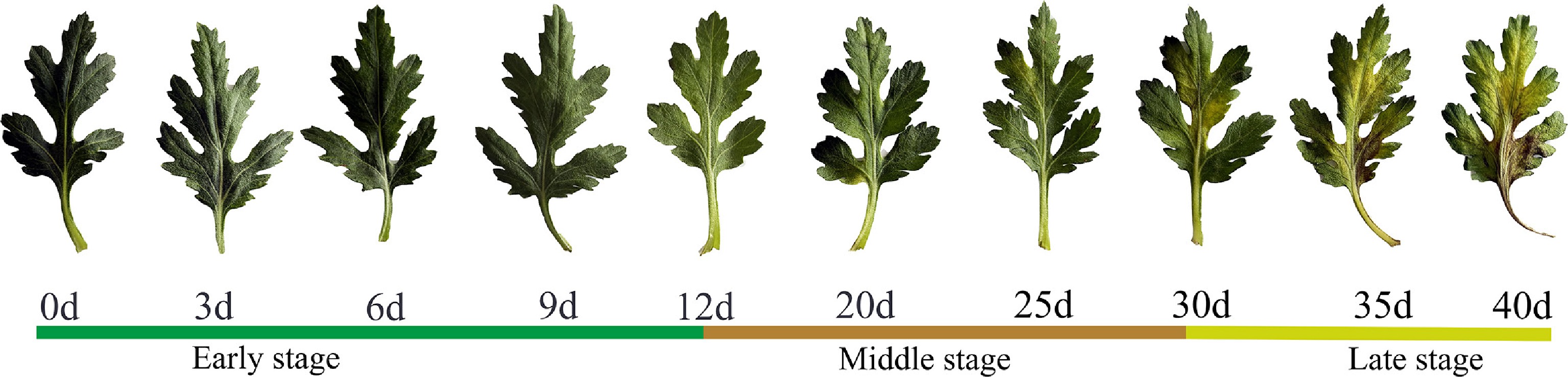
Figure 1.
Leaf senescence phenotypes of cut chrysanthemum during vase life. Leaves were sampled at 0, 3, 6, 9, 12, 20, 25, 30, 35, and 40 d after vase treatment. The horizontal color bar classifies the whole senescence process into three stages: early stage (0–12 d), middle stage (12–30 d), and late stage (30–40 d). Note: d, days after vase insertion.
To elucidate the physiological basis of early senescence in chrysanthemum cut flowers during vase holding, six key physiological indices were systematically determined at 0, 3, 6, 9, and 12 d after harvest according to the phenotypic changes of chrysanthemum cut flower leaves (Fig. 2). MDA content (Fig. 2a) and H2O2 content (Fig. 2f) both displayed continuous increases throughout the vase period. The MDA content increased by approximately 105.5% at 12 d compared with that at 0 d. Similarly, H2O2 content at 12 d increased by approximately 92.7% compared with that at 0 d, showing a comparable accelerating pattern. These results indicated progressive oxidative damage to cellular membranes during leaf senescence. Chlorophyll content (Fig. 2b) exhibited a slowly decreasing trend during senescence. The chlorophyll content remained at a high level with no significant difference at 0 and 3 d. A significant decrease was observed after 6 d, and the content dropped 10.9% at 12 d compared to 0 d. The change in chlorophyll content serves as a core physiological marker of leaf senescence.
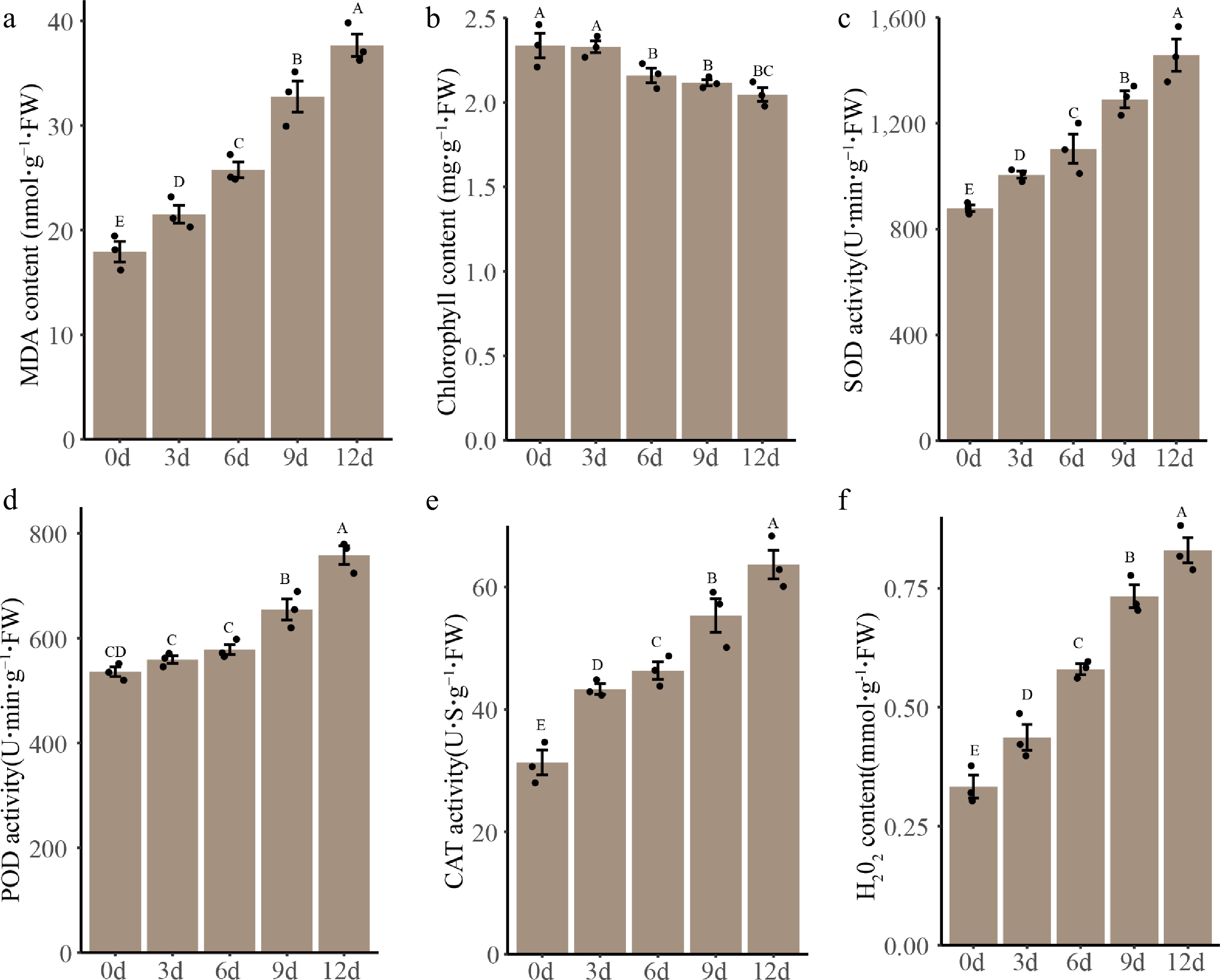
Figure 2.
Changes in physiological indices of cut chrysanthemum leaves during early vase treatment. (a) MDA content. (b) Chlorophyll content. Activities of antioxidant enzymes including (c) SOD, (d) POD, and (e) CAT. (f) H2O2 content. All data are presented as mean ± standard deviation (SD) of three biological replicates. Different uppercase letters indicate significant differences at the p < 0.05 level according to Duncan's multiple range test. FW: fresh weight.
The activities of three major antioxidant enzymes all increased continuously. SOD activity (Fig. 2c) increased by approximately 62.7% at 12 d compared with that at 0 d, with a prominent upward trend after 6 d. POD activity (Fig. 2d) remained relatively stable from 0 to 6 d, then rose sharply at 12 d, with an increase of approximately 44.1% compared with that at 0 d. CAT activity (Fig. 2e) increased steadily from 0 to 12 d, increasing by approximately 109.3% compared with that at 0 d, with accelerated elevation after 6 d. These findings demonstrated that the antioxidant defense system was continuously activated to mitigate oxidative stress during early leaf senescence.
Transcriptome reveals stage-specific regulatory patterns
-
To further investigate the mechanism of early senescence stages, and based on the above physiological changes, we divided this stage into two stages. Transcriptome sequencing was performed on samples at 0, 6, and 12 d to explore the molecular mechanisms underlying vase-induced early senescence. Differentially expressed genes (DEGs) were identified and subjected to GO and KEGG enrichment analyses. A multi-dimensional integrative analysis was conducted to characterize temporal transcriptomic shifts and co-expression networks (Fig. 3).
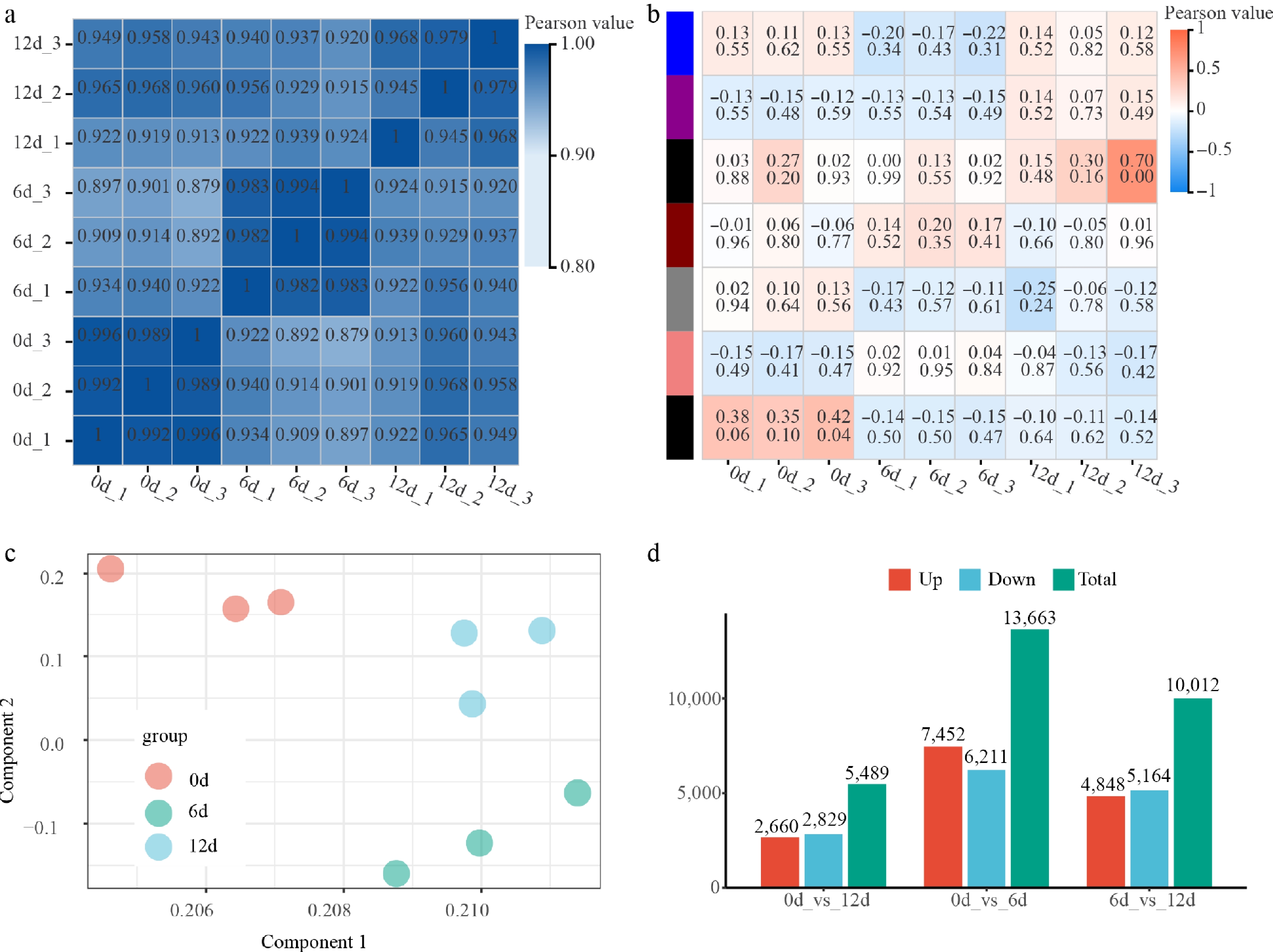
Figure 3.
Multi-dimensional transcriptomic analysis of gene co-expression modules and temporal expression dynamics across different time points. (a) Correlation heatmap of gene co-expression modules. (b) Module-trait association heatmap. (c) Principal component analysis (PCA) of global transcriptomes. (d) Differential gene expression (DGE) statistics between time points.
A gene co-expression module correlation heatmap (Fig. 3a) showed that all nine modules exhibited extremely high intra- and inter-module Pearson correlation coefficients (0.89–0.99), verifying the robustness of module partitioning and the consistency of co-expression relationships. Module–trait association analysis (Fig. 3b) identified distinct temporal correlation patterns. The black module was most strongly positively correlated with the 12 d groups, suggesting an association with senescence processes. In contrast, another module was highly correlated with 0 d samples, implying a role in early or basal regulatory programs. Principal component analysis (PCA) of the global transcriptome (Fig. 3c) clearly separated samples by time point, with biological replicates clustering tightly together. Samples from 0 d were closely grouped, whereas 12 d samples showed greater dispersion, indicating increased transcriptomic heterogeneity at the second early senescence stage (6–12 d).
Differential expression analysis (Fig. 3d) quantified transcriptomic divergence between time points. The comparison between 0 and 6 d yielded the largest number of DEGs (7,452 upregulated, 6,211 downregulated), indicating the most dramatic transcriptomic remodeling occurs in this stage, with the most substantial changes occurring at 6 d.
Stage-specific functional enrichment of DEGs during early leaf senescence
-
To dissect the molecular regulatory programs underlying senescence, DEGs from the first (0–6 d) and second (6–12 d) stages were separately subjected to KEGG pathway and GO enrichment analyses (Fig. 4).
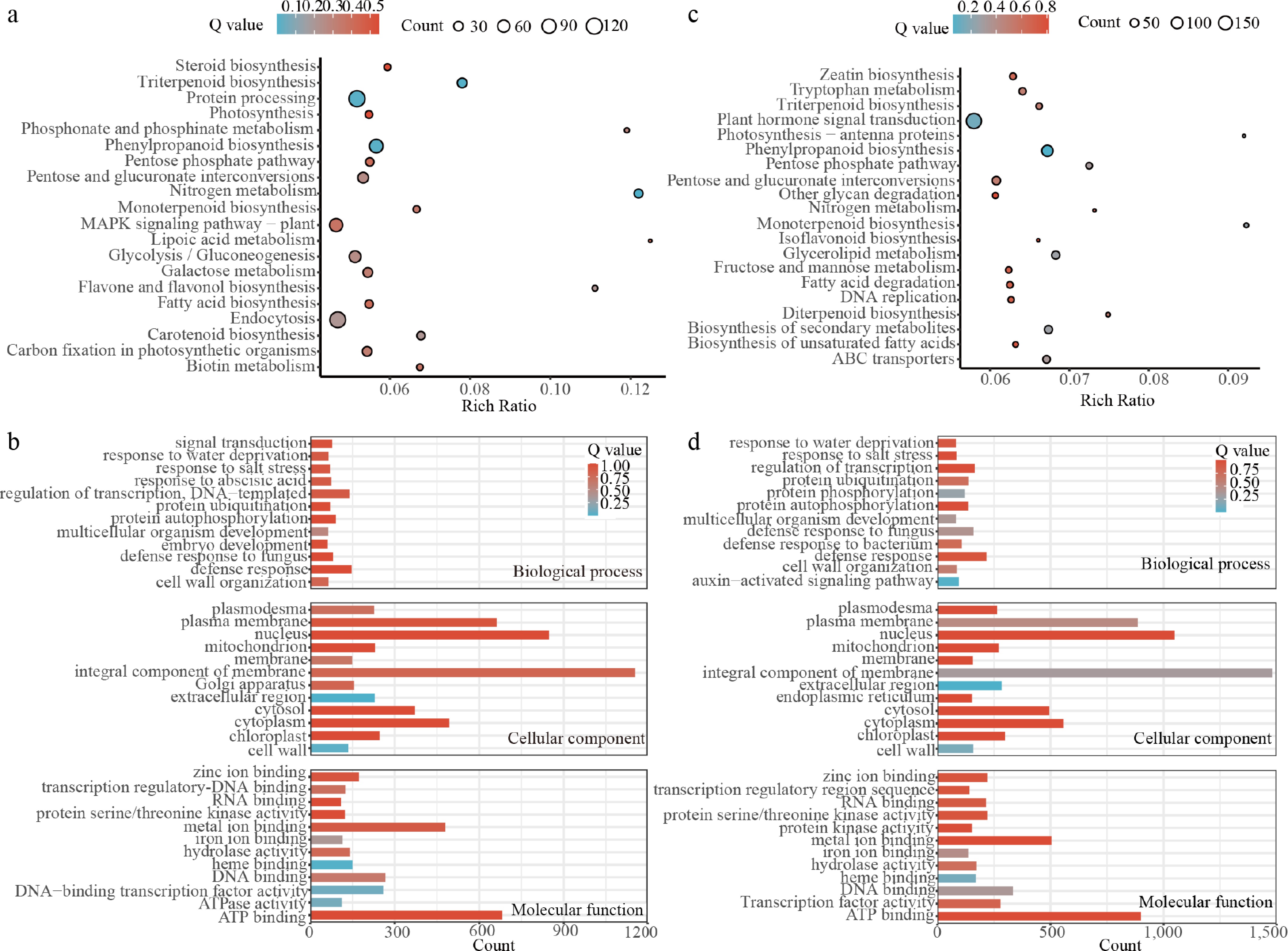
Figure 4.
KEGG pathway and GO enrichment analysis of DEGs during early leaf senescence in chrysanthemum cut flowers during vase treatment. (a) KEGG pathway enrichment of DEGs in the first stage (0–6 d); (b) GO enrichment of DEGs in the first stage (0–6 d); (c) KEGG pathway enrichment of DEGs in the second stage (6–12 d); (d) GO enrichment of DEGs in the second stage (6–12 d). The color of the dots/bars represents the Q value (FDR-adjusted p-value), and the size of the dots/length of the bars represents the number of enriched DEGs.
KEGG analysis of first-stage DEGs (Fig. 4a) revealed significant enrichment in pathways related to stress defense, secondary metabolism, and photosynthetic maintenance. The most significantly enriched pathway was the plant MAPK signaling pathway, followed by fatty acid biosynthesis, phenylpropanoid biosynthesis, protein processing in the endoplasmic reticulum, and photosynthesis. Terpene-related biosynthesis, carbon metabolism, and antioxidant biosynthesis pathways were also enriched. These results indicated that the early vase stage is characterized by active stress responses, global metabolic reprogramming, and the preservation of core photosynthetic function.
GO enrichment analysis (Fig. 4b) further supported this pattern. For biological processes (BP), DEGs were enriched in defense response, response to water deprivation, salt stress, abscisic acid signaling, transcriptional regulation, and protein phosphorylation. For cellular components (CC), DEGs were mainly located in the integral membrane component, plasma membrane, mitochondrion, and nucleus. For molecular functions (MF), DEGs were enriched in ATP binding, zinc ion binding, protein serine/threonine kinase activity, and DNA-binding transcription factor activity. Together, these results reflected the rapid activation of stress signaling and transcriptional regulation during early postharvest adaptation.
In contrast to the early defense-oriented transcriptome, second-stage DEGs showed a pronounced functional shift toward senescence regulation. KEGG analysis (Fig. 4c) identified plant hormone signal transduction as the most significantly enriched pathway, followed by photosynthesis-antenna proteins, phenylpropanoid biosynthesis, zeatin biosynthesis, and tryptophan metabolism. Lipid metabolism, secondary metabolism, and ABC transporters were also enriched, indicating that the late stage is dominated by hormone-dependent senescence progression, photosynthetic degradation, and programmed cell death.
GO enrichment (Fig. 4d) reinforced this senescence signature. DEGs remained enriched in stress responses and transcriptional regulation but with a marked increase in gene numbers compared with the early stage. Cellular component terms highlighted the integral membrane component, extracellular region, plasma membrane, and endoplasmic reticulum, consistent with extensive cellular remodeling. Molecular functions were concentrated in ATP binding, transcription factor activity, and kinase activity, indicating large-scale activation of senescence regulatory networks.
Identification and functional characterization of core senescence-associated genes
-
To identify the core regulatory genes driving continuous senescence rather than stage-specific responses, DEGs from the first (0 vs 6 d) and second (6 vs 12 d) stages were compared, yielding 6,194 shared DEGs across both phases (Fig. 5a). A total of 7,469 DEGs were specific to the first stage and 3,818 to the second stage, indicating that the core set constitutes a major component of senescence-related transcriptomic changes.
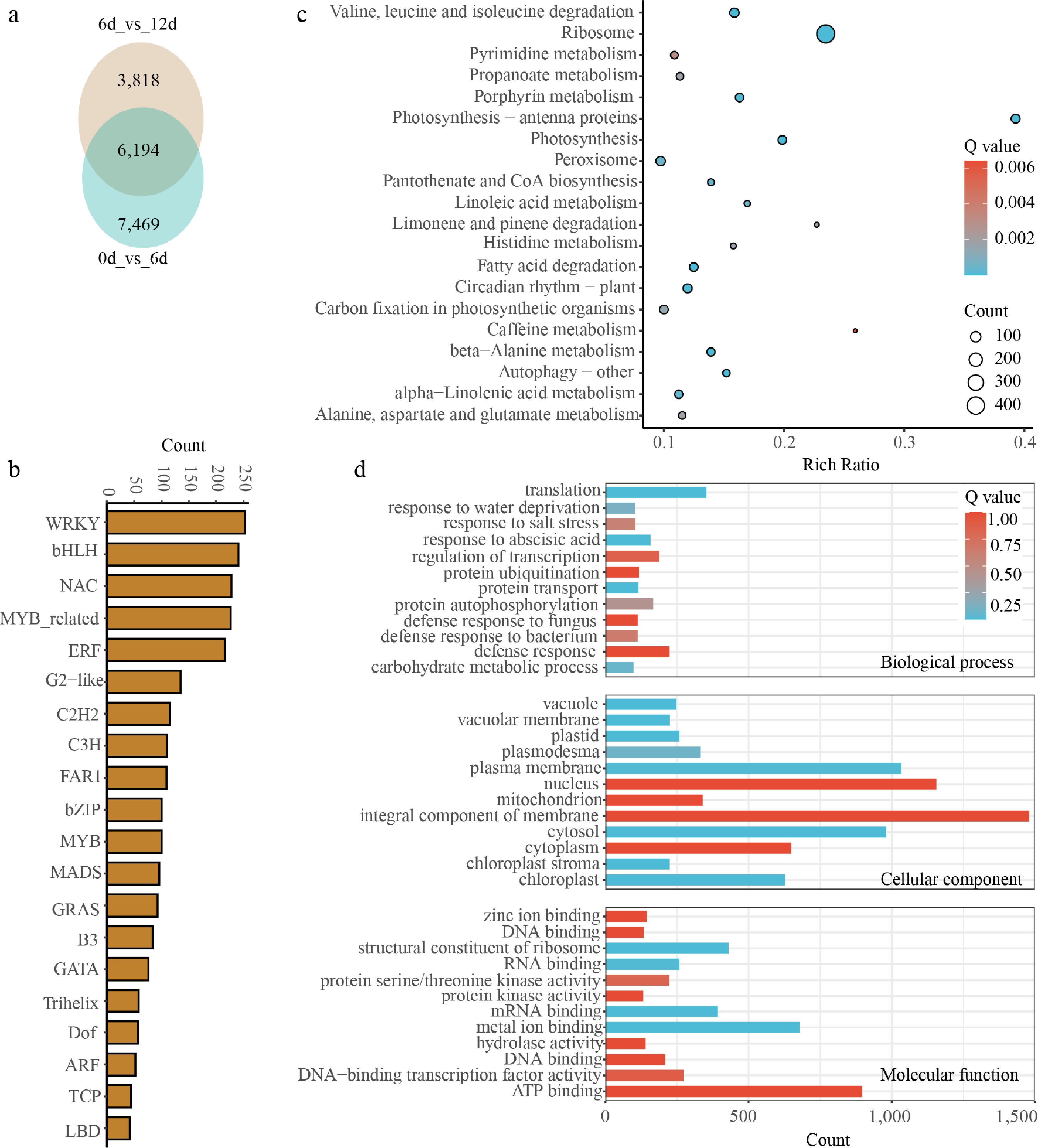
Figure 5.
Identification and functional analysis of the core senescence-associated gene set shared across early vase stages in chrysanthemum cut flowers. (a) Venn diagram showing the overlap of DEGs between the first (0 vs 6 d) and second (6 vs 12 d) early senescence stages. (b) Distribution of transcription factor (TF) families within the core shared DEGs. (c) KEGG pathway enrichment analysis of the core shared DEGs. The dot size represents the number of enriched genes, and the color represents the Q value (FDR-adjusted p-value). (d) GO enrichment analysis of the core shared DEGs, covering biological process, cellular component, and molecular function.
Transcription factor (TF) classification within the core gene set (Fig. 5b) revealed the dominant regulatory families. The most abundant family was WRKY (258 genes), followed by bHLH (241), NAC (228), MYB-related (227), and ERF (216). All these families are well-documented regulators of senescence, stress responses, and hormone signaling, supporting their central roles in orchestrating the senescence program.
KEGG enrichment of core DEGs (Fig. 5c) highlighted pathways including valine, leucine and isoleucine degradation (branched-chain amino acid catabolism), ribosome function, photosynthesis, peroxisome, and autophagy. This indicated that nutrient recycling, photosynthetic decline, and oxidative stress responses are initiated early and sustained throughout senescence, rather than being restricted to late stages. GO analysis (Fig. 5d) confirmed consistent enrichment in stress responses, abscisic acid signaling, transcriptional regulation, and protein ubiquitination across the entire senescence process. Core DEGs were primarily localized to the integral membrane component, nucleus, and plasma membrane, with molecular functions centered on ATP binding, transcription factor activity, and kinase activity, reflecting persistent transcriptional and post-translational regulation.
Expression dynamics and qRT-PCR validation of core regulatory genes
-
Hierarchical clustering was performed to visualize the expression patterns of key TFs and functional genes at 0, 6, and 12 d (Fig. 6). Biological replicates showed high consistency, supporting the reliability of expression trends. In the TF cluster, one group including CmHSFC1, CmHSF30, CmERF052, and several BBX family members was continuously upregulated, peaking at 6 and 12 d. Another group, including circadian clock genes CmLHY, CmCDF2, light signaling regulators CmPIF7, CmCOL5, and certain bHLH genes, was highly expressed at 0 d but significantly downregulated thereafter. In the functional gene cluster, photosynthesis-related genes such as CmCAB3C, CmCAB1B, and CmLHCB1.1 were downregulated in parallel with the light- and clock-related TFs. In contrast, the chlorophyll degradation gene CmNYC1, antioxidant gene CmPRDX4, ABA signaling components CmPP2A13, CmPP2C37, CmSnRK2.3, and gibberellin metabolism gene CmGA20ox1 were coordinately upregulated. The core clock gene CmTOC1 was also significantly downregulated, consistent with CmLHY and CmCDF2.
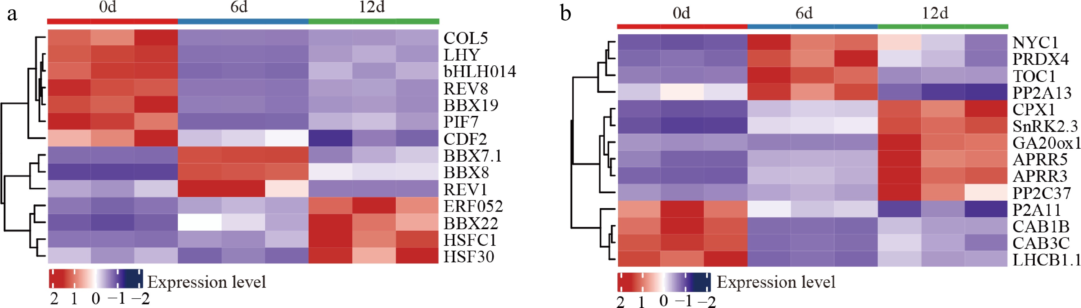
Figure 6.
Hierarchical clustering heatmap of the expression dynamics of core senescence-associated genes during vase treatment in chrysanthemum cut flowers. (a) Expression patterns of key transcription factors across 0, 6, and 12 d of vase life; (b) expression patterns of core functional genes. The color scale represents the z-score-normalized gene expression level, with red indicating high expression and blue indicating low expression.
To validate transcriptomic accuracy, six representative core DEGs were analyzed using qRT-PCR (Fig. 7). Each selected gene, covering antioxidant regulation, chlorophyll degradation, hormone signal transduction, and HSF family members, can fully reflect the main molecular changes during early leaf senescence. Expression trends were fully consistent with RNA-seq results. CmHSFC1 and CmERF052 were continuously and strongly upregulated, reaching approximately 30-fold and 12-fold higher expression at 12 d than at 0 d. CmGA20ox1 expression increased nearly 6-fold by 12 d. CmNYC1 was transiently induced at 6 d (about 5-fold) and returned to basal levels at 12 d. CmLHY and CmbHLH014 were significantly downregulated, with CmLHY decreasing to approximately 20% of initial levels and CmbHLH014 dropping to around 10% at 6 d. These results confirmed the high reliability of the transcriptome data, providing a solid foundation for further mechanistic investigation.
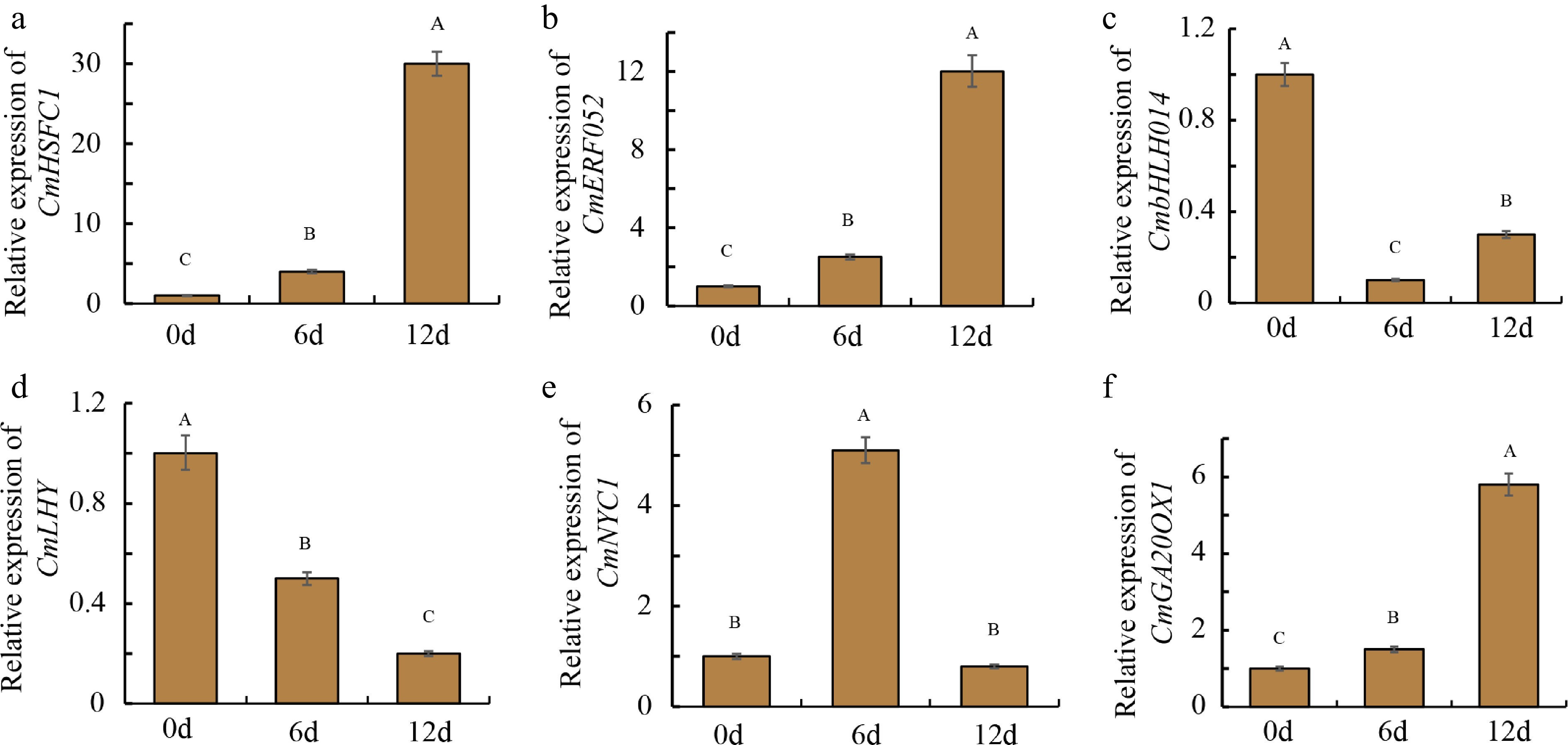
Figure 7.
qRT-PCR validation of the expression dynamics of six core senescence-associated genes during early senescence in chrysanthemum cut flowers. Error bars represent the standard error of three biological replicates. Different uppercase letters indicate significant differences at the p < 0.05 level.
Silencing of CmHSFC1 regulates leaf senescence and chlorophyll degradation
-
Among these differentially expressed TFs, the expression of CmHSFC1 (evm.TU.scaffold_10636.70) showed the strongest induction, with a Log2(12 d/0 d) value of 5.01 (Fig. 6a; Supplementary Table S2), suggesting its potential involvement in senescence regulation. To investigate the role of CmHSFC1 in leaf senescence, we performed virus-induced gene silencing (VIGS) in chrysanthemum. As shown in Fig. 8a, no obvious phenotypic difference was observed between CmHSFC1-silenced lines (CmHSFC1-PCVA-1, CmHSFC1-PCVA-2) and the control (EV-PCVA) at 0 d. However, after 8 d of treatment, CmHSFC1-silenced leaf discs exhibited less yellowing and senescence symptoms compared with the EV-PCVA control.
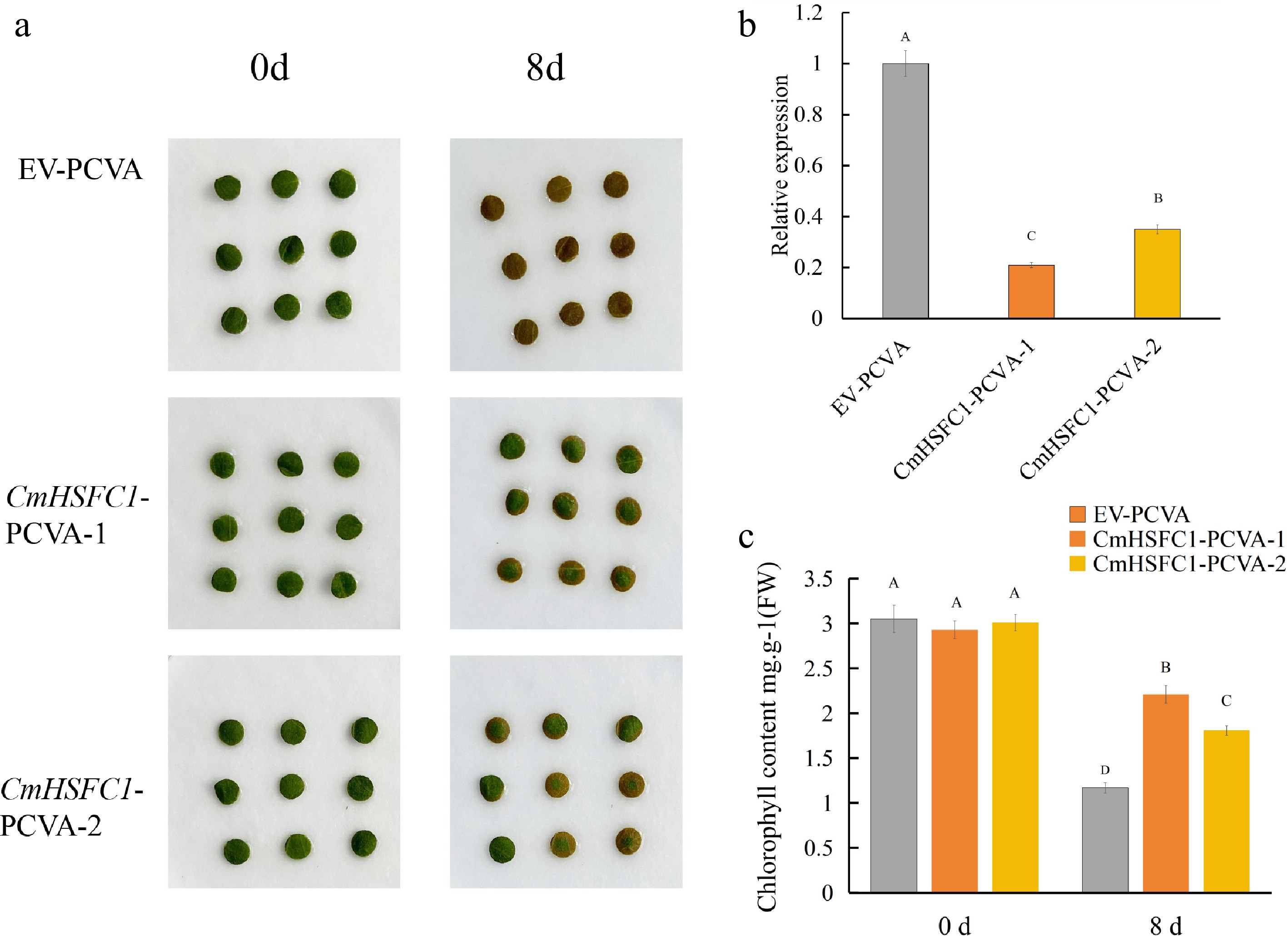
Figure 8.
Silencing of CmHSFC1 delays leaf senescence in chrysanthemum. (a) Phenotypic observation of leaf discs from CmHSFC1-silenced (CmHSFC1-PCVA-1, CmHSFC1-PCVA-2) and empty vector control (EV-PCVA) plants at 0 and 8 d of dark-induced senescence. (b) Relative expression level of CmHSFC1 in silenced and control lines, verified by qRT-PCR. (c) Chlorophyll content in leaf discs of silenced and control lines at 0 and 8 d of of dark-induced senescence. Data are presented as means ± SD (n = 3). Different uppercase letters indicate significant differences at the p < 0.05 level.
qRT-PCR analysis confirmed that the transcript level of CmHSFC1 was significantly reduced in both silenced lines, with CmHSFC1-PCVA-1 showing an 80% reduction and CmHSFC1-PCVA-2 showing a 65% reduction relative to the control (Fig. 8b). Consistent with the phenotypic changes, chlorophyll content, a key marker of leaf senescence, was comparable among all lines at 0 d. After 8 d of treatment, chlorophyll content decreased in all groups, but the chlorophyll content of the CmHSFC1 silencing lines was significantly higher than that of the EV-PCVA control (Fig. 8c). Collectively, these results demonstrate that silencing of CmHSFC1 delays leaf senescence in chrysanthemum.
-
As one of the most economically valuable ornamental crops worldwide, chrysanthemum cut flowers frequently undergo rapid and premature leaf senescence during postharvest vase treatment, which severely shortens their display life and reduces their market competitiveness[24]. The vase life of chrysanthemums is relatively long, but once cut, chrysanthemums reach the late stage of senescence; this process cannot be reversed. Therefore, research and regulation of their early senescence stage are particularly important[36].
In the present study, early leaf senescence of chrysanthemum cut flowers 'Jinba' was accompanied by persistent chlorophyll degradation, membrane lipid peroxidation, ROS overaccumulation, and continuous induction of antioxidant enzyme activities. MDA and H2O2 contents increased dramatically after 6 d of vase treatment, coinciding with accelerated chlorophyll loss, indicating that oxidative stress became increasingly severe with increasing vase treatment time. Meanwhile, activities of SOD, POD, and CAT rose progressively, suggesting that the antioxidant system is mobilized to scavenge excess ROS and mitigate oxidative damage (Fig. 2). These results indicate that ROS begin to accumulate in chrysanthemum leaves at the early vase treatment stage, even though the leaves remain green visually.
Transcriptomic analysis further revealed distinct and highly ordered gene expression programs between the first (0–6 d) and second (6–12 d) senescence stages. During the first stage, differentially expressed genes (DEGs) were significantly enriched in MAPK signaling, secondary metabolism, and stress defense pathways, indicating that plants prioritize stress perception, signal transduction, and metabolic reprogramming to cope with multiple postharvest stresses including wounding, water deficit, and oxidative stress. Enriched GO terms related to defense response, abscisic acid signaling, and protein phosphorylation further reflect the rapid activation of stress-responsive transcriptional regulatory networks (Fig. 4a, b).
In contrast, the second-stage transcriptome shifted dramatically toward pathways involved in plant hormone signal transduction, chlorophyll catabolism, and senescence execution. Plant hormone signaling emerged as the most prominent KEGG pathway, consistent with the well-documented roles of ethylene, ABA, cytokinins, and other phytohormones in modulating leaf senescence[7]. Ethylene and ABA promote senescence progression, whereas cytokinins delay leaf yellowing by maintaining chlorophyll content and photosynthetic efficiency[4]. Concurrently, photosynthesis-related genes were sharply downregulated, while chlorophyll catabolic genes (NYC1) and senescence-associated genes (PRDX4) were significantly upregulated, marking the onset of large-scale cellular degradation and nutrient remobilization[37]. These stage-specific expression patterns confirm that leaf senescence is a highly organized biological process governed by sequential transcriptional reprogramming (Fig. 4c, d).
Analysis of DEGs shared across both senescence stages identified WRKY, bHLH, NAC, MYB-related, and ERF as the most abundant transcription factor families, all of which are widely implicated in stress responses and senescence regulation[38,39] (Fig. 5). Among them, NAC transcription factors, especially homologs of ORE1 and NAP, act as master regulators that directly activate chlorophyll degradation and the expression of senescence-associated genes[40]. Recent studies have demonstrated that JA induces sponge tissue cell death through the CmNAC055-CmNAC087-CmACX5 regulatory cascade, which is functionally conserved across plant species. WRKY and ERF family members integrate hormonal and stress signals to fine-tune the expression of downstream senescence regulators. The dynamic interaction between CmbHLH1L and CmNLP6/7L on the scaffold protein CmbHLH63 serves as a molecular switch that controls stress-induced leaf senescence in chrysanthemum[24].
Notably, the expression levels of multiple core circadian clock genes significantly change during leaf senescence, including LHY, TOC1, and CDF2, which were significantly downregulated during senescence. The circadian clock system tightly coordinates photosynthetic gene expression, redox homeostasis, and stress responses; its disruption accelerates photosynthetic decline and promotes premature senescence[41,42] (Fig. 6). More and more evidence suggests that the core genes of the biological clock finely regulate the senescence process of leaves through the temporal dimension, and their disordered expression is an important molecular trigger for accelerated postharvest leaf senescence. The PRR9-dependent circadian regulation of ORE1 has been verified in Arabidopsis, implying a conserved mechanism linking clock dysfunction to leaf senescence in chrysanthemum[43]. Our transcriptomic data highlight that dysregulation of the circadian clock represents a key molecular event underlying postharvest leaf senescence.
Given the central roles of transcription factors in modulating leaf senescence, we systematically screened all TF-encoding genes among DEGs. In total, 1,358 TFs were upregulated, and 1,739 TFs were downregulated (Supplementary Table S2). These TFs were mainly distributed in the WRKY, NAC, bHLH, and ERF families (Fig. 5b). Among these differentially expressed TFs, CmHSFC1 (evm.TU.scaffold_10636.70) showed the strongest induction, with a Log2(12 d/0 d) value of 5.01 (Fig. 6a; Supplementary Table S2), suggesting its potential involvement in senescence regulation.
Heat shock transcription factors (HSFs) are best known for their functions in heat stress responses, yet increasing evidence has linked HSFs to oxidative stress signaling and senescence modulation[44]. In this study, CmHSFC1 was continuously upregulated during vase treatment, and VIGS-mediated silencing of CmHSFC1 markedly delayed leaf senescence and retained higher chlorophyll content. These results demonstrate that CmHSFC1 functions as a positive regulator of chrysanthemum leaf senescence (Fig. 8). HSFs can act as ROS sensors and regulate the expression of antioxidant and senescence-related genes[45]. Similar regulatory patterns have been reported in tall fescue, where FaHSFC1b mediates crosstalk between heat tolerance and leaf senescence[44,46]. Our findings expand the functional diversity of HSFs in ornamental plants and identify CmHSFC1 as a promising target for delaying postharvest leaf senescence. Collectively, this study confirms that leaf senescence in chrysanthemum cut flowers is driven by the combined effects of oxidative stress and hierarchical transcriptional regulation.
At the molecular level, the core SAGs identified in this study provide valuable targets for genetic improvement. Silencing senescence-promoting regulators such as CmHSFC1 may enable the breeding of chrysanthemum cultivars with prolonged vase life. But this study only used the chrysanthemum variety 'Jinba' for verification, and the stability of VIGS silencing efficiency and phenotypic traits under different environmental conditions and different varieties needs further verification. Future research should focus on dissecting the upstream signaling pathways and direct downstream targets of CmHSFC1, as well as the molecular crosstalk between HSF-mediated regulatory networks and hormone-dependent senescence programs.
-
Chrysanthemum cut flower leaf senescence during early postharvest vase treatment (0–12 d) follows a two-stage pattern characterized by early stress defense and late senescence execution. Physiologically, this process involves progressive chlorophyll loss, membrane damage, ROS accumulation, and sustained antioxidant activation. Transcriptionally, sequential reprogramming of stress signaling, hormone pathways, photosynthetic genes, and transcription factor networks governs senescence progression. The HSF family member CmHSFC1 acts as a novel positive regulator of leaf senescence, whose silencing delays yellowing and preserves chlorophyll. These findings provide mechanistic insights and gene resources for improving postharvest quality and vase life of chrysanthemum cut flowers.
-
The authors confirm their contributions to the paper as follows: designed the experiments: Huang Y, Ren Y, Jiang B; performed the experiments: Huang Y, Ren Y, Chen Y, Zhang L, Yan Y; analyzed the data: Huang Y, Ren Y, Chen Y, Jiang B; wrote the manuscript: Huang Y. All authors reviewed the results and approved the final version of the manuscript.
-
All data generated or analyzed during this study are included in this published article and its supplementary information files. Further inquiries can be directed to the corresponding authors.
-
The authors declare that they have no conflict of interest.
-
accompanies this paper online at: https://doi.org/10.48130/opr-0026-0025.
-
# Authors contributed equally: Yi Ren, Bingbing Jiang, Yi Chen
- Supplementary Table S1 Primers used in the present study.
- Supplementary Table S2 Differential gene annotation and expression levels.
- Copyright: © 2026 by the author(s). Published by Maximum Academic Press, Fayetteville, GA. This article is an open access article distributed under Creative Commons Attribution License (CC BY 4.0), visit https://creativecommons.org/licenses/by/4.0/.
-
About this article
Cite this article
Ren Y, Jiang B, Chen Y, Zhang L, Yan Y, et al. 2026. Physiological and transcriptomic analyses reveal the mechanisms and key regulator CmHSFC1 in leaf senescence during the early stages of chrysanthemum vase life. Ornamental Plant Research 6: e034 doi: 10.48130/opr-0026-0025 

Physiological and transcriptomic analyses reveal the mechanisms and key regulator CmHSFC1 in leaf senescence during the early stages of chrysanthemum vase life
- Received: 25 April 2026
- Revised: 01 July 2026
- Accepted: 10 August 2026
- Published online: 09 October 2026
Abstract: Leaf senescence is a major factor limiting the vase life of chrysanthemum cut flowers, for which the early senescence stage is vital but remains not fully studied. Exploring its mechanism enables extending the vase life of chrysanthemum cut flowers. In this study, physiological results showed that early senescence displayed continuous chlorophyll degradation, elevated MDA and H2O2 contents, and gradually increased activities of SOD, POD, and CAT. Transcriptomic analysis revealed stage-specific transcriptional reprogramming: the first stage (0–6 d) was dominated by stress responses, MAPK signaling, and metabolic adaptation, whereas the second stage (6–12 d) was mainly involved in plant hormone signal transduction and senescence execution. A total of 6,194 core DEGs shared by both stages were identified, with WRKY, bHLH, NAC, and ERF as the predominant transcription factor families. The expression of circadian clock genes was significantly disrupted, while multiple senescence-associated genes were continuously upregulated. Among these differentially expressed TFs, CmHSFC1 exhibited the strongest induction. Moreover, VIGS-mediated silencing of CmHSFC1 markedly delayed leaf senescence and chlorophyll loss. These results reveal the sequential regulatory network of postharvest early leaf senescence in chrysanthemum and identify CmHSFC1 as a positive key regulator of early senescence, providing valuable gene resources for the genetic improvement of cut-flower vase life.
-
Key words:
- Chrysanthemum /
- Leaf senescence /
- Vase life /
- Transcriptome /
- CmHSFC1 /
- VIGS