












-
Veterinary antibiotics (VAs) are effective agents used to treat and prevent bacterial infections in animals, thereby protecting animal health and welfare. However, VAs enter agricultural soils both directly, through grazing animals[1,2], and indirectly, via application of manure or compost fertilizers[1−5]. As emerging pollutants, VAs pose significant environmental risks[6] and have been detected in agricultural environments worldwide[3,4]. Several studies have reported the negative effects of VAs on crop and non-crop plants, impacting growth and physiological performance[7−10]. Antibiotics can alter key physiological traits such as plant height, root and shoot length, and photosynthetic pigment content. They have also been shown to interfere with abscisic acid synthesis and nutrient cycling[11−13]. Additionally, various antibiotics present in soil can be absorbed and accumulated in plant tissues, causing detrimental effects on functional traits that vary depending on the plant species, antibiotic type, and concentration[14]. Higher plants are widely used as bioindicators to study the toxicity of environmental contaminants[15,16]. Among edible crops, Trifolium alexandrinum and Eruca vesicaria are globally important and frequently exposed to antibiotics via manure fertilization. Therefore, assessing the phytotoxicity of VA on these species is essential for food safety and environmental health.
Despite increasing concern over the widespread occurrence of VA in agricultural soils, their potential impacts on edible plant species and associated oxidative stress responses remain poorly characterized, particularly under combined antibiotic exposures. Most existing studies have focused on single compounds or non-edible model plants, thereby limiting ecological and food safety relevance. Therefore, the present study was designed to address these knowledge gaps. We hypothesized that: (i) veterinary antibiotics would cause dose-dependent impairments in plant growth and physiological functioning; (ii) combined antibiotic exposure would produce greater phytotoxicity than single-compound treatments; and (iii) plant enzymatic activities serve as sensitive biochemical indicators of physiological stress, reflecting metabolic adjustments and detoxification responses under contaminant exposure. This study provides novel insights into the phytotoxicity of VAs by: (i) directly comparing responses of two agronomically important edible species, T. alexandrium and E. vesicaria; (ii) evaluating both single and combined antibiotic exposures to capture potential mixture effects; and (iii) integrating plant physiological, biochemical, and enzymatic endpoints to identify functional indicators of antibiotic-induced stress. By simultaneously examining growth, metabolic, and enzymatic responses under controlled soil conditions, this work advances mechanistic understanding of VA phytotoxicity in crop plants, addressing critical knowledge gaps relevant to agro-environmental risk assessment and food safety.
-
Soil used in this study was collected from a greenhouse at the University of Science and Technology Bannu, KP, Pakistan. The soil had no known history of antibiotic exposure. After collection, soil was air-dried, homogenized, and passed through a 2 mm sieve to remove stones, plant residues, and debris. Processed soil was stored under controlled laboratory conditions until experimental use. Initial physicochemical properties of the soil were determined prior to treatment application. The soil exhibited an alkaline pH of 8.4 and electrical conductivity (EC) of 500 µS·cm−1.
Preparation of antibiotic treatments
-
Three VA—enrofloxacin (ENR), penicillin (PCN) and sulfamethazine (SMZ) (Sigma-Aldrich USA) were tested individually and as a mixture. Each antibiotic stock solution was prepared at 1 mg·mL−1. Equal volumes of the three stock solutions were mixed to prepare the antibiotic mixture. Therefore, the MIX contained ENR, PCN, and SMZ in a 1:1:1 ratio, with each antibiotic representing one-third of the total applied concentration. Experimental soil was spiked with antibiotics to achieve seven concentration levels for individual antibiotics (0, 20, 50, 100, 250, and 500 mg·kg−1 dry soil). The selected antibiotic concentrations were chosen to cover environmentally detected levels in manure-amended agricultural soils as well as elevated exposure conditions representing worst-case accumulation scenarios arising from repeated manure application, incomplete degradation, and strong soil sorption of antibiotics[9,17]. Stock solutions were diluted with methanol as required and thoroughly mixed into the soil for uniform distribution. Control soils received an equivalent volume of methanol without antibiotics. Treated soils were left under a fume hood for 24 h to allow complete evaporation of methanol. One kg of treated soils was transferred into plastic pots (10 × 10 cm) and arranged in a completely randomized design with three replicates per treatment. Pots were maintained under controlled greenhouse conditions.
Plant material and sowing
-
Seeds of locally grown cultivars of Trifolium alexandrinum L. and Eruca vesicaria ssp. sativa were obtained from the District Agriculture Office, Bannu, Pakistan. Seeds were sown at a density of 15 seeds per pot in pots and allowed to grow under controlled greenhouse conditions (25 ± 2 °C; 16 h light/8 h dark photoperiod; relative humidity maintained) for 30 d.
Seed germination test and seedling vigor assessment
-
Seed germination was assessed using the filter paper method. Petri dishes and filter papers were sterilized prior to use. Seeds were surface-sterilized with 0.1% sodium hypochlorite (NaOCl) for 3 min and rinsed three times with distilled water. Fifteen seeds were placed in each sterilized Petri dish on filter paper moistened with 5 mL of antibiotic solutions at the respective concentrations. Dishes were incubated in a growth chamber at 28 °C. Germination rate (%) was calculated using the formula: Germination rate was calculated as:
$ \mathrm{Germination\; rate}=\dfrac{\mathrm{No.\; of\; seeds\; germinated}}{\text{Total no. of seeds}}\times100 $ Seedling vigor was evaluated concurrently with germination following standard seed testing protocols. At 6 d after sowing, vigorous seedlings—defined as those exhibiting complete morphology and no visible defects—were counted. At 12 d, seedlings were classified as vigorous, normal (solid or weak), or abnormal based on morphological characteristics. Results were expressed as the percentage of vigorous seedlings per pot at each antibiotic concentration.
Measurements of phenotypic traits, biomass, and relative water content
-
Phenotypic traits, including plant height, root length, shoot length, and leaf morphology, were assessed using ten randomly selected plants per treatment, with three replicates. Root length was recorded at the three-leaf stage (12–15 d after germination), while shoot length was measured every 2 d from day 3 to day 15. Leaf area was calculated according to[18].
$ \mathrm{Leaf\; area\; (cm^2)=Length\; of\; leaf\times Width\; of\; leaf\times0.75} $ Seedling biomass was determined by measuring fresh weight (FW) and dry weight (DW) following standard methods[19]. Seedlings were first weighed fresh, and then dried at 70 °C for 12 h to determine dry weight. Relative water content (RWC, %) was calculated using the formula:
$ {\bf {RWC}}=\rm\dfrac{Seedling\;fresh\;weight-Seedling\;dry\;weight}{Seedling\;turgid\;weight-Seedling\;dry\;weight}\times 100 $ Determination of chlorophyll and carotenoid pigments
-
One gram of fresh leaves was homogenized in 20 mL of 80% acetone. The homogenate was centrifuged at 5,000 rpm for 5 min, and the absorbance of the supernatant was measured at 645, 663, and 470 nm to quantify chlorophyll a, chlorophyll b, and carotenoids, respectively. Pigment concentrations were calculated using standard equations[20].
Determination of proline content
-
Proline concentration was determined spectrophotometrically using the ninhydrin method described by Bates et al.[21]. Fresh leaf tissues were homogenized in 3% (w/v) sulfosalicylic acid. The extract was mixed with 2 mL of acid ninhydrin reagent and 2 mL of glacial acetic acid, and the mixture was incubated at 100 °C for color development. The reaction mixture was subsequently extracted with toluene, and the absorbance of the toluene phase was recorded at 520 nm.
$\rm Proline\;({\text µ}mol\;{g}^{-1}\;FW)=\dfrac{({\text µ}g\;proline\;{mL}^{-1}\times mL\;to\; luene)}{115.5}\div \dfrac{g\;sample}{5}$ DPPH radical scavenging activity
-
The antioxidant activity of plant extracts was evaluated using the DPPH assay. A reaction mixture containing 900 µL of DPPH solution and 100 µL of plant extract at concentrations of 100, 250, and 500 µg was incubated in the dark at 37 °C for 1 h. Absorbance was measured at 517 nm. Controls included 3% methanol (blank), DPPH with methanol (negative control), and ascorbic acid (positive control). Percent inhibition of DPPH radical was calculated as:
$ {\rm{Percent \;inhibition}} = \dfrac{\mathrm{Absorbance}\;\mathrm{of}~\mathrm{control}-\mathrm{Absorbance}~\mathrm{of}~\text{sample}}{\mathrm{Absorbance}~\mathrm{of}~\text{control}} \times 100 $ Preparation of crude enzyme extracts and enzyme activity assays
-
Crude enzyme extracts were prepared from 1 g of fresh seedlings by homogenization in 100 mmol·L−1 potassium phosphate buffer (pH 7.5) for urease and esterase, or acetate buffer (pH 5.5) for acid phosphatase, containing 1 mmol·L−1 EDTA, 3 mmol·L−1 DL-dithiothreitol, and 5% (w/v) insoluble polyvinyl pyrrolidone. Homogenates were centrifuged at 5,000 rpm for 20 min, and supernatants were aliquoted and stored at −80 °C.
Acid phosphatase activity was assayed at 37 °C using para-nitrophenyl phosphate (PNPP) as substrate in acetate buffer (pH 5.5)[22]. The reaction mixture contained 900 µL substrate and 100 µL enzyme extract. The reaction was stopped with 4 mL 0.1 N KOH, and absorbance was measured at 405 nm. One unit of activity corresponds to hydrolysis of 1 µmol substrate per minute.
Esterase activity was measured using p-nitrophenyl acetate (PNPA) as substrate[23]. The reaction mixture (880 µL) contained 0.1 mol·L−1 potassium phosphate buffer (pH 8.0), 0.2% sodium deoxycholate, 0.1% gum arabic, and 20 µL enzyme extract. After preincubation at 37 °C for 5 min, 100 µL of 8 mmol·L−1 PNPA was added. Following 5 min incubation, the reaction was terminated with 0.5 mL 3 mol·L−1 HCl, centrifuged, and 1 mL 2 mol·L−1 NaOH was added to the supernatant. Absorbance was measured at 420 nm. One unit corresponds to hydrolysis of 1 µmol substrate per minute.
Urease activity was determined by the indophenol method[24]. The reaction mixture contained 200 µL enzyme extract, 300 µL phosphate buffer (pH 8.21) with 300 mmol·L−1 urea, phenolic mixture, and alkaline reagent. After incubation at 37 °C for 15 min, 200 µL phenolic mixture and 300 µL NaOH were added. Absorbance was measured at 630 nm, and one unit of urease activity corresponds to hydrolysis of 1 µmol substrate per minute.
Statistical analysis
-
All experiments were conducted in triplicate, and data are presented as the mean ± standard deviation (SD). Statistical analyses were performed using OriginPro (OriginLab Corporation, Northampton, MA, USA). Differences among treatments at each antibiotic concentration were analyzed using one-way analysis of variance (ANOVA) followed by Tukey's honestly significant difference (HSD) post hoc test. Differences were considered statistically significant at p < 0.05.
-
The phytotoxic effects of VAs on seed germination of T. alexandrinum and E. vesicaria are presented in Fig. 1a, b, respectively. Germination percentage showed a clear concentration-dependent decline under both individual and combined antibiotic treatments. At lower concentrations (20–50 mg·kg−1), germination remained largely unaffected, with values exceeding 95% in T. alexandrinum and approximately 90%–95% in E. vesicaria, indicating minimal toxicity at these levels. However, as antibiotic concentrations increased (≥ 100 mg·kg−1), a significant reduction in germination was observed. In T. alexandrinum (Fig. 1a), germination declined gradually, with ENR showing the strongest inhibitory effect, followed by SMZ and PCN, while the mixture treatment exhibited the greatest overall suppression at higher concentrations. At 500 mg·kg−1, germination decreased to approximately 90% under ENR and further to ~80% under the mixture treatment. In contrast, E. vesicaria (Fig. 1b) exhibited a more pronounced sensitivity to antibiotic stress. Germination declined sharply beyond 100 mg·kg−1, particularly under ENR and mixture treatments. At 500 mg·kg−1, germination dropped to nearly 70% under ENR and as low as ~60% under the mixture, confirming higher susceptibility of this species compared to T. alexandrinum. Across both species, ENR consistently exerted the strongest inhibitory effect, while PCN showed comparatively moderate toxicity. SMZ displayed intermediate effects. Notably, combined antibiotic exposure resulted in greater suppression than individual treatments, suggesting possible additive or synergistic phytotoxic effects.
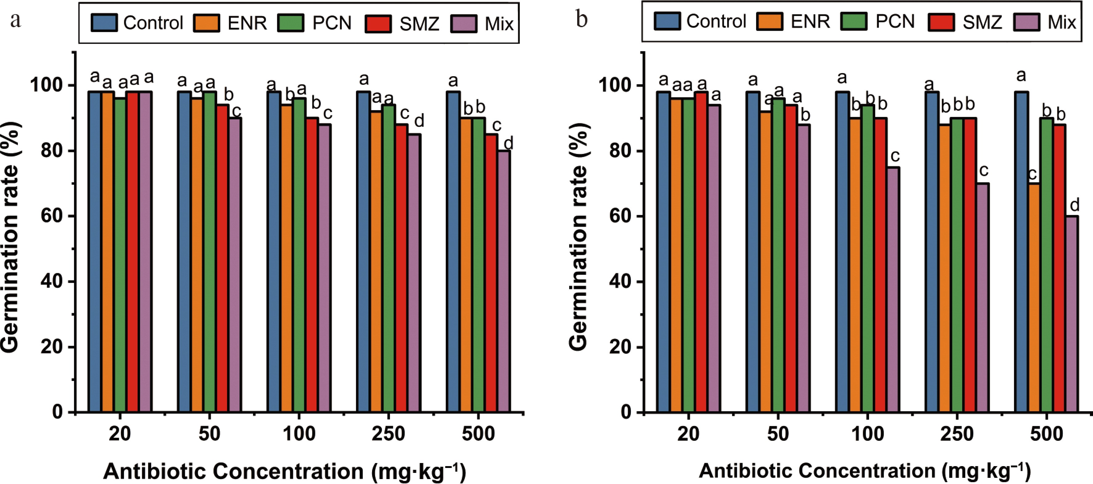
Figure 1.
Effects of enrofloxacin (ENR), penicillin (PCN), sulfamethazine (SMZ), and their mixture (Mix) on seed germination rate of (a) T. alexandrinum, and (b) E. vesicaria exposed to different antibiotic concentrations (20, 50, 100, 250, and 500 mg·kg−1). Values are expressed as mean ± SD (n = 3). At each concentration, differences among treatments were analyzed using one-way ANOVA followed by Tukey's honestly significant difference (HSD) post hoc test. Different lowercase letters above the bars indicate significant differences among treatments within the same concentration (p < 0.05).
Seedling vigor followed a similar declining trend with increasing antibiotic concentration. At lower concentrations (20–50 mg·kg−1), both species maintained high vigor (> 85%), comparable to control conditions. However, at intermediate to high concentrations (100–500 mg·kg−1), a marked reduction in vigorous seedlings was observed, particularly in E. vesicaria. The mixture treatment caused the most severe decline, reducing vigorous seedlings to approximately 35% in T. alexandrinum and 20% in E. vesicaria at the highest concentration. Overall, these findings demonstrate a clear dose-dependent phytotoxic effect of VAs, with E. vesicaria being more sensitive than T. alexandrinum, and antibiotic mixtures posing a greater risk than individual compounds.
Effect of VAs on seedling biomass and relative water content
-
Seedling biomass, expressed as fresh weight (FW) and dry weight (DW), exhibited a clear concentration-dependent decline under VA exposure (Supplementary Table S1, S2). The magnitude of reduction varied among antibiotics and between plant species, with E. vesicaria showing greater sensitivity than T. alexandrinum. In T. alexandrinum (Supplementary Table S1), FW decreased gradually with increasing antibiotic concentration. Under ENR treatment, FW declined from 3.83 g in the control to 1.3 g at 500 mg·kg−1, similar but slightly stronger reductions were observed under PCN and SMZ treatments. The mixture treatment exerted a comparatively greater inhibitory effect, reducing FW to 0.1 g (≈ 97% reduction relative to control) at 500 mg·kg−1, indicating enhanced phytotoxicity under combined exposure. In contrast, E. vesicaria (Supplementary Table S2) exhibited a sharper decline in biomass parameters. Under ENR, FW showed only a slight reduction (2.25 to 0.62 g at 500 mg·kg−1), but DW decreased markedly. The most pronounced effect was observed under the mixture treatment, where FW and DW declined drastically, representing reductions of more than 90% compared to control values.
Relative water content (RWC) followed trends similar to biomass, indicating impaired plant water status under antibiotic stress. In T. alexandrinum, RWC decreased from 96.4% in the control to 72.2% under ENR at 500 mg·kg−1 (≈ 25% reduction). PCN and SMZ treatments caused moderate declines, while the mixture treatment reduced RWC to ~67%, confirming cumulative stress effects. In E. vesicaria, RWC reductions were more pronounced. Under ENR, RWC declined from 98.4% to 69.2% (≈ 30% reduction) at 500 mg·kg−1. PCN and SMZ treatments further reduced RWC to 63.9% and 60.4%, respectively. The mixture treatment resulted in the most severe dehydration effect, lowering RWC to 40% at 500 mg·kg−1, indicating substantial disruption of plant water balance. Overall, the results demonstrate that VA exposure significantly impairs seedling biomass accumulation and water status in a dose-dependent manner. The greater reductions observed in E. vesicaria compared to T. alexandrinum highlight species-specific sensitivity, while the pronounced effects under mixture treatments suggest additive phytotoxic interactions among antibiotics.
Effect of VAs on plant phenotypic traits
-
Plant phenotypic traits, including root length, shoot length, plant height, and leaf area, were significantly influenced by VA exposure in both T. alexandrinum and E. vesicaria (Fig. 2). Overall, a clear concentration-dependent inhibition of growth was observed, with more pronounced effects at higher antibiotic levels. At lower concentrations (20–50 mg·kg−1), no substantial differences in morphological traits were observed compared to the control, indicating minimal phytotoxic effects at these levels. However, at moderate to high concentrations (≥ 100 mg·kg−1), significant reductions in growth parameters became evident. Root length was particularly sensitive to VA. At 500 mg·kg−1 ENR, root length decreased by approximately 25% in T. alexandrinum (Fig. 2a) and 45% in E. vesicaria (Fig. 2b), demonstrating greater susceptibility of the latter species. A similar trend was observed for shoot length, which declined by ~22% in T. alexandrinum (Fig. 2c) and ~42% in E. vesicaria (Fig. 2d) under the same treatment. Leaf area also showed a marked reduction with increasing VA concentrations (Supplementary Table S1, S2). At higher doses, E. vesicaria exhibited a reduction of up to ~48%, whereas T. alexandrinum showed a comparatively moderate decrease (~30%). The inhibitory effects were further intensified under combined antibiotic treatments, where leaf area reductions exceeded 50% in E. vesicaria at 500 mg·kg−1, while T. alexandrinum showed reductions in the range of 35%–40% (Supplementary Table S1, S2). Among the tested antibiotics, SMZ exerted the strongest inhibitory effect on plant growth. At 500 mg·kg−1 SMZ, root length in E. vesicaria was drastically reduced to approximately 0.22 inches, whereas T. alexandrinum maintained relatively greater root growth (~1.86 inches), further confirming interspecific variation in tolerance. Shoot growth followed a comparable pattern. Morphological observations supported these quantitative findings (Supplementary Fig. S1). Seedlings exposed to higher VA concentrations appeared visibly stressed, exhibiting reduced height, thinner roots, chlorosis, and smaller, often wilted leaves. In contrast, seedlings grown under control and low-concentration treatments maintained normal morphology and vigor. By 15 d after exposure, most growth parameters, including plant height, root thickness, and leaf development, were significantly suppressed under high VA treatments. Interestingly, under extreme mixture exposure (500·mg kg−1), T. alexandrinum exhibited pronounced stress symptoms such as pale coloration, stunted growth, and reduced vigor, in some cases exceeding the visible damage observed in E. vesicaria. Nevertheless, overall survival and growth trends indicated that E. vesicaria remains the more sensitive species. In severe cases, particularly under high concentrations of individual antibiotics and mixtures, many seedlings failed to survive to later developmental stages, indicating strong phytotoxic effects. Overall, these results demonstrate that VA exposure adversely affects early plant growth and development in a dose-dependent manner. E. vesicaria is comparatively more sensitive, showing greater reductions in morphological traits, whereas T. alexandrinum exhibits relatively higher tolerance and resilience under similar stress conditions.
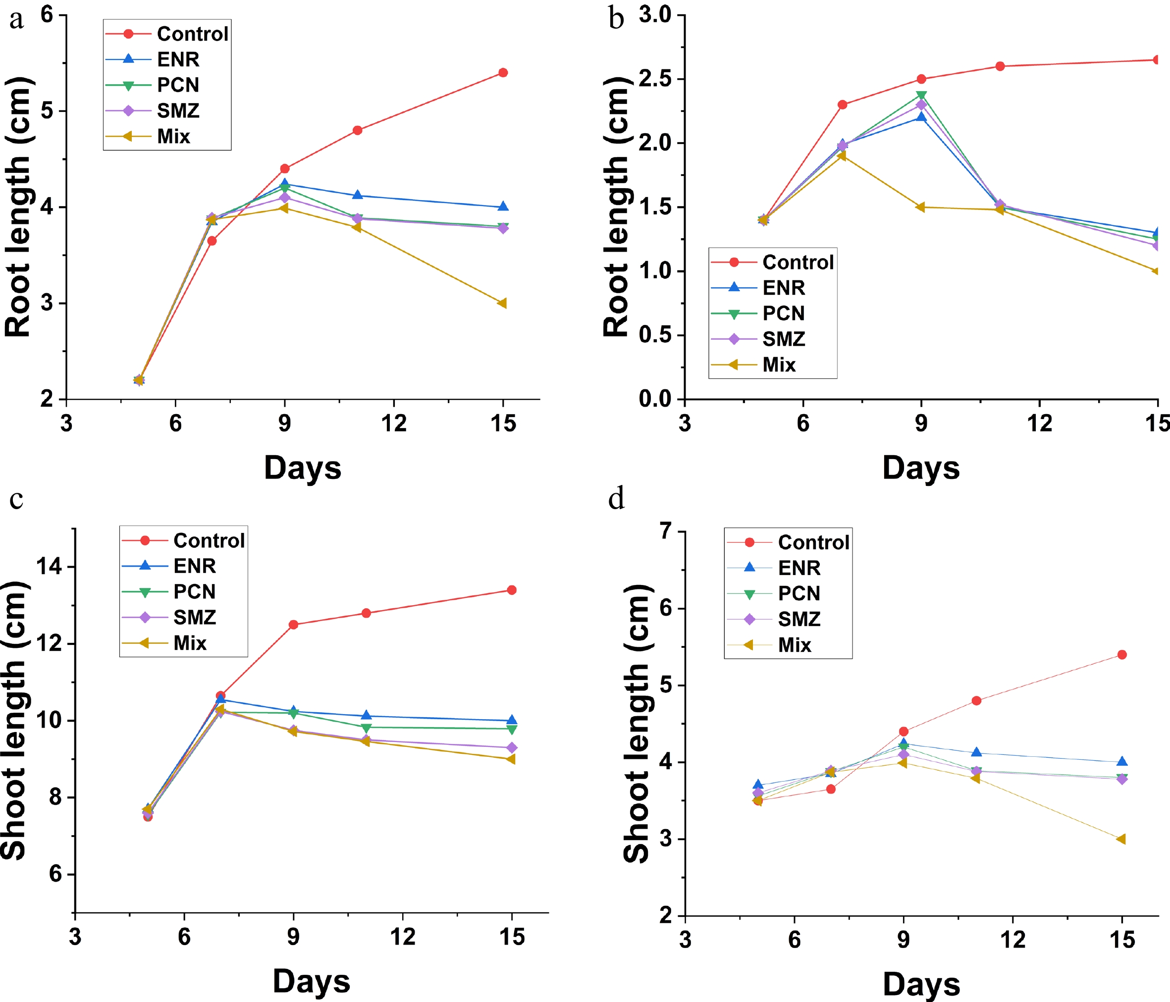
Figure 2.
Effects of enrofloxacin (ENR), penicillin (PCN), sulfamethazine (SMZ), and their combined mixture (Mix) on seedling growth of T. alexandrinum and E. vesicaria during a 15-d exposure period. (a) Root length of T. alexandrinum. (b) Root length of E. vesicaria. (c) Shoot length of T. alexandrinum. (d) Shoot length of E. vesicaria. Values are presented as mean ± SD (n = 3).
Effect of VAs on photosynthetic pigments
-
Exposure to VAs caused a clear, concentration-dependent decline in photosynthetic pigments, including chlorophyll a, chlorophyll b, and carotenoids, in both plant species (Fig. 3a–f). However, the magnitude of reduction was consistently greater in E. vesicaria compared to T. alexandrinum, indicating higher sensitivity of the former. For chlorophyll a (Fig. 3a, b), a gradual decrease was observed with increasing antibiotic concentrations across all treatments. At higher concentrations (≥ 250 mg·kg−1), ENR caused a marked reduction, reaching approximately 40%–45% in E. vesicaria, whereas T. alexandrinum showed a comparatively lower decline (~25%–30%). The antibiotic mixture exerted the strongest inhibitory effect, particularly at 500 mg·kg−1, where chlorophyll a dropped to its lowest levels in both species.
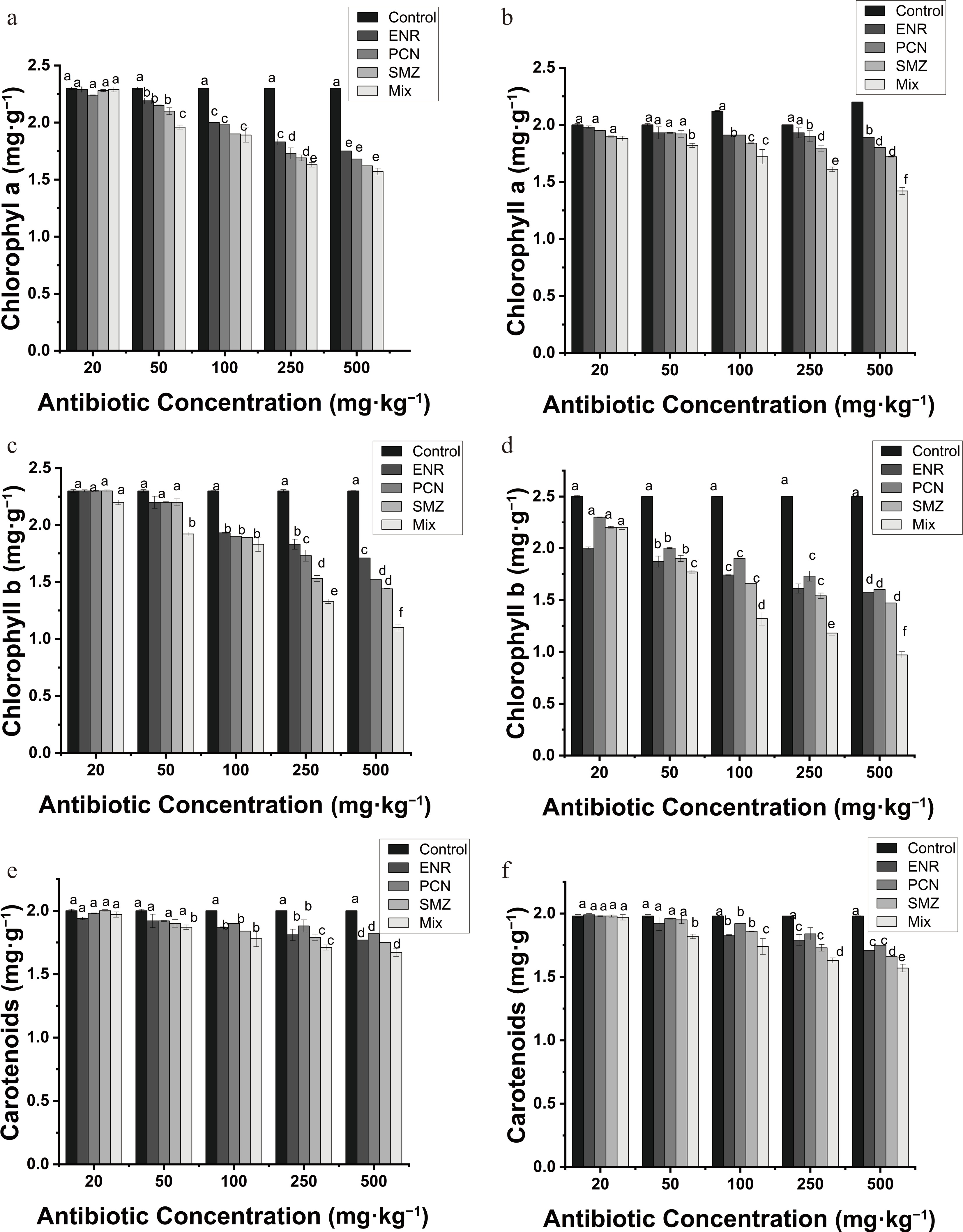
Figure 3.
Effects of enrofloxacin (ENR), penicillin (PCN), sulfamethazine (SMZ), and their mixture (Mix) on (a), (b) chlorophyll a, (c), (d) chlorophyll b, and (e), (f) carotenoid contents in the T. alexandrinum and E. vesicaria exposed to different antibiotic concentrations (20, 50, 100, 250, and 500 mg·kg−1). Values are expressed as mean ± SD (n = 3). For each concentration, differences among treatments were analyzed using one-way ANOVA followed by Tukey's HSD post hoc test. Different lowercase letters above the bars indicate significant differences among treatments within the same concentration (p < 0.05).
A similar trend was evident for chlorophyll b (Fig. 3c, d), though the reductions were slightly more pronounced than for chlorophyll a. In E. vesicaria, chlorophyll b decreased sharply at higher concentrations, especially under mixed antibiotic treatment, showing reductions exceeding 50% at 500 mg·kg−1. In contrast, T. alexandrinum exhibited moderate declines, indicating relatively better preservation of pigment integrity under stress. Carotenoid content (Fig. 3e, f) also declined progressively with increasing antibiotic concentration. While the reduction pattern followed that of chlorophylls, carotenoids appeared slightly more stable at lower concentrations (20–100 mg·kg−1). However, at elevated levels (≥ 250 mg·kg−1), significant decreases were observed, particularly under the mixture treatment. At 500 mg·kg−1, carotenoid content decreased by approximately 30%–40% in E. vesicaria and 20%–30% in T. alexandrinum. Across all pigments, the mixture of antibiotics consistently produced the strongest inhibitory effects, followed by ENR, PCN, and SMZ. The greater reduction in pigment content in E. vesicaria suggests a higher susceptibility of its photosynthetic machinery to antibiotic-induced stress. In contrast, T. alexandrinum maintained relatively higher pigment levels, reflecting a more robust tolerance mechanism. Overall, these results demonstrate that VAs significantly impair photosynthetic capacity by reducing pigment content, with severity depending on concentration, antibiotic type, and plant species. The pronounced sensitivity of E. vesicaria highlights its vulnerability, whereas T. alexandrinum shows adaptive resilience under comparable stress conditions.
Effect of VAs on antioxidant activity (DPPH radical scavenging)
-
The DPPH free radical scavenging activity of seedlings was significantly affected by VA exposure, showing a clear concentration-dependent decline in both plant species (Fig. 4a, b). Overall, E. vesicaria exhibited a more pronounced reduction in antioxidant capacity compared to T. alexandrinum, indicating higher susceptibility to oxidative stress. At lower concentrations (20–50 mg·kg−1), only minor variations in DPPH scavenging activity were observed across treatments, remaining close to control levels. However, as the antibiotic concentration increased (≥ 100 mg·kg−1), a consistent decline became evident. At 500 mg·kg−1, DPPH scavenging activity decreased markedly in all treatments, with the strongest inhibition observed under the antibiotic mixture.
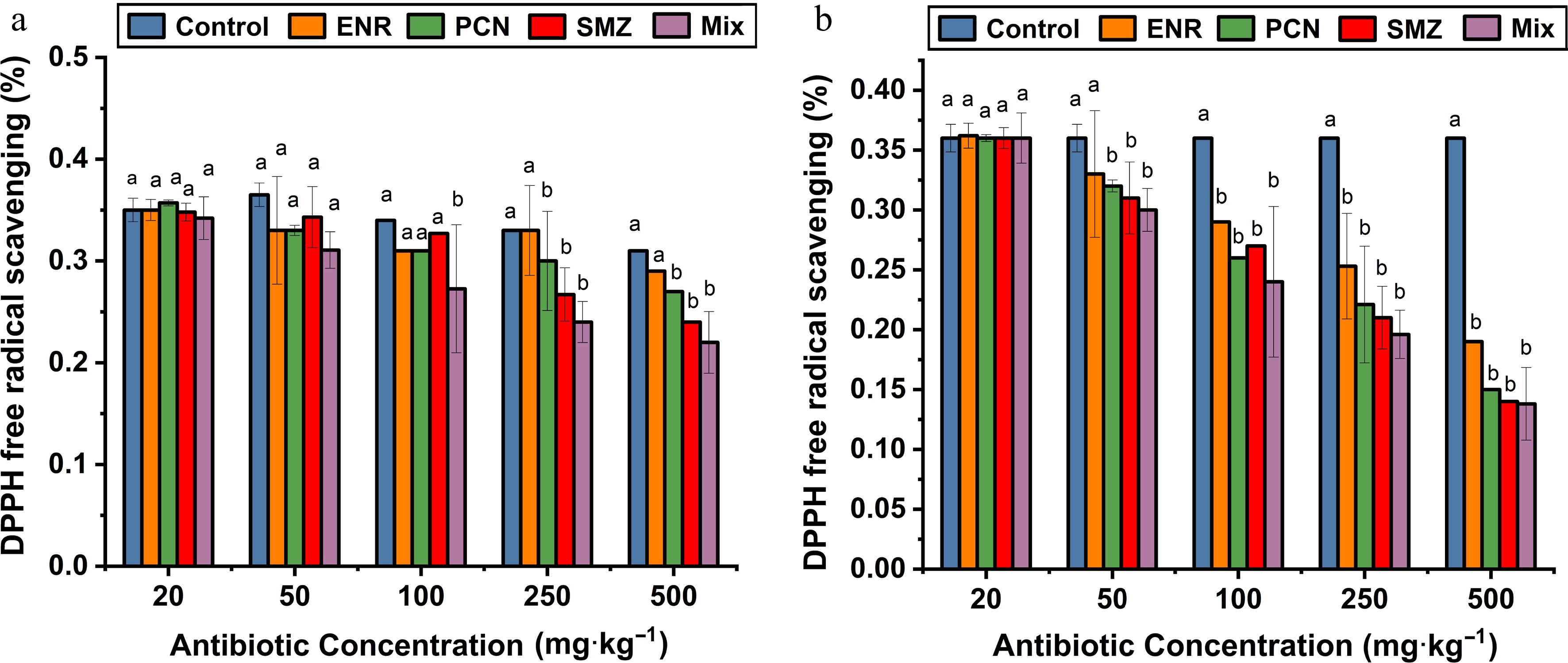
Figure 4.
Effects of enrofloxacin (ENR), penicillin (PCN), sulfamethazine (SMZ), and their mixture (Mix) on DPPH free radical scavenging activity of (a) T. alexandrinum and (b) E. vesicaria exposed to different antibiotic concentrations (20, 50, 100, 250, and 500 mg·kg−1). Values are presented as mean ± SD (n = 3). At each concentration, differences among treatments were analyzed using one-way ANOVA followed by Tukey's honestly significant difference (HSD) post hoc test. Different lowercase letters indicate significant differences among treatments within the same concentration (p < 0.05).
As shown in Fig. 4a, DPPH scavenging activity decreased with increasing concentration; the extent of reduction in T. alexandrinum was smaller, with values declining from ~0.36 in the control to ~0.14–0.18 under mixture treatment at the highest concentration. This indicates relatively better maintenance of antioxidant defense in T. alexandrinum. In E. vesicaria (Fig. 4b), DPPH activity declined from approximately ~0.35 in control conditions to ~0.22 under mixture treatment at 500 mg·kg−1, representing a substantial reduction in antioxidant potential. ENR also caused a pronounced decrease, followed by PCN and SMZ, which showed comparatively moderate effects. Across both species, the inhibitory effects followed the trend: mixture > ENR > PCN ≈ SMZ, consistent with patterns observed for enzymatic and pigment responses. The progressive decline in DPPH scavenging activity suggests that increasing antibiotic concentrations impair the plant's antioxidant defense system, likely due to disruption of ROS-scavenging mechanisms. Overall, these findings indicate that VA exposure induces oxidative stress by reducing non-enzymatic antioxidant capacity, with E. vesicaria being more vulnerable, while T. alexandrinum exhibits comparatively greater resilience under antibiotic stress.
Effect of VAs on proline content
-
Proline accumulation increased significantly in antibiotic-treated plants compared with control seedlings (Fig. 5). Control plants exhibited the lowest proline levels, whereas proline content increased approximately fourfold as VA concentrations increased from 250 to 500 mg·kg−1, indicating a concentration-dependent response. In T. alexandrinum, all three antibiotics induced comparable proline accumulation at lower concentrations (20–100 mg·kg−1), while higher concentrations (> 250 mg·kg−1) resulted in markedly greater increases. The MIX treatment produced higher proline accumulation than individual antibiotic applications across all tested concentrations. In E. vesicaria, enhanced proline accumulation was observed even at low antibiotic levels, demonstrating greater sensitivity to VA exposure, with ENR producing the strongest stimulatory effect among the tested antibiotics. Overall, proline content increased progressively with rising antibiotic concentrations in both species, with MIX treatments eliciting the most pronounced response.
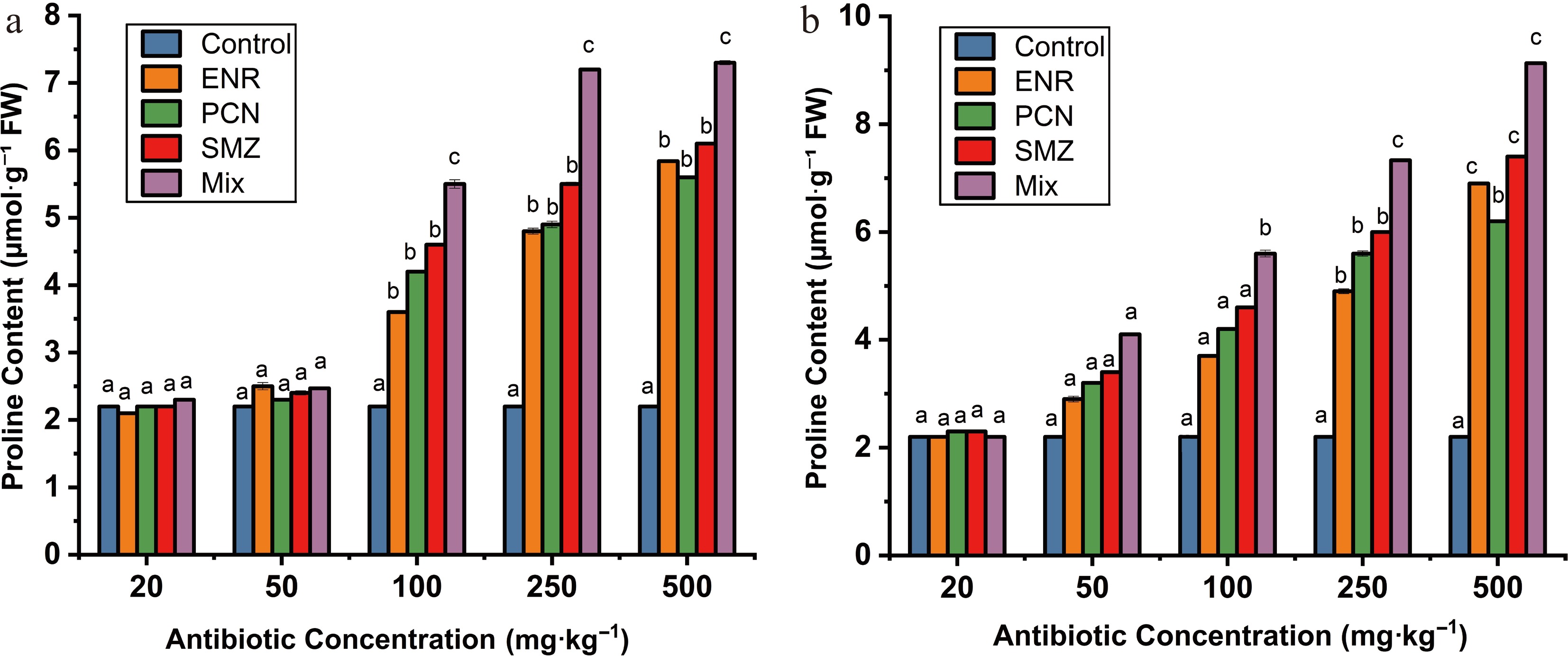
Figure 5.
Effects of enrofloxacin (ENR), penicillin (PCN), sulfamethazine (SMZ), and their mixture (Mix) on proline content in (a) T. alexandrinum and (b) E. vesicaria exposed to different antibiotic concentrations (20, 50, 100, 250, and 500 mg·kg−1). Values are presented as mean ± SD (n = 3). At each concentration, differences among treatments were analyzed using one-way ANOVA followed by Tukey's honestly significant difference (HSD) post hoc test. Different lowercase letters indicate significant differences among treatments within the same concentration (p < 0.05).
Effect of VAs on enzyme activities
-
The activities of key metabolic enzymes—acid phosphatase, esterase, and urease—were significantly affected by VA exposure, showing a clear concentration-dependent decline in both plant species (Fig. 6a–f). Across all treatments, E. vesicaria exhibited greater sensitivity than T. alexandrinum, as reflected by steeper reductions in enzyme activities. Acid phosphatase activity (Fig. 6a, b) decreased progressively with increasing antibiotic concentration. At lower concentrations (20–50 mg·kg−1), only slight reductions were observed; however, at ≥ 100 mg·kg−1, a marked decline became evident. At 500 mg·kg−1, ENR reduced acid phosphatase activity by approximately 35%–45% in E. vesicaria, whereas the reduction in T. alexandrinum remained comparatively moderate (~20%–30%). The antibiotic mixture exerted the strongest inhibitory effect across all concentrations, resulting in the lowest activity levels in both species. Esterase activity (Fig. 6c, d) showed the highest sensitivity to VA stress among the enzymes studied. A sharp decline was observed beyond 100 mg·kg−1, with substantial inhibition at higher concentrations. At 500 mg·kg−1 ENR, esterase activity decreased by nearly 40%–50% in E. vesicaria and about 25%–30% in T. alexandrinum. The MIX treatment caused the most pronounced suppression, with reductions exceeding 60%–70% in E. vesicaria, indicating severe disruption of metabolic processes. In contrast, PCN and SMZ showed relatively milder effects.
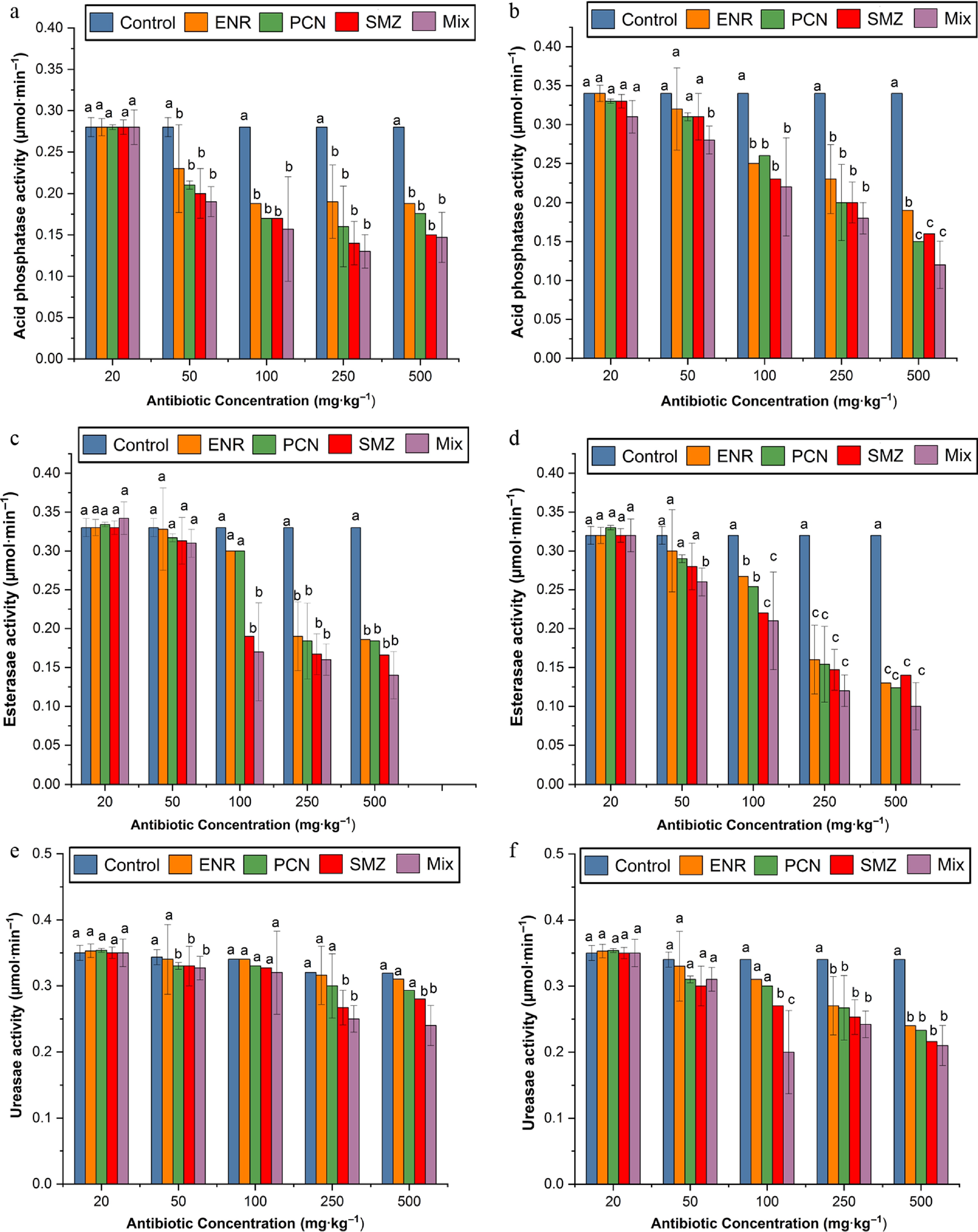
Figure 6.
Effects of enrofloxacin (ENR), penicillin (PCN), sulfamethazine (SMZ), and their mixture (Mix) on (a), (b) acid phosphatase a, (c), (d) esterase, and (e), (f) urease activity in the T. alexandrinum and E. vesicaria exposed to different antibiotic concentrations (20, 50, 100, 250, and 500 mg·kg−1). Values are expressed as mean ± SD (n = 3). For each concentration, differences among treatments were analyzed using one-way ANOVA followed by Tukey's HSD post hoc test. Different lowercase letters above the bars indicate significant differences among treatments within the same concentration (p < 0.05).
Urease activity (Fig. 6e, f) was comparatively less sensitive but still followed a consistent declining trend with increasing antibiotic concentration. At higher concentrations (≥ 250 mg·kg−1), ENR caused a noticeable reduction (~40%–50%) in E. vesicaria, while T. alexandrinum exhibited smaller decreases (~20%–30%). The combined antibiotic treatment again produced the strongest inhibition, though the overall magnitude of decline was lower than that observed for esterase. Overall, enzyme inhibition followed the general order: MIX > ENR > PCN ≈ SMZ, with esterase being the most sensitive enzyme, followed by acid phosphatase and urease. The greater susceptibility of E. vesicaria suggests a higher degree of metabolic disruption under antibiotic stress, whereas T. alexandrinum maintained relatively higher enzymatic activity, indicating better physiological tolerance. These findings demonstrate that VAs interfere with essential enzymatic processes, potentially impairing nutrient metabolism and overall plant health, particularly under high-dose and combined antibiotic exposure.
-
In the present study, a clear dose-dependent inhibition of plant growth and development was observed following exposure to VAs. High concentrations of VAs, applied either individually or in combination, significantly reduced seed germination or adversely affected key morphological traits of both E. vesicaria and T. alexandrinum. The germination assay demonstrated that VA toxicity varied depending on plant species and antibiotic type, indicating species-specific sensitivity to antibiotic stress. Both filter paper and pot experiments were conducted to evaluate seed germination and early seedling responses. The two approaches provide complementary information by assessing phytotoxic effects under controlled conditions and in antibiotic-spiked soil. Among the tested species, E. vesicaria exhibited greater sensitivity to VA exposure compared with T. alexandrinum, although toxicity levels differed among antibiotics. Among the tested antibiotics, ENR showed comparatively stronger phytotoxic effects. Similar phytotoxic effects of sulfonamides have been reported in several plant species, confirming their broad impact on plant physiological processes[25,26]. Exposure to higher antibiotic concentrations (> 100 mg·kg−1) markedly inhibited plant growth, producing seedlings with chlorotic symptoms, reduced plant height, and shorter, thinner root systems. These morphological impairments are consistent with previous findings demonstrating that VAs disturb root activity, nutrient uptake, and cellular metabolism at elevated concentrations[25]. Species-dependent variability in plant responses observed in this study further supports earlier reports indicating that antibiotic toxicity depends on plant physiology, uptake efficiency, and detoxification capacity[9,10]. In contrast, lower VA concentrations did not significantly affect overall plant growth, suggesting a threshold-dependent response. However, plants exposed to higher doses developed weak and chlorotic leaves accompanied by reduced leaf area and RWC, which are characteristic indicators of physiological stress. Reduced RWC likely results from impaired root function and altered membrane permeability under antibiotic stress conditions[27]. Furthermore, chlorophyll and carotenoid contents declined significantly at elevated VA concentrations, indicating disruption of the photosynthetic apparatus. Antibiotics can interfere with plastid ribosomes due to their evolutionary similarity to bacterial ribosomes, thereby inhibiting protein synthesis required for chlorophyll biosynthesis and photosystem maintenance[9]. Fluoroquinolone antibiotics, including ENR analogues, have also been reported to inhibit photosynthetic electron transport and energy transfer processes, ultimately reducing plant growth and biomass accumulation[28]. Similar reductions in photosynthetic pigments following antibiotic exposure have been documented in rice and other crop species[29]. Overall, the findings demonstrate that MIX treatment exerts strong phytotoxic effects influencing germination, morphology, water relations, and photosynthetic performance. Similar observations have been reported for antibiotic mixtures in agricultural soils, where combined exposure generally results in greater phytotoxicity than single antibiotics[30−32]. Additionally, the experimental soil used in this study had an alkaline pH (8.4), which may have influenced the bioavailability and phytotoxicity of the tested antibiotics. Soil pH affects antibiotic adsorption, degradation, and mobility, thereby altering their uptake by plants. Consequently, the observed phytotoxic effects may reflect both the intrinsic toxicity of the antibiotics and the influence of soil pH on their environmental behavior[1,5]. Future studies should investigate antibiotic phytotoxicity across a wider range of soil pH conditions to better understand how soil chemistry modulates plant exposure and toxicity.
VAs-induced oxidative stress and proline accumulation
-
Antibiotic stress, though less studied than drought or salinity, disrupts plant homeostasis by causing oxidative damage, protein disruption, and metabolic imbalance[33]. A central adaptive response is proline accumulation, which acts as an osmoprotectant, antioxidant, protein stabilizer, and stress signal. Antibiotics such as streptomycin, kanamycin, and ciprofloxacin induce ROS overproduction, and proline mitigates this oxidative stress[34]. Proline also stabilizes proteins misfolded by antibiotic-induced inhibition of ribosomal activity in chloroplasts and mitochondria[33−36] and can be catabolized to provide energy and nitrogen during recovery[33].
In our study, VAs triggered a dose-dependent increase in proline levels, with ENR being the most potent stressor. E. vesicaria showed greater sensitivity to ENR than T. alexandrinum, highlighting species-specific responses. Similarly, VAs significantly reduced DPPH free radical scavenging activity, particularly under ENR treatment, suggesting a weakened non-enzymatic antioxidant defense system. Together, these results indicate that VA exposure (≥ 100 mg·kg−1) induces oxidative stress both by impairing enzymatic scavengers and diminishing radical-quenching capacity. These findings align with previous reports of antibiotic-induced oxidative stress in duckweed exposed to amoxicillin[28]. Overall, our results demonstrate that VAs compromise redox balance and stimulate proline accumulation while suppressing radical scavenging activity, underscoring proline's central role in mitigating antibiotic toxicity in plants.
VAs alter plant enzyme activities
-
Plant enzymes play essential roles in metabolic regulation and physiological adaptation, contributing to growth, nutrient remobilization, and stress tolerance. Several hydrolytic enzymes, including acid phosphatase, esterase, and urease, are recognized as sensitive biochemical indicators of biotic and abiotic stresses such as drought, salinity, and chemical contamination[37−43]. In this study, exposure to VAs in soil significantly altered the activities of these key plant enzymes. Acid phosphatase, which is crucial for phosphate metabolism and intracellular phosphorus regulation, was significantly inhibited in E. vesicaria at higher concentrations of ENR, suggesting disruption of phosphorus homeostasis under antibiotic stress. Similar inhibitory effects on phosphatase activity have been reported under various antibiotic and contaminant exposures[39]. Esterases, a diverse group of hydrolases catalyzing the hydrolysis of ester bonds, are widely distributed in plants and commonly used as biomarkers of physiological stress due to their rapid response to environmental disturbances[42]. In the present study, esterase activity was highly sensitive to VAs, showing inhibition even at low antibiotic concentrations. Previous reports indicate that esterase responses vary depending on stress type and intensity[39,40]. Urease, a nickel-dependent enzyme that catalyzes urea hydrolysis and contributes to nitrogen metabolism, also exhibited a clear concentration-dependent decline in activity following VA exposure. Urease activity is known to respond to salinity and other environmental stresses[43]. Collectively, the observed alterations in enzymatic activities reflect metabolic stress responses within plant tissues. Inhibition of esterase, acid phosphatase, and urease suggests disruption of cellular metabolism and nutrient remobilization processes, indicating impaired metabolic regulation and detoxification capacity. These biochemical changes likely contributed to the reduced growth and physiological performance observed in VA-treated plants.
-
This study specifically evaluates plant biochemical responses, indicating that the observed toxicity primarily reflects direct physiological effects on plant metabolism. The findings demonstrate species-specific susceptibility to VAs and highlight plant enzymatic activities as sensitive biochemical indicators of antibiotic-induced stress, providing insight into the metabolic disruptions underlying phytotoxic responses. This study demonstrates that exposure to a mixture of antibiotics produced greater phytotoxic effects than most individual antibiotics under the tested conditions, providing useful baseline information on the potential risks of combined antibiotic contamination. However, further studies using factorial mixture designs are needed to distinguish additive, synergistic, or antagonistic interactions.
The authors acknowledge the University of Science and Technology, Bannu, Pakistan, for providing research facilities. This research received no external funding.
-
The authors confirm their contributions to the paper as follows: study conception and design: Nadir S, Khan S; experimental work: Khan F, Khan S; data collection: Khan F, Khan S; data analysis: Khan H, Zaman L; draft manuscript preparation: Nadir S, Khan S; writing–review and editing: Khan H, Zaman L; supervision: Nadir S, Khan S. All authors reviewed the results and approved the final version of the manuscript.
-
All data generated or analyzed during this study are included in this published article [and its supplementary information files].
-
The authors declare that they have no conflict of interest.
-
accompanies this paper online at: https://doi.org/10.48130/cas-0026-0025.
- Supplementary Table S1 Seedling vigor %, fresh weight, dry weight, leaf area and RWC of T. alexandrinum.
- Supplementary Table S2 Seedling vigor %, fresh weight, dry weight, leaf area and RWC of E. vesicaria.
- Supplementary Fig. S1 (a) Image showing impact of ENR (A), PCN (B), SMZ (C), or mixture (D) on morphological and phenotypic characteristics of T. alexandrium. (b) Image showing impact of ENR (A), PCN (B), SMZ (C), or mixture (D) on morphological and phenotypic characteristics of E.vesicaria.
- Copyright: © 2026 by the author(s). Published by Maximum Academic Press, Fayetteville, GA. This article is an open access article distributed under Creative Commons Attribution License (CC BY 4.0), visit https://creativecommons.org/licenses/by/4.0/.
-
About this article
Cite this article
Nadir S, Khan S, Khan S, Khan H, Khan F, et al. 2026. Species-specific phytotoxic and enzymatic responses of edible crops to single and combined veterinary antibiotics. Circular Agricultural Systems 6: e026 doi: 10.48130/cas-0026-0025 

Species-specific phytotoxic and enzymatic responses of edible crops to single and combined veterinary antibiotics
- Received: 21 April 2026
- Revised: 03 July 2026
- Accepted: 27 July 2026
- Published online: 22 October 2026
Abstract: Veterinary antibiotics (VAs) are emerging agroecosystem contaminants introduced through manure and wastewater reuse, yet their phytotoxic effects on crop physiological and biochemical functioning remain insufficiently understood. This study evaluated the individual and combined phytotoxic effects of enrofloxacin (ENR), penicillin (PCN), sulfamethazine (SMZ), and their ternary mixture (MIX) on two agronomically important plant species, Trifolium alexandrinum and Eruca vesicaria, under controlled soil conditions. Antibiotics were applied at 20–500 mg·kg−1 to evaluate dose-dependent responses in growth, physiological traits, and stress-related biochemical indicators. VA exposure caused significant, concentration-dependent inhibition of seed germination and plant growth. Biomass production, root and shoot elongation, leaf area, relative water content, and photosynthetic pigment synthesis declined markedly, particularly at ≥ 250 mg·kg−1. E. vesicaria exhibited greater susceptibility than T. alexandrinum, with toxicity ranking as MIX > ENR > SMZ > PCN, demonstrating enhanced phytotoxicity under combined antibiotic exposure. Biochemical analyses revealed substantial reductions in antioxidant capacity accompanied by approximately fourfold increases in proline accumulation, indicating oxidative stress-mediated phytotoxicity. Enzymatic activities in plant extracts were differentially affected, with esterase showing the highest sensitivity, acid phosphatase moderate inhibition, and urease comparatively minor responses across treatments. Collectively, these findings demonstrate species-specific vulnerability and identify plant enzymatic activities as sensitive indicators of physiological stress. Comparative analysis of single vs combined antibiotic exposures further highlights mixture toxicity in agroecosystems, informing risk assessment and sustainable management of antibiotic residues in agricultural soils.
-
Key words:
- Edible crops /
- Veterinary antibiotics /
- Phytotoxicity /
- Oxidative stress /
- Enzymatic stress response