














-
Plants must constantly balance growth with defense against environmental stresses. This growth-defense trade-off is essential for optimizing resource allocation and ensuring fitness under adverse conditions[1,2]. Acidic soils occupy a large proportion of global agricultural land, where aluminum (Al3+) toxicity represents a major factor limiting crop production. When soil pH drops below 5.0, soluble Al3+ rapidly inhibits root elongation and severely impairs water and nutrient uptake, posing a serious threat to global agricultural output and food security[3,4]. To counteract Al3+ toxicity, plants have evolved multiple defense mechanisms, among which MATE (Multidrug and Toxic Compound Extrusion) transporters play pivotal roles[5−7]. MATE proteins are characterized by 12 transmembrane domains and mediate the efflux of diverse substrates, thereby modulating plant development and stress adaptation[8−15]. A well- characterized mechanism of MATE-mediated Al3+ detoxification is the facilitation of root citrate secretion, which chelates toxic Al3+ in the rhizosphere[16,17]. Beyond organic acid efflux, certain MATE transporters participate in the vacuolar sequestration of flavonoids, including anthocyanins and proanthocyanidins[18−20]. These compounds can scavenge reactive oxygen species (ROS) under Al3+ stress, enhancing cellular redox homeostasis[21]. Additionally, some MATE members transport plant hormones such as auxin, directly linking them to growth and stress signaling pathways[22,23].
The interplay between auxin signaling and Al3+ tolerance pathways is particularly intriguing. As a key growth regulator, auxin modulates Al3+ tolerance through polar transport and cellular distribution[24−26]. Conversely, Al3+ stress disrupts auxin homeostasis. Notably, auxin signaling directly regulates Al3+-responsive genes, including MATE genes, which in turn modulate auxin transport and accumulation[23,27−30]. This functional versatility positions MATE transporters as potential hubs balancing growth and stress responses. However, whether and how a single MATE protein directly coordinates this critical trade-off remains unclear. For instance, overexpression of BoMATE enhances Al3+ tolerance but reduces plant biomass[31], whereas AtDTX50 improves drought tolerance without a growth penalty[32]. These findings highlight the context-dependent and functional diversity of the MATE family.
Current research has focused largely on transcriptional regulation of MATE genes, yet the potential for post-translational modifications to dynamically control MATE function has been largely overlooked. This is particularly important because plants facing acute Al3+ stress require immediate responses. Reversible phosphorylation offers a distinct advantage over transcriptional control by modulating protein activity within minutes, bypassing the time-consuming processes of transcription and translation[33−36]. Such a mechanism would enable rapid activation of detoxification and growth curtailment upon stress perception. However, whether post-translational modifications coordinate growth-defense trade-offs through a single MATE transporter in perennial woody plants remains unknown.
Precise transcriptional control of MATE genes is equally critical for optimizing the growth-defense balance. Constitutive or excessive expression of a growth-suppressing MATE transporter would impose continuous fitness costs even without stress. A finely tuned expression level, achieved through an activator-repressor transcriptional module, can ensure sufficient transporter activity during stress while preventing excessive fitness costs. Thus, the integration of rapid post-translational switches with precise transcriptional fine-tuning likely represents an evolutionarily optimized strategy for balancing immediate stress responses with long-term fitness.
Mulberry (Morus alba) is an economically important woody plant that exhibits exceptional resilience to environmental stresses, including extreme soil acidity (pH < 4.5)[37,38]. This inherent tolerance makes it an important plant for utilizing marginal land and an ideal model for deciphering sophisticated Al3+ tolerance mechanisms. Here, we identified an Al3+-induced MATE gene, MaDTX19, from mulberry, and hypothesized that it acts as a central regulator integrating Al3+ stress and growth signaling, with activity controlled by a post-translational switch. Our objectives were to characterize the role of MaDTX19 in regulating plant growth and Al3+ tolerance, validate the function of Ser4 phosphorylation in modulating MaDTX19 activity and protein-protein interactions, and elucidate the upstream transcriptional regulators and downstream pathways underlying its role in the growth-defense trade-off. Collectively, our findings reveal a phosphorylation-dependent mechanism by which a single MATE transporter coordinates growth-defense balance in a woody perennial, providing a genetic target for breeding Al3+-tolerant mulberry cultivars on acidic marginal lands.
-
Seedlings of mulberry ('Guisang you62', M. atropurpurea), Arabidopsis thaliana (Col-0 ecotype), and Nicotiana benthamiana were cultivated in growth chambers. Mulberry and tobacco were cultured at 26 °C with 50%−60% relative humidity under a 16 h light/8 h dark photoperiod. Arabidopsis was grown at 22 °C under the same humidity and photoperiod.
Phylogenetic and motif analysis
-
MATE family protein sequences from A. thaliana and M. notabilis were retrieved from NCBI. Multiple sequence alignment was performed using ClustalW in MEGA 6, and a neighbor-joining phylogenetic tree was constructed with 1,000 bootstrap replicates. Conserved motifs were identified using MEME (
http://meme.nbcr.net/meme/intro.html ) with default parameters. Transmembrane domains of MaDTX19 were predicted using TMHMM Server v.2.0.Gene cloning and Arabidopsis transformation
-
Total RNA was extracted from mulberry leaves with TRIzol™ reagents (Invitrogen, Carlsbad, CA, USA) and reverse-transcribed using oligo (dT) primers (Invitrogen) and M-MLV Reverse Transcriptase (Promega, Madison, WI, USA). Target genes were amplified and cloned into plant expression vectors, and the resulting constructs were then transformed into Agrobacterium tumefaciens GV3101. Arabidopsis transformation was performed using the floral dip method. Transgenic lines were selected on 1/2 MS medium containing 50 mg·L−1 kanamycin and verified by PCR. Homozygous T3 lines were used for subsequent experiments.
Generation of transgenic hairy roots
-
Target genes were inserted into the binary vector PLGNL under the control of the CaMV35S promoter and individually introduced into A. rhizogenes strain K599. Two-week-old seedlings of 'Guisang You 62' were wounded at the stem and infected with A. rhizogenes K599 harboring the target genes or an empty vector. Infected seedlings were co-cultured on MS medium in the dark for 2 d. Subsequently, they were transferred to selective MS medium supplemented with 50 mg·L−1 kanamycin and 100 mg·L−1 terramycin. Transgenic hairy roots emerged approximately four weeks later and were verified by PCR.
Subcellular localization analysis of MaDTX19
-
The coding sequence (CDS) of MaDTX19 (without stop codon) was fused to GFP in the pCAMBIA1300 vector to construct 35S::MaDTX19-GFP. The recombinant vector and empty vector were transformed into A. tumefaciens GV3101 and infiltrated into N. benthamiana leaves. GFP fluorescence was observed 24–72 h post-infiltration under a Zeiss LSM880 laser scanning confocal microscope (Zeiss, Germany) (excitation/emission: 488/505–530 nm; chloroplast autofluorescence: 650 nm).
RT-qPCR
-
Total RNA was extracted as described above. RT-qPCR assays were performed using SYBR Premix Ex Taq II (TaKaRa Bio, Dalian, China) on a Bio-Rad CFX96™ System according to the manufacturer's protocols. EF1-a and ACTIN served as reference genes, and relative expression was calculated using the 2−ΔΔCᴛ method. Three independent biological replicates, each with three technical replicates, were analyzed per sample.
Promoter activity analysis
-
The MaDTX19 promoter (pMaDTX19, a 2,000-bp fragment upstream of the start codon) was amplified from mulberry genomic DNA and fused to the β-glucuronidase (GUS) reporter gene in pBI121. One-week-old transgenic Arabidopsis seedlings carrying pMaDTX19::GUS were transferred to 1/2 MS medium supplemented with 50 μmol·L−1 ABA, 50 μmol·L−1 IAA, or 0.8 mmol·L−1 AlCl3. Seedlings on normal medium served as controls. GUS staining was performed following the standard protocol[39], and stained tissues were photographed.
Aluminum stress treatment and determination of Al3+ content
-
Surface-sterilized Arabidopsis seeds were sown on 1/2 MS solid medium containing 0, 0.4, 0.8, or 1.2 mmol·L−1 AlCl3. After stratification at 4 °C for 3 d, plates were transferred to growth chambers. Germination rates were scored daily, and root lengths were measured 7−10 d after germination. For physiological assays, four-week-old soil-grown Arabidopsis and mulberry seedlings were treated with 80 mmol·L−1 AlCl3 solution, with water treatment as a control. The AlCl3 concentrations were selected based on preliminary experiments and previous studies[40].
Al3+ accumulation in roots was visualized by hematoxylin staining. Roots were rinsed with distilled water and then stained with a freshly prepared solution containing 0.1% hematoxylin and 0.01% KI for 30 min. After thorough washing with distilled water, roots were photographed under a microscope.
For Al3+ content determination, plant samples were dried at 70 °C to constant weight, digested in 5% HNO3 at 120 °C for 2 h, and Al3+ concentration was measured by inductively coupled plasma mass spectrometry (ICP-MS; Agilent 7700x, USA).
Determination of physiological and biochemical parameters
-
Malondialdehyde (MDA) and proline contents were determined using assay kits (both from Solarbio, Beijing, China) according to the manufacturer's instructions. Citric acid content was assayed using a Citric Acid (CA) Content Assay Kit (Boxbio, Beijing, China). Anthocyanin was extracted with acidified methanol (1% HCl, v/v) and quantified by spectrophotometry[41], with three biological replicates per sample.
4-(Dimethylamino)cinnamaldehyde (DMACA) staining for proanthocyanidins
-
Samples were immersed in DMACA solution (containing 1% DMACA, 10% HCl, and 50% methanol) for 5−10 min. After staining, samples were rinsed with 70% ethanol and observed under a microscope. Magenta staining indicated proanthocyanidin accumulation.
Reactive oxygen species (ROS) detection and antioxidant enzyme activity assay
-
Histochemical staining for H2O2 and O2·− was performed with 3,3'-diaminobenzidine (DAB) and nitrotetrazolium blue chloride (NBT), respectively, using a Plant Tissue ROS Detection Kit (Solarbio, Beijing, China). Stained leaves were decolorized with 95% ethanol and photographed. Activities of superoxide dismutase (SOD), catalase (CAT), and peroxidase (POD) were determined with commercial assay kits (Solarbio, Beijing, China) following the manufacturer's methods.
Chlorophyll fluorescence imaging
-
Maximum photochemical efficiency of photosystem II (Fv/Fm) was measured with an IMAGING-PAM M-Series chlorophyll fluorescence system (Walz, Germany) after 30 min of dark adaptation. The minimal fluorescence (Fo) was measured under weak measuring light, and the maximal fluorescence (Fm) was determined after a saturating pulse of light. Fv/Fm was calculated as (Fm – Fo)/Fm.
Yeast one-hybrid assay (Y1H)
-
The pMaDTX19 fragment was cloned into the pAbAi vector to generate pAbAi-pMaDTX19. Then, linearized pAbAi-pMaDTX19 plasmids were integrated into Y1HGold yeast to create bait-reporter yeast strains. The coding sequence of MaMYB61 was cloned into pGADT7 to produce AD-MaMYB61. The plasmid AD-MaMYB61 was transformed into the bait yeast competent cells, and transformants were spread onto SD/-Leu/AbA selection medium and cultured at 28 °C for 3−5 d.
Luciferase reporter assay (LUC)
-
pMaDTX19 was cloned into the pGreenII 0800 vector, driving LUC expression. The CDSs of MaMYB61 and MaMYB308 were cloned into the pGreen62-SK effector vector, respectively, under the 35S promoter. Combinations of reporter and effector plasmids were transformed into A. tumefaciens GV3101 and co-infiltrated into N. benthamiana leaves. Luminescence was observed using a CCD (Charge-Coupled Device) camera 24−72 h later. Relative LUC activity was measured using a Dual-Luciferase Reporter Assay Kit (TransGen Biotech, China) and calculated as the ratio of firefly LUC to Renilla luciferase (REN).
Electrophoretic mobility shift assay (EMSA)
-
Recombinant MaMYB61 protein was expressed in E. coli BL21 and purified using Ni-NTA affinity chromatography. Biotin-labeled probes containing the predicted MaMYB61-binding site in the MaDTX19 promoter were synthesized. Binding reactions were performed with purified protein and labeled probes, with unlabeled probes as competitors. Complexes were resolved on native polyacrylamide gels, transferred to nylon membranes, and detected using a chemiluminescent EMSA kit (Beyotime, China).
Membrane yeast two-hybrid assay
-
MaDTX19 was cloned into the pBT3-STE (bait) to generate the recombinant plasmid (pBT3- MaDTX19), and the CDSs of MaAECC8, Ma5NG4, and MaWAT1 were cloned into pPR3-N (prey) to generate pPR3-MaAECC8, pPR3-Ma5NG4, and pPR3-MaWAT1, respectively. Bait and prey plasmids were co-transformed into yeast strain NMY51. The transformed cells were selected on SD/-Leu/-Trp (DDO) medium and tested for interaction on SD/-Ade/-His/-Leu/-Trp (QDO) medium. Yeast growth on QDO medium indicates a positive interaction.
Bimolecular fluorescence complementation assay (BiFC)
-
The CDS of MaDTX19 was fused to the C-terminal half of YFP (YFPC) in pSPYCE to generate a recombinant plasmid (MaDTX19-YFPC). The CDSs of MaAECC8, Ma5NG4, and MaWAT1 were fused to the N-terminal half (YFPN) in pSPYNE, respectively, to generate MaAECC8-YFPN, Ma5NG4-YFPN, and MaWAT1-YFPN. The Agrobacterium strain harboring the MaDTX19-YFPC plasmid was mixed with strains containing MaAECC8-YFPN, Ma5NG4-YFPN, or MaWAT1-YFPN plasmids, respectively. Mixtures were infiltrated into N. benthamiana leaves, and YFP fluorescence was detected 24–72 h post-infiltration using a Zeiss LSM880 confocal microscope (Zeiss, Germany) with excitation at 514 nm and emission collected at 520–560 nm.
Luciferase complementation assay (LCA)
-
The CDSs of MaDTX19, MaDTX19SD (Ser4→Asp), and MaDTX19SA (Ser4→Ala) were cloned into the pCAMBIA1300-nLUC (N-terminal LUC), respectively. The CDSs of MaAECC8, Ma5NG4, and MaWAT1 were cloned into the pCAMBIA1300-cLUC (C-terminal LUC), respectively. Agrobacterium strains harboring nLUC and cLUC constructs were paired and infiltrated into N. benthamiana leaves. After 48–72 h, the leaves were sprayed with 1 mmol·L−1 luciferin and incubated in the dark for 5−10 min. Luminescence was captured using a CCD camera.
Western blot analysis
-
Total protein was extracted from root tips of MaDTX19-OE Arabidopsis treated with 0.8 mmol·L−1 AlCl3 for various time points. Proteins were separated by SDS-PAGE and transferred to a PVDF membrane. For phosphorylation detection, the membrane was immunoblotted with a custom-made Anti-Phospho-(Ser/Thr) antibody (Abcam). For protein stability, total protein was extracted from Arabidopsis lines overexpressing MaDTX19-GFP, MaDTX19SA-GFP, or MaDTX19SD-GFP under Al3+ stress. Western blot was performed using an anti-GFP antibody (Abcam), and signals were visualized using chemiluminescence.
Auxin content and distribution analysis
-
The auxin-responsive reporter DR5::GFP was transformed into WT and MaDTX19-OE Arabidopsis. Seedlings were treated with 0 or 0.4 mmol·L−1 AlCl3 for 24 h, and GFP signals in root tips were examined under a confocal microscope. For auxin quantification, root tips were excised, and IAA content was quantified using an IAA ELISA Kit (Hengyuan Technology, China) according to the manufacturer's instructions.
Exogenous IAA treatment
-
For seedling assays, surface-sterilized WT and MaDTX19-OE Arabidopsis seeds were sown on half-strength MS medium supplemented with 0.5 μmol·L−1 IAA (dissolved in DMSO). Control seedlings were grown on medium containing equivalent DMSO. Plates were placed vertically under standard light conditions (16 h light/8 h dark, 22 °C). After 10 d, hypocotyl length was recorded. For adult plants, four-week-old WT and MaDTX19-OE Arabidopsis were sprayed with 0.5 μmol·L−1 IAA solution containing 0.01% Tween-20, and control plants received the same solution without IAA. Fresh weight and dry weight were measured 7 d after treatment, with at least 15 seedlings or ten adult plants per genotype and treatment.
Statistical analysis
-
All experiments were performed with at least three independent biological replicates. Data are presented as the mean ± standard deviation (SD). Statistical analyses were performed using SPSS software (v20.0). Student's t-test was applied for comparisons between two groups. One-way analysis of variance (ANOVA) followed by Duncan's multiple range test was used for multiple-group comparisons. Different lowercase letters or asterisks indicate statistically significant differences (* p < 0.05; ** p < 0.01; *** p < 0.001).
-
To identify key MATE family members involved in Al3+ stress responses, we analyzed transcriptomic data from Al3+-stressed mulberry seedlings. Among the six MATE family members whose transcript abundance was upregulated under Al3+ stress, MaDTX19 exhibited the most pronounced upregulation (Supplementary Fig. S1). Phylogenetic analysis placed MaDTX19 within Group II of the MATE family, closely related to Arabidopsis AtDTX18 and AtDTX19 (Fig. 1a). MaDTX19 contains 12 typical transmembrane domains (Fig. 1b) and localizes to the plasma membrane (Fig. 1c), consistent with a role in substrate efflux.
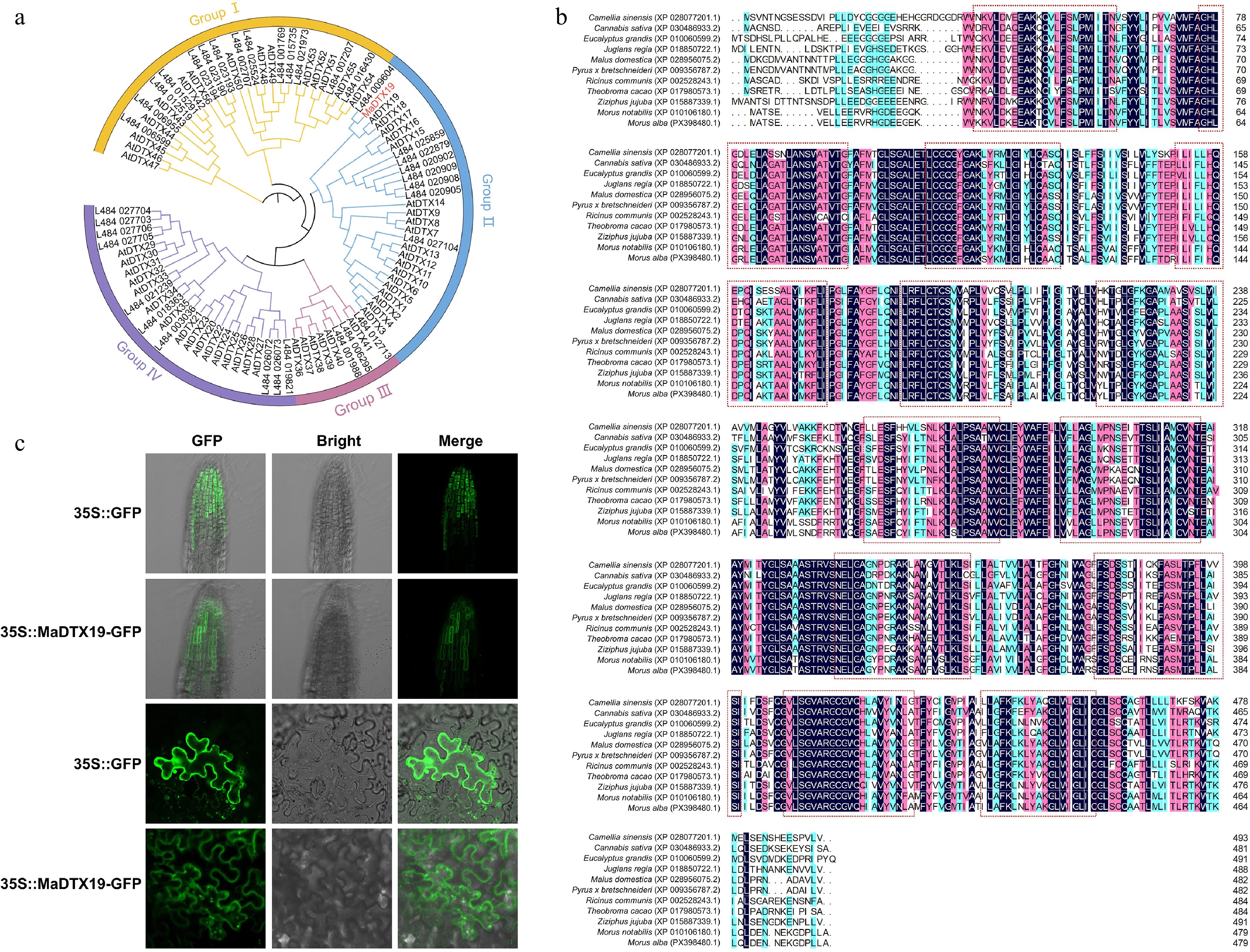
Figure 1.
Molecular characterization and subcellular localization of MaDTX19. (a) Phylogenetic analysis of MaDTX19 and MATE family proteins from Arabidopsis and mulberry. The phylogenetic tree was constructed using the neighbor-joining method with 1,000 bootstrap replicates. MaDTX19 (highlighted in red) clusters within MATE Group II with AtDTX18 and AtDTX19. (b) Transmembrane domain prediction of MaDTX19 and its homologs. The transmembrane regions are marked with dashed boxes. (c) Subcellular localization of MaDTX19 in N. benthamiana leaf epidermal cells and Arabidopsis root tips.
RT-qPCR analysis demonstrated that MaDTX19 transcript levels were rapidly and strongly induced by Al3+ stress (Fig. 2a). Treatments with ABA and IAA also significantly upregulated MaDTX19 expression (Fig. 2b, c), suggesting integration with both stress and hormonal signaling pathways. Promoter-GUS analysis in transgenic Arabidopsis further showed that pMaDTX19 activity was markedly enhanced by Al3+, ABA, and IAA treatments (Fig. 2d). These results establish MaDTX19 as an Al3+-inducible MATE gene responsive to multiple stress-related signals.
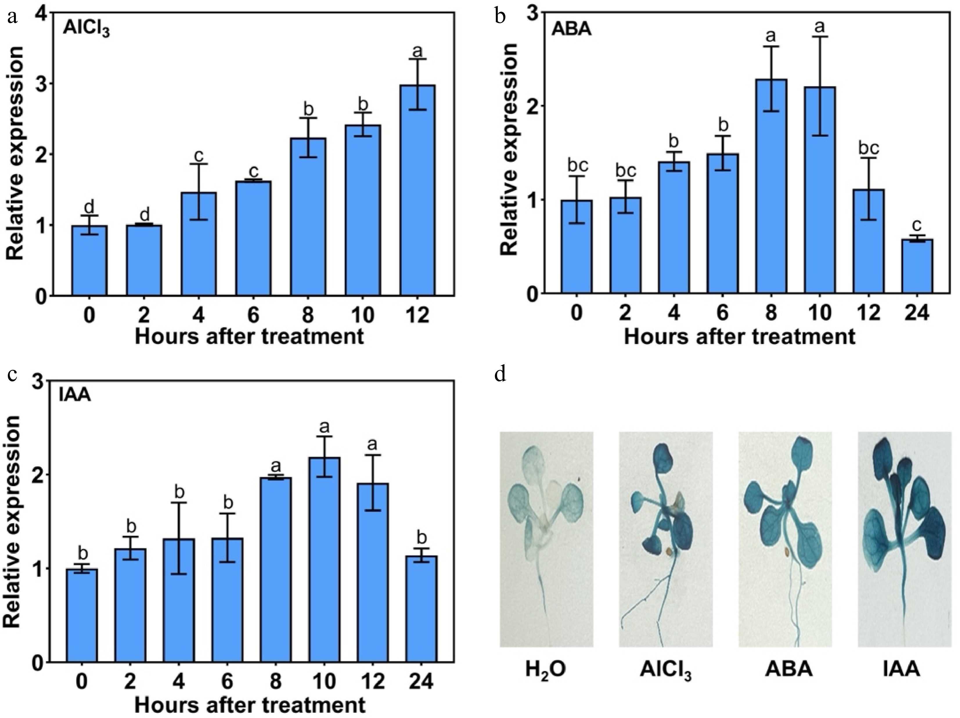
Figure 2.
Expression pattern of MaDTX19 in response to Al3+ stress and phytohormone treatments. (a) Time-course analysis of MaDTX19 transcript levels in mulberry seedlings under 80 mmol·L−1 Al3+ stress, detected by RT-qPCR. MaDTX19 expression in mulberry seedlings treated with (b) 50 μmol·L−1 ABA and (c) 50 μmol·L−1 IAA. (d) GUS staining analysis of pMaDTX19::GUS transgenic Arabidopsis seedlings under different treatments (H2O, 0.8 mmol·L−1 AlCl3, 50 μmol·L−1 ABA, 50 μmol·L−1 IAA). All RT-qPCR data are presented as mean ± SD of three biological replicates with three technical replicates each. Different letters indicate significant differences (p < 0.05).
Ectopic expression of MaDTX19 suppresses growth and development
-
To explore whether MaDTX19 affects plant development, we heterologously expressed it in Arabidopsis. Under both continuous light and dark conditions, transgenic seedlings exhibited significantly shorter hypocotyls compared to wild-type (WT) (Fig. 3a, b). When grown under standard photoperiods, mature MaDTX19-overexpressing (OE) plants displayed a striking dwarf phenotype with reduced plant height and total biomass (fresh and dry weight) (Fig. 3c−f). Moreover, reproductive development was also impaired in transgenic lines, as evidenced by shorter siliques and lower thousand-seed weight (Fig. 3g−i). Thus, MaDTX19 acts as a negative regulator of plant growth and development.
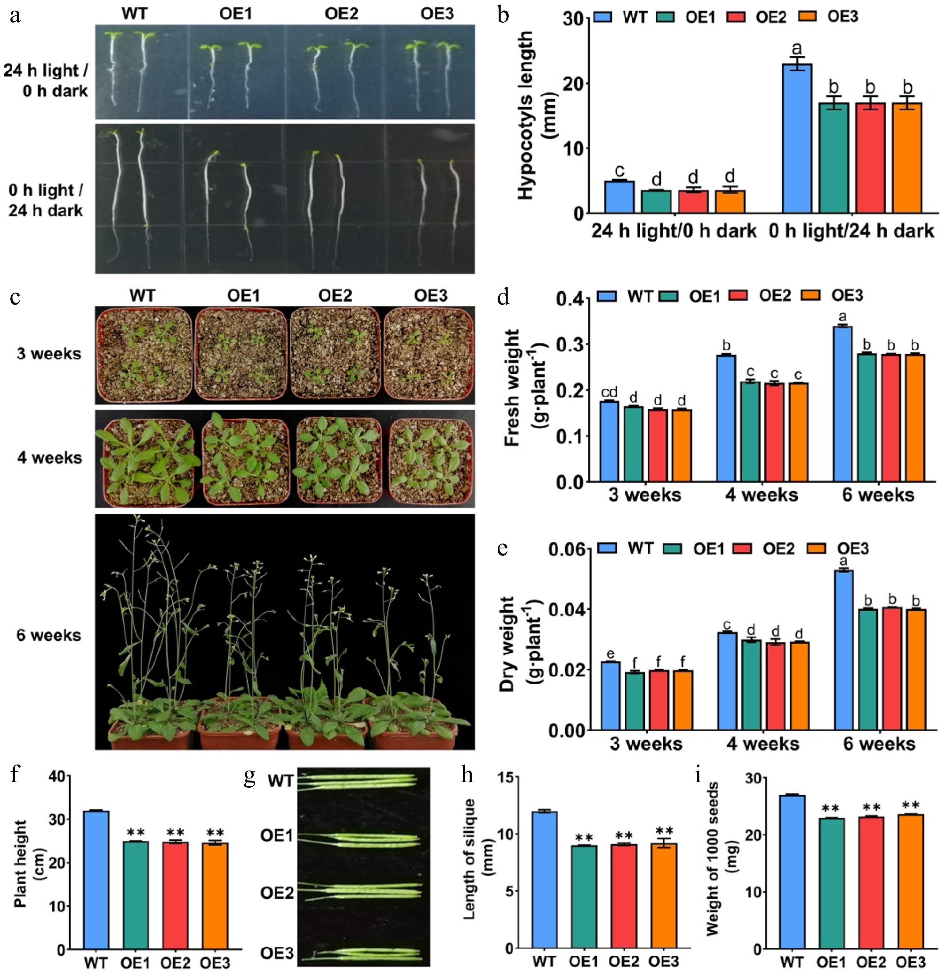
Figure 3.
Ectopic expression of MaDTX19 suppresses plant growth and development in Arabidopsis. (a) Hypocotyl phenotypes of seven-day-old wild-type (WT) and MaDTX19-overexpressing (OE) Arabidopsis seedlings grown under (a) continuous light (24 h light/0 h dark) and dark (0 h light/24 h dark) conditions. (b) Hypocotyl length of seven-day-old WT and OE Arabidopsis seedlings grown under continuous light and dark. (c) Representative phenotypes of WT and MaDTX19-OE plants under standard photoperiod. Quantitative analysis of growth and reproductive traits: (d) fresh weight, (e) dry weight, (f) plant height, (g) silique phenotype, (h) silique length, and (i) thousand-seed weight. Data represent the mean ± SD. Asterisks indicate significant differences between WT and OE transgenic lines (t-test, ** p < 0.01). Different letters indicate statistically significant differences among treatments (p < 0.05).
MaDTX19 confers Al3+ tolerance by enhancing citrate secretion and antioxidant capacity
-
To assess the role of MaDTX19 in Al3+ stress tolerance, germination and growth phenotypes of WT and MaDTX19-OE Arabidopsis seedlings under AlCl3 stress were compared. Transgenic seeds exhibited significantly higher germination rates and faster germination kinetics than WT (Fig. 4a), and OE seedlings maintained longer primary roots across multiple Al3+ concentrations (Fig. 4b, c), confirming that MaDTX19 confers Al3+ tolerance at the seedling stage. Under Al3+ stress, OE lines accumulated higher citrate levels in root tips (Fig. 4d) and showed less Al3+ accumulation in the meristematic zone by hematoxylin staining (Fig. 4e). We further examined Al3+ distribution at the whole-plant level. Both root and shoot Al3+ contents were significantly lower in OE lines compared to WT, with a more pronounced reduction in shoots. Consequently, the translocation factor (shoot Al3+/root Al3+ ratio) was markedly reduced in transgenic plants (Fig. 4f), suggesting that MaDTX19 reduces both Al3+ uptake by roots and subsequent translocation to shoots by enhancing chelation or compartmentalization within root tissues.
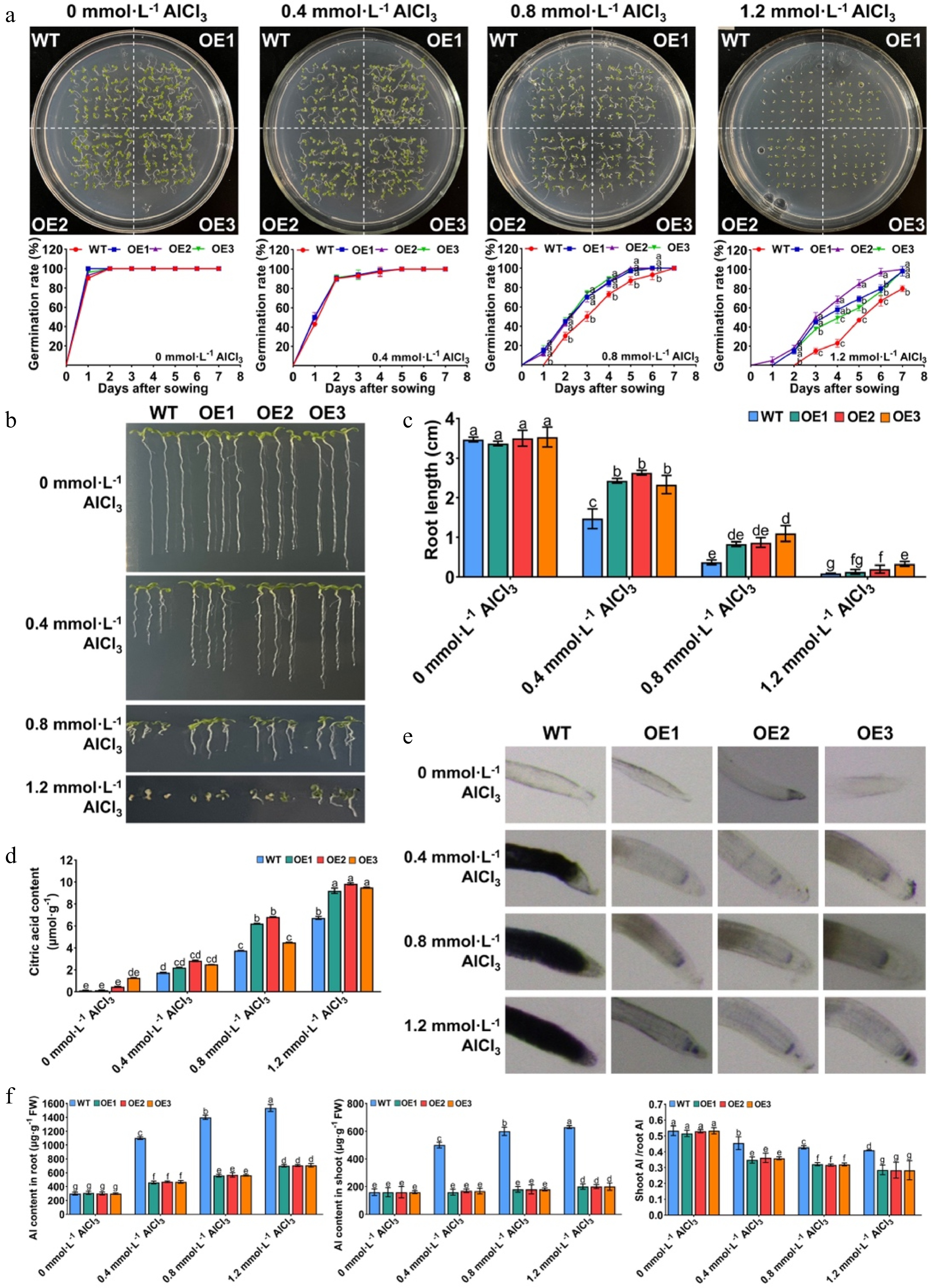
Figure 4.
MaDTX19 enhances Al3+ tolerance by promoting citrate secretion and reducing Al3+ accumulation in transgenic Arabidopsis. (a) Germination kinetics of WT and MaDTX19-OE seeds under 0, 0.4, 0.8, 1.2 mmol·L−1 AlCl3 stress. Germination rate was recorded daily for 7 d. Primary root length of seedlings grown on MS medium supplemented with (b) 0, 0.4, 0.8, 1.2 mmol·L−1 AlCl3 and (c) its quantitative analysis. (d) Citrate content in root tips under AlCl3 stress. (e) Hematoxylin staining of root tips (red color indicates Al3+ accumulation). (f) Al3+ content in roots and shoots, and the calculated translocation factor (shoot Al3+/root Al3+ ratio) of WT and OE lines under Al3+ stress. OE1-3 represent different MaDTX19-OE lines. Data are presented as mean ± SD. Different lowercase letters above bars indicate significant differences (p < 0.05).
Under Al3+ stress, OE plants exhibited milder chlorosis and wilting, with significantly higher survival rates than WT (Fig. 5a, b). Physiological analyses showed that OE plants accumulated higher levels of proline (Fig. 5c) and maintained lower MDA levels (Fig. 5d). Chlorophyll fluorescence measurements showed that the maximum photochemical efficiency of photosystem II (Fv/Fm) was significantly higher in transgenic plants (Fig. 5e, f), indicating that MaDTX19 overexpression effectively alleviated the Al3+-induced inhibition of photosystem II photochemical efficiency. ROS staining showed weaker DAB and NBT signals in OE leaves under Al3+ stress (Fig. 5g), accompanied by higher activities of CAT, SOD, and POD (Fig. 5h–j). These findings indicate that MaDTX19 confers Al3+ tolerance by enhancing antioxidant capacity and maintaining redox homeostasis.
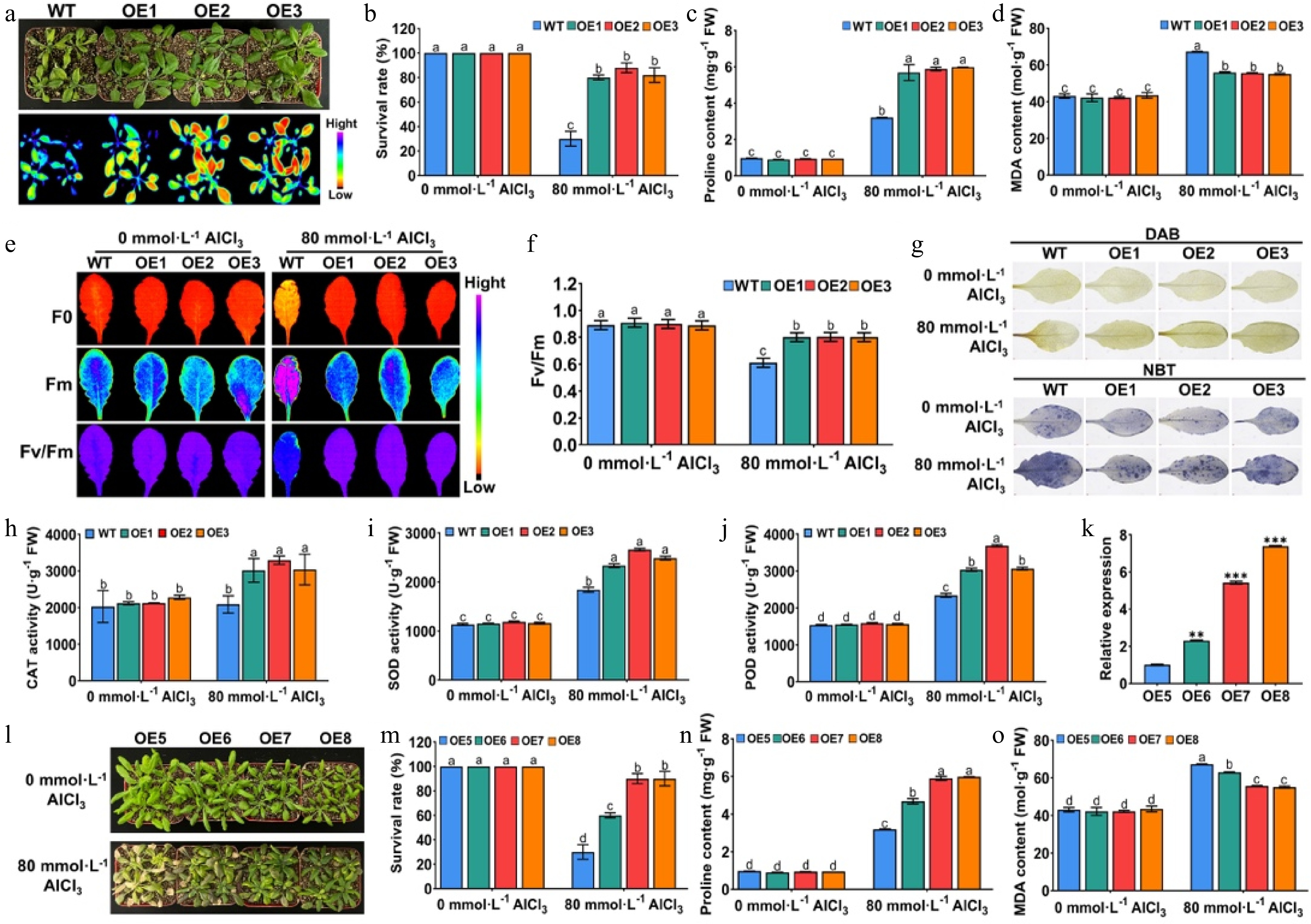
Figure 5.
MaDTX19 confers Al3+ tolerance by enhancing antioxidant capacity and protecting photosynthetic function in transgenic Arabidopsis. (a) Phenotypes and chlorophyll fluorescence images and (b) survival rate of four-week-old WT and MaDTX19-OE plants under 80 mmol·L−1 AlCl3 stress. (c) Proline and (d) MDA content in leaves under AlCl3 stress. (e) Chlorophyll fluorescence images and (f) quantitative analysis of maximum photochemical efficiency of PSII (Fv/Fm). (g) Histochemical staining of H2O2 (DAB) and O2·− (NBT) in leaves under AlCl3 stress. (h) Activities of CAT, (i) SOD, and (j) POD in leaves under AlCl3 stress. Correlation analysis of growth inhibition and Al3+ tolerance in transgenic lines with graded MaDTX19 expression levels: relative expression of MaDTX19 in (k) independent OE lines, (l) phenotypes under Al3+ stress, (m) survival rate, (n) proline content, and (o) MDA accumulation. OE1-8 represent different MaDTX19-OE lines. Data are presented as mean ± SD. Different lowercase letters above bars indicate significant differences (p < 0.05).
To determine whether the degree of growth inhibition correlates with Al3+ tolerance, we examined multiple independent transgenic lines with graded MaDTX19 expression levels. Lines with greater growth suppression exhibited higher survival rates and proline content, but lower MDA accumulation under Al3+ stress (Fig. 5k–o). This relationship plateaued beyond a certain expression threshold, suggesting an optimal MaDTX19 activity level that maximizes tolerance while minimizing fitness costs. These data indicate that growth inhibition and Al3+ tolerance are dosage-dependently coupled.
MaDTX19 promotes the accumulation of anthocyanin and proanthocyanidin
-
Given that some MATE transporters mediate vacuolar sequestration of flavonoids, we examined whether MaDTX19 influences this pathway. MaDTX19-OE seedlings grown on sucrose-containing medium exhibited notably darker cotyledon pigmentation (Fig. 6a) and significantly increased anthocyanin accumulation (Fig. 6b). Leaves and petioles of OE seedlings exhibited deeper purple-red coloration, with significantly increased anthocyanin levels (Fig. 6c). DMACA staining of seed coats revealed intensified magenta staining in OE seeds (Fig. 6d), indicating enhanced proanthocyanidin biosynthesis. These results demonstrate that MaDTX19 enhances anthocyanin accumulation.
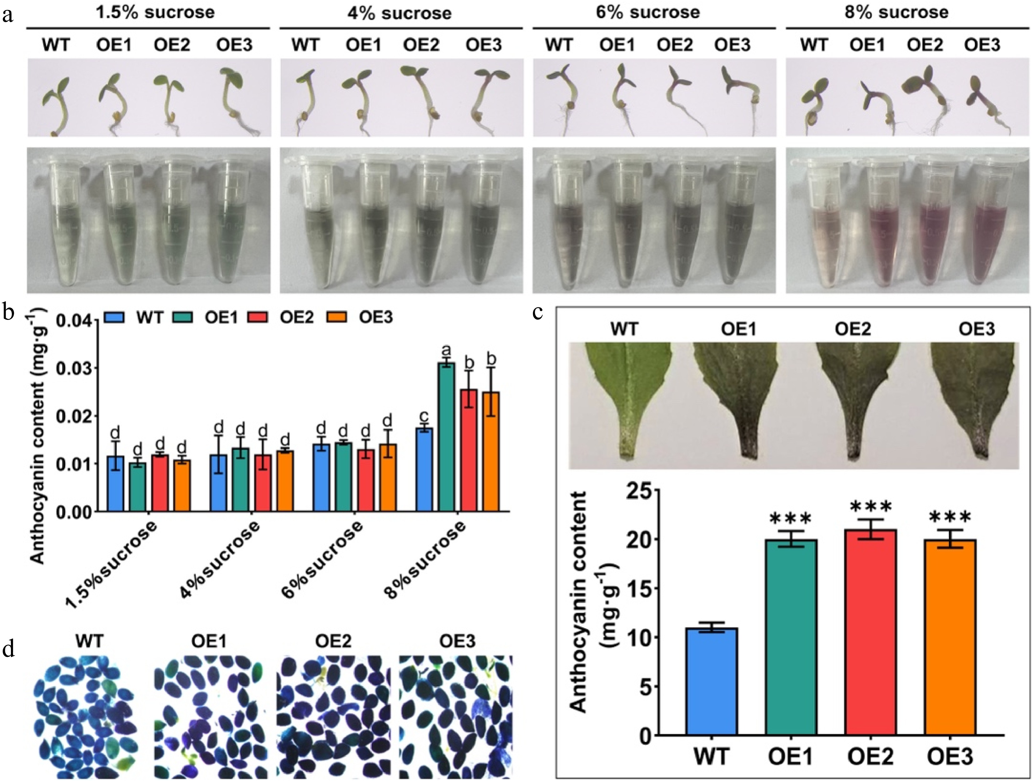
Figure 6.
MaDTX19 promotes anthocyanin and proanthocyanidin accumulation in transgenic Arabidopsis. (a) Cotyledon phenotypes and (b) quantitative analysis of anthocyanin content of WT and MaDTX19-OE seedlings grown on MS medium with 1.5%, 4%, 6%, and 8% sucrose. (c) Phenotypes and anthocyanin content of leaves and petioles of WT and MaDTX19-OE Arabidopsis seedlings. (d) DMACA staining of proanthocyanidins in seed coats (magenta color indicates proanthocyanidin accumulation). OE1-3 represent different MaDTX19-OE lines. Data represent the mean ± SD. Asterisks denote significant differences (t-test, *** p < 0.001), while different letters indicate significant differences at p < 0.05.
MaDTX19 enhances Al3+ tolerance in mulberry hairy roots
-
To validate the function of MaDTX19 in its native woody species, we generated A. rhizogenes-mediated hairy root transformations in mulberry. Under Al3+ stress, seedlings harboring MaDTX19-OE hairy roots showed milder symptoms than empty-vector controls, which displayed severe chlorosis and wilting (Fig. 7a). ROS accumulation was substantially lower in transgenic leaves (Fig. 7b), corresponding with higher activities of POD, SOD, and CAT (Fig. 7c–e) and higher Fv/Fm values (Fig. 7f, g). Survival rate assays confirmed that MaDTX19-OE hairy root lines had a higher survival rate than controls under prolonged Al3+ exposure (Fig. 7h). Hematoxylin staining demonstrated significantly less Al3+ accumulation in the root meristematic zones of transgenic hairy roots (Fig. 7i), accompanied by elevated citrate content in root tips (Fig. 7j). Taken together, these findings demonstrate that MaDTX19 confers Al3+ tolerance in mulberry through both ROS scavenging and citrate-mediated Al3+ chelation.
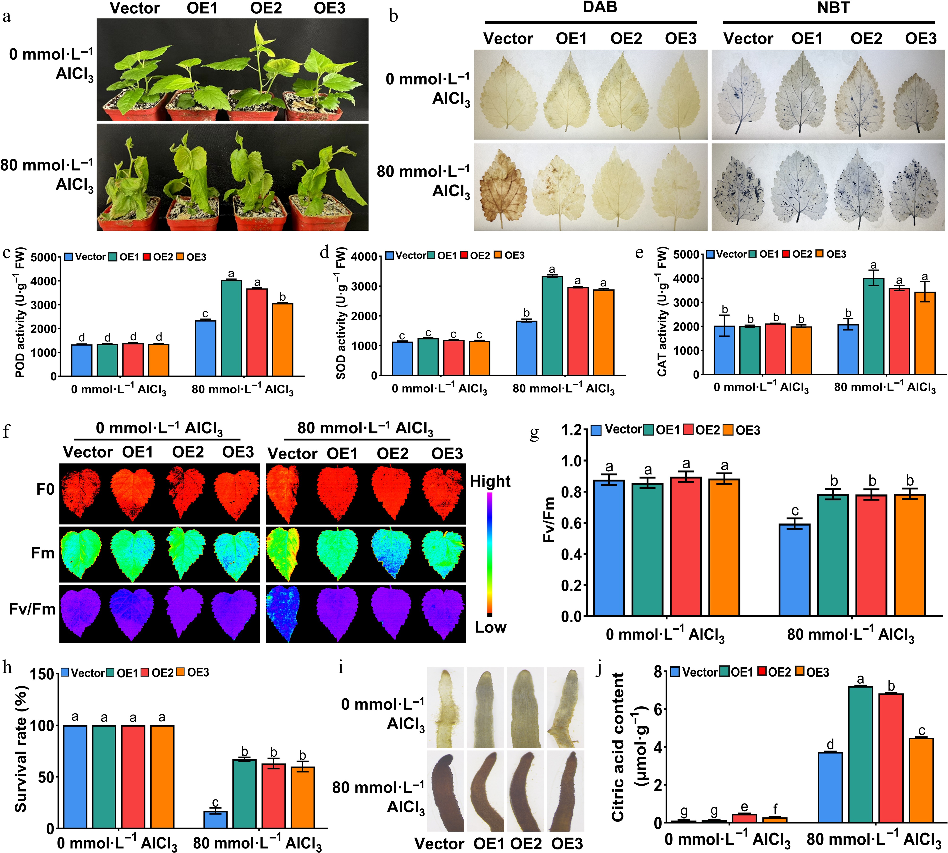
Figure 7.
Overexpression of MaDTX19 enhances Al3+ tolerance in hairy root-transformed mulberry seedlings. (a) Phenotypes of empty vector (Vector) control and MaDTX19-OE (OE1-3) mulberry seedlings under 80 mmol·L−1 AlCl3 stress. (b) DAB and NBT staining of mulberry leaves under AlCl3 stress. (c) POD, (d) SOD, and (e) CAT activities in leaves under AlCl3 stress. (f) Chlorophyll fluorescence images and (g) Fv/Fm values of seedlings under AlCl3 stress. (h) Survival rate of seedlings under AlCl3 stress. (i) Hematoxylin staining of hairy root tips. (j) Citrate content in hairy root tips under AlCl3 stress. Data represent mean ± SD. Different letters indicate significant differences (p < 0.05).
Al3+ stress induces MaDTX19 phosphorylation at Ser4
-
Bioinformatic analysis predicted that the fourth serine residue (Ser4) of MaDTX19 is a potential phosphorylation site. Using a phospho-specific antibody, we detected increased phosphorylation of MaDTX19 in OE Arabidopsis roots with prolonged Al3+ treatment (Fig. 8a), confirming stress-induced phosphorylation at Ser4. To further investigate the regulatory effect of Ser4 phosphorylation on MaDTX19 protein stability, we generated phosphodeficient (Ser4-to-Ala, SA) and phosphomimetic (Ser4-to-Asp, SD) mutants. Western blot analysis using a GFP antibody indicated that the abundance of MaDTX19-GFP and MaDTX19SD-GFP increased gradually under Al3+ stress, whereas MaDTX19SA-GFP showed no obvious changes (Fig. 8b). Thus, the Ser4 site of MaDTX19 serves as a phosphorylation modification site in response to Al3+ stress, with its phosphorylation status directly regulating MaDTX19 protein stability.
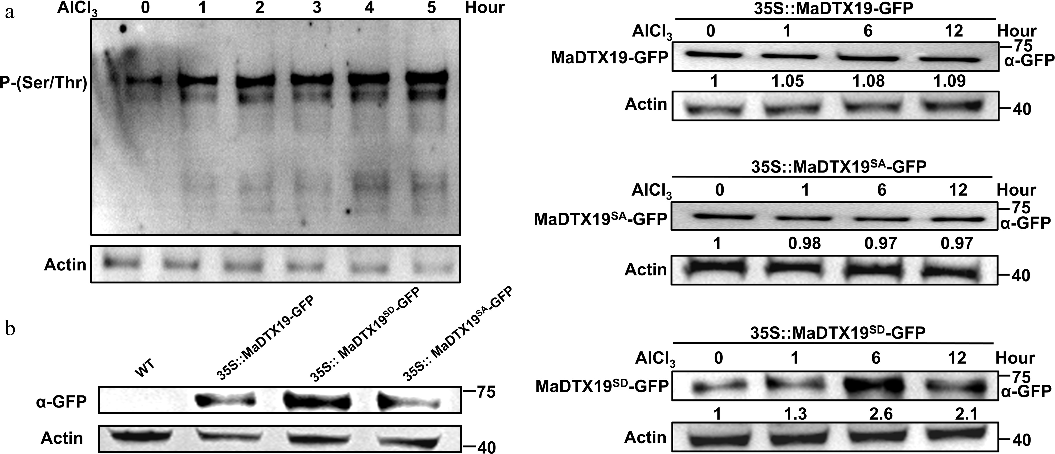
Figure 8.
Al3+ stress induces in vivo phosphorylation of MaDTX19 at Ser4 and modulates its protein stability. (a) Time-course analysis of Ser4 phosphorylation level of MaDTX19 in MaDTX19-OE Arabidopsis root tips under 0.8 mmol·L−1 AlCl3 stress, detected by western blot using an anti-phospho-(Ser/Thr) antibody. (b) Protein stability of MaDTX19-GFP, MaDTX19SA-GFP, and MaDTX19SD-GFP in Arabidopsis root tips under AlCl3 stress detected by western blot using an anti-GFP antibody.
Ser4 phosphorylation is required for both growth suppression and Al3+ tolerance
-
To explore whether Ser4 phosphorylation is functionally required, we transformed MaDTX19, MaDTX19SA, and MaDTX19SD constructs into Arabidopsis. Phenotypic analyses showed that both MaDTX19 and MaDTX19SD overexpressing plants exhibited growth inhibition compared to WT plants, characterized by reduced rosette leaf number and shorter siliques. In contrast, MaDTX19SA overexpressing plants showed no significant difference from WT (Fig. 9a, b). These results demonstrate that Ser4 phosphorylation is indispensable for the growth regulatory function of MaDTX19.
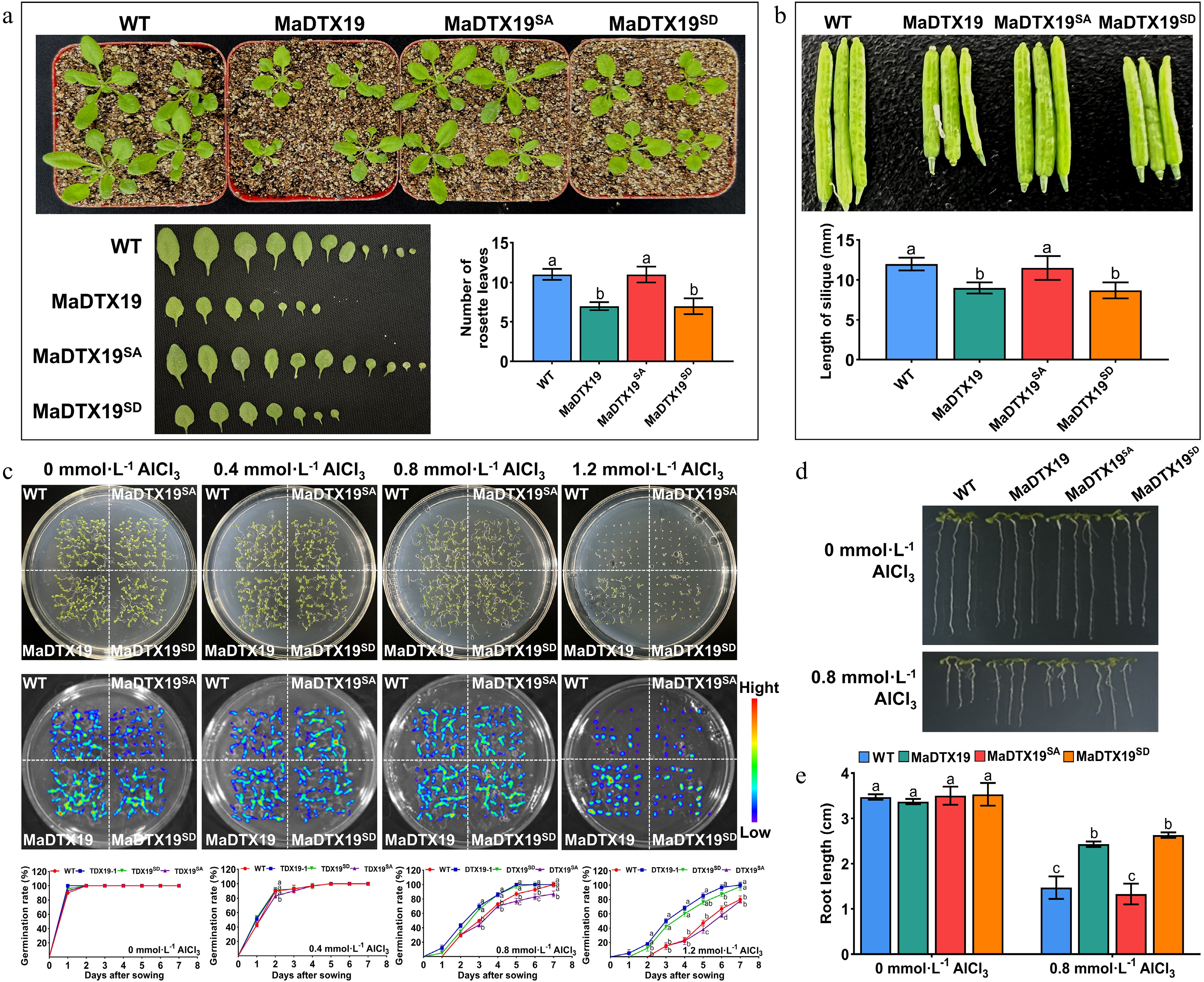
Figure 9.
Ser4 phosphorylation is essential for MaDTX19-mediated growth inhibition and Al3+ tolerance. (a) Growth phenotypes of four-week-old WT, MaDTX19-OE, MaDTX19SD-OE, and MaDTX19SA-OE Arabidopsis under normal conditions. (b) Silique length of the same plants under normal conditions. (c) Seed germination rate under 0, 0.4, 0.8, 1.2 mmol·L−1 AlCl3 stress. (d) Primary root phenotypes of seedlings grown on MS medium supplemented with 0 or 0.8 mmol·L−1 AlCl3. (e) Primary root length of seedlings on MS medium supplemented with 0 or 0.8 mmol·L−1 AlCl3. Different lowercase letters above the bars indicate statistically significant differences among treatments (p < 0.05).
In Al3+ tolerance assays, both MaDTX19 and MaDTX19SD overexpressing plants exhibited significantly enhanced Al3+ tolerance, with higher seed germination rates and longer primary roots compared to WT. In contrast, the MaDTX19SA overexpressing plants showed Al3+ sensitivity comparable to WT, with lower germination rates and shorter roots (Fig. 9c). In addition, the primary roots of MaDTX19 and MaDTX19SD overexpressing seedlings were shorter than WT plants under Al3+ stress (Fig. 9d, e). These results demonstrate that Ser4 phosphorylation is indispensable for both growth suppression and Al3+ tolerance.
Phosphorylation enhances MaDTX19 interaction with auxin-related proteins
-
To elucidate the mechanistic basis of the phosphorylation-dependent switch, we examined the MaDTX19 interaction network. Membrane yeast two-hybrid, BiFC, and LCA assays revealed that MaDTX19 interacts with three auxin-related proteins: the auxin-induced protein Ma5NG4, the auxin-responsive protein MaWAT1, and the auxin efflux carrier component MaAECC8 (Fig. 10a–c). Importantly, the phosphomimetic MaDTX19SD mutant exhibited markedly stronger interaction with Ma5NG4, MaWAT1, and MaAECC8. Conversely, the phosphodeficient MaDTX19SA mutant showed significantly weakened binding (Fig. 10d). Thus, Ser4 phosphorylation enhances MaDTX19 association with auxin transport components, likely disrupting auxin homeostasis and accounting for growth inhibition.
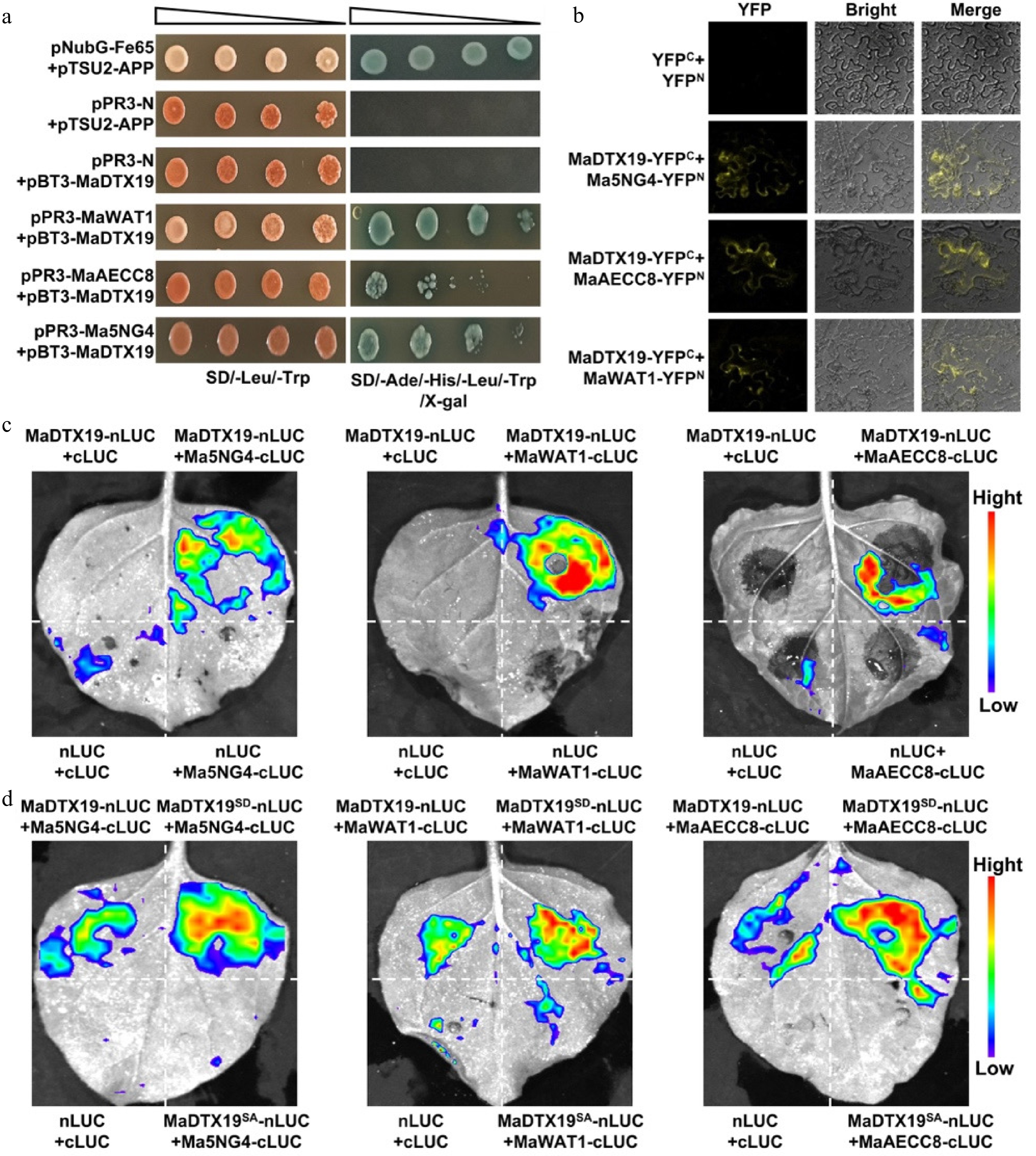
Figure 10.
Ser4 phosphorylation enhances the interaction of MaDTX19 with auxin-related proteins. (a) Membrane yeast two-hybrid assay showing the interaction of MaDTX19 with Ma5NG4, MaWAT1, and MaAECC8. Growth on QDO medium indicates a positive interaction. (b) Bimolecular fluorescence complementation (BiFC) assay confirming the interactions in N. benthamiana leaf epidermal cells. YFP fluorescence indicates protein-protein interaction. (c) Luciferase complementation assay (LCA) validating the interactions. (d) LCA comparing the interaction strength of MaDTX19 (WT), MaDTX19SD (phosphomimetic), and MaDTX19SA (phosphodeficient) with auxin-related proteins. Luminescence intensity reflects interaction strength.
MaDTX19 alters auxin homeostasis and spatial distribution in root tips
-
Using the auxin-responsive reporter DR5::GFP, we analyzed auxin distribution in root tips. Under Al3+-free conditions, weak GFP fluorescence signals were confined to the meristematic and elongation zones of WT root tips, where auxin levels remained basal. In MaDTX19-OE plants, GFP fluorescence was markedly stronger and extended beyond these growth zones into non-growing tissues, including the root epidermis and cortex. Under Al3+ stress, GFP fluorescence intensity increased and spread to non-growing regions in both WT and MaDTX19-OE root tips, with stronger enhancement and wider spreading in MaDTX19-OE roots than in WT (Fig. 11a). Quantification showed higher auxin content in MaDTX19-OE root tips under both conditions (Fig. 11b). These results indicate that MaDTX19 overexpression causes auxin redistribution, with increased accumulation in non-growing regions and altered auxin distribution in the meristematic zone. This likely depletes the auxin pool required for cell division and elongation, providing a mechanistic explanation for the observed dwarf phenotype.
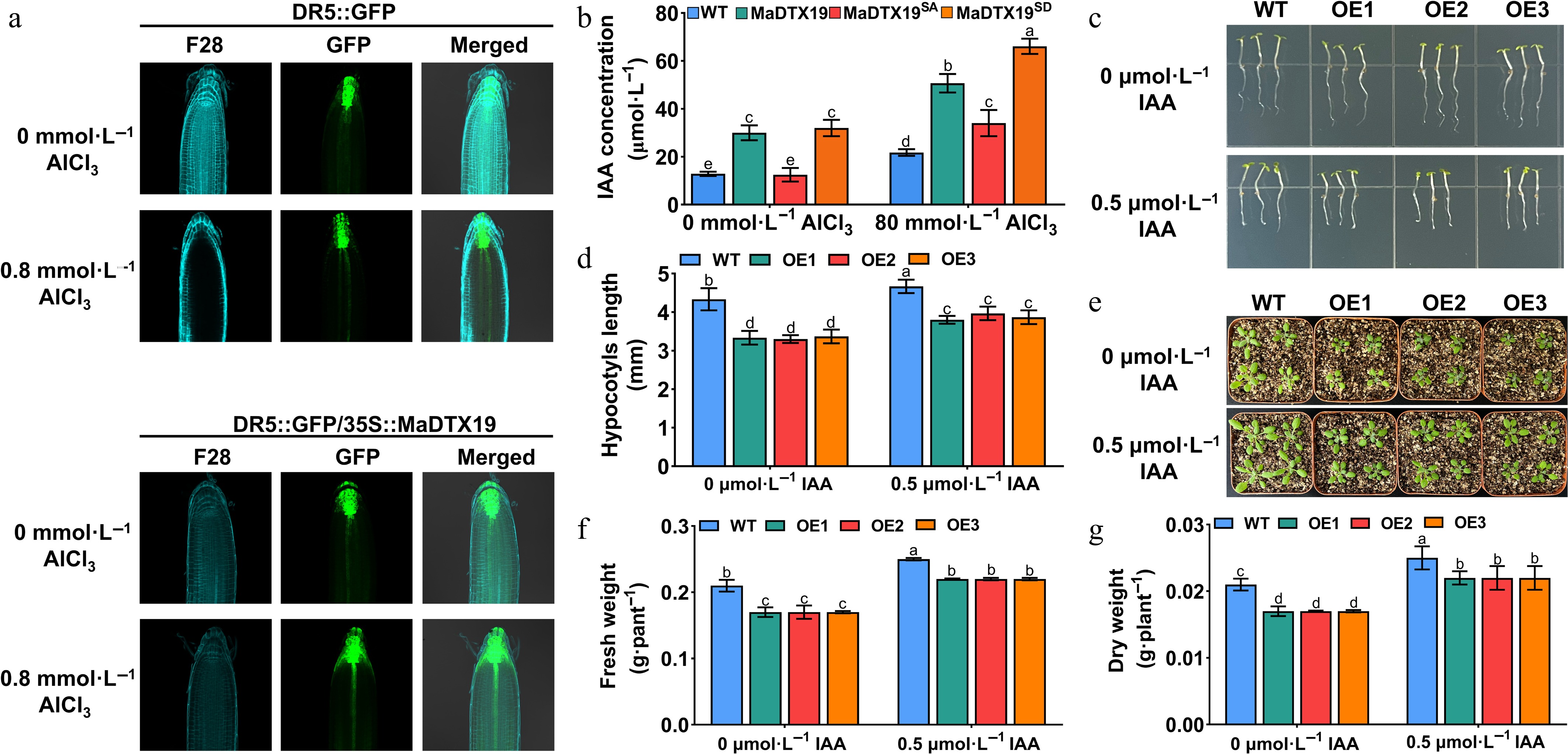
Figure 11.
MaDTX19 modulates auxin homeostasis and spatial distribution in root tips under Al3+ stress. (a) DR5::GFP fluorescence imaging of auxin distribution in root tips of WT and MaDTX19-OE Arabidopsis seedlings under 0 and 0.8 mmol·L−1 AlCl3 stress. (b) Quantitative analysis of auxin (IAA) content in root tips of WT and MaDTX19-OE seedlings under the same conditions. Effects of exogenous IAA treatment on growth phenotypes of WT and MaDTX19-OE seedlings: (c) hypocotyl phenotype, (d) hypocotyl length, (e) seedling phenotype, (f) fresh weight, and (g) dry weight under standard light conditions. Data are mean ± SD. Different letters indicate significant differences (p < 0.05).
To test whether auxin supplementation could rescue the growth defect, we applied exogenous IAA to WT and MaDTX19-OE seedlings under standard light conditions. IAA treatment slightly promoted growth in both genotypes, as reflected by modest increases in hypocotyl length, fresh weight, and dry weight. However, OE seedlings remained significantly smaller than WT controls across all measured parameters, with no obvious rescue of the dwarf morphology (Fig. 11c−g). This limited recovery is consistent with auxin mislocalization rather than simple auxin deficiency as the primary defect, as exogenous IAA would still be subject to the same disrupted polar transport machinery.
A MaMYB61/MaMYB308 transcriptional module fine-tunes MaDTX19 expression
-
Bioinformatic analysis predicted that MaMYB61 potentially binds to the MaDTX19 promoter (pMaDTX19). Y1H assays confirmed direct binding of MaMYB61 to pMaDTX19 (Fig. 12a), and LUC reporter assays demonstrated that MaMYB61 significantly enhanced pMaDTX19 activity (Fig. 12b, c), suggesting a positive regulatory role of MaMYB61 in MaDTX19 expression. EMSA further confirmed sequence-specific binding of MaMYB61 to the promoter (Fig. 12d).
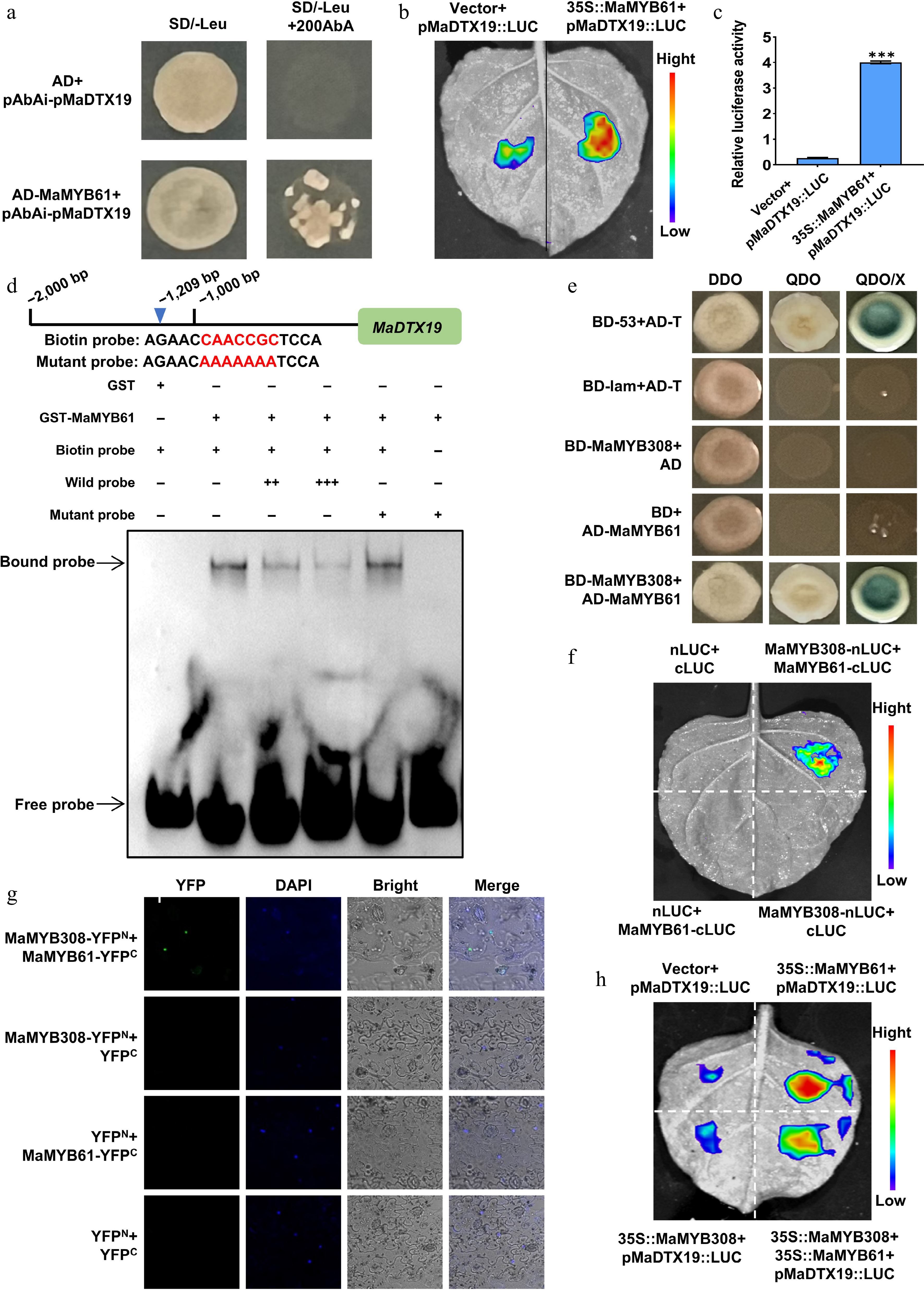
Figure 12.
The MaMYB61/MaMYB308 module fine-tunes MaDTX19 expression via transcriptional activation and repression. (a) Yeast one-hybrid (Y1H) assay confirming the binding of MaMYB61 to the MaDTX19 promoter (pMaDTX19). Growth on SD/-Leu/AbA medium indicates a positive interaction. (b) Dual-luciferase reporter assay showing that MaMYB61 activates pMaDTX19 in tobacco leaf epidermal cells. (c) Quantitative analysis of the LUC/REN ratio. (d) EMSA confirming sequence-specific binding of MaMYB61 to the MaDTX19 promoter. Labeled probes were incubated with recombinant MaMYB61 protein; unlabeled probes were used as competitors. (e) Y2H assay confirming the physical interaction between MaMYB61 and MaMYB308. (f) LCA confirming the physical interaction between MaMYB61 and MaMYB308. (g) BiFC assay localizing the MaMYB61-MaMYB308 interaction to the nucleus. DAPI was used as the nuclear marker. (h) Dual-luciferase reporter assay showing that MaMYB308 attenuates MaMYB61-mediated activation of pMaDTX19. All data are mean ± SD of three independent biological replicates. Asterisks indicate significant differences (t-test, *** p < 0.001).
Previous studies reported interactions between MYB61 and MYB308 in plants. Physical interaction between MaMYB61 and MaMYB308 was detected by Y2H assays (Fig. 12e) and further confirmed by LCA (Fig. 12f). BiFC analysis additionally showed that the two proteins interacted within the nucleus (Fig. 12g). Intriguingly, LUC assays showed that MaMYB308 attenuated MaMYB61-mediated transactivation of pMaDTX19 (Fig. 12h). These results define a MaMYB61-MaMYB308 transcriptional module in which MaMYB61 activates and MaMYB308 represses MaDTX19 expression, enabling dynamic fine-tuning in response to stress intensity.
-
Balancing growth and defense is a fundamental challenge for plants, especially long-lived perennials like mulberry that face persistent abiotic stresses, yet the molecular mechanisms governing this balance remain incompletely understood. While transcription factors and hormone signals are well-documented regulators of the growth-defense balance, relatively few studies have explored how transporters integrate stress signals and allocate resources through post-translational modifications[1,2]. Here, we show that MaDTX19 functions as a phosphorylation-dependent molecular hub that mediates resource reallocation from growth to defense under Al3+ stress. Two interconnected regulatory layers, Ser4 phosphorylation and transcriptional control by the MaMYB61-MaMYB308 module, coordinate this trade-off in a woody perennial.
Ser4 phosphorylation integrates growth suppression with Al3+ tolerance
-
The ability of a single phosphorylation event at Ser4 to govern both growth inhibition and Al3+ tolerance distinguishes MaDTX19 from previously characterized MATE transporters. Transcriptional upregulation of MATE genes such as SbMATE and HvAACT1 enhances citrate secretion and Al3+ exclusion, but these transporters do not simultaneously modulate plant growth[16,34,35,42]. MaDTX19 achieves dual functionality through phosphorylation-dependent rewiring of its protein interaction network. The phosphorylated form binds more strongly to auxin-related proteins (Ma5NG4, MaWAT1, MaAECC8), disrupting auxin transport and suppressing growth while concurrently activating Al3+ tolerance pathways. This phosphorylation-dependent switch enables coordinated suppression of growth and activation of detoxification.
The biological significance of this switch lies in its temporal advantage. Transcriptional reprogramming takes time, whereas reversible phosphorylation acts within minutes[12,29,30,43]. For plants experiencing acute Al3+ toxicity, where root elongation is inhibited within 30 min of stress onset, a phosphorylation-dependent switch provides an immediate mechanism for resource reallocation[23,27]. This rapid response is particularly relevant for woody perennials that cannot escape stress. The reversibility of phosphorylation also allows growth to resume quickly once stress subsides.
Recent studies have documented phosphorylation-dependent regulation of MATE transporters in other species, but these focus on modulation of transport activity rather than coordination of growth-defense trade-offs[12,18,23]. MaDTX19 therefore represents a distinct paradigm, wherein phosphorylation toggles between growth and defense modes through rewiring of protein-protein interactions. We acknowledge that the upstream kinase responsible for Ser4 phosphorylation remains unidentified. Given that ABA induces MaDTX19 expression, ABA signaling may participate in activating this kinase, although direct evidence is lacking. We also recognize that our auxin distribution data establish a strong correlation with growth inhibition, but we cannot exclude the possibility that some auxin changes are secondary consequences of broader physiological alterations.
Dual detoxification pathways and transcriptional fine-tuning
-
MaDTX19 confers Al3+ tolerance through two synergistic pathways, citrate secretion and flavonoid-mediated antioxidant defense. Citrate secreted into the rhizosphere chelates Al3+ and reduces its entry into root cells, while the flavonoid-antioxidant system scavenges residual ROS[6,16,17,21,23,31]. This outside-in strategy mirrors the detoxification logic observed in other Al3+-tolerant species. Integration of both pathways through a single phosphorylation-dependent transporter distinguishes MaDTX19 from other MATE family members. In contrast to MaDTX19, which consolidates citrate secretion, flavonoid accumulation, and growth modulation into one phosphorylation-gated protein, SbMATE functions primarily as a citrate transporter[44], AtDTX50 participates in ABA efflux[32], and BoMATE enhances Al3+ tolerance but imposes a growth penalty[31].
The MaMYB61-MaMYB308 transcriptional module adds an additional layer of regulation. MaMYB61 activates MaDTX19 transcription, while MaMYB308 attenuates this activation through protein-protein interaction. This activator-repressor configuration is conceptually analogous to the MYC2-MYB21/24 module in JA signaling. The temporal dynamics of this module are particularly informative. MaMYB61 is rapidly induced by Al3+, whereas MaMYB308 shows delayed induction, consistent with a negative feedback mechanism that curtails excessive MaDTX19 expression once the acute response is mounted (Supplementary Fig. S2). Compared to STOP1, which directly activates multiple MATE genes in Arabidopsis and other species, the MYB61-MYB308 module represents a distinct regulatory architecture employing an activator-repressor pair for dynamic expression tuning. Our Y1H, LUC, and EMSA assays confirm that MaMYB61 directly binds and activates the MaDTX19 promoter. We acknowledge that increased citrate and flavonoid levels, while consistent with a role for MaDTX19 in their transport, do not constitute direct biochemical evidence of transport activity. Direct transport assays are needed to establish substrate specificity.
A two-tier strategy for environmental plasticity and the adaptive trade-off
-
Based on these findings, we propose a working model for MaDTX19-mediated regulation of the growth-defense continuum in mulberry (Fig. 13). Under optimal conditions, low MaDTX19 expression and basal Ser4 phosphorylation maintain normal auxin transport and growth. Upon Al3+ stress, the MaMYB61-MaMYB308 module rapidly induces MaDTX19 transcription, while a yet-unidentified kinase phosphorylates Ser4, switching MaDTX19 to its active conformation. Phosphorylated MaDTX19 then exerts dual functions, inhibiting growth by interacting with auxin transport components and activating Al3+ tolerance through citrate secretion and flavonoid-mediated antioxidant defense. The MaMYB308-mediated negative feedback prevents excessive MaDTX19 expression, minimizing sustained fitness costs. This two-tier strategy, with rapid phosphorylation for immediate response and transcriptional modulation for long-term optimization, enables mulberry to balance short-term survival with long-term growth. Quantitative analysis of transgenic lines with graded MaDTX19 expression supports this model. Lines with greater growth suppression showed higher survival rates and lower oxidative damage under Al3+ stress. Notably, this relationship plateaued beyond a certain expression threshold, suggesting an optimal MaDTX19 activity level that maximizes tolerance while minimizing fitness costs.
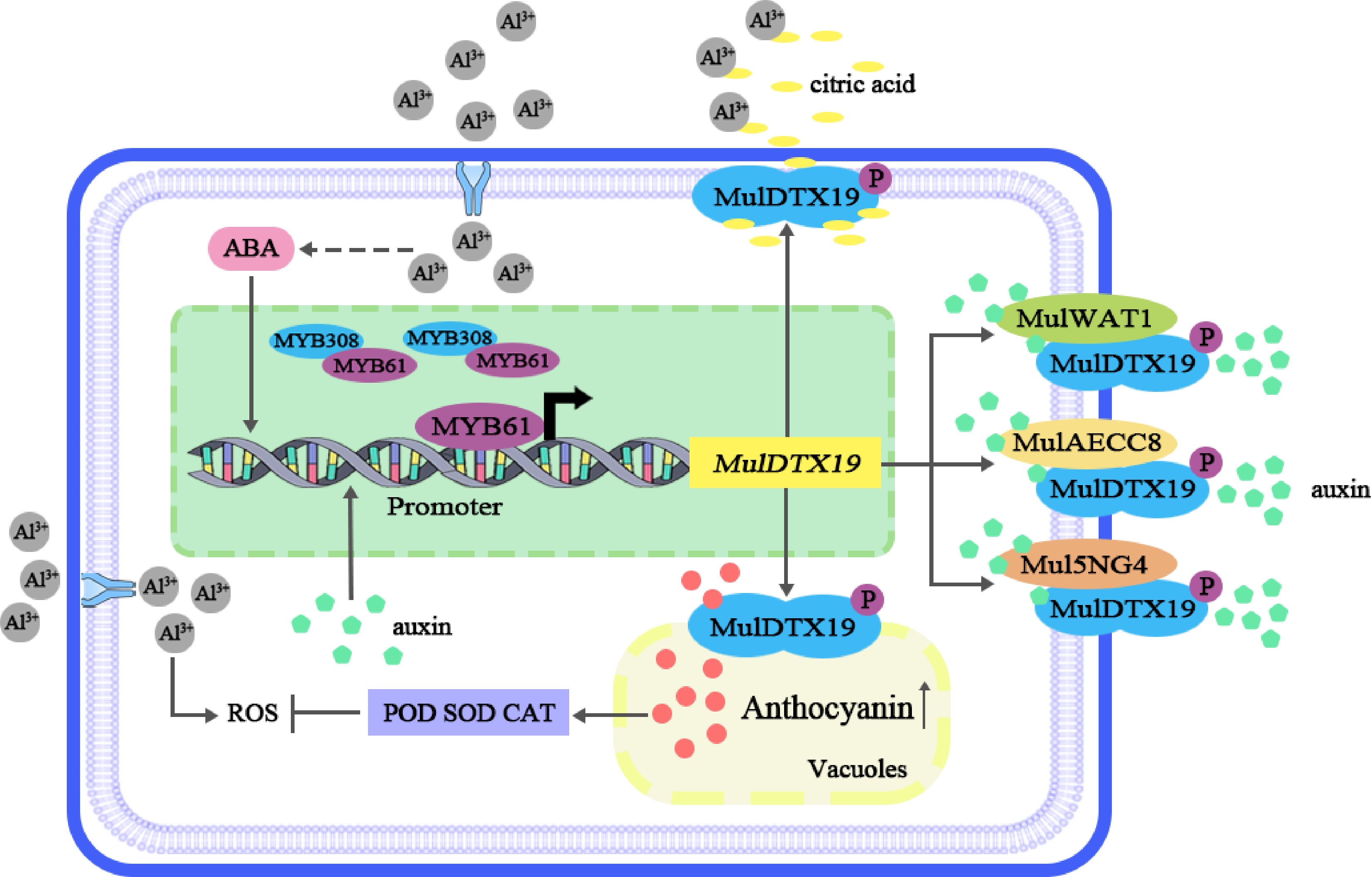
Figure 13.
Hierarchical regulatory model of MaDTX19-mediated growth-defense trade-off in mulberry under Al3+ stress. Under optimal conditions, low MaDTX19 expression and basal Ser4 phosphorylation maintain normal auxin transport and growth. Upon Al3+ stress, the MaMYB61-MaMYB308 module induces MaDTX19 transcription, while a yet-unidentified kinase phosphorylates Ser4, switching MaDTX19 to its active conformation. Phosphorylated MaDTX19 then exerts dual functions: inhibiting growth by interacting with auxin transport components (MaAECC8, Ma5NG4, MaWAT1) and activating Al3+ tolerance through citrate secretion and flavonoid-mediated antioxidant defense. The MaMYB308-mediated negative feedback prevents excessive MaDTX19 expression, minimizing sustained fitness costs.
Nevertheless, we acknowledge that our observations across transgenic lines with graded expression levels, while supportive of the adaptive trade-off interpretation, do not constitute definitive proof. Separation-of-function mutants that uncouple Al3+ tolerance from growth suppression would provide the strongest genetic evidence for the adaptive nature of this trade-off. Without such evidence, alternative explanations cannot be formally excluded.
-
Ser4 phosphorylation of MaDTX19 serves as a core regulatory node that synchronously regulates growth inhibition and Al3+ tolerance in mulberry. Phosphorylated MaDTX19 modulates auxin homeostasis to restrain plant growth and enhances citrate exudation and antioxidant capacity to confer Al3+ tolerance. The MaMYB61-MaMYB308 transcriptional module fine-tunes MaDTX19 expression to optimize fitness. These findings reveal a post-translational regulatory mechanism of tree adaptation to Al3+ stress, and provide promising genetic targets for molecular breeding of Al3+-tolerant mulberry cultivars.
-
The authors confirm contributions to the paper as follows: study conception and design: Ji X, Gai Y; methodology: Han S, Zhao T, Liu C; validation: Sun M, Han SM; formal analysis: Liu C, Zhao J; investigation: Han S, Zhao T, Zhao J, Wang Y; visualization: Liu Z; draft manuscript preparation: Han S, Zhao T; writing – review & editing: Ji X, Gai Y; supervision: Ji X, Gai Y; project administration: Ji X; funding acquisition: Ji X, Gai Y. All authors reviewed the results and approved the final version of the manuscript.
-
All data generated or analyzed during this study are included in this published article and its supplementary information files.
-
The authors declare that they have no conflict of interest.
-
accompanies this paper online at: https://doi.org/10.48130/forres-0026-0031.
-
# Authors contributed equally: Shengmei Han, Teng Zhao
- Supplementary Fig. S1 Expression pattern of MaMATE in response to Al3+ stress.
- Supplementary Fig. S2 Dynamic expression patterns of MaMYB61 and MaMYB308 under Al3+ stress.
- Copyright: © 2026 by the author(s). Published by Maximum Academic Press, Fayetteville, GA. This article is an open access article distributed under Creative Commons Attribution License (CC BY 4.0), visit https://creativecommons.org/licenses/by/4.0/.
-
About this article
Cite this article
Han S, Zhao T, Liu C, Sun M, Zhao J, et al. 2026. MaDTX19 phosphorylation governs growth-defense trade-off under aluminum stress in mulberry. Forestry Research 6: e032 doi: 10.48130/forres-0026-0031 

MaDTX19 phosphorylation governs growth-defense trade-off under aluminum stress in mulberry
- Received: 13 May 2026
- Revised: 03 September 2026
- Accepted: 09 October 2026
- Published online: 28 October 2026
Abstract: The growth-defense trade-off is essential for plant adaptation to abiotic stress, yet the post-translational mechanisms underlying its dynamic regulation remain unclear. Here, we show that phosphorylation of the MATE transporter MaDTX19 at serine 4 (Ser4) acts as a critical regulatory node governing this trade-off in mulberry under Al3+ stress. MaDTX19 expression was strongly upregulated by Al3+ stress, and its encoded protein underwent concurrent phosphorylation at Ser4. The phosphomimetic MaDTX19SD mutant, but not the phosphodeficient MaDTX19SA mutant, exhibited both growth inhibition and Al3+ tolerance, indicating that Ser4 phosphorylation is essential for these functions. Mechanistically, Ser4 phosphorylation enhanced the interaction of MaDTX19 with auxin-related proteins (Ma5NG4, MaWAT1, and MaAECC8), disrupting auxin homeostasis and suppressing growth. Concurrently, phosphorylated MaDTX19 promoted root citrate secretion and flavonoid accumulation while enhancing antioxidant enzyme activities, thereby conferring Al3+ tolerance through coordinated extracellular chelation and intracellular ROS scavenging. We further identified a MaMYB61-MaMYB308 transcriptional module in which MaMYB61 activates MaDTX19 transcription through direct promoter binding, whereas MaMYB308 attenuates this activation by protein-protein interaction, allowing dynamic fine-tuning of MaDTX19 expression to limit excessive fitness costs. Our study uncovers a phosphorylation-dependent mechanism by which a single MATE transporter coordinates growth-defense balance in a woody perennial, providing a genetic target for breeding Al3+-tolerant mulberry cultivars on acidic marginal lands.
-
Key words:
- Mulberry /
- MaDTX19 /
- Aluminum stress /
- Phosphorylation /
- Growth-defense trade-off