












-
Feeding is the foundation of all life activities and the only way for the body to obtain energy and nutrition. The disorder in food intake induces energy imbalance, which leads to fish maldevelopment or excessive obesity, thereby damaging the health[1]. Over the past few decades, numerous central and peripheral appetite-regulating factors such as proopiomelanocortin (POMC)[2], neuropeptide Y (NPY)[3], agouti-related peptide (AGRP)[4], leptin[5], motilin[6] and ghrelin[7], have been shown to play important roles in the regulation of feeding. The anorexic role of Neuromedin U (NMU) in mammals and birds has attracted increasing attention[8−10]. In fish, our recent research on Siberian sturgeon (Acipenser baerii Brandt) has found that NMU exerts an inhibitory effect on food intake regulation[11]. However, the interaction between NMU and nmurs remains complex, so further research is significant to elucidate the interaction between NMU and its receptors in the context of appetite regulation.
Like other neuropeptides, NMU-related biological effects are triggered by mobilization of G protein-coupled receptors (GPCRs) upon membrane binding[12]. Before 2000, NMU was considered an orphan ligand[13]. In 2000, researchers used reverse pharmacology to confirm that human NMU is a ligand for the GPCR (FM-3 or GPR66) and discovered that the FM-3 amino acid sequence shares high homology with the growth hormone secretagogue receptor (GHSR) and neurotensin receptor. They also found FM-3 could not be activated by several peptides similar to the NMU sequence, such as neurotransmitters B, C, K, and N, motilin, vasoactive intestinal polypeptide, pancreatic polypeptide, neurotensin, as well as Ghrelin[14]. This established FM-3 as a specific receptor of NMU, later named NMUR1[15]. Simultaneously, NMUR2 (FM-4) was identified as the second specific receptor of NMU in humans and rats[16,17]. NMUR1 (FM-3) and NMUR2 (FM-4) are two receptors of NMU belonging to G protein-coupled receptors (GPCR)[18], which have been widely identified in mammals and birds, including mice[19], rat[14], rabbit[20], pig[21], and chicken[22]. The sequences of nmur1 and nmur2 have been identified in many fishes, but so far, those receptors have been functionally characterized only in goldfish[23] and zebrafish[24]. In mammals, nmur1 is mainly expressed in the peripheral tissues, while nmur2 is primarily found in the central nervous system[20,25,26]. However, in goldfish[27], nmur1 and nmur2 are highly expressed in the brain and pituitary, suggesting that the mechanisms of NMU in central and peripheral regulation of feeding in fish may differ significantly from those in mammals.
In higher vertebrates, it is well known that the inhibitory effect of NMU on food intake is closely related to nmurs[12,28]. Interestingly, mammalian NMUR1 primarily mediates the anorexic effects of NMU in peripheral tissues, whereas in the central nervous system, this effect is achieved through the activation of NMUR2[29,30]. Peripheral acute or chronic administration of NMU leads to a dose-dependent reduction in food intake and body weight, an effect that is reversed in NMUR1 knockout mice[29]. Additionally, NMU induces contraction of the stomach, ileum, and colon in dogs via NMUR1, indicating that peripheral NMU may regulate appetite by promoting gastrointestinal peristalsis through NMUR1[31]. However, research on NMU-NMUR interactions primarily focuses on NMUR2. NMUR2 is predominantly expressed in several reward-related tissues of the rodent brain, suggesting its role in reward regulation[30]. Peripheral injection of NMU increases nmur2 expression in the paraventricular nucleus (PVN) and dorsal raphe nucleus (DRN)[28]. I.c.v. injection of NMU reduces feeding, weight, spontaneous activity, and body temperature of wild-type mice, effects that are absent in nmur2-deficient mice[32]. Similarly, selective RNAi knockdown of nmur2 in the PVN of the hypothalamus in rats significantly increases the consumption and preference for high-fat diets, and promotes weight gain[32]. Long-term central injection of NMU significantly increases the body weight and fat content in NMUR2 knockout female mice under high-fat conditions[33]. Furthermore, subcutaneous injection of NMUR2-specific agonist results in elevated NMUR2 levels in the brain and significantly reduces high-fat diet intake in rats, leading to notable weight loss[34−38]. These studies confirm that the anorexic and weight-reducing effects of NMU in the central nervous system and peripheral tissues of mammals are mediated through the differential activation of NMURs. However, in fish, the mechanism by which NMU suppresses appetite via nmurs remains unknown. Our study lays a molecular basis for further investigating the physiological roles of NMU in fish and helps to reveal the conserved actions of NMU-NMUR signaling across vertebrates.
The Siberian sturgeon is a widely cultured species in the world. Exploring the feeding regulation system of fish is of great significance for sturgeon farming. To date, there is no study available on how fish NMURs respond to appetite signals. Therefore, through this study, we explored the tissue-specific distribution of nmurs in the Siberian sturgeon, how nmurs respond to the changes in nutritional status of the fish, and whether nmur1 or nmur2 regulates feeding behavior through different mechanisms in the central and peripheral tissues. Therefore, in this study, we sequenced nmur1 and nmur2 and investigated their tissue-specific distribution in Siberian sturgeon. We further explored how nmurs respond to changes in nutritional status in fish and whether nmur1 and nmur2 regulate feeding behaviour through distinct mechanisms in central and peripheral tissues. This study laid a foundation for explaining the molecular mechanism of NMU regulating the physiological function of Siberian sturgeon and enriching the understanding of the appetite regulatory network in fish.
-
Healthy Siberian sturgeons (107.02 ± 13.02 g) were purchased from Runzhao Fisheries Co. Ltd (Sichuan, China). Fish were acclimated in the Aquaculture Laboratory of Sichuan Agricultural University (Chengdu, China) indoor tanks (60 × 50 × 40 cm), which were supplied with a continuous flow of freshwater at 19 ± 1 °C. The rearing systems were maintained under a natural light period (12 h light:12 h dark). Before the experiment, all fish were cultured with constantly aerated fresh water for at least two weeks and fed commercial sinking pellets (nutritional composition: crude protein ≥ 44%, crude fat ≥ 6%, crude ash ≤ 16%, moisture ≤ 12%, and Lys ≥ 1.8%, Tongwei, China) once a day at 14:00, at a rate of 3% of body weight (BW). In the experiment, all fish were anesthetized with 0.02% tricaine methanesulfonate (MS-222) before sampling, i.p. and i.c.v. injection. All experiments were conducted in accordance with the Animal Care and Use Committee of Sichuan Agricultural University's Animal Experiment Guidelines, with license number DKY-S20150812.
Cloning and bioinformatics analysis of nmur1 and nmur2
-
We collected the whole brain from six Siberian sturgeons for nmur1 and nmur2 cloning. Total RNA was extracted using the RNA iso Plus reagent (Takara, China). The quality and quantity of RNA were determined using electrophoresis and NanoDrop 2000c (Bio-Rad, USA). The high-quality RNA (A260/A280 ratio between 1.8 and 2.0) was used for synthesizing cDNA with the PrimeScript™ RT Reagent Kit (Takara, China). The thermal cycling program included an initial denaturation at 95 °C for 5 min, followed by 34 cycles of 94 °C for 30 s (denaturation), 54.7 °C for 60 s (annealing), and 72 °C for 1 min (extension), with a final extension at 72 °C for 7 min and a hold at 4 °C. The products were electrophoresed in a 1.5% agarose gel, and DNA bands were purified using Universal DNA Purification (TIANGEN, China). Purified PCR products were ligated into the pMD-19T vector (Takara, China) and transformed into competent cells, E. coli DH5α (Takara, China). Sequencing was performed at Beijing Genomics Institute (BGI, China). Cloning primers used for nmur1 and nmur2 cDNA amplification were designed using Primer Premier 5.0 software and listed in Supplementary Table S1.
The nmur1 and nmur2 nucleotide sequences were analyzed using BLAST (
https://blast.ncbi.nlm.nih.gov/Blast.cgi ). We used DNASTAR 7.1.0 software and SignalP 5.0 Server (https://services.healthtech.dtu.dk/service.php?SignalP-5.0 ) to predict the nmur1 and nmur2 ORF and the cleavage site of the signal peptide, respectively. Moreover, multiple alignments of amino acid sequences and phylogenetic analysis were performed by ClustalW (www.ddbj.nig.ac.jp/services/clustalw.html ). The phylogenetic tree was constructed by MEGA 11 software, and the reliability was assessed by 1,000 bootstrap replicates. Domain analysis was performed using the TBtools software (v2.042). Finally, the protein tertiary structures were predicted using SWISS-MODEL (https://swissmodel.expasy.org/interactive ) and visualized using PyMOL 3.0 software.Tissue distribution pattern of nmur1 and nmur2
-
To analyze the tissue distribution pattern of nmur1 and nmur2, 22 different tissue samples were collected from Siberian sturgeon individually (n = 6) after fasting for 24 h. The tissue samples included telencephalon, hypothalamus, mesencephalon, cerebellum, medulla oblongata, esophagus, stomach, pyloric caeca, spiral valve intestine, duodenum, rectum, heart, liver, pancreas, spleen, trunk kidney, undifferentiated gonad, swim bladder, eye, gill, muscle, and skin. All tissues were washed with sterile ice-cold saline (0.68% NaCl solution), snap-frozen in liquid nitrogen, and stored at −80 °C until total RNA isolation.
Pre-prandial, fasting, and re-feeding experiments
-
Siberian sturgeons (n = 84) were randomly assigned to seven groups with three tanks per group (four fish per tank) to determine the pre-prandial changes in nmur1 and nmur2 mRNA expression. During the experiment, two groups were fed according to a normal feeding procedure that served as the control. The other five groups were sampled either before, during, or after the scheduled feeding time (14:00) on the same day. Out of five fasting groups, two groups were sampled at 3 h (–3 h, 11:00) and 1 h (–1 h, 13:00) before the feeding, while the other three groups were sampled at 0 h, 14:00, 1 h after feeding time point (+1 h, 15:00), and 3 h after feeding time point (+3 h, 17:00), respectively. In addition, the other two fed groups were sampled 1 and 3 h (+1 h and +3 h) after the feeding time point. Three individuals were randomly selected from each tank, and hypothalamus, spiral valve intestine, and duodenum were collected after euthanizing the fish. Then the tissue samples were snap-frozen in liquid nitrogen and stored at -80 °C until total RNA isolation (Supplementary Fig. S1).
Siberian sturgeons (n = 180) were randomly dispersed in 15 groups with three tanks per group (four fish per tank) to determine the changes in nmur1 and nmur2 mRNA expression induced by long-term-fasting and re-feeding regime. In the experiment, six unfed groups were sampled on different fasting days 1, 3, 6, 10, 15, and 17, respectively. Simultaneously, six fed groups were sampled on days 1, 3, 6, 10, 15, and 17, which served as the control for the respective days. The remaining three groups were refed daily between days 10 and 17 of fasting and sampled on days 10, 15, and 17 (refed for 1, 5, and 7 d). Three individuals were randomly selected from each tank. The hypothalamus and liver were sampled 1 h after the feeding point. Sample collection and preservation were described above (Supplementary Fig. S1).
Long-term i.p. and i.c.v. injections of NMU-25
Peptide
-
Siberian sturgeon NMU mature peptide—NMU-25 (FRDDDDLQGPGGIQSR-GYFLFR-PRN)—was synthesized with a purity of 98% and a molecular weight of 2,927.19 Da by Shanghai Bootech BioScience & Technology Co., Ltd. The peptide was dissolved in saline (0.68% NaCl solution) and stored at −20 °C for long-term i.p. and i.c.v. injection experiments. The dosage of NMU-25 is referenced from our previous research in Siberian sturgeon[11].
Effects of long-term i.p. and i.c.v. of NMU-25 on the expression of nmur1 and nmur2
-
Previous studies have shown that NMU-25 is a functional peptide segment of pre-NMU, which plays a role in inhibiting appetite[11]. Therefore, we injected NMU-25 to explore the response of nmurs in Siberian sturgeon. The investigations were performed with long-term i.p. and i.c.v. injections of NMU-25. Siberian sturgeons (n = 48) were randomly divided into four groups with three tanks per group (four fish per tank). The first group received long-term i.p. injection with NMU-25, and the second group was injected with saline for 7 d as a control. Similarly, the third group received a single i.c.v. injection with NMU-25, while the last group was injected with saline as a control. Before the experiment, fish were anesthetized with MS-222 (0.02%). Initially, fish were weighed and were i.p. injected with saline or 100 ng/g BW NMU-25 solution for 7 d and i.c.v. injected with saline or 10 pmol/g BW NMU-25 solution for 1 d at 13:30 (The NMU-25 dose of i.p. and i.c.v. injection experiments were based on our previous research[11]). The i.p. injection site is located at the posterior margin of the base of the abdominal fin, with an injection depth of approximately 1 cm. The i.c.v. was injected by a 10 μL glass syringe with a needle of 0.5 mm outside diameter within 1 min. The injection site and method were based on the study by Defang Chen et al.[39]. Then, fish were fed the pre-weighed sinking pellet diet at an excess of 3% body weight ratio at 14:00 (0 h). At 15:00 (1 h after feeding), the uneaten feed was collected and weighed after drying to determine the food intake. Simultaneously, the hypothalamus and spiral valve intestine of the i.c.v. injection group were sampled for subsequent experiments. For the long-term i.p. injection group, fish were injected for seven consecutive days as described above, and the hypothalamus and spiral valve intestine were sampled on the seventh day at 15:00 (1 h after feeding) (Supplementary Fig. S2). The uneaten feed was collected at 15:00 (i.e., 1 h after feeding) every day to determine food intake (Supplementary Fig. S2).
Real-time qPCR analysis
-
The expression of Siberian sturgeon nmur1 and nmur2 mRNA was detected by real-time quantitative PCR (qPCR) with SYBR Premix Ex TapTMII (TaKaRa, Japan). We used β-actin and gapdh as housekeeping genes to normalize the expression levels. For all standard curves, the primer amplification efficiencies of the genes were 90%–110% and 0.99 < R2 < 1.000, respectively. The qPCR cycle conditions were as follows: 95 °C denaturation for 5 min; 95 °C denaturation for 30 s, Tm annealing for 1 min, and 72 °C extension for 1 min, repeated 39 cycles; 72 °C extension for 5 min, and then 4 °C cooling. The target genes were normalized to the housekeeping genes, and the expression levels were calculated using the 2−ΔΔCᴛ method. The qPCR primers used are shown in Supplementary Table S1.
Statistical analyses
-
All data are expressed as mean ± standard error of the mean (SEM). Statistical analyses were performed using SPSS (v27.0) statistical software (SPSS Inc., Chicago, IL, USA). Student's t-tests were used for the comparison between the two groups. For multiple group designs, we first conducted homogeneity of variance and normality analysis using the Levene test and Shapiro-Wilk test. We performed one-way ANOVA analysis with Tukey's multiple comparisons test. All reported p-values are based on two-sided tests, and p < 0.05 was considered to be statistically significant.
-
In this experiment, we cloned the nmurs CDS sequences of Siberian sturgeon, including 1,287 bp of nmur1 (Fig. 1a) and 1,257 bp of nmur2 (Fig. 1b), encoding 428 and 418 amino acids, respectively. We found that neither NMUR1 nor NMUR2 has a signal peptide (Fig. 1c, d), while both receptors are GPCRs, formerly orphan receptors, with seven transmembrane domains (TMDs) (Fig. 1e, f). Further analysis showed that the TMDs of nmurs were all composed of the same three motifs, whose sites and sizes were relatively conserved in different species. Except for fish, NMUR2 of other species contains a C-terminal superfamily of unknown function, which may be necessary for its activity (Fig. 1g). Amino acid sequence alignment revealed that NMUR1 is more conserved among species than NMUR2. The identity of NMUR1 between Siberian sturgeon and other species is as follows: human (50.87%), chicken (60.22%), Komodo dragon (60.22%), Bufonid (62.61%), and Goldfish (64.57%). The identity of NMUR2 between Siberian sturgeon and other species is as follows: human (40.77%), chicken (39.64%), Komodo dragon (42.37%), Bufonid (41.00%), and Goldfish (58.31%). The amino acid sequences of Siberian sturgeon NMUR1 and NMUR2 have the highest identity with those of Acipenser ruthenus, with values of 91.74% and 95.22%, respectively. Meanwhile, NMURs of different species had the highest conservation within the TMD, while there is little homology between the N and C termini. The ERY motif and the two cysteine residues used to form disulfide bonds are also highly conserved in all species (Fig. 2a, b). By constructing a phylogenetic tree, it was found that NMUR1 and NMUR2 of Siberian sturgeon clustered into a clade with other fish and Acipenser ruthenus, respectively (Fig. 2c).
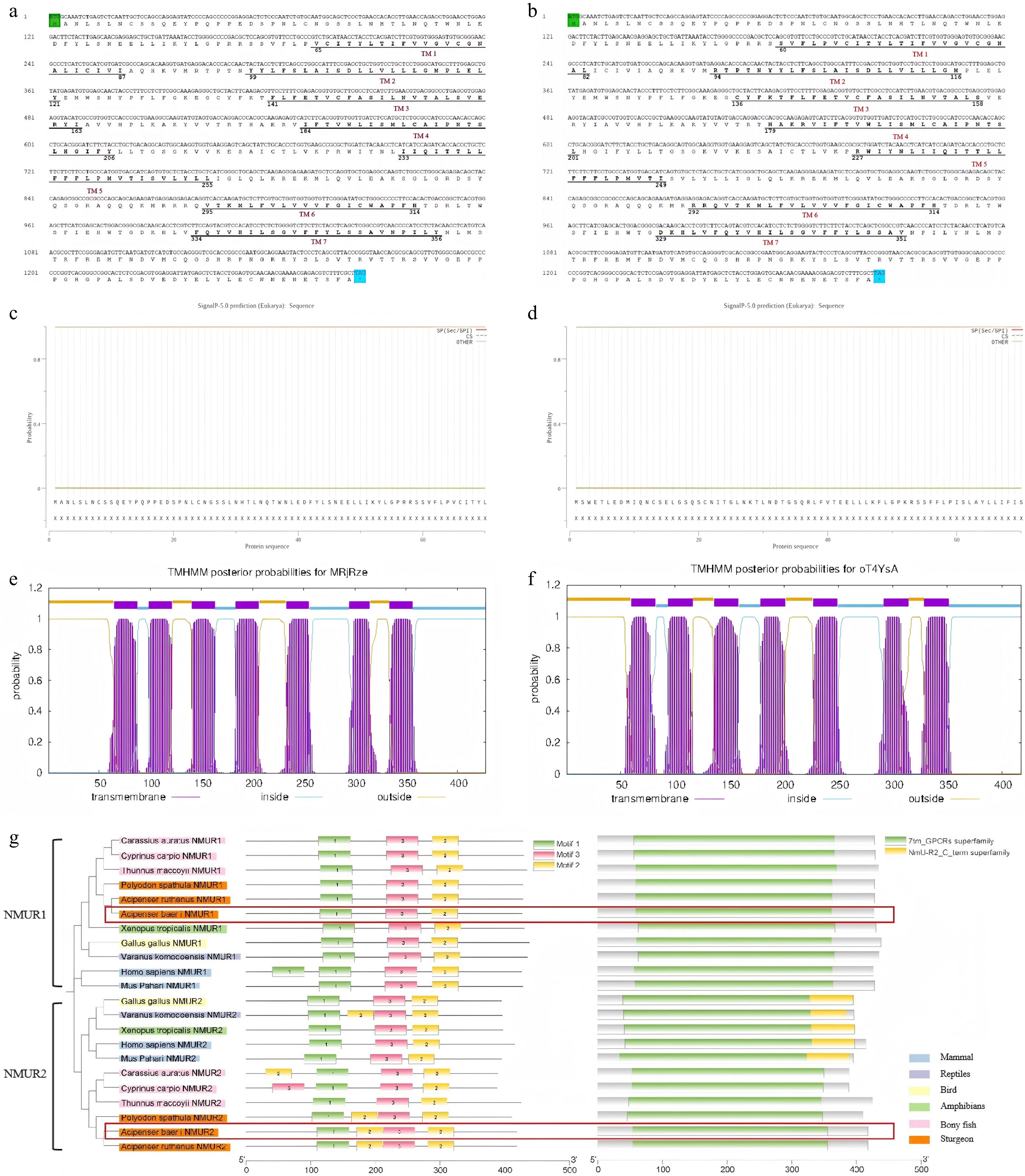
Figure 1.
Sequence analysis of nmr1 and nmur2 nucleotide sequences of Siberian sturgeon. Comparative nucleotide-amino acid map of (a) nmur1 and (b) nmur2. Signal peptide prediction of (c) NMUR1 and (d) NMUR2. Transmembrane domain (TMD) prediction of (e) NMUR1 and (f) NMUR2. (g) TMD analysis of NMUR1 and NMUR2 in Siberian sturgeon. The green shaded region and the blue shaded region indicate the initiator codon and the termination codon, respectively, while the putative seven transmembrane domains (TM1–TM7) are indicated by horizontal bars. Figures represent the start and end sites of the TMDs.
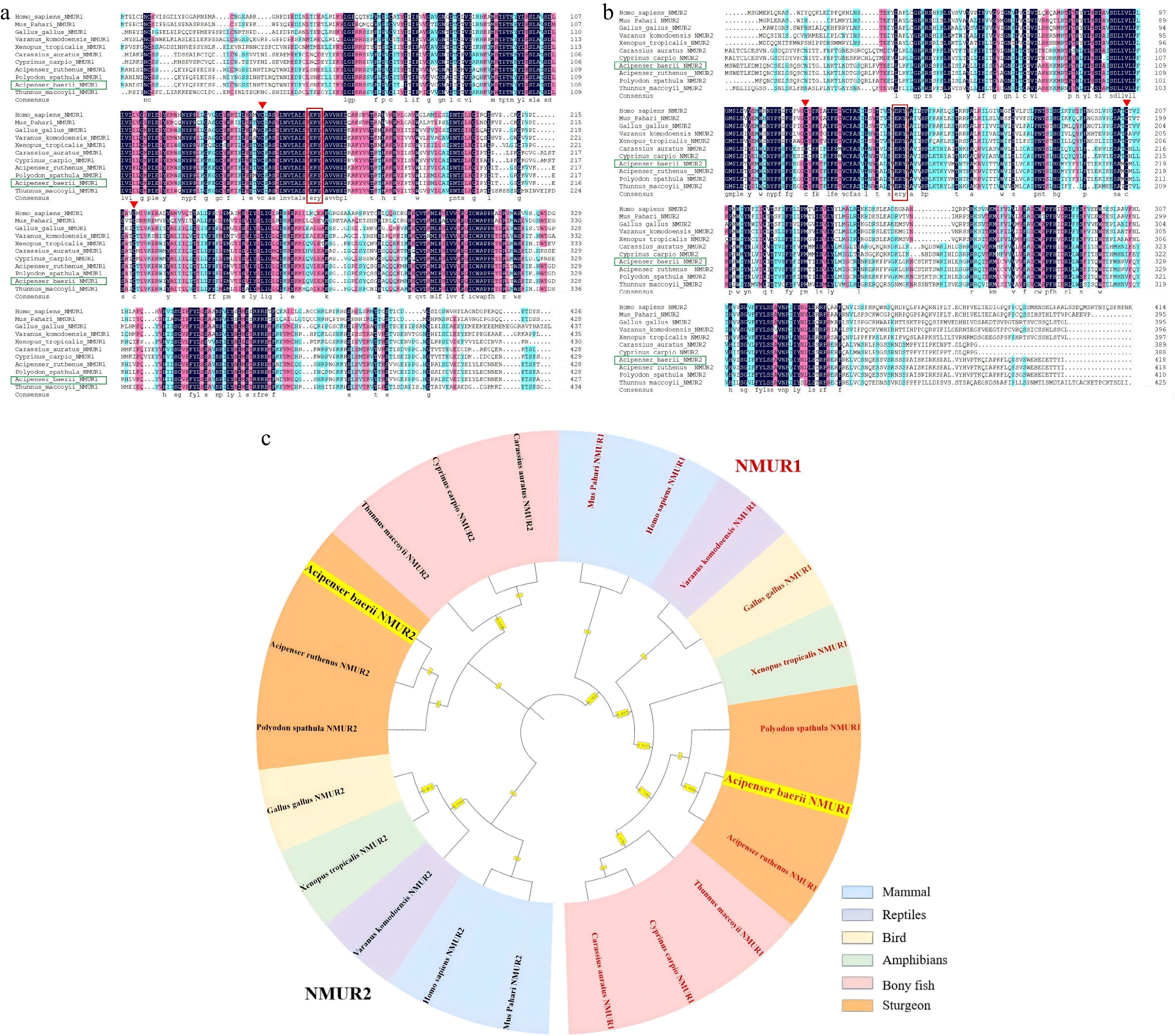
Figure 2.
Differences of NMUR1 and NMUR2 among different species. Amino acid sequence alignment of Siberian sturgeon (a) NMUR1 and (b) NMUR2 with other vertebrates. The ERY motifs are indicated in red boxes. Conserved cysteine residues are indicated by triangles. Identity: black (100%), red (75%), blue (50%). (c) Phylogenetic analysis of NMUR1 and NMUR2 in vertebrates. The phylogenetic tree was constructed using MEGA 5 and the maximum likelihood method. Data were resampled by 1,000 bootstrap replicates. Human (Homo sapiens NMUR1: NP_006047.3, NMUR2: NP_064552.3), Mouse (Mus Pahari NMUR1: XP_021054936.1, NMUR2: XP_021069648.1), Chicken (Gallus gallus NMUR1: NP_001305365.1, NMUR2: NP_001305366.1), Komodo dragon (Varanus komodoensis NMUR1: XP_044312154.1, NMUR2: XP_044306959.1), Bufonid (Xenopus tropicalis NMUR1: XP_002937396.1, NMUR2: XP_002940961.1), Goldfish (Carassius auratus NMUR1: XP_026054730.1, NMUR2: XP_026117111.1), Sterlet (Acipenser ruthenus NMUR1: XP_033893944.2, NMUR2: XP_058849317.1), Mississippi paddlefish (Polyodon spathula NMUR1: XP_041120650.1, NMUR2: XP_041130621.1), Southern Bluefin Tuna (Thunnus maccoyii NMUR1: NMUR2: XP_042258388.1).
Analysis of the tertiary structure of the nmurs revealed that the nmurs of mammals, birds, and fish all mainly composed of seven transmembrane α-helices, two β-sheets, and several loops (Fig. 3). However, there are no significant differences in the tertiary structure of NMUR1 (Fig. 3a–d) and NMUR2 (Fig. 3e–h), and they are extremely conserved among different species.
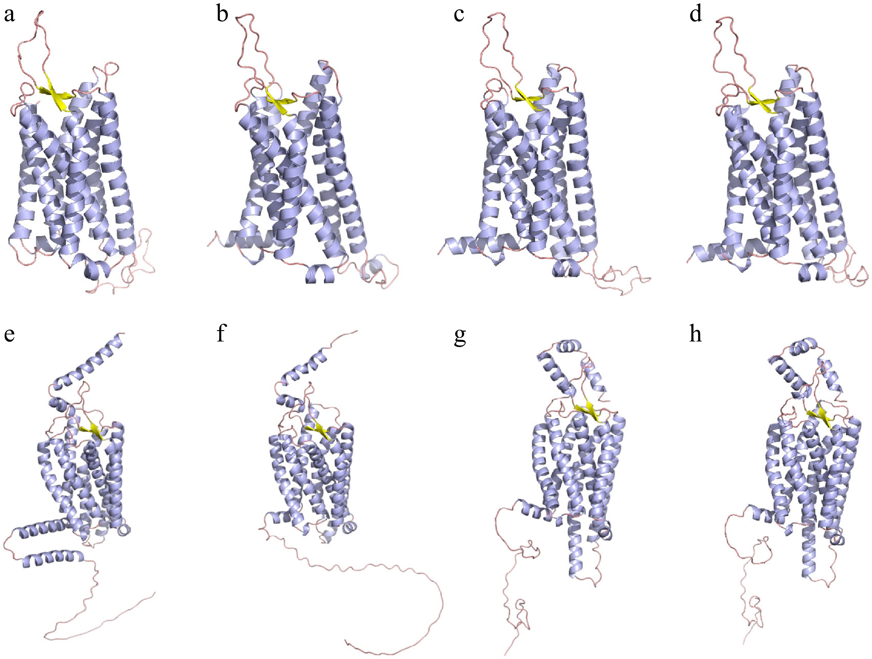
Figure 3.
The prediction of protein tertiary structure of (a)–(d) NMUR1 and (e), (f) NMUR2. (a) Homo sapiens - NMUR1, (b) Gallus gallus - NMUR1, (c) Acipenser baerii - NMUR1, (d) Acipenser ruthenus - NMUR1. (e) Homo sapiens - NMUR2, (f) Gallus gallus - NMUR2, (g) Acipenser baerii - NMUR2, (h) Acipenser ruthenus - NMUR2. The α-helices, β-Sheet and loop are depicted by purple, yellow and orange ribbons, respectively.
Distribution of nmur1 and nmur2 mRNA in different tissues
-
In this study, the expression of nmur1 and nmur2 mRNA was detected in 22 tissues by qPCR, including heart, liver, pancreas, spleen, kidney, esophagus, stomach, pyloric caecum, spiral valve intestine, duodenum, rectum, undifferentiated gonad, swim bladder, eye, gills, muscle, skin and brain (telencephalon, hypothalamus, midbrain, cerebellum and medulla oblongata). The highest mRNA levels of nmur1 and nmur2 were observed in muscle and spleen, respectively, but both mRNA levels were lowest in the liver. In addition, nmur1 and nmur2 are widely distributed in tissues closely related to feeding regulation, such as the hypothalamus, duodenum, and valvula intestinalis (Fig. 4).
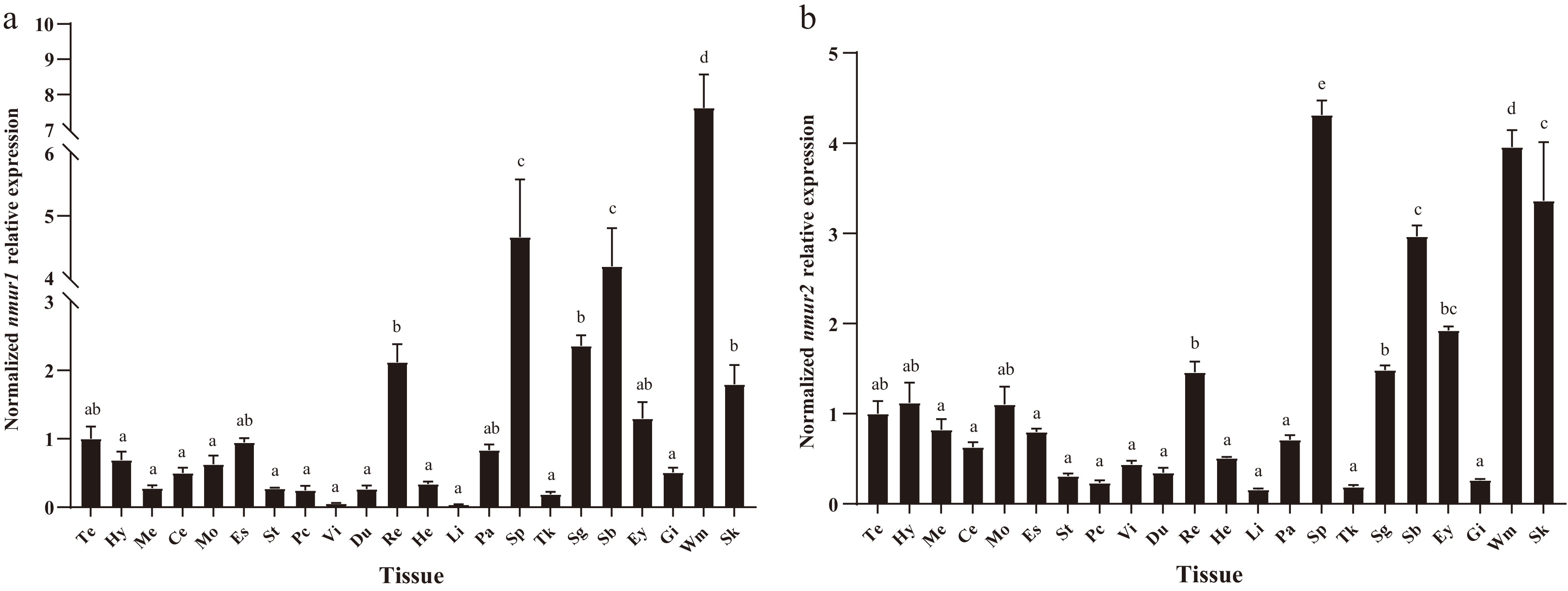
Figure 4.
Tissue distribution of (a) nmur1 and (b) nmur2 mRNA in Siberian sturgeon. The results were expressed as relative expression levels normalised to β-actin and GAPDH levels. Te: Telencephalon, Hy: Hypothalamus, Me: Mesencephalon, Ce: Cerebellum, Mo: Medulla oblongata, Es: Esophagus, St: Stomach, Pc: Pyloric caeca, Vi: Spiral valve intestine, Du: Duodenum, Re: Rectum, He: Heart, Li: Liver, Pa: Pancreas, Sp: Spleen, Tk: Trunk kidney, Sg: Sexual gland, Sb: Swim bladder, Ey: Eye, Gi: Gill, Wm: Muscle, Sk: Skin. Data are presented as mean ± SEM, n = 6 per tissue. Different letter above the bars indicates statistically significant differences ( p < 0.05, One-way ANOVA).
Feeding-induced changes of nmur1 and nmur2 mRNA in hypothalamus and peripheral tissues
-
The nmur1 and nmur2 mRNA were found to be abundant in the peripheral tissue and various brain tissues. As the hypothalamus is well known to be the center of feeding regulation, and the digestive tract contains various enzymes responsible for food digestion, the hypothalamus, spiral valve intestine, and duodenum tissues were selected to study the effect of feeding on nmur1 and nmur2 mRNA expression. The mRNA expression levels of nmur1 and nmur2 in the hypothalamus of the unfed group were upregulated in a time-dependent manner. Compared to the fasting group, after 1 h of feeding, the level of nmur1 in the hypothalamus was significantly downregulated, and the level of nmur2 was significantly downregulated after 1 and 3 h of feeding. (Fig. 5a, b). In peripheral tissues, 3 h of fasting significantly upregulated nmur1 and nmur2 expression levels in the duodenum and spiral valve intestine. In the duodenum and spiral valve intestine, the expression of nmur1 and nmur2 was significantly reduced at postprandial 1 and 3 h compared to the unfed group (Fig. 5c–f).
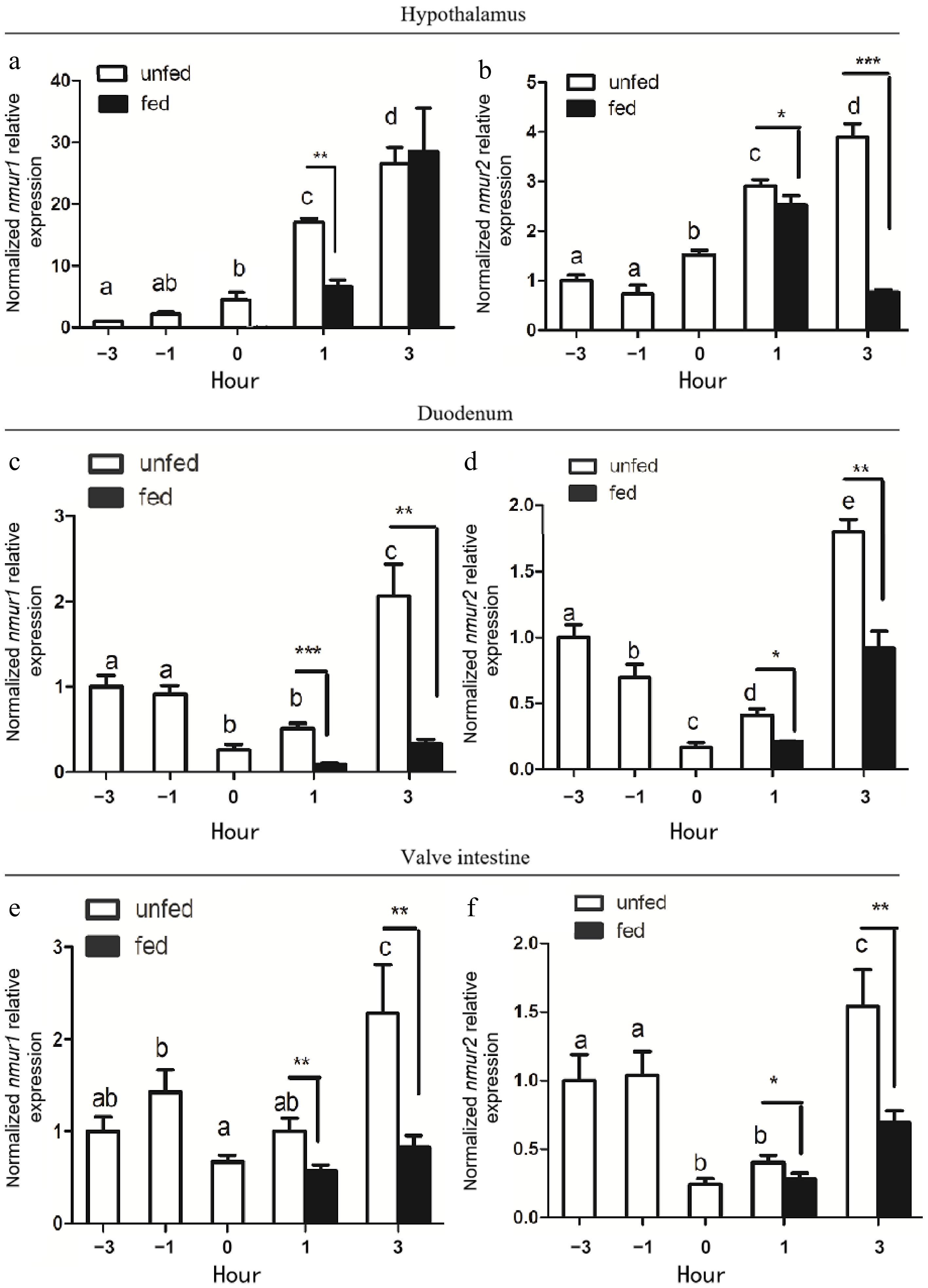
Figure 5.
Pre-prandial changes in the expression of (a), (c), (e) nmur1 and (b), (d), (f) nmur2 mRNA in (a), (b) the hypothalamus, (c), (d) duodenum, and (e), (f) valve intestine of Siberian Sturgeon. Data are presented as mean + SEM, n = 9. The results were expressed as relative expression levels normalized to β-actin and GAPDH levels. Bars with different letters represent significant differences between unfed groups (p < 0.05, One-way ANOVA). Asterisks represent significant differences between fed and unfed groups at a set time (* p < 0.05, ** p < 0.01, *** p < 0.001; Student's t-test).
Fasting and re-feeding induced changes of nmur1 and nmur2 mRNA in the hypothalamus and liver
-
We simulated the long-term food deprivation of fish in the natural environment by fasting and re-feeding experiments to explore the expression pattern of nmurs in Siberian sturgeon. In the state of long-term food deprivation, animals maintain the basic energy demand of the body by decomposing intraperitoneal and muscle fat through the liver after glycogen depletion. The hypothalamus is the center of appetite regulation in animals, so we chose the hypothalamus and liver for this experiment. In fasted groups, we found a significant increase in nmur1 mRNA in the hypothalamus on the first day and thereafter a significant decrease on the third, tenth, and 17th day. When fasted fish were re-fed on the tenth day onwards, the nmur1 mRNA expression was significantly increased compared to the unfed fish on the 10th, 15th, and 17th d in the hypothalamus (Fig. 6a). In the liver, compared to fed groups, we found a significant increase of nmur1 mRNA in unfed groups at all time points, and the expression of nmur1 was significantly reduced to normal levels after re-feeding (Fig. 6c). Similarly, compared to the fed groups, a significant increase in nmur2 mRNA expression was observed at all time points in the unfed groups. However, expression levels returned to normal following re-feeding in both the hypothalamus and liver (Fig. 6b, d).
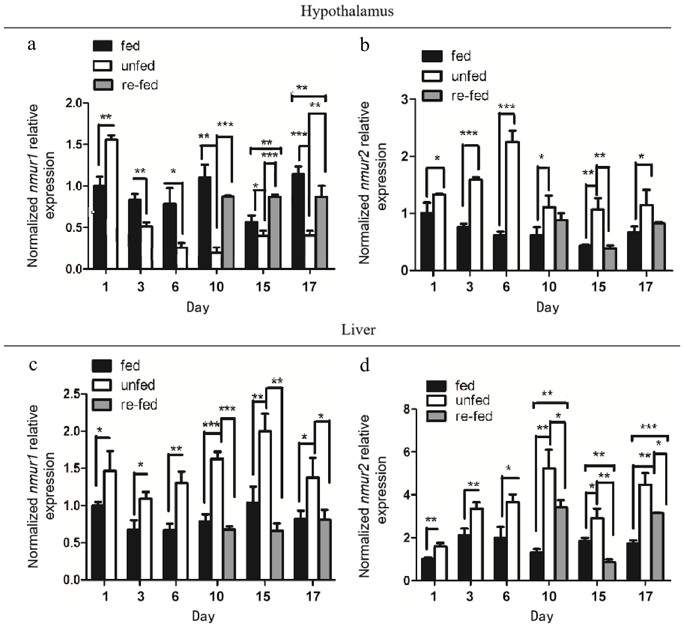
Figure 6.
Relative changes in (a), (c) nmur1 and (b), (d) nmur2 mRNA expression in (a), (b) hypothalamus, and (c), (d) liver of Siberian sturgeon during long-term fasting and re-feeding. Data are presented as mean ± SEM, n = 9 per treatment. The mRNA expression levels were normalized to β-actin and GAPDH expression levels. Asterisks represent significant differences between the fed and unfed groups (* p < 0.05, ** p < 0.01, *** p < 0.001; One-way ANOVA).
Effect of long-term i.p. and i.c.v. injection of NMU-25 on food intake and the expression of nmur1 and nmur2
-
Our results showed that single i.c.v. injection of 10 pmol/g BW NMU-25 solution or i.p. injection of 100 ng/g BW NMU-25 solution for seven consecutive days could significantly reduce the food intake (until 1 h after feeding) in Siberian sturgeon (Figs 7a, 8a). To examine receptor-specific expression in response to NMU-25-induced feeding inhibition, we assessed nmur1 and nmur2 expression following NMU-25 administration. After 7 d of daily intraperitoneal injections (100 ng/g BW), nmur1 expression significantly decreased, while nmur2 increased in the hypothalamus and valve intestine (Fig. 7b, c). In contrast, intracerebroventricular injection (10 pmol/g BW) led to a significant increase in nmur1 and a decrease in nmur2 expression in the same tissues (Fig. 8b, c).
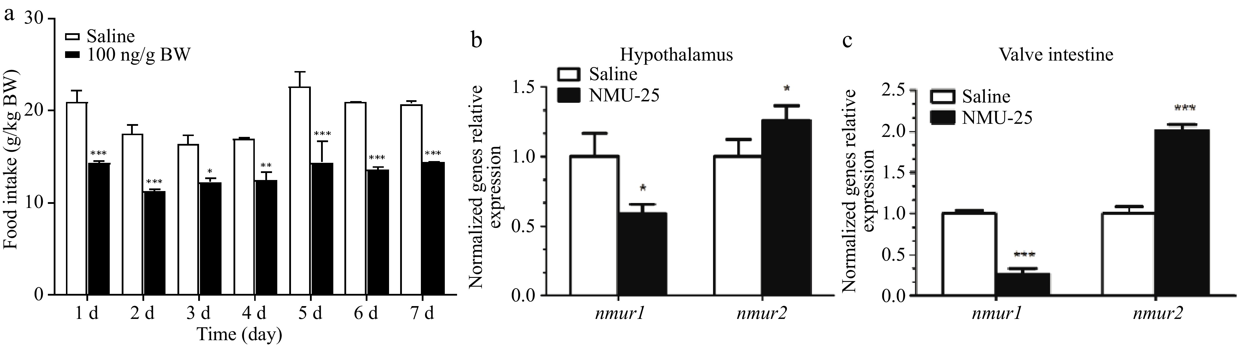
Figure 7.
Effects of long-term i.p. injection of NMU-25 (100ng/g BW) on (a) the food intake, (b) the expression of nmur1 and nmur2 mRNA in the hypothalamus, and (c) the valve intestine of Siberian sturgeon. Data are means ± SEM, n = 12 per treatment. The mRNA expression levels are normalized to β-actin and GAPDH expression levels. Asterisks represent significant differences between the two groups (* p < 0.05, ** p < 0.01, *** p < 0.001; Student's t-test).
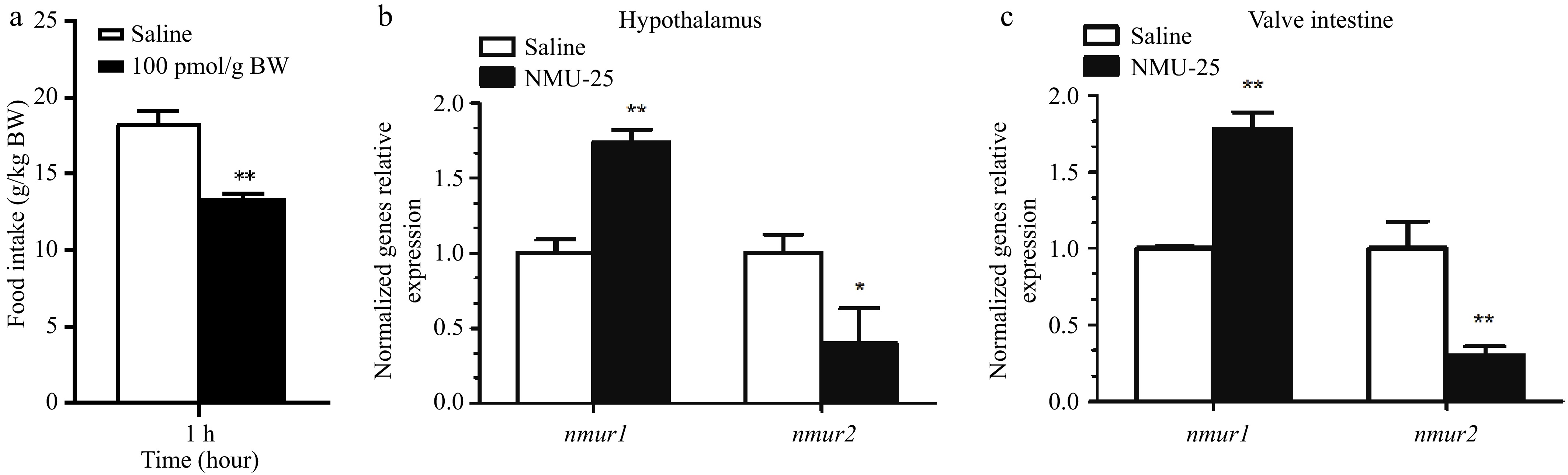
Figure 8.
Effects of i.c.v. injection of NMU-25 (10 pmol/g BW) on the expression of nmur1 and nmur2 mRNA in (a) hypothalamus and (b) valve intestine of Siberian sturgeon. Data are means ± SEM, n = 12 per treatment. The mRNA expression levels are normalized to β-actin and GAPDH expression levels. Asterisks represent significant differences between the two groups (* p < 0.05, ** p < 0.01, *** p < 0.001; Student's t-test).
-
This study identified nmur1 and nmur2 in Siberian sturgeon, which showed relatively low homology with nmurs of mammals, birds, reptiles, amphibians, and bony fish. However, nmurs had high homology with other sturgeon species, which may be attributed to the unique evolutionary status of sturgeon. Vertebrate ancestors experienced two rounds of whole-genome duplications[40]. Unlike other fish species, sturgeon has undergone three whole-genome duplications (WGD), and the last WGD event happened independently in the sturgeon lineage. This resulted in the formation of an octoploid state in sturgeon[6], suggesting the complexity and diversity of Siberian sturgeon genes. Additionally, the conservation of nmur1 is greater than that of nmur2. Sequence alignment revealed that the nmurs of these species all contain seven conserved transmembrane domains (TMDs), which are typical features of GPCRs and essential molecular structures required for ligand binding. NMURs of all species share the same ERY motif and two conserved cysteine residues, which are important for G protein activation[41], ligand binding[42], and the formation of disulfide bonds to stabilize protein structure[43,44], suggesting that NMURs of different species may perform similar biological functions.
This study found that nmur1 and nmur2 mRNA were widely distributed in all tested tissues of Siberian sturgeon, including various brain tissues and the digestive tract. Studies in mammals have found that nmur1 and nmur2 are widely distributed in various immune cells, such as eosinophils, T lymphocytes, and natural killer cells, mediating the role of NMU in antigen presentation and inflammation regulation[45−48]. The spleen produces and stores abundant immune cells. The nmur1 and nmur2 of Siberian sturgeon were highly expressed in the spleen, suggesting NMU may regulate the expression of nmurs to exert immune regulatory function. In addition, studies on rodents have found that NMUs can also regulate smooth muscle contraction through NMURs[34,49]. In Siberian sturgeon, nmurs are also highly expressed in the muscle of Siberian sturgeons, indicating that NMUs may regulate muscle movement by activating NMURs. Similar to our results, the nmurs of goldfish[23] are also highly expressed in the brain and the pituitary. However, in peripheral tissues, unlike the widespread expression of nmurs in the Siberian sturgeon gastrointestinal tract, nmur1 rather than nmur2 is expressed in the intestine of goldfish to regulate gastrointestinal motility[23]. This suggests that nmur1 and nmur2 may jointly participate in intestinal peristalsis and food digestion in Siberian sturgeons[50]. In mammals and birds, nmur1 is mainly distributed in peripheral tissues, while nmur2 is predominantly found in the central nervous system[25,26]. The expression level of porcine nmur2 mRNA in the central nervous system, such as the cerebral cortex, medulla oblongata, spinal cord, hypothalamus, midbrain, and pituitary, is significantly higher than that in peripheral tissues such as stomach, spleen, lung, heart, ovary, and adrenal gland[21]. Conversely, nmur1 mRNA is primarily expressed in the thymus, duodenum, testis, pancreas, stomach, and kidney of pigs[21]. Similarly, chicken nmur1 mRNA is mainly distributed in peripheral tissues, including the gastrointestinal tract and adrenal gland, while nmur2 mRNA is predominantly found in the central nervous system, especially in the hypothalamus[22]. These observations suggest that the function of NMURs in fish may differ from those of mammals and birds, and the high expression of nmurs in the brain and gastrointestinal tract of Siberian sturgeon may be related to food intake and energy homeostasis.
Recently, the role of NMU-NMURs in feeding regulation has attracted much attention. However, most of the studies focused on NMU, with few studies examining the role of transcriptional regulation of nmur1 and nmur2 in feeding regulation. Therefore, we conducted pre-prandial, fasting, and re-feeding experiments to determine how nmurs respond to feed deprivation and re-feeding in Siberian sturgeon. It is well known that the hypothalamus is the center of feeding regulation, particularly the arcuate nucleus and paraventricular nucleus. The duodenum contains numerous intestinal glands and various digestive enzymes, while the abundant blood vessels and spiral valves of the spiral valve intestine are unique features of cartilaginous and ancient bony fish[51]. These tissues effectively respond to changes in the body’s nutritional status. Our previous study showed NMU may exert bidirectional roles in the regulation of food intake, tentatively acting as an orexigenic factor in short-term feeding regulation and as an anorexigenic factor in the long-term feeding regulation in Siberian sturgeon[11]. After the feeding point, we found that the levels of nmur1 and nmur2 mRNA in the hypothalamus and intestine of unfed fish were upregulated in a time-dependent manner. The nmurs decreased significantly after 1 h of feeding compared with the fasting group, consistent with the trend of nmu[11]. This finding aligns with the trend observed in the orexigenic factor motilin and its receptors in the Yangtze sturgeon after short-term fasting[6]. These results suggest that the altered mRNA expression suggests nmurs may be involved in the response to NMU signalling. During long-term food deprivation, animals maintain the basic energy demands by breaking down intraperitoneal and muscle fat through the liver after glycogen depletion, as most nutritional utilization and metabolic regulation occur in the liver[52,53]. In the long-term fasting experiment, we observed a significant increase in hypothalamic nmur1 mRNA expression in the fasting group on day 1 compared to the fed group. This was followed by a marked decrease in expression between days 3 and 17, with expression levels returning to normal after refeeding. These changes may be linked to the sturgeon's characteristic ability to tolerate prolonged periods of starvation[54]. Sturgeon can endure prolonged periods without food, and successful foraging is often unpredictable, making necessary endocrine changes crucial for regulating the body's energy homeostasis[55,56]. Additionally, nmur1 levels in the liver and nmur2 levels in the hypothalamus and liver were significantly increased in the fasting group but returned to normal after refeeding. This suggests that in long-term appetite regulation, NMU may exert an inhibitory effect on appetite by activating nmur2 in the central nervous system and activating nmur1 in the periphery. Previous studies have shown that fasting increases the expression of appetite factors such as ghrelin[39], galanin[9], and agouti-related protein (AgRP)[57], while decreasing the expression of appetite-suppressing factors such as cholecystokinin (CCK)[58], urocortin3 (UCN3)[59], and NMU[27]. Overall, the differential expression of nmur1 and nmur2 in Siberian sturgeon under various nutritional statuses may be closely related to specific tissues and durations.
Due to limited knowledge of NMU receptors and their signaling systems in Siberian sturgeon, we conducted experiments involving long-term i.p. injection and i.c.v. injection of NMU-25 to explore the response patterns of peripheral and central nmurs. A previous study demonstrated that long-term i.p. and i.c.v. injection of NMU-25 can significantly reduce the food intake of Siberian sturgeon[11]. Interestingly, research on mammals found no response to peripheral acute or chronic administration of NMU in nmur1 knockout mice, whereas levels of anorexic-related peptides glucagon-like peptide-1 (GLP-1) and Peptide YY (PYY) in plasma of normal mice increased significantly, and the food intake and body weight decreased in a dose-dependent manner[29]. In dogs, peripheral NMU could inhibit appetite by activating nmur1 to promote gastrointestinal peristalsis[31], suggesting that peripheral NMU mainly activates nmur1 to regulate appetite in mammals. Contrary to mammals, our study found that nmur2 expression was significantly up-regulated in the hypothalamus and spiral valve intestine after long-term i.p. injection, a potential contribution of nmur2 activation to the peripheral anorexigenic effect of NMU in Siberian sturgeon. However, in the i.c.v. injection experiment, the receptor activation pattern of NMU’s appetite-suppressive effect once again differed from that of mammals, which rely on the mediation of nmur2. Long-term i.c.v. injection of NMU did not influence the body weight and appetite of female mice with nmur2 deficiency, but significantly reduced the body weight and total energy intake of diet-induced obese mice[33]. Short-term central injection of NMU failed to reduce feeding, spontaneous activity, and body temperature of mice with nmur2 deficiency[33]. Additionally, Acute and long-term central injection of NMU had no effect on body weight and food intake in nmur2-deficient mice[32]. Knockout of nmur2 from the hypothalamic paraventricular nucleus increased high-fat diet intake and promoted weight gain[60]. Takayama et al. delivered nmur2-selective agonist to the brain via nasal administration and observed a dose-dependent reduction in weight and food intake in mice[38]. In contrast, our results revealed that nmur1 expression was significantly up-regulated in the hypothalamus and spiral valve intestine after i.c.v. injection of NMU-25, suggesting a possible correlation between nmur1 activation and the anorexigenic effect of NMU-25 in the central nervous system of Siberian sturgeons, although the causal relationship awaits investigation. Overall, long-term i.p. injection of NMU-25 induced a significant upregulation of nmur2 transcript levels, while i.c.v. injection of NMU-25 significantly upregulated nmur1 transcript levels. These findings indicate that, unlike the observations in mammals, NMU-25 in the central system may play an anorexic role by activating nmur1, while in the periphery it may act by activating nmur2 in fish.
However, our experiment only reflects the differential responses to the anorexic effect of NMU in central and peripheral tissues of Siberian sturgeon at the mRNA level. Further experiments are needed to confirm our conclusion and explore the potential specific molecular mechanism, such as co-injection of receptor agonists/inhibitors or the use of knockout/overexpression and other experimental techniques.
-
To sum up, the CDS sequences of nmur1 and nmur2 of Siberian sturgeon were cloned for the first time, and they showed high homology with those of other species. NMURs across species share the same ERY motif and two conserved cysteine residues. Both nmur1 and nmur2 of Siberian sturgeon were widely expressed in the central nervous system and peripheral tissues. Moreover, we observed that nmurs of Siberian sturgeon can respond to different nutritional statuses, and their responses are closely related to specific tissues and duration, suggesting that NMU may regulate appetite by activating specific receptors in different tissues. Furthermore, our study found that, in Siberian sturgeon, NMU in the central system may play an anorexic role by activating nmur1, while in the periphery, it may exert its effect by activating nmur2. This study reveals the important biological role of nmurs in regulating animal appetite at the molecular level. Future studies should further investigate the central and peripheral responses of the receptor system to NMU in the regulation of animal feeding, and clarify the underlying molecular mechanisms involved in NMU's role in appetite control.
Thanks to Runzhao Fisheries (Sichuan, China) for providing us with experimental fish. The authors thank all the participants who devoted their time to participate in this study.
-
These animal experiments were approved by the Animal Care and Use Committee of Sichuan Agricultural University (DKY-S20150812).
-
The authors confirm their contributions to the paper as follows: experimental design and supervision: Tang N; experimental implementation: Wu H, Kang Q; data analysis and visualization: Wu H, Zhou B; supervision: Zhang X, Li Z; resources and reagents: Li Z, Tang N, Wu H, Chen D; writing − original draft: Wu H; writing − review and editing: Jiang H, Tang N. All authors reviewed the results and approved the final version of the manuscript.
-
The data used to support the findings of this study are available from the corresponding author upon reasonable request.
-
The authors declare that there is no conflict of interest.
-
accompanies this paper online at: https://doi.org/10.48130/jai-0026-0008.
- Supplementary Table S1 Primers used for this study.
- Supplementary Fig. S1 Preprandial, fasting and re-feeding experiments.
- Supplementary Fig. S2 Long-term i.p. and i.c.v. injection experiments.
-
Copyright: © 2026 by the author(s).
Journal of Applied Ichthyology published by Maximum Academic Press on behalf of John Wiley & Sons Ltd. This is an open access article under theterms of the Creative Commons Attribution License, which permits use, distribution and reproduction in any medium, provided the original work is properly cited. -
About this article
Cite this article
Wu H, Kang Q, Li Z, Zhang X, Zhou B, et al. 2026. Molecular characterization of neuromedin U receptors, nmur1 and nmur2, and their differential responses to the anorexic effect of NMU in central and peripheral tissues of Siberian sturgeon (Acipenser baerii Brandt). Journal of Applied Ichthyology 2026: e009 doi: 10.48130/jai-0026-0008 

Molecular characterization of neuromedin U receptors, nmur1 and nmur2, and their differential responses to the anorexic effect of NMU in central and peripheral tissues of Siberian sturgeon (Acipenser baerii Brandt)
- Received: 25 April 2026
- Revised: 09 August 2026
- Accepted: 28 August 2026
- Published online: 29 September 2026
Abstract: Neuromedin U (NMU) exerts an anorexigenic role by activating neuromedin U receptors (nmurs) in mammals. In fish, nmurs have been functionally characterized only in goldfish and zebrafish, but the information about nmurs in appetite regulation is limited. We cloned the coding sequence (CDS) of nmur1 and nmur2, which were widely expressed in muscle, brain, and gastrointestinal tissues of Siberian sturgeon. The expression levels of nmurs were significantly increased in the hypothalamus and intestinal tissues after postprandially in unfed fish, but nmurs significantly decreased 1 or 3 h after feeding. The expression of nmur1 in the liver and nmur2 in both the liver and hypothalamus were significantly increased after long-term fasting, and returned to normal levels after refeeding. The nmurs of Siberian sturgeon can respond to different nutritional statuses and are closely related to specific tissues and durations. In addition, after long-term intraperitoneal injection of NMU-25, the expression of nmur2 was significantly increased in the hypothalamus and spiral valve intestine, while the expression of nmur1 increased significantly after intracerebroventricular injection. In contrast to mammals, the inhibitory effect of NMU-25 on appetite may be mediated by nmur1 in the central nervous system, while in peripheral tissues, it may primarily be achieved through nmur2 in Siberian sturgeon. In conclusion, our study laid a foundation for further exploring the molecular mechanism of NMU in fish appetite control.
-
Key words:
- Appetite regulation /
- numr1 /
- numr2 /
- Nutritional status /
- Siberian sturgeon