











-
Mango (Mangifera indica L.) is a tropical fruit known for its delicious flesh and rich nutrients, earning it the title 'King of Tropical Fruits'[1]. However, the mango industry faces significant challenges due to postharvest diseases, particularly in tropical regions with high temperatures and humidity. Mango anthracnose caused by Colletotrichum gloeosporioides is a significant disease affecting growth and postharvest storage, with incidence rates reaching 100% under favorable conditions[2]. Effective disease management strategies are crucial to promote the healthy development of the mango industry and reduce economic losses[3].
Postharvest mango disease management involves physical[4], chemical[5−7], and biological control methods. Excessive chemical fungicide use has led to drug resistance[8] and safety concerns like residues and pollution, necessitating safer alternatives. Biological control provides a safer, effective, and eco-friendly alternative[9]. It leverages microorganisms, induced resistance, and plant-derived antimicrobials to curb pathogen growth, reducing fruit rot and disease incidence[10,11]. Antagonistic bacteria are a group of microorganisms that exhibit significant antagonism against pathogenic bacteria without causing harm to the fruits themselves[12]. They can induce fruits to activate their intrinsic defense systems to resist exogenous pathogen invasion, holding broad application prospects in the control of postharvest fruit diseases. For example, previous studies have shown that Bacillus subtilis IAGS174, a rhizospheric bacterial strain, employs multiple defense mechanisms to synergistically restrict pathogens and control Fusarium wilt in tomato[13]. In mango fruit, previous research findings have indicated that the antagonistic yeast Candida. metapsilosis showed high inhibition to Cladosporium gloeosporioides of spore germination at 33.23%. Bacillus atrophaeus TE7 could inhibit the growth and development of C. cladosporioides and enhance the activities of defense response-related enzymes in mango fruit[14]. Bacillus amyloliquefaciens GSBa-1 primarily enhances the resistance of mangoes to anthracnose by synergistically activating ROS in the peel and phenylpropanoid metabolism, thereby reducing the incidence of anthracnose effectively[15]. In addition, our laboratory previously isolated and screened a strain of Bacillus siamensis (designated N-1), an efficient biocontrol agent with strong inhibitory activity against phytopathogenic fungi. This strain can induce disease resistance in litchi and pitaya, and improve the postharvest storage and nutritional quality of the fruits[16,17]. However, research on its mechanism of action in mango disease resistance is limited; most previous studies only focused on the phenotypic effects of biocontrol, while the specific mechanism in postharvest disease prevention remains to be further studied, which is crucial for its application as a biocontrol agent in various fruits.
Plants have developed complex defense mechanisms to counter pathogen invasion[18]. Reactive oxygen species (ROS) are key signaling molecules regulating growth, development, and stress responses, and play fundamental roles in plant responses to pathogen infection, including the expression of defense-related genes[19]. Accumulation of ROS is a common phenomenon observed in a wide range of plant–pathogen interactions. For example, a rapid increase in reactive oxygen species (ROS) was observed in tobacco after inoculation with Botrytis cinerea[20]. But excessive ROS production can cause oxidative damage[21]. To mitigate this, plants utilize antioxidant enzymes to scavenge ROS and minimize cellular damage[22]. Additionally, plants produce pathogenesis-related proteins that degrade fungal cell wall components, inhibiting pathogen growth[23,24]. The activity of defense-related enzymes and gene expression play crucial roles in regulating plant immunity, as evidenced by ScCHIVII1 overexpression, enhancing disease resistance in sugarcane[25]. These indicate that the activities of these defense enzymes and the relative gene expression play important roles in plant disease resistance.
To elucidate the potential of N-1 as an antagonistic bacterium for inhibiting postharvest fruit diseases. Tainong mango was used as the test material. After treatment, the disease incidence, disease index, and fruit quality and ripening-related indices were measured, and the effects of N-1 on mango's physiological defense responses (stabilize ROS levels, enhance the activities of defense enzymes, phenolic metabolite accumulation) and molecular responses (defense-related gene expression) were further investigated. It aimed to uncover the regulatory mechanism of postharvest disease resistance induction in mangoes by N-1, providing a theoretical basis for their scientific and rational postharvest treatment.
-
'Tainong' mangoes at the commercially mature stage (approximately 80% ripeness) were harvested from a commercial orchard in Lingshui Li Autonomous County, Hainan Province, China, and transported to the Tropical Fruit and Vegetable Preservation Technology Laboratory at Hainan University within 3 h. Mangoes with uniform size, maturity, and external color, and without any signs of disease or mechanical injury, were selected for analysis. Before treatment, the fruit were immersed in 0.1% (w/v) sodium hypochlorite solution for 20 min, then air-dried at room temperature.
N-1 stored in an ultra-low-temperature freezer was reactivated, and spore density was adjusted to 1 × 10⁸ CFU·mL−1[26]. Mangoes were then randomly assigned to two treatment groups: one immersed in nutrient broth (NB, control) and the other in the N-1 suspension, each for 20 min. After that, fruits were air-dried and stored at 20 °C with 85%–90% relative humidity for further evaluation. Samples were collected at 0, 4, 6, 8, 9, 12, 15, and 21 d after treatment, flash-frozen in liquid nitrogen, and stored at −80 °C for subsequent analyses.
Test methods
Determination of disease index and disease incidence
-
For inoculation, a small puncture was made on the fruit surface using a sterile pipette tip, followed by gentle removal of the peel to expose the wound site. The treatment group was inoculated with 15 μL of the N-1 suspension, while the control group received 15 μL of sterile water. After incubation at room temperature for 12 h, each wound site was inoculated with 30 μL of C. gloeosporioides spore suspension. The inoculated fruits were then incubated in a growth chamber set at 25 °C and 85%–90% relative humidity. Lesion expansion and disease severity indices were monitored and recorded at predetermined time intervals.
Fruit decay was assessed at 0, 6, 9, 12, and 15 d after treatment. Disease incidence and disease index were calculated based on the proportion of decayed fruits in each replicate (12 fruits per replicate).
Respiration rate and ethylene release measurement
-
Respiration rate was measured using a portable O2/CO2 headspace analyzer (Dansensor® CheckPoint 3), and ethylene production was quantified by gas chromatography[27]. Five fruits from each treatment were randomly selected, weighed, and placed into a 2 L airtight container. The container was sealed with plastic film and held at room temperature for 2 h. After equilibration, the headspace gas was mixed thoroughly, and then CO2 concentration was measured. Each measurement was repeated three times and expressed as mL·kg−1·h−1.
For ethylene analysis, approximately 1 mL of headspace gas was extracted using a gas-tight syringe and injected into a gas chromatograph. Ethylene production was quantified by comparing retention time and peak area with those of standard ethylene gas.
Determination of firmness and color
-
Fruit firmness was assessed using a texture analyzer equipped with a needle probe, and results were expressed in Newtons (N). Surface color was measured with a colorimeter, and the a* value was recorded.
Determination of TSS, TA, and AsA contents
-
TSS was determined using a digital saccharimeter and expressed as a percentage. TA was quantified following a modified method by Yu et al.[28]. Briefly, 2 g of pulp tissue was weighed, homogenized, and centrifuged at 12,000 r/min for 20 min at 4 °C. The supernatant was collected, mixed with 1% phenolphthalein indicator, and titrated with 0.1 M NaOH until neutralization. The volume of NaOH consumed was recorded, with distilled water used as the blank control, and TA was expressed as a mass fraction (%). AsA content was determined using the 2,6-dichlorophenolindophenol (DCPIP) titration method[29].
Determination of total phenols and flavonoids
-
Total phenolic and flavonoid contents in uninoculated mango fruits were quantified using the Folin–Ciocalteu method[30]. Absorbance for total phenols was measured at 280 nm, and concentrations were calculated from a gallic acid standard curve. Flavonoid content was determined by measuring absorbance at 325 nm and quantifying values using a standard curve. All results are expressed as g·kg−1 fresh weight (FW).
Determination of ROS accumulation
-
The O2•− production rate was determined following the method described by Tang et al.[31]. Briefly, 0.2 g of fruit peel tissue was homogenized in 4 mL of phosphate buffer in an ice bath and centrifuged at 12,000 r/min for 20 min. The supernatant was incubated at 25 °C for 1 h, after which p-aminobenzenesulfonic acid and α-naphthylamine solutions were added. The mixture was then incubated for an additional 20 min at 25 °C, and absorbance was recorded at 530 nm. Results were expressed as nmol·min−1·g−1 FW.
H2O2 content was quantified using a commercial assay kit (BC3590, Beijing Solarbio Science & Technology Co., Ltd) according to the manufacturer's instructions and expressed as mmol·g−1 FW.
Determination of MDA levels and cell membrane permeability
-
MDA concentration was measured using the thiobarbituric acid method[32], with absorbance recorded at 450, 532, and 600 nm. MDA levels were expressed as μmol·g−1 FW. Cell membrane permeability was determined using a DDS-11A conductivity meter. The initial conductivity of the samples and the maximum conductivity (measured after boiling the samples for 15 min) were recorded.
Determination of defense-related enzyme activities
-
The activities of POD, PPO, CAT, SOD, and APX were determined with slight modifications[33], and results were expressed as U·g−1·min−1. PAL activity was measured following a modified protocol according to Sun et al.[34]. Briefly, borate buffer and L-phenylalanine were added to the reaction system. One vial contained 0.5 mL of crude enzyme extract, while a second vial containing 0.5 mL of enzyme solution boiled for 5 min served as a control. Absorbance values were measured at 290 nm, with A1 representing the crude enzyme and A2 representing the boiled control. One unit of PAL activity was defined as an increase of 0.01 in absorbance per gram of fresh mango tissue per hour, expressed as U·g−1·min−1. CHT activity was determined using a commercial assay kit (BC0820, Beijing Solarbio Science & Technology Co, Ltd) according to the manufacturer's instructions. Results were expressed as U·g−1·min−1[35]. GLU activity in mango peel was assessed with slight modifications from. Briefly, 0.2 g of tissue was homogenized in extraction buffer and centrifuged at 12,000 × g for 30 min at 4 °C. The supernatant was collected, and 100 µL of 0.4% laminarin solution was added. Two separate tubes contained 0.1 mL of either crude enzyme or enzyme boiled for 5 min. The DNS reagent was subsequently added to terminate the reaction. Absorbance changes were measured at 540 nm, and enzyme activity was expressed as U·g−1·min−1.
Quantitative real-time PCR (qRT-PCR) analysis
-
Total RNA was extracted from mango tissue using a modified CTAB method. RNA quality was evaluated by 1% agarose gel electrophoresis. cDNA was synthesized via reverse transcription using the MonScript™ RTIII All-in-One Mix with dsDNase kit according to the manufacturer's instructions. The qRT-PCR was subsequently performed using the 2× Q3SYBR qPCR Master Mix kit, with MiActin serving as the internal reference gene. Specific primers were designed using Primer6 software. Primer sequences are provided in Table 1. Relative gene expression levels were calculated using the 2−ΔΔCᴛ method.
Table 1. Primer sequences used in qRT-PCR.
Gene Forward primer(5'-3') Reverse primer(5'-3') MiPOD TTTCCAGCAAACCAATCAGCG TCCCTTAGCATCCTTGACGG MiSOD CAAGCCCTTCGTTGTTGTCG CTTCACAGTTGTGGGACCATC MiAPX GCTGAGAAGAACTGTCTCCTATC CAACAATATCCTGGTCACTGAGTCC MiPAL GACGTCGCATAGGAGAACGA TCCAGCATTCAAGAACCTTATGA MiCHT CAAGCTGTGCTGGAAAGAGC TTCCACAGGCACCGTAGTTG MiGLU TATTCAGTAACAGGGCGTGGA AATCCGCAACTCTTGGTCTC MiActin ATCTGCTGGAAGGTGCTGAG CCAAGCAGCATGAAGATCAA Statistical analysis
-
Data are presented as mean ± standard deviation (SD). Statistical analyses were performed using GraphPad Prism 8.0.2. All experiments were conducted with at least three independent replicates. Differences between groups were evaluated using Student's t-test, with significance levels indicated as * p < 0.05 and ** p < 0.01.
-
As shown in Fig. 1a, the brown lesions on anthracnose-inoculated sites of control fruits had expanded markedly at 12 d after treatment. However, lesions on fruits treated with B. siamensis N-1 showed minimal progression. Disease incidence in the N-1-treated group was significantly lower than in the control. Specifically, with the incidence reaching 25% at 12 d after treatment (a reduction of 55.55% compared with the control) and 77.78% at 15 d (a reduction of 22.22% compared with the control) (Fig. 1b). Consistent with these findings, the disease index was also significantly reduced in N-1-treated fruits; on days 12 and 15, treated fruits exhibited markedly lower disease indices than controls (Fig. 1c). As a climacteric fruit, respiratory metabolism is a key physiological process during postharvest ripening. Both control and N-1-treated fruits exhibited an initial increase in respiratory rate followed by a decline, displaying a characteristic respiratory peak. However, N-1 treatment significantly delayed the onset of the respiratory peak, and the treated fruits maintained a relatively lower respiratory rate during the later stages of storage. On days 15 and 18, the respiratory rate of N-1-treated fruits was 61.5% and 55.6% lower than that of the control, respectively (Fig. 1d). Ethylene production generally increased over time, but by day 21, N-1-treated fruits released significantly less ethylene than the control group (Fig. 1e). Firmness, an essential indicator of fruit maturity, decreased alongside water content during storage. Notably, N-1 treatment mitigated the decline in firmness, and from day 15 onward, the firmness of treated fruits remained significantly higher than that of the control (p < 0.05) (Fig. 1f). Collectively, N-1 treatment effectively suppressed the colonization and spread of C. gloeosporioides, thereby enhancing mango fruit resistance to anthracnose. In addition, it also delayed fruit ripening and helped maintain key postharvest quality attributes.
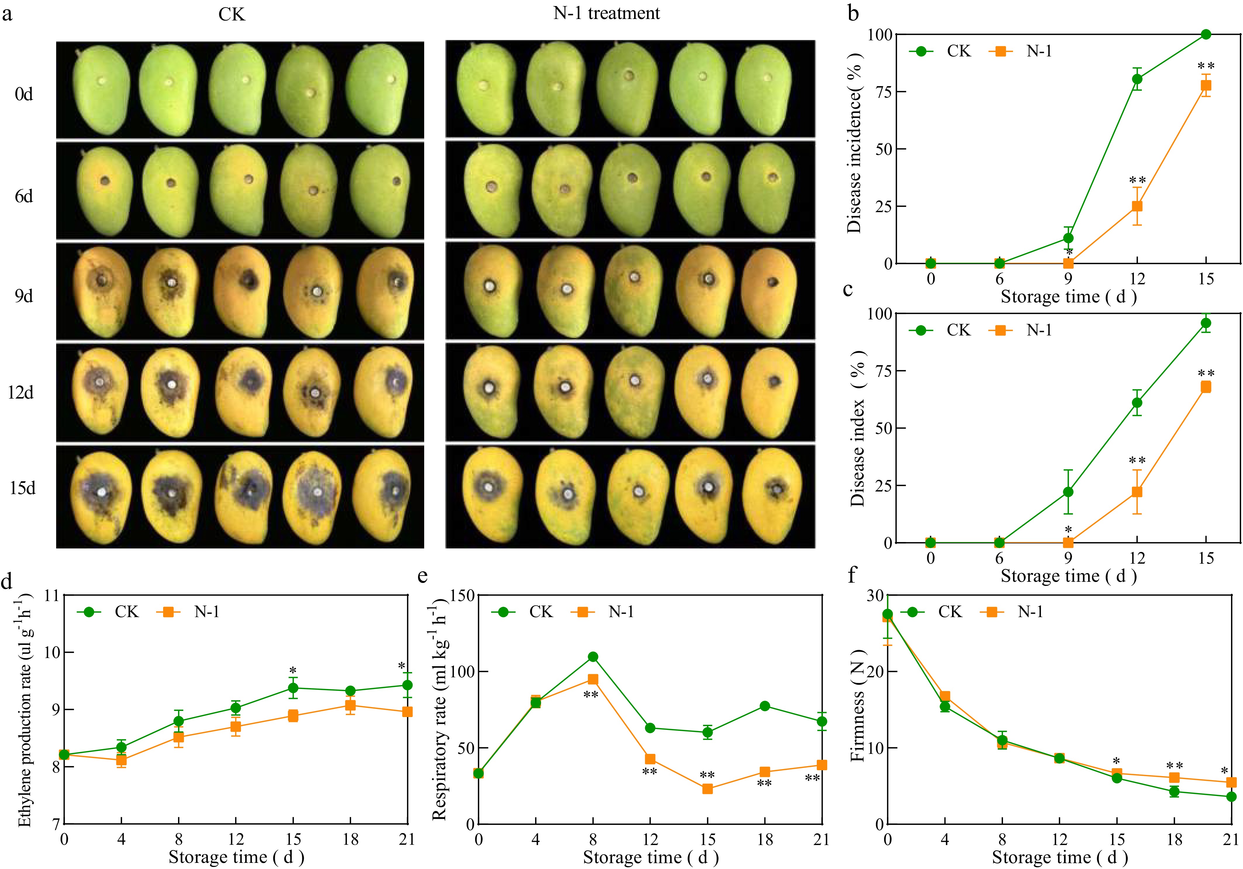
Figure 1.
Changes in (a) fruit appearance, (b) disease incidence, (c) disease index, (d) respiratory rate, (e) ethylene production rate, and (f) firmness of mango fruit after inoculation at 25 °C. Vertical bars represent the standard error of the mean, and the asterisks indicate a significant difference between two groups at the corresponding sampling point (* p < 0.05, ** p < 0.01).
N-1 enhances postharvest storage and nutritional quality
-
The color change of mangoes from green to yellow was observed, with the treatment group exhibiting a slower color change, particularly during mid-storage, compared to the control group (Fig. 2a). TSS content in both treatments initially increased, followed by a decline. Especially, the N-1 treatment group maintained a higher TSS level in the later stages, with a significant 39.3% increase observed in the treatment group compared to the control group on 21 d (p < 0.05) (Fig. 2b). The AsA content exhibited a continuous decline in both groups throughout storage, with the N-1 treatment group displaying significantly higher levels during the mid-storage period (4−12 d) compared to the control group (p < 0.01) (Fig. 2c). Similarly, the TA content decreased progressively in both groups, with the N-1 treatment group maintaining significantly higher levels during the later stages of storage (15−24 d) compared to the control group (p < 0.01) (Fig. 2d). The total phenol content remained relatively stable throughout storage, with the treatment group generally exhibiting higher levels compared to the control group. Notably, on 21 d, the total phenol content in the treatment group was significantly higher than that in the control group (p < 0.01) (Fig. 2e). In contrast, the total flavonoid content followed a trend of initial increase followed by a decline. The control group exhibited a steady increase in flavonoid content throughout storage. In contrast, the treatment group showed a 16.5% significant increase in flavonoid content compared to the control group at the end of storage (p < 0.01) (Fig. 2f). These findings suggest that N-1 treatment effectively maintained the storage quality of mango fruits.
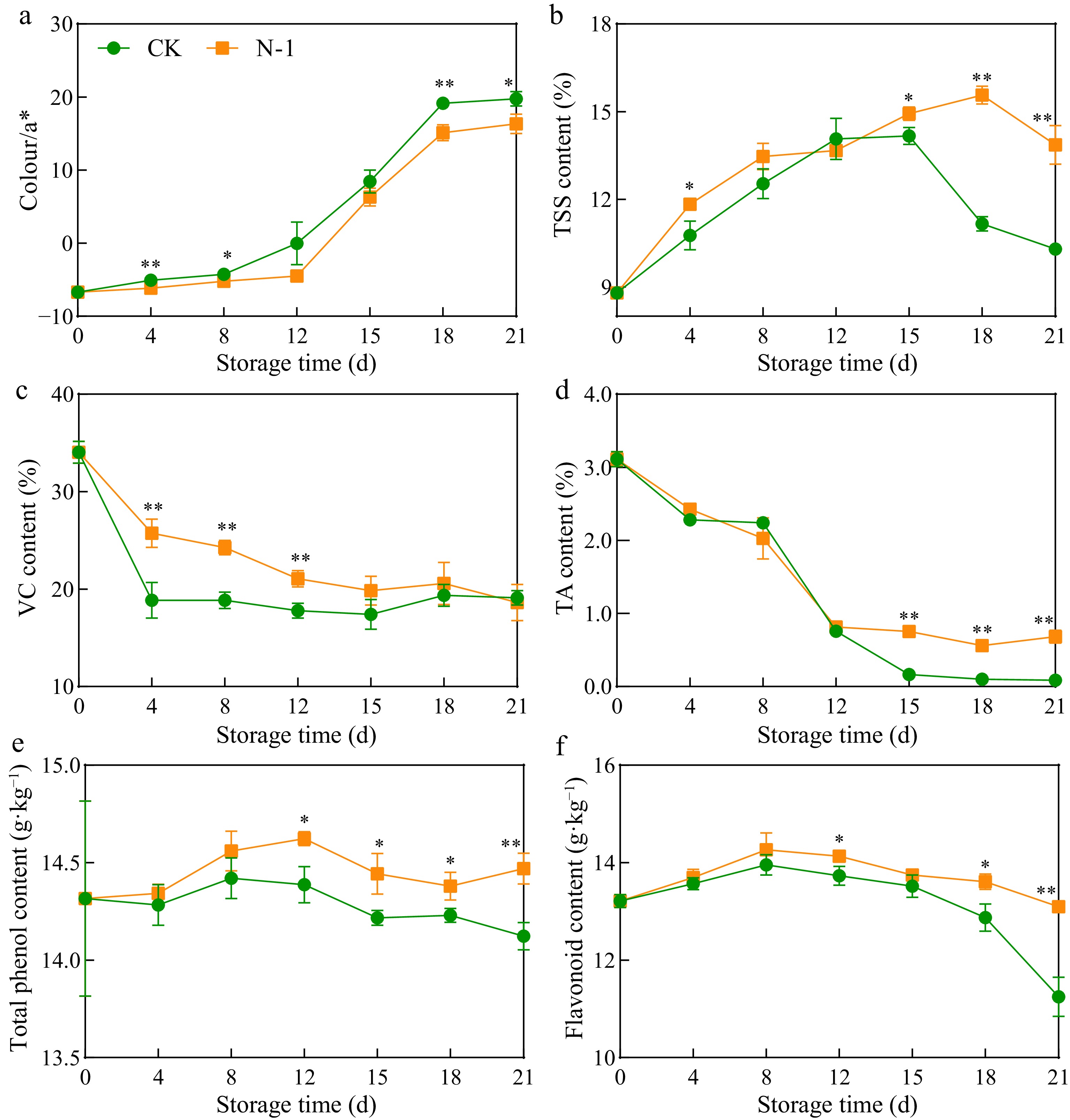
Figure 2.
Changes in (a) color, (b) TSS content, (c) AsA content, (d) TA content, (e) total phenol, and (f) flavonoid contents of mango fruit during storage at 25 °C. Vertical bars represent the standard error of the mean, and the asterisks indicate a significant difference between the two groups at the corresponding sampling point (* p < 0.05, ** p < 0.01).
N-1 maintains ROS homeostasis
-
The O2− production rate exhibited an increasing trend, with the control group consistently showing significantly higher rates (31%) compared to N-1 treated fruits, particularly at the end of storage (Fig. 3a). H2O2 content, indicative of reactive oxygen species metabolism, was significantly higher in the control group during early and mid-storage (0−15 d), with a notable 29% increase in the control group on 8 d (p < 0.01) (Fig. 3b). The MDA content positively correlated with storage time, with significantly higher levels in the control group during late storage. The MDA content in the control group was 1.96, 1.53, and 1.4 times that of N-1-treated fruits at 15, 18, and 21 d, respectively (p < 0.01) (Fig. 3c). Relative conductivity increased initially and then decreased, with N-1-treated fruits showing significantly lower conductivity on 18 d (p < 0.01) (Fig. 3d).
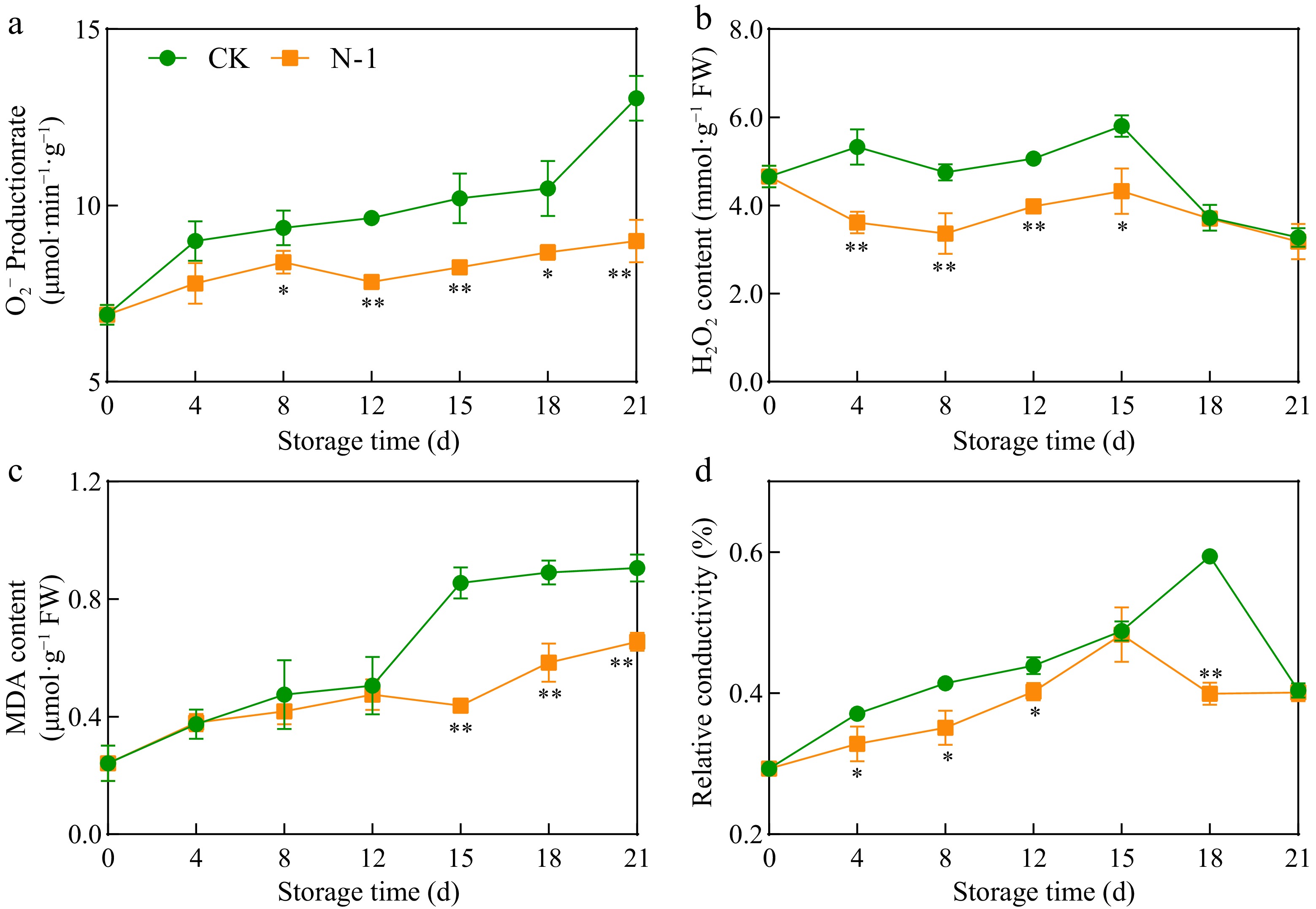
Figure 3.
Changes in (a) O2− production rate, (b) H2O2 content, (c) MDA content, and (d) relative conductivity in mango fruit during storage at 25 °C. Vertical bars represent the standard error of the mean, and the asterisks indicate a significant difference between two groups at the corresponding sampling point (* p < 0.05, ** p < 0.01).
N-1 enhances the activity of defense-related enzymes
-
The activities of PPO and POD in mango fruits exhibited an initial increase followed by a decline during storage (Fig. 4a, b). Notably, PPO activity was significantly higher in the treated group during early storage (0−12 d), peaking at 2.19 U·g−1·min−1 on day 12. In contrast, POD activity in the control group peaked on day 4 (0.408 U·g−1·min−1), whereas the treated group reached a higher peak on day 8 (0.436 U·g−1·min−1, 185.2% of control). The activities of CAT and SOD also followed a trend of initial increase and subsequent decrease. N-1 treatment activated CAT earlier, enhancing its activity against free radicals, with significantly higher CAT activity observed in the treatment group after 8 d (Fig. 4c). SOD activity peaked at 32.18 U·g−1·min−1 on day 8 in the control group. In contrast, the treatment group reached a higher peak of 49.16 U·g−1·min−1 on day 15 (Fig. 4d).
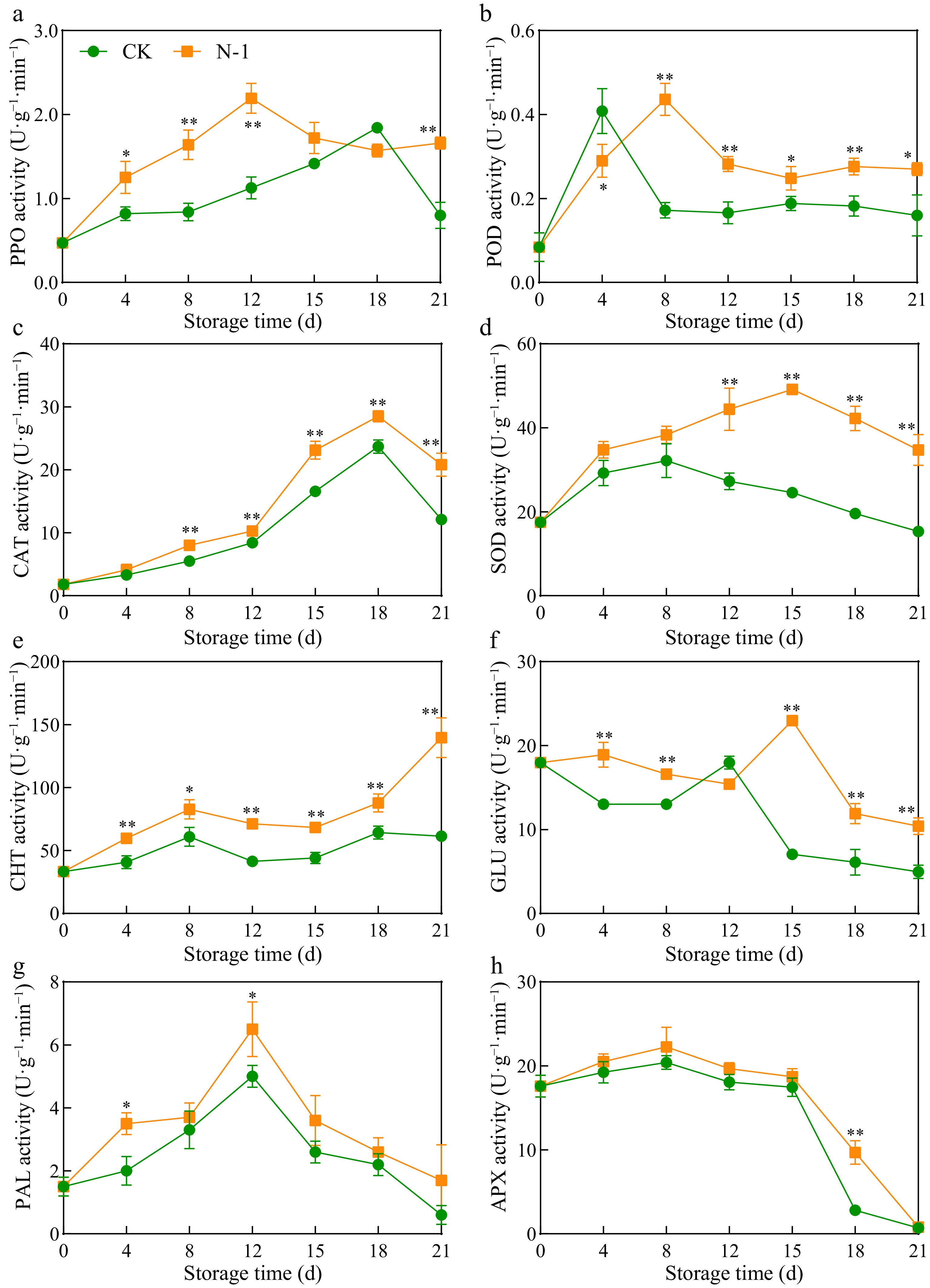
Figure 4.
Activities of (a) PPO, (b) POD, (c) CAT, (d) SOD, (e) CHT, (f) GLU, (g) PAL, and (h) APX in mango fruit during storage at 25 °C. Vertical bars represent the standard error of the mean, and the asterisks indicate a significant difference between two groups at the corresponding sampling point (* p < 0.05, ** p < 0.01).
CHT activity was significantly higher in treated fruits throughout storage, peaking at 139.6 U·g−1·min−1 on day 21, which was substantially higher than the control group (Fig. 4e). GLU activity exhibited an initial increase followed by a decline, with significantly higher activity in the treated group during early (4−8 d) and late (15−21 d) storage (Fig. 4f). The treated group reached a peak of 22.98 U·g−1·min−1 on day 15, 3.75 times that of the control group, indicating N-1's treatment on day 21 significantly inducing effect on defense-related enzymes. PAL activity followed a trend of initial increase and subsequent decrease, with consistently higher activity in the treated group (Fig. 4g). The APX activity in the fruits exhibited an overall decreasing trend, with the treated group showing a slower decline rate than the control group; notably, the APX activity in the treated group was extremely significantly higher than that in the control group at 18 d (p < 0.01) (Fig. 4h), suggesting N-1 induces APX activity enhancement.
N-1 modulates disease resistance and antioxidant gene expression
-
N-1 treatment upregulated the expression of genes encoding antioxidant and disease-resistant enzymes in mango fruit (Fig. 5). Specifically, compared with the control group, the expression levels of MiPOD, MiSOD, and MiAPX in the treatment group were elevated. MiPAL expression was significantly higher in the treatment group during late storage (15−18 d), while MiCHT expression was significantly higher in the treatment group during mid-storage (4−12 d). MiGLU activity peaked at 15 d in the treatment group, 1.54 times that of the control group, indicating enhanced defense-related gene expression.
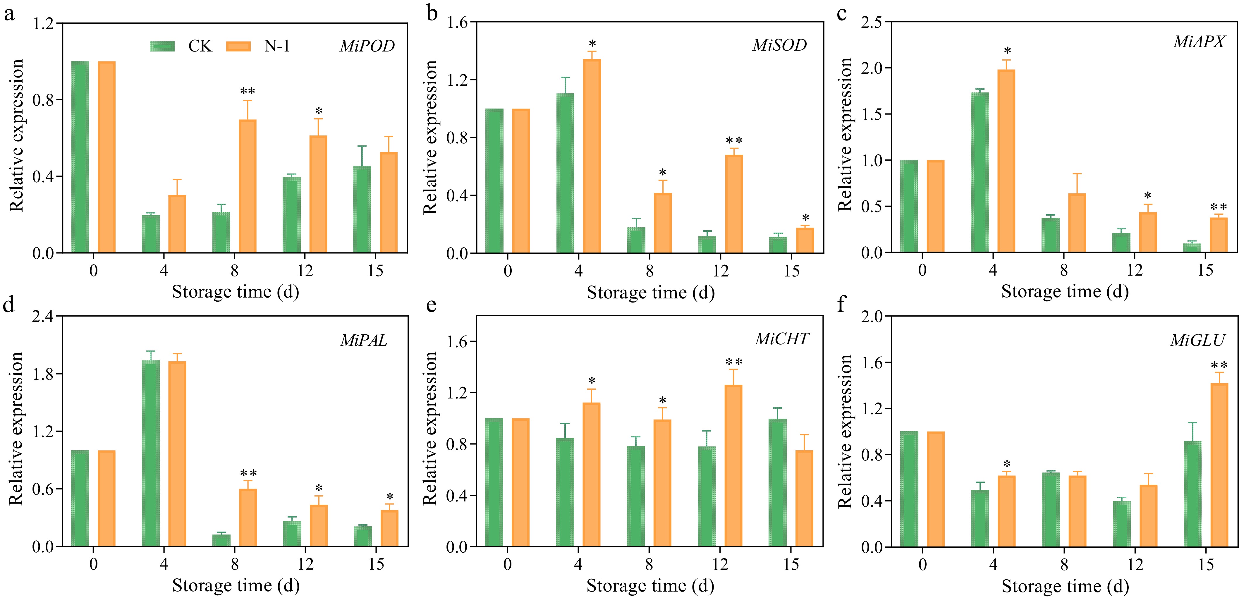
Figure 5.
Relative expressions of (a) MiPOD, (b) MiSOD, (c) MiAPX, (d) MiPAL, (e) MiCHT, (f) MiGLU in mango fruit during storage at 25 °C. Vertical bars represent the standard error of the mean, and the asterisks indicate a significant difference between the two groups at the corresponding sampling point (* p < 0.05, ** p < 0.01).
Correlation analysis of mango fruit quality and disease resistance parameters
-
Correlation analysis revealed significant relationships between disease incidence, disease index, and various quality indicators, ROS accumulation, enzyme activities, and gene expression (Fig. 6). Disease incidence and index were positively correlated with ethylene release rate (p < 0.05), but negatively correlated with firmness, AsA, and TA (p < 0.05). Additionally, disease incidence and index were positively correlated with O2− production rate, H2O2 content, and cell membrane permeability (p < 0.01). Conversely, disease incidence was negatively correlated with defense-related enzyme activities, including PPO, POD, SOD, APX, PAL, and CHT (p < 0.05), and gene expression levels of MiPOD, MiSOD, MiAPX, MiPAL, and MiCHT (p < 0.05). Enzyme activities of APX and POD were positively correlated with MiAPX and MiPOD expression levels, respectively (p < 0.05).
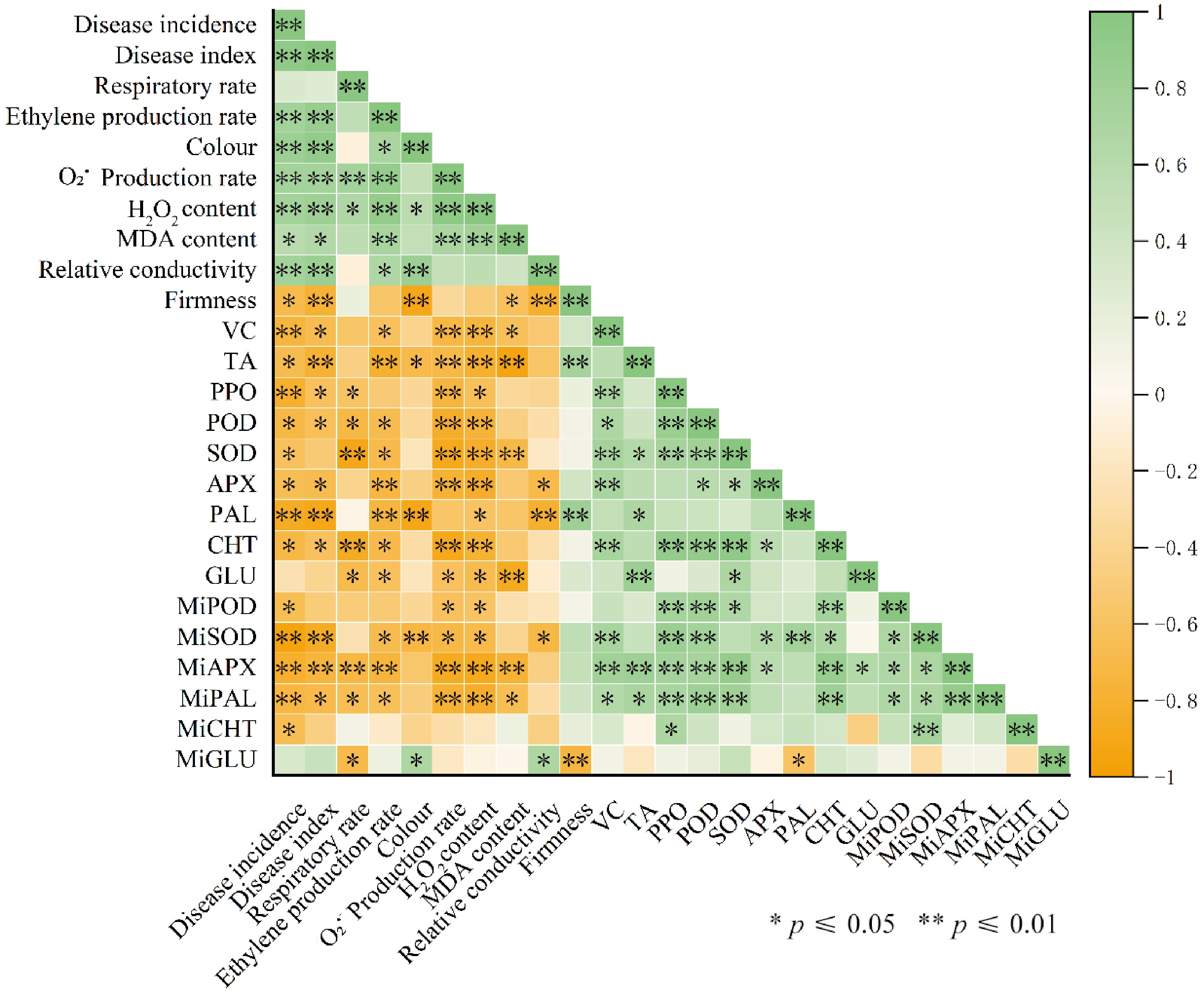
Figure 6.
Correlation analysis between disease incidence, ROS accumulation, enzyme activities, and gene expressions.
-
Mangoes are highly favored by consumers for their delicious flesh. However, the perennial high temperature and humidity in Hainan expose mangoes to various diseases during growth, transportation, and storage. Notably, anthracnose is a latent disease, and many broad-spectrum antibacterial chemical preservatives exhibit poor efficacy in controlling anthracnose on tropical fruits. Therefore, it is imperative to explore a more effective and eco-friendly approach to extend the storage life of mangoes. Our findings demonstrated that N-1 significantly reduced the disease incidence of mango fruits. Consistent with our results, Wang et al.[17] confirmed that Bacillus siamensis could enhance the disease resistance of litchi fruits. In addition, treatment with the N-1 strain maintained the reactive oxygen species homeostasis in fruits and markedly improved the disease resistance of pitaya fruits[16].
Stress-induced elevation of hydrogen peroxide content in plants is an indicator of the intensity of ROS metabolism in fruits[36]. We observed that after N-1 treatment, the production rates of H2O2 and O2− were significantly lower in the treatment group than in the control group (Fig. 4a, b). Meanwhile, MDA content and relative electrical conductivity in the N-1 treatment group were maintained at low levels (Fig. 4c). This could be attributed to the fact that N-1 treatment alleviated the lipid peroxidation of fruit cell membranes, thereby modulating the cellular redox state and triggering the activation of multiple defense mechanisms, which is consistent with the findings of previous studies[37].
However, excessive ROS accumulation can induce damage to cell membranes. To counteract this issue, plants activate both enzymatic and non-enzymatic ROS-scavenging systems. Strain N-1 significantly enhanced the activities of major ROS-scavenging enzymes (SOD, CAT, and POD)[38] in mango fruit, which is consistent with the findings of a previous research by Liu et al.[39]. Furthermore, extensive studies have demonstrated that ROS homeostasis, as a core regulatory node in plant defense responses against pathogenic infection, is accompanied by elevated activities of key enzymes[39,40]. N-1 markedly upregulated the activities of APX and also markedly upregulated the activities of PPO associated with plant antioxidant defense.
Plants possess an advanced immune system with a multitude of receptors capable of sensing pathogen-associated molecular patterns, triggering tailored defense responses[41]. Meng et al.[42] demonstrated that the combined treatment with 1-methylcyclopropene and salicylic acid (SA) enhanced the resistance of mango fruits to anthracnose, with the core mechanism involving the induction of disease resistance-related enzymes (phenylalanine ammonia-lyase, 4-coumarate-CoA ligase, peroxidase, polyphenol oxidase, chitinase, and β-1,3-glucanase). This enzymatic induction subsequently triggers the accumulation of total phenolics and flavonoids, which confer disease resistance in mango fruits. In the present study, treatment with strain N-1 significantly elevated the activity of PAL — a key enzyme involved in the phenylpropanoid metabolism pathway[43], and promoted the accumulation of phenolic compounds (total phenolics and flavonoids) in mango fruits. These polyphenolic metabolites can modulate the storage performance and disease resistance of fruits through their processes of biosynthesis, accumulation, and degradation[44]. Furthermore, N-1 treatment also significantly enhanced the activities of GLU and CHT. In plants, GLU also functions to degrade fungal cell wall components. CHT exerts a direct antifungal effect by hydrolyzing the key structural components of fungal cell walls, thereby boosting fruit disease resistance[45,46]. The synergistic action of these two enzymes collectively reinforces the disease resistance of mango fruits.
Beyond disease resistance, postharvest quality attributes are crucial for consumer acceptance. Fruit color, firmness, TSS, TA, and AsA are key indicators of visual appeal, flavor, and nutritional value[47]. Climacteric fruits such as mangoes undergo rapid color change during ripening, while TSS and TA levels reflect the balance of sweetness and acidity, contributing to overall flavor[48]. AsA is one of the most essential vitamins in human nutrition. Studies have indicated that during cold storage, the TSS content in jujube fruits increases while the TA content decreases, thereby maintaining the nutritional quality and flavor of jujube fruit. Similarly, N-1 treatment effectively preserved the contents of AsA, TA, and TSS in mango fruit. These results indicate that N-1 not only enhances disease resistance but also maintains postharvest quality and nutritional value, thereby improving shelf life and consumer acceptability.
The synthesis of enzymes is ultimately regulated by gene expression, whereby genes control the transcription and translation of genetic information into functional enzymes. At the molecular level, qRT-PCR analysis revealed that N-1 treatment upregulated the expression of key defense and antioxidant genes, including MiPOD, MiSOD, MiAPX, MiPAL, MiCHT, and MiGLU, corroborating previous findings in other fruit species[49]. Correlation analysis further demonstrated that disease incidence was negatively associated with the activities of PPO, POD, SOD, APX, PAL, and CHT, as well as the expression levels of the corresponding genes, while being positively correlated with O2− production rate, H2O2 content, and membrane permeability. These relationships highlight the central role of enzymatic defenses and gene expression in enhancing mango resistance to anthracnose, while excessive ROS accumulation compromises disease resistance. Collectively, our results provide insights into how N-1 regulates the postharvest disease resistance mechanism in mango. These findings lay a theoretical foundation for developing safe and effective postharvest treatments.
-
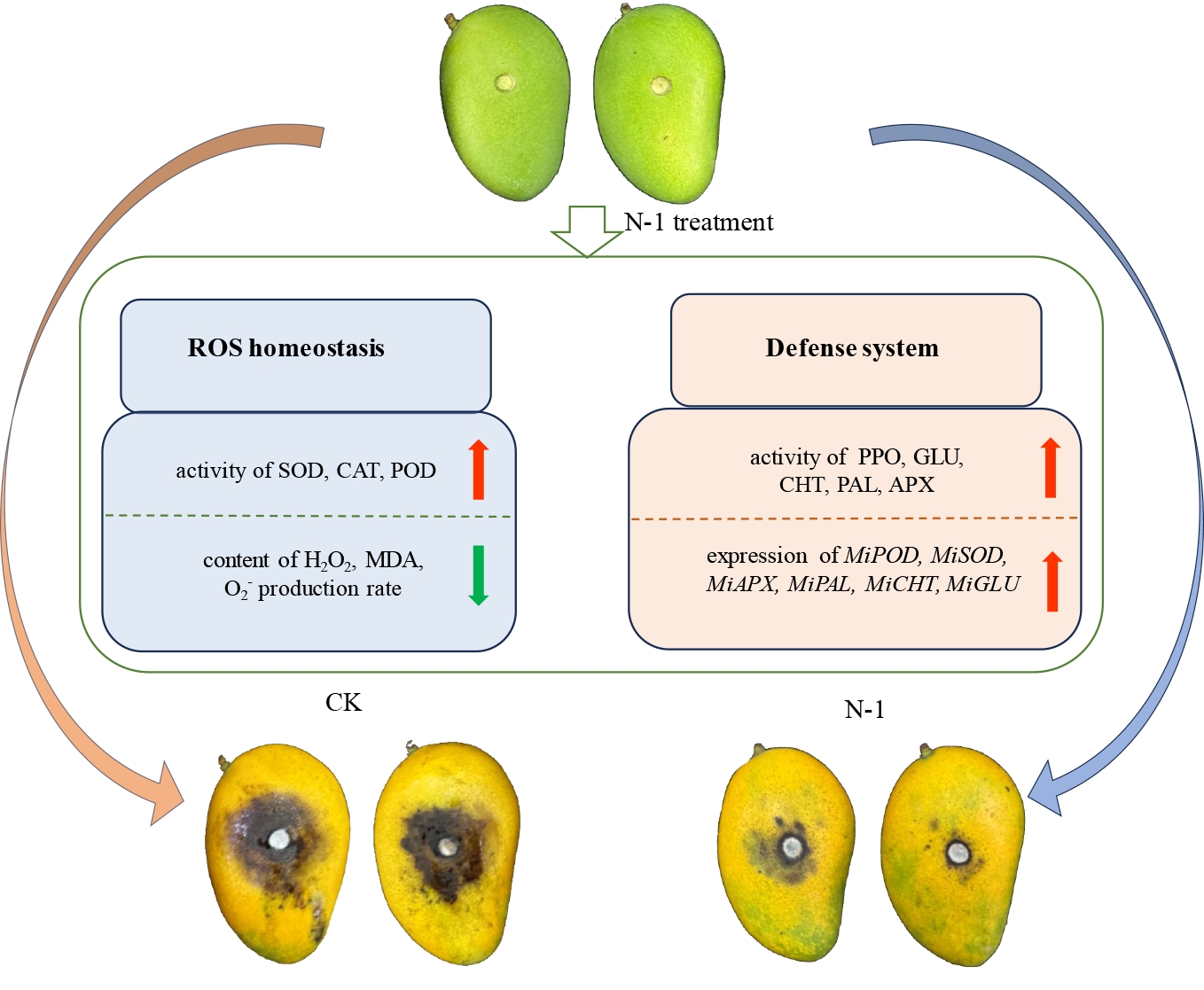
This study demonstrated that the antagonistic bacterium N-1 not only significantly inhibited the mycelial growth of Colletotrichum gloeosporioides but also remarkably improved the storage quality and nutritional quality of postharvest mangoes. The disease resistance of mangoes was enhanced by this antagonistic bacterium through regulating reactive oxygen homeostasis and elevating the activities of defense-related enzymes. Specifically, N-1 promoted the accumulation of antioxidant substances (total phenols and flavonoids), which contributed to the regulation of ROS homeostasis, and simultaneously upregulated the activities of ROS-scavenging enzymes, including APX, SOD, and CAT. In addition, N-1 enhanced the activities of antioxidant and disease resistance-associated enzymes (PPO, POD, PAL, CHT, and GLU) as well as the expression levels of their encoding genes, thereby strengthening the resistance of mangoes to anthracnose. These findings elucidated the mechanism underlying the disease resistance induction of mangoes by N-1 and provided novel insights into the biocontrol mechanism of antagonistic bacteria in enhancing the postharvest disease resistance of mangoes.
-
The authors confirm contribution to the paper as follows: conceptualization, writing – original draft: He Y, Li R; methodology: He Y, Cui Y, He J; software, data curation: He Y; validation, visualization: He Y, Cui Y; formal analysis, investigation: He Y, He J; writing – review and editing: Aslam MM, Li W; supervision: Li R, Shao Y, Li W; project administration, funding acquisition: Li R. All authors reviewed the results and approved the final version of the manuscript.
-
All data generated or analyzed during this study are included in this article.
-
The authors declare that they have no conflict of interest.
-
Received 11 December 2025; Accepted 17 April 2026; Published online 25 August 2026
- Copyright: © 2026 by the author(s). Published by Maximum Academic Press on behalf of Hainan University. This article is an open access article distributed under Creative Commons Attribution License (CC BY 4.0), visit https://creativecommons.org/licenses/by/4.0/.
-
About this article
Cite this article
He Y, Cui Y, He J, Aslam MM, Li R, et al. 2026. Bacillus siamensis (N-1) enhances postharvest disease resistance in Tainong mango by maintaining ROS homeostasis and regulating defense enzyme activities. Tropical Plants 5: e030 doi: 10.48130/tp-0026-0022 

Bacillus siamensis (N-1) enhances postharvest disease resistance in Tainong mango by maintaining ROS homeostasis and regulating defense enzyme activities
- Received: 11 December 2025
- Revised: 16 February 2026
- Accepted: 17 April 2026
- Published online: 25 August 2026
Abstract: Mango is a prominent tropical fruit of Hainan, yet its postharvest susceptibility to spoilage, particularly anthracnose caused by Colletotrichum gloeosporioides, limits the development of the mango industry. In this study, treatment with Bacillus siamensis N-1 reduced disease incidence by 22.22% to 55.55% compared with the control group, delayed fruit softening and color change, and enhanced nutritional quality by increasing ascorbic acid (AsA), titratable acidity (TA), and total soluble solids (TSS). Moreover, N-1 treatment significantly reduced ethylene production, respiratory rate, hydrogen peroxide (H2O2) content, superoxide anion (O2−) generation, malondialdehyde (MDA) accumulation, and membrane electrolyte leakage. Compared with the CK group, the MDA content in the treatment group was reduced by 28.6% to 49.0%. while elevating total phenols and flavonoid contents. At the enzymatic level, treatment with N-1 also enhanced the activities of ascorbate peroxidase (APX) and superoxide dismutase (SOD), the key enzymes in the reactive oxygen species (ROS) scavenging system. It also enhanced the activities of antioxidant-related enzymes, including peroxidase (POD), catalase (CAT), and polyphenol oxidase (PPO), as well as disease resistance-related enzymes such as chitinase (CHT), β-1,3-glucanase (GLU), and phenylalanine ammonia-lyase (PAL). CHT activity in the treated group increased significantly by 127.21% relative to the control group at the late storage stage. And simultaneously elevated the expression levels of their corresponding genes. In conclusion, N-1 treatment could enhance the disease resistance of mango fruit by maintaining reactive oxygen species (ROS) homeostasis and upregulating the activities of defense enzymes, offering a promising strategy for the biological preservation in mango fruit.
-
Key words:
- Mango fruit /
- Bacillus siamensis /
- Postharvest quality /
- ROS /
- Defense enzymes