












-
Carbohydrates are fundamental constituents of food, directly influencing its nutritional value, sensory attributes, and processing behavior. Among them, fructose is a major soluble monosaccharide widely distributed in fruits, honey, and processed foods, contributing significantly to sweetness and consumer acceptance. Its phosphorylated derivative, fructose-6-phosphate (F6P), is a key intermediate in glycolysis and hexose metabolism, linking sugar's composition to biochemical transformations that occur during food processing and storage. The determination of fructose and F6P is therefore important not only for understanding metabolic pathways but also for evaluating food's quality, authenticity, and biochemical changes in complex matrices[1,2].
Despite their importance, the quantitative analysis of sugar phosphates such as F6P remains analytically challenging. Current approaches are dominated by liquid chromatography (LC)–mass spectrometry (MS)-based methods, including stable isotope-assisted LC-MS/MS techniques, which offer high sensitivity and specificity for metabolite profiling and flux analysis[3]. However, these methods typically require labor-intensive sample preparation, expensive instrumentation, and advanced technical expertise, limiting their routine application in food analysis laboratories and high-throughput screening scenarios[4]. Furthermore, comprehensive metabolite coverage often necessitates multiplatform analytical strategies, increasing both the operational complexity and the cost[5−7]. These limitations highlight the need for simple, rapid, and cost-effective analytical methods suitable for routine food analysis.
Analysis of the biochemical and kinetic parameters of enzymes such as glucosamine-6-phosphate deaminase (GlcN-6-P deaminase) is crucial for understanding the central carbon pathways which convert glucosamine-6-phosphate (GlcN-6-P) to F6P. Monitoring such enzymatic transformations is relevant for studying metabolic function, fermentation processes, and enzymes' functionality. However, rapid and selective detection methods for the formation of F6P in these systems remain limited, hindering efficient functional evaluation of such enzymes[8].
The Seliwanoff reaction, first reported almost 140 years ago,[9] is a classical colorimetric assay that selectively detects ketohexoses via acid-catalyzed dehydration and subsequent reaction with resorcinol to produce a characteristic orange-red chromophore. This reaction has long been applied for qualitative and quantitative detection of fructose in food samples because of its simplicity and visual readout[10]. However, its application to sugar phosphates, particularly F6P, has not been systematically optimized, and challenges remain in achieving selectivity in the presence of structurally similar aldoses and amino sugars, which are commonly found in food matrices.
In addition, fructose levels vary widely across different fruits and plant-derived foods, depending on the species, cultivar, and ripening stage, further complicating accurate quantification in real samples[11−16]. These variations, combined with matrix interference, demand analytical methods that are both selective and adaptable to diverse food systems.
To address these challenges, this study revisits and systematically optimizes the Seliwanoff reaction to establish a robust photometric assay for the selective detection of fructose and F6P. The method is designed to provide high specificity toward ketohexoses while maintaining operational simplicity. Its applicability is demonstrated through a quantitative analysis of fructose in various fruit samples and real-time monitoring of the formation of F6P during enzymatic conversion of GlcN-6-P. The developed workflow integrates food sample analysis and enzyme activity monitoring into a unified, rapid colorimetric detection platform. This work presents a practical and accessible analytical strategy for detecting ketohexose, offering significant potential for applications in food chemistry, quality control, and evaluations of enzyme activity.
-
Unless otherwise specified, all chemicals were purchased from Sigma-Aldrich (China) or Sangon Biotech (China) and were of analytical grade.
Seliwanoff reaction
-
Resorcinol (0.18 g) was dissolved in 100 mL of 33% hydrochloric acid. The solution was protected from light, freshly prepared, and used immediately. Briefly, 50 µL of a sample (the standard solution, fruit extract, or enzymatic reaction mixture) was transferred into a microcentrifuge tube, followed by the addition of 100 µL of Seliwanoff reagent. The mixture was vortexed thoroughly and incubated in a metal bath at 95 °C for 10 min. The reaction was then immediately quenched in an ice bath for 2 min. After centrifugation at 4,000 r/min for 5 min, 100 µL of the supernatant was transferred to a 96-well microplate. Absorbance was measured at 480 nm using a microplate reader.
Construction of standard curves
-
Standard solutions of glucosamine, fructose, glucose, galactose, mannose, glucosamine-6-phosphate, and F6P were prepared as a series of concentrations ranging from 0 to 1.5 mmol/L (0, 0.2, 0.4, 0.6, 0.8, 1.0, and 1.5 mM). For each concentration, 50 µL of the standard solution was subjected to the Seliwanoff colorimetric assay under the conditions described above. The absorbance at 480 nm was then recorded. Standard curves for each compound were constructed by plotting the concentration (mM) versus the absorbance at 480 nm.
Fruit sample preparation and fructose determination
-
Twelve common fruits, including pitaya (Selenicereus undatus), grape (Vitis vinifera), apple (Malus domestica), pear (Pyrus spp.), mango (Mangifera indica), mulberry (Morus spp.), kiwi (Actinidia chinensis), orange (Citrus × aurantium), strawberry (Fragaria × ananassa), pineapple (Ananas comosus), cantaloupe (Cucumis melo), and watermelon (Citrullus lanatus), were selected for analysis. For each fruit, 5 g of the edible portion was weighed and thoroughly homogenized to obtain a uniform slurry. Distilled water was added to each homogenate to a final volume of 5 mL (w/v = 1:1), and the mixture was thoroughly vortexed. The samples were then centrifuged at 5,000 r/min for 10 min, and the supernatant was collected. Through preliminary experiments, 100-, 200-, 400-, and 800-fold dilutions were compared. The results showed that the 400-fold dilution ensured that the absorbance of all fruit samples fell within the linear range of the fructose standard curve with good reproducibility. Accordingly, 5 µL aliquots of each supernatant was diluted 400-fold with distilled water. Subsequently, 50 µL of the diluted solution was added to the Seliwanoff reaction system (the final reaction volume was consistent with the standard assay conditions), and the absorbance was measured at 480 nm. Fructose content (mg/g fresh weight) in each sample was calculated on the basis of the established fructose standard curve. All experiments were performed in triplicate.
Recombinant protein expression and purification
-
The expression and purification procedure was performed following a strategy similar to previously reported recombinant enzyme production methods[17], with minor modifications to optimize yield and stability. Briefly, recombinant Escherichia coli BL21(DE3) harboring the ThteGPDA gene was cultured overnight in 5 mL of lysogeny broth (LB) medium supplemented with the appropriate antibiotic at 37 °C and 200 r/min. The culture was then inoculated (1:100, v/v) into fresh LB medium and grown until the optical density at 600 nm (OD600) reached 0.6 under the same conditions. Protein expression was induced with 1 mmol/L Isopropyl β-D-1-thiogalactopyranoside (IPTG), followed by incubation at 18 °C for 18 h. Cells were harvested by centrifugation and resuspended in a lysis buffer (50 mmol/L Tris-HCl, 500 mmol/L NaCl, 1% Triton X-100, pH 7.0), and disrupted by sonication on ice. After centrifugation (12,000 r/min, 20 min, 4 °C), the supernatant was collected and heat-treated at 65 °C for 30 min to remove thermolabile proteins, followed by a second round of centrifugation. The clarified supernatant was applied to a Ni2+-NTA affinity column pre-equilibrated with a binding buffer. After washing with a buffer containing 20 mmol/L imidazole, the target protein was eluted with 250 mmol/L imidazole. The eluted fractions were concentrated by ultrafiltration and exchanged into a storage buffer (50 mmol/L Tris-HCl, 100 mmol/L NaCl, pH 7.0). Protein purity was analyzed by sodium dodecyl sulfate–polyacrylamide gel electrophoresis (SDS-PAGE), and the concentration was determined via the Bradford assay.
Enzymatic characterization
-
The optimal pH and temperature of ThteGPDA were determined. For optimal pH determination, 45 µL of the purified enzyme solution (2.3 mg/mL) was mixed with 5 µL of the substrate solution (40 mmol/L GlcN6P), and the reaction was carried out in buffer systems with different pH values ranging from 5.0 to 10.5. For determining the optimal temperature, reactions were performed at various temperatures ranging from 25 to 85 °C, while all other conditions were kept constant. After incubation, 50 µL of the reaction mixture was subjected to the Seliwanoff colorimetric assay, and absorbance was measured at 480 nm. The concentration of F6P was calculated using a standard curve for F6P. Relative enzyme activity was expressed as a percentage of the maximum activity, which was defined as 100%. All experiments were performed in triplicate.
Enzyme activity assay
-
The enzymatic reaction was carried out in a total volume of 50 µL, consisting of 45 µL of the purified enzyme solution (2.3 mg/mL) and the 5 µL of substrate solution (40 mmol/L GlcN6P). The reaction was initiated by addition of the substrate, and the sample was incubated at 60 °C for 2 h. The reaction was terminated by heating at 95 °C for 10 min to inactivate the enzyme. The mixture was then centrifuged at 12,000 r/min for 5 min, and the supernatant was collected for subsequent analysis. A 1-µL aliquot of the supernatant was subjected to thin-layer chromatography (TLC) for product profiling. For quantitative analysis, 45 µL of the supernatant was mixed with 100 µL of Seliwanoff reagent and incubated at 95 °C for 10 min. After cooling to room temperature, absorbance was recorded at 480 nm for product quantification.
-
Colorimetric analysis using a 96-well microplate showed that, within the concentration range of 0−1.5 mM, the color intensity of fructose and F6P increased progressively with increasing concentrations. In contrast, glucosamine, glucose, galactose, mannose, and glucosamine-6-phosphate exhibited no noticeable color development, and their absorbance values were similar to those of the blank control (Fig. 1a). Sucrose also produced a visible color response under the same conditions, but the color intensity was much weaker than that of fructose at equivalent concentrations, indicating that its contribution to the overall signal is negligible. These observations are consistent with the known selectivity of the Seliwanoff reaction toward ketohexoses, which undergo rapid acid-catalyzed dehydration to form colored products, whereas aldoses react much more slowly[9,10].
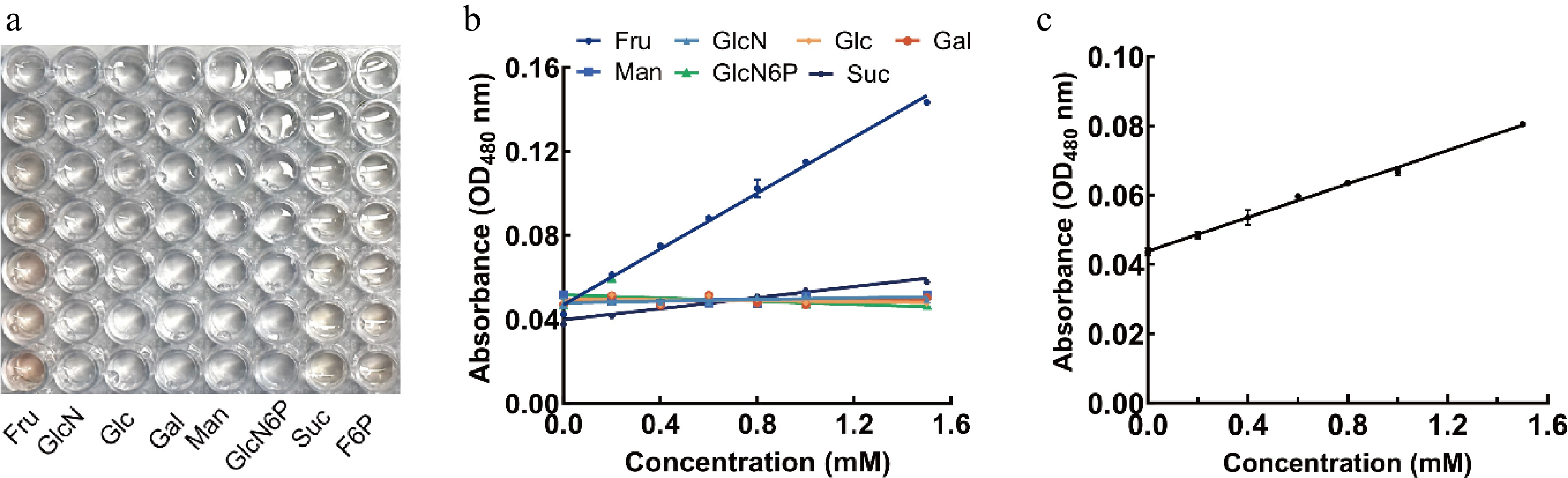
Figure 1.
Color changes and calibration curves of the Seliwanoff colorimetric assay for detecting sugar. (a) Color changes of seven sugars in a 96-well plate system (0−1.5 mM). (b) Calibration curve of fructose. (c) Calibration curve of fructose-6-phosphate (F6P).
On the basis of these results, further absorbance measurements were made to construct standard curves. Fructose exhibited a strong linear relationship within the tested concentration range (R2 = 0.9937) (Fig. 1b), and F6P also showed a good linear correlation (R2 = 0.9906) (Fig. 1c). The slightly lower slope observed for F6P compared with fructose may be attributed to the presence of the phosphate group, which can influence dehydration efficiency and chromophore formation under acidic conditions.
Compared with the conventional Seliwanoff method, the optimized assay demonstrated improved selectivity toward fructose and F6P, enabling effective discrimination from structurally related sugars. Similar resorcinol-based spectrophotometric approaches have been reported for detecting fructose; however, their application to phosphorylated sugars has been limited. The strong linear relationships obtained in this study indicate that the optimized method is suitable for reliable quantitative analyses and for determining enzyme activity in subsequent experiments.
Determination of fructose in fruit samples
-
In the 96-well microplate assay, distinct colorimetric responses were observed among different fruit samples with each sample assayed in triplicate wells as biological replicates (Fig. 2a). Apple, grape, and pear displayed strong color development, whereas pitaya, strawberry, and mulberry showed relatively weak signals, indicating substantial variation in fructose levels among fruits. A quantitative analysis based on the absorbance measurements and the established standard curve revealed that the fructose content in 12 fruit samples ranged from 11.6 to 83.7 mg/g fresh weight (Fig. 2b, c). Grapes exhibited the highest level (83.7 ± 1.9 mg/g), whereas mulberries showed the lowest (11.6 ± 0.6 mg/g). Intermediate levels were observed in watermelon, orange, kiwi, and cantaloupe. These experimental workflows and results are consistent with the analytical approach illustrated in the graphical workflow.
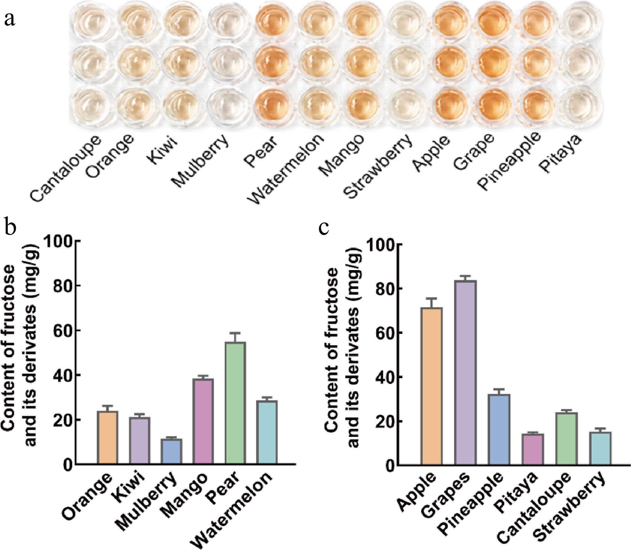
Figure 2.
Fructose determination in fruit samples using the optimized Seliwanoff assay. (a) Colorimetric responses of different fruit extracts in the 96-well microplate assay; (b) calibration-based quantification of fructose content; (c) distribution of fructose content among 12 fruit samples.
These findings are consistent with previous reports demonstrating that fructose, as a major soluble monosaccharide in fruits, varies significantly across species and cultivars[12,13], with higher accumulation typically observed in grape and pear and lower levels in berry fruits[14]. Moreover, fructose and glucose generally constitute the dominant soluble sugars in fruits, and their accumulation is strongly influenced by the cultivar, environmental conditions, and ripening stage[11,15]. The optimized Seliwanoff assay presented here enables the reliable quantification of fructose in complex fruit matrices, demonstrating strong applicability for real sample analysis. Compared with conventional chromatographic approaches, this method provides a rapid and straightforward alternative for the routine screening of fructose.
ThteGPDA-catalyzed conversion of GlcN6P to F6P
-
To verify the catalytic activity of ThteGPDA in converting GlcN6P to F6P, TLC was used to analyze the reaction products (Fig. 3a). Compared with the negative control, the GlcN6P spot disappeared, and a new product spot appeared at an Retardation factor (Rf) value of 0.12. This spot corresponded to the migration position of the F6P standard, confirming the formation of F6P.
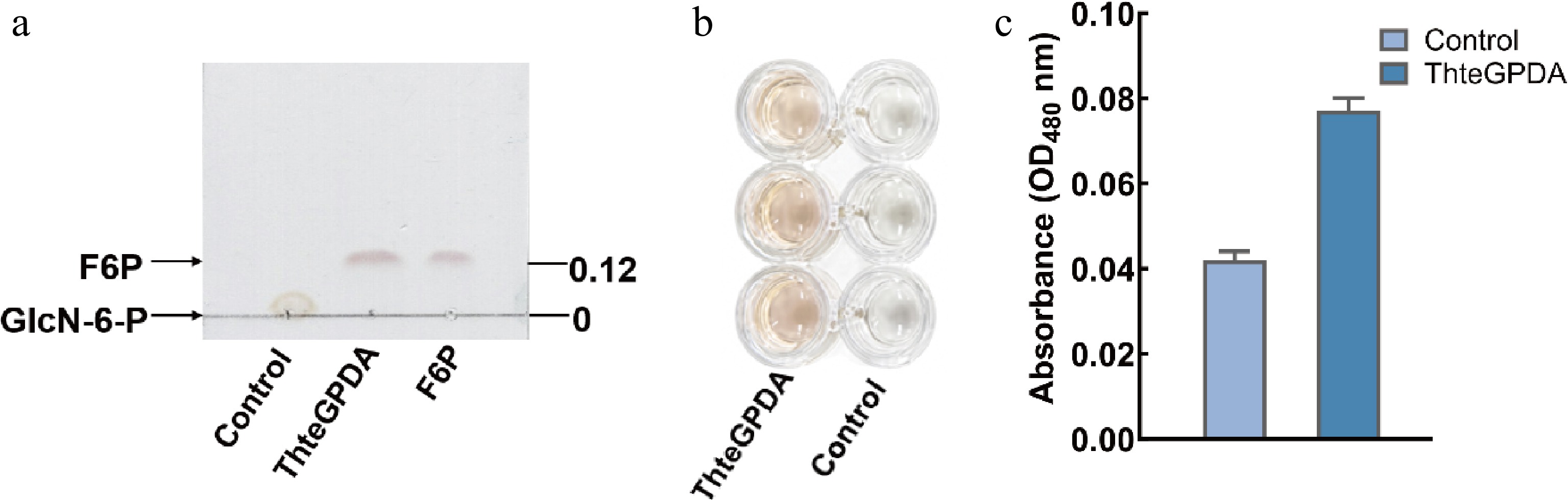
Figure 3.
Catalytic conversion of GlcN6P to F6P by ThteGPDA. (a) TLC analysis of the reaction mixture. Lanes correspond to the negative control (GlcN6P without the enzyme), the enzymatic reaction system, and the F6P standard. (b) Representative visual color change in the Seliwanoff assay. (c) Absorbance response at 480 nm of the reaction system compared with the control.
The reaction mixture was further evaluated using the optimized Seliwanoff assay (Fig. 3b, c), which showed the development of a distinct orange-red color and a significantly increased absorbance at 480 nm compared with the control. This colorimetric response is characteristic of ketohexoses and provides direct evidence of product formation. Quantitative analysis was performed using the F6P calibration curve after appropriate dilution, yielding an F6P concentration of 2.76 ± 0.26 mmol/L (n = 3). The overall workflow and analytical strategy are consistent with the experimental design illustrated in the graphic summary.
Conventional analytical methods for detecting sugar phosphates, such as derivatization-based LC-MS approaches[18−20] and targeted phosphosugar profiling techniques[21], typically require complex sample preparation and sophisticated instrumentation. In addition, comprehensive metabolite analysis often relies on high-resolution MS platforms and multistep workflows, which limit throughput and accessibility[22]. Recent metabolomics studies further highlight that the accurate detection of sugar phosphates frequently requires the integration of multiple LC-MS methodologies to ensure coverage and reliability[23]. Although the colorimetric method presented here does not offer the structural resolution of MS-based techniques, its high selectivity toward ketohexoses and ease of operation make it particularly suitable for rapid screening, routine analysis, and preliminary enzymatic studies in food and biochemical systems.
A direct cost comparison further highlights the advantage of our method. Commercial fructose assay kits require expensive enzymes (hexokinase, phosphoglucose isomerase), cofactors (adenosine triphosphate [ATP], nicotinamide adenine dinucleotide phosphate [NAD(P)H]), and precise temperature control. In contrast, our colorimetric assay is inexpensive, uses stable reagents, and generates a detectable signal within 10 min without multistep handling procedures. Thus, although commercial kits remain suitable for absolute quantification in complex matrices, our method offers an economical alternative for routine screening.
Biochemical characterization of ThteGPDA
-
SDS-PAGE analysis showed that the purified ThteGPDA exhibited a single distinct band at approximately 28 kDa, consistent with the predicted molecular weight (Fig. 4a), indicating high purity of the target protein. With GlcN6P as the substrate, the effects of pH and temperature on enzyme activity were systematically investigated. As shown in Fig. 4b, ThteGPDA exhibited a clear pH-dependent activity profile, with the maximum activity observed at pH 7.0. The enzyme retained relatively high activity within the pH range of 6.0−7.0, whereas activity decreased markedly under more acidic or alkaline conditions, retaining only approximately 22% residual activity at pH 5.5.
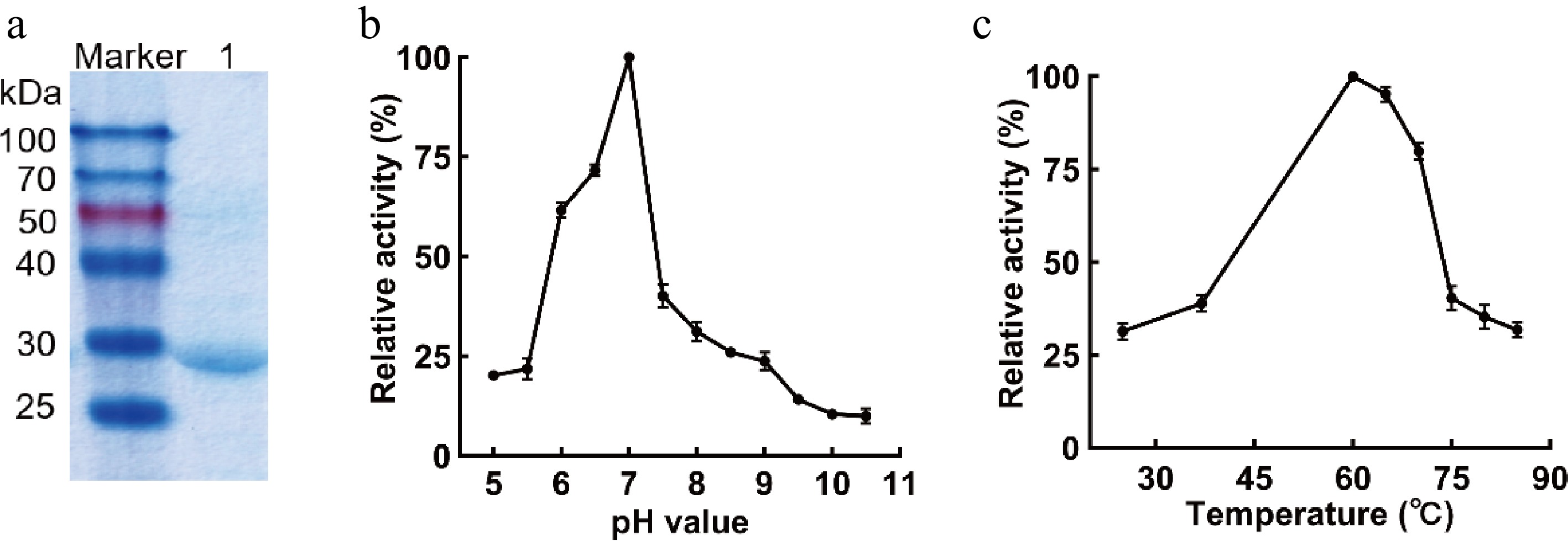
Figure 4.
Biochemical properties of ThteGPDA. (a) SDS-PAGE analysis of recombinant ThteGPDA purification. Lane marker, protein molecular weight marker; Lane 1, purified ThteGPDA obtained by Ni2+-NTA affinity chromatography; (b) pH-dependent activity profile of ThteGPDA; (c) temperature-dependent activity profile of ThteGPDA.
The effect of temperature on enzyme activity is presented in Fig. 4c. ThteGPDA reached its optimal activity at 60 °C and maintained relatively high activity between 60 and 70 °C. However, enzyme activity decreased rapidly above 70 °C, with only approximately 34% residual activity observed at 80 °C, indicating partial thermal instability at elevated temperatures. Compared with previously reported GlcN6P deaminases, enzymes from different sources exhibit considerable variation under their optimal catalytic conditions[8,18]. Previous studies have shown that this class of enzymes generally displays higher catalytic activity under neutral to mildly alkaline conditions[19], and their catalytic properties are strongly influenced by the source organism, structural organization, and allosteric regulation mechanisms[6]. The ThteGPDA characterized in this study exhibited optimal activity at a neutral pH (7.0) and a maximum activity temperature of 60 °C, suggesting moderate thermal adaptation. This behavior may reflect evolutionary adaptation to the native environmental conditions of the source organism and indicates potential suitability for applications requiring moderate temperature stability, such as enzymatic assays in food-related systems.
-
In this study, a robust photometric analytical system based on the Seliwanoff colorimetric reaction was systematically optimized for the selective detection of fructose and F6P. The developed assay exhibited high specificity toward ketohexoses and demonstrated excellent linearity within the concentration range of 0−1.5 mM, enabling reliable quantitative analysis.
The method was successfully applied for determinto the determination of fructose in diverse fruit samples, revealing a wide variation in content (11.6−83.7 mg/g fresh weight), consistent with known compositional differences in food matrices. In addition, the assay proved effective for monitoring enzymatic reactions, allowing a rapid evaluation of ThteGPDA's activity. Through TLC validation and quantitative analysis, ThteGPDA was confirmed to catalyze the conversion of GlcN6P to F6P, with a product concentration of 2.76 ± 0.26 mM.
Compared with conventional chromatographic and MS-based methods, the proposed approach offers significant advantages in terms of simplicity, speed, and cost-effectiveness, without requiring derivatization or advanced instrumentation. Although it does not provide structural resolution comparable with LC-MS techniques, its high selectivity and operational convenience make it particularly suitable for rapid screening, routine analysis, and preliminary enzyme characterization. This optimized Seliwanoff-based assay provides a practical and accessible tool for carbohydrate analysis and evaluations of enzyme activity, with broad potential applications in food chemistry, quality control, and metabolic studies.
-
The authors confirm their contributions to the paper as follows: study conception and design, draft manuscript preparation: Liu L, Voglmeir J; data collection: Zhang L; analysis and interpretation of results: Zhang L, Sun ZR. All authors reviewed the results and approved the final version of the manuscript.
-
All data generated or analyzed during this study are included in this published article.
-
The authors declare that they have no conflict of interest.
- Copyright: © 2026 by the author(s). Published by Maximum Academic Press on behalf of Nanjing Agricultural University. This article is an open access article distributed under Creative Commons Attribution License (CC BY 4.0), visit https://creativecommons.org/licenses/by/4.0/.
-
About this article
Cite this article
Zhang L, Sun ZR, Voglmeir J, Liu L. 2026. An optimized Seliwanoff-based photometric assay for selective detection of fructose and fructose-6-phosphate: applications in food analysis and enzyme activity monitoring. Food Materials Research 6: e013 doi: 10.48130/fmr-0026-0012 

An optimized Seliwanoff-based photometric assay for selective detection of fructose and fructose-6-phosphate: applications in food analysis and enzyme activity monitoring
- Received: 30 April 2026
- Revised: 25 May 2026
- Accepted: 01 June 2026
- Published online: 25 August 2026
Abstract: Fructose and fructose-6-phosphate (F6P) are key ingredients and intermediates in food, yet their rapid and selective quantification remains analytically challenging, particularly in complex matrices. In this study, we revisited and systematically optimized the classical Seliwanoff reaction to establish a robust photometric assay for the selective detection of ketohexoses. The optimized system exhibited high specificity toward fructose and F6P over structurally related aldoses and amino sugars, with a reliable linear response in the range of 0−1.5 mmol/L (R2 > 0.99). The method was successfully applied to quantify fructose in diverse fruit samples, revealing substantial variation in its content (11.6−83.7 mg/g fresh weight), consistent with known compositional differences in food matrices. Furthermore, the assay enabled rapid monitoring of the formation of F6P during glucosamine-6-phosphate deaminase-catalyzed reactions, which was independently validated by thin-layer chromatography and quantitative analysis (2.76 ± 0.26 mM). This strategy offers a simple, rapid, and cost-effective alternative for detecting ketohexose without requiring complex instrumentation or derivatization workflows. Overall, this optimized Seliwanoff-based assay provides a practical analytical tool for food-related carbohydrate analysis and enzymatic activity evaluation, with broad applicability in food chemistry and metabolic studies.
-
Key words:
- Fructose /
- Fructose-6-phosphate /
- Seliwanoff reaction /
- Colorimetric assay /
- Food carbohydrate analysis