













-
Vascular dementia (VaD) is the second most common type of dementia after Alzheimer's disease, with its core pathological mechanism being cerebrovascular lesions leading to insufficient blood supply in the brain, resulting in neuronal damage, death, and loss of synaptic connections[1]. Clinically, VaD typically manifests as memory loss, attentional deficits, and executive dysfunction, attributed to a complicated pathology network yet to be fully unveiled[2]. However, there are no specific medications for VaD, and clinical treatment primarily relies on controlling vascular risk factors and managing cognitive symptoms[3]. In-depth research into its pathological mechanisms is crucial for slowing disease progression and identifying novel therapeutic targets.
Acute cerebral ischemia is the core pathological trigger of VaD, which leads to brain tissue damage and neurotransmitter system imbalance through multiple mechanisms[4]. Following cerebral ischemia, the sharp drop in cerebral blood flow triggers neuronal energy metabolism disturbance and mitochondrial dysfunction, resulting in ATP depletion and ion pump inactivation, thereby inducing cytotoxic edema and neuronal death. Ischemia-reperfusion injury further exacerbates the pathological process, with reactive oxygen species (ROS) bursts and excessive microglial activation, forming a vicious cycle that expands neuronal damage[5]. During this process, significant disturbances occurring in the neurotransmitter system induce neural circuit dysfunction, with cholinergic system dysfunction being the most prominent. Typically, the striatum (STR), as the core brain area of the basal ganglia, is densely innervated by cholinergic nerves and shows high sensitivity to ischemic injury, triggering complex neurological dysfunction potentially through the basal ganglia-cortical circuit[6]. Therefore, the occurrence and development of cognitive impairment in VaD are regulated by multiple factors, including neuroinflammation and cholinergic system dysfunction, suggesting the need to block the vicious cycle to jointly attenuate cognitive impairment.
Huperzine A (HupA) is a naturally occurring reversible acetylcholinesterase (AChE) inhibitor, which is one of the commonly used drugs for clinical treatment of cognitive impairment[7,8]. Its mechanism of action is predominantly thought to be the increase of acetylcholine (ACh) in the brain via AChE inhibition, which contributes to the enhancement of cholinergic neurotransmission[7]. Interestingly, it has also been increasingly reported to have neuroprotective, antioxidant, anti-apoptotic, and anti-inflammatory effects[9], suggesting the existence of multi-dimensional mechanisms underlying its cognitive regulatory effects[10]. In consideration of the fact that chronic diseases such as VaD involve multi-node dysfunctions, we reasoned that dissecting an integral mechanism underlying HupA polypharmacology could enlighten synergistic targets and new strategies for cognitive improvement.
In this study, using metabolic profiling, neurochemical assay, functional imaging, and neurocircuit mapping techniques, we show that HupA improves cognitive function in VaD mice through dual mechanisms: on the one hand, it improves synaptic loss via ACh-α7nAChR axis inside the STR; on the other hand, it recruits a STRdGABA→SNrGABA neural circuit to synergistically improve the cognitive behavior in VaD mice. Our findings on this neurometabolic regulation provide an integral perspective for the neuropharmacology of HupA and suggest potential combinatorial targets for cognitive therapy.
-
Male C57BL/6J mice (6−8 weeks old) were purchased from Charles River Laboratories (Charles River, China) and housed in a stable environment maintained at 22 ± 2 °C, 55% ± 5% humidity, and under a 12-h light-dark cycle (lights on: 07:00−19:00). Animals had free access to standard chow and drinking water throughout the experimental period. All animal procedures were approved by the Animal Ethics Committee (2024-05-039) of China Pharmaceutical University (Nanjing, China).
Bilateral common carotid artery occlusion (BCCAO) model and HupA treatment
-
A mouse model of global cerebral ischemia and hypoperfusion was established using a previously described method of bilateral common carotid artery occlusion (BCCAO) with appropriate modifications[11]. Mice were anesthetized with 4% isoflurane (RWD Life Science, China) for induction and maintained under continuous anesthesia with 1.5% isoflurane (30% oxygen). After disinfecting the ventral neck with 75% ethanol and shaving, a 1.0−1.5 cm longitudinal midline incision was made. Subcutaneous tissue and the platysma muscle were bluntly dissected to expose both common carotid arteries (CCAs). Non-invasive elastic vascular clamps were applied to occlude the left and right CCAs for 20−30 min to induce transient cerebral ischemia. Following clamp removal to restore perfusion, one carotid artery was irreversibly ligated to simulate chronic hypoperfusion. The muscle and skin layers were sutured sequentially with 5−0 sutures, and the surgical site was disinfected with povidone-iodine. Mice were placed in a 37 °C recovery chamber until fully awake and then returned to their home cages with access to soft food and water to minimize postoperative stress.
To investigate the efficacy of HupA, HupA injections (0.2 mg·mL−1, WEPON Pharmaceutical Company, Zhejiang, China) were diluted with saline to 0.05 mg·mL−1 for intramuscular (i.m.) administration in mice (0.1 mg·kg−1). For intracerebral in situ injection, the HupA solution was diluted with artificial cerebrospinal fluid (aCSF) to 0.1 μg·μL−1, mixed thoroughly, filtered through a 0.22 μm filter for sterilization, and then injected unilaterally at a volume of 1 μL per side.
Laser speckle imaging
-
Under isoflurane anesthesia (4% induction, 1.5% maintenance with 30% O2), the mouse head was shaved. A 1.5 cm sagittal incision was made to expose the skull between the anterior fontanelle and the sphenoid suture. After cleaning with saline, the skull surface was illuminated with a 780 nm laser using a laser speckle imaging system (MOOR, UK). A high-resolution CCD camera was focused on the area, and exposure parameters were optimized for real-time blood flow imaging.
Behavioral tests
-
Mice were acclimated to the testing room 1 h prior to behavioral experiments. The apparatus was thoroughly cleaned with 75% ethanol between trials to eliminate olfactory cues. All tests were conducted and recorded by experimenters blinded to the treatment groups. Behaviors were recorded with a video tracking system (Logitech, Switzerland) and data were analyzed using ANY-maze software (Stoelting, USA).
Y-maze
-
The Y-maze test was used to assess spatial working memory and exploratory behavior in rodents. At the beginning of the test, each mouse was placed in the central area of the Y-maze and allowed to explore freely for 5−10 min. The sequence and number of entries into each arm were recorded. The spontaneous alternation rate was calculated, with a higher rate indicating better spatial working memory. Data analysis included recording the number of entries and time spent in each arm, and calculating metrics such as entries into the novel arm and active avoidance rate.
Novel object recognition (NOR) test
-
The NOR test was used to evaluate recognition memory. The test consisted of three phases: habituation, familiarization, and testing. During the habituation phase (2−3 d), mice were placed in an empty test chamber for 10 min daily. In the familiarization phase, two identical objects (A and A') were placed in the chamber, and mice were allowed to explore for 5−10 min. For the short-term memory test (conducted 10 min to 1 h after familiarization), one object (A') was replaced with a novel object (B). The long-term memory test was conducted 24 h after familiarization, with object A replaced by novel object C. The time spent exploring the novel (Tn) and familiar (Tf) objects was recorded. Discrimination index (DI) was calculated by (Tn – Tf)/(Tn + Tf). Recognition index (RI) was calculated by RI = [Tn/(Tn + Tf)] × 100%.
Open field test (OFT)
-
The floor of the open field arena (40 cm × 40 cm) was divided into a central zone and a peripheral zone. Before testing, the arena floor was thoroughly cleaned with 75% ethanol and allowed to dry. Each mouse was gently placed in the center of the arena, facing the central zone. Spontaneous locomotor activity was video-recorded for 8 min. After the test, the mouse was removed, and the arena floor was cleaned again with 75% ethanol.
Morris water maze (MWM) test
-
The MWM test was conducted using a white, opaque circular PVC pool (120 cm in diameter, 50 cm high) filled with water (30 cm deep, maintained at 23 ± 1 °C). A hidden transparent Plexiglas platform (10 cm in diameter) was submerged 1 cm below the water surface. The pool was conceptually divided into four quadrants (NE, SE, NW, SW). Distinct spatial cues were placed around the room. The procedure included: a 1-d habituation session (platform removed, 60-s free swim), five consecutive days of training (four trials per day from random start points, recording escape latency and path length to find the hidden platform within 60 s; mice not finding the platform were guided to it and allowed to stay for 10 s), a probe test on day 6 (platform removed, recording time spent in the target quadrant and number of platform crossings during a 60-s trial), and an optional reversal learning test (platform relocated). Lighting and noise levels were kept constant. The pool was cleaned, and mice were dried after each trial. Data from mice showing abnormal behavior (e.g., floating > 10 s) were excluded.
Neurological function assessment
-
Neurological deficit was evaluated using the Bederson scoring system[12]. Mice were placed on a standard platform. While gently lifted by the tail to suspend the body at a 30° angle to the platform surface, with the front paws positioned 10 cm above the surface, the forelimb extension and motor coordination were observed and recorded. A series of three tests was then performed, and neurological function was scored as follows: 0 points (no deficit): Symmetrical forelimb extension, normal vibrissae reflex, and symmetrical resistance to lateral pushing. 1 point (mild deficit): Characteristic flexion reflex (ventral retraction) of the contralateral forelimb when suspended, while the ipsilateral forelimb remains extended. 2 points (moderate deficit): Forelimb flexion as above, plus a significant reduction (> 50%) in resistance to lateral pushing when in a prone position. 3 points (severe deficit): Spontaneous circling behavior towards the contralateral side during free movement (> 3 rotations per min).
Primary neuron isolation
-
Round coverslips (14 mm diameter) were placed at the edge of wells in a 12-well plate, air-dried in a biosafety cabinet, then moved to the center of the wells. They were coated with 0.1 mg·mL−1 poly-D-lysine (Sigma, USA) solution in sterile D-Hanks buffer and incubated at 37 °C for ≥ 2 h. After rinsing with sterile water, coverslips were stored at 4 °C until use. Pregnant dams at embryonic day 16 were anesthetized with isoflurane and euthanized by cervical dislocation. After abdominal disinfection with ethanol, embryos were removed via cesarean section and placed in a culture dish containing HBSS buffer on ice. Under a stereomicroscope, embryonic heads were quickly removed, brains were extracted, hemispheres were separated along the sagittal fissure, meninges were carefully peeled off, and striatal tissues were dissected and transferred to a 15 mL centrifuge tube containing 6 mL of ice-cold D-Hanks solution.
For cell dissociation, a papain digestion solution (80 U enzyme activity, containing 2 mg L-cysteine and 6 µL saturated NaHCO3) was prepared with pre-warmed D-Hanks buffer, activated at 37 °C for 20 min, and filter-sterilized. This solution was added to the tissue samples for digestion at 37 °C for 20 min. After centrifugation at 200 g to remove the enzyme solution, pre-warmed complete medium (Neurobasal medium supplemented with 0.5 mmol·L−1, Glutamine, 1 × B27, 1% penicillin/streptomycin, and 0.5% FBS) was added to stop the reaction. Tissue was gently triturated 10 times using a blue pipette tip. After settling for 2 min, the supernatant was collected. The dissociation step was repeated twice. The cell suspension was counted and seeded onto pre-coated coverslips at densities of 1.3 × 10⁵ cells/cm2 (high density) or 9.2 × 10⁴ cells/cm2 (low density). After 3−4 h, the medium was replaced with serum-free Neurobasal medium (Gibco, USA). Cells were cultured at 37 °C with 5% CO2, and half of the medium was changed every 2 d. All procedures followed sterile techniques, with key steps performed on ice or using pre-warmed (37 °C) reagents to ensure cell viability.
Oxygen-glucose deprivation/reoxygenation (OGD/R) model
-
Cultured primary striatal neurons were washed three times with PBS and then placed in glucose-free medium (Gibco, USA) at 37 °C under hypoxic conditions (95% N2, 5% CO2) for a specified period to induce OGD. For reperfusion, the glucose-free medium was replaced with normal DMEM medium (Gibco, USA) containing glucose, and cells were returned to a normoxic incubator (95% air, 5% CO2) for 24 h. For drug administration, HupA injection was added to the blank culture medium to a final concentration of 10 μmol·L−1.
Liquid chromatography-mass spectrometry (LC-MS) analysis
-
Mice were euthanized by cervical dislocation. The head was sprayed with alcohol, the scalp was incised, and the skull was opened to extract the whole brain. The brain was rapidly dissected into regions, which were placed in 1.5 mL Eppendorf tubes and stored at −80 °C.
For analysis, brain tissue samples were thawed on ice. Approximately 15 mg of tissue was weighed into a clean 1.5 mL tube. An appropriate number of grinding beads were added, and the sample was homogenized using a tissue homogenizer in dry mode (60 Hz, 60 s) once. Then, 100 µL of ultrapure water was added, and homogenization was performed in wet mode (60 Hz, 60 s) twice. Subsequently, 800 µL of precipitation reagent containing internal standards was added, and the mixture was vortexed for 10 min, followed by centrifugation at 18,000 r·min−1 for 10 min at 4 °C. A 750 µL aliquot of the supernatant was transferred to a new tube and dried using a vacuum concentrator. The residue was reconstituted in 200 µL of pure methanol, vortexed for 10 min, and centrifuged at 18,000 r·min−1 for 10 min at 4 °C. A 120 µL aliquot of the supernatant was transferred to a new tube and centrifuged again under the same conditions. Finally, 80 µL of the supernatant was transferred to an injection vial for analysis. For calibration standards, 10 µL of working solution was mixed with 90 µL of blank matrix and processed alongside the samples.
-
Chromatographic separation was performed on a Waters Atlantis T3 column (3 µm, 2.1 mm × 100 mm, USA). Mobile phase A was 0.05% formic acid and 5 mmol·L−1 ammonium formate in water (500 mL H2O + 250 µL formic acid + 250 µL of 5 mol·L−1 ammonium formate solution). Mobile phase B was acetonitrile. The flow rate was 0.3000 mL·min−1, and the column temperature was 40 °C. The gradient program settings are detailed in Supplementary Table S1.
MS analysis was performed using an LC-20AD liquid chromatograph (Kyoto, Japan) coupled with API 4000+ liquid-chromatography/mass spectrometry system (Sciex, USA). Data acquisition and processing used Analyst 1.5.1 software. The ion source was a Turbo Ionspray with electrospray ionization (ESI) in positive ion mode using multiple reaction monitoring (MRM). Key parameters were: IonSpray Voltage (IS) 5,500 V; Temperature (TEM) 550 °C; Collision Gas (CAD) 8 psi; Curtain Gas (CUR) 20 psi; Nebulizer Gas (GS1) 55 psi; Heater Gas (GS2) 50 psi. Detailed MRM parameters for neurotransmitters are listed in Supplementary Table S2.
Viral injection and chemogenetic manipulation
-
Each mouse was anesthetized with 5% isoflurane (RWD Life Sciences, China) and maintained under anesthesia with 2.5% isoflurane. Mice were then placed in a stereotaxic apparatus (Harvard Instruments, USA). A microinjection pump (Harvard, USA) was used to microinject 100 nL volumes of specific virus (BrianVTA, China) into the target region at a rate of 200 nL·min−1. Following injection, the microliter syringe was left in place for 10 min to ensure complete viral diffusion. Coordinates for each brain region are as follows: STRd: (AP, 1.0 mm; ML, 2.4 mm; DV, 2.65 mm) GPe: (AP, −0.46 mm; ML, 2.0 mm; DV, 4.0 mm), GPi (AP, −1.3 mm; ML, 1.8 mm; DV, 4.5 mm), SNr: (AP, −3.28 mm; ML, 1.5 mm; DV, 4.8 mm). AP, ML, DV denote anteroposterior, medial-lateral, and dorsal-ventral positions relative to the bregma point, respectively.
To monitor GABAergic neuronal activity in the STRd, a GABAergic neuron-specific Cre-expressing virus (AAV2/9-GAD67-CRE-mCherry-WRPE-hGH) and a Cre-dependent calcium signal probe (AAV2/9-EF1a-DIO-jGCaMP7s-WPRE-hGH) were bilaterally injected into the STRd. To identify downstream projection targets of the STRd, a pan-neuronal Cre expression virus (AAV2/9-hSyn-CRE-WPRE-hGH-pA) and a Cre-dependent anterograde AAV virus (AAV2/9-EF1a-DIO-YPet-2A-mGFP-WPRE-pA) were bilaterally injected into the STRd. To achieve chemogenetic control of projections from the STRd to the Gpe region, AAV2/1-hSyn-CRE-EGFP-WPRE-hGH pA was bilaterally injected into the STRd, while AAV2/9-EF1a-DIO-hM3Dq-mCherry-WPREs was bilaterally injected into the Gpe. To achieve chemogenetic control of projections from the STRd to the Gpi regions, AAV2/1-Gad-CRE-EGFP-WPRE-hGH pA was bilaterally injected into the STRd, while AAV2/9-EF1a-DIO-hM4Di-mCherry-WPREs was bilaterally injected into the Gpi. To achieve chemogenetic modulation of projections from the STRd to the SNr, AAV2/1-hSyn-CRE-EGFP-WPRE-hGH pA was bilaterally injected into the STRd, while AAV2/9-EF1a-DIO-hM4Di-mCherry-WPREs was bilaterally injected into the SNr. To activate GABAergic neurons in the SNr, AAV2/1-hSyn-CRE-EGFP-WPRE-hGH pA and AAV2/9-EF1a-DIO-hM3Dq-mCherry-WPREs were bilaterally injected into the SNr. To activate cholinergic neurons in the STRd region, AAV2/R-ChAT-CRE-P2A-EGFP-WPRE-hGH and AAV2/9-EF1a-DIO-hM3Dq-mCherry-WPREs were bilaterally injected into the STRd. To achieve chemogenetic regulation of the projection from STRd to SNrGABA neurons, AAV2/1-VGAT1-CRE-WPRE-hGH polyA was injected into the STRd, and AAV2/9-hSyn-DIO-hM3D(Gq)-EGFP-WPREs was injected into the SNr. For chemogenetic manipulation of D1-MSNs, AAV2/9-hSyn-DIO-hM3D(Gq)-P2A-EGFP-WPRE-polyA and AAV2/9-D1-CRE-mCherry-WPRE-hGH-polyA were injected into the STRd. Detailed information on all adeno-associated viruses is provided in Supplementary Table S3.
Stereotaxic implantation of catheters
-
For local drug administration, dual-guide cannulae (inner diameter 0.25 mm) were stereotaxically implanted bilaterally, targeting the STRd (AP +1.0 mm, ML ±2.4 mm, DV −2.65 mm). The cannulae were secured to the skull with dental cement, and dummy stylets were inserted. Mice were returned to their home cages for a 4-d recovery period.
Fiber photometry
-
Mice were anesthetized with isoflurane (induction concentration 4%, maintenance concentration 1%−2%), followed by scalp incision for viral injection into the target brain region. After a 10-min interval for viral diffusion, the injection needle was withdrawn. An optometric optical fibre (300 μm in diameter, NA 0.39, RWD Life Science, China) was implanted along the same trajectory, ensuring precise co-localization of the fiber tip with the viral expression region. The fiber and ceramic ferrule were then fixed to the skull plate using dental cement. Signal acquisition commenced at least 2 weeks post-surgery.
In vivo two-photon calcium imaging in mouse brain
-
Mice were anesthetized with isoflurane (4% for induction, 1.5% for maintenance), fixed in a stereotaxic apparatus, and maintained at body temperature using a heating pad. A cranial window approximately 1.4 mm in diameter was created over the dorsal striatum using stereotaxic coordinates (AP, 1.0 mm; ML, 2.4 mm; DV, 2.65 mm). A small amount of superficial striatal tissue was gently aspirated. Imaging experiments were conducted at least 1 month post-surgery to ensure complete animal recovery. Calcium imaging of neurons within the dorsal medial striatum was performed using a two-photon microscope (Olympus, Japan). First, we scanned the distribution of labeled neuronal somata across different layer depths to identify a focal plane suitable for functional imaging. We designated the position of the shallowest observed vascular structure (directly imaged by the two-photon instrument) as the top layer. Observation then proceeded deeper into the cortex. When somata were no longer detectable at a given depth, that depth was designated as the bottom layer. Images were acquired sequentially from top to bottom. At each 1 μm depth interval, we captured a 512 × 512 pixel distribution map of the cell, thereby obtaining information on neuronal distribution at different depths. Once the imaging focal plane selection was complete, functional imaging could proceed by synchronizing the stimulation event with functional imaging. After acquiring calcium signal images of the neuronal soma, we processed and analyzed the images using the Fiji image processing software. For data analysis, we selected 40 images acquired before stimulation, 90 images during stimulation, and 40 images after stimulation to analyze the stimulus event. Response strength was calculated based on the change in relative soma fluorescence intensity, where a higher response strength indicates stronger soma preference for movement direction. The calculation formula is ΔF/F = (Ft − F0)/(F0 − Fb). Ft represents the average fluorescence intensity of the cell body within the manually delineated region of interest (ROI). F0 denotes the average fluorescence intensity of the ROI cell body in the control group without stimulation. Fb indicates the average background fluorescence intensity in the absence of drug administration. Calculating ΔF/F yields a response curve over time, enabling analysis of calcium signal changes in the neuronal cell body.
Immunofluorescence
-
After euthanasia by cervical dislocation, brain tissue was harvested and fixed in 4% paraformaldehyde at 4 °C for 24 h. Fixed tissues were dehydrated via sucrose gradient immersion in 10%, 20%, and 30% sucrose in 1× PBS at 4 °C until they sank. Tissues were then blotted dry on filter paper, embedded, and sectioned using a cryostat (Leica, Germany). Sections were washed three times with PBS and blocked for 1 h at room temperature in blocking solution (PBS containing 10% normal donkey serum, 0.3% Triton X-100). Sections were then incubated with primary antibodies diluted in blocking solution at 4 °C for 24 h. After washing five times with PBS containing 0.1% Tween-20 (PBST), sections were incubated with appropriate fluorescent secondary antibodies for 1 h at room temperature protected from light. Following five washes with PBST, nuclei were counterstained with DAPI (Abcam, ab104139). For immunofluorescence staining, the following antibodies were used: rabbit anti-c-Fos antibody (Abcam, Cat.# ab190289), rabbit anti-β-Tubulin III (tuBB3) antibody (Cell Signaling Technology, Cat.#5568S), and mouse anti-α7nAChR antibody (Abcolon, Cat.# M20041). All antibodies were selected based on their compatibility with immunofluorescence applications, and their corresponding catalog numbers are consistent with the official designations of the respective suppliers for traceability. Fluorescent imaging and data acquisition were performed using an Olympus FV3000 microscope (Olympus, Japan) with Olympus SW software. Image processing was consistent within each dataset.
Hematoxylin and eosin (H&E) staining
-
H&E staining was performed by Nanjing Youmeng Biotechnology Co., Ltd. (Nanjing, China). Sections were sequentially immersed in 70% ethanol for 1 min, hematoxylin solution for 5 min, rinsed under running water for 5 min, differentiated with 1% hydrochloric acid ethanol for 5 s, rinsed under running water for 10 min, and stained with eosin solution for 1 min. Subsequently, sections were dehydrated sequentially in 70%, 90%, and 100% ethanol for 1 min each, cleared with xylene for 5 min, and finally mounted with neutral resin. After staining, slides were examined under a bright-field microscope, and images were captured for documentation and analysis.
Nissl staining
-
Nissl staining was performed by Nanjing Youmeng Biotechnology Co., Ltd. (Nanjing, China). Sections were sequentially immersed in 100% ethanol for 1 min, 95% ethanol for 1 min, and rinsed with distilled water for 1 min. They were then stained in 0.1% toluidine blue solution for 5 min, rapidly rinsed with distilled water to remove excess dye, followed by 95% ethanol differentiation for 5 s, 100% ethanol dehydration for 1 min, followed by xylene clearing for 5 min. Finally, mount with neutral resin. After staining, observe the distribution of neuronal Nissl bodies under a bright-field microscope, and images were captured for documentation and analysis.
Golgi staining
-
Golgi staining was performed using the FD Rapid GolgiStain Kit (FD NeuroTechnologies) according to the manufacturer's protocol. Sholl analysis of dendritic complexity was conducted using the Simple Neurite Tracer (SNT) plugin in ImageJ, with a Sholl circle spacing of 10 μm.
Statistical analysis
-
Experimental data were processed and statistically analyzed using GraphPad Prism 9.5.1 (GraphPad Software Inc, Canada) and ImageJ (NIH, USA). Statistical significance between two groups was assessed using a two-tailed Student's t-test. Comparisons among multiple groups were performed by one-way analysis of variance (one-way ANOVA). Continuous variables, such as biochemical parameters and behavioral metrics, are presented as the mean ± standard error of the mean (SEM). Statistical significance between groups was evaluated and graphically indicated as follows: * P < 0.05, ** P < 0.01, *** P < 0.001, **** P < 0.0001; non-significant differences are denoted as 'ns'.
-
To investigate the therapeutic effect of HupA on VaD, we established a mouse model using the BCCAO method[11](Fig. 1a). We validated the model by cerebral blood flow imaging (Supplementary Fig. S1a), neurological function score (Supplementary Fig. S1b and S1c), body weight monitoring (Supplementary Fig. S1d), and a series of behavioral tests (Supplementary Fig. S1e–S1q). Next, we evaluated the impact of HupA (0.1 mg·kg−1, i.m.) on cognitive function in BCCAO-induced VaD mice (Fig. 1b). Behavioral tests showed no significant difference in locomotor activity between the two groups in the OFT (Fig. 1c and d; Supplementary Fig. S2a). In the MWM test, HupA-treated mice took significantly less time to locate the platform than model group mice (Fig. 1e; Supplementary Fig. S2b). In the NORT, HupA-treated mice spent significantly more time exploring the novel object (Fig. 1f and g; Supplementary Fig. S2c). Also, in the Y-maze test, the HupA treatment group showed clear improvement in both the time spent in the open arms and spontaneous alternation rate (Fig. 1h and i; Supplementary Fig. S2d). These behavioral data collectively confirm that HupA improved BCCAO-induced cognitive dysfunction.
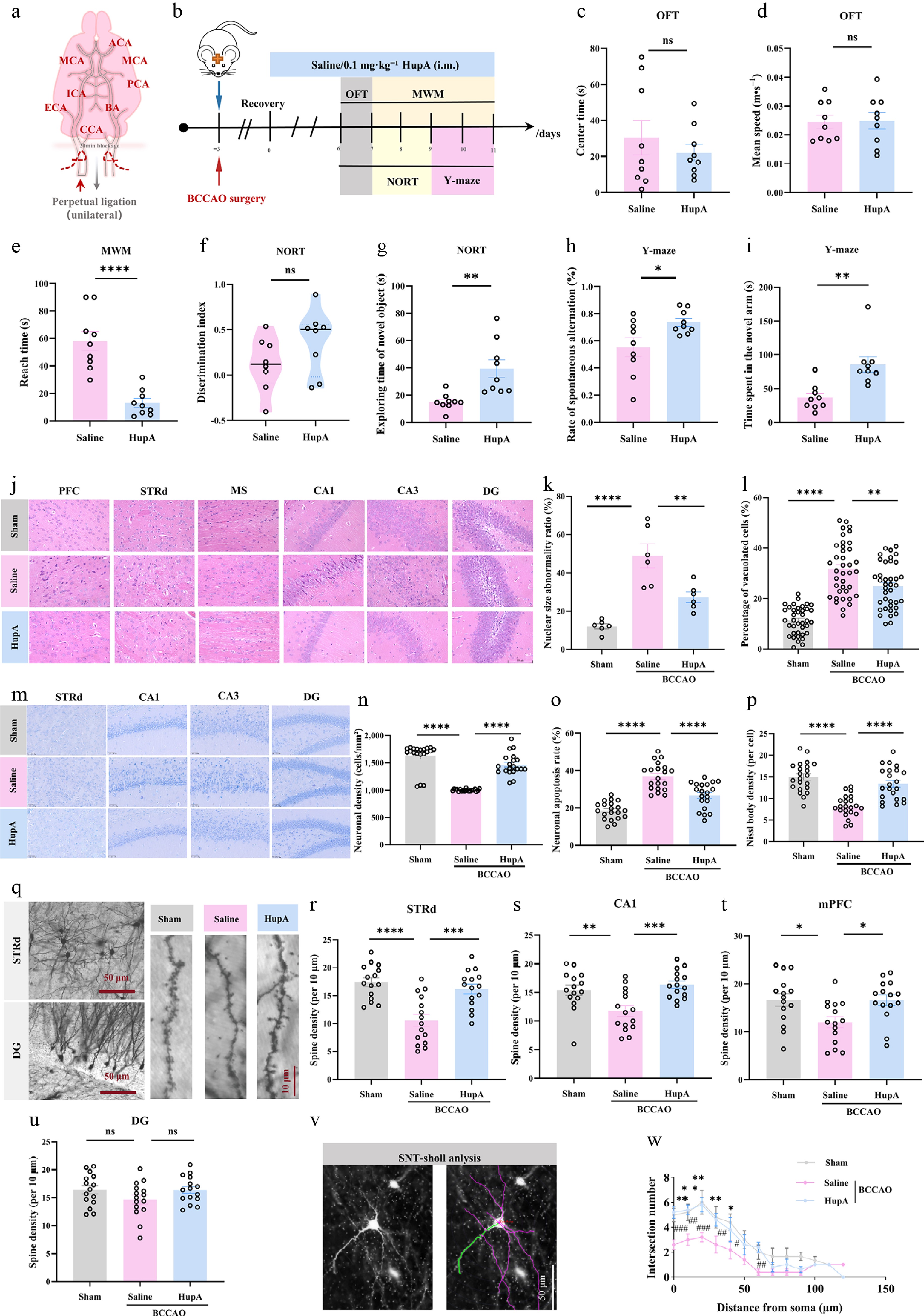
Figure 1.
Intramuscular injection of HupA attenuates cognitive impairment in VaD mice. (a) Schematic diagram of bilateral common carotid artery occlusion (BCCAO) surgery for vessel blockade in mice. (b) Schematic diagram of the Huperzine A (HupA) dosing regimen and behavioral testing timeline in BCCAO mice. i.m., intramuscular. (c) Time spent in the central area during the Open Field Test (OFT) (n = 9). (d) Average movement speed during OFT (n = 9). (e) Time to reach the platform in the Morris Water Maze test (n = 9). (f) Discrimination index in the Novel Object Recognition Test (NORT) (n = 9). (g) Exploration time of the novel object in NORT (n = 9). (h) Spontaneous alternation rate in the Y-maze test (n = 9). (i) Exploration time in the novel open arm of the Y-maze test for each group (n = 9). (j) Representative H&E staining images of different brain regions from each group. Scale bar = 100 μm. PFC, prefrontal cortex. STRd, dorsal striatum. MS, medial septum. CA1, cornu ammonis area 1. CA3, cornu ammonis area 3. DG, dentate gyrus. (k) Percentage of nuclei with abnormal size (n = 6). (l) Percentage of vacuolated cells (n = 38 cells). (m) Representative Nissl staining images of different brain regions. Scale bar = 100 μm. (n) Neuronal density per mm2 (n = 20). (o) Proportion of apoptotic neurons (n = 20). (p) Density of Nissl bodies per cell (n = 22 cells). (q) Representative Golgi staining images from each group. Scale bar = 50 and 10 μm. (r)–(u) Dendritic spine density in the STRd, CA1, mPFC, and DG regions (n = 15). (v) Schematic diagram of Sholl analysis. Scale bar = 50 μm. (w) Results of Sholl analysis. Data are presented as mean ± SEM; * P < 0.05, ** P < 0.01, *** P < 0.001, **** P < 0.0001; # P < 0.05, ## P < 0.01, ### P < 0.001; (c)–(i) two-tailed unpaired Student's t-test; (k), (l), (n)–(p), (r)–(u) one-way analysis of variance.
Next, we evaluated the impact of HupA from histopathological aspects. H&E staining revealed that, compared to the model group, the HupA treatment group exhibited fewer intracellular vacuoles, reduced interstitial edema, and less pronounced nuclear/cytoplasmic displacement and deformation in brain tissue (Fig. 1j–l). Nissl staining similarly showed that HupA intervention markedly alleviated the abnormal distribution of Nissl bodies and neuronal loss caused by BCCAO (Fig. 1m–p). Golgi staining analysis indicated that BCCAO significantly reduced dendritic spine density in the STRd, prefrontal cortex, and hippocampal CA1 region, while HupA treatment restored spine density in these brain areas to levels comparable to the sham group (Fig. 1q–u). Sholl analysis further demonstrated that HupA treatment reversed the BCCAO-induced decline in neuronal complexity (Fig. 1v and w). Furthermore, Iba1 staining showed that HupA treatment mitigated microglial overactivation (Supplementary Fig. S2e–S2k). Together, HupA treatment effectively alleviated BCCAO-induced neuronal damage, neuroinflammation, and cognitive decline.
HupA improves cognitive function by elevating striatal ACh levels
-
Our previous study demonstrated that HupA showed good distribution to major brain regions such as the STR after intraperitoneal injection into rats[13]. To elucidate the mechanism of action of HupA in the VaD model, first we examined its distribution patterns across various brain regions in normal and BCCAO model mice. We observed that HupA was distributed to major brain regions including the hippocampus, medial prefrontal cortex (mPFC), and STR, and the concentrations were largely similar in normal and BCCAO groups (Fig. 2a), suggesting that BCCAO did not affect its brain distribution. Subsequently, using LC-MS analysis, we measured the changes in ACh and several other neurotransmitters such as γ-aminobutyric acid (GABA), norepinephrine (NE), and aspartic acid (Aspa) across different brain regions (Fig. 2b; Supplementary Fig. S3a–S3c). We found that HupA injection significantly increased ACh concentration in multiple brain regions of VaD mice, typically including the cerebellum, STR and parietal cortex (Fig. 2b). In comparison to the change in control groups, HupA induced more pronounced increase in ACh concentration in the VaD groups, with significant rises observed in the striatum, cerebellum, and parietal cortex (Fig. 2b).
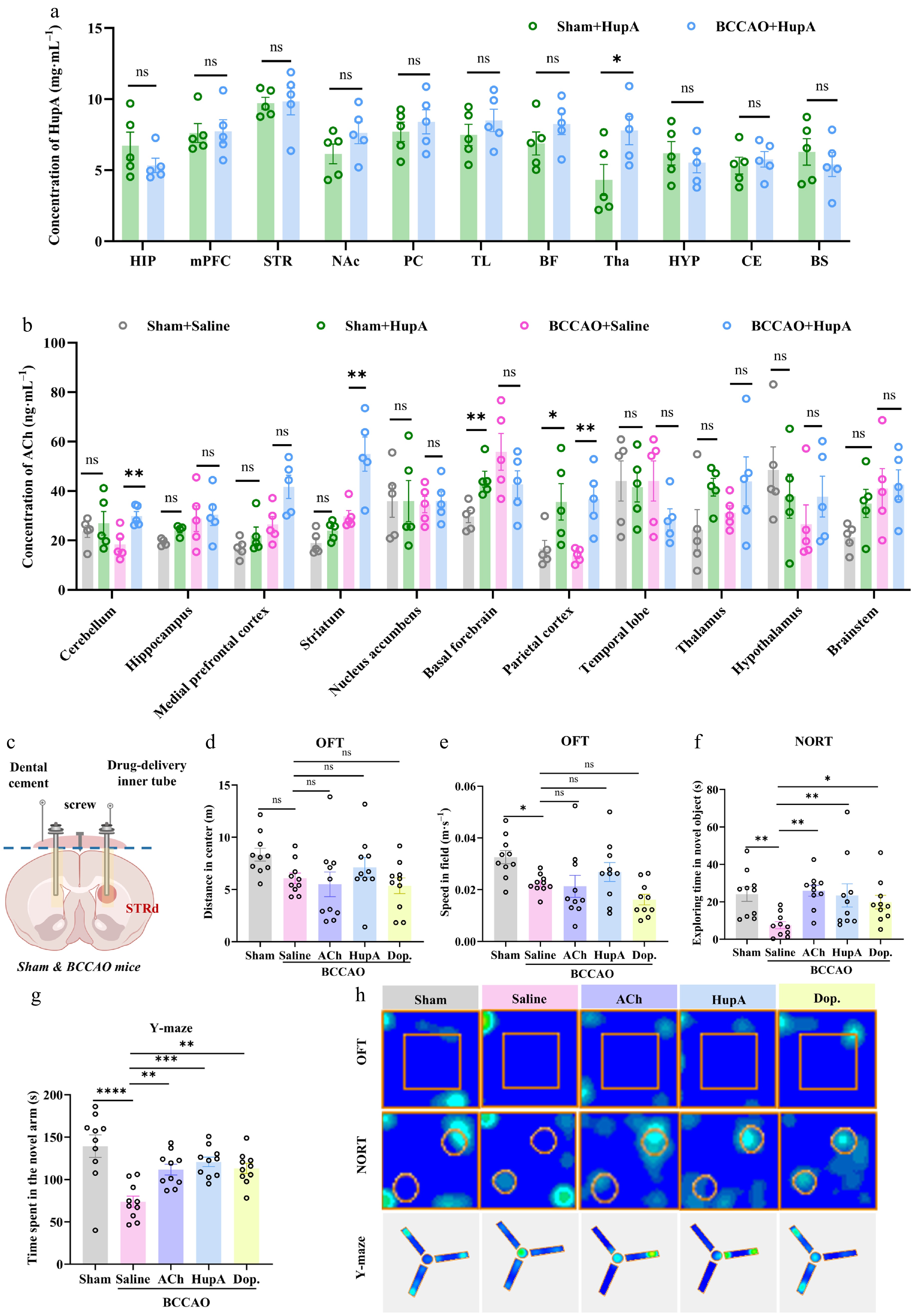
Figure 2.
HupA administration into the STRd confers cognitive improvement in VaD mice. (a) Changes in Huperzine A (HupA) concentration in different brain regions after intramuscular injection of HupA to sham or bilateral common carotid artery occlusion (BCCAO) mice (n = 5). HIP, hippocampus. mPFC, medial prefrontal cortex. STR, striatum. NAc, nucleus accumbens. PC, piriform cortex. TL, temporal lobe. BF, basal forebrain. Tha, thalamus. HYP, hypothalamus. CE, central nucleus of amygdala. BS, brainstem. (b) Changes in ACh concentration in different brain regions of sham or BCCAO mice with or without HupA treatment (n = 5). (c) Schematic diagram of dorsal striatal cannula burial and in situ drug delivery. (d) Time spent in the central area during the Open Field Test (OFT) (n = 10). ACh, acetylcholine. Dop, donepezil. (e) Average movement speed during OFT (n = 10). (f) Exploration time in the novel open arm of the Y-maze test (n = 10). (g) Exploration time of the novel object in the Novel Object Recognition Test (NORT) (n = 10). (h) Representative heatmaps depicting the movement tracks of mice during behavioral tests. Data are expressed as mean ± SEM; * P < 0.05, ** P < 0.01, *** P < 0.001, **** P < 0.0001; (a), (b) two-tailed unpaired Student's t-test; (d)–(g) one-way analysis of variance.
Recent studies have shown that the STRd plays a crucial role in learning and memory, decision-making, and executive functions[14−16]. To verify whether the cognitive improvement induced by HupA primarily involved the STRd, we performed targeted HupA injection into the STRd of BCCAO mice and evaluated behavioral performance (Fig. 2c). OFT results indicated that local HupA injection did not affect locomotor ability or anxiety-like behavior (Fig. 2d, e and h). Y-maze results showed that BCCAO mice receiving STRd HupA injection spent significantly more time exploring the open arms, nearly recovering to the level seen in the normal control group (Fig. 2f and h). The NOR test revealed that STRd HupA injection significantly increased the exploration time of the novel object in BCCAO mice (Fig. 2g and h). These results demonstrate that HupA action within the STRd is sufficient to recapitulate the cognitive improvement effects.
ACh is an important neurotransmitter in the STR, primarily released by cholinergic interneurons[10,14]. Given that HupA could notably increase ACh in this region, we further tested the effects of local injections of exogenous ACh or the AChE inhibitor donepezil (Dop) into the STRd. Behavioral tests showed that ACh or Dop treatment did not affect basic motor function (Fig. 2d, e and h), but similarly improved the performance of BCCAO mice in the Y-maze and NOR tests (Fig. 2f, g and h). Therefore, exogenously elevating ACh levels in the STRd is sufficient to mimic HupA's therapeutic effect, further suggesting that HupA likely improves cognition by enhancing striatal cholinergic transmission.
The neurobiological effect of ACh depends on its interaction with canonical receptors including muscarinic receptors (M-type receptors) and nicotinic receptors (N-type receptors)[17]. To further identify specific ACh receptor subtypes involved in HupA's cognitive improvement, we co-administered HupA locally with either the classical muscarinic ACh receptor (mAChR) antagonist atropine or the nicotinic ACh receptor (nAChR) antagonist benzethonium chloride (Benz) in the VaD model (Fig. 3a). Behavioral tests showed no differences in locomotor ability among the control and different treatment groups (Fig. 3b, c and h). Compared to the HupA treatment group, the HupA+atropine group showed no significant difference in novel object discrimination index or exploration time. In contrast, the HupA+Benz group exhibited significantly decreased discrimination index and exploration time, indicating that nAChR antagonism effectively counteracted HupA's pharmacological activity (Fig. 3d, e and h). Similarly, in the Y-maze test, spontaneous alternation rate was significantly decreased by Benz but not atropine, further supporting a dependence of nAChR for HupA-induced cognitive improvement (Fig. 3f–h). In consistence with the behavioral phenotypes, histopathological analysis also showed consistent changes that Benz intervention markedly weakened HupA's neuroprotective effect, with both H&E and Nissl staining indicating decreased neuronal density and aggravated tissue damage (Fig. 3i and j); in contrast, atropine intervention showed no significant effect on the action of HupA (Fig. 3i and j). Taken together, these data indicate that HupA's cognitive improvement mainly occurs through activating nAChRs.
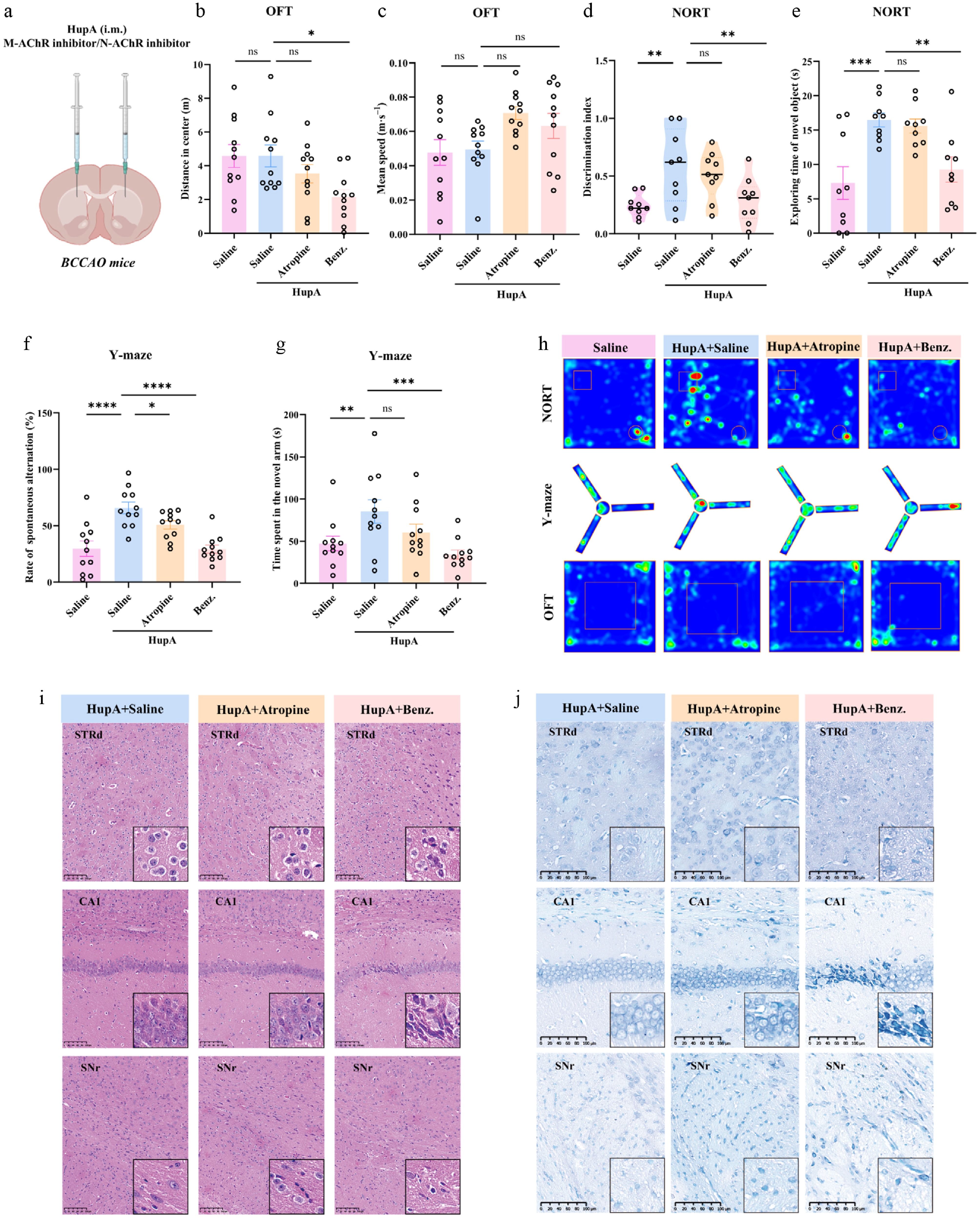
Figure 3.
N-type AChRs mediate the improvement of cognitive dysfunction in VaD mice by HupA. (a) Schematic diagram of in situ administration into the dorsal striatum (STRd) in BCCAO mice. Drug administration: atropine (0.5 mmol·L−1, 1 μL per side, bilateral injection). (b) Distance traveled in the center during the Open Field Test (OFT) (n = 11). Benz., benzethonium chloride. Drug administration: Benz (1 mmol·L−1, 0.25 μL per side, bilateral injection). (c) Movement speed during OFT (n = 11). (d) Discrimination index in the Novel Object Recognition Test (NORT) (n = 9). (e) Exploration time of the novel object in NORT (n = 9). (f) Spontaneous alternation rate in the Y-maze (n = 11). (g) Exploration time in the novel arm of the Y-maze (n = 11). (h) Representative heatmaps depicting the movement tracks of mice during behavioral tests. CA1, cornu ammonis area 1. SNr, substantia nigra pars reticulata. (i) Representative H&E staining images of different brain regions. Scale bar = 100 μm. (j) Representative Nissl staining images of different brain regions; scale bar = 100 μm. Data are expressed as mean ± SEM; * P < 0.05, ** P < 0.01, *** P < 0.001, **** P < 0.0001; (b)–(g) one-way analysis of variance.
α7nAChR mediates the cognitive improvement effect of HupA in VaD mice
-
By re-analyzing a published single-cell dataset[18], we found the Chrna7 gene, which encodes the α7 nicotinic acetylcholine receptor (α7nAChR), was significantly reduced following ischemic stroke (Supplementary Fig. S4a and S4c). However, whether this subtype is involved in the action of HupA in VaD mice remains unclear. To answer this question, we used the highly selective α7nAChR antagonist methyllycaconitine citrate (MLA) and examined its effect in combination with HupA. In the sham-operated controls, MLA injection alone significantly reduced the time mice spent exploring the open arms of the Y-maze, indicating that blocking α7nAChR directly impairs working memory performance and exploratory behavior. Furthermore, when co-administered with MLA, the effect of HupA in promoting open arm exploration was compromised (Fig. 4a and b). The NOR test yielded similar results, where α7nAChR blockade compromised HupA's cognitive benefits (Fig. 4c and d). These results collectively demonstrate an important role of α7nAChR in HupA-mediated cognitive enhancement.
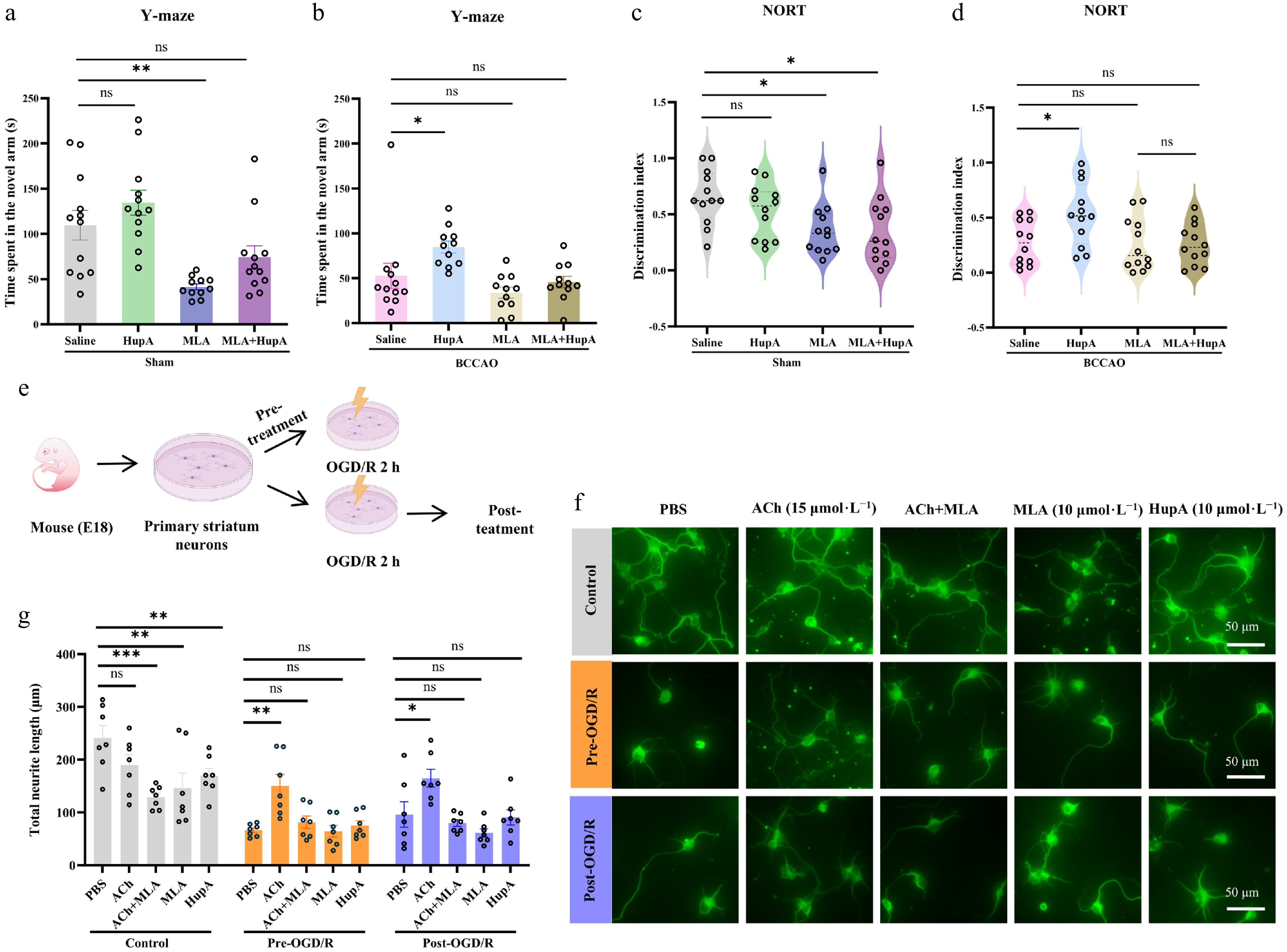
Figure 4.
α7nAChR mediate improvement of cognitive dysfunction in VaD mice by HupA. (a), (b) Exploration time in the novel arm of the Y-maze (n = 11). Drug administration: MLA (methyllycaconitine citrate, 2 μmol·L−1, 1 μL per side, bilateral injection). (c), (d) Discrimination index in the NORT (n = 11). Drug administration: MLA (methyllycaconitine citrate, 2 μmol·L−1, 1 μL per side, bilateral injection). (e) Schematic diagram of study involving the mouse striatal primary neuron extraction, oxygen-glucose deprivation/reoxygenation (OGD/R) and pharmacological treatment. (f) Fluorescence images of neurons after pharmacological treatment. Scale bar = 50 μm. (g) Neurite length after drug treatment (n = 7). Data are presented as mean ± SEM; * P < 0.05, ** P < 0.01, *** P < 0.001, **** P < 0.0001; (a)–(d), (g) one-way analysis of variance.
Based on the animal experiments above, we further verified the role of α7nAChR in a cell model (Fig. 4e). In primary striatal neurons, we administered ACh, the α7nAChR antagonist MLA, or their combination to examine the effects on neuronal growth. Neuronal staining results showed that MLA alone inhibited neurite outgrowth (Fig. 4f and g), indicating that α7nAChR is the key receptor through which ACh promotes neuronal axon growth. Further, in an oxygen-glucose deprivation/reoxygenation (OGD/R) model, we found that ACh treatment significantly increased neurite length in OGD/R model neurons, whereas this increase was completely inhibited in the presence of MLA (Fig. 4f and g), verifying the dependence on α7nAChR function. In contrast, HupA or MLA alone had no significant effect on neurite length in OGD/R model neurons (Fig. 4f and g), suggesting an indirect mechanism by which HupA promotes neural growth via elevating endogenous ACh to act on α7nAChR.
HupA enhances calcium activity in striatal GABAergic neurons
-
In the STRd, over 90% of neurons are known as GABAergic[19]. A complex mutual regulatory relationship exists between cholinergic and GABAergic neurons, and they jointly participate in key brain functions such as motor control and reward processing[20,21]. We injected a ChAT-specific Cre virus into the STRd of BCCAO mice to specifically manipulate striatal cholinergic neurons (Supplementary Fig. S5a). Behavioral results demonstrated that directly activating STRd cholinergic neurons significantly increased novel object exploration time and discrimination index in the NOR test (Supplementary Fig. S5c and S5d), as well as the open arm exploration time in Y-maze (Supplementary Fig. S5e and S5f), which was in line with the activation of ACh signaling by HupA. Furthermore, virus-mediated spine labeling revealed that HupA increased the dendritic spine density of GABAergic neurons in the STRd region (Supplementary Fig. S6a–S6d). To assess the impact of HupA on GABAergic neuron excitability, we injected a Cre-dependent virus (rAAV-GAD67-CRE-mCherry-WRPE-hGH) for GABAergic neuron-specific Cre expression and a Cre-dependent calcium indicator (rAAV-EF1a-DIO-jGCaMP7s-WPRE-hGH) into the STRd to detect calcium signal activity in these neurons (Fig. 5a). We confirmed that the virus was successfully expressed in the STRd, with the optical fiber positioned above this brain region (Fig. 5b). Quantitative analysis revealed that HupA induced a rapid and robust increase in striatal GABAergic calcium activity, with a mean peak amplitude of 2.2 ± 0.5 times the baseline level (Fig. 5c–e) and a time to peak of 30 ± 5 min (Fig. 5f), resulting in sustained calcium elevation with an AUCCa of 1,713 ± 518 (Fig. 5g). Pharmacokinetic analysis of plasma drug concentrations showed that HupA reached its peak plasma concentration (Cmax) at approximately 15 ± 2 min, which immediately preceded the peak calcium response (Fig. 5f). These temporal quantitative data suggest that, upon reaching the brain region, HupA rapidly elevates local ACh levels to activate striatal GABAergic neuronal activity.
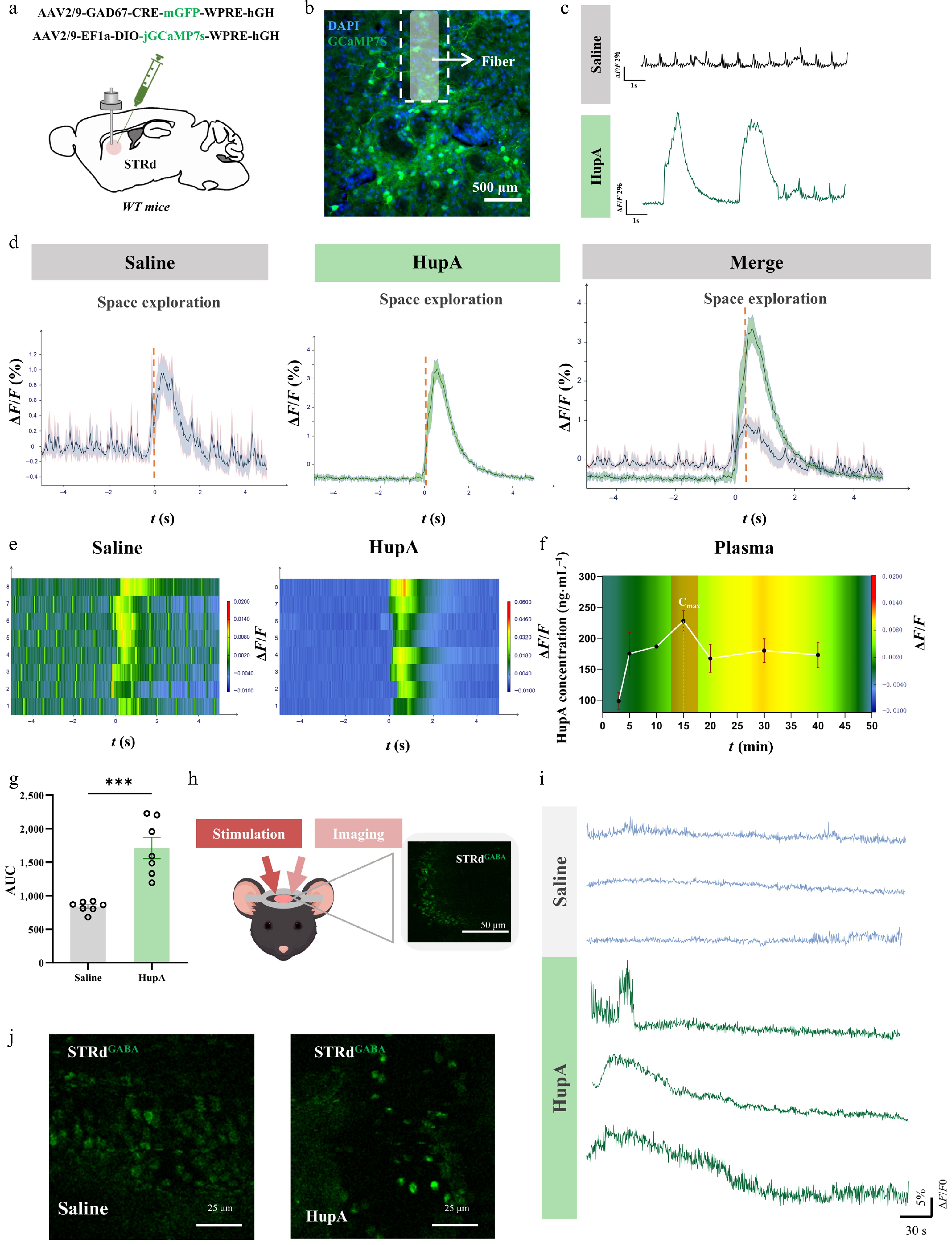
Figure 5.
Intramuscular injection of HupA activates STRd GABA neurons. (a) Schematic diagram of virus injection into the dorsal striatum (STRd) in WT mice. (b) Diagram confirming virus injection site and cannula placement site in the STRd. Scale bar = 500 μm. (c) Characteristic calcium signal waveforms before and after HupA injection. (d) Comparison of calcium signals before and after HupA injection. (e) Representative heatmaps of calcium signals before and after HupA injection. (f) Time-course plot of calcium signaling and plasma drug concentration following HupA injection (0−50 min). The calcium signals were plotted as a heatmap. (g) Statistical values of the area under the curve (AUC) for fiber photometry signals in each group (n = 7). (h) Schematic diagram of in vivo two-photon experiment in normal mice. Scale bar = 50 μm. (i) Characteristic calcium signal waveforms in saline and HupA groups (n = 3). (j) Fluorescence images of calcium signals in GABAergic neurons in the STRd. Scale bar = 25 μm. Data are presented as mean ± SEM; * P < 0.05, ** P < 0.01, *** P < 0.001, **** P < 0.0001; (g) two-tailed unpaired Student's t-test.
Next, we used two-photon calcium imaging to further confirm the activity in STRd GABAergic neurons in normal mice. First, we injected fluorescein isothiocyanate-dextran (FITC-Dextran) to label blood vessels, accurately locating striatal coordinates under the two-photon imaging field. During real-time calcium signal recording, we simultaneously performed HupA injection (Fig. 5h). We observed prominent calcium activity spikes in the striatal region post-injection, indicating that HupA can rapidly activate neuronal activity in this brain area (Fig. 5i and j). Taken together, both fiber photometry and two-photon imaging results demonstrate that HupA injection enhances calcium activity in striatal GABAergic neurons.
Striatal GABAergic neurons are primarily divided into two subtypes: D1-type expressing Medium Spiny Neurons (D1-MSNs) and D2-type expressing Medium Spiny Neurons (D2-MSNs)[22,23]. To identify the potential subtype involved in cognitive function, we used chemogenetics to specifically activate D1-MSNs in BCCAO-induced VaD mice (Supplementary Fig. S7a–S7c). OFT results showed no difference in locomotor activity between the Clozapine-N-oxide (CNO) group and the saline group (Supplementary Fig. S7d–S7f). In the NOR test, the CNO group had significantly increased novel object exploration time and discrimination index (Supplementary Fig. S7g and S7h). In the Y-maze test, compared to the saline group, the CNO group showed increased exploration time in the novel arm and spontaneous alternation rate (Supplementary Fig. S7i and S7j). We also investigated the effect of D1-MSNs activation on neuronal injury. H&E staining results showed that compared to the saline group, the CNO group had a decreased proportion of neurons with abnormal nuclear size and reduced neuronal swelling rate (Supplementary Fig. S7k, S7m and S7n). Nissl staining results indicated that, although neuronal density showed no significant difference between the saline and CNO groups, the density of Nissl bodies was increased in the CNO group (Supplementary Fig. S7l, S7o and S7p). These results suggest that specifically activating D1-MSNs effectively alleviates BCCAO-induced brain tissue damage and cognitive decline, which implies that the D1-MSN subtype of striatal GABAergic neurons may play a role in HupA's improvement of cognitive impairment.
HupA improves cognitive function via a STRdGABA to SNrGABA neural circuit
-
The striatal neurons are deeply involved in various functions such as cognition, emotion regulation, and motor control through complex network connections with multiple brain regions including the prefrontal cortex, thalamus, and basal ganglia[24]. To map the neural circuitry underlying HupA's action, we first used viral tracing to identify major efferent targets of the STRd (Fig. 6a). We observed a robust viral expression in the STRd injection site, while green fluorescent signals were detected in brain regions including the nucleus accumbens (NAc), substantia nigra pars reticulata (SNr), external globus pallidus (GPe), and internal globus pallidus (GPi) (Fig. 6b), suggesting these areas as potential downstream targets of STRd projections. To identify which of these projections are functionally engaged by HupA, we performed c-Fos mapping on these STRd projection regions in HupA-treated mice. Immunofluorescence staining results showed that, after HupA treatment, c-Fos expression levels significantly increased in the SNr, Gpe, dentate gyrus (DG), primary somatosensory cortex (SSp), hippocampal CA1 region, and medial septum (MS), indicating enhanced neuronal activity in these areas responding to HupA. In contrast, c-Fos expression was decreased in the GPi (Fig. 6c and d). These results reveal that the SNr, Gpe, DG, SSp, CA1, and MS may serve as downstream effector regions activated by STRd under HupA's influence, while the GPi may exhibit inhibitory changes in this process.
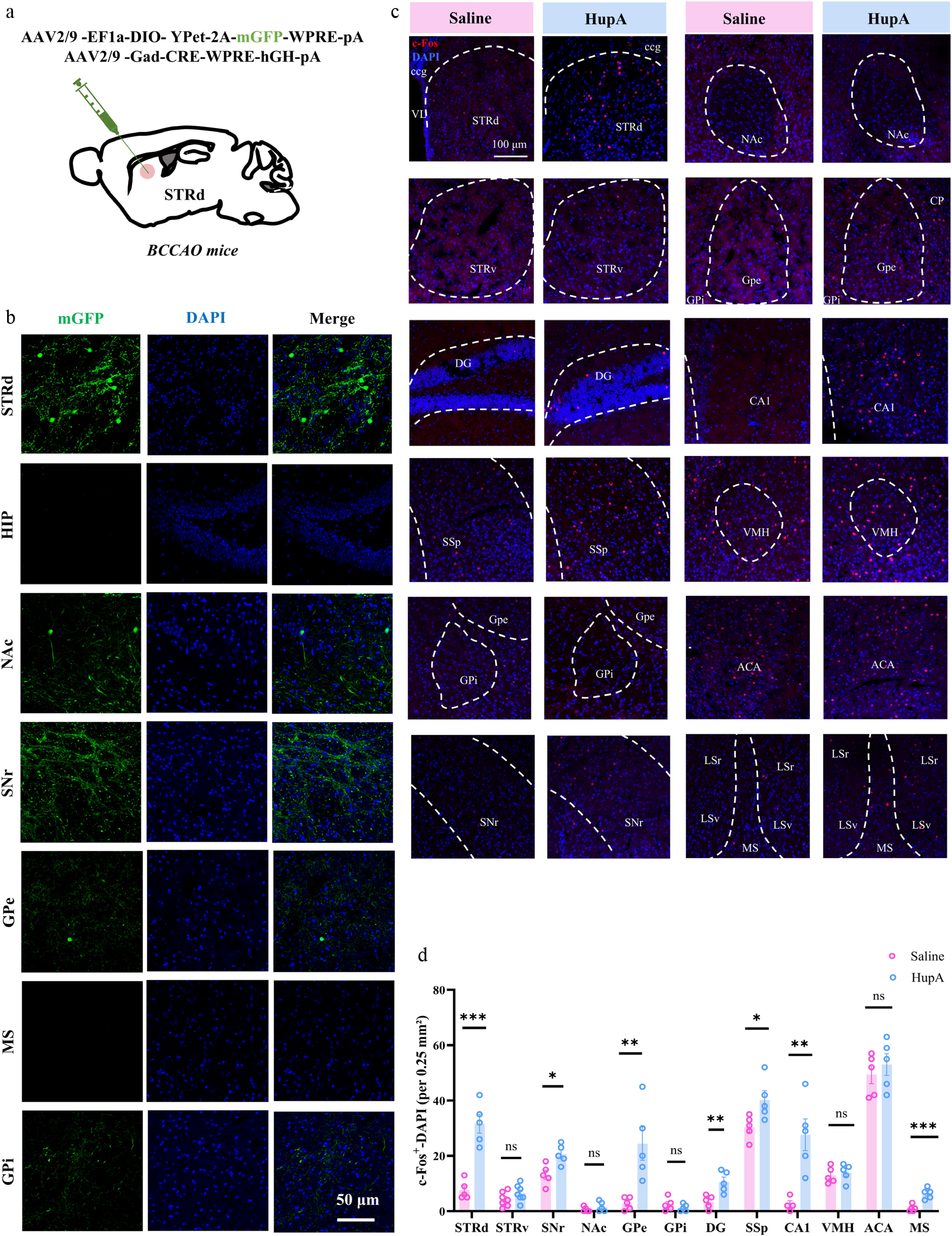
Figure 6.
HupA activates SNr neurons downstream of the striatum. (a) Schematic of the anterograde tracing virus strategy in BCCAO mice. (b) Schematic of anterograde tracing results from the striatum. Scale bar = 50 μm. HIP, hippocampus. STRd, dorsal striatum. NAc, nucleus accumbens. SNr, substantia nigra pars reticulata. GPi, globus pallidus internus. GPe, globus pallidus externus. MS, medial septum. (c) Representative c-Fos immunostaining images of the STRd, GPi, GPe, SNr, NAc, ventral striatum (STRv), dentate gyrus (DG), primary somatosensory cortex (SSp), cornu ammonis area 1 (CA1), ventromedial hypothalamus (VMH), anterior cingulate area (ACA), and MS brain regions. Scale bar = 100 μm. (d) Statistical analysis of the number of c-Fos+ neurons per 0.25 mm2 area (n = 5). Data are presented as mean ± SEM; * P < 0.05, ** P < 0.01, *** P < 0.001; (d) two-tailed unpaired Student's t-test.
To screen for the downstream brain region whereby STRd exerts its therapeutic effect, we further used chemogenetics to specifically modulate STRd projections to different downstream nuclei, ensuring precise viral expression (Fig. 7a–c). We focused on the basal ganglia output nuclei based on the previous observation that HupA concentration was well correlated with the change in ACh level in the STRd, as well as a marked pathological improvement in this region. Behavioral results showed that chemogenetic inhibition of the STRd→SNr pathway significantly increased the discrimination index and novel object exploration time in the NOR test (Fig. 7d and e), and also significantly improved the spontaneous alternation rate and open arm exploration time in the Y-maze test (Fig. 7f and g). No significant behavioral differences, however, were observed upon activation of the STRd→GPe pathway or inhibition of the STRd→GPi pathway (Fig. 7d–i). These results indicate that inhibiting the STRd→SNr circuit, but not the STRd→GPi/GPe circuits, might contribute to the improved cognitive function in HupA-treated VaD mice.
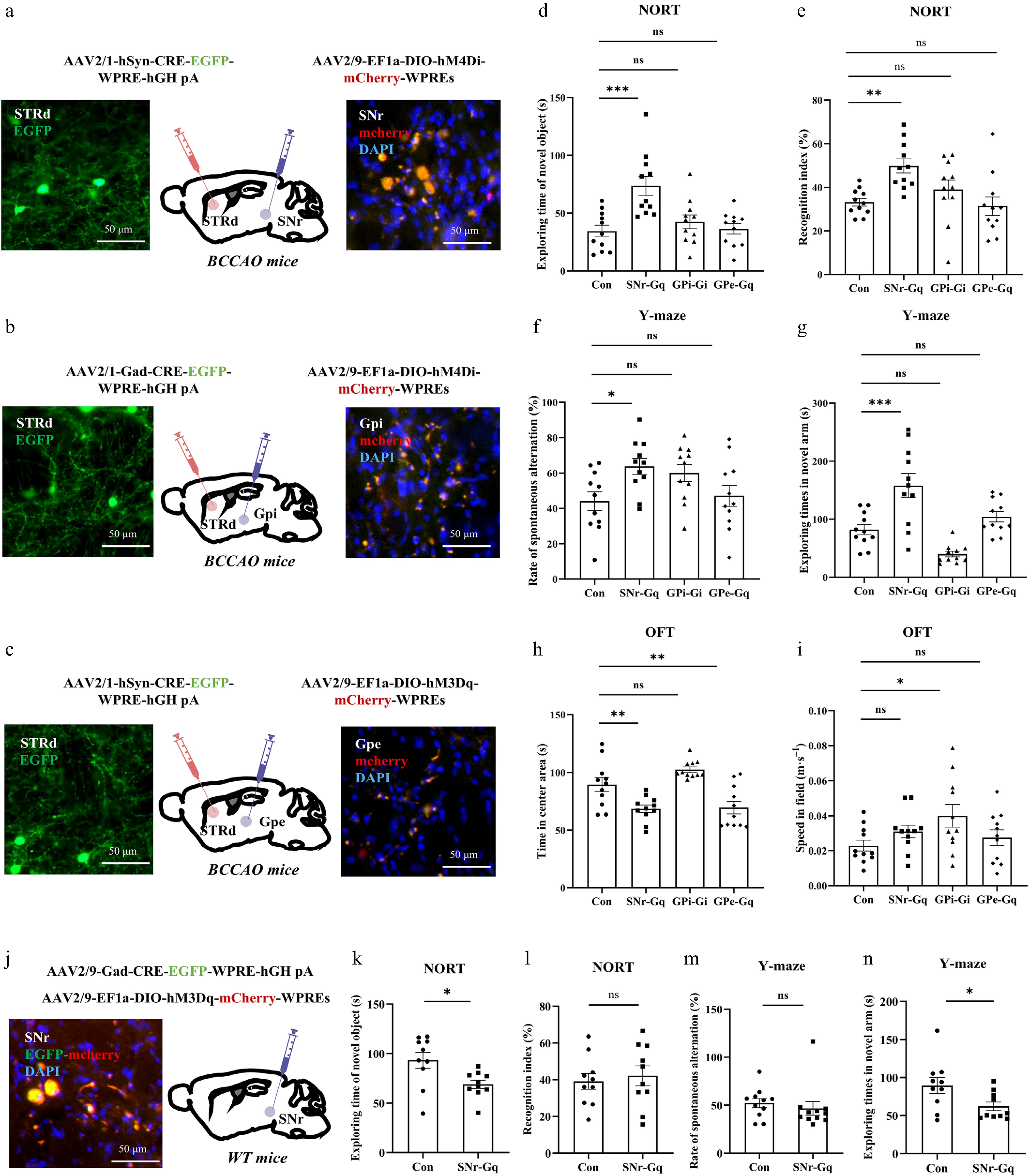
Figure 7.
HupA improves cognitive impairment in VaD mice via the STRdGABA→SNrGABA circuit. (a)–(c) Chemogenetic regulation strategy for STRd projections and representative images of virus expression. Scale bar = 50 μm. (d) Exploration time of the novel object in NORT (n = 11). (e) Discrimination index in NORT (n = 11). (f) Exploration time in the open arm of the Y-maze (n = 11). (g) Spontaneous alternation rate in the Y-maze (n = 11). (h) Time spent in the central area during OFT (n = 11). (i) Movement speed during OFT (n = 11). (j) Chemogenetic strategy for SNr. Scale bar = 50 μm. (k), (l) Exploration time and discrimination index of the novel object in the NORT (n = 10). (m), (n) Exploration time in the open arm and spontaneous alternation rate in the Y-maze (n = 10). Data are presented as mean ± SEM; * P < 0.05, ** P < 0.01, *** P < 0.001; (k)–(n) two-tailed unpaired Student's t-test; (d)–(i) one-way analysis of variance.
To further investigate the neuronal subtype responsible within the SNr, we employed a chemogenetic strategy to directly modulate the activity of GABAergic neurons in the SNr (Fig. 7j). Behavioral results showed that directly activating GABAergic neurons in the SNr reduced exploration time with novel object and in the Y-maze open arm (Fig. 7k–n), suggesting that activation of SNr GABAergic neurons induces a degree of cognitive impairment.
To further determine whether the STRdGABA-SNrGABA circuit is necessary for HupA's cognitive improvement effects in VaD mice, we conducted an occlusion experiment by chemogenetically activating SNr GABAergic neurons via hM3Dq-mediated DREADD signaling while simultaneously administering HupA to BCCAO mice, and confirmed the specificity of viral expression (Fig. 8a–c). Behavioral assessment revealed that CNO-mediated activation of SNr GABAergic neurons significantly compromised HupA-induced cognitive improvement in both the NORT and Y-maze tests, with behavioral performance returning to model group levels (Fig. 8d–i). These data establish that the functional engagement of the SNr GABAergic neurons downstream of the STRd is indispensable for the cognitive benefits conferred by HupA in VaD mice.
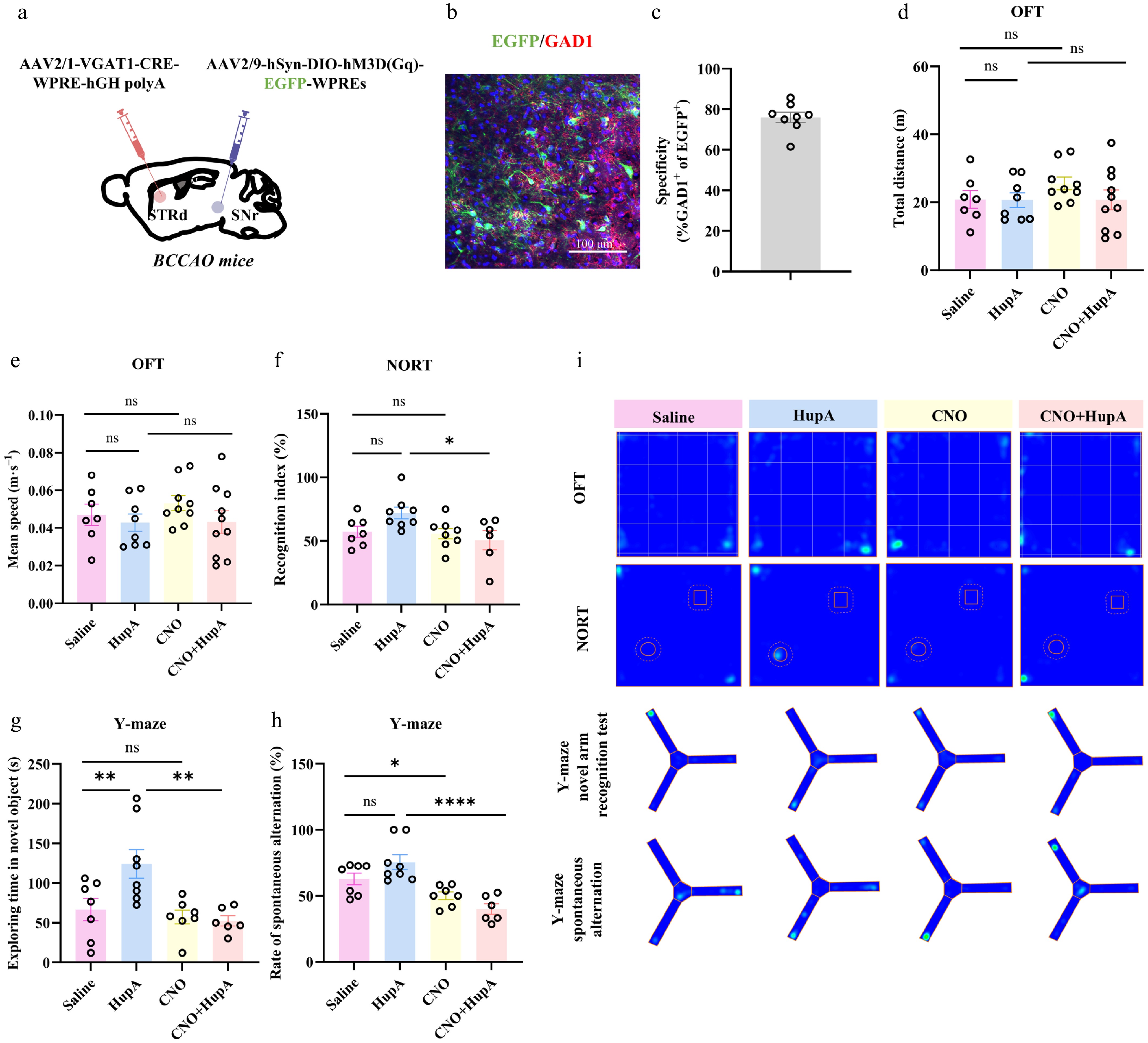
Figure 8.
Chemogenetic activation of SNr GABAergic neurons blunts HupA effect. (a) Schematic diagram of virus injection into VaD mice. (b) Specificity verification of virus expression. (c) Percentage of EGFP+ cells co-labeled with GAD1 (n = 8). (d) Total distance traveled during OFT for each group (n = 7−10). (e) Movement speed during OFT (n = 7−10). (f) The novel object recognition index in NORT for each group (n = 6−8). (g) Exploration time in the novel open arm of the Y-maze test for each group (n = 6−8). (h) Spontaneous alternation rate in the Y-maze test for each group (n = 6−8). (i) Representative heatmaps depicting the movement tracks of mice during the behavioral tests. Data are presented as mean ± SEM; * P < 0.05, ** P < 0.01, **** P < 0.0001; (d)–(h) two-way analysis of variance.
-
Dissecting the mechanisms of natural medicines provides a promising reverse pharmacology approach to the identification of new targets for chronic diseases such as dementia[25,26]. HupA is well established as a natural AChE inhibitor that elevates ACh levels. Our prior work in rats successfully modeled this link using a physiologically based pharmacokinetic-pharmacodynamic (mPBPK-PD) approach. Despite such a close relationship between ACh regulation and HupA, the detailed downstream mechanism linking ACh dynamics to the neurobehavioral impacts of HupA remains incompletely understood. In this study, we first sought to understand whether and how the neuroprotective effects of HupA were linked to ACh level modulation in the VaD mouse model. By cellular studies and in vivo regulation, we found that the antagonism of α7nAChR significantly weakened the effects of HupA on neuronal survival and structural protection, while mAChR inhibition had no significant effect. These findings not only further confirm the core position of STR as a key region for the cognitive improvement effects of HupA, but also clarify that ACh-α7nAChR axis activation serves as a critical basis for the ability of HupA to enhance neuronal activity and cognitive function. Although MLA is a highly potent and selective antagonist of α7nAChR, off-target pharmacological effects cannot be excluded. Future studies using a targeted genetic knockdown model would be necessary to strengthen the role of α7nAChR in the action of HupA. It is notable that current studies largely ascribe the neuroprotective effects of natural compounds to a direct effect on neuroinflammation[27−29]. However, our findings highlight that the indirect mode whereby natural components like HupA could regulate the neurotransmitter network to confer neuroprotection also deserves attention. Given that ACh balance and its sensation by AChR receptors are fundamental in the regulation of various neurobehavioral events[30,31], these findings also provide important experimental rationale for neuroprotective strategies targeting specific cholinergic receptors such as α7nAChR.
Although HupA is known to modulate the cholinergic system, the detailed neural circuit mechanism by which HupA confers cognitive improvement is largely elusive. We explored the neural circuit mechanism by which HupA improves cognitive function in VaD mice by anterograde cross-synaptic virus tracing, c-Fos mapping, and chemogenetic manipulation in normal and BCCAO mice. All these strategies enabled us to dissect the neural circuit mechanism through which STRd ACh signaling, enhanced by HupA, translates into synergistic cognitive improvement via a STRdGABA→SNrGABA neural circuit. Future studies using optogenetics to stimulate cholinergic terminals in the STRd while simultaneously recording from GABAergic neurons (via patch-clamp or fiber photometry in GCaMP-expressing GABA neurons) would provide a causal link between cholinergic and GABAergic neurons.
By collecting multi-layer data from pharmacokinetics, metabolomics, neurophysiology, and neurocircuit aspects, we propose a dual mechanism of HupA in improving cognitive impairment in VaD mice. These two mechanisms are not entirely independent, and it is reasonable that ACh-mediated neuroprotection could synergize with STRd-SNrGABA circuit modulation to improve cognitive function in VaD mice. In particular, ACh-initiated α7nAChR signaling could increase spine density in the STRd, thereby enabling circuit-level activation for cognitive improvement. Future studies would be needed to further dissect how these two effects are integrated at the temporal and circuit level.
VaD, as the second most common type of dementia worldwide, faces severe challenges in its treatment due to the lack of targeted therapies. The core pathological mechanism of the disease lies in white matter degeneration caused by cerebral vascular injury, leading to cognitive and motor dysfunction[32]. Due to the involvement of multiple cell types and complex signaling pathways in the pathological process of vascular dementia[33], as well as the emerging implication of systemic factors such as the gut microbiome[34], single target interventions are often difficult to comprehensively correct its disrupted intercellular communication and repair disorders. Therefore, establishing a clinically relevant animal model and adopting a multi-target regulatory strategy is of significant necessity. The VaD mouse model in our study exhibits significant spatial memory deficits accompanied by multiple pathological markers such as neuronal damage, neuroinflammation, and circuit dysfunction. This enabled us to uncover an integral neurometabolic mechanism of HupA efficacy beyond the traditional view on ACh levels. Our findings based on this model support that pharmacological modulation of ACh-centered neurometabolic nodes, as exemplified by HupA, could provide an effective approach to combat both neuronal injury and circuit dysfunction in the pathological network of VaD.
HupA is predominantly thought to act via increasing the concentration of ACh. Of interest, by neurotransmitter panel profiling, we found that HupA simultaneously regulated several other neurotransmitters such as GABA, Glu, and NE in multiple brain regions of mice, especially in the STR brain region. This regulatory pattern suggests that, in addition to single neurotransmitter regulation, HupA may affect neural network function by synergistically regulating the neurotransmitter system to confer the benefit of cognition improvement. This is an intriguing concern, given that GABA and Glu, for example, have also been implicated in the regulation of neuronal injury and synaptic plasticity[35,36]. Therefore, future studies are warranted to clarify how the changes of other signaling network engaged by other neurotransmitter could collectively contribute to the cognitive improvement conferred by HupA, which could uncover more combinatorial targets for the treatment of VaD and other types of cognitive decline.
To address whether the changes in GABA, Glu, and NE are direct downstream consequences of α7nAChR activation or represent parallel neuromodulatory pathways, additional analyses are required to examine the temporal correlations among neurotransmitter changes following HupA administration. Specifically, as shown in our previous study[13], time-course profiling of ACh, GABA, Glu, and NE in the STRd using LC-MS/MS at multiple time points after HupA injection would reveal the temporal dynamics and hierarchical relationships among these neurotransmitters. To further substantiate this link, it would be interesting to test whether pharmacological blockade of α7nAChR with MLA attenuates the HupA-induced changes in GABA and NE levels. Furthermore, because HupA is a natural alkaloid, we cannot completely rule out the possibility of direct interactions with other receptor systems. Future studies employing broader receptor profiling or proteomic approaches will be necessary to fully elucidate the multi-neurotransmitter regulatory mechanism underlying the cognitive improvement induced by HupA.
-
In conclusion, our work in BCCAO mouse model and primary neurons collectively show that HupA confers cognitive effects via a dual mechanism involving ACh-α7nAChR axis mediated neuronal protection and ACh activated STRdGABA→SNrGABA circuit to synergistically improve cognitive network in VaD mice. These findings enrich the mechanistic understanding of HupA in the treatment of dementia and cognitive decline. Also, our work underscores the necessity to design a multi-target strategy for VaD, which may be guided by the neurometabolic coupling mechanism in cognitive regulation.
-
All animal procedures were approved bythe Animal Ethics Committee (2024-05-039) of China Pharmaceuti-cal University (Nanjing, China).
-
The authors confirm their contributions to the paper as follows: conceived the project and designed the study: Zheng X, Hao H; carried out most of the biochemical, molecular experiments, and behavioral assessments: Zhang Y, Li Z; data analysis: Zhang Y, Li Z, Nie R, Wu H, Cai X, Zhang Y, Lin X, Duan R, Liu S; contributed to manuscript revisions: Li J; provided experimental assistance: Zhou P; provided suggestions and guidance for the research: Fu Y; wrote the manuscript: Zheng X, Li Z, Zhang Y. All authors reviewed the results and approved the final version of the manuscript.
-
The datasets generated and/or analyzed during the current study are available from the corresponding author upon reasonable request.
-
The authors have no conflicts of interest to declare.
-
accompanies this paper online at: https://doi.org/10.48130/targetome-0026-0042.
-
#Authors contributed equally: Yangfan Zhang, Zemin Li
- Supplementary Table S1 Gradient elution procedure of neurotransmitter.
- Supplementary Table S2 Mass spectrometry parameters of HupA and neurotransmitters.
- Supplementary Table S3 Summary of virus information used in this study.
- Supplementary Fig. S1 Model validation of BCCAO-induced VaD mice.
- Supplementary Fig. S2 HupA treatment alleviates BCCAO-induced excessive microglial activation and neuroinflammation.
- Supplementary Fig. S3 Effect of HupA on other neurotransmitters in different brain regions of mice after intramuscular injection.
- Supplementary Fig. S4 Re-analysis of public single-cell RNA sequencing data.
- Supplementary Fig. S5 Direct activation of STRd cholinergic neurons ameliorates cognitive impairment in VaD mice.
- Supplementary Fig. S6 Effect of intramuscular injection of HupA on dendritic spine growth of GABAergic neurons in the STRd region.
- Supplementary Fig. S7 The D1-MSNs subtype mediates the cognitive impact of STRdGABA neurons in VaD mice.
- Copyright: © 2026 by the author(s). Published by Maximum Academic Press on behalf of China Pharmaceutical University. This article is an open access article distributed under Creative Commons Attribution License (CC BY 4.0), visit https://creativecommons.org/licenses/by/4.0/.
-
About this article
Cite this article
Zhang Y, Li Z, Nie R, Wu H, Li J, et al. 2026. Dual cognition-improving mechanisms of huperzine A in vascular dementia mice via neurometabolic regulation. Targetome 2(5): e043 doi: 10.48130/targetome-0026-0042 

Dual cognition-improving mechanisms of huperzine A in vascular dementia mice via neurometabolic regulation
- Received: 04 February 2026
- Revised: 18 May 2026
- Accepted: 25 June 2026
- Published online: 10 September 2026
Abstract: Vascular dementia (VaD) is a poorly treated disease that involves complicated pathology such as neuronal damage and neural circuit dysfunction. Huperzine A (HupA) is commonly used for clinical treatment of cognitive impairment, the mechanism of which is prevalently attributed to the increase in synaptic acetylcholine (ACh) levels. Here we elucidate an integral neurometabolic mechanism for HupA in the treatment of cognitive impairment in VaD. HupA effectively alleviated bilateral common carotid artery occlusion (BCCAO)-induced neuronal damage, neuroinflammation, and cognitive decline in mice, accompanied by a notable restoration of ACh homeostasis in the dorsal striatum (STRd). HupA or ACh injection into the STRd recapitulated the cognitive effects, while an α7nAChR antagonist blocked the neuroprotective effects in both primary neurons and VaD mice. By neural circuit mapping and functional manipulation, we further identified that HupA-induced ACh accumulation in the STRd recruits a STRdGABA→SNrGABA neural circuit to synergistically improve the cognitive network in VaD mice. Overall, we reveal a dual mechanism orchestrated by STRd ACh signaling through which HupA confers cognitive improvement in VaD mice. These findings underscore the therapeutic value of targeting neurometabolic nodes for the treatment of VaD and potentially other forms of dementia.