












-
Plastic production grew from 234 million tons in 2000 to 460 million tons in 2019, and is projected to reach 1,231 million tons by 2060, with 19–53 million tons of plastic waste entering aquatic environments annually[1,2]. Aquatic environments act as primary sinks and transformation sites for plastic debris, receiving an estimated 4.8–12.7 million tons of plastic waste from land in 2010 alone[3], which subsequently fragments into microplastics (MPs, 1 μm–5 mm) and nanoplastics (NPs, < 1 μm) through photo-oxidation, abrasion, and biological activity[4,5]. These particles, spanning polyethylene (PE), polypropylene (PP), polystyrene (PS), polyvinyl chloride (PVC), polyamide (PA), and polyethylene terephthalate (PET)[6], are not homogeneous in their hazard: Nanoscale particles exhibit greater biological reactivity and barrier penetrability owing to their high specific surface area[7,8], whereas micrometer-sized particles primarily affect reproduction through interference with feeding and energy depletion[9]. Consequently, MNPs should be assessed within a reproductive and developmental risk framework that extends beyond conventional toxicity metrics.
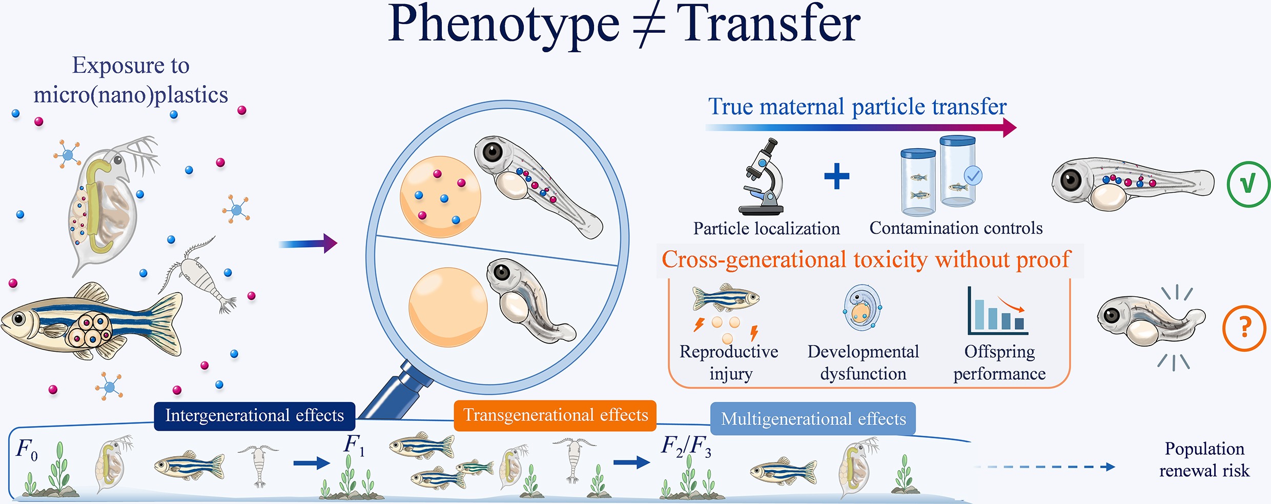
Existing studies have predominantly measured adult survival, tissue injury, and molecular stress markers[10−12]. Although informative, these endpoints do not resolve the more ecologically relevant question: Whether parental MNP exposure impairs the offspring's quality and population renewal. A deeper obstacle, however, is conceptual. Similar offspring phenotypes can arise through nonequivalent causal routes, including true maternal particle transfer, parental physiological injury, chorion-associated exposure, culture medium carryover, and co-contaminant vector effects[13−15]. Without explicit criteria to distinguish these pathways, the literature conflates distinct phenomena under the same interpretive umbrella.
In this review, we propose and apply an evidence hierarchy framework that separates four noninterchangeable concepts: Intergenerational evidence (F0 exposure followed by F1 changes), multigenerational evidence (continuous exposure across F0–F2 or beyond), transgenerational evidence (persistence in unexposed generations after clean culture), and true maternal particle transfer (direct particle localization inside eggs or embryos, with orthogonal confirmation and contamination controls). By applying this framework across cladocerans, rotifers, copepods, fish, and bivalves, we distinguish transfer confidence from exposure design categories, evaluate mechanistic pathways linking MNP exposure to offspring outcomes, and identify critical knowledge gaps that constrain environmental risk extrapolation.
-
To place maternal particle transfer within the broader field of MNP ecotoxicology, we conducted a structured literature search covering studies published from 2010 to May 20, 2026. Searches were performed using combinations of terms related to MPs, NPs, micro(nano)plastics, uptake, bioaccumulation, reproductive toxicity, developmental toxicity, maternal transfer, parental transfer, intergenerational effects, multigenerational effects, transgenerational effects, co-exposure, and aquatic organisms.
The retrieved literature was screened according to the scope of this review, with particular attention to the exposure design, particle localization, contamination controls, and offspring endpoints. Studies demonstrating particles in eggs, embryos, or offspring tissues were distinguished from those reporting offspring phenotypes alone, and continuous multigenerational exposure was distinguished from persistence after clean-generation or recovery conditions. Because this review was designed as a critical evidence classification synthesis rather than a formal systematic review or meta-analysis, the structured search was used primarily to support the interpretation of the evidence rather than quantitative evidence pooling.
The broader literature dataset was further analyzed using VOSviewer version 1.6.19 to construct a keyword co-occurrence network. Before network construction, synonymous and closely related keyword variants were standardized. Keywords occurring at least 10 times were retained. The final network contained 40 keywords, 395 co-occurrence links, and 6 clusters. Node size represents keywords' occurrence frequency, edge thickness represents co-occurrence strength, and colors indicate keyword clusters (Fig. 1).
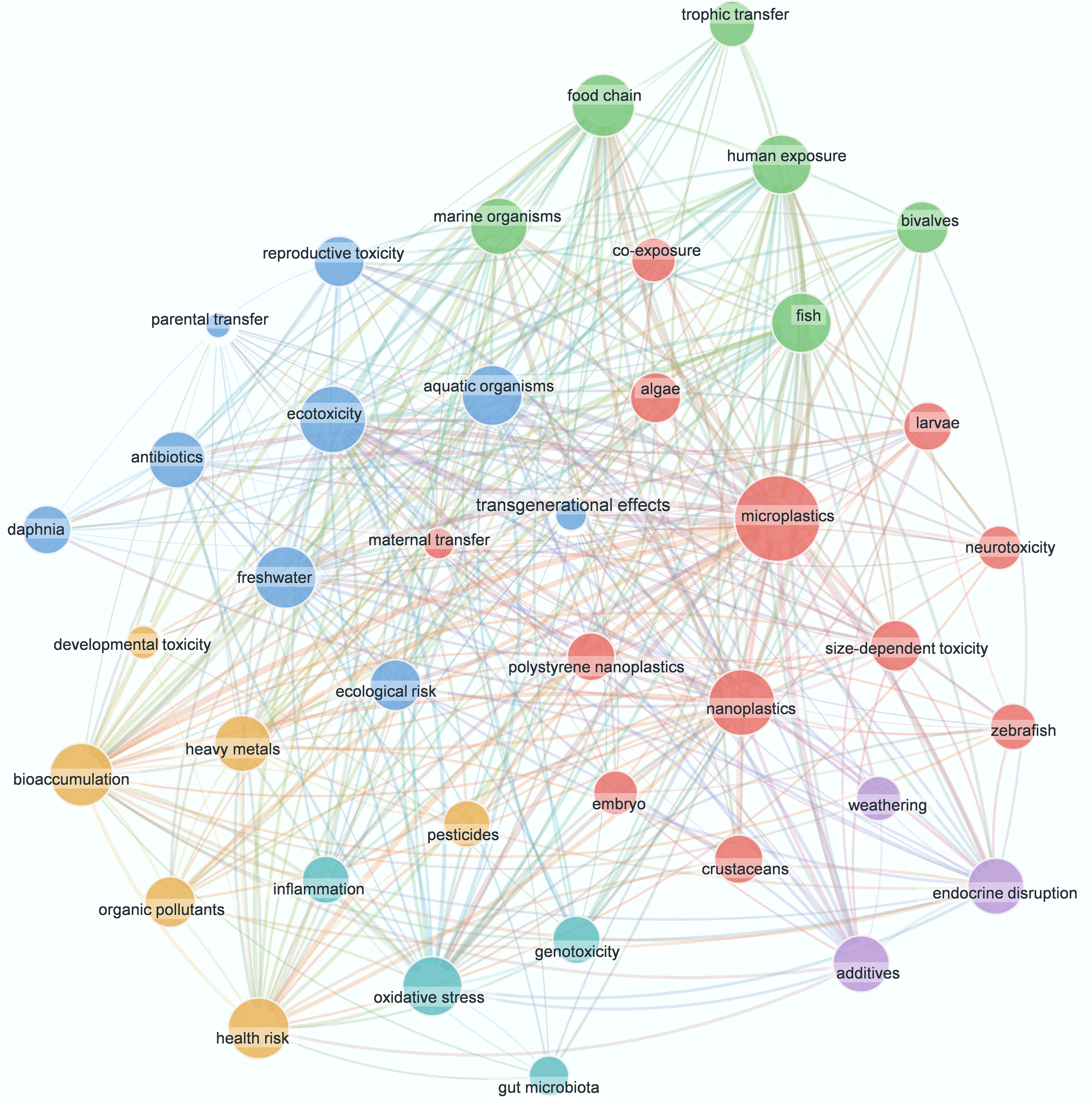
Figure 1.
Keyword co-occurrence network showing the research landscape of MNP ecotoxicology from 2010 to 2026. The network contains 40 keywords and 395 co-occurrence links distributed across six clusters. Node size indicates keywords' occurrence frequency, edge thickness indicates co-occurrence strength, and colors represent keyword clusters. The network is used only to position cross-generational transfer-related research within the broader MNP ecotoxicology field and does not provide causal evidence for particle transfer.
The network shows that the broader MNP ecotoxicology literature is dominated by themes related to exposure, bioaccumulation, organismal toxicity, oxidative stress, endocrine disruption, and developmental toxicity. In contrast, maternal transfer, parental transfer, and transgenerational effects occur less frequently and occupy more specialized positions, associated mainly with reproductive toxicity, embryo and larval development and aquatic model organisms. The network is therefore used only to position these transfer-related terms within the broader MNP ecotoxicology landscape, rather than to infer causal relationships or transfer certainty.
The relatively specialized positions of these cross-generational terms are consistent with the still limited and methodologically heterogeneous evidence base for maternal particle transfer. Importantly, offspring phenotypes following parental exposure should not automatically be interpreted as evidence of true maternal particle transfer. This distinction between what has been observed (offspring effects) and what has been demonstrated (particle localization inside offspring tissues) constitutes the organizing principle of this review.
-
A critical distinction underpins this section: Exposure design categories and the strength of transfer evidence are not interchangeable. Table 1 classifies selected cross-generational studies according to their experimental design, intergenerational aspects (F0 exposure → F1 changes), multigenerational aspects (continuous exposure across F0–F2 or beyond), or transgenerational aspects (persistence in unexposed generations after clean culture), with co-exposure added only as a qualifier when MNPs are combined with other contaminants. This classification describes what the experimental design can demonstrate, not whether particles have moved from parent to offspring. The latter requires direct particle localization in eggs, embryos, or offspring tissues, which we examine below across major taxonomic groups.
Table 1. Representative cross-generational MNP studies in aquatic organisms selected to distinguish true maternal particle transfer, intergenerational evidence, multigenerational evidence, and transgenerational evidence
Model Plastic material Exposure design Evidence category Localization evidence Main methodological limitation Biological endpoints Ref. Daphnia galeata PS-NPs; 52 nm; 5 mg/L F0 exposed for 5 d; embryos remained in the brood chamber during exposure; no clean-generation follow-up Intergenerational evidence Fluorescence microscopy; confocal Z-stack No clean-generation design Adult survival and pregnancy rate decreased; embryos showed abnormal development and low hatching; no persistent offspring survival/lipid effect after hatching [16] Daphnia magna Pd-doped PS NPs; 200 nm; 0.1 and 1 mg/L F0–F3 continuous exposure; 21 d per generation; no clean-generation condition Multigenerational evidence ICP-MS via Pd tracer No clean-generation design; no tissue-level particle localization Adult survival largely unchanged; F3 fertility increased at 1 mg/L, whereas F3 offspring body size and lipid content decreased; useful low-effect case [17] Daphnia magna Polyester MP fibers; 6 μm; 10 and 100 particles/L Parallel F0–F2 continuous and clean-generation lines Transgenerational evidence SEM; transfer check; DNA methylation sequencing Particle transfer was not directly confirmed Reproduction and molting decreased; clean F1/F2 lines retained fitness loss and altered DNA methylation; particle transfer was not confirmed. [18] Daphnia magna PE-MP/BP-3 fragments; 19.31 ± 5.16 μm; 5 mg/L F0 exposed for 21 d; F1–F3 reared in clean M4 medium Transgenerational evidence; co-exposure WGBS; phenotypic tracking No direct particle localization; combined MP/BP-3 exposure Population growth decreased in F0–F1 but recovered in F2–F3; DNA methylation signatures persisted across F0–F3. [19] Brachionus
koreanusFluorescent PS-NPs; 50 nm; 1 and 10 μg/mL F0 maternal exposure; eggs/offspring transferred to clean seawater before assessment Intergenerational evidence; strong support for maternal particle transfer Fluorescence microscopy; egg-only and dye leaching controls Fluorescence-based detection without orthogonal particle confirmation NP signal increased in eggs; F1 maturation was delayed, reproduction/lifespan declined, and ROS increased. [20] Brachionus
plicatilisAmino-modified PS-NPs
(PS-NH2); 0.5 and 2.5 mg/LF0–F3 continuous exposure with recovery lineage follow-up Multigenerational evidence Bioaccumulation imaging; transcriptomics No orthogonal confirmation of particle transfer Population growth was suppressed; egg and offspring numbers decreased across generations; reproductive strategy shifted from r- to K-selection. [21] Tigriopus japonicus PS-NPs; 50 nm; 23 μg/L;
Hg, 1 μg/LF0–F2 continuous co-exposure; no clean-generation condition Multigenerational evidence; co-exposure CV-AFS for Hg; RNA-seq No clean-generation design or direct offspring localization PS-NPs increased Hg body burden; survival and fecundity decreased; development time and reproduction-related transcriptome were disrupted. [22] Danio rerio Fluorescent PS-NPs; 100 nm; 10 μg/L; EHS, 1–100 μg/L F0 exposed for 28 d; F1 embryos/larvae cultured in clean water Intergenerational evidence; co-exposure Fluorescence imaging and SEM for PS-NP distribution; GC-MS/MS for EHS quantification F1 particle uptake was observed, but endogenous maternal transfer was not confirmed. PS-NPs and EHS accumulated in F1 embryos, predominantly in the yolk sac; PS-NPs subsequently redistributed to other tissues and enhanced EHS transfer to offspring [23] Danio rerio Fluorescent PS-NPs; 54.5 ± 2.8 nm; 100 μg/L; TDCIPP, 0.4–10 μg/L F0 lifecycle exposure, 2 hpf–150 dpf; F1 assessed in clean water to 5 dpf Intergenerational evidence; co-exposure Fluorescence microscopy; LC-MS/MS No orthogonal particle localization in F1 PS-NPs promoted TDCIPP transfer to F1 eggs; Hatching/development and locomotor behavior worsened with disruption of the dopamine pathway. [24] Oryzias melastigma PS-MPs; 13 μm; 2–200 μg/L; Phe, 50 μg/L F0 females exposed 60 d; mated with unexposed males; F1 cultured in clean seawater Intergenerational evidence; co-exposure Optical microscopy; HPLC Optical localization lacks polymer-specific confirmation MPs increased Phe accumulation in ovaries/embryos; ovarian maturity and egg output decreased; F1 heart rate, hatching, and body size were impaired. [25] Note: Models are ordered by cladocerans, rotifers/copepods, and fish. Intergenerational evidence = F0 exposure followed by F1 changes; multigenerational evidence = continuous exposure across F0, F1, F2, or later generations; transgenerational evidence = persistence in unexposed generations after a clean-generation or recovery design; true maternal particle transfer = particles directly localized in eggs, embryos, or offspring tissues with orthogonal particle confirmation and adequate controls for external contamination. Co-exposure is shown only as a qualifier. Localization evidence lists the reported detection or analytical methods and does not by itself establish transfer certainty. CV-AFS, cold-vapor atomic fluorescence spectrometry; dpf, days post-fertilization; EHS, ethylhexyl salicylate; GC-MS/MS, gas chromatography-tandem mass spectrometry; Hg, mercury; HPLC, high-performance liquid chromatography; ICP-MS, inductively coupled plasma mass spectrometry; LC-MS/MS, liquid chromatography-tandem mass spectrometry; Phe, phenanthrene; RNA-seq, RNA sequencing; SEM, scanning electron microscopy; TDCIPP, tris(1,3-dichloro-2-propyl) phosphate; WGBS, whole-genome bisulfite sequencing. Additional evidence is provided in Supplementary Table S1. Taxon-specific evidence
-
Evidence for maternal particle transfer varies markedly across taxa, reflecting differences in reproductive anatomy, exposure interfaces, and experimental tractability. Three broad patterns emerge: Direct localization with pathway quantification in cladocerans, uneven and often conflicting evidence in rotifers and copepods, and a predominance of indirect intergenerational effects in fish, where direct particle entry into offspring tissues remains rarely demonstrated.
In Daphnia magna, confocal and label-free hyperspectral imaging after exposure to 25 nm PS-NPs (50 mg/L) showed that particles entered the brood chamber and were subsequently detected in eggs, embryos, and ovarian oocytes, with the brood chamber route accounting for approximately 88% of embryo exposure versus 12% through gut–ovary–oocyte transfer[26]. Size selectivity is clear: 25- and 50-nm particles enter eggs or embryos more readily than 100-nm particles, whereas 250-nm and even 10-μm particles reach the brood chamber but are not internalized by early embryos[26]. Similar patterns have been observed in Daphnia galeata (52-nm PS-NPs, 5 mg/L), where particles appeared in the thoracic limbs, ovaries, and brood chambers alongside reduced parental survival, abnormal embryonic development, and decreased hatching[16]. Notably, however, observations of brood chamber fluorescence alone do not distinguish external embryo contact from genuine internalization—a distinction that requires orthogonal confirmation (e.g., Raman spectroscopy) to rule out dye leakage or surface adsorption[26,27]. The key conclusion for cladocerans is that two distinct processes operate in parallel: Brood chamber-mediated external embryo exposure and a quantitatively minor but mechanistically important endogenous gut–ovary–oocyte transfer route[26] (Fig. 2).
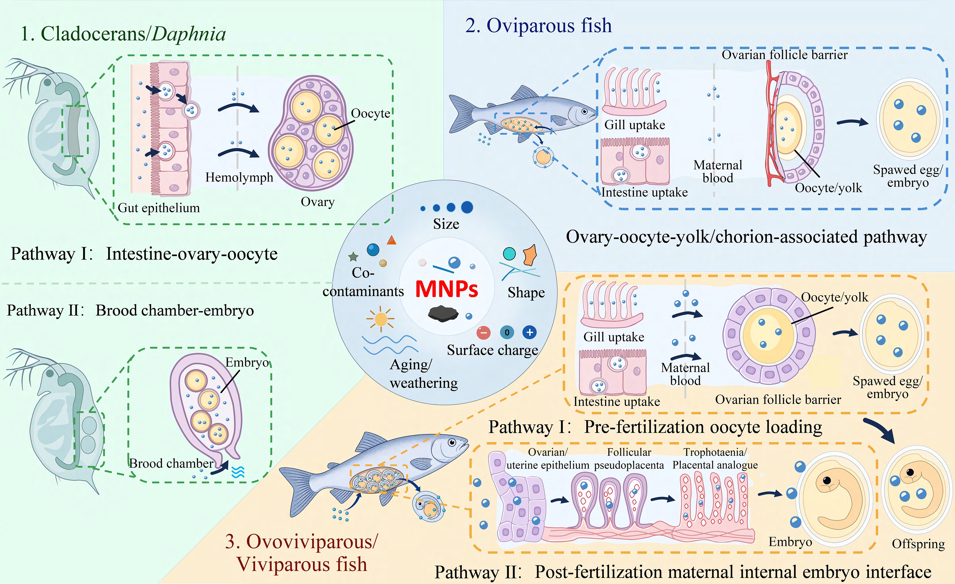
Figure 2.
Conceptual summary of taxon-specific maternal particle transfer and embryo exposure interfaces of MNPs in aquatic organisms. Background colors distinguish cladocerans, oviparous fish, and ovoviviparous/viviparous fish. Arrows indicate proposed directions of particle movement or embryo exposure. In cladocerans, Pathway I represents intestine–ovary–oocyte transfer and Pathway II brood chamber-mediated embryo exposure. In ovoviviparous/viviparous fish, Pathways I and II represent prefertilization oocyte loading and post-fertilization maternal internal embryo exposure, respectively. MNPs, micro(nano)plastics.
These organisms are attractive for multigenerational studies because of their rapid lifecycles, yet the evidence for endogenous maternal particle transfer remains heterogeneous. In Brachionus koreanus, parents exposed to 50-nm PS-NPs (1 and 10 μg/mL) and transferred to clean seawater produced offspring (eggs) with concentration-dependent particle fluorescence, and egg-only exposure plus dye leaching controls yielded no comparable signals[20], a design that provides stronger support for endogenous transfer. Conversely, in Brachionus plicatilis exposed to 70-nm PS-NPs for two generations and then cultured in clean water for two further generations, particles were found in feces and on the surface of F1 eggs, but not convincingly within embryonic tissues[28]. This pattern points toward egg surface contact and life history carryover rather than true endogenous transfer. Copepod studies further complicate the picture: Proteomic changes persisted in F2 after clean recovery in a two-generation exposure to 6-μm PS-MPs[29], yet such persistence does not by itself demonstrate particle entry into offspring. Overall, many rotifer and copepod offspring effects may reflect continuous exposure, altered parental physiology, or egg surface contamination rather than proven particle transfer[20,29] (Fig. 2).
The relevant interfaces shift from brood structures to the ovary, oocytes, chorion, and yolk sac, yet the evidentiary bar for true maternal particle transfer has rarely been met. In zebrafish (Danio rerio), dietary exposure of parents to ~42-nm PS-NPs (~1 mg/g body weight, 7 d) yielded particle fluorescence in the yolk sac, intestine, and liver of F1 embryos, but only in maternal and biparental exposure groups, not after paternal exposure alone[30]. This pattern implicates a yolk-associated maternal route, but fluorescence alone does not confirm the particles' identity. A 42-d exposure of F0 females to 100-nm PS-NPs resulted in particle detection in F0 and F1 tissues, reduced mature oocyte numbers, altered F1 hatching and heart rate, and disrupted expression of sex hormone and sex differentiation genes[31]. Critically, however, only a handful of oviparous fish studies have directly localized particles inside F1 tissues using orthogonal methods (e.g., confocal microscopy plus Raman spectroscopy). Most reported intergenerational effects are therefore more parsimoniously attributed to parental reproductive injury, altered yolk provisioning, or chorion-associated exposure rather than to demonstrated particle transfer[31−33].
Correlative evidence from other fish species reinforces this cautionary interpretation. In Paramisgurnus dabryanus, PE-MPs (1 and 10 mg/L, 15 or 30 d) were observed in the gonads alongside ovarian follicle disruption and altered F1 hatching and body length[34]. In marine medaka (Oryzias melastigma), 10-μm PS-MPs (60 d) accumulated mainly in the gills, intestines, and liver—not in the gonads—yet gonadosomatic index (GSI) declined, egg production fell, and F1 fertilization, hatching, and heart rate were altered[32]. Even when F1 offspring were reared in clean seawater after a full lifecycle of parental exposure to 2 μm PS-MPs, they showed an increased heart rate, earlier hatching, and delayed growth[33]. Such findings indicate that toxicity in the offspring can arise from gonadal injury and altered yolk quality even in the absence of detectable particle transfer to the ovary[32−34] (Fig. 2).
Ovoviviparous and viviparous fish represent a theoretically more permissive pathway because embryos develop within the maternal body and are exposed to the maternal internal environment for extended periods. After pregnant guppies (Poecilia reticulata) were exposed to ~23-nm PS-NPs (50 μg/L, 30 d), the pregnancy rate dropped from 87.5% to 37.3%, embryo number decreased, and embryonic protein and carbohydrate levels shifted, alongside elevated reactive oxygen species (ROS) and antioxidant enzyme activities[35]. These phenotypes are consistent with altered maternal provisioning and redox status, but do not prove particle entry into embryos. Detection of multiple polymer types, including PS, PE, PET, PP, and polymethyl methacrylate (PMMA), in embryos of the viviparous blue shark Prionace glauca[36] extends the ecological relevance of this concern, yet continuous localization from maternal blood or ovarian fluid into embryonic tissues remains to be demonstrated (Fig. 2).
Checklist for evaluating claims of true maternal particle transfer in aquatic organisms
-
Across all taxa, Criteria 1 and 2 represent the minimum requirements for classifying a study as demonstrating true maternal particle transfer. Criterion 3 is additionally required when persistence in unexposed generations is claimed, whereas Criterion 4 strengthens the quantitative assessment of transfer magnitude and biological relevance:
1. Direct particle localization in eggs, embryos, or offspring tissues using orthogonal methods (e.g., confocal microscopy + Raman spectroscopy or pyrolysis gas chromatography/mass spectrometry [Py-GC/MS]);
2. Controls for external contamination: No detectable particles in the culture medium after exposed parents are transferred to a clean medium; no surface adsorption on egg/embryo chorion (or signal removed by washing); dye leakage controls (e.g., unexposed embryos co-incubated with labeled particle supernatants show no signal);
3. Clean-generation follow-up when transgenerational persistence is claimed: At least one generation reared in an uncontaminated medium with verified absence of particles in water and on the offspring's surfaces;
4. Quantification of particle burden in offspring's tissues: The particle burden should be quantified whenever technically feasible (e.g., particles per mg tissue) to distinguish incidental detection from quantitatively meaningful transfer.
If a study reports only the offspring's phenotypes (e.g., reduced hatching, altered behavior) without direct particle localization and adequate controls for external contamination, the findings should be described according to the exposure design as intergenerational, multigenerational, or transgenerational effects, or carryover of parental effects rather than as maternal particle transfer.
Transfer pathways across biological interfaces
-
Maternal particle transfer and embryo exposure pathways of MNPs can be conceptualized as four biological interfaces, which differ fundamentally in their anatomical basis, barrier properties, and evidentiary requirements. These are as follows: (1) brood chamber-mediated external embryo exposure, (2) endogenous gut–ovary–oocyte transfer, (3) pre- and post-spawning yolk sac/chorion-associated interfaces in oviparous fish, and (4) maternal internal embryo interfaces in ovoviviparous or viviparous fish. Each is evaluated below against the evidence hierarchy defined earlier (Fig. 2).
Brood chamber–embryo interface
-
In cladocerans, the brood chamber is not a closed maternal barrier. Its opening allows ambient particles to enter and directly contact the egg membrane, embryonic surface, or mouthparts of late embryos[26,16]. This mechanism explains how particles too large for endogenous transfer (e.g., 250 nm or 10 μm) can still exert extra-embryonic effects on development. Crucially, however, brood chamber fluorescence alone does not distinguish external contact from embryo internalization. Studies reporting only chamber-localized signals should not be classified as evidence for endogenous maternal transfer without orthogonal confirmation (e.g., Raman spectroscopy) to rule out surface adsorption[26,27] (Fig. 2).
Gut–ovary–oocyte interface
-
This is a stricter pathway: Particles must cross the intestinal epithelium, enter circulation or the coelomic fluid, reach the ovary, and be internalized by oocytes. In D. magna, 25-nm PS-NPs were directly observed in gut, ovary, and oocytes using label-free hyperspectral imaging, which circumvents artifacts of fluorescence leakage[26]. Quantitatively, this route contributed only ~12% of embryo exposure versus ~88% via the brood chamber, and particles measuring ≥ 50 nm were poorly detected in the ovary, highlighting the strong size selectivity of ovarian barriers[26]. The cellular mechanisms underlying epithelial crossing, endocytosis, paracellular transport, or passive entry after injury are size- and surface-dependent and are discussed further in the mechanism section (Fig. 2).
Yolk sac and chorion–embryo interface in oviparous fish
-
This interface operates in two temporally distinct phases. Before spawning, the relevant barrier is the ovarian follicle, comprising vascular supply, follicular cells, and the zona radiata, through which particles or particle-bound contaminants may enter the oocyte or associate with yolk precursors (e.g., vitellogenin, lipid droplets)[37]. The observation of F1 yolk sac signals after maternal but not paternal exposure supports the biological plausibility of this yolk-associated route[30]. After spawning, the embryo is no longer connected to the maternal environment, and the interface shifts to exogenous chorion contact. Chorion adsorption may delay hatching or induce local hypoxia[38], but does not by itself demonstrate particle entry into embryonic tissues. Internalization of smaller NPs into the yolk sac, liver, and other organs has been documented in direct embryo exposure studies, with accompanying effects on heart rate, body length, and locomotor activity[39]. The key distinction is therefore between maternal provisioning (pre-spawning, potentially true transfer) and external embryonic contact (post-spawning; exposure, not transfer) (Fig. 2).
Maternal internal embryo interface in ovoviviparous and viviparous fish
-
Here, embryos are retained within the maternal reproductive tract for extended periods, creating a two-stage pathway. The first stage parallels that of oviparous fish: Particles may enter maternal circulation and reach ovarian follicles or oocytes before fertilization. The second stage is distinctive: After fertilization, retained embryos may continue to interact with the maternal internal environment through interfaces such as residual yolk, ovarian or uterine fluid, trophotaenia, or placenta-like structures. Gestational exposure in guppies[35] and particle detection in blue shark embryos[36] support the relevance of this pathway, yet continuous localization from maternal blood or ovarian fluid into embryonic tissues remains absent. Future evidence must demonstrate not only particles in maternal compartments but also their traversal of embryo-associated barriers and presence within embryonic tissues (Fig. 2).
-
This section describes phenotypic and life history endpoints observed in the offspring following parental MNP exposure. Mechanistic pathways (oxidative stress, endocrine disruption, epigenetics, etc.) are addressed separately in Section entitled 'Mechanisms linking MNP exposure to offspring effects'. Here, we focus on three interconnected endpoints: Parental reproductive injury, embryonic development, and offspring's growth and reproduction, organized along the life history continuum from gamete formation to population recruitment.
Parental reproductive injury and gamete quality
-
Parental gonadal damage represents the earliest point at which MNP exposure can compromise the offspring's quality[9]. In crustaceans, MNPs delay first spawning, reduce total offspring output, and induce gonadal tissue injury[9], reflecting a forced reallocation of energy from reproduction to survival. Similar patterns are evident in fish. Female zebrafish exposed to 0.5-μm PS-MPs (50–500 μg/L, 60 d) showed gonadal particle accumulation, altered GSI, ovarian damage, and reduced reproductive rate[40]. In males, 1-μm PS-MPs (10–1,000 μg/L, 21 d) induced testicular oxidative stress and upregulation of apoptosis-related genes (p53, Bax, caspases), with increased germ-cell apoptosis[41].
At the gamete level, the consequences extend to heritable cellular integrity. In zebrafish, acute exposure to 45-nm PS-NPs (5 mg/L, 96 h) altered sperm chromatin compaction and motility, shifted oocyte-stage proportions, and changed the expression of genes involved in meiosis and DNA repair[42]. Paternal exposure alone can be consequential: Brief pre-fertilization exposure of European whitefish (Coregonus lavaretus) sperm to 50-nm carboxylated PS-NPs reduced sperm motility, and offspring hatched earlier, weighed less, and swam less effectively[43]. In bivalves, even when particles remain largely in the digestive tract rather than gonads, as observed in Pacific oysters (Magallana gigas) exposed to 2- and 6-μm PS microspheres during gamete maturation, oocyte number and diameter, sperm swimming speed, and D-larval yield declined[44], indicating that energy allocation and gamete quality are independent routes to offspring injury.
Embryonic development and early life toxicity
-
The embryonic period represents a particularly vulnerable developmental window for MNPs' effects[45,46]. Cell division, organogenesis, cardiovascular, and neuromotor development occur simultaneously, and interference can manifest as delayed hatching, altered heart rate, reduced body length, malformation, or abnormal movement, whether from direct particle contact, reduced yolk quality, parental endocrine disruption, or residual culture medium exposure[38,47].
Direct embryo exposure studies confirm that once nanoparticles enter internal compartments, distributing to the yolk sac, liver, heart, and brain, they can induce oxidative stress, inflammation, and apoptosis alongside developmental and behavioral changes[39,45]. More ecologically relevant are maternal exposure designs. Female zebrafish exposed to europium-labeled 50-nm PS-NPs (1–100 μg/L, 120 d) and paired with unexposed males produced F1 embryos with reduced head area and body length, increased malformation at 72 h post-fertilization (hpf), and reduced swimming distance at 144 hpf[48]. These effects cannot be explained solely by direct embryonic exposure; they are likely to reflect maternal alterations in egg composition, mitochondrial status, or energy provisioning[48].
Offspring growth, behavior, and reproduction
-
For population-level risk, the critical question is whether F1 individuals can complete feeding, growth, sexual maturation, and reproduction. Many MNP exposures cause no severe embryonic malformation yet produce delayed reproduction[18,19], altered life history timing, or reduced metabolic reserves in later life[33]. For population renewal, F1 reproductive capacity and F2 recruitment are more relevant than any single embryonic endpoint.
In D. magna, exposure to 6-μm MP fibers (10–100 particles/L) reduced total offspring number and increased infertility risk across continuous F0–F2 exposure; even offspring reared in clean water after parental exposure showed delayed reproduction[18]. In a stricter transgenerational design, F0 animals were exposed to PE/benzophenone-3 (BP-3) fragments (5 mg/L, 21 d), whereas the F1–F3 generations were reared in clean media. Survival did not differ across generations, but reproduction showed early injury followed by recovery or delayed manifestation, alongside DNA methylation changes persisting from F0 to F3[19]. Such designs are more informative than single-generation embryo tests because they connect parental exposure to population renewal.
Exposure design and evidence classification
-
Terminology determines interpretive strength (Fig. 3). Intergenerational effects refer to changes in F1 after exposure of F0. Multigenerational effects involve continuous exposure across F0, F1, and beyond. Transgenerational effects require persistence in generations not directly exposed, after excluding residue carryover, surface adsorption, and renewed exposure. By this standard, clean-generation tracking and contamination controls provide stronger evidence for transgenerational effects[18,19], whereas continuous exposure designs better reflect chronic environmental pressure. The evidence classification framework proposed here is intended as an interpretive tool rather than a fixed consensus standard and may be refined as analytical methods and cross-generational evidence continue to develop.
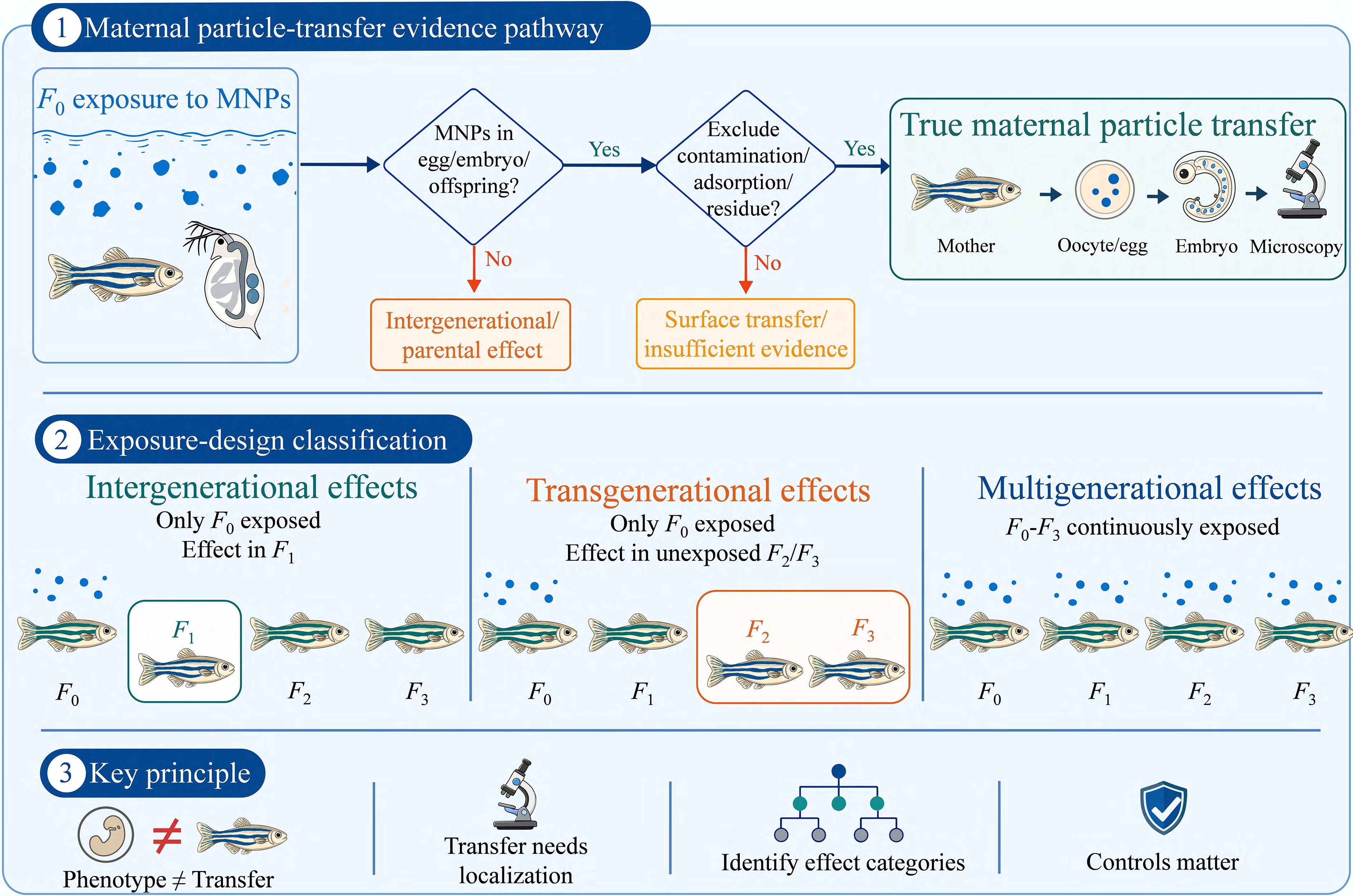
Figure 3.
Evidence-classification framework distinguishing true maternal particle transfer from intergenerational evidence, multigenerational evidence, and transgenerational evidence. The diagram separates transfer confidence from exposure design: True particle transfer requires particle localization in eggs, embryos, or offspring's tissues plus the controls for contamination, adsorption, dye leakage, and renewed exposure, whereas continuous exposure across generations is interpreted as multigenerational evidence unless a clean-generation or recovery design is present.
Notably, cross-generational responses are not necessarily monotonic; higher concentrations or additional generations do not consistently yield stronger effects[17,49]. Variation may reflect particle size, surface properties, exposure design, compensatory responses, or reproductive trade-offs[50,51]. The findings should therefore be interpreted as exposure design-dependent life history responses unless direct localization and clean-generation criteria are met. Representative studies are summarized in Table 1.
-
The mechanistic pathways connecting parental MNP exposure to offspring-related outcomes can be organized into three interconnected layers (Fig. 4). The first layer comprises initial interface events, including corona formation and barrier uptake, which determine whether the particles reach reproductive tissues. The second layer encompasses core cellular stress responses, including oxidative stress, mitochondrial dysfunction, inflammation, and apoptosis, which directly compromise gamete and embryo quality. The third layer involves systemic and persistent signals, including endocrine disruption, epigenetic regulation, and microbiota dysbiosis, which can propagate effects across generations. Co-contaminant vector effects act as modifiers across all three layers by altering the effective dose and tissue distribution of the associated pollutants.
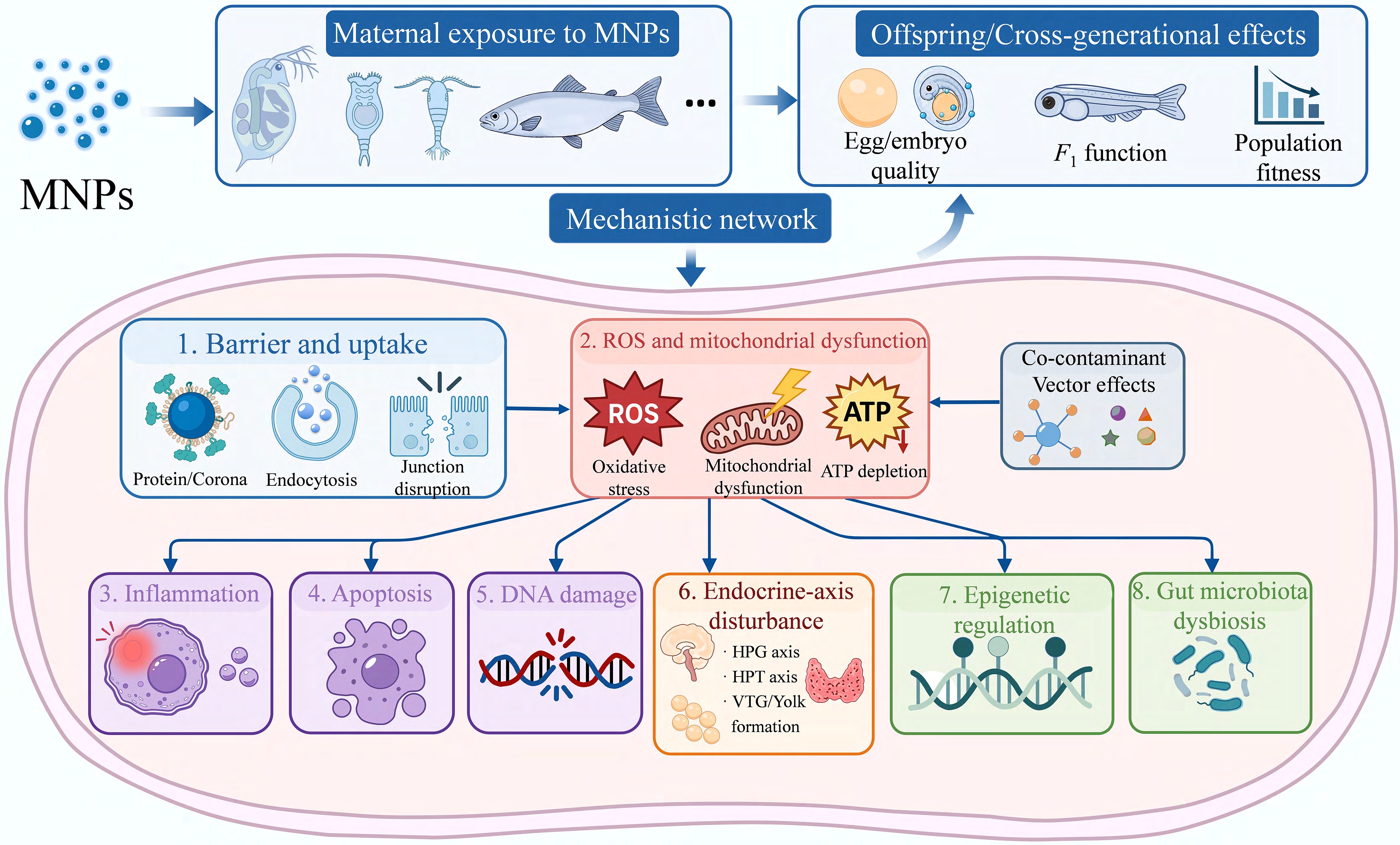
Figure 4.
Potential mechanistic pathways linking parental MNP exposure to offspring and multigenerational phenotypes, independent of demonstrated particle transfer. The mechanistic network includes barrier uptake, oxidative stress and mitochondrial dysfunction, inflammation, apoptosis, DNA damage, endocrine axis disturbance, epigenetic regulation, and gut microbiota dysbiosis. Co-contaminant vector effects may modify exposure and tissue distribution across these pathways. Arrows indicate proposed mechanistic connections rather than a confirmed linear causal sequence. MNPs, micro(nano)plastics; ROS, reactive oxygen species; ATP, adenosine triphosphate; HPG, hypothalamic–pituitary–gonadal; HPT, hypothalamic–pituitary–thyroid; VTG, vitellogenin.
Corona formation and barrier uptake
-
Upon entering an organism, MNP surfaces rapidly adsorb biomolecules (proteins, lipids, polysaccharides) to form a bio-corona that determines the surface charge, hydrophobicity, and receptor recognition, thereby governing mucus retention, cellular internalization, and access to reproductive compartments[52−55]. Barrier crossing occurs through transcellular endocytosis (clathrin-, caveolin-, or phagocytosis-mediated), paracellular leakage following tight-junction disruption, or passive entry after tissue injury[56]. Positively charged particles (e.g., PS-NH2) exhibit stronger retention and greater inhibition of population growth than carboxylated or pristine particles[21]. In zebrafish, PS-NPs alter intestinal villi, tight-junction genes, and the gut microbiota, with concomitant changes in brain neurotransmitters and F1 embryonic endpoints[57]. These barrier effects may provide an indirect link between intestinal exposure and reproductive or embryonic consequences even when direct particle transfer is limited.
Oxidative stress, mitochondrial dysfunction, and energy reallocation
-
Oxidative stress provides a direct mechanistic link between particle exposure and reproductive cost[58,59]. Membrane perturbation, electron transport chain leakage, and antioxidant depletion generate ROS that first compromise adenosine triphosphate (ATP) supply and redox balance in oocytes, embryos, and other high-metabolism tissues[58]. Mitochondrial dysfunction is central: Lipid peroxidation reduces membrane integrity, the loss of mitochondrial membrane potential lowers oxidative phosphorylation efficiency, and respiratory chain Complexes I and III leak additional electrons, amplifying ROS production[60]. Since oocyte maturation, cleavage, and early organogenesis depend on a stable ATP supply, mitochondrial stress translates parental exposure into reduced embryo quality[48,59].
Long-term studies illustrate how oxidative responses convert to life history outcomes. Daphnia pulex exposed to 75-nm PS-NPs for three generations showed altered antioxidant and vitellogenin genes in F0–F1, with reduced antioxidant responses and altered growth/reproduction in F2[61]. Moina macrocopa exposed to 1-μm PS beads for three generations showed decreased F2 survival, growth inhibition, and delayed reproduction alongside shifts in malondialdehyde (MDA), catalase (CAT), and superoxide dismutase (SOD)[62]. The interpretive value of oxidative markers depends on their position within a complete energy allocation chain from parental maintenance and yolk accumulation to embryonic development, larval movement, and reproductive investment.
Inflammation and immune homeostasis
-
Inflammation converts local particle irritation into tissue dysfunction. Larger particles trigger mechanical abrasion and epithelial injury; internalized nanoparticles induce lysosomal damage, potassium efflux, and mitochondrial danger signals, activating the NOD-like receptor family pyrin domain-containing 3 (NLRP3) inflammasome via nuclear factor kappa B (NF-κB) priming and caspase-1-mediated interleukin-1β (IL-1β) and interleukin-18 (IL-18) maturation[63]. For reproduction, chronic low-grade inflammation is more consequential than acute peaks, as it consumes energy and alters nutrient transport and tissue microenvironments[56].
Although the intestine and gill are primary inflammatory sites, consequences extend to the gonads and neuroendocrine systems. In Nile tilapia (Oreochromis niloticus) exposed to multiple polymer types, particles accumulated in the intestine, gill, liver, gonad, and brain, with altered immune and biochemical markers[64]. Zebrafish intestinal studies likewise show that PS-NPs affect immune factors, barrier genes, and the microbiota[57]. Parental intestinal inflammation can thus alter nutrient absorption, metabolites, and endocrine signals, modifying yolk supply and the F1 developmental environment, a route that explains offspring effects even when particles are undetected in the embryos[65−67].
Apoptosis, DNA damage, and developmental quality
-
Apoptosis and DNA damage primarily explain reduced gamete quality, embryonic abnormalities, and early offspring mortality[41,47]. Particle-induced membrane injury, oxidative DNA base damage, and chromatin disruption activate p53, Bax/Bcl-2, and caspase pathways[41]. When damage occurs in germ cells or early embryos, consequences extend beyond local cell loss to impaired oocyte maturation, reduced sperm function, abnormal cleavage, and unstable organogenesis[42].
In zebrafish embryos exposed to ~106-nm PS-NPs (200–400 mg/L), survival and hatching decreased, body length shortened, and locomotor behavior was altered, accompanied by downregulation of anti-apoptotic genes (bcl2, bcl-XL) and altered base excision repair genes (lig1, pold, parp1)[47]. Short-term nanoparticle exposure also directly affects germ-cell chromatin compaction, motility, and oocyte-stage proportions in zebrafish[42]. The key intergenerational question is whether sublethal or misrepaired germ-cell damage survives fertilization. A normal early F1 phenotype does not necessarily preclude later functional defects[43].
Endocrine disruption and maternal provisioning
-
Endocrine disruption directly alters hormonal signals governing reproduction and development. The hypothalamic–pituitary–gonadal (HPG) axis controls gametogenesis, steroidogenesis, and vitellogenin (VTG) production, which links maternal liver function to yolk supply and embryonic starting quality[66−68]. The hypothalamic–pituitary–thyroid (HPT) axis regulates hatching, neurodevelopment, and basal metabolism, while the growth hormone (GH)/insulin-like growth factor (IGF) axis controls growth and energy allocation[69,70].
By altering cholesterol transport, steroidogenic enzyme expression, VTG regulation[31,71], and thyroid hormone metabolism[69], MNPs can compromise offspring from the embryonic starting point. For example, parental zebrafish exposed to 1-μm PS-MPs showed intestinal accumulation and altered gonadal steroidogenesis genes, yet hatching and malformation rates in F1 remained unchanged[71], indicating that endocrine shifts can precede overt developmental toxicity. Co-exposure scenarios further illustrate this: PS-NPs promoted tris(1,3-dichloro-2-propyl) phosphate (TDCIPP) transfer to zebrafish eggs and aggravated thyroid hormone disruption in F1[69], while microcystin-LR (L = leucine, R = arginine) and PS-NP co-exposure reduced F1 growth through interference with the GH/IGF axis[70].
Epigenetic regulation and microbiota dysbiosis
-
Strict transgenerational effects—persistence in unexposed generations—are difficult to explain by particle residues alone and may instead involve epigenetic reprogramming[18,19,72] and microbiota-mediated signals[73]. DNA methylation, histone modification, and noncoding RNAs can alter gene expression thresholds during gametogenesis and early embryogenesis[74]. The gut microbiota affects liver, brain, and gonadal function through short-chain fatty acids, bile acids, and immune signals[57,65].
MNP-induced metabolic stress changes substrates' availability (e.g., S-adenosylmethionine [SAM] for methylation, acetyl coenzyme A [acetyl-CoA] for acetylation), affecting DNA methyltransferases and histone-modifying enzymes[74]. In D. magna, F0 exposure to PE/BP-3 fragments produced reproduction-related DNA methylation changes persisting to F3[19]. Microbiota-mediated persistence is particularly relevant under combined stress: Diaphanosoma celebensis exposed to PS-NPs under elevated temperature (30 °C) showed reproductive failure in F2 with altered host–microbiota interactions[73]. Thus, an altered parental microbiome can affect egg quality and the early developmental environment without direct particle inheritance[65].
Co-contaminant vector effects
-
Under co-exposure conditions, MNPs alter the effective dose, tissue distribution, and biological targets of associated contaminants, the core of vector effects, rather than merely transporting pollutants[75]. Whether MNPs enhance co-contaminants' toxicity depends on their adsorption–desorption equilibrium in the gut, intestinal absorption efficiency, barrier damage, and distribution direction, not on adsorption capacity alone[75,76].
Three lines of evidence illustrate distinct vector mechanisms. First, hydrophobic organic contaminants such as PS-NPs (70 nm) increased TDCIPP accumulation in parental zebrafish tissues and offspring, aggravating thyroid disruption in F1[69]. Pristine PA MPs promoted TDCIPP transfer to F1, whereas aged PA, with stronger contaminant binding and lower intestinal injury, weakened this effect[77]. Second, regarding metals, in the copepod Tigriopus japonicus, PS-MPs (0.023 mg/L) increased Hg body burden across three generations, aggravating developmental delay and fertility reduction[22]. Ultraviolet (UV)-aged PS-NPs promoted Hg accumulation more strongly than pristine particles, reducing F1 and F2 survival[78]. Third, regarding pharmaceuticals, in D. magna, co-exposure to PS-MPs and carbamazepine reduced F1 offspring number and altered genes involved in reproduction and detoxification (vtg1, vtg2, cyp4, and gst)[79].
Critically, enhanced contaminant transfer does not by itself demonstrate particles' entry into offspring's tissues. Only simultaneous quantification of particles and contaminants in parental tissues, eggs, embryos, and culture media can distinguish a vector's action from altered contaminant bioavailability or parental toxicity signaling.
-
The transfer efficiency and offspring consequences of MNPs are not intrinsic properties of the particles alone but emerge from interactions among three sets of factors: (1) Particle-intrinsic properties (size, morphology, polymer type), (2) surface and interfacial characteristics (charge, functional groups, bio-corona, aging status), and (3) system-level conditions (exposure design, environmental context). These modifiers operate across different scales and have distinct implications for experimental design and risk extrapolation.
Particle-intrinsic properties: size, morphology, and polymer type
-
Particle size is among the most consistently reported determinants of barrier-crossing[26,46,80]. Nanoparticles (< 1 μm) are more likely to enter cells, yolk-associated spaces, and embryonic tissues, whereas micrometer-scale particles tend to remain in the digestive tract and external developmental interfaces, exerting effects through feeding inhibition, mechanical irritation, and nutritional dilution[9]. Morphology further modulates these size-dependent effects: Spherical particles are convenient for standardization, but fibers (accounting for 52%–73% of environmental anthropogenic particles), fragments, films, and weathered mixtures dominate real-world exposures[81]. Nevertheless, cross-generational studies have overwhelmingly used spherical PS particles (75.8%) and pristine materials (83.3%), with only ~3% using fibers[81], a representativeness gap that severely limits environmental extrapolation.
Polymer type, though less mechanistically resolved, influences density, hydrophobicity, additive content, and contaminant affinity. The predominant reliance on PS (71.1% of cross-generational studies) leaves PE, PP, PET, PVC, PA, and PMMA critically understudied[81]. Tire wear particles illustrate how complex materials can invert toxicity rankings across generations: In B. plicatilis, leachate was most toxic in single-generation exposure, but nano-sized tire wear particles became more toxic than the leachate across F3–F6 multigenerational exposure[82], a shift that is not predictable from single-generation assays or from pristine PS particles.
Surface and interfacial characteristics: charge, corona, and aging
-
Surface charge, functional groups, and zeta potential determine MNPs' affinity for membranes, mucus, and biomolecules[52−55]. Positively charged particles generally exhibit higher uptake, retention, and endocytic activity than carboxylated or pristine particles, resulting in stronger feeding inhibition, reduced ATP, and altered reproductive strategies[21]. Bio-coronas further modify these intrinsic surface properties, giving different biological identities to the same particle in different media[52]. For transgenerational studies, reporting nominal particle size is insufficient; zeta potential, hydrodynamic size, aggregation state, and corona composition should be routinely characterized.
Aging and weathering add another layer of complexity. Photooxidation, mechanical abrasion, and biofilm formation increase surface roughness, oxygen-containing functional groups, and additive release, altering aggregation, contaminant adsorption, and bioavailability[7,78]. However, aging does not uniformly enhance toxicity. PET photodegradation leachate reduced locomotor capacity in F1 without direct exposure[83], yet wastewater-incubated PS-MPs showed weakened multigenerational effects[84], and aged PA MPs weakened the promotion of TDCIPP transfer to F1[77]. The direction and magnitude of aging effects depend on the leachate's composition, particle ingestion, organic matter coating, and endpoint sensitivity, precluding any simple generalization that aging increases toxicity.
System-level conditions: exposure design and environmental context
-
Exposure concentration, duration, window, and generation design collectively determine whether particle contact translates into persistent offspring effects[17,18]. Short-term exposures are suitable for uptake and ROS studies[10−12]; long-term or multigenerational designs are required to assess reproductive output and population growth[19,49]. Age and brood order can also alter outcomes: In D. magna, adult F0 exposure to PE/BP-3 fragments reduced growth and reproduction more strongly than neonatal exposure, and the first F1 brood showed advanced reproduction and reduced offspring length compared with later broods[50].
Cross-generational response trajectories fall into four nonexclusive patterns, namely recovery[19,53], persistence[18,72], cumulative aggravation[49,82], and life history trade-offs[33], each with distinct ecological implications. Recovery does not necessarily indicate the absence of risk, because it may depend on compensatory energy allocation. Persistence across unexposed generations may support a transgenerational interpretation when carryover of residue and renewed exposure have been excluded. Cumulative aggravation may emerge under repeated or continuous exposure, whereas life history trade-offs indicate that numerical reproductive output may be maintained at the expense of offspring's quality. These patterns are exposure- and context-dependent rather than inherent properties of a given particle type.
The environmental context—temperature, pH, food availability[85], organic matter[86], and co-contaminants—modifies MNPs' aggregation, settling, surface charge, and ingestion probability, often amplifying or diminishing the effects observed under standard laboratory conditions. Warming (30 °C) combined with PS-NPs induced reproductive failure in F2 of Diaphanosoma celebensis with altered host microbiota[73]. Acidification (pH 7.3) in Paracyclopina nana produced reduced fertility in clean-generation F1 and F2, with DNA methylation patterns resembling those of continuous multigenerational exposure, yet fluorescence tracking did not support persistent particle transfer[72]. Co-contamination further complicates the risk profiles: MNPs can enhance (e.g., Hg in copepods[87]), reduce, or redistribute the toxicity of co-occurring pollutants, and environmentally weathered particles often carry complex additive and metal burdens (Cd, Pb, Cr, Cu, Zn) that are absent from pristine laboratory particles[88]. Such findings underscore that extrapolating from single-particle clean-water assays to real aquatic environments is inherently limited[72,73,88].
-
A central challenge in MNP-associated reproductive toxicology is not simply the documentation of toxic effects but the shortage of experimental designs that support causal interpretation and environmental risk extrapolation. Several priorities are particularly important for strengthening future MNP-related reproductive toxicity studies.
First, evidence for true maternal particle transfer must be strengthened by orthogonal localization and contamination controls. Many studies infer transfer from F1 phenotypes without directly localizing particles in eggs, embryos, or offspring's tissues. Future research should combine fluorescence tracking with polymer-confirmatory methods (Raman spectroscopy, Py-GC/MS, electron microscopy) and include systematic controls for surface adsorption, dye leakage, and culture medium carryover[89].
Second, particles' representativeness and characterization must improve. The field's reliance on monodisperse, pristine PS spheres diverges sharply from environmental reality, which is dominated by polydisperse fibers, fragments, aged mixtures, and particles with complex contaminant loads. Reporting should routinely include the particle size distribution, morphology, surface charge, aggregation state, aging status, additive release, and bio-corona composition to enable cross-study comparisons and environmental contextualization.
Third, endpoint chains and mechanistic validation must move beyond correlative markers. Measures of ROS, SOD, VTG, DNA methylation, and transcriptomic shifts describe responses but do not demonstrate causality. Functional interventions, antioxidant rescue, endocrine blockade, mitochondrial restoration, and microbiota transplantation should be used to link molecular changes to phenotypic outcomes. Likewise, endpoints must extend beyond single embryonic measures (hatching rate, malformation) to include parental gonadal burden, gamete quality, feeding and locomotion in F1, reproductive capacity of F1, and population growth rates. Only with such continuous endpoint chains can individual toxicity observations be translated into population-level risk.
Collectively, these priorities point toward a more integrated framework of identifying which particles, through which biological interfaces and under which environmentally relevant conditions, produce predictable and measurable population-level consequences.
-
From an ecological risk perspective, a key concern is not simply whether particles are maternally transferred but whether population renewal is compromised. The available evidence indicates that weakened reproductive output, impaired gamete quality, altered embryonic development, and reduced offspring fitness can arise through multiple nonequivalent routes, including true maternal particle transfer, parental physiological injury, external embryonic contact, continuous multigenerational exposure, and co-contaminant vector effects. These routes are often conflated in the literature, yet they demand distinct interpretive frameworks and experimental designs. Several recurring limitations currently constrain causal inference and environmental risk extrapolation. First, evidence for true maternal particle transfer remains methodologically thin: Most studies infer transfer from the offspring's phenotypes rather than demonstrating particle localization in eggs, embryos, or offspring's tissues with orthogonal confirmation and contamination controls. Second, experimental particles diverge sharply from environmental reality—the field relies overwhelmingly on monodisperse, pristine PS spheres, whereas real exposures involve polydisperse fibers, fragments, aged mixtures, and complex contaminant loads. Third, mechanistic studies predominantly report correlative markers (ROS, gene expression, DNA methylation) without functional validation linking molecular changes to fitness-relevant outcomes. Moving forward, MNP-associated reproductive risk assessments should prioritize three shifts: (1) Distinguishing true maternal particle transfer from broader cross-generational toxicity through localization-based evidence criteria; (2) replacing model particles with environmentally relevant materials and comprehensive physicochemical characterization; and (3) extending endpoint chains from single embryonic measures to population-level parameters, such as parental reproductive output, growth and locomotion in F1, reproductive capacity of F1, and the population growth rate. These changes would help shift evaluations of MNP risk from a catalog of observed effects toward a more predictive framework for environmental risk assessments.
We thank the reviewers for their comments on this work.
-
It accompanies this paper at https://doi.org/10.48130/newcontam-0026-0023.
-
Not applicable.
-
The authors confirm their contributions to this study as follows: Chengbin Li: conceptualization, methodology, writing – original draft; Yanping Li: methodology, writing – original draft; Sitong Huo: formal analysis, methodology; Jiang Yu: formal analysis; Xuerui Zhou: formal analysis; Kejing Zhang: formal analysis; Neng Yan: conceptualization, writing – review and editing, funding acquisition, supervision; Jianbo Shi: conceptualization, writing – review and editing, Supervision. All authors reviewed the results and approved the final version of the manuscript.
-
This article is a review and does not report new primary data. All information discussed in this manuscript is derived from published literature cited in the references.
-
The authors declare no competing financial interest.
-
Full list of author information is available at the end of the article.
- The supplementary files can be downloaded from here.
- Copyright: © 2026 by the author(s). Published by Maximum Academic Press, Fayetteville, GA. This article is an open access article distributed under Creative Commons Attribution License (CC BY 4.0), visit https://creativecommons.org/licenses/by/4.0/.
-
About this article
Cite this article
Li C, Li Y, Huo S, Yu J, Zhou X, et al. 2026. Maternal particle transfer versus cross-generational toxicity of micro(nano)plastics in aquatic organisms: a critical evidence-classification review. New Contaminants 2: e026 doi: 10.48130/newcontam-0026-0023 

Maternal particle transfer versus cross-generational toxicity of micro(nano)plastics in aquatic organisms: a critical evidence-classification review
- Received: 13 July 2026
- Revised: 15 August 2026
- Accepted: 31 August 2026
- Published online: 17 September 2026
Abstract: Micro(nano)plastics (MNPs) have become persistent and widespread contaminants in aquatic ecosystems, raising concerns about their capacity to impair reproductive success and population renewal in exposed organisms. However, the interpretation of cross-generational reproductive risks remains fragmented because similar offspring phenotypes can arise from nonequivalent causal routes, including direct maternal particle transfer, parental physiological injury, or external exposure at embryonic interfaces. Here, we propose an evidence hierarchy framework to distinguish true maternal particle transfer from broader cross-generational toxicity, encompassing intergenerational, multigenerational, and transgenerational effects. Our synthesis reveals that transfer pathways differ markedly among aquatic taxa, ranging from brood chamber exposure and gut–ovary–oocyte transfer in cladocerans to ovary–oocyte–yolk/chorion interfaces in fish, yet conclusive evidence for endogenous particle entry into offspring's tissues remains scarce and requires rigorous orthogonal localization and contamination controls. Mechanistically, we link MNP exposure to offspring outcomes through a network of barrier uptake, oxidative and mitochondrial stress, inflammation, apoptosis, endocrine disruption, epigenetic regulation, microbiota dysbiosis, and contaminant vector effects, which collectively compromise gamete quality, embryonic development, and subsequent life history performance. Our synthesis indicates that offspring toxicity following parental exposure cannot be equated with maternal particle transfer, and that environmentally realistic risk assessment must prioritize particle localization evidence, clean-generation designs, and population-level fitness endpoints over single-generation toxicity descriptions.