











-
Intensive aquaculture subjects fish to a range of husbandry-related stressors, including high stocking density, poor water quality, and nutritional imbalance, which may compromise host health and increase vulnerability to infectious diseases. Antibiotics and other veterinary drugs remain important tools for disease control; however, excessive use can contribute to antimicrobial resistance, disturbance of the intestinal microbiota, greater pathogen susceptibility, and food safety concerns[1]. These concerns have promoted the development of alternative approaches to protect fish health and reduce antibiotic use.
Plant-derived compounds are increasingly investigated for their potential to support fish health and strengthen disease resistance. Beneficial effects of extracts from loquat (Eriobotrya japonica), tulsi (Ocimum sanctum), and dandelion (Taraxacum officinale) have been reported in kelp grouper (Epinephelus bruneus)[2], rohu (Labeo rohita)[3], and rainbow trout (Oncorhynchus mykiss)[4], respectively. Because such extracts contain numerous bioactive constituents, it is often difficult to determine which individual compounds are responsible for the observed biological effects.
Compared with crude plant extracts, chemically defined compounds allow more accurate dose determination and facilitate the evaluation of their specific biological effects. Individual plant-derived compounds with defined chemical structures have therefore been investigated more directly. Carvacrol and thymol from oregano (Origanum heracleoticum)[5], cinnamaldehyde from Chinese cinnamon (Cinnamomum cassia)[6], and dasyscyphin C from false daisy (Eclipta prostrata)[7] have all shown potential applications in fish disease management. Compared with crude extracts, purified compounds allow more precise control of exposure levels and clearer evaluation of compound-specific biological effects.
Parthenolide (PTL), a bioactive compound derived from feverfew (Tanacetum parthenium), exhibits anticancer and inflammation-modulating activities[8]. Studies in mammalian models further indicate that PTL can suppress bacterial growth and inflammatory cytokine production and alleviate immune-mediated tissue injury[9,10]. These properties raise the possibility that PTL may influence both pathogens and host-associated microbial communities.
The intestinal microbiota contributes to nutrient utilization, immune regulation, and disease susceptibility in fish[11], and disruption of this community is frequently associated with impaired intestinal function[12]. Mammalian studies have shown that PTL can reshape the intestinal microbial community and enhance short-chain fatty acid generation while alleviating intestinal inflammation[13]. In our previous study, dietary fecal microbiota transplantation restored the intestinal microbial community of florfenicol-treated koi carp (Cyprinus rubrofuscus) within 7 d, while Aeromonas levels declined. Metabolomic analysis further associated PTL with this recovery process[14]. These observations prompted us to investigate the effects of purified PTL in fish more directly.
Aeromonas veronii is an opportunistic bacterial pathogen associated with hemorrhagic septicemia, ulcers, and fin rot in several fish species, including koi carp, river sleeper (Odontobutis potamophila), and sea bass (Lateolabrax maculatus)[15−17]. Accordingly, purified PTL was investigated for its activity against A. veronii and for short-term modulation of the gut microbial community in koi carp during a 7-d feeding trial. Bacterial proliferation, biofilm development, and swarming and swimming behavior were examined in vitro, while serum biochemical responses, microbial community profiles, and post-challenge survival were assessed in vivo.
-
The A. veronii strain KCL-1, obtained earlier from clinically affected koi carp[15], was cultured in Luria–Bertani broth at 30 °C for 12 h. Bacterial suspensions used for the in vitro PTL assays and challenge experiments were adjusted to 1 × 10⁶ CFU/mL.
In vitro antibacterial activity of PTL against A. veronii
Growth inhibition assay and growth curve analysis
-
The growth-inhibitory activity of PTL against A. veronii was examined by a micro-broth dilution assay adapted from Agbo et al.[18]. A PTL (6,000 μg/mL; Sigma-Aldrich, St. Louis, MO, USA; purity ≥ 98%) stock solution was prepared in 0.5% DMSO and diluted with LB medium to obtain the required working solutions. For each treatment, 190 μL of A. veronii suspension (1 × 10⁶ CFU/mL) was mixed with 10 μL of PTL working solution in a 96-well plate, giving final PTL concentrations of 9.375, 18.75, 37.5, 75, 150, and 300 μg/mL. Control wells received the same bacterial inoculum with an equal volume of PTL-free LB medium, and all treatments were run in triplicate. Bacterial growth was followed for 48 h at 30 °C by recording OD600 at 2-h intervals.
Bacterial biofilm inhibition assay
-
For microscopic observation, 14-mm glass coverslips were placed in 24-well plates. Each well contained 950 μL of A. veronii suspension (1 × 10⁶ CFU/mL) supplemented with 50 μL of PTL working solution prepared in LB medium, resulting in final PTL concentrations of 18.75, 37.5, or 75 μg/mL. For the control, LB medium lacking PTL was added at an equivalent volume. After incubation for 24 h at 30 °C, the coverslips were washed with sterile PBS, immersed in anhydrous methanol for 15 min, allowed to dry, and then treated with 0.2% crystal violet for 20 min. Biofilm staining characteristics were visualized using a Leica DM4000B microscope (Leica, Germany).
Biofilm biomass was quantified using a parallel 96-well assay. A total volume of 200 μL was used per well, comprising 190 μL of bacterial suspension and 10 μL of PTL working solution, with the same final PTL concentrations as those used for microscopic observation. Incubation, fixation, and crystal violet staining were carried out under the conditions described above. After staining, excess dye was removed by washing the wells twice with sterile PBS. The biofilm-associated crystal violet was then solubilized in 33% acetic acid, and absorbance was measured at 590 nm on a microplate reader, with three replicate wells included for each condition.
Swarming and swimming assays
-
Bacterial motility was evaluated on semisolid media containing PTL at 18.75, 37.5, or 75 μg/mL. Swarming agar was prepared with 1 g tryptone, 0.5 g NaCl, 0.5 g agar, and 0.5 g glucose in 100 mL water, while the swimming agar consisted of 1 g tryptone, 3 g NaCl, and 0.3 g agar in the same volume of water; formulations without PTL served as controls. After solidification, a 5-μL aliquot of an A. veronii culture was applied to the center of each plate. Following incubation at 30 °C for 24 h, bacterial motility was evaluated by measuring the diameter of the migration zone. Three independent replicates were included for each condition.
In vivo feeding trials
Diet preparation and fish acclimation
-
Details of the experimental diet composition are provided in Supplementary Table S1. After grinding, the diet was supplemented with PTL at 15, 75, or 150 mg/kg and processed into 2-mm pellets. The pellets were allowed to dry at 20–22 °C before being kept at −20 °C until use. Koi carp (~39 g, ~14 cm) without visible signs of disease were obtained from Tianjin Longjin Aquaculture Co., Ltd and acclimated under laboratory conditions for 2 weeks. During this period, the basal diet was offered twice daily at a daily ration equivalent to 2% of body weight. Fish were acclimated in water at 25 ± 1 °C with approximately 8 mg/L dissolved oxygen and a pH of 7.5 ± 0.2.
Short-term tolerance assessment of PTL in koi carp
-
The short-term feeding experiment comprised four dietary treatments: Control, L-PTL (15 mg/kg), M-PTL (75 mg/kg), and H-PTL (150 mg/kg), with three replicate tanks per treatment. These dietary levels provided approximately 0, 0.3, 1.5, and 3 mg PTL/kg body weight per day, respectively. Feeding activity, general growth condition, swimming behavior, external appearance, and signs of distress or illness were checked daily throughout the 7-d trial. On day 7, six individuals per tank were sedated in 100 mg/L MS-222 (Sigma-Aldrich, St. Louis, MO, USA). Following surface disinfection with 75% alcohol, approximately 0.5 mL of blood was collected from each fish by caudal venipuncture. Serum samples obtained from the six individuals in the same tank were combined into one pooled sample for biochemical assays. The concentrations of albumin (ALB) and total bile acid (TBA), together with the activities of gamma-glutamyltransferase (γ-GT), alanine aminotransferase (ALT), alkaline phosphatase (ALP), and aspartate aminotransferase (AST), were measured with commercial kits supplied by Changchun Huili Biotechnology Co., Ltd (Jilin, China).
Influence of PTL on intestinal microbiota
-
A total of 144 koi carp were allocated to 16 tanks, with nine fish housed in each tank. Four dietary groups, including Control, L-PTL (15 mg/kg), M-PTL (75 mg/kg), and H-PTL (150 mg/kg), were represented by four tanks. After the 7-d feeding period, fish were immersed in 100 mg/L MS-222 for anesthesia, and the entire intestine was aseptically excised. The external surface of the intestine was rinsed with sterile PBS before the luminal contents were sampled. Material collected from six fish in the same tank was combined into one composite sample and immediately frozen in liquid nitrogen.
Microbial DNA extraction from each composite intestinal sample was performed with the QIAamp DNA Stool Mini Kit (Qiagen, GmbH, Hilden, Germany). PCR was then used to target the V3–V4 hypervariable region of the bacterial 16S rRNA gene. Amplicons were purified with Agencourt AMPure XP magnetic beads. Library fragment size and concentration were checked on an Agilent 2100 Bioanalyzer, after which libraries meeting the quality criteria underwent HiSeq sequencing.
Paired-end sequences were assembled with FLASH v1.2.11[19]. OTU clustering at 97% sequence similarity was conducted using UPARSE v7.0.1090[20], followed by taxonomic annotation against the Greengenes v13.5 database[21]. Shannon and Chao1 indices were calculated to evaluate alpha diversity. Variation in microbial community structure was examined using unweighted UPGMA clustering, partial least squares discriminant analysis (PLS-DA), and non-metric multidimensional scaling (NMDS). Data processing and visualization were carried out with QIIME v1.8.0[22] and R v3.4.1. Linear discriminant analysis effect size (LEfSe) analysis[23] was applied to identify bacterial taxa showing differential abundance among dietary groups.
Microbial functional potential was predicted using PICRUSt (Phylogenetic Investigation of Communities by Reconstruction of Unobserved States)[24]. Prior to prediction, OTU abundances were normalized based on predicted 16S rRNA gene copy numbers. The normalized OTU table was then used to infer metagenomic functional content, and the predicted genes were assigned to KEGG orthologs in the Kyoto Encyclopedia of Genes and Genomes (KEGG;
www.kegg.jp/ ) for subsequent functional analysis[25]. KEGG functional categories with p < 0.05 were considered significantly different between groups.Bacterial challenge experiment
-
A separate group of 144 juvenile koi carp (approximately 3 g per fish) was obtained from the Tianjin Zhonghuan Ornamental Fish Market (Tianjin, China). Fish were divided among the Control, L-PTL, M-PTL, and H-PTL groups, with 12 fish in each of three replicate units, and were fed the corresponding diets for 7 d. Each fish was then challenged by intraperitoneal administration of 15 μL of A. veronii suspension (1 × 10⁶ CFU/mL), equivalent to approximately 5 × 10³ CFU/g body weight. Post-challenge mortality was recorded daily for 14 d.
Statistical analysis
-
Statistical analysis was performed in GraphPad Prism 7.0 (San Diego, CA, USA) for all data analyses. The analytical approach was selected according to normality and equality of variances. Data distribution was checked using the Shapiro–Wilk test. When both normality and homogeneity of variance were satisfied, group differences were evaluated by one-way ANOVA with Tukey's post hoc comparisons. Welch's ANOVA was adopted when variance homogeneity was not met. Data that failed to satisfy parametric requirements were analyzed using the Kruskal–Wallis test followed by Dunn's post hoc comparisons. Results are expressed as mean ± standard deviation (SD). Exact p values are reported where applicable, with p < 0.05 considered statistically significant. For the main outcomes, effect estimates are presented as between-group differences with 95% confidence intervals (CIs), and the corresponding effect estimates, 95% CIs, and p values are summarized in Supplementary Table S2.
-
The 48-h growth profiles showed a clear concentration-related effect of PTL on A. veronii. At 9.375 μg/mL, the lag phase was extended to approximately 12 h, whereas exposure to 18.75–75 μg/mL prolonged this phase to approximately 24 h (Fig. 1a). Increasing PTL concentrations progressively slowed bacterial growth during the logarithmic phase. By 48 h, cultures exposed to 0–18.75 μg/mL had reached comparable final densities, whereas significant growth suppression occurred at 37.5, 75, 150, and 300 μg/mL compared with the Control group (p = 0.0020, p = 0.0013, p = 0.0009, and p = 0.0009, respectively; Fig. 1b). At 37.5 μg/mL, the mean difference in OD600 relative to the Control group was −0.5670 (95% CI: −0.6090 to −0.5250), making this the lowest concentration producing a significant inhibitory effect (Supplementary Table S2). Bacterial growth was nearly completely suppressed at 150 and 300 μg/mL.
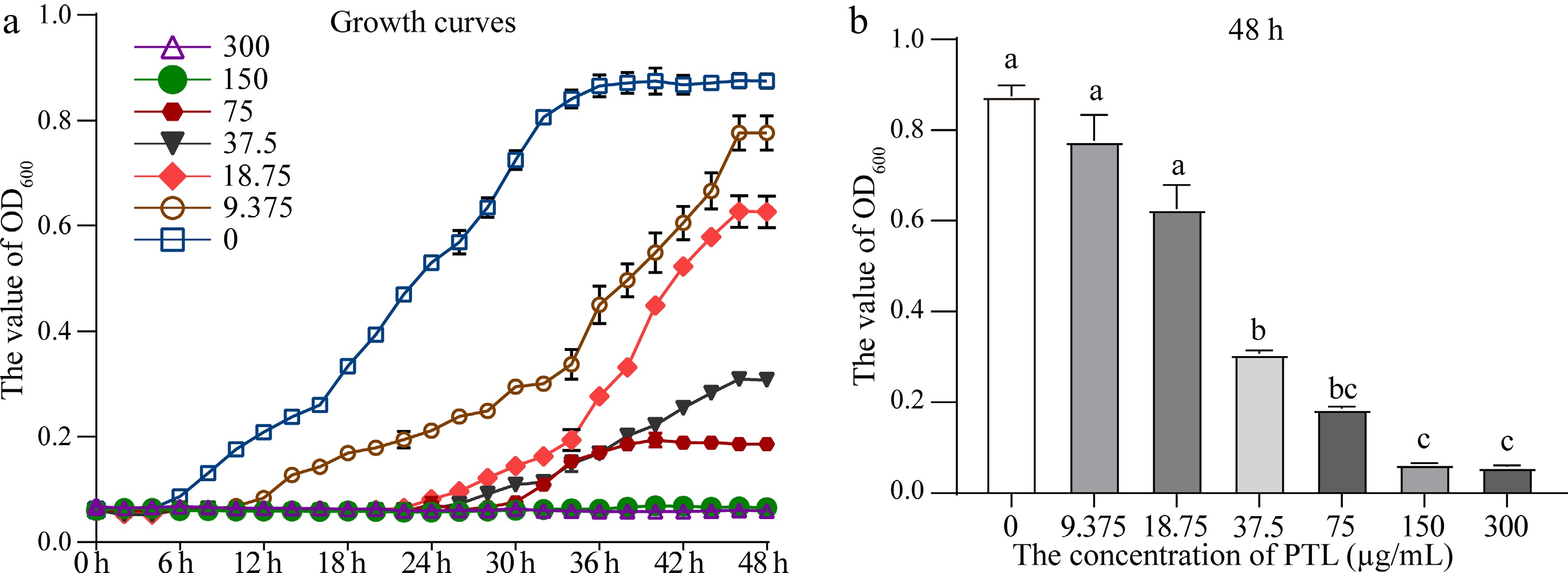
Figure 1.
Growth response of Aeromonas veronii to PTL. (a) 48-h growth profiles at the tested PTL concentrations. (b) OD600 at 48 h. Values are means of three replicates with standard deviations; different letters denote significant differences (p < 0.05).
Effects of PTL on A. veronii biofilm formation
-
Microscopic examination showed a dense and continuous biofilm in the Control group (Fig. 2a). Biofilm density decreased slightly at 18.75 μg/mL PTL, although the overall structure remained largely intact (Fig. 2b). At 37.5 μg/mL, biofilm coverage was markedly reduced and staining became lighter (Fig. 2c), whereas 75 μg/mL PTL severely disrupted biofilm development, preventing formation of a continuous structure (Fig. 2d). Quantitative measurement of biofilm biomass confirmed a concentration-dependent reduction, with significant decreases at 18.75, 37.5, and 75 μg/mL compared with the Control group (all p < 0.0001; Fig. 2e). At 75 μg/mL, the mean difference in biofilm biomass relative to the Control group was −1.0750 (95% CI: −1.1080 to −1.0420; Supplementary Table S2).
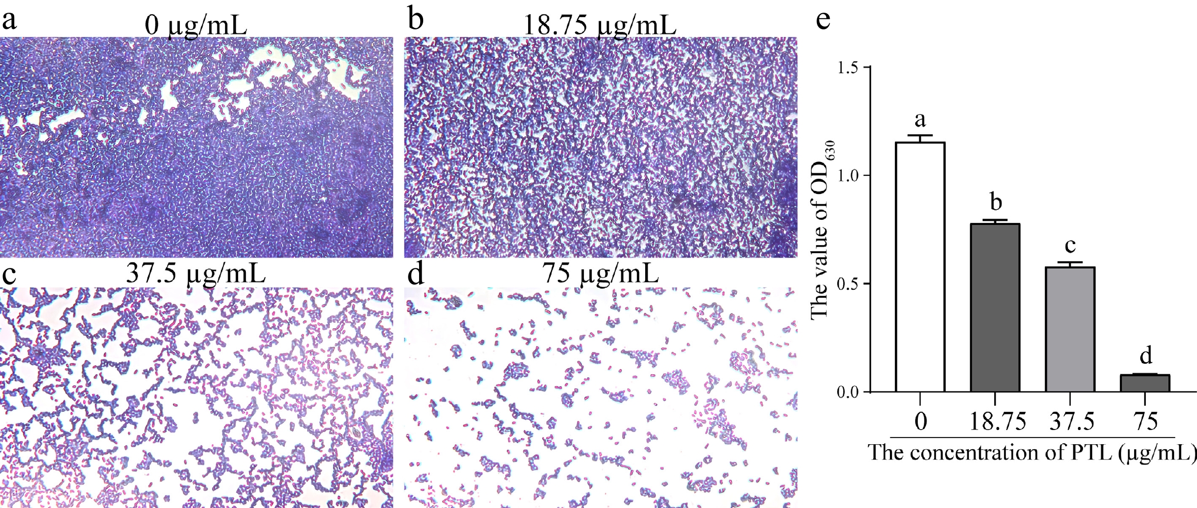
Figure 2.
PTL-mediated changes in Aeromonas veronii biofilm development. (a)–(d) Biofilms visualized after crystal violet staining following treatment with 0, 18.75, 37.5, or 75 μg/mL PTL. (e) Quantitative assessment of biofilm biomass. Data represent mean values with standard deviations from three replicates; groups marked with different letters differ significantly (p < 0.05).
Effects of PTL on the motility of A. veronii
-
PTL reduced both swarming and swimming motility of A. veronii. Swarming was significantly reduced at 37.5 and 75 μg/mL PTL compared with the Control group (p = 0.0071 and p = 0.0004, respectively), corresponding to inhibition rates of 19.89% and 42.80% (Table 1). Swimming was significantly reduced at 18.75, 37.5, and 75 μg/mL compared with the Control group (all p < 0.0001), with inhibition rates of 45.15%, 50.98%, and 74.46% (Table 1). At 75 μg/mL, the mean differences in swarming and swimming diameters relative to the Control group were −0.2833 cm and −0.9500 cm, with corresponding 95% CIs of −0.3603 to −0.2063 and −1.0260 to −0.8741, respectively (Supplementary Table S2).
Table 1. Influence of PTL on the swarming and swimming behavior of Aeromonas veronii.
PTL concentration (μg/mL) 0 (Control) 18.75 37.5 75 Swarming Diameter (cm) 0.66 ± 0.021a 0.55 ± 0.071ab 0.53 ± 0.035b 0.38 ± 0.035c Inhibition rate (%) 0 15.81 19.89 42.80 Swimming Diameter (cm) 1.28 ± 0.035a 0.70 ± 0.071b 0.63 ± 0.021b 0.33 ± 0.035c Inhibition rate (%) 0 45.15 50.98 74.46 Note: Values are presented as mean ± SD (n = 3). Within each row, entries bearing different superscript letters differ significantly (p < 0.05). Short-term tolerance of dietary PTL in koi carp
-
All fish survived the 7-d feeding period and maintained normal feeding activity, growth condition, swimming behavior, and external appearance, with no overt signs of distress or illness. Serum ALB was lower in the M-PTL group than in the Control group (mean difference, −3.8570; 95% CI, −6.8320 to −0.8826; p = 0.0395), whereas no significant pairwise differences were detected among the four groups for ALT, AST, ALP, γ-GT, or TBA (Fig. 3).
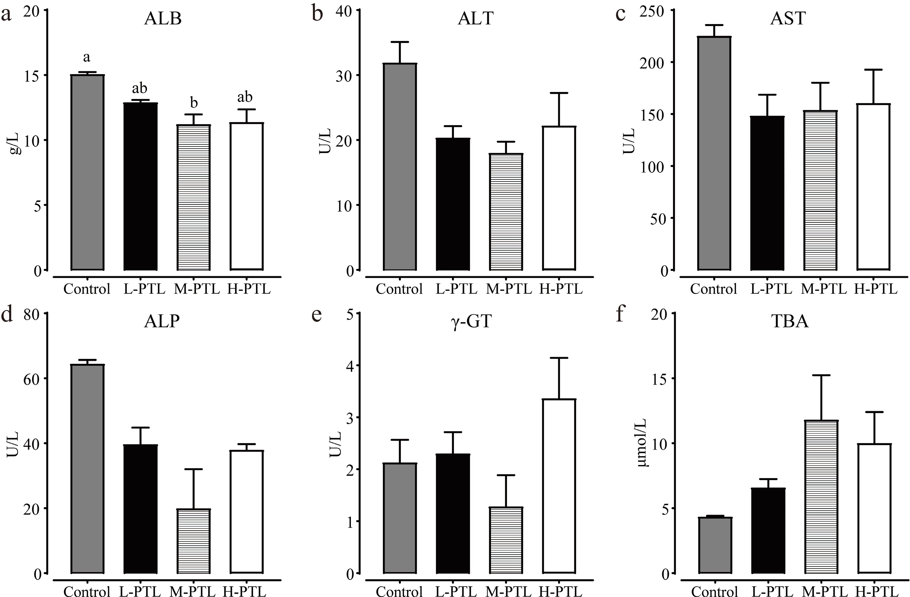
Figure 3.
Serum biochemical indicators in koi carp following 7-d dietary PTL exposure: (a) albumin (ALB), (b) alanine aminotransferase (ALT), (c) aspartate aminotransferase (AST), (d) alkaline phosphatase (ALP), (e) gamma-glutamyltransferase (γ-GT), and (f) total bile acid (TBA). Data are presented as mean values with standard deviations (n = 3); groups assigned different letters differ significantly (p < 0.05).
Influence of PTL administration on intestinal microbiota
-
Sequencing of the 16 intestinal samples generated 696,656 effective tags and 478 OTUs. Thirty-nine OTUs were shared by all four groups, while 159, 15, 17, and 18 OTUs were unique to the Control, L-PTL, M-PTL, and H-PTL groups, respectively (Fig. 4a). The Shannon index was significantly lower in H-PTL than in the Control group (p = 0.0014; Fig. 4b), with a mean difference of −1.7248 (95% CI: −2.4890 to −0.9606; Supplementary Table S2). Chao1 did not differ between the Control and L-PTL (p = 0.1876), M-PTL (p > 0.9999), or H-PTL (p > 0.9999), whereas L-PTL had a lower Chao1 value than H-PTL (p = 0.0178; Fig. 4c). PLS-DA and UPGMA clustering placed the L-PTL and M-PTL samples closer to each other than to the Control and H-PTL samples (Fig. 4d, e).
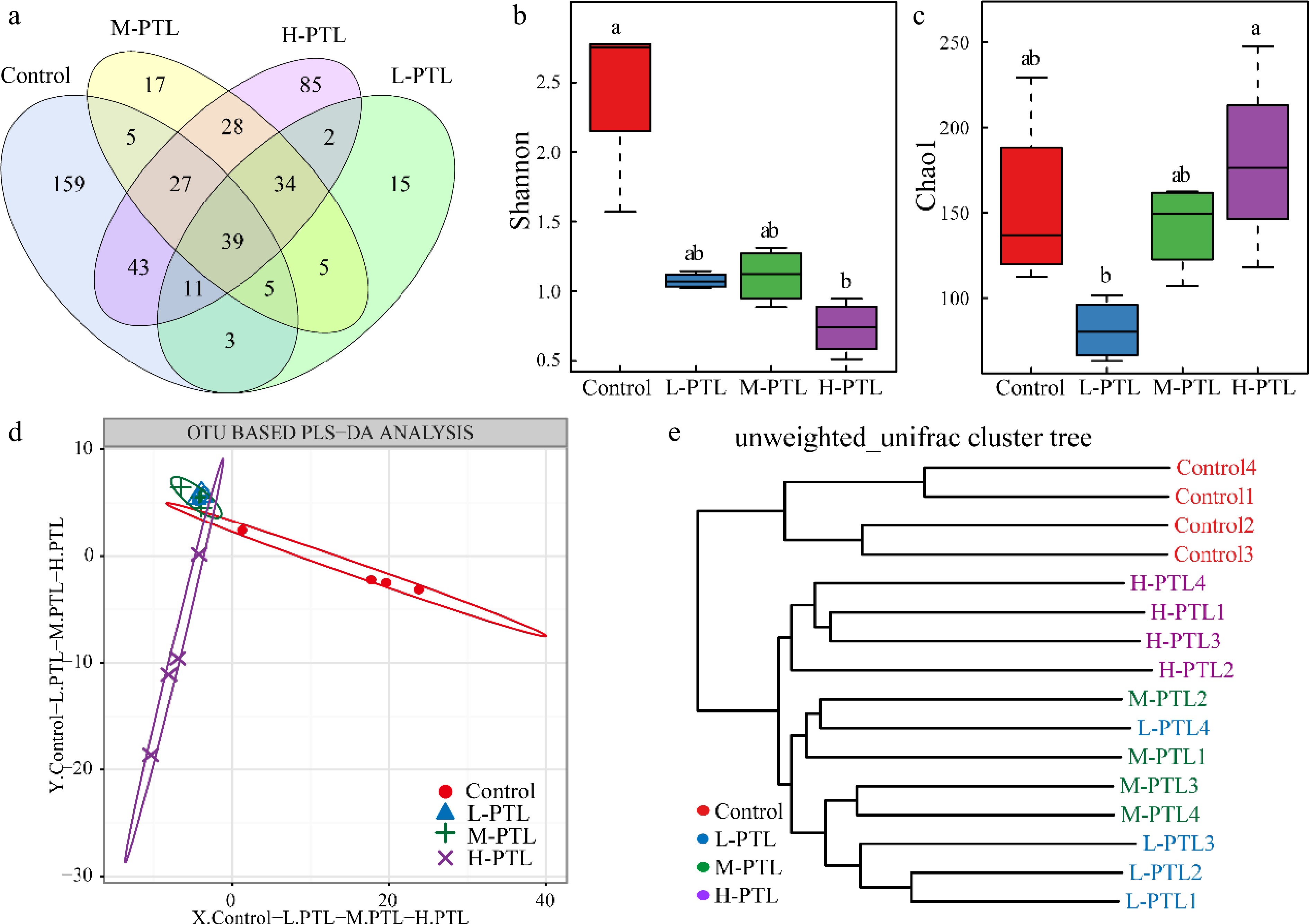
Figure 4.
Intestinal microbial diversity in koi carp after dietary PTL supplementation. (a) Distribution of OTUs among the four groups. (b) Shannon diversity. (c) Chao1 richness. (d) PLS-DA. (e) Unweighted UniFrac-based UPGMA clustering. Results are expressed as mean values with standard deviations (n = 4); groups labeled with different letters differ significantly (p < 0.05).
At the phylum level, Proteobacteria, Bacteroidetes, Cyanobacteria, and Fusobacteria dominated the intestinal microbiota in all groups (Fig. 5a). Compared with the Control, H-PTL had a lower relative abundance of Bacteroidetes (p = 0.0140) and a higher abundance of Fusobacteria (p = 0.0022). Cyanobacteria were also reduced in L-PTL relative to the Control (p = 0.0022), whereas no significant change was detected in M-PTL (p = 0.2688) or H-PTL (p = 0.7117). No significant pairwise differences in Proteobacteria abundance were detected among the four groups (Fig. 5b). At the genus rank, Cetobacterium and Bacteroides represented the dominant taxa. Cetobacterium was enriched in H-PTL relative to Control (p = 0.0022), whereas Bacteroides occurred at a higher relative abundance in L-PTL than in H-PTL (p = 0.0451; Fig. 5c, d).
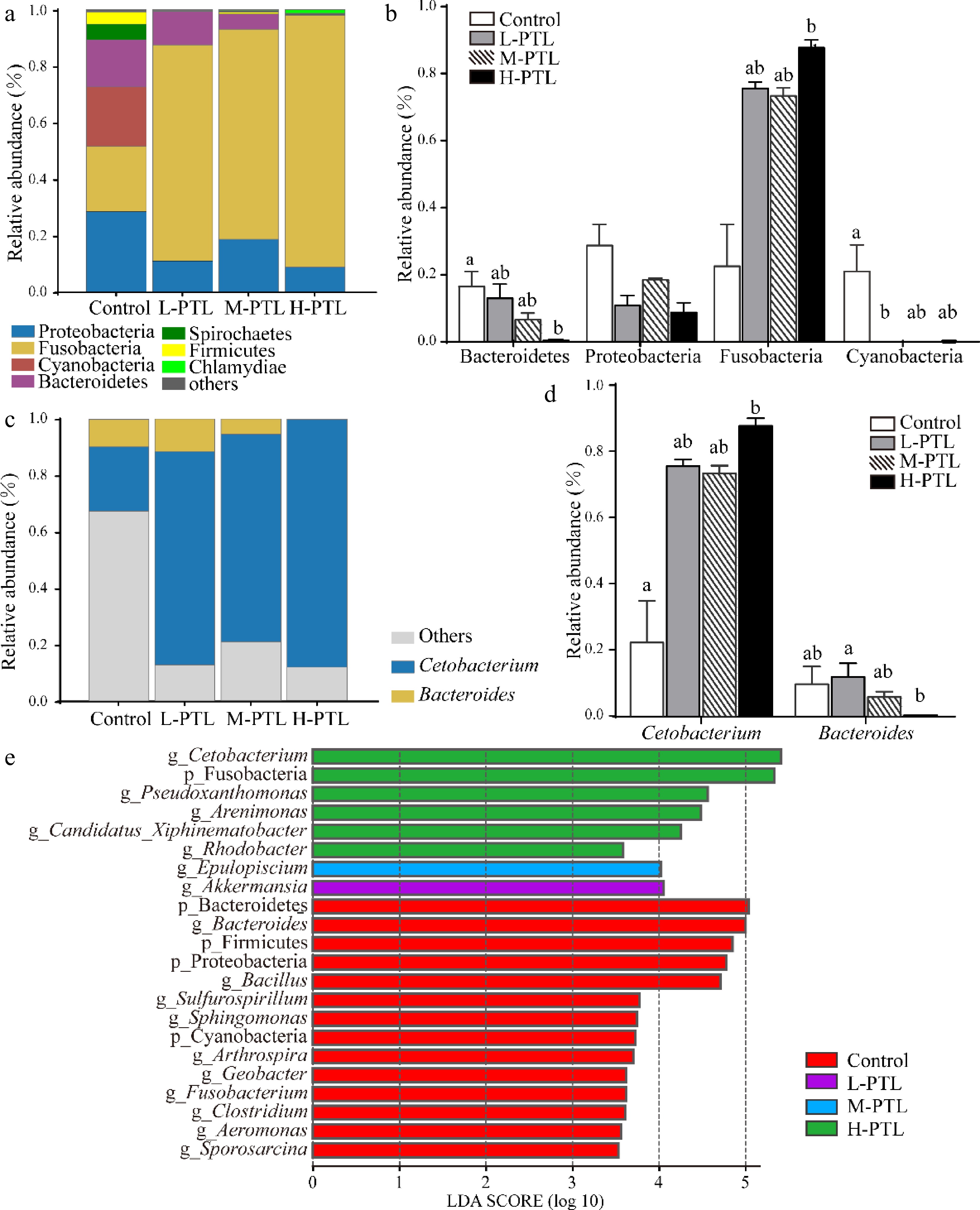
Figure 5.
Bacterial community profiles and treatment-associated taxa in the intestinal microbiota. (a) Distribution of bacterial phyla. (b) Abundance of the major phyla. (c) Distribution of bacterial genera. (d) Abundance of the major genera. (e) Taxa discriminating among groups identified by LEfSe (LDA score > 3.5). Bars represent the mean ± SD (n = 4); groups not sharing the same letter differ significantly (p < 0.05).
LEfSe analysis (LDA score > 3.5; Fig. 5e) revealed distinct taxonomic enrichment among treatments. The Control group was characterized by Bacteroidetes, Firmicutes, Proteobacteria, Cyanobacteria, and 10 genera (Bacteroides, Bacillus, Sulfurospirillum, Sphingomonas, Arthrospira, Geobacter, Fusobacterium, Clostridium, Aeromonas, and Sporosarcina). Akkermansia and Epulopiscium were enriched in L-PTL and M-PTL, respectively, whereas H-PTL was characterized by Fusobacteria and the genera Cetobacterium, Pseudoxanthomonas, Arenimonas, Rhodobacter, and Candidatus Xiphinematobacter.
PICRUSt prediction indicated that metabolism represented the dominant functional category across all groups (Fig. 6a). Relative to the Control group, L-PTL showed greater predicted representation of genes associated with energy metabolism, cofactors and vitamins, terpenoids and polyketides, lipid metabolism, and carbohydrate metabolism. M-PTL showed similar changes together with increased representation of genes associated with xenobiotic biodegradation and metabolism. H-PTL was characterized by increased predicted representation of genes related to energy metabolism, cofactors and vitamins, carbohydrate metabolism, and xenobiotic biodegradation and metabolism (Fig. 6b–d).
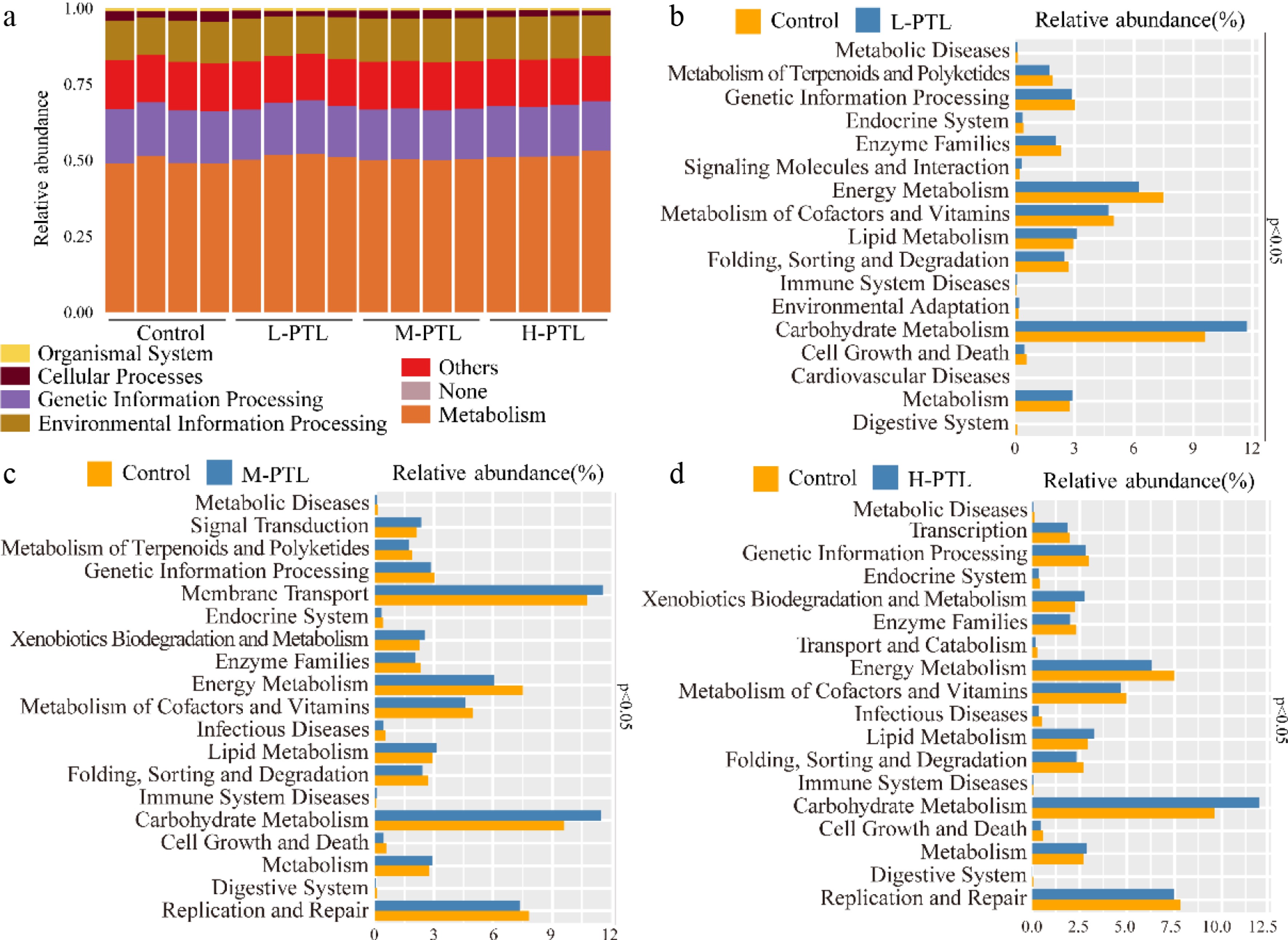
Figure 6.
Predicted microbial functions inferred with PICRUSt. (a) Relative abundance of predicted genes assigned to KEGG level 1 categories. (b)–(d) KEGG level 2 functional categories differing significantly in predicted relative abundance between (b) Control and L-PTL, (c) Control and M-PTL, and (d) Control and H-PTL (p < 0.05).
Bacterial challenge in koi carp
-
In the bacterial challenge experiment, mortality in the M-PTL group occurred only on day 5 and remained unchanged thereafter. Mortality in the Control, L-PTL, and H-PTL groups began on day 1 and reached plateaus on days 8, 6, and 9, respectively (Fig. 7). Final cumulative mortality was 16.67%, 8.33%, 2.78%, and 13.89% in the Control, L-PTL, M-PTL, and H-PTL groups, respectively. Cumulative mortality was significantly lower in the M-PTL group than in the Control (p = 0.0046) and H-PTL (p = 0.0167) groups, whereas no significant differences were detected among the other pairwise comparisons. The absolute difference in cumulative mortality between the Control and M-PTL groups was 13.89 percentage points (95% CI: 4.9960 to 22.7900; Supplementary Table S2).
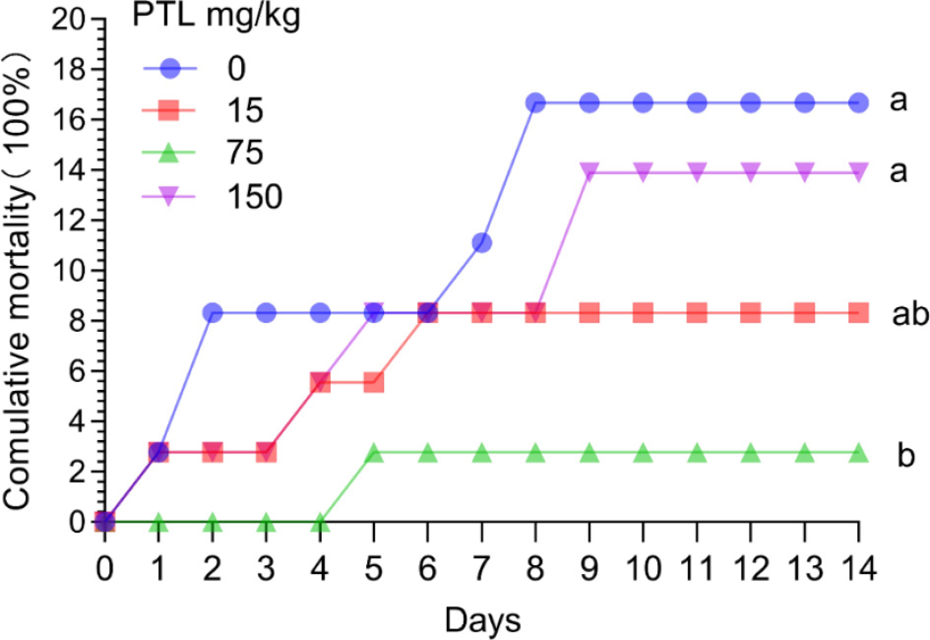
Figure 7.
Post-challenge mortality of koi carp after 7 d of dietary PTL treatment. Fish received PTL at 0, 15, 75, or 150 mg/kg before exposure to Aeromonas veronii. Data are shown as the mean ± SD (n = 3); different letters denote significant differences in the final cumulative mortality (p < 0.05).
-
PTL delayed the growth of A. veronii in a concentration-dependent manner. After 48 h, growth was significantly suppressed at 37.5–300 μg/mL, and 37.5 μg/mL represented the lowest tested concentration producing a significant inhibitory effect. Similar antibacterial activity of PTL has been reported against Bacillus subtilis and S. aureus[26], supporting its broader antibacterial potential.
Biofilm formation provides bacteria with a protective extracellular matrix that can increase tolerance to antimicrobial agents and host defenses. PTL has previously been reported to interfere with established biofilms and amyloid-like structural fibers in B. cereus and Escherichia coli[27]. In the present study, increasing PTL concentrations progressively reduced A. veronii biofilm biomass. This effect may involve interference with extracellular matrix production or accumulation, although the underlying mechanism remains to be determined.
Swarming and swimming motility are important for bacterial spread, colonization, and biofilm formation. In this study, PTL markedly reduced both forms of motility in A. veronii, which may restrict bacterial movement and colonization and thereby contribute to the observed inhibition of biofilm formation. PTL also restricts swarming behavior and biofilm development in Pseudomonas aeruginosa[28], supporting the possibility that reduced motility contributes to the inhibitory effects observed in A. veronii.
Short-term tolerance to dietary PTL was evaluated using both overt clinical observations and serum biochemical responses. No mortality or obvious discomfort occurred during the 7-d feeding period, and γ-GT, ALT, TBA, ALP, and AST remained unchanged. Serum ALB, however, decreased in the M-PTL group. Reduced serum ALB has previously been associated with decreased hepatic protein synthesis in fish[29], suggesting that the observed reduction in ALB may reflect altered protein metabolism. Because this response lacked a dose-dependent pattern and was not accompanied by significant changes in the other measured biochemical parameters, its physiological significance remains uncertain. Overall, these observations support short-term tolerance of PTL under the present experimental conditions, while longer exposure and additional physiological or histopathological assessments are required for a more comprehensive safety evaluation.
Short-term dietary administration is widely used to capture early host-microbiota responses to bioactive compounds without confounding long-term adaptive effects. Herein, a 7-d feeding period was selected to evaluate the acute modulatory effects of PTL on intestinal microbial homeostasis and disease resistance. From an applied perspective, short-term administration is particularly relevant for practical scenarios such as prophylactic or emergency intervention during bacterial outbreaks, where rapid modulation of host defense and gut microbiota is required rather than long-term nutritional effects. This design is supported by our previous work, in which PTL was identified as a microbiota-associated metabolite associated with rapid restoration of intestinal microbial homeostasis following antibiotic-induced dysbiosis[14], further supporting its possible role in short-term microbiota modulation.
It is widely acknowledged that a diverse intestinal microbiota enhances host defenses against pathogens[30]. The reduction in Shannon diversity observed in H-PTL suggests that the highest PTL dose (3 mg/kg fish weight) may adversely affect intestinal microbial diversity. In contrast, although the Shannon and Chao1 indices in the L-PTL and M-PTL groups did not differ significantly from the Control group, beta-diversity analysis showed that samples from these two groups clustered more closely together, suggesting greater similarity in their microbial community composition[31].
Analysis of microbial composition showed that the Control group was enriched in several taxa with potentially beneficial properties, such as Bacteroides and Bacillus, as well as genera containing species previously regarded as opportunistic pathogens, including Fusobacterium, Clostridium, Aeromonas, and Sporosarcina[14]. The enrichment of these potentially adverse taxa may influence host–microbiota interactions during infection, although their specific roles in koi carp remain to be determined.
In the H-PTL group, several genera with diverse potential ecological or physiological roles were enriched, including Cetobacterium, Rhodobacter, Arenimonas, and Pseudoxanthomonas. Some species or strains of Cetobacterium and Rhodobacter have been reported to exert probiotic effects in aquatic animals. Cetobacterium produces vitamin B12 and ferments carbohydrates, supporting digestion and nutrition in freshwater fish[32]. Supplementation with Cetobacterium somerae or its fermentation products enhances tight junctions in zebrafish (Danio rerio) and Nile tilapia (Oreochromis niloticus) and improves intestinal health[33,34]. Similarly, Rhodobacter species, such as R. azotoformans and R. sphaeroides, improve microbiota stability in Chinese mitten crab (Eriocheir sinensis) and shrimp (Penaeus vannamei)[35,36]. Arenimonas has been associated with ecological functions related to nutrient cycling and environmental stability[37]. In contrast, some members of Pseudoxanthomonas have been associated with infectious diseases in humans[38]. The enrichment of Pseudoxanthomonas in the H-PTL group may therefore indicate a potentially adverse shift in the intestinal microbial community at high PTL doses.
In the L-PTL group, Akkermansia was significantly enriched. Akkermansia species, including A. muciniphila and A. glycaniphila, utilize intestinal mucins as substrates, a process associated with mucus renewal and maintenance of barrier function[39]. Akkermansia muciniphila has been associated with benefits related to obesity, diabetes, and inflammation[40,41]. In fish, Akkermansia abundance decreased under environmental stresses, such as copper exposure, high salinity, or drug-induced intestinal damage[42−44]. Our findings suggest that dietary supplementation with PTL may help maintain Akkermansia abundance, potentially supporting intestinal barrier integrity and host health.
Epulopiscium spp. have been reported in the intestinal microbiota of common carp and grouper and have been associated with the utilization of complex carbohydrates[45,46]. Because microbial fermentation of carbohydrates can generate short-chain fatty acids that contribute to intestinal health and disease resistance in fish[47], the enrichment of Epulopiscium spp. in the M-PTL group suggests that dietary PTL may favor a microbial community with enhanced carbohydrate-processing potential, which may contribute to intestinal health and nutrient utilization in koi carp.
PICRUSt analysis predicted increased representation of KEGG functional categories related to lipid, carbohydrate, and energy metabolism following PTL supplementation. Similar PICRUSt-based studies in fish have identified gut microbial functions related to carbohydrate, lipid, and energy metabolism, supporting their potential roles in nutrient processing and energy metabolism[48]. These predicted functional changes therefore suggest that PTL may enhance the metabolic potential of the intestinal microbiota.
In aquaculture, natural compounds derived from plants are increasingly recognized for their potential to enhance disease resistance in fish. Huang et al.[49] reported that plumbagin exhibited antibacterial activity against Streptococcus agalactiae and improved the survival of infected zebrafish. Similarly, geniposide significantly inhibited white spot syndrome virus replication and improved the survival of infected crayfish (Procambarus clarkii), with a maximum viral replication inhibition rate exceeding 98%[50]. In our study, 7-d dietary administration of PTL improved the survival of koi carp following A. veronii challenge. This protective effect was most pronounced in the M-PTL group (75 mg/kg feed), which exhibited the lowest cumulative mortality. These results suggest that the medium PTL dose may be more effective in enhancing resistance to bacterial infection in koi carp.
In this study, the in vivo effects of dietary PTL were evaluated following a short-term feeding period, and the functional implications of the observed microbiota changes are primarily based on taxonomic associations and predicted microbial functions. Future studies should incorporate longer-term feeding trials and higher-resolution metagenomic and metabolomic analyses to clarify the long-term effects of PTL and the mechanisms underlying its modulation of intestinal health and disease resistance.
-
PTL exerted multiple inhibitory effects on A. veronii, including suppression of bacterial proliferation, biofilm development, and motility, with 37.5 μg/mL representing the lowest tested concentration that significantly affected growth after 48 h. Dietary PTL altered the intestinal microbial community and its predicted functional profile in a dose-dependent manner. Low and moderate doses were associated with the enrichment of selected taxa with potentially beneficial properties and changes in predicted metabolic pathways, whereas the high dose was associated with reduced microbial diversity and increased abundance of several taxa with potentially adverse implications. Following A. veronii challenge, the medium dietary dose of PTL (75 mg/kg feed, corresponding to 1.5 mg/kg body weight) produced the lowest cumulative mortality, significantly lower than that in the Control and H-PTL groups. These findings support further evaluation of PTL as a potential functional compound for bacterial disease management in aquaculture.
We thank Mr. Qijie Zhang of Tianjin Longjin Aquaculture Co., Ltd for providing the koi carp used in this study.
-
Animal procedures were conducted in accordance with the National Institutes of Health Guide for the Care and Use of Laboratory Animals (NIH Publications No. 8023, revised 1978) and were approved by the Animal Experiment Ethics Committee of Tianjin Agricultural University (approval No. 2024LLSC20, approved on 12 March 2024).
-
The authors confirm their contributions to the paper as follows: investigation and writing − original draft: Han Z; methodology: Han Z, Luo Z, Yang H; data curation: Han Z, Luo Z; conceptualization: Liu J; resources: Liu J, Zhou X; funding acquisition, project administration, and supervision: Sun J; writing − review and editing: Luo Z, Yang H, Sun J. All authors reviewed the results and approved the final version of the manuscript.
-
The raw 16S rRNA gene sequencing data generated in this study have been deposited in the NCBI Sequence Read Archive (SRA) under BioProject accession number PRJNA1475345. The data are publicly available through the NCBI SRA database.
-
Junfeng Liu was employed by Tianjin Yushengtang Biotechnology Co., Ltd, and Xixun Zhou was employed by Yueyang Yumeikang Biotechnology Co., Ltd. The remaining authors declare that the research was conducted in the absence of any commercial or financial relationships that could be construed as a potential conflict of interest.
-
accompanies this paper online at: https://doi.org/10.48130/jai-0026-0011.
- Supplementary Table S1 Ingredient of the basal diet for koi carp.
- Supplementary Table S2 Effect estimates, 95% confidence intervals, and p values for the main comparisons.
-
Copyright: © 2026 by the author(s).
Journal of Applied Ichthyology published by Maximum Academic Press on behalf of John Wiley & Sons Ltd. This is an open access article under theterms of the Creative Commons Attribution License, which permits use, distribution and reproduction in any medium, provided the original work is properly cited. -
About this article
Cite this article
Han Z, Luo Z, Liu J, Zhou X, Yang H, et al. 2026. Efficacy of parthenolide against Aeromonas veronii and its impact on the intestinal microbiota of koi carp (Cyprinus rubrofuscus). Journal of Applied Ichthyology 2026: e011 doi: 10.48130/jai-0026-0011 

Efficacy of parthenolide against Aeromonas veronii and its impact on the intestinal microbiota of koi carp (Cyprinus rubrofuscus)
- Received: 22 June 2026
- Revised: 16 August 2026
- Accepted: 15 October 2026
- Published online: 28 October 2026
Abstract: Growing antibiotic resistance has encouraged the exploration of alternative compounds for aquaculture. Parthenolide (PTL), a plant-derived bioactive molecule, exhibits antimicrobial and inflammation-modulating activities. The antibacterial effects of PTL on Aeromonas veronii and its short-term influence on the intestinal microbiota of koi carp (Cyprinus rubrofuscus) were investigated. In vitro, PTL suppressed bacterial growth and reduced biofilm development, swarming, and swimming motility. After 48 h, growth inhibition was significant at 37.5–300 μg/mL. During a 7-d feeding trial, koi carp received diets containing 15 mg/kg (L-PTL), 75 mg/kg (M-PTL), or 150 mg/kg (H-PTL) PTL. No mortality or overt signs of discomfort occurred, and serum biochemical parameters remained unchanged except for a reduction in albumin in M-PTL. Analysis of the intestinal microbiota by 16S rRNA gene sequencing showed lower Shannon diversity in H-PTL than in the Control group, whereas samples from L-PTL and M-PTL exhibited greater similarity in community structure. LEfSe analysis identified enrichment of Akkermansia in L-PTL and Epulopiscium in M-PTL, while H-PTL was characterized by enrichment of several taxa, including Pseudoxanthomonas. Following A. veronii challenge, dietary PTL reduced cumulative mortality, with M-PTL showing the lowest value. Overall, these results indicate that PTL exerts antibacterial activity against A. veronii and modulates the fish intestinal microbiota in a dose-dependent manner.
-
Key words:
- Parthenolide /
- Aeromonas veronii /
- Koi carp /
- Antibacterial efficacy /
- Intestinal microbiota