













-
The loss of retinal neurons, i.e., neurodegeneration, is a common feature of most visually debilitating retinal pathologies, either as a primary mechanism or secondary to inflammatory and vascular insults. However, despite extensive efforts, translation of promising neuroprotective strategies into the clinic is currently lacking, highlighting the need for improved understanding of the molecular mechanisms driving retinal degeneration and identification of novel therapeutic targets.
Pro-neurotrophin engagement of the p75 neurotrophin receptor (p75NTR) has been strongly implicated in outer and inner retinal neurodegeneration[1,2]. However, p75NTR does not evidently localize to retinal ganglion cells (RGCs) themselves[3], and the effect of the receptor on inner retinal neurodegeneration in the setting of ischemic retinopathies is dependent on paracrine mechanisms primarily involving Müller cells[4,5]. The effect of pro-neurotrophin-stimulated p75NTR signaling has been investigated in specific RGC loss models such as traumatic optic neuropathy, i.e., damage to the RGC axons in the optic nerve by crush or transection, or by induced ocular hypertension (OHT) mimicking glaucoma. Following optic nerve crush (ONC), increased p75NTR protein levels and co-localization with the macroglia marker glial fibrillary acidic protein (GFAP) are observed[6]. Pharmacological inhibition of p75NTR in Sprague–Dawley rats and p75NTR knockout in mice have been reported to reduce RGC death following optic nerve transection (ONT)[7]. Similarly, small-molecule inhibition of p75NTR protected against ONT and OHT in Wistar rats[8], and optic nerve regeneration was also slightly increased in p75NTR knockout mice following crush[9]. In contrast, others have observed no protective effect of pharmacological p75NTR antagonism on RGC loss following OHT[10], suggesting that the contribution of p75NTR signaling may depend on the specific experimental context and therapeutic strategy.
Sortilin (SORT1) functions as a co-receptor for proneurotrophin-mediated p75NTR signaling and is required for induction of apoptosis and inflammatory signaling by pro-neurotrophins[11]. Sortilin is a member of the Vps10p-domain receptor family and regulates intracellular trafficking and degradation of multiple ligands and receptors, including progranulin[12,13]. Consequently, sortilin has received increasing interest as a therapeutic target in cardiovascular and metabolic disease[14,15], cancer[16,17], and neurodegenerative disorders[18]. The role of sortilin in retinal neuroprotection is less well characterized, but sortilin-deficient mice show reduced rates of apoptosis in the developing retina[19], and sortilin loss is protective against light-dependent photoreceptor death in vivo[20]. Importantly, a remarkable neuroprotective effect on the inner retina following pharmacological sortilin inhibition in streptozotocin-induced diabetic mice was recently demonstrated[21]. However, studies in central and peripheral nervous system injury models suggest that the contribution of sortilin to neuronal degeneration is highly context-dependent[19,22,23].
Based on the established role of p75NTR signaling in optic nerve injury, the function of sortilin as a p75NTR co-receptor, and our previous findings of sortilin-dependent neuroprotection in diabetic retinal injury, we investigated whether pharmacological inhibition or constitutive loss of sortilin could protect against acute inner retinal neurodegeneration induced by ONC. In addition, we examined sortilin and p75NTR expression and injury-associated molecular responses following ONC.
-
Male C57Bl/6JRj mice (Janvier, Le Genest-Saint-Isle, France) aged 8–10 weeks were used for examination of sortilin levels following ONC and therapeutic experiments with sortilin inhibitors. Male mice were chosen due to our previous implication of sortilin in diabetes-induced neurodegeneration in male mice[21].
Sort1-/- animals were C57Bl/6J-Sort1tm1Tew/JBomTac mice originally created by replacing 126 bp from exon 14 and 303 bp of the subsequent intron sequence of sortilin with a neo-cassette using standard embryonic stem cell technology[19]. Homozygous Sort1-/- mice were used for breeding in-house and underwent regular backcrossing and genotyping. Age-matched C57Bl/6JBomTac mice (Taconic Biosciences, Germantown, New York) were used as WT controls.
Mice were kept on a 12/12 h light/dark cycle at the Animal Facilities at the Department of Biomedicine, Aarhus University, Denmark. Mice had ad libitum access to Altromin maintenance feed and water. The animal experiments were approved by the Danish Animal Inspectorate (Case #2020-15-0201-00556).
Anesthesia
-
Before surgery or non-invasive imaging, mice were anesthetized with an intraperitoneal (i.p.) injection of a mixture of ketamine (Ketador 60–100 mg/kg [Richter Pharma AG, Wels, Austria]) and medetomidine hydrochloride (Cepetor 0.5–1 mg/kg [ScanVet Animal Health A/S, Fredensborg, Denmark]). Pupils were dilated with a drop of 1% tropicamide (Mydriacyl, Alcon Nordic A/S, Copenhagen, Denmark). During anesthesia, the eyes were lubricated with carbomer eye gel (Viscotears 2 mg/mL, Alcon Nordic). Immediately after procedures, anesthesia was reversed by atipamezole 0.5–1 mg/kg (Antisedan, Orion Pharma, Copenhagen, Denmark), and animals were placed on a heating plate until they moved spontaneously.
Optic nerve crush
-
ONC was performed in accordance with previous reports[24,25] (Supplementary Fig. S1). Surgeries were performed in one eye under an OPMI 1 FR PRO surgical microscope (Zeiss, Jena, Germany). A partial temporal peritomy was created with fine Vannas–Tübingen scissors (Fine Science Tools GmbH, Heidelberg, Germany) and the optic nerve was exposed by blunt dissection using Dumont angled forceps (Dumont, Montignez, Switzerland). The exposed optic nerve was crushed 1–2 mm behind the globe using Dumont #N7 curved, self-closing forceps for 5 s. Fundoscopy was performed using a coverslip to ensure patency of the central retinal artery. In the opposite eye, a sham procedure was performed limited to a conjunctival peritomy and blunt dissection.
In preliminary experiments, fluorescein angiography using i.p. injection of sodium fluorescein (Fluorescein 0.05 mg/g [Paranova Danmark A/S, Herlev, Denmark]) and fundus fluorescence imaging with the MICRON® IV imaging system (Phoenix Research Laboratories, Pleasanton, CA, USA) was performed to further ensure that the ONC method did not affect retinal perfusion.
Therapeutic compounds and intravitreal injection
-
Pharmaceutical sortilin inhibition was evaluated in two experiments. In the first experiment, the effect of 2 μL (1 μg/μL) intravitreally injected goat anti-sortilin polyclonal antibody (AF2934, R&D Systems, Minneapolis, MN, USA) was compared with normal goat IgG polyclonal antibody (AB-108-C, R&D Systems). Both antibodies were diluted in PBS (Biowest, Nuaillé, France). In the second experiment, the small-molecule inhibitor AF38469 (MedChemExpress LLC, Monmouth Junction, NJ, USA) was compared with the buffer solution. A stock solution of 100 μg/μL in DMSO (VWR Chemicals, Radnor, PA, USA) was diluted 1:100 in PBS. A 2 μL sample of this 1 μg/μL solution was injected, and a 1% DMSO in PBS solution was used as a control.
Intravitreal injections were performed immediately following surgery in the eye subjected to ONC. The other eye was used as a non-injected control, i.e., subjected to the sham procedure, but not intravitreally injected. A 30G disposable needle (B. Braun, Melsungen, Germany) was used to puncture the sclera near the limbus, and a 33G blunt-ended needle of a Hamilton syringe (Hamilton Company, Reno, NV, USA) was then inserted into the opening and used to inject 2 μL of either therapeutic compound or control solution. The surgeon was blinded to treatment allocation during the procedure.
Optical coherence tomography
-
For evaluation of inner retinal neurodegeneration, circular peripapillary optical coherence tomography (OCT) B-scans were acquired using the MICRON® IV image-guided OCT 2 system (Phoenix Research Laboratories). Examinations were performed during the first 2 weeks following crush, where robust thinning of retinal layers occurs[26]. The average thickness of the NGI (retinal nerve fiber layer, ganglion cell, and inner plexiform layer) complex was determined by loading the OCT images into InSight software (Phoenix Research Laboratories) and manually segmenting retinal layers. For characterization of retinal layers in Sort1-/- mice and WT, additional horizontal, linear B-scans through the optic disc were acquired and segmented.
Flat mounting and immunostaining
-
Mice were sacrificed 2 weeks following crush surgery, as robust loss of RGCs is present at this time point[26]. Following sacrifice, eyes were enucleated and fixed in 4% paraformaldehyde (VWR Chemicals) overnight at 4 °C. The anterior segment and lens were removed, and the neuroretina was carefully peeled off the retinal pigment epithelium (RPE)/choroid and immersed in PBS buffer. Before immunostaining, flat mounts were blocked and permeabilized overnight at 4 °C in retina blocking buffer (RBB) containing PBS with 1% BSA (Sigma-Aldrich, St. Louis, Burlington, MA, USA) and 0.5% Triton X100 (Sigma-Aldrich). Subsequently, the retinas were incubated with rabbit anti-RNA-binding protein with multiple splicing (RbPMS) 1–1.6 mg/mL (NBP2-20112, Novus Biologicals, LLC, Centennial, CO, USA) diluted 1:400–1:500 in RBB for 48 h at 4 °C. Next, the retinas were washed and incubated with Alexa-568 Donkey anti-rabbit 2 mg/mL (A10042, Thermo Fisher Scientific, Waltham, MA, USA) and diluted 1:400 in RBB for 24 h at 4 °C. Following the washing steps, the flat mounts were transferred to Super-Frost Plus glass slides (Thermo Fisher Scientific) and mounted with the RGC layer facing upwards using Fluoromount-G™ Mounting Medium (Thermo Fisher Scientific).
Wide-field fluorescence imaging and quantification of retinal ganglion cells
-
Image acquisition was performed at the Bioimaging Core Facility, Health, Aarhus University, Denmark. Retinal sections were imaged with the Olympus VS120 upright widefield fluorescence microscope (Olympus, Tokyo, Japan) equipped with a Spectra X 7IR LED multi-spectral light engine and a Semrock pentafilter (DAPI/FITC/Cy3/Cy5/Cy7 Penta LED HC Filter Set #F68-050) with a Hamamatsu ORCA-FLASH 4.0 V2 (QE 82%) camera (Hamamatsu, Shizuoka, Japan). Images were taken with the Olympus UPlanSApo 20x/0.75 air objective and associated VS-ASW imaging software. Images were captured with fixed settings and processed similarly.
Quantification of RbPMS-positive cells was performed in the QuPath 0.5.1 image analysis software[27] using the automatic cell detection tool applied to whole retinas or by manual counting within a defined midperipheral area. Quantification was performed similarly within experimental cohorts.
Quantification of Sort1 and Ngfr expression using RT-qPCR
-
Groups of C57Bl/6JRj mice were sacrificed without any intervention or at 3, 7, and 14 day post-crush (dpc). Six mice were included in each group. In the control group sacrificed without intervention, one eye was used for Western blotting and one eye for qPCR. In the mice subjected to crush, the contralateral eye served as a sham. Eyes were dissected immediately, and the neuroretina was flash-frozen and stored at −80 °C until processing. For RNA extraction, neuroretinas were lysed using the RLT+ buffer (QIAGEN, Hilden, Germany) with 10 mM TCEP (Macherey-Nagel, Düren, Germany) and subsequently homogenized using a QIAshredder (QIAGEN). RNA was purified with the RNeasy micro kit according to protocol (QIAGEN). DNase treatment was performed using the DNA-free™ Kit (Thermo Fisher Scientific) according to protocol ('routine DNase treatment'). The iScript cDNA synthesis kit (Bio-Rad, Hercules, CA, USA) was used for first-strand synthesis with 144 ng RNA as input.
qPCR reactions were prepared using RealQ Plus Master Mix Green (Amplicon, Odense, Denmark) with 1 µL cDNA diluted 1:20 and a total reaction volume of 10 µL. Final primer concentrations were 1 µM. Reactions were run on the LightCycler 480 (Roche Diagnostics, Basel, Switzerland). A standard curve was prepared using a mix of cDNA samples in three-fold serial dilutions. Relative concentrations were calculated using the standard curve method with the LightCycler 480 software (Roche Diagnostics).
Primers used are presented in Supplementary Table S1. Glyceraldehyde 3-phosphate dehydrogenase (Gapdh) was used as an endogenous control, as it had been shown to be stable between ONC and sham groups in pilot experiments. Determined efficiencies were 109.5%, 93.1%, and 98.6% for Sort1, Ngfr, and Gapdh, respectively. Specificity was confirmed using melt curve analysis and/or gel electrophoresis. No-template and minus reverse transcriptase control Cq values were above the detection limit of Cq 35.
Quantification of protein levels using Western blot
-
In an experiment evaluating changes in sortilin and p75NTR following ONC, C57Bl/6JRj mice were sacrificed without any intervention or at 3, 7, and 14 dpc. Six mice were included in each group. In the control group sacrificed without intervention, one eye was used for Western blotting and one eye for qPCR. In the mice subjected to crush, the contralateral eye served as a sham. In an experiment evaluating the effects of sortilin deficiency on protein changes following ONC, Sort1-/- and aged-matched C57Bl/6JBomTac mice were sacrificed without any intervention or at 7 dpc. Five Sort1-/- and seven WT animals were used as baseline controls. Six mice of both genotypes were subjected to a crush in one eye, while the contralateral eye served as a sham.
Following sacrifice, eyes were dissected immediately, and the neuroretina flash-frozen and stored at −80 °C until processing. For Western blot analysis, the neuroretinas were first thawed on ice. Subsequently, 100 µL radioimmunoprecipitation assay buffer (Thermo Fisher) with cOmplete™ Mini protease inhibitor cocktail (Roche Diagnostics) was added to each sample tube, and tissue was homogenized using a metal bead and a Bullet Blender® (Next Advance Inc., Troy, NY, USA); samples were placed in the Bullet Blender for 30 s at 6,000 rpm, then incubated for 2 min on ice. This procedure was repeated twice if necessary. The homogenates were centrifuged at 13,000 g at 6 °C for 10 min, after which the supernatant was transferred to a new microcentrifuge tube.
Protein concentrations were determined using the Bradford Assay Dye Reaction Concentrate (Bio-Rad) according to the manufacturer's instructions, and 19–20 µg of total protein was loaded onto a 12% (sortilin, p75NTR, and anti-tumor necrosis factor α [TNFα]) or 4%–15% (brain-derived neurotrophic factor [BDNF], GFAP, and p75NTR) CriterionTM TGX Stain-FreeTM gel (Bio-Rad). Gels were run for 1 h at 100 V and subsequently UV-activated to enable total protein visualization. Proteins were transferred onto a polyvinylidene difluoride membrane (Bio-Rad) using the Trans-Blot Turbo Transfer system (Bio-Rad), and total protein was visualized using the ChemiDoc Imaging system (Bio-Rad). Membranes were blocked for 1 h at room temperature in Tris-buffered saline (Thermo Fisher Scientific) with 0.1% Tween-20 (Sigma-Aldrich) (TBS-T) containing 5% w/v skim-milk powder (VWR Chemicals). Membranes were incubated at 4 °C overnight with anti-sortilin antibodies (ab16640, 1.0 mg/mL, Abcam, Cambridge, UK) 1:1000, anti-nerve growth factor receptor (NGFR) antibodies (AF1157, 0.2 mg/mL, R&D Systems) 1:1,000, anti-TNFα antibodies (ab6671, 1 mg/mL, Abcam) 1:2,000, anti-BDNF antibodies (abcam108319, 0.28 mg/mL, Abcam) 1:5,000, or anti-GFAP antibodies (ab5804, 2 mg/mL, Millipore, Merck Life Science A/S, Søborg, Denmark) 1:5,000. For determining the effect of sortilin deficiency on protein levels following optic nerve crush, anti-NGFR antibodies (07-476, 1 mg/mL, Millipore) were used at a dilution of 1:1,000 to detect p75NTR. After a 3 × 5 min wash in TBS-T, the membranes were subsequently incubated for 1 h at room temperature with HRP-conjugated secondary antibodies diluted 1:10,000 (Goat-anti-rabbit, Bio-Rad; Rabbit-anti-goat, 0.5 g/l DAKO, Agilent Technologies, Santa Clara, CA). ClarityTM Western ECL Substrate (179-5060, Bio-Rad) was used for detection, and imaging was performed with the ChemiDoc MP imaging system (Bio-Rad). Quantification was performed using Image Lab Software (Bio-Rad). Band intensities were normalized to total protein content measured on the stain-free membranes following protein transfer. To account for inter-gel variability, common control samples were included within each gel set and used for inter-gel normalization relative to a designated reference gel. A scaling factor derived from the mean signal intensity of the control samples was subsequently applied to all samples within the corresponding gel set.
Statistical analysis
-
Values are presented as mean ± SD unless stated otherwise. Statistical analyses were performed using the R statistical software package R-4.4.1 (
www.r-project.org ). A significance level of α = 0.05 was used, and confidence intervals are presented accordingly.Data were assessed for normality by QQ-plot inspection. Comparisons between two groups were performed using Student's t-test, while comparisons of multiple groups were performed using one- or two-way ANOVA followed by Tukey's test. Linear mixed-effects models for repeated measures were implemented using the lmer package when the same animals were subjected to serial measurements. Genotype or treatment and time were included as fixed effects and the animal as a random effect.
-
Retinal neurodegeneration is a hallmark of many vision-threatening diseases, and given the emergence of sortilin as a promising therapeutic target, we investigated the effect of sortilin inhibition in a model of acute inner retinal neurodegeneration induced by ONC. The polyclonal AF2934 antibody blocks nerve growth factor (NGF) and sortilin interaction, as shown by the ability to inhibit pro-NGF-stimulated neurite elongation[28]. We have earlier observed a protective effect of intravitreally injected AF2934 on inner retinal neurodegeneration in streptozotocin-induced diabetic mice[21]. Furthermore, this antibody has been shown to protect mice from mechanically induced allodynia[29]. We thus wanted to investigate whether intravitreal injection of this antibody could protect against inner retinal neurodegeneration induced by ONC.
Following baseline OCT measurements, ONC was performed in one eye and sham surgery in the other eye. This was followed immediately by intravitreal injection in the eye subjected to ONC. OCT measurements were performed 14 dpc, immediately before sacrifice and harvest of eyes for flat mounting and quantification of RGCs (Fig. 1a). A total of 15 8-week-old animals were randomized to receive either the therapeutic AF2934 antibody or IgG control (2 µL of 1 µg/µL). Reduction in NGI thickness from baseline measurements was not significantly different, being 2.1% [−1.69; 5.91] (p = 0.36) smaller in the AF2934 group compared with the control group. Likewise, RGC density in flat mounts was not significantly different, with −6 cells/mm2 [−239; 228] (p = 0.998) in the AF2934 group compared with the control group (Fig. 1b–e).
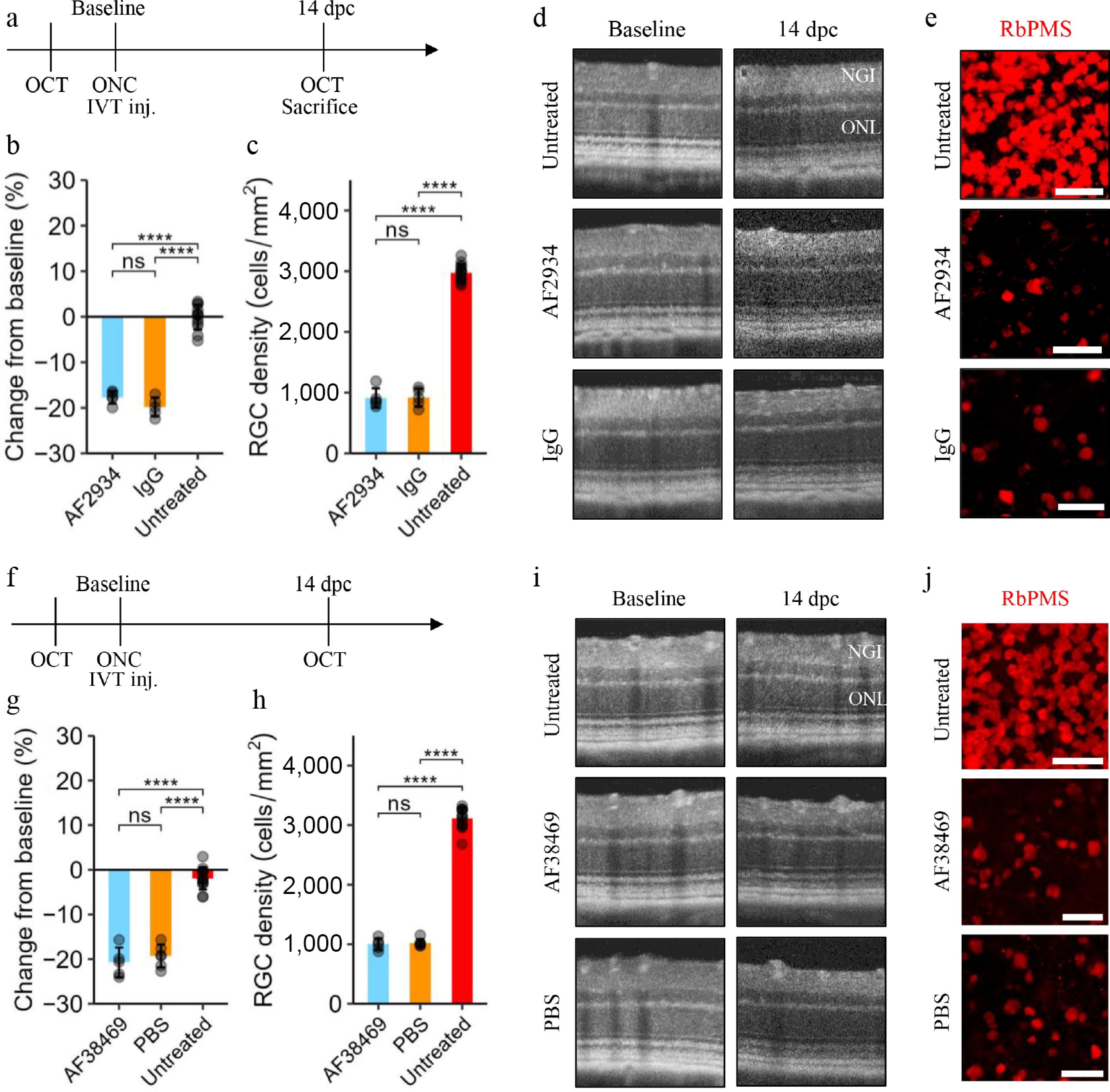
Figure 1.
Sortilin inhibition using a polyclonal antibody or small-molecule inhibitor does not preserve inner retinal structure. (a) Experimental plan: following baseline optical coherence tomography (OCT) scans, C57BL6/JRj mice were subjected to optic nerve crush (ONC) in one eye and a sham procedure in the other eye. Immediately after, mice were randomized to receive intravitreal (IVT) injection of either the anti-sortilin polyclonal antibody (pAb) AF2934 or IgG control (2 µL of 1 µg/µL) in the crush eye, while the other served as an untreated sham control. Mice were sacrificed following OCT at 14 day post-crush (dpc). (b) Bar plot (mean ± SD) showing quantification of the change from baseline of the combined thickness of the nerve fiber, ganglion cell, and inner nuclear layer, denoted the NGI thickness. Statistical comparisons were performed using one-way ANOVA followed by Tukey's test (n > 5). (c) Bar plot (mean ± SD) showing quantification of the density of RNA-binding protein with multiple splicing (RbPMS) positive retinal ganglion cells (RGCs) in retinal flat mounts. Statistical comparisons were performed by one-way ANOVA followed by Tukey's test (n > 5). (d) Representative OCT scans and (e) areas from anti-RbPMS (red) immunostained retinal flat mounts are shown. Scale bars: 50 µm. (f) Experimental plan: as in (a), C57BL6/JRj mice were randomized to receive IVT injection of the small-molecule inhibitor AF38469 (2 µL of 1 µg/µL) or vehicle (PBS-1% DMSO) in the crush eye. (g) Bar plot (mean ± SD) showing quantification of the change from baseline of NGI thickness. Statistical comparisons were performed by one-way ANOVA followed by Tukey's test (n > 5). (h) Bar plot (mean ± SD) showing quantification of RGC density in retinal flat mounts. Statistical comparisons were performed by one-way ANOVA followed by Tukey's test (n > 5). (i) Representative OCT scans and (j) areas from anti-RbPMS (red) immunostained retinal flat mounts are shown. Scale bars: 50 µm. Significance levels: * [0.01, 0.05]; ** [0.001, 0.01]; *** [0.0001, 0.001]; **** [0, 0.0001].
To investigate whether a higher dose would confer benefit, a separate experiment was performed using a higher dose (2 µL of 5 µg/µL) and with quantification of NGI thickness in OCT scans acquired 5, 10, and 15 dpc. Twelve mice were included in each group. Mice were randomized to receive either AF2934 or IgG control. A greater NGI thickness reduction from baseline amounting to −2.8% [−5.2; 0.3] (p = 0.034) was observed in the high-dose AF2934 group compared with the IgG group at 3 dpc, but otherwise no difference was observed (Supplementary Fig. S2).
Sortilin inhibition using the small-molecule inhibitor AF38469 does not preserve inner retinal thickness or retinal ganglion cell density following optic nerve crush
-
The small-molecule inhibitor AF38469[30], blocking the interaction between sortilin and the NGF pro-domain[31], has been used to study, e.g., the cancer-promoting effects of sortilin[16]. Therefore, the therapeutic potential of intravitreal AF38469 delivery in the ONC model was evaluated: Following baseline OCT measurements, ONC was performed in one eye and sham surgery in the other eye. This was followed immediately by intravitreal injection in the eye subjected to ONC. OCT measurements were performed 14 dpc, immediately before sacrifice and harvest of eyes for flat mounting and quantification of RGCs (Fig. 1f). A total of 15 8-week-old animals were randomized to receive either AF38469 or a vehicle control (2 µL of 1 µg/µL). No therapeutic effect was observed: reduction in NGI thickness from baseline measurements was not significantly different, being 1.41% [−2.66; 5.49] (p = 0.66) larger in the AF38469 group compared with the control group. Likewise, RGC density in flat mounts was not significantly different, with −22 cells/mm2 [−243; 200] (p = 0.996) in the AF38469 group compared with the control group (Fig. 1g–j).
Sortilin deficiency does not protect the inner retina following optic nerve crush
-
Pharmacological inhibition may be highly dependent on dose and timing. Thus, to confirm our findings, we also performed experiments in Sort1-/- mice. Sortilin-deficient mice are viable and without known adverse phenotypes. Sortilin-deficient mice show reduced neuronal loss during development.[19] However, they have normal fundoscopic appearance, and quantification of retinal layers in OCT scans acquired from sortilin-deficient and age-matched WT animals did not reveal structural differences (Supplementary Fig. S3). These findings indicate preserved retinal structure in adulthood, although potential adaptive changes during development cannot be excluded, as described for Ngfr-/- mice[32].
Following baseline OCT measurements, ONC was performed in the right eye and sham surgery in the left eye in cohorts of sortilin-deficient and age-matched WT animals. Twelve animals were included in each group; two and one animal, respectively, were sacrificed prematurely due to humane endpoints. Follow-up OCT was performed 5, 10, and 15 dpc. Following sacrifice at 15 dpc, eyes were harvested for flat mounting and quantification of RGC density (Fig. 2a). Change in NGI thickness from baseline was not significantly different at any time point between sortilin-deficient and WT control mice, being 1.0% [−2.7; 4.7] (p = 0.59) smaller in the sortilin-deficient mice (Fig. 2b, d). The difference between the sortilin-deficient and the WT control mice was 2 cells/mm2 [−25; 28] (p = 0.996) (Fig. 2c). However, absolute RGC densities were lower than previously observed in C57BL/6JRj mice, indicating a substrain-dependent difference. To confirm the genotype-independent effect, the experiment was repeated in an independent cohort of C57BL/6JBomTac mice including six sortilin-deficient animals and nine age-matched WT controls. Consistent with the initial experiment, RGC densities were similar between genotypes 15 day post-crush. In addition, RGC densities in the sham eyes were similar between the groups (Fig. 2e, f) and comparable to those in C57Bl/6JRj. These findings demonstrate that, while absolute RGC densities differ between substrains, no genotype-dependent differences were observed.
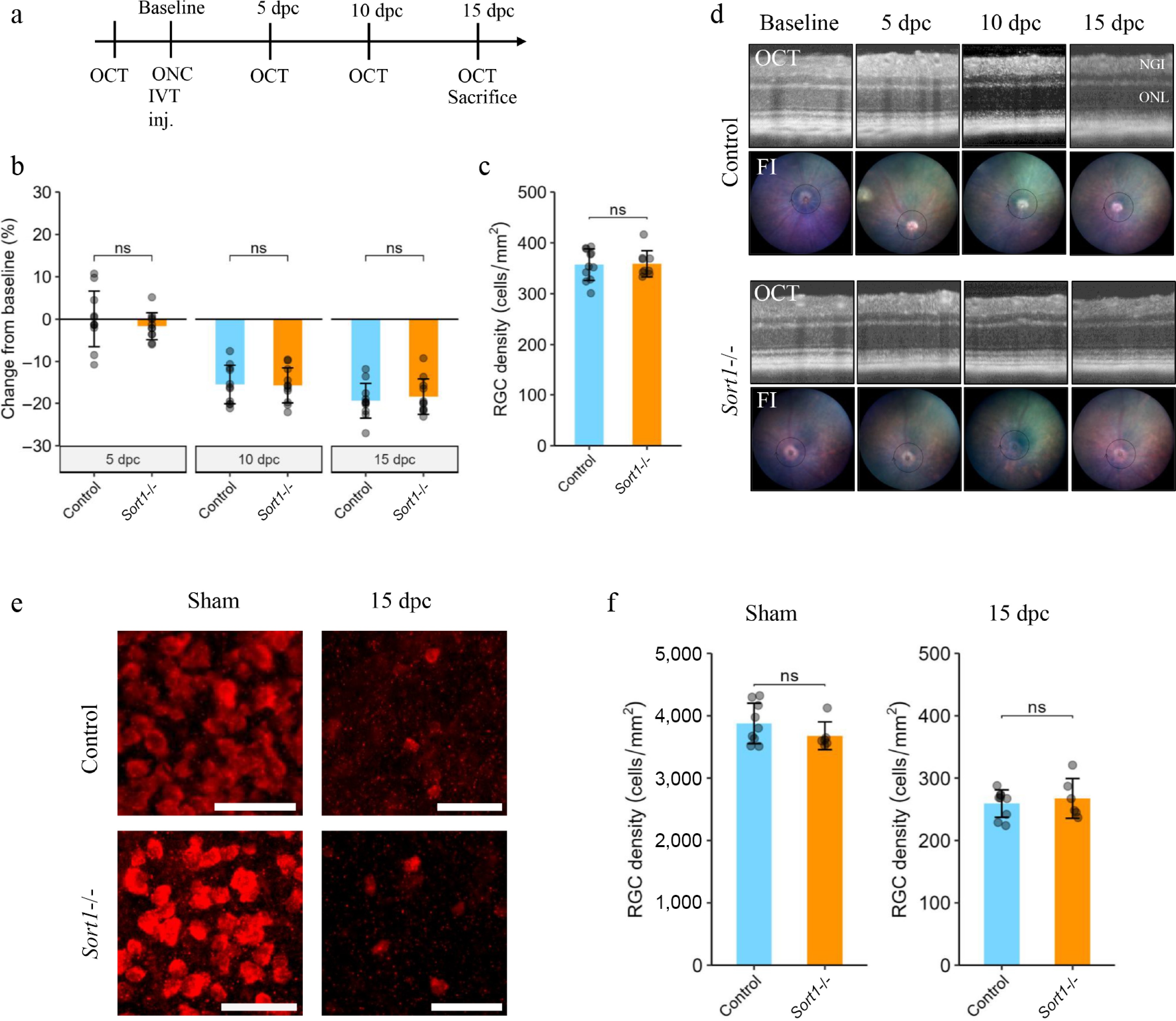
Figure 2.
Sortilin deficiency does not affect inner retinal pathology following optic nerve crush. (a) Experimental plan: 9-week-old Sort1-/- mice and age-matched C57Bl/6JBomTac wild-type mice were subjected to optic nerve crush (ONC) in the right eye. Optical coherence tomography (OCT) scans were performed at baseline and at 5, 10, and 15 day post-crush (dpc). (b) Bar plot (mean ± SD) showing quantification of change from baseline of the combined thickness of the nerve fiber, ganglion cell, and inner nuclear layer denoted the NGI thickness. Statistical comparisons were performed using linear, mixed-effects model to account for repeated measurements (n = 10–11). C. Bar plot (mean ± SD) showing quantification of RNA-binding protein with multiple splicing (RbPMS) positive retinal ganglion cells (RGCs) at 15 dpc. Statistical comparison was performed using Student's t-test (n = 10–11). (d) Representative fundus images (FI) and OCT scans at the respective dpc. (e) Representative areas from anti-RbPMS (red) immunostained retinal flat mounts in a separate replication cohort. Scale bars: 50 µm. (f) Bar plots (mean ± SD) showing quantification of RbPMS-positive RGCs 15 dpc in eyes subjected to crush (right) and the contralateral sham eyes (left). Statistical comparisons were performed using Student's t-test (n = 6–9). Significance levels: * [0.01, 0.05]; ** [0.001, 0.01]; *** [0.0001, 0.001]; **** [0, 0.0001].
Regulation of sortilin and p75NTR following optic nerve crush
-
Changes in ocular sortilin expression have not previously been evaluated following ONC, but an ~1.5-fold increase in p75NTR mRNA in the first days following ONC in mice[9] as well as an increase in p75NTR protein have been reported[6]. To identify neuroretinal alterations in sortilin and p75NTR mRNA and protein following ONC, groups of male C57Bl/6JRj mice were sacrificed before any intervention (8-weeks-old) and at 3, 7, and 14 dpc. Successful crush was confirmed by uniform NGI thickness reduction on OCT scans (Supplementary Fig. S4).
Relative neuroretinal Sort1 mRNA expression (Fig. 3a) was not significantly different in eyes subjected to ONC compared with controls. Similarly, no significant differences were observed compared with baseline levels. Ngfr expression was significantly increased at 3 and 7 dpc compared with the controls and the contralateral sham-treated eye but decreased to normal levels at 14 dpc (Fig. 3b). The increase at 3 dpc amounted to 44.9% [8.9; 81.0] (p = 0.021).
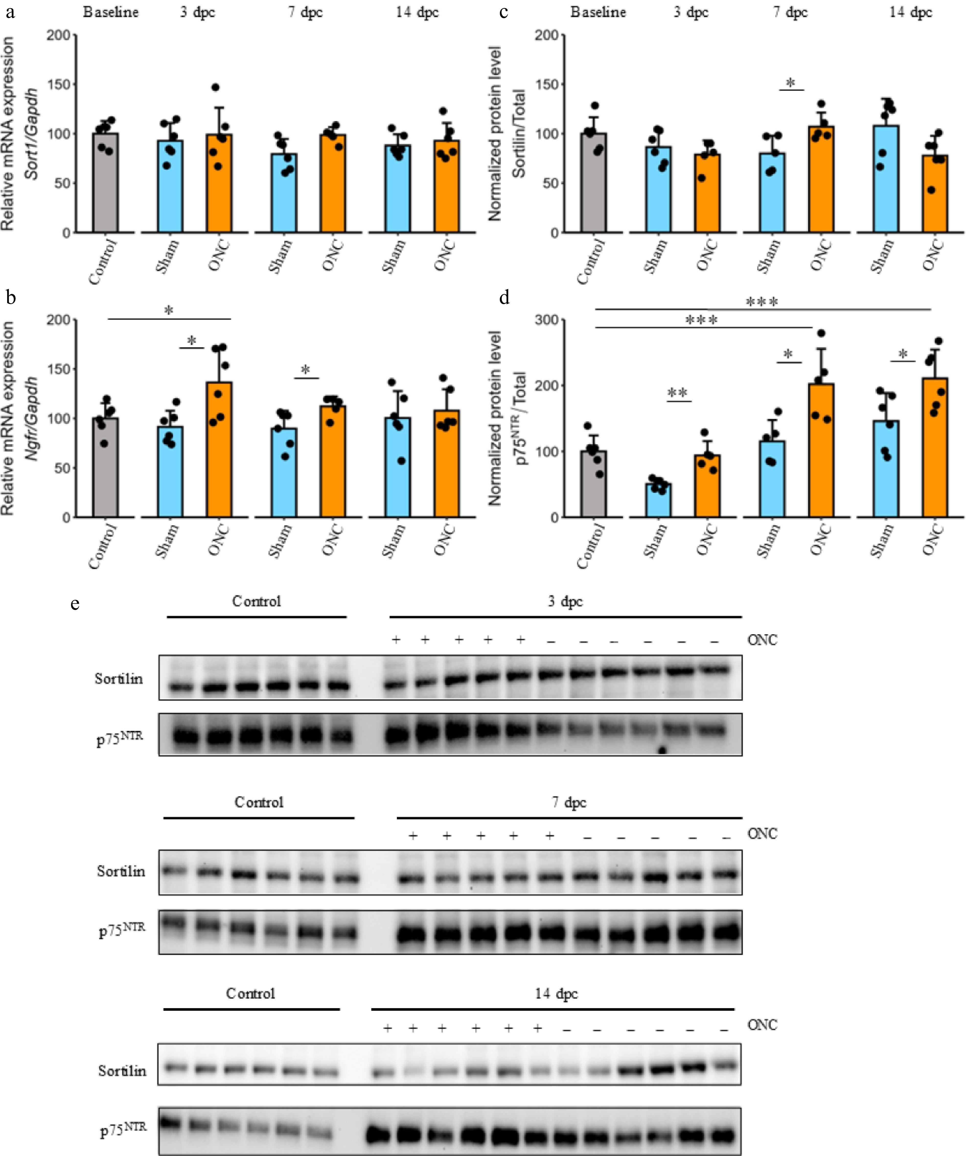
Figure 3.
Sortilin and p75NTR expression following optic nerve crush. Neuroretinal samples from control eyes not subjected to any procedure, as well as eyes subjected to optic nerve crush (ONC) or a sham procedure at 3, 7, and 14 day post-crush (dpc), were analyzed. (a), (b) Bar plots showing expression of Sort1 and Ngfr related to the endogenous control Gapdh. (c), (d) Bar plots (mean ± SD) showing quantification of sortilin and p75NTR protein levels related to total protein. (e) Western blots used for quantification. Statistical comparisons of the difference between the control and ONC group at different time points and comparisons between the ONC and the sham group at different time points were performed using two- or one-way ANOVA followed by Tukey's test and Dunnett's test, respectively (n = 5–6 at each time point). Significance levels: * [0.01, 0.05]; ** [0.001, 0.01]; *** [0.0001, 0.001]; **** [0, 0.0001].
Western blotting showed a small but significant 27.0% (p = 0.03) increase in sortilin protein levels in the ONC eyes compared with controls at 7 dpc (Fig. 3e), but no increase relative to baseline at any time point (Fig. 3c). The p75NTR protein level was significantly increased in retinas from eyes subjected to ONC 7 and 14 dpc compared with baseline and the contralateral sham eye (Fig. 3d). The increase relative to baseline amounted to 101.98% [43.1; 160.9] (p < 0.0001) at 7 dpc and 110.6% [54.4; 166.8] (p = 0.0003) at 14 dpc. Complete blots are provided as supplemental information (Supplementary Fig. S5).
Influence of sortilin deficiency on protein levels following optic nerve crush
-
Western blot analysis was performed to assess protein levels of p75NTR, TNFα, BDNF, and GFAP in whole retinal lysates from WT and sortilin-deficient mice under baseline conditions and 7 day following ONC or sham surgery (Fig. 4).
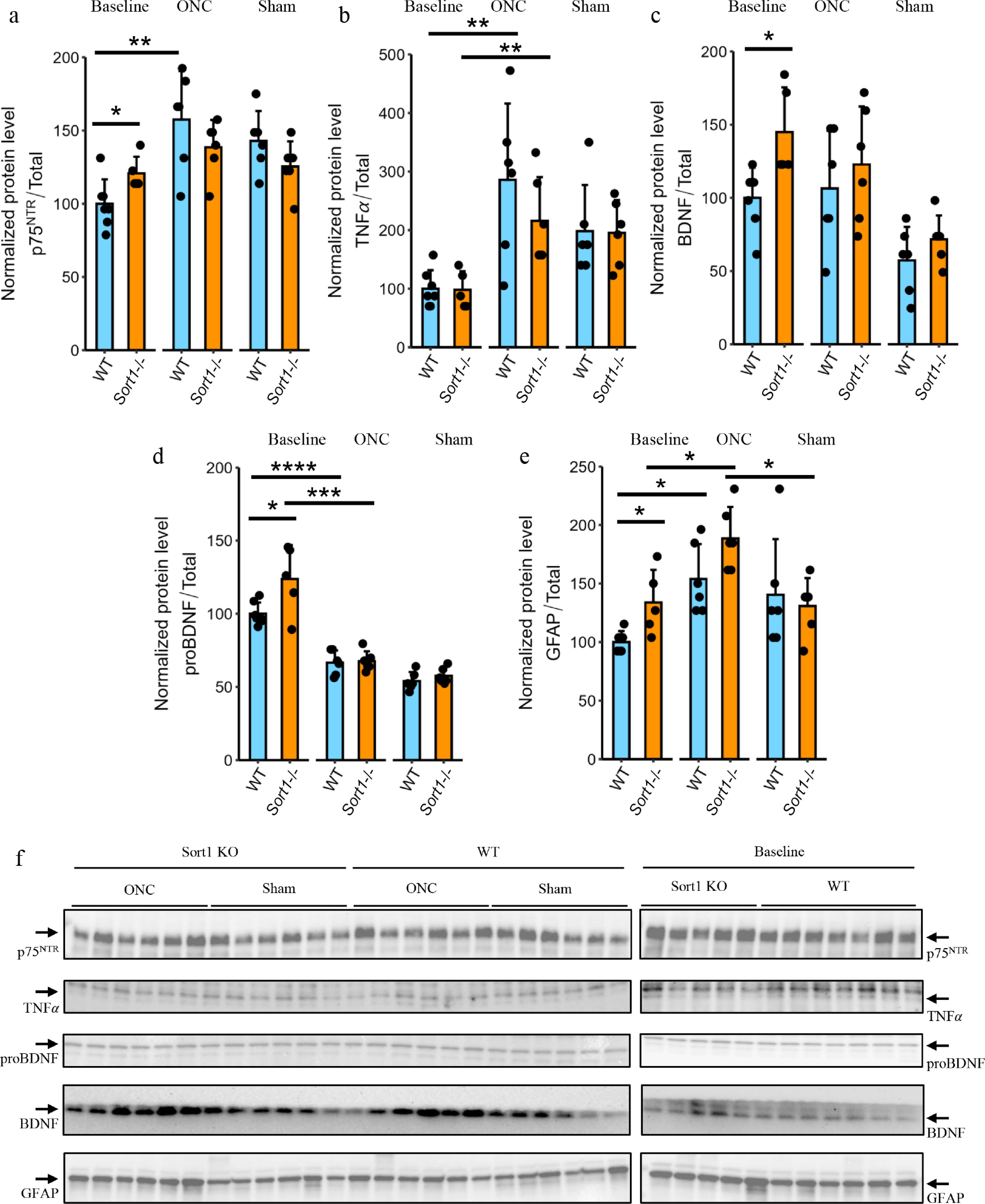
Figure 4.
Influence of sortilin deficiency on protein levels following optic nerve crush. Neuroretinal samples from Sort1-/- and age-matched C57Bl/6JBomTac wild-type (WT) mice not subjected to any procedure or with eyes subjected to optic nerve crush (ONC) or sham procedure and sacrificed 7 day post-crush (dpc) were analyzed. (a)–(e) Bar plots (mean ± SD) showing quantification of protein levels related to total protein in Western blots for p75NTR, TNFα, proBDNF, BDNF, and GFAP, respectively. (f) Western blots used for quantification. Statistical comparisons of the difference between WT and Sort1-/- animals in the different groups and the difference from baseline levels were performed using two-way ANOVA followed by Tukey's test (n = 5–7). Significance levels: * [0.01, 0.05]; ** [0.001, 0.01]; *** [0.0001, 0.001]; **** [0, 0.0001].
At baseline, sortilin-deficient retinas exhibited significantly increased levels of p75NTR (20.8% [1.47; 40.0], p = 0.037) and GFAP (33.8% [6.71; 61.0], p = 0.02) compared to WT, indicating differences in basal protein expression. ProBDNF (~32 kDa) and BDNF (~15 kDa) levels were also elevated in sortilin-deficient retinas (23.7% [3.09; 44.3], p = 0.0283 and 44.9% [12.2; 77.6], p = 0.012, respectively), whereas TNFα levels were comparable between genotypes (−2% [−43.2; 39.2], p = 0.916).
Following ONC, p75NTR levels increased significantly in WT retinas (57.5% [26.2; 88.8], p = 0.00193), while the small increase in sortilin-deficient mice was insignificant (17.8% [−3.96; 39.5], p = 0.0973). The increase was significantly larger in WT animals (39.1% [16.4; 61.7], p = 0.017). A similar pattern was observed for TNFα and GFAP, where protein levels increased after ONC in both WT (186% [74.5; 297], p = 0.00366 and 53.8% [25.4; 82.3], p = 0.00177, respectively) and sortilin-deficient (118% [36.1; 200], p = 0.0098 and 54.6% [17.2; 92.1], p = 0.00925, respectively) retinas. A tendency toward a less pronounced TNFα increase in sortilin-deficient retinas was observed, although not significantly (−65.6 [−203; 71.8], p = 0.312).
ProBDNF levels decreased following ONC in both WT (−33.4% [−43.2; −23.7], p < 0.0001) and sortilin-deficient (−56.2% [−78.4; −33.9], p = 0.0003) retinas to similar levels (0.97% [−8.76; 10.7], p = 0.829). The decrease was slightly larger in sortilin-deficient retinas (−12.0 [−21.0; −2.95], p = 0.0144). No consistent genotype-dependent differences were observed in mature BDNF levels. Complete blots are provided as Supplemental information (Supplementary Fig. S6).
-
In the present study, we did not observe a protective effect of pharmacological sortilin inhibition or constitutive sortilin deficiency on inner retinal thinning or RGC loss following ONC. However, sortilin deficiency was associated with a distinct alteration of the retinal baseline state and partial modulation of injury response. At baseline, sortilin-deficient retinas exhibited increased levels of GFAP, p75NTR, and proBDNF, indicating altered glial activation and proneurotrophin signaling. Following ONC, both WT and sortilin-deficient retinas showed robust injury responses, but the increase in p75NTR was attenuated in sortilin-deficient retinas. Similarly, a tendency toward an attenuated TNFα response was observed. Despite these molecular differences, RGC loss and structural degeneration were comparable between genotypes.
Evaluation of neuroretinal expression of neurotrophins and their receptors in optic nerve injury models is somewhat limited, and this is the first investigation of changes in sortilin levels following ONC. The lack of robust alterations at both the transcript and protein levels is compatible with a lack of structural rescue, although changes in cellular localization or cell-specific expression patterns cannot be excluded from the available data. Our finding of a transient increase in transcript levels of Ngfr is comparable with the ~1.5-fold increase observed in the first days following ONC in a mouse study[9]. Similarly, the increase in p75NTR proteins observed in retinas from eyes subjected to ONC is in line with a study showing a ~3-fold increase in p75NTR protein, most pronounced 14 d following ONC in Long Evans rats[6]. The difference in magnitude may depend on the specific experimental procedure, animal species, and protein quantification, including normalization to total protein in our study instead of a single housekeeping protein. Based on established models of proneurotrophin signaling, in which proBDNF engages the p75NTR/sortilin receptor complex to promote apoptotic and inflammatory responses, increased proBDNF and p75NTR levels would be expected to enhance degenerative signaling[33]. However, despite elevated baseline levels of proBDNF and p75NTR, sortilin deficiency did not result in increased RGC loss, suggesting that increased ligand and receptor availability alone is insufficient to drive degenerative signaling in the absence of sortilin. Conversely, the injury-induced increase in TNFα appeared attenuated in sortilin-deficient retinas without improving survival. Together, these observations suggest that modulation of the proneurotrophin/p75NTR axis is not limiting for ONC-induced degeneration in this model, and therefore a major contribution of altered sortilin-p75NTR signaling to retinal ganglion cell survival following ONC appears unlikely.
Sortilin is an enigmatic receptor with pleiotropic functions[13]. A major focus for the therapeutic use of sortilin inhibition is the dependence on sortilin for pro-neurotrophin signaling through p75NTR[11]. Activation of the sortilin/p75NTR receptor complex can induce apoptosis[11] or inflammation[3] depending on the cell type. In the eye, p75NTR is not evidently present on adult RGCs[3]. Rather, the protective effect of blocking p75NTR in models of acute and chronic neurodegeneration seems to rely on inhibiting p75NTR activation on Müller cells. Hence, intraocular injection of pro-NGF induces RGC loss with upregulation of TNFα expression in Müller cells, and this effect is blocked by sortilin deficiency[4]. In models of acute and chronic optic nerve injury, p75NTR signaling promotes inner retinal neurodegeneration[34]: p75NTR antagonism or knockout has been shown to ameliorate RGC loss following ONT[7,8] and OHT[8] as well as in induced diabetes[5], and these effects are dependent on a non-autonomous mechanism[5,7].
We recently showed that inhibition of the p75NTR co-receptor sortilin also confers neuroprotection in induced diabetes[21] and sought in the present study to examine if this would translate into a protective effect in a model of acute inner retinal neurodegeneration. However, we did not observe any effect using pharmacological inhibition or knockout of sortilin under the experimental conditions applied. A true effect may exist outside the effect size that our experiments were powered to identify, and the pharmacological effect may be sensitive to the chosen agent and its pharmacokinetics[7]. However, replication using different approaches suggests that any existing effect is not robust. A potential limitation to the external validity is the use of male mice, precluding identification of a gender specific effect. Another limitation is that the study did not investigate functional measures such as electroretinography or optomotor responses representing highly relevant subjects for future studies; however, these parameters would be expected to correlate closely with the pronounced structural damage observed in the applied model system[35]. Finally, the effect on optic nerve regeneration was not specifically examined. This also represents an important avenue for future investigation.
The absence of neuroprotection likely reflects the nature of the ONC model, in which RGC degeneration is driven by rapid, intrinsic injury-induced pathways that may proceed independently of glial-mediated and proneurotrophin-dependent signaling. In this context, sortilin-dependent pathways may influence the retinal environment but appear insufficient to alter the course of ONC-induced cell death. The loss of RGC following optic nerve injury and glaucoma has been hypothesized to involve reduced neurotrophic support secondary to disrupted axonal transport[34]. The neurotrophin BDNF and its cognate receptor TrkB seem to play a particularly important role in RGC survival[36]. Hence, it is interesting that sortilin also regulates BDNF secretion and degradation in a complex way[37−39] and that it, in certain contexts, enhances Trk receptor transport to facilitate neurotrophin signaling[40]. In the present study, sortilin deficiency was associated with increased baseline levels of both BDNF and proBDNF, suggesting altered neurotrophin homeostasis in the uninjured retina. Since mature BDNF generally supports neuronal survival through TrkB activation, whereas proBDNF can promote degenerative signaling through p75NTR/sortilin-associated pathways, the net functional impact of these changes cannot be inferred from protein levels alone. Importantly, retinal structure remained unchanged in uninjured adult Sort1-/- mice, and these endogenous alterations were insufficient to substantially modify retinal degeneration following ONC.
In summary, sortilin inhibition does not preserve inner retinal structure following ONC. However, sortilin influences glial activation, inflammatory signaling, and proneurotrophin dynamics in the retina. These effects do not translate into neuroprotection in the ONC model but may be more relevant in disease settings characterized by chronic stress and sustained neuroinflammation, such as diabetic retinopathy.
-
Animals were handled in accordance with the 'Statement for the Use of Animals in Ophthalmic and Vision Research' from the Association for Research in Vision and Ophthalmology (ARVO). All animal experiments were performed under the approval of the Danish Animal Inspectorate (Case# 2020-15-0201-00556, approval date 09-06-2020).
-
The authors confirm their contributions to the paper as follows: study conception and design: Jakobsen TS, Askou AL, Corydon TJ; resources: Jakobsen TS, Askou AL, Corydon TJ, Nykjaer A; data collection: Jakobsen TS, Lindholm AB, Askou AL; analysis and interpretation of results, draft review and editing: Jakobsen TS, Lindholm AB, Askou AL, Corydon TJ; draft manuscript preparation: Jakobsen TS; supervision: Askou AL, Corydon TJ, Bek T. All authors reviewed the results and approved the final version of the manuscript.
-
The datasets generated during and/or analyzed in the current study are available from the corresponding author on reasonable request.
-
The authors thank Tina Hindkjær for expert technical assistance, and Stella Solveig Nolte for assistance with breeding of the knockout animals. The Aarhus University Health Bioimaging Core Facility is thanked for the use of equipment and technical assistance. The Animal Facility, Department of Biomedicine, Aarhus University, is thanked for housing the mice. This work was supported by the Faculty of Health Sciences, Aarhus University (PhD scholarships to TSJ and ABL), Fight for Sight, Denmark (TSJ), Ophthalmologist Else Bruntse's foundation (TSJ), the Synoptik Foundation (TSJ), the A.P. Moller Foundation (TSJ), the Dagmar Marshalls Foundation, the Independent Research Fund Denmark (Grant no. 2034-00036B [TJC]), the Riisfort foundation (ALA), the Danish National Research Foundation (Grant no. DNRF133 [AN]), and the Lundbeck Foundation (Grant no. R480-2024-1094).
-
The authors declare that they have no conflict of interest.
-
accompanies this paper online at: https://doi.org/10.48130/vns-0026-0032.
-
# Authors contributed equally: Thomas J. Corydon, Anne Louise Askou
- Supplementary Fig. S1 Validation of optic nerve crush model.
- Supplementary Fig. S2 Sortilin inhibition using a higher dose of polyclonal antibody does not preserve inner retinal thickness.
- Supplementary Fig. S3 Retinal structure of Sort1-/- (C57Bl/6J-Sort1tm1Tew/JBomTac) mice and age-matched C57Bl/6JBomTac wild-type mouse.
- Supplementary Fig. S4 NGI thickness in animals used for Western blot and RT-qPCR.
- Supplementary Fig. S5 Western blot analysis for sortilin and p75NTR in retinal tissue lysate following optic nerve crush.
- Supplementary Fig. S6 Western blot analysis for p75NTR, TNFα, BDNF, and GFAP in retinal tissue lysate following optic nerve crush.
- Supplementary Table S1 RT-qPCR primers.
- Copyright: © 2026 by the author(s). Published by Maximum Academic Press, Fayetteville, GA. This article is an open access article distributed under Creative Commons Attribution License (CC BY 4.0), visit https://creativecommons.org/licenses/by/4.0/.
-
About this article
Cite this article
Jakobsen TS, Lindholm AB, Bek T, Nykjaer A, Corydon TJ, et al. 2026. Sortilin deficiency alters baseline retinal homeostasis and injury-induced signaling without affecting optic nerve crush-induced neurodegeneration. Visual Neuroscience 43: e033 doi: 10.48130/vns-0026-0032 

Sortilin deficiency alters baseline retinal homeostasis and injury-induced signaling without affecting optic nerve crush-induced neurodegeneration
- Received: 22 October 2025
- Revised: 24 May 2026
- Accepted: 25 May 2026
- Published online: 22 July 2026
Abstract: Retinal neurodegeneration is a hallmark of many vision-threatening diseases, and the receptor sortilin has emerged as a promising therapeutic target due to its involvement in p75NTR-associated neurodegenerative signaling. The effect of sortilin inhibition on acute inner retinal neurodegeneration induced by optic nerve crush was therefore investigated. Pharmacological sortilin inhibition using intravitreal delivery was evaluated in C57BL/6JRj male mice subjected to unilateral crush. Furthermore, the effect of constitutive sortilin deficiency was examined using Sort1-/- mice, and changes in sortilin, p75NTR, and associated injury markers were analyzed. Neither pharmacological inhibition nor constitutive loss of sortilin protected against inner retinal thinning or retinal ganglion cell loss following optic nerve crush. A transient 1.4-fold increase in p75NTR mRNA was observed early after injury, accompanied by a two-fold increase in protein levels. While sortilin expression remained largely unchanged, sortilin deficiency was associated with an altered baseline retinal state. Following optic nerve crush, the induction of p75NTR was significantly attenuated in sortilin-deficient retinas. In summary, sortilin inhibition does not preserve inner retinal structure following optic nerve crush, but alters molecular markers associated with glial activation, inflammatory signaling, and neurotrophin-related pathways. Sortilin-dependent pathways may be more relevant in disease contexts characterized by chronic stress and neuroinflammation.
-
Key words:
- Neurodegeneration /
- Sortilin /
- Neurotrophins /
- Optic nerve crush