












-
Pepper (Capsicum annuum L.) is a widely cultivated and economically important vegetable crop worldwide, highly valued by consumers due to its unique flavor and rich nutritional content[1]. However, pepper cultivation is often threatened by various soil-borne pathogens, which severely compromise its yield and quality[2]. Among these, bacterial wilt, caused by R. solanacearum, is one of the most devastating soil-borne bacterial diseases[3], characterized by a broad host range, rapid transmission, and high mortality rate, thereby posing a significant threat to pepper production[4]. This pathogen was reported to infect more than 200 plant species across over 50 families, causing catastrophic losses particularly to Solanaceous crops[5].
Due to the complex biology of R. solanacearum, conventional chemical and agronomic control strategies are largely ineffective and environmentally unsustainable[6]. The development and deployment of resistant cultivars remain the most effective approach to manage bacterial wilt[7]. In addition to classical genetic resistance, resistant cultivars also play a crucial role in selectively shaping the assembly and symbiotic functions of root-associated microbial communities[8,9]. Increasing evidence indicates that resistant genotypes selectively recruit and enrich beneficial microorganisms, thereby establishing root-associated microbial communities that contribute to the suppression of soil-borne diseases[10,11]. Mechanistically, this selective enrichment is achieved through multiple pathways, including the secretion of specific chemical compounds, activation of immune signaling pathways, and regulation of hormone-mediated processes[12−14]. These recruited or enriched root-associated microbiomes, particularly those in the rhizosphere and root compartments, have the potential to directly or indirectly modulate disease resistance[15].
In recent years, various beneficial microorganisms with biocontrol functions have been successfully isolated from multiple resistant plant species. Among them, beneficial microbes such as Pseudomonas and Bacillus are well recognized for their biocontrol potential and ability to enhance pepper resistance against bacterial wilt[16,17]. These biocontrol strains possess several advantages, such as the secretion of plant growth-promoting hormones, improved nutrient utilization efficiency, competitive niche occupation, the production of antimicrobial compounds, enhancement of antioxidant and defense enzyme activities, and activation of plant immune responses[12,15,18,19]. In addition, some antagonistic strains produce diverse enzymes and secondary metabolites that act as effective natural bactericides or fungicides, showing a great potential for sustainable and environmentally friendly management of soil-borne diseases[20]. Furthermore, certain microbial taxa exhibit high sensitivity to host genotypic variation, with their community composition showing significant associations with plant genotype; such microbes are often referred to as heritable microbiota[21]. These heritable and resistance-related microbial taxa provide valuable germplasm resources for developing disease-resistant crop varieties and microbial-based biocontrol agents.
In this study, we aimed to elucidate the microbiological, physiological, and molecular mechanisms underlying resistance to R. solanacearum in pepper. We first compared the disease responses and root-associated bacterial communities of resistant and susceptible cultivars following pathogen infection. Subsequently, we isolated and identified a potential biocontrol strain, P. aeruginosa P1202, and assessed its effects on plant growth, antioxidant enzyme activity, photosynthetic system, and transcriptional regulation of defense-related genes. Our findings provide insights into the interactions among beneficial microbes, plant hosts, and pathogens, and offer a promising strategy for the biocontrol of bacterial wilt in pepper cultivation.
-
The experiment was conducted in an artificial climate chamber of the College of Horticulture, Hebei Agricultural University, Baoding, Hebei, China. Two pepper inbred lines, HR01 (resistant) and HS03 (susceptible), exhibiting contrasting resistance to bacterial wilt, were used as experimental materials. These lines, provided by Dr. Du, originated from two independent segregating populations derived from local farmer cultivars and were stabilized through successive self-pollination[22].
Seeds of both cultivars were surface-sterilized with 0.5% sodium hypochlorite (NaClO) for 5 min and subsequently rinsed five times with sterile distilled water to remove the residual disinfectant. The sterilized seeds were sown in nutrient pots filled with sterilized field soil collected from a vegetable cultivation site in Baoding, Hebei, China. To ensure substrate homogeneity, the soil was passed through a 3 mm mesh prior to sterilization. Seedlings were grown in a greenhouse maintained at 25 ± 2 °C with 75% relative humidity under a 14 h light/10 h dark photoperiod.
For each cultivar, 80 healthy seedlings with uniform growth were selected and divided into two groups: control (C, 40 plants) and inoculated treatment (T, 40 plants). Plants in the treatment group were inoculated with a suspension of R. solanacearum, while control plants received mock inoculation with sterile solution. Root samples were collected to determine the root-related traits. Root samples were gently washed with sterile water, blotted dry with sterile filter paper, and weighed to determine root fresh weight.
For P. aeruginosa inoculation, the susceptible cultivar HS03 was used. Four treatments were established: (i) control, (ii) plants pretreated with P. aeruginosa for 2 weeks (P.a), (iii) plants inoculated with R. solanacearum for 16 h (+R.s), and (iv) plants pretreated with P. aeruginosa for 2 weeks followed by R. solanacearum inoculation for 16 h (P.a+R.s). The biocontrol strain P1202 was applied at an optical density at 600 nm (OD600) of 0.5. For each treatment, uniformly growing seedlings were selected, and roots were wounded approximately 2 cm below the base of the primary root, followed by application of 20 mL of bacterial suspension per plant. Treatments were applied once per week for two consecutive weeks. After the treatment period, disease severity was evaluated using a standard disease rating method as described below. For each treatment, three plants with similar growth status and disease severity were selected as biological replicates for subsequent analyses. Physiological parameters were measured for each replicate. Fresh root tissues were collected and stored at −80 °C for further analysis. All measurements were performed with three technical replicates.
Pathogen culture and inoculation assay
-
The R. solanacearum strain used in this study was provided by the Fujian Academy of Agricultural Sciences, Fuzhou, Fujian, China. The preserved strain was revived and cultured in nutrient broth (NB) medium at 28 °C with shaking (200 rpm) for 24 h. Bacterial cells were harvested by centrifugation, washed twice, and resuspended in sterile 10 mM MgCl2 solution. The bacterial suspension was adjusted to an OD600 of 0.6 and subsequently used for inoculation.
For inoculation, five-week-old pepper seedlings (with 7−8 leaves) were inoculated using a root-wounding method. The procedure was adapted with minor modifications from a previously described method[23]. Briefly, the soil near the base of each plant was gently pierced to a depth of approximately 3 cm with a sterile glass rod to slightly wound the lateral roots, after which 20 mL of the bacterial suspension was applied. Control plants received the same treatment but were irrigated with a sterile 20 mL solution of MgCl2. After inoculation, the greenhouse environment was maintained at 30 ± 2 °C with a relative humidity of > 90%.
Disease symptoms were first recorded at 7 days post-inoculation (dpi) and subsequently assessed every day until 14 dpi, when symptom progression had stabilized. Disease severity was evaluated using a standardized 0–4 rating scale as follows: 0 = no symptoms; 1 = 1%–25% of leaves wilted or dropped; 2 = 26%–50% of leaves wilted or dropped, with mild growth retardation; 3 = 51%–75% of leaves wilted or dropped, with severe growth retardation; 4 = 76%–100% of leaves wilted or dropped, or plant death.
The disease index was calculated according to the following formula:
DI = 100 × [(Σ Disease grades × The number of seedlings at each disease grades) / (4 × The number of total seedlings)].
Rhizosphere and root endophytic bacterial community analyses
-
For rhizosphere soil samples, large soil particles loosely attached to the roots were gently shaken off, and approximately 15 g of root segments with tightly adhering rhizosphere soil were collected and placed into sterile centrifuge tubes containing 0.86% NaCl solution. The tubes were kept on ice for 30 min, with gentle shaking every 5 min. Roots were then removed, and the suspension was centrifuged at 4,000 × g for 30 min at 4 °C. The resulting soil pellet was collected and used for DNA extraction. This method was adapted from a previously reported method[24].
For root samples, fresh roots were surface-sterilized by immersion in 75% ethanol for 1 min, followed by 2.5% sodium hypochlorite for 5 min, and rinsed three times with sterile distilled water. The sterilized roots were blotted dry with sterile filter paper, immediately frozen in liquid nitrogen and stored for DNA extraction.
Each treatment included three biological replicates, with three root samples or three rhizosphere soil samples per replicate. For rhizosphere bacterial community analysis, the V3–V4 region of the 16S rRNA gene was amplified using primers 341F and 806R. For endophytic bacterial community analysis, the V5–V7 region of the 16S rRNA gene was amplified using primers 799F and 1193R (Supplementary Table S1). Sequencing was performed on the Illumina platform. Raw sequence data were processed using the demultiplexing and Cutadapt plugins to remove adapters and primers, followed by quality filtering, denoising, merging, and chimera removal with the DADA2 plugin[25]. Amplicon sequence variants (ASVs) were inferred using the DADA2 algorithm at 100% sequence resolution.
Bioinformatic analyses
-
Representative ASVs were taxonomically classified using the Ribosomal Database Project (RDP) naive Bayesian classifier against the SILVA reference database. Calculation of Shannon diversity indices and principal component analysis (PCA) of bacterial communities were performed using the QIIME2 platform[26]. To identify differentially abundant taxa, linear discriminant analysis (LDA) effect size (LEfSe) was performed using the Python LEfSe package, with an LDA score threshold of 2. To further validate the relative abundance of representative taxa identified by LEfSe, the relative abundances of key bacterial genera were compared using the Kruskal–Wallis test followed by Dunn's multiple-comparison test, with p < 0.05 considered statistically significant.
Isolation and identification of bacteria
-
Root samples were collected as described above, and serial dilutions were prepared using sterile distilled water. Aliquots of the dilutions were spread on Luria–Bertani (LB) agar plates and incubated at 28 °C for 24–48 h. Distinct bacterial colonies were selected based on morphological characteristics and repeatedly streaked onto fresh LB agar plates until pure single colonies were obtained.
Genomic DNA was extracted from each purified isolate using a commercial bacterial DNA extraction kit (Tiangen, China) according to the manufacturer's instructions. The nearly full-length 16S rDNA gene was amplified using universal primers 27F and 1492R (Supplementary Table S1), generating an amplicon of approximately 1,500 bp (Supplementary Table S2). Polymerase chain reaction (PCR) amplification was performed in a 50 µL reaction mixture, and the products were verified by electrophoresis on a 1.0% agarose gel for quality control prior to sequencing. The amplicons were subjected to Sanger sequencing, and the resulting sequences were compared with reference sequences in the NCBI database using the BLAST program. Representative isolates were further analyzed by constructing a phylogenetic tree using the neighbor-joining (NJ) method implemented in MEGA software to determine their taxonomic positions and evolutionary relationships.
Dual-culture antagonistic assay and fermentation broth inhibition test
-
The antagonistic activity of bacterial isolates against R. solanacearum was first evaluated in vitro using a dual-culture assay. Briefly, the pathogen was spot-inoculated onto the center of nutrient agar plates. Each candidate antagonistic isolate was then streaked at three equidistant points near the periphery of the same plate. After incubation at 28 °C in the dark for 24 h, the radius of pathogen growth inhibition toward the antagonists was measured along two perpendicular diameters.
The fermentation broth assay was adapted from a previously described procedure[27]. Antagonistic isolates were pre-cultured in NB at 28 °C with shaking at 160 rpm for 24 h to prepare seed cultures. They were then transferred at an inoculum concentration of 1% (v/v) into LB fermentation medium and incubated under the same conditions for another 24 h. The resulting cultures were centrifuged at 8,000 rpm for 5 min, and the supernatant was filter-sterilized three times through a 0.22 μm membrane to obtain a cell-free fermentation broth. To prepare pathogen-embedded plates, R. solanacearum was mixed at a ratio of 1:200 with sterilized LB medium cooled to approximately 50 °C, and the mixture was poured over a basal water-agar layer. After solidification, a well was created at the center of each plate, into which 150 μL of sterile fermentation broth was added. Each isolate was tested in three replicates. Plates were incubated at 28 °C for 48 h, and the diameter of the inhibition zone was measured using the cross-streak method.
Biocontrol efficacy against bacterial wilt in vivo
-
The biocontrol efficacy of the selected antagonistic isolate was further assessed in vivo. The susceptible cultivar HS03 was grown under natural light conditions with day/night temperatures of 28/22 °C, respectively. The antagonistic isolate was first cultured in liquid NB medium at 28 °C with shaking at 200 rpm for 24 h. The culture was harvested, centrifuged, washed three times, and resuspended in sterile 10 mM MgCl2 solution to an OD600 of 0.5. The experimental design included two treatments: control and plants pretreated with P. aeruginosa (P.a). These plants were challenged with R. solanacearum using the root-wounding method. Briefly, the roots were wounded approximately 2 cm below the base of the primary root before inoculation, and 20 mL of the bacterial suspension was applied to the wounded roots. Disease development was monitored weekly, and the disease index was evaluated at 7 dpi using a standard wilt rating scale (0–4). Disease symptoms were first recorded at 7 dpi and subsequently assessed every day until 14 dpi.
Chlorophyll fluorescence measurement
-
Chlorophyll fluorescence (Chl-F) data were obtained using the multifunctional plant photosynthetic phenotype imaging system (PlantExplorerPRO+, PhenoVation, Netherlands). Chl-F parameter values were converted into Chl-F pseudocolor images for visualization using the PV64b_Analysis software. Chl-F data and images were measured at 16 h post-inoculation (hpi) to capture early physiological responses before the appearance of visible disease symptoms. The effective quantum yield of photosystem II (PSII) is calculated as ΦPSII = (FM' − FS)/FM'. The relative electron transport rate (rETR) is calculated as rETR = ΦPSII × PPFD × (0.5). Here, FM' denotes the maximum fluorescence in a light-adapted state, FS the current fluorescence in a light-adapted state, and PPFD the photosynthetic photon flux density. This method was adapted from a previously described procedure[28].
Transcriptomic sample collection and analysis
-
Root samples of HS03 from four treatment groups were collected for transcriptome sequencing: the control plants, plants pretreated with P. aeruginosa for 2 weeks (P.a), plants inoculated with R. solanacearum for 16 h (+R.s), and plants pretreated with P. aeruginosa for 2 weeks followed by R. solanacearum inoculation for 16 h (P.a+R.s), as described in the section entitled "Pathogen culture and inoculation assay". Samples were collected at 16 hpi, representing an early stage of infection before visible disease symptoms developed. This time point was selected based on our preliminary experiments, which identified significant changes in Chl-F parameters at 16 hpi, indicating that early physiological responses to pathogen infection had already been initiated. Three biological replicates were included for each treatment. Raw sequencing reads were filtered to generate high-quality clean reads, which were then aligned to the reference genome Zunla-1_V3.0 using HISAT2. Gene expression levels were quantified using fragments per kilobase of transcript per million mapped reads (FPKM). Analysis of differential expressed genes (DEGs) was conducted using DESeq2 R package, with genes exhibiting |log2fold-change| ≥ 1 and adjusted p value ≤ 0.05 identified as DEGs. Functional enrichment analysis of DEGs was performed using the Gene Ontology (GO) and Kyoto Encyclopedia of Genes and Genomes (KEGG) databases. Pathways with an adjusted p value ≤ 0.05 were considered significantly enriched. This analysis was adapted from a previously described procedure[29].
RNA isolation and qRT-PCR
-
Total RNA was extracted using a total RNA extraction Kit (Promega), following the manufacturer's instructions. Approximately 100 ng of RNA was used for cDNA synthesis using HiScript III RT SuperMix (Vazyme, China), which includes all necessary components for reverse transcription and a gDNA wiper Mix to remove genomic DNA contamination. Quantitative real-time PCR (qRT-PCR) was performed using the RNA Fluorescence Quantitative Detection Kit (Vazyme, China), following the manufacturer's instructions. Gene-specific primers were designed for qRT-PCR analysis (Supplementary Table S1). The PCR conditions involved an initial denaturation step at 95 °C for 10 min, followed by 40 cycles of denaturation at 95 °C for 10 s, annealing at 56 °C for 30 s, and an extension at 72 °C for 30 s. A melt-curve analysis of the amplification products was conducted at the end of the PCR, with the melting cycle consisting of 95 °C for 15 s, 60 °C for 1 min, 95 °C for 15 s, and 60 °C for 15 s. Relative gene transcript levels were calculated using the comparative 2−ΔΔCᴛ method.
Determination of POD, SOD, and CAT activities
-
Enzyme activities were determined using commercial kits (Solarbio, China) according to the manufacturer's instructions. Approximately 0.1 g of fresh root tissue was homogenized in 1 mL extraction buffer under ice-bath conditions and centrifuged at 8,000 × g for 10 min at 4 °C. The supernatant was collected as a crude enzyme extract.
Peroxidase (POD) activity was measured by mixing 5 μL enzyme extract with 240 μL working solution, and absorbance was recorded at 470 nm after 30 and 90 s.
Superoxide dismutase (SOD) activity was determined after incubation at 37 °C for 30 min in the dark, followed by absorbance measurement at 560 nm.
Catalase (CAT) activity was measured at 240 nm by recording the absorbance changes at 5 and 65 s using a UV-transparent plate.
All enzyme activities were calculated according to the corresponding kit instructions and expressed as U mg−1 fresh weight (FW).
Statistical analysis
-
Statistical analyses of microbial diversity and differential taxa were performed in R software. For normally distributed data, Student's t-test and one-way analysis of variance (ANOVA) followed by Tukey's post hoc test were applied. For non-normally distributed data, the Kruskal–Wallis test followed by Dunn's post hoc test was used. All figures were visualized and polished using GraphPad Prism v9.0, with ordination plots and microbial network diagrams produced in R for enhanced visualization.
-
Distinct phenotypic differences were observed between the control (C) and the inoculated treatment (T) groups of the resistant cultivar HR01 and the susceptible cultivar HS03 at 14 dpi. Compared with their control counterparts, inoculated HS03 plants (HS03_T) showed severe wilting symptoms, whereas inoculated HR01 plants (HR01_T) exhibited only mild growth retardation, without obvious wilting (Fig. 1a). Consistently, disease index analysis confirmed that HS03_T had a significantly higher disease index than HR01_T (Fig. 1b). Moreover, compared with the control plants, root fresh weight was significantly reduced in HS03_T, whereas no significant change was observed in HR01_T (Fig. 1c), suggesting that pathogen infection had a more severe impact on the root system of HS03. Furthermore, the relative abundance of R. solanacearum in the roots was significantly higher in HS03_T than in HR01_T (Fig. 1d), indicating that pathogen colonization was effectively suppressed in HR01.
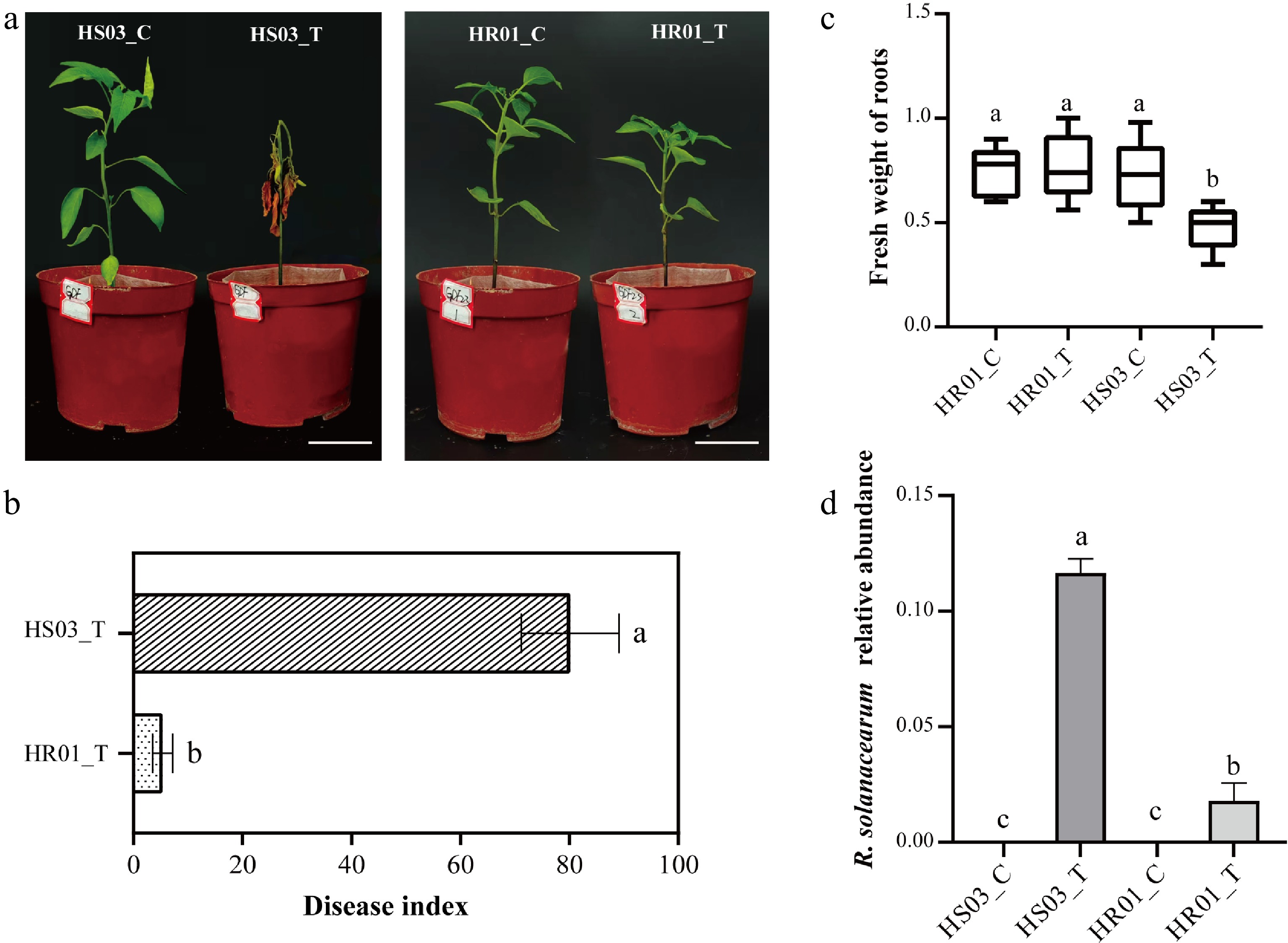
Figure 1.
Phenotypic characteristics of HR01 and HS03 following R. solanacearum inoculation. (a) Disease symptoms of HR01 and HS03 at 14 dpi. (b) Disease index of HR01 and HS03 at 14 dpi. (c) Fresh weight of roots of HR01 and HS03 at 14 dpi. (d) The relative abundance of R. solanacearum in the roots of HR01 and HS03 at 14 dpi. Data are presented as mean ± SD (n = 6). Statistical analyses were performed using the independent-samples t-test and one-way ANOVA followed by Tukey's post hoc test for normally distributed data, or the Kruskal–Wallis test followed by Dunn's post hoc test for non-normally distributed data. Different letters indicate a significant difference (p < 0.05). HR01_C, control group of the resistant cultivar HR01; HR01_T, inoculated group of the resistant cultivar HR01; HS03_C, control group of the susceptible cultivar HS03; HS03_T, inoculated group of the susceptible cultivar HS03. Scale bar = 5 cm.
Effects of R. solanacearum infection on the composition and diversity of rhizosphere and root endophytic bacterial communities
-
To investigate the effects of R. solanacearum infection on root-associated bacterial community differences in the two pepper cultivars, the rhizosphere and root endophytic bacterial communities of inoculated and control plants were analyzed using 16S rRNA amplicon sequencing. Upset plot analysis showed that, in the rhizosphere, the number of ASVs decreased in HS03 after inoculation (HS03_T vs HS03_C), whereas a slight increase was observed in HR01 (HR01_T vs HR01_C). Moreover, 3,029 ASVs were shared among the four treatments, indicating that the core rhizosphere bacterial community remained relatively stable (Fig. 2a). In the root endophytic bacteria, ASV richness declined in both HS03 and HR01 following inoculation, although the reduction was more pronounced in HS03, indicating that pathogen infection had a stronger disruptive impact on the root endophytic bacterial community of HS03 (Fig. 2b). Compared to the control plants, Shannon index showed no significant change in the rhizosphere of either HR01 or HS03 (Fig. 2c). In contrast, a significant reduction in Shannon index was observed in the root endophytic bacteria of both cultivars (Fig. 2d). PCA revealed a clear separation of bacterial communities after inoculation, indicating substantial shifts in community composition (Fig. 2e, f). In the rhizosphere, the dominant bacterial phyla in both cultivars were Proteobacteria and Acidobacteriota, with the most abundant orders being Burkholderiales and Vicinamibacterales, and the representative genera including RB41 and members of Vicinamibacteraceae (Fig. 2e; Supplementary Fig. S1). In the endophytic bacterial community, Proteobacteria was the dominant phylum across both cultivars. At the order level, the root endophytic bacteria of HS03_C were primarily composed of Burkholderiales, Rhizobiales, and Pseudomonadales, whereas HR01_C harbored Burkholderiales, Rhizobiales, and Sphingomonadales. At the genus level, HS03_C was dominated by Pseudomonas, Serratia, and Sphingobium, while HR01_C was enriched in Allorhizobium–Neorhizobium–Pararhizobium–Rhizobium (ANPR), Ralstonia, and Sphingobium. Following inoculation, however, Burkholderiales and Ralstonia became the dominant order and genus, respectively, in both cultivars (Fig. 2f; Supplementary Fig. S1), suggesting a strong restructuring of the root endophytic bacterial community under R. solanacearum stress.
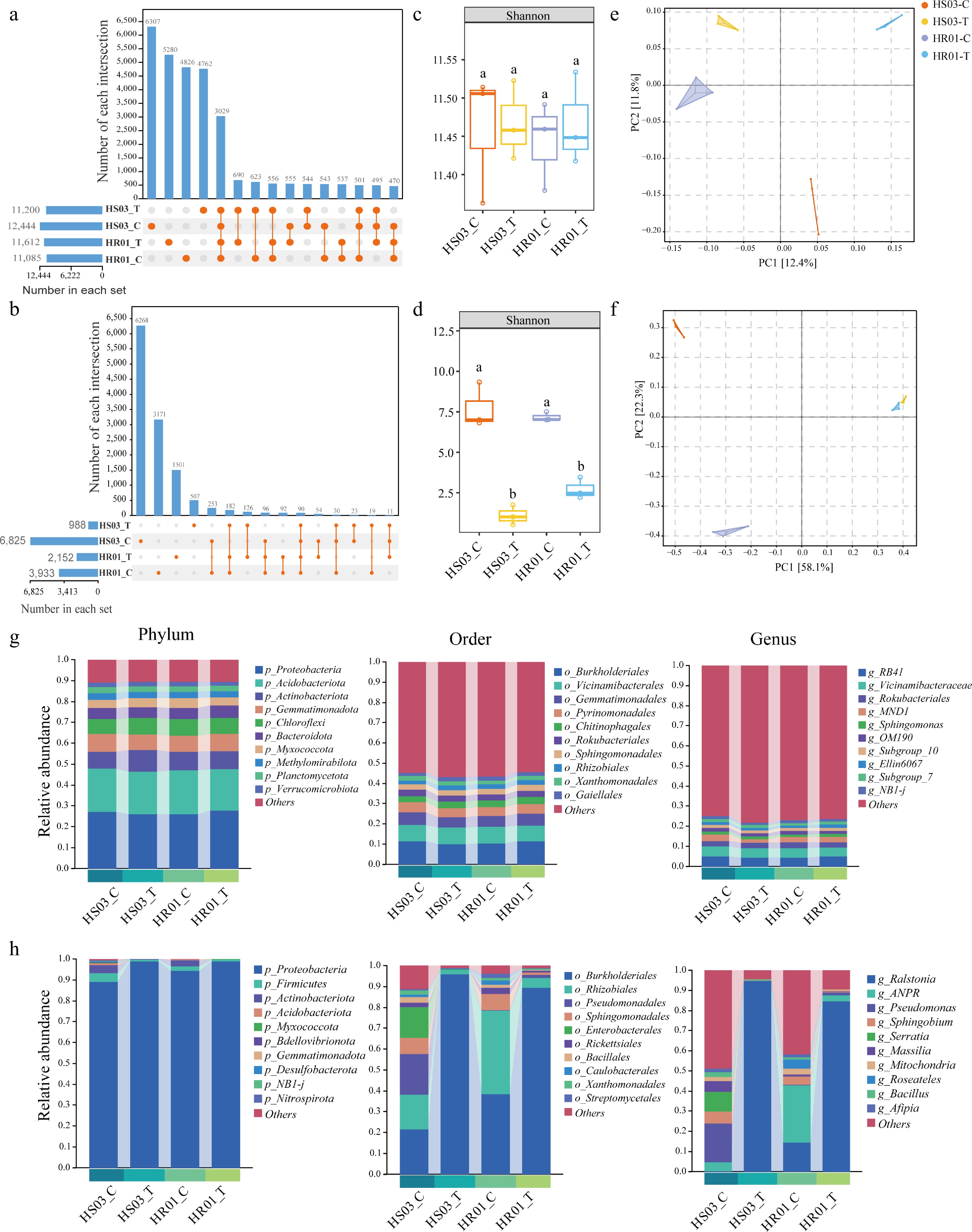
Figure 2.
Diversity and composition of rhizosphere and root bacterial communities in HR01 and HS03. (a), (b) Upset plots showing the number of ASVs detected in the rhizosphere (a) and root (b) bacterial communities. The bar plot on the left indicates the total number of taxa in each group; the matrix with orange (present) and gray (absent) dots below represents intersections among groups; the top bar plot displays the number of shared taxa in each intersection. (c), (d) Shannon index in the rhizosphere (c) and roots (d). (e), (f) PCA analysis in the rhizosphere (e) and roots (f). (g), (h) Relative abundances at the phylum, order, and genus level in the rhizosphere (g) and roots (h). Data are presented as mean ± SD from three biological replicates (n = 3). Statistical analyses were performed using the Kruskal–Wallis test followed by Dunn's post hoc test. Different letters indicate a significant difference (p < 0.05). HR01_C, control group of the resistant cultivar HR01; HR01_T, inoculated group of the resistant cultivar HR01; HS03_C, control group of the susceptible cultivar HS03; HS03_T, inoculated group of the susceptible cultivar HS03.
Differential bacterial biomarkers between resistant and susceptible cultivars following R. solanacearum inoculation
-
To further identify differential bacterial biomarkers, LEfSe analysis (LDA score > 2) was performed across all treatments. In the rhizosphere, eight distinctly abundant taxa were significantly enriched in HS03_C and HR01_C. Ten distinctly abundant taxa were significantly enriched in HS03_T, including Ralstonia (belonging to Burkholderiaceae), bacteriap25 (an uncharacterized genus within the phylum Myxococcota), Gaiella (belonging to Gaiellaceae), Bacillus (belonging to Bacillaceae), Streptomyces (belonging to Streptomycetaceae), Nordella, Aquicella, Nocardioides (belonging to Propionibacteriales), Skermanella (belonging to Azospirillaceae), and Anaeromyxobacter (belonging to Anaeromyxobacteraceae). In contrast, six genera, such as Ellin6067, Azospira (belonging to Rhodocyclaceae), Qipengyuania (belonging to Sphingomonadales), Dinghuibacter, Altererythrobacter (belonging to Sphingomonadales), and Arenimonas, were enriched in the inoculated resistant plants (HR01_T) (Fig. 3a; Supplementary Fig. S2). In the roots, 22 genera were enriched in the control groups, including Pseudomonas (belonging to Pseudomonadaceae), Massilia, Sphingobium, Bacillus (belonging to Bacillaceae), and Variovorax. Furthermore, Ralstonia was the dominant enriched genus in HS03_T, whereas Acidovorax was notably enriched in HR01_T (Fig. 3b; Supplementary Fig. S2).
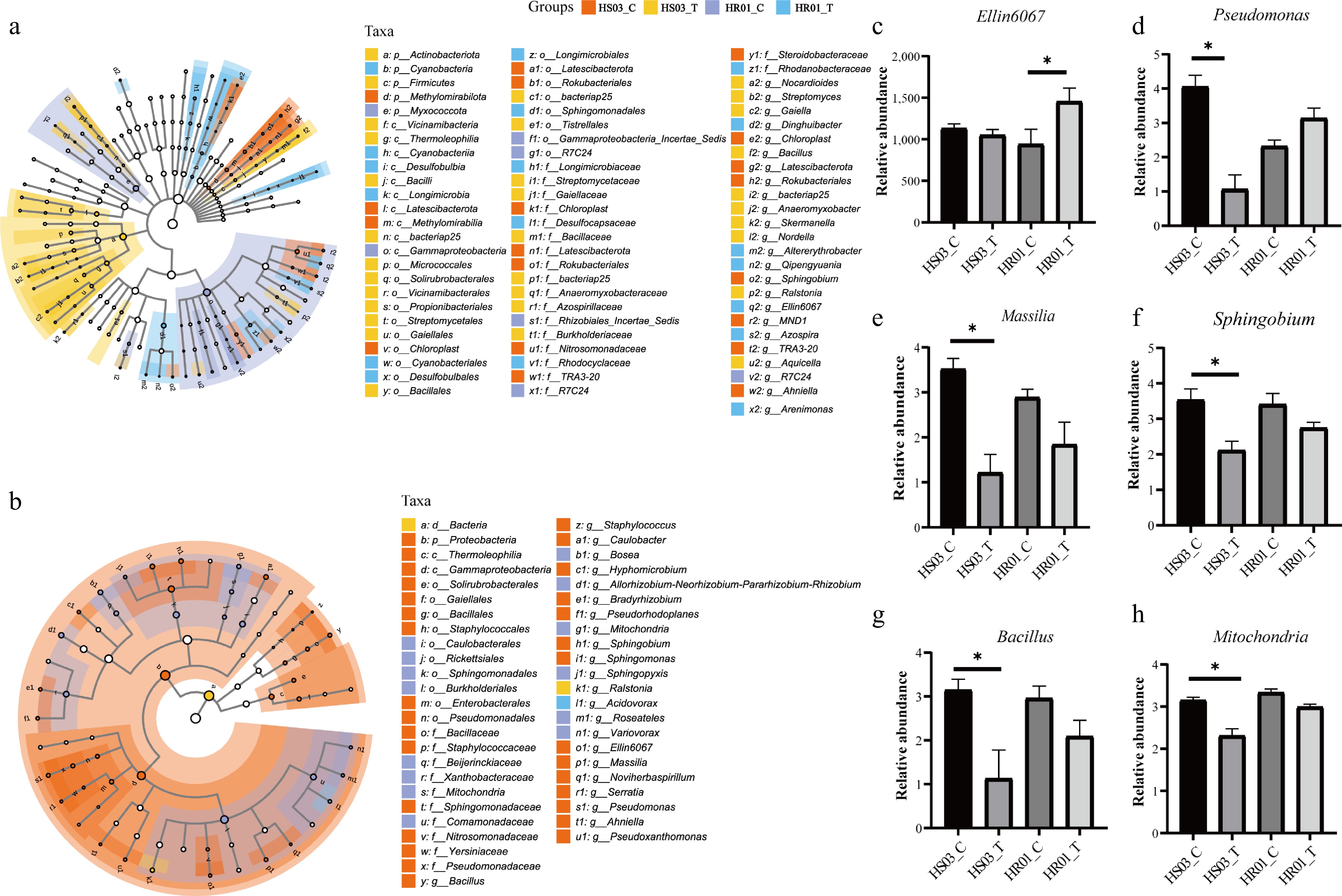
Figure 3.
Differential bacterial biomarkers in rhizosphere and root communities. (a), (b) LEfSe cladograms showing the phylogenetic distribution of bacterial lineages in the rhizosphere soil (a) and roots (b). (c) The relative abundance of significantly enriched genera in the rhizosphere soil. (d)–(h) The relative abundance of significantly enriched genera in the roots. Taxonomic annotations: p, phylum; c, class; o, order; f, family; g, genus; s, species. HR01_C, control group of the resistant cultivar HR01; HR01_T, inoculated group of the resistant cultivar HR01; HS03_C, control group of the susceptible cultivar HS03; HS03_T, inoculated group of the susceptible cultivar HS03. Data were log10-transformed and expressed as mean ± SD from three biological replicates (n = 3). Statistical analyses were performed using the Kruskal–Wallis test with Dunn's post hoc pairwise comparisons (*p < 0.05).
Further analysis of the relative abundances of differential biomarkers revealed that Ellin6067 became the dominant differential genus in the rhizosphere of HR01_T, showing a significant increase in relative abundance after inoculation in HR01 but no notable change in HS03 (Fig. 3c). In the root samples, the relative abundances of genera, including Pseudomonas, Massilia, Sphingobium, Bacillus, and Mitochondria, were significantly reduced in HS03 after inoculation. In contrast, no substantial differences were observed between the control and inoculated groups of HR01, although Pseudomonas exhibited an increasing trend upon infection (Fig. 3d–f). These results suggest that the loss of beneficial microbes and the overproliferation of Ralstonia in HS03 disrupt microbial homeostasis, whereas the maintenance or enrichment of beneficial taxa in HR01 contributes to enhanced disease resistance.
Screening and identification of biocontrol strains against R. solanacearum
-
To identify potential biocontrol agents, 80 bacterial strains were isolated from the rhizosphere soil and roots of bacterial communities of the resistant cultivar HR01 and screened for their antagonistic activity against R. solanacearum using dual-culture assays. Among these, strain P1202, isolated from root tissue, exhibited the largest inhibition zone against R. solanacearum and produced a characteristic copper-green pigment on LB medium (Fig. 4a). Furthermore, the cell-free fermentation broth of strain P1202 formed a clear inhibition zone of approximately 2 cm in diameter against the pathogen (Fig. 4b). Phylogenetic analysis based on the 16S rRNA gene sequence, together with NCBI BLAST analysis, showed that strain P1202 shared 100% sequence identity with Pseudomonas aeruginosa strain isol7. Combined with its morphological characteristics, strain P1202 was tentatively identified as Pseudomonas aeruginosa (Fig. 4c; Supplementary Fig. S3). These results indicate that strain P1202 possesses strong antagonistic potential, and it was therefore selected for subsequent biocontrol efficacy evaluation.
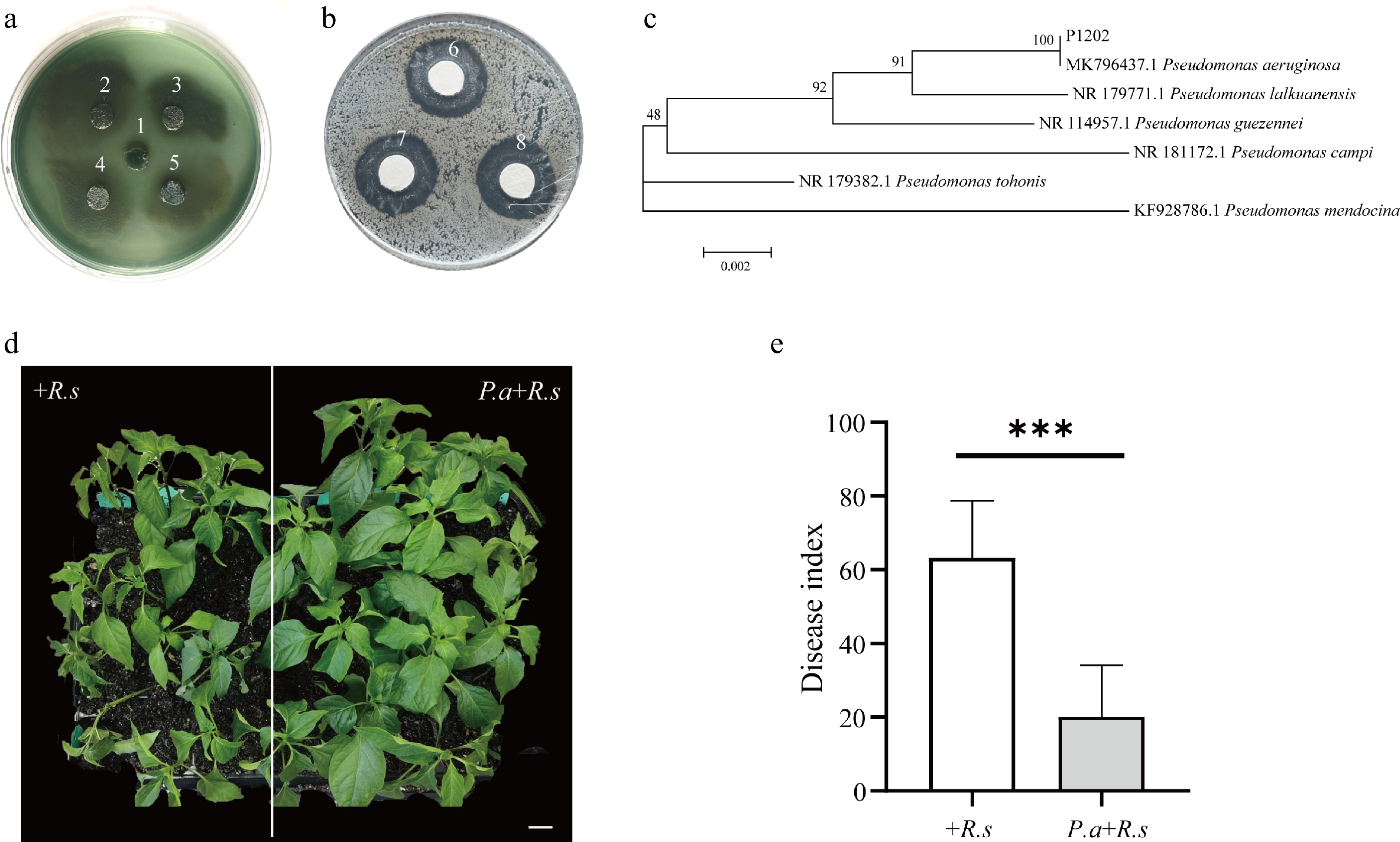
Figure 4.
Antagonistic activity and biocontrol potential of P. aeruginosa strain P1202. (a) Antagonistic activity of strain P1202 against R. solanacearum in a dual-culture assay. (b) Inhibitory effect of the cell-free fermentation broth of strain P1202 against R. solanacearum. In panels (a) and (b): 1, R. solanacearum; 2–5, P. aeruginosa strain P1202; 6–8, R. solanacearum exposed to the cell-free fermentation broth of P1202. (c) Phylogenetic tree based on the 16S rRNA sequence of strain P1202. (d) Disease symptoms of pepper plants in control (+R.s) and strain P1202 treatment groups (P.a+R.s) at 7 dpi. (e) Disease index of pepper plants in control (+R.s) and strain P1202 treatment groups (P.a+R.s) at 7 dpi. Data are presented as mean ± SD. Statistical analysis was performed using a two-tailed Student's t-test (n = 3 independent experiments, ***p < 0.001). Scale bar = 3 cm.
Application of P. aeruginosa enhances resistance of susceptible pepper to bacterial wilt
-
To evaluate its biocontrol efficacy in vivo, P. aeruginosa strain P1202 was applied to the rhizosphere soil and allowed to colonize in HS03 for two weeks prior to pathogen inoculation. Following pathogen inoculation, plants inoculated with R. solanacearum alone (+R.s) developed severe wilting symptoms, whereas plants pretreated with P. aeruginosa strain P1202 (P.a+R.s) exhibited markedly reduced disease severity (Fig. 4d). The disease index in the +R.s group reached 61.5, which was significantly higher than that in the P.a+R.s group (19.2) (Fig. 4e). These results demonstrate that P. aeruginosa strain P1202 effectively suppresses bacterial wilt development and significantly enhances disease resistance in the susceptible cultivar HS03.
Application of P. aeruginosa promotes plant growth and enhances the antioxidant enzyme system in pepper
-
To further investigate the physiological mechanisms underlying the enhanced resistance induced by P. aeruginosa, we first examined the changes in plant growth and related physiological parameters. After two weeks of the application of P. aeruginosa (P.a), the overall growth of pepper plants was significantly improved compared with the untreated control plants (Fig. 5a). Specifically, plant height and seedling fresh weight were significantly increased in the treated plants than in the control plants (Fig. 5b, c).
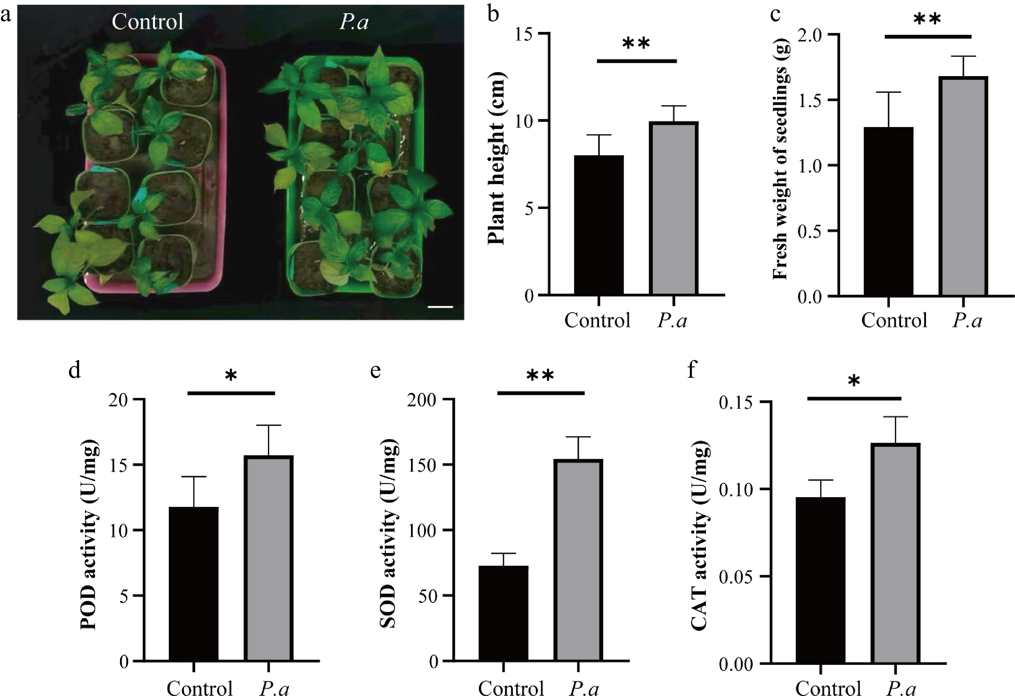
Figure 5.
Pseudomonas aeruginosa promotes plant growth and enhances antioxidant enzyme activities in pepper plants. (a) Phenotypes of control plants and plants pretreated with P. aeruginosa for two weeks (P.a). (b) Plant height of control and P.a plants. (c) Fresh weight of seedlings of control and P.a plants. (d) POD activity in control and P.a plants. (e) SOD activity in control and P.a plants. (f) CAT activity in control and P.a plants. Data are presented as mean ± SD from eight biological replicates (n = 8). Statistical analysis was performed using a two-tailed Student's t-test (*p < 0.05, **p < 0.01). Scale bar = 3 cm.
Moreover, the application of P. aeruginosa markedly enhanced the antioxidant enzyme activities in pepper plants. Compared with the control group, the activities of POD, SOD, and CAT in the P. aeruginosa-treated plants were significantly increased (Fig. 5d–f). These results suggest that P. aeruginosa treatment not only promotes plant growth but also strengthens the antioxidant defense capacity of pepper plants.
Application of P. aeruginosa enhanced PSII photochemical activity in pepper under R. solanacearum infection
-
To evaluate the effects of P. aeruginosa treatment on the photosynthetic performance of pepper plants during the early stage of R. solanacearum infection, the Chl-F parameter ΦPSII, which reflects the electron transport efficiency of PSII, and rETR were measured. ΦPSII and rETR showed rapid responses during the early stage of infection. At 16 hpi, both parameters were significantly higher in the +R.s and P.a+R.s treatments than in their respective uninoculated controls (Fig. 6a, b). Furthermore, compared with the control group, the P.a group showed significantly increased ΦPSII and rETR (Fig. 6c). Following R. solanacearum inoculation, the P.a+R.s treatment also led to significantly higher ΦPSII and rETR than the +R.s treatment (Fig. 6d). These results indicate that P. aeruginosa pretreatment enhances PSII photochemical efficiency and electron transport capacity, maintaining a higher level of PSII photochemical activity under R. solanacearum infection.
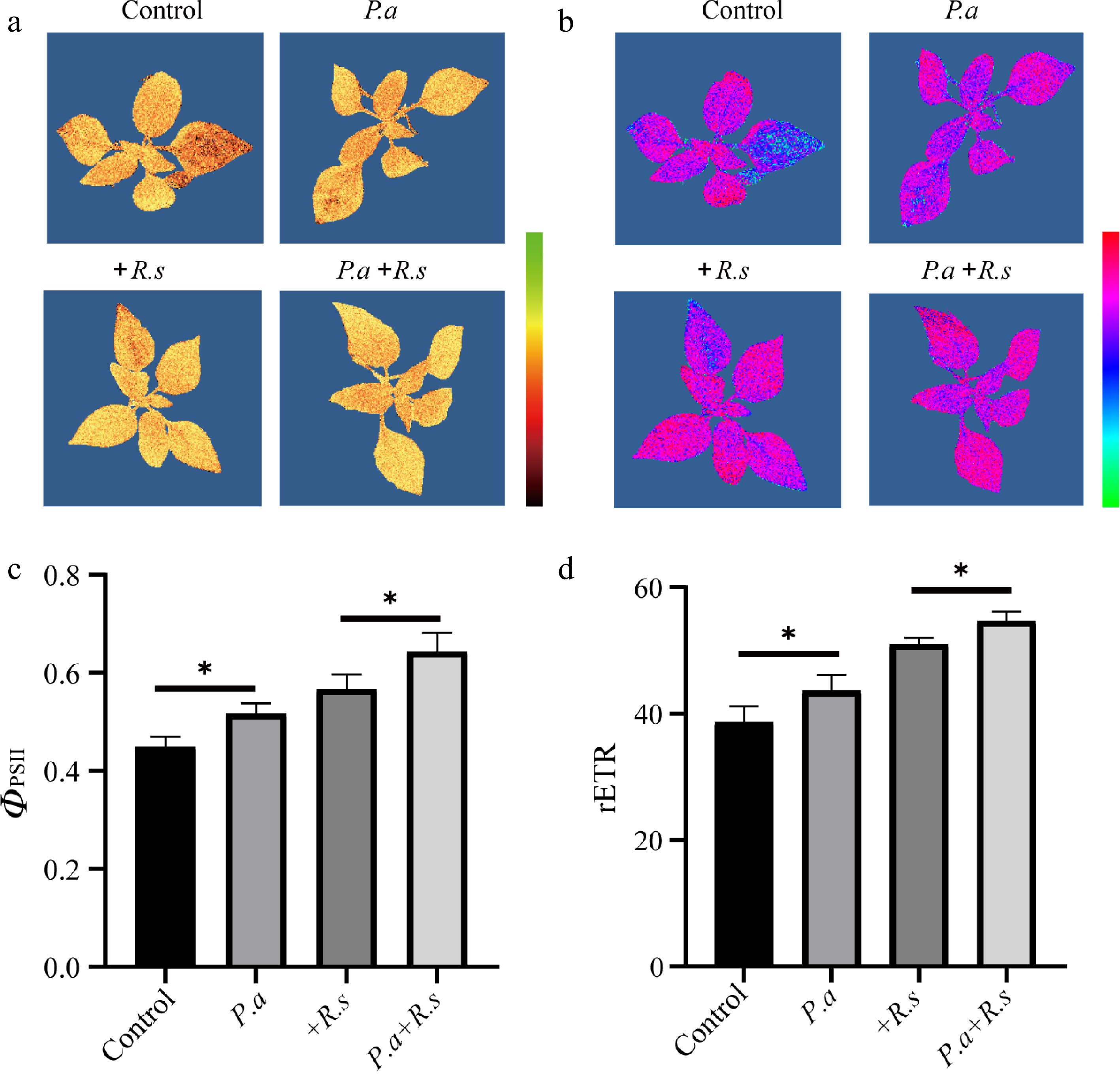
Figure 6.
Chlorophyll fluorescence responses of pepper plants under different treatments. (a), (b) Pseudocolor images of ΦPSII (a) and rETR (b) in four treatments: control plants, plants inoculated with R. solanacearum for 16 h (+R.s), plants pretreated with P. aeruginosa for 2 weeks (P.a), and plants pretreated with P. aeruginosa for 2 weeks followed by R. solanacearum inoculation for 16 h (P.a+R.s). (c), (d) Quantitative analysis of ΦPSII (c) and rETR (d) in control, +R.s, P.a, and P.a+R.s plants. Color cards indicate that the value gradually decreases from top to bottom. Data are presented as mean ± SD from eight biological replicates (n = 8). Statistical analysis was performed using a two-tailed Student's t-test (*p < 0.05).
Transcriptomic responses of pepper to R. solanacearum under P. aeruginosa treatment
-
To elucidate the molecular mechanisms underlying the enhanced resistance induced by P. aeruginosa during the early stage of R. solanacearum infection, transcriptome sequencing was performed on four treatment groups. PCA analysis revealed clear separation among the treatments (Supplementary Fig. S4), indicating that the treatments markedly affected global gene expression patterns in pepper. A total of 1,959 and 646 differentially expressed genes (DEGs) were identified in control vs P.a and +R.s vs P.a+R.s, respectively (Supplementary Fig. S5). GO enrichment analysis showed that the DEGs were mainly associated with biological process (BP), molecular function (MF), and cellular component (CC). In the BP category, significantly enriched terms included "defense response" and "cell wall organization". In the MF category, enriched terms included "DNA-binding transcription factor activity" and "transcription cis-regulatory region binding". In the CC category, "extracellular region" was among the significantly enriched terms. Similar enrichment patterns were observed in both comparisons (Fig. 7a, b). KEGG analysis revealed that in the control vs P.a comparison, DEGs were significantly enriched in the "plant–pathogen interaction", "zeatin biosynthesis", and "phenylpropanoid metabolism" pathways (Fig. 7c). In contrast, in the +R.s vs P.a+R.s comparison, DEGs were predominantly enriched in the "plant–pathogen interaction" pathway (Fig. 7d). These results suggest that P. aeruginosa induces extensive transcriptional reprogramming, with a strong emphasis on defense-related pathways, thereby priming the host for enhanced resistance.
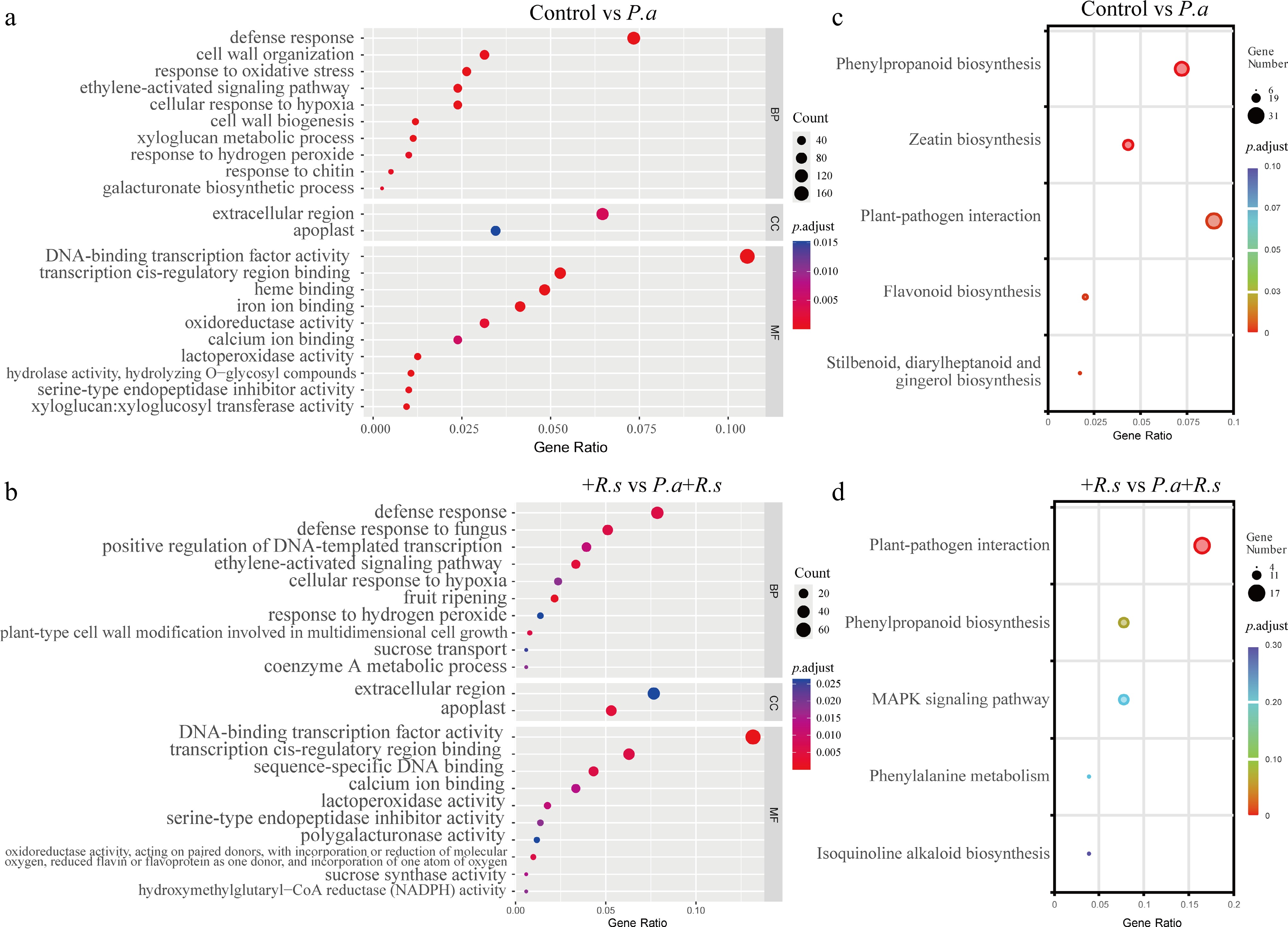
Figure 7.
Functional enrichment analysis of DEGs. (a), (b) GO enrichment analysis of DEGs between the control group and the group pretreated with P. aeruginosa for 2 weeks (control vs P.a) (a), and between the group inoculated with R. solanacearum for 16 h and the group pretreated with P. aeruginosa for 2 weeks followed by R. solanacearum inoculation for 16 h (+R.s vs P.a+R.s) (b). (c), (d) KEGG enrichment analysis of DEGs between the control group and the group pretreated with P. aeruginosa for 2 weeks (control vs P.a) (c), and between the group inoculated with R. solanacearum for 16 h and pretreated with P. aeruginosa for 2 weeks followed by R. solanacearum inoculation for 16 h (+R.s vs P.a+R.s) (d).
P. aeruginosa activates defense-related genes in the plant–pathogen interaction pathway in pepper
-
To further investigate the genes involved in the plant–pathogen interaction pathway, several key DEGs identified from the RNA-seq analysis were selected for qRT-PCR analysis. The results showed that, compared with the control plants, P. aeruginosa-treated plants (P.a) showed significantly upregulated expression levels of WRKY33 (WRKY transcription factor 33), CNGC (cyclic nucleotide-gated channel), CaM (calmodulin), and MPK3 (mitogen-activated protein kinases 3). At 16 hpi, the expression levels of these genes were also significantly higher in plants pretreated with P. aeruginosa followed by R. solanacearum inoculation (P.a+R.s) than in plants inoculated with R. solanacearum alone (+R.s) (Fig. 8). These findings suggest that P. aeruginosa enhances pepper resistance to R. solanacearum by inducing the expression of defense-related genes involved in the plant–pathogen interaction pathway.
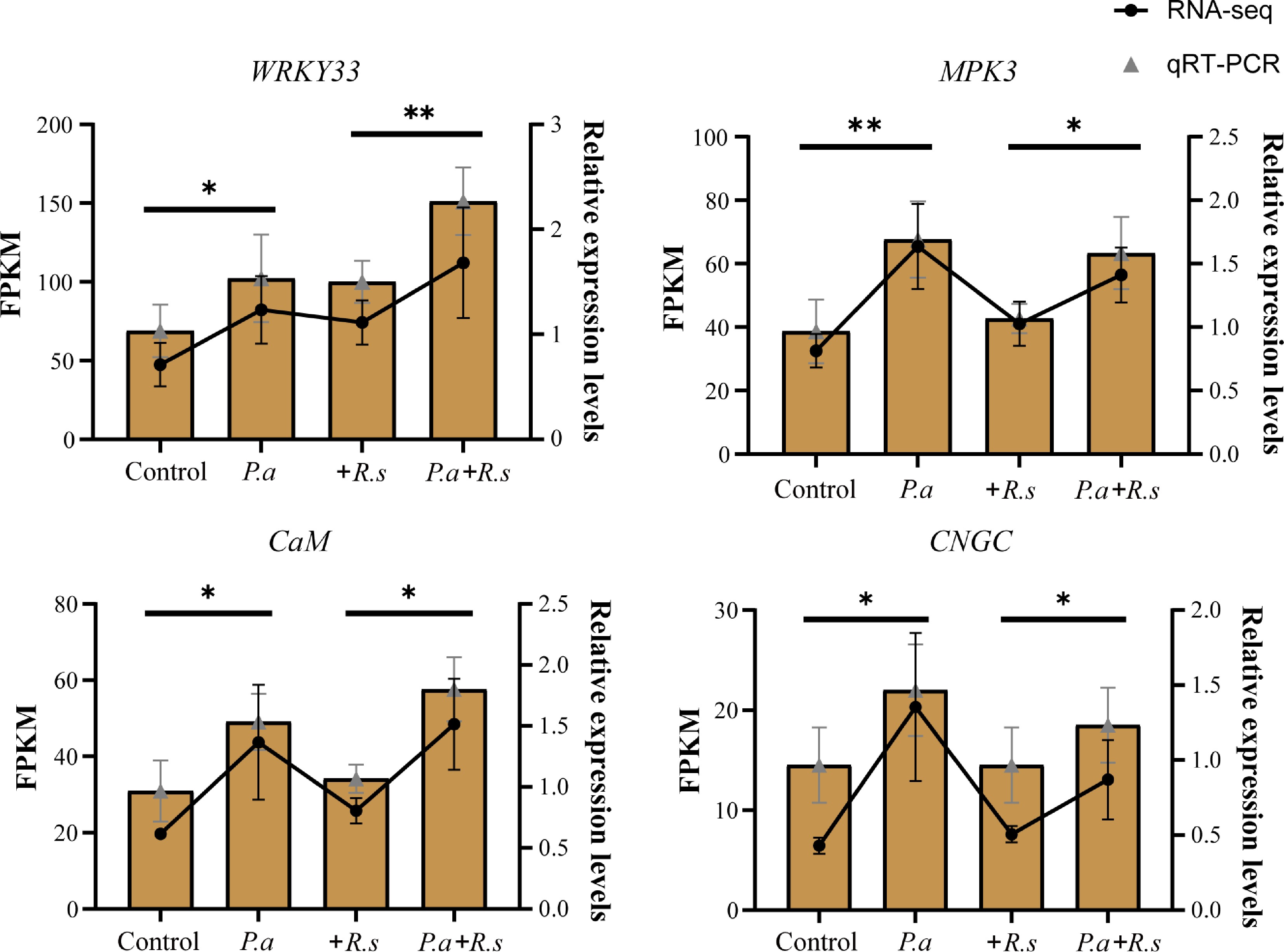
Figure 8.
Expression patterns of defense-related genes in pepper roots under different treatments. Expression patterns of key genes (WRKY33, MPK3, CaM, and CNGC) under different treatments (control, P.a, +R.s, and P.a+R.s). FPKM values were log-normalized and clustered by rows. Lines represent transcriptome (RNA-seq) data, and bars represent qRT-PCR data. Data are presented as mean ± SD from three biological replicates (n = 3). Statistical significance was determined using a two-tailed Student's t-test (*p < 0.05, **p < 0.01).
-
Host genotype plays a critical role in disease resistance and in shaping the structure, acquisition, assembly, and function of root-associated bacterial communities[30,31]. In this study, we demonstrate that under R. solanacearum infection, HR01 maintained robust root health, exhibited low levels of pathogen colonization, and preserved root biomass, whereas HS03 developed severe disease symptoms accompanied by substantial pathogen proliferation. In addition, the relative abundance of R. solanacearum in the roots was significantly higher in HS03 than in HR01 (Fig. 1). These findings indicate that the resistant genotype effectively restricts pathogen growth and disease progression, which may be closely associated with differences in root-associated microbial communities.
Rhizosphere and root endophytic bacteria are known to possess traits critical for responding to biotic and abiotic stresses[24,32,33]. These microbial communities are shaped by plant genotype, the soil's physicochemical properties, and environmental factors, forming a complex ecological network that influences plant health and disease resistance[34]. Our results showed that R. solanacearum infection significantly reshaped the composition and diversity of both rhizosphere and root endophytic bacterial communities in HS03 and HR01. After inoculation, the rhizosphere bacterial communities of both cultivars remained relatively stable; however, the root endophytic bacterial communities exhibited a sharp decline in diversity and underwent substantial structural reorganization, with Ralstonia becoming the dominant genus inside the roots of both cultivars (Fig. 2). This pattern is likely influenced by the root-wounding inoculation method used in this study, which facilitates rapid pathogen entry and colonization. Under natural infection conditions, where root tissues remain intact, the colonization process and the resulting microbial community dynamics may differ. Nevertheless, in both the rhizosphere and endophytic bacteria, HS03 consistently harbored higher Ralstonia loads than HR01, further underscoring the cultivar-dependent nature of host defense. Biomarker analysis further identified Ralstonia as the dominant genus in HS03_T, both in the rhizosphere and in the root endophytic bacteria. In contrast, HR01_T was enriched with taxa such as Ellin6067 in the rhizosphere and Acidovorax in the root endosphere (Fig. 3). Ellin6067 has previously been enriched in tomato rhizosphere soil treated with biological control agents against bacterial wilt[35], suggesting that they may contribute to rhizosphere-mediated disease resistance. Similarly, members of the genus Acidovorax have been reported as plant-associated beneficial bacteria. For example, Acidovorax radicis N35 isolated from wheat roots exhibited plant growth-promoting properties[36], indicating its potential application as a microbial inoculant. Furthermore, after inoculation, within the root endophytic bacterial community, a significant decline was observed in several beneficial genera, including Massilia, Bacillus, and Pseudomonas, in HS03, whereas no comparable reductions were detected in HR01. Interestingly, Pseudomonas exhibited an increasing trend in HR01, suggesting its potential importance in mediating resistance (Fig. 3).
Screening of root-associated antagonistic bacteria represents an effective strategy for managing soil-borne pathogens[37]. From the resistant cultivar HR01, 80 bacterial isolates with antagonistic potential were obtained from both rhizosphere soil and root tissues. Among these, strain P1202, which originated from the root of HR01, exhibited the strongest inhibition against R. solanacearum. Morphological and molecular characterization identified this strain as P. aeruginosa (Fig. 4). Although some Pseudomonas species are recognized as opportunistic human pathogens, many members of this genus have been widely applied in agriculture as biocontrol agents because they produce diverse bioactive metabolites, including antibiotics, siderophores, and volatile organic compounds, which collectively suppress pathogen growth and stimulate plant defense responses[38,39]. Consistent with these reports, strain P1202 exhibited strong antibacterial activity against R. solanacearum in vitro and significantly reduced bacterial wilt symptoms in pepper under greenhouse conditions. However, given the potential biosafety concerns associated with P. aeruginosa, comprehensive biosafety assessments, including toxicity evaluation, environmental persistence, and effects on non-target microorganisms, are still required before practical application. Interestingly, the cell-free fermentation broth of strain P1202 showed an even larger inhibition zone against R. solanacearum than the live bacterial culture, further confirming its direct inhibitory capacity (Fig. 4). Considering the potential biosafety concerns associated with the direct application of live P. aeruginosa, cell-free fermentation products may provide a safer and more practical approach for agricultural application compared with live bacterial inoculants, as they retain antibacterial activity while avoiding the introduction of viable bacterial cells into the environment. In addition, the present study did not include a commercial biocontrol product or a well-characterized biocontrol strain as a positive control. Therefore, the relative efficacy of strain P1202 compared with currently available biocontrol agents remains to be determined. Future studies should compare strain P1202 with representative commercial biocontrol products or well-established biocontrol strains to better evaluate its practical application potential.
Beyond their direct antagonistic activity, these beneficial bacteria can also promote plant growth and enhance host resilience by improving nutrient uptake, modulating immune responses, and increasing tolerance to various environmental stresses[40,41]. In this study, application of strain P1202 promoted plant growth, as evidenced by the increased plant height and fresh weight. These results are consistent with previous reports demonstrating that P. aeruginosa can enhance plant growth in crops such as tomato, potato, taro, and strawberry[42,43]. Antioxidant enzymes play a crucial role in enhancing root antioxidant capacity and maintaining reactive oxygen species (ROS) homeostasis[44]. In this study, application of strain P1202 significantly enhanced antioxidant enzyme activities in pepper roots (Fig. 5), which are positively associated with resistance. Pathogen infection can rapidly alter photosynthetic processes during the early stage of host–pathogen interactions, and the responses of ΦPSII and rETR may vary among plant genotypes with different levels of disease resistance[28]. The increased ΦPSII and rETR observed in P. aeruginosa-pretreated plants suggest enhanced PSII photochemical activity and photosynthetic electron transport during early pathogen challenge (Fig. 6). Such enhancement may facilitate ATP and NADPH production, thereby providing additional energy and reducing power to support plant growth and defense responses.
Notably, the 16 hpi time point also coincided with extensive transcriptional reprogramming. Comparative transcriptome analysis between the control and P.a, as well as between the +R.s and P.a+R.s treatments, revealed that DEGs were predominantly enriched in the GO term "defense response" and the KEGG pathway "plant–pathogen interaction". These results indicate that P. aeruginosa treatment activates plant defense-related processes (Fig. 7). Further analysis of genes involved in the "plant–pathogen interaction" pathway revealed that P. aeruginosa treatment upregulated several key defense-related genes, including WRKY33, CNGC, CaM, and MPK3 (Fig. 8). CNGCs are important plasma membrane Ca2+ channels that mediate rapid calcium influx in response to pathogen perception, whereas CaM functions as a calcium sensor that transduces Ca2+ signals to downstream targets[45,46]. The activation of calcium signaling is closely associated with MAPK cascades, and MPK3 serves as a central regulator linking early signal perception to transcriptional reprogramming[47]. Activated MPK3 can subsequently regulate transcription factors such as WRKY33, thereby promoting the expression of defense-related genes[48]. The coordinated upregulation of these genes suggests that P. aeruginosa pretreatment may enhance early pathogen perception and facilitate the rapid activation of the Ca2+-MAPK-WRKY signaling module, leading to a faster and more effective immune response upon R. solanacearum infection. However, downstream defense responses, such as ROS burst, callose deposition, and pathogenesis-related (PR) gene expression, were not evaluated in this study. Further investigation of these immune outputs will help elucidate the molecular mechanisms underlying P. aeruginosa-mediated disease resistance. Although the observed physiological changes and transcriptomic responses suggest that strain P1202 interacts with the host plant, direct colonization was not investigated in this study. Future work will focus on characterizing the colonization ability and spatial distribution of strain P1202 in pepper roots using approaches such as GFP labeling, or strain-specific molecular detection. This will help to further elucidate the relationship between root colonization and the induction of plant resistance.
Collectively, our findings provide strong evidence that beneficial root-associated microbes derived from resistant cultivars, particularly P. aeruginosa strain P1202, can enhance host resistance through multiple mechanisms. These mechanisms include antagonistic activity against R. solanacearum, promotion of plant growth, stimulation of antioxidant enzyme activities, enhancement of PSII photochemical activity, and activation of defense-related gene expression (Fig. 9). Our results highlight the potential of P. aeruginosa as an effective pepper biocontrol agent against bacterial wilt.
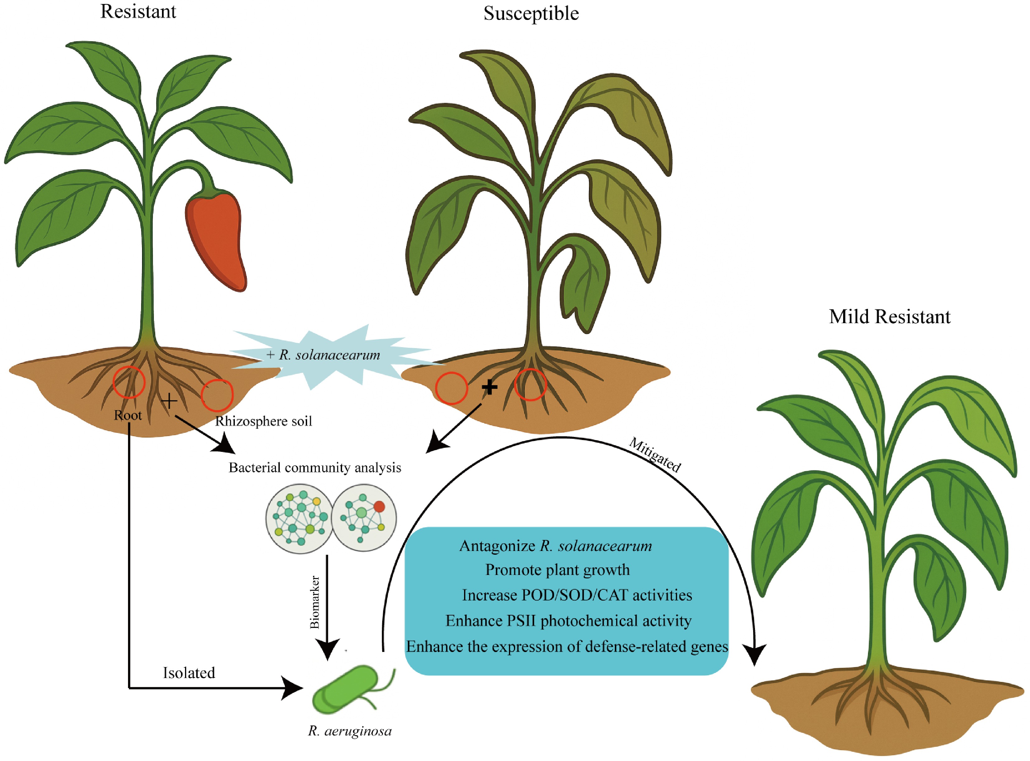
Figure 9.
Schematic diagram of the enhancement of pepper resistance to bacterial wilt by P. aeruginosa. The model illustrates cultivar-dependent differences in root-associated bacterial communities during R. solanacearum infection, as well as the biocontrol mechanisms by which P. aeruginosa application enhances resistance in susceptible pepper plants.
-
This study revealed that resistance to bacterial wilt in pepper is closely associated with host genotype and root-associated bacterial communities. Inoculation with R. solanacearum resulted in lower disease severity and reduced pathogen colonization in the resistant cultivar HR01 than in the susceptible cultivar HS03, while also reshaping the rhizosphere and root endophytic bacterial communities, particularly in the roots, with a notable decline in beneficial genera in HS03 but relative stability in HR01. P. aeruginosa P1202, a strain isolated from HR01, exhibited strong antagonistic activity against R. solanacearum. Application of this strain significantly enhanced pepper resistance, promoted plant growth, increased antioxidant enzyme activities, and enhanced early-stage PSII photochemical activity. Transcriptomic analysis further showed that P. aeruginosa activated defense-related pathways and upregulated key genes. These results demonstrate that P. aeruginosa enhances pepper resistance to bacterial wilt through coordinated regulation of bacterial communities, plant physiology, and defense signaling pathways, providing a promising strategy for sustainable disease management.
This work was supported by the Foundation of S&T Program of Hebei (262N6302D), Hebei Agriculture Research System (HBCT2023100207), and Innovation Capability Enhancement Special Project of the Baoding Science and Technology Bureau (grant no. 2494N005).
-
The authors confirm their contributions to the paper as follows: study design and manuscript revision: Cheng F, Li H; writing – original draft preparation: Cheng F, Cao Y, Dong R; data curation and investigation: Liu W, Pei Z, Sun Y, Xing R; writing – review and editing: Cheng F, Du W, Du P, Chen X; project administration, and supervision: Du W, Li H. All authors read and approved the final version of this manuscript.
-
Data will be made available on request.
-
The authors declare that they have no conflict of interest.
-
accompanies this paper online at: https://doi.org/10.48130/vegres-0026-0024.
-
# Authors contributed equally: Fengqing Cheng, Haili Li, Yang Cao
- Supplementary Table S1 The information of the primers.
- Supplementary Table S2 Strain P1201 identification based on 16S rRNA gene sequencing.
- Supplementary Fig. S1 Community composition of rhizosphere soil (a-c) and roots (d-f) bacterial communities.
- Supplementary Fig. S2 Bar charts showing taxa with LDA scores ≥ 2 in rhizosphere soil (A) and roots (B) bacterial communities.
- Supplementary Fig. S3 NCBI BLAST alignment of the 16S rRNA gene sequence of strain P1202.
- Supplementary Fig. S4 Principal component analysis (PCA) of four treatment groups.
- Supplementary Fig. S5 Identification of differentially expressed genes (DEGs) under different treatments.
- Copyright: © 2026 by the author(s). Published by Maximum Academic Press, Fayetteville, GA. This article is an open access article distributed under Creative Commons Attribution License (CC BY 4.0), visit https://creativecommons.org/licenses/by/4.0/.
-
About this article
Cite this article
Cheng F, Li H, Dong R, Liu W, Cao Y, et al. 2026. A root-associated Pseudomonas aeruginosa strain enhances pepper resistance to bacterial wilt through antimicrobial activity and activation of plant defense responses. Vegetable Research 6: e031 doi: 10.48130/vegres-0026-0024 

A root-associated Pseudomonas aeruginosa strain enhances pepper resistance to bacterial wilt through antimicrobial activity and activation of plant defense responses
- Received: 01 May 2026
- Revised: 10 July 2026
- Accepted: 27 July 2026
- Published online: 15 September 2026
Abstract: Bacterial wilt caused by Ralstonia solanacearum (R. solanacearum) poses a severe threat to pepper production. However, resistant cultivars can recruit root-associated microbial communities to cope with soil-borne pathogen infection. In this study, we analyzed the physiological traits, rhizosphere soil and root endosphere bacterial communities of the resistant cultivar HR01 and the susceptible cultivar HS03 under R. solanacearum stress. Infection significantly reduced the root biomass and altered the bacterial composition in HS03 compared with HR01, particularly in root endophytic bacteria. Biomarker analysis showed that Ralstonia dominated both the rhizosphere soil and roots of HS03_T, whereas HR01_T was enriched with taxa such as Ellin6067 in the rhizosphere and Acidovorax in the roots. Notably, Pseudomonas abundance decreased in HS03 but increased in HR01 after inoculation. Among 80 bacterial isolates obtained from HR01, strain P1202 was identified as Pseudomonas aeruginosa (P. aeruginosa), exhibiting the largest inhibition zone against R. solanacearum in dual-culture assays; its cell-free fermentation filtrate also showed strong inhibitory effects. Pot experiments demonstrated that strain P1202 application effectively alleviated the disease symptoms of HS03 and also promoted pepper growth, enhanced antioxidant enzyme activities, and improved early-stage photochemical activity of photosystem II (PSII). Transcriptomic and quantitative real-time polymerase chain reaction (qRT-PCR) analyses revealed activation of defense-related pathways (e.g., plant–pathogen interaction) and upregulation of key genes (WRKY33, CNGC, CaM, and MPK3). These results suggest that P. aeruginosa enhances pepper resistance to bacterial wilt through direct antimicrobial activity and induction of pepper defense responses, highlighting its potential as an effective biocontrol agent.
-
Key words:
- Pepper /
- Bacterial wilt /
- Root endophytic bacteria /
- Rhizosphere soil /
- Ralstonia solanacearum /
- Pseudomonas aeruginosa